FT-IR vs. Microscopy for Microplastic Identification: A Comprehensive Guide for Environmental and Biomedical Research
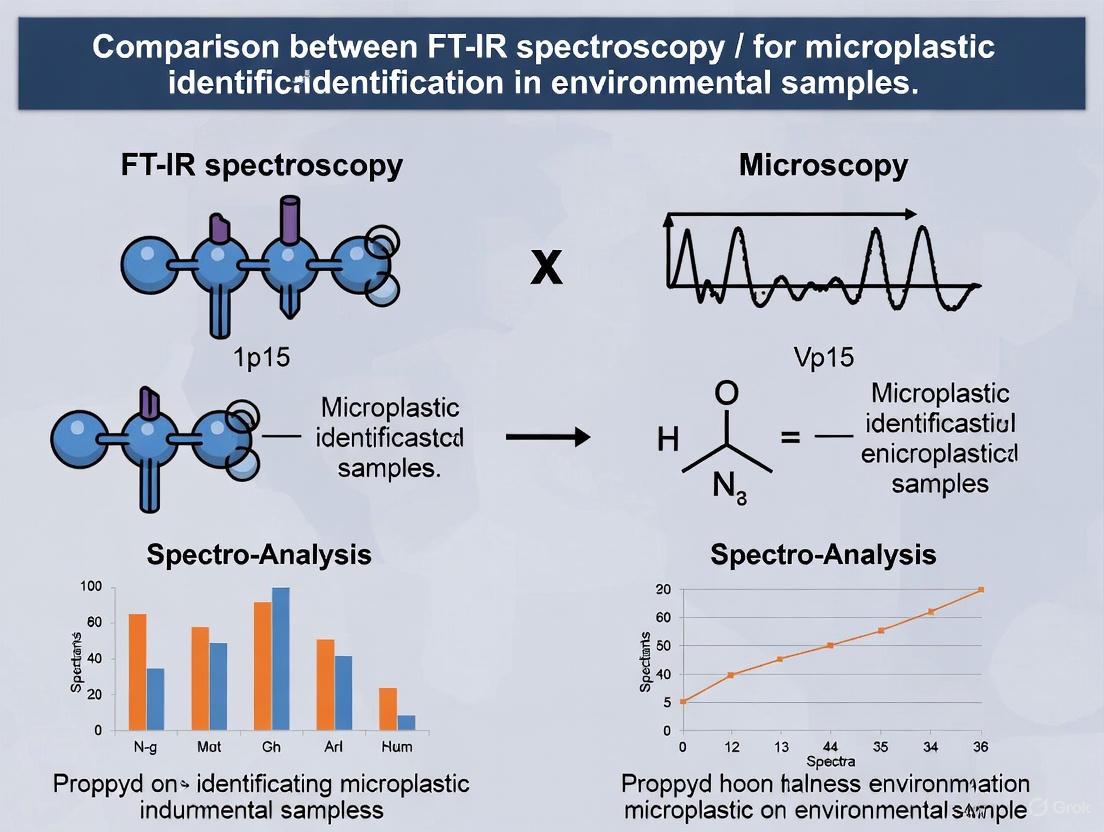
This article provides a detailed comparative analysis of Fourier-Transform Infrared (FT-IR) spectroscopy and microscopy for microplastic identification in environmental samples, tailored for researchers and drug development professionals.
FT-IR vs. Microscopy for Microplastic Identification: A Comprehensive Guide for Environmental and Biomedical Research
Abstract
This article provides a detailed comparative analysis of Fourier-Transform Infrared (FT-IR) spectroscopy and microscopy for microplastic identification in environmental samples, tailored for researchers and drug development professionals. It explores the foundational principles of both techniques, delves into advanced methodological applications including automated systems and machine learning integration, and addresses key troubleshooting and optimization challenges. By presenting a rigorous validation and comparative framework, this review synthesizes current capabilities, limitations, and future directions, offering evidence-based guidance for selecting appropriate analytical strategies in environmental monitoring and assessing potential implications for human health.
Understanding the Core Technologies: Principles of Microscopy and FT-IR in Microplastic Analysis
The pervasive nature of microplastics—plastic particles smaller than 5 mm—in global ecosystems presents a formidable analytical challenge for environmental and health researchers [1]. These particles originate from diverse sources, including product degradation and engineered microbeads, and exhibit high mobility in air, water, and soil [2]. Their small size and chemical stability enable bioaccumulation, where they can transport toxic chemicals and potentially enter human organs through ingestion and inhalation [3]. Addressing this environmental threat requires precise identification and characterization methods, with Fourier-Transform Infrared (FT-IR) spectroscopy and optical microscopy emerging as pivotal, yet fundamentally different, analytical approaches [4] [1]. This guide provides an objective comparison of these techniques to inform methodological selection for microplastics research.
Fundamental Principles: FT-IR Spectroscopy vs. Optical Microscopy
FT-IR Spectroscopy
FT-IR spectroscopy is a vibrational technique that provides a molecular "fingerprint" of a sample [5]. When infrared light interacts with a microplastic particle, specific chemical bonds within the polymer absorb characteristic wavelengths of IR light. This creates a unique spectrum that allows for definitive identification of the polymer type, such as polyethylene (PE), polypropylene (PP), or polystyrene (PS) [3]. Modern FT-IR microscopy (µ-FT-IR) integrates spectroscopy with visual examination, enabling chemical analysis of very small structures [5]. Measurement can be performed in transmission, reflection, or attenuated total reflectance (ATR) mode, with ATR being particularly widespread due to its minimal sample preparation requirements [5].
Optical Microscopy
Optical microscopy for microplastic analysis relies primarily on visual assessment of physical characteristics such as size, shape, and color under magnification [4] [1]. This method is considered a traditional approach for the identification and quantification of microplastics [1]. However, it does not provide any chemical composition information, making definitive identification of polymer types impossible based on visual characteristics alone [1].
Comparative Performance Evaluation
The table below summarizes the core performance characteristics of both techniques based on experimental findings.
Table 1: Performance Comparison of Optical Microscopy and FT-IR Spectroscopy for Microplastic Analysis
| Analytical Parameter | Optical Microscopy | FT-IR Spectroscopy |
|---|---|---|
| Chemical Specificity | None; cannot identify polymer type [1] | High; provides unique molecular fingerprint for precise polymer identification [3] [5] |
| Detection Limit (Particle Size) | Effective for larger particles (>1 mm) [6] | Can identify particles down to 1 µm, or even less with advanced ATR microscopy [5] [1] |
| Quantitative Data | Particle counts and size estimation (with low accuracy for small particles) [1] | Particle counts, size, and chemical composition; enables concentration calculations [2] |
| Analysis Speed | Simple and rapid for visual inspection [1] | Slower per particle, but automation enables high-throughput [4] [2] |
| Accuracy/Reliability | Low; accuracy drops to 30-44% for particles <1-2 mm [6] | High; considered a confirmation method, though accuracy depends on spectral libraries and processing [4] [6] |
Experimental Protocols and Data Output
Standard Workflow for Optical Microscopy
Samples are typically filtered from a liquid matrix (water) and dried [2]. The filter is then placed under a light microscope, and particles are manually counted and characterized based on morphological features. The lack of chemical confirmation is a critical limitation, as non-plastic particles (e.g., organic matter, minerals) can be easily misidentified as microplastics [1].
Standard Workflow for FT-IR Microscopy
The FT-IR analysis workflow involves more steps but yields chemically verified results. The following diagram illustrates a typical workflow for microplastic analysis using FT-IR microscopy:
- Sample Preparation: A known volume of liquid is vacuum-filtered onto a suitable substrate (e.g., 0.2-micron Anodisc or gold-coated filters) [3]. The filter is dried, often with no further preparation needed [2].
- Instrumental Analysis: The filter is placed in the FT-IR microscope. For ATR analysis, a germanium crystal is pressed onto the particle of interest. The system exposes the sample to IR light and detects the absorbed wavelengths to create a spectrum [5].
- Spectral Identification: The unknown spectrum is automatically compared against commercial or open-source spectral libraries (e.g., KnowItAll, Open Specy) [6]. The match is quantified with a Hit Quality Index (HQI), though visual confirmation of high-scoring matches is recommended to avoid false positives from spectral artifacts [6].
- Data Output: Advanced software can automate the entire process, using focal plane array (FPA) detectors to scan large areas of the filter, collect thousands of spectra, and generate reports cataloging particle count, size, and polymer identity [4] [2].
Table 2: Experimental Data from a Model Study Using FT-IR Imaging
| Polymer Type Identified | Number of Particles Detected | Key IR Absorption Bands (cmâ»Â¹) |
|---|---|---|
| Polyethylene (PE) | 5 | ~2920, ~2850 (C-H stretch), ~1470 (C-H bend), ~720 (C-H rock) [3] |
| Polystyrene (PS) | 4 | ~3025 (aromatic C-H stretch), ~1600 (C=C stretch), ~1490 (C=C stretch) [3] |
| Polyethylene Terephthalate (PET) | 8 | ~1720 (C=O stretch), ~1240, ~1090 (C-O stretch) [3] |
| Polyvinyl Chloride (PVC) | 8 | ~1250 (C-H bend), ~690 (C-Cl stretch) [3] |
Advanced Considerations and Methodological Refinements
Addressing the Limitations of FT-IR
While FT-IR is a powerful tool, its accuracy is not infallible. Several factors can affect performance:
- Spectral Library Quality: The accuracy of automated matching varies significantly (64.1% to 98.0%) depending on the library and processing routines [6]. Using derivative correction during processing can greatly reduce misidentification of natural materials (e.g., cotton) as synthetics [6].
- Environmental Fouling: Weathered particles with biofilms or adsorbed contaminants can have altered spectra. While fouling generally reduces the HQI of library matches, the effect is inconsistent across different polymer types [6].
- Analysis Mode: Studies comparing manual, semi-automated, and fully automated µFTIR analysis have found that a semi-automated method—using a combination of ultrafast mapping and subsequent manual checking—strikes the best balance, minimizing both false positives and false negatives while analyzing a large proportion of particles [4].
The Evolving Analytical Landscape
The microplastic analysis market is rapidly advancing, driven by environmental concerns and stricter regulations [7] [8]. Technological progress is focused on improving efficiency and accuracy through:
- Automation and AI: Automated analysis based on FPA-FTIR microscopy and AI-based image analysis reduces time demands and the risk of human bias inherent in manual methods [4] [7].
- Portable Devices: Development of portable, affordable FT-IR devices aims to increase accessibility and enable real-time environmental monitoring [8].
- Data Processing: Advanced statistical methods like Principal Component Analysis (PCA) are being leveraged to rapidly classify microplastic types without manual sorting, improving efficiency and reducing error [3].
The Scientist's Toolkit: Essential Research Reagents and Materials
Table 3: Key Materials and Reagents for Microplastic Analysis via FT-IR
| Item | Function in Analysis |
|---|---|
| Vacuum Filtration System | To extract and concentrate microplastics from large liquid volumes onto a filter for analysis [2]. |
| FT-IR Microscope | The core instrument that combines light microscopy with FT-IR spectroscopy for chemical imaging and identification [5]. |
| ATR Crystal (e.g., Germanium) | Enables Attenuated Total Reflectance measurement, providing high-resolution data with minimal sample preparation [5]. |
| Specialized Filters (Anodisc, Gold-coated) | Act as substrates for filtered samples; choice of filter depends on the IR measurement mode (transmission or reflectance) [3]. |
| Spectral Library Software | Commercial (e.g., Omnic) or open-source (e.g., Open Specy, siMPle) databases for matching unknown spectra to known polymer references [6]. |
| Oxidizing Agent (e.g., Hâ‚‚Oâ‚‚) | Used in sample pretreatment to remove organic biological material that could obscure the polymer signal [6]. |
| Borax (B4Na2O7.10H2O) | Borax (Sodium Tetraborate) for Research |
| LLP-3 | LLP-3, MF:C32H23ClN2O4, MW:535.0 g/mol |
The choice between optical microscopy and FT-IR spectroscopy for microplastic analysis is not one of simple preference but of analytical rigor. Optical microscopy offers speed and low cost for initial visual assessment but fails to provide chemical confirmation, leading to high error rates, especially for particles below 1 mm [6] [1]. In contrast, FT-IR spectroscopy delivers definitive polymer identification, is capable of analyzing smaller particles, and, with automation, can provide comprehensive quantitative data on particle count, size, and type [4] [3] [2]. For researchers requiring accurate and reliable data to assess ecological risks, track pollution sources, and inform policy, FT-IR spectroscopy, particularly in a semi-automated workflow, represents the current methodological standard. The continued advancement of FT-IR technology and data processing protocols will further solidify its role as an indispensable tool in confronting the global challenge of microplastic pollution.
The accurate analysis of microplastics in environmental samples is a critical step in understanding and mitigating this form of pollution. The process fundamentally relies on three core tasks: visual identification of potential plastic particles, their sizing, and the determination of their morphology (shape). While traditional light microscopy serves as an initial tool for these tasks, its significant limitations have led to the adoption of more advanced, spectroscopic techniques. This guide objectively compares the performance of visual microscopy against Fourier-Transform Infrared (FT-IR) microscopy for these fundamental analyses, framing the discussion within the broader context of selecting the optimal method for reliable microplastic research.
Table 1: Method Comparison at a Glance
Table comparing key performance metrics of Visual Microscopy and FT-IR Microscopy for microplastic analysis.
| Performance Metric | Visual Microscopy | FT-IR Microscopy |
|---|---|---|
| Identification Basis | Visual characteristics (color, shape, opacity) | Molecular vibration (chemical fingerprint) |
| Identification Accuracy | Low (up to 70% error rate, higher for smaller particles) [9] | High (91-95% accuracy for particles >50 µm) [10] |
| Minimum Detectable Size | Subject to human vision limit; unreliable for <500 µm [10] | ~10-25 µm (μ-FTIR) [9] [11] |
| Sizing Capability | Manual measurement; prone to human error | Automated, software-driven measurement (length, width, aspect ratio) [12] |
| Morphology Analysis | Subjective classification (fiber, fragment, etc.) | Objective classification with quantitative shape descriptors [12] |
| Polymer Typing | Not possible; cannot distinguish polymer from natural particles | High-confidence polymer identification [4] [13] [3] |
| Analysis Throughput | Fast initial screening, but slow if counting/measuring | High throughput with automated imaging and particle analysis [4] [2] |
Experimental Protocols for Method Evaluation
Protocol for Visual Identification and Counting
The traditional method for visual analysis involves a series of manual steps that are common in many studies [10] [4].
- Sample Preparation: Environmental samples (water, sediment) are processed through density separation or filtration to isolate particulates. The collected material is filtered onto a membrane filter [9] [14].
- Microscopy: The filter is placed under a stereo- or optical microscope. Potential microplastics are manually identified based on visual cues: the absence of cellular structures, consistent color, and homogeneous texture [10].
- Sizing and Morphological Classification: Using a calibrated eyepiece graticule, a researcher manually measures the length and width of each particle. Particles are subjectively categorized into shape classes such as fiber, fragment, granule, or film [14].
- Data Recording: The count, size, shape, and color of all suspected microplastics are recorded. This method is often followed by chemical confirmation for a subset of particles due to its inherent uncertainty [10].
Protocol for FT-IR Microscopy Analysis
FT-IR microscopy automates and chemically verifies the fundamental analysis. The following protocol is adapted from validated methods [4] [12].
- Sample Preparation and Substrate: Samples are filtered onto substrates that minimize spectral interference, such as silicon oxide, aluminum oxide, or gold-coated filters [9] [11] [3]. Silicon filters, in particular, allow for direct analysis without transfer, reducing particle loss [11].
- Automated Imaging and Spectral Collection: The filter is placed in an automated μ-FTIR microscope. The software first captures a visual mosaic image of the entire filter or defined "count fields" [12]. Using Focal Plane Array (FPA) detection, the instrument then collects thousands of infrared spectra simultaneously across the sample area in transmission or reflectance mode [9] [3].
- Spectral Identification and Particle Analysis: The collected spectra are compared against reference libraries of known polymers (e.g., using a Pearson correlation coefficient). A match score (typically >65-70%) confirms the plastic nature and identifies the polymer type [4] [13] [12].
- Automated Sizing and Morphology: The integrated software calculates the length and width of each identified particle and uses the aspect ratio (AR) to objectively classify morphology: spherical (AR ≤1), ellipsoidal (AR ≥2), or cylindrical/fibrous (AR ≥3) [12].
Visual Workflow and Decision Logic
The following diagram illustrates the typical analytical workflow for microplastic analysis, highlighting the divergent paths of the two methods and their respective outcomes.
The Scientist's Toolkit: Essential Research Reagents and Materials
Successful microplastic analysis, particularly with FT-IR microscopy, depends on the use of specific materials and reagents to ensure accuracy and minimize contamination.
Table listing key reagents, materials, and their functions in microplastic analysis.
| Item | Function in Analysis | Key Consideration |
|---|---|---|
| Silicon Filter | Ideal substrate for μ-FTIR in transmission mode; mostly transparent in mid-IR range, allowing direct analysis [11]. | Minimizes spectral interference, reduces sample preparation steps and particle loss [9] [11]. |
| Gold-coated Filter | Substrate for FT-IR measurement in reflectance mode, extending the spectral range to lower wavenumbers [3]. | An alternative to silicon filters when reflectance measurement is preferred. |
| Aluminum Oxide Filter | Another suitable filter substrate for micro-FTIR particle analysis with minimal background interference [12]. | Chemically inert and provides a consistent background. |
| Sodium Chloride (NaCl) | Used to prepare high-density solutions (e.g., 5 M NaCl) for density separation of microplastics from inorganic particles [14]. | A cost-effective salt for separating less dense polymers from sediment and water samples. |
| Hydrogen Peroxide (Hâ‚‚Oâ‚‚) / Enzymes | Used in purification to digest natural organic matter (e.g., algae, biomass) that would otherwise cause spectral interference [9]. | Selection depends on sample type; enzymatic digestion is often gentler on microplastic surfaces. |
| FT-IR Spectral Library | A curated database of reference spectra from known polymers; essential for automated identification of unknown particles [9] [13]. | Quality and comprehensiveness of the library directly impact identification accuracy. |
| CL264 | CL264, MF:C19H23N7O4, MW:413.4 g/mol | Chemical Reagent |
| TAI-1 | TAI-1|Hec1 Inhibitor | TAI-1 is a potent, first-in-class Hec1 inhibitor for cancer research. It disrupts Hec1-Nek2 interaction. This product is for research use only and not for human use. |
The fundamental tasks of visual identification, sizing, and morphological analysis of microplastics can be approached with either traditional light microscopy or FT-IR microscopy. The choice of method has profound implications for data quality. Visual microscopy offers rapid screening but is plagued by high error rates and an inability to provide chemical information. In contrast, FT-IR microscopy automates these fundamental tasks and, crucially, adds a layer of chemical verification, transforming subjective visual assessment into objective, data-rich analysis. For research requiring high-confidence results in particle count, size distribution, and polymer-specific morphological data, FT-IR microscopy is the unequivocally superior technique.
In the critical field of environmental science, accurately identifying and characterizing microplastics has become essential for assessing pollution levels and ecological impacts. Among analytical techniques, Fourier-Transform Infrared (FT-IR) Spectroscopy has emerged as a cornerstone technology for molecular identification and polymer fingerprinting. This guide objectively compares FT-IR's performance against alternative methodologies within environmental microplastics research, providing researchers with experimental data and protocols to inform their analytical strategies.
Fundamentals of FT-IR for Polymer Analysis
FT-IR spectroscopy analyzes the interaction of infrared light with materials at a molecular level. When samples are exposed to infrared light, each polymer absorbs specific wavelengths, creating a unique spectral pattern that serves as a chemical fingerprint for identification [3]. This molecular fingerprinting capability makes FT-IR particularly valuable for microplastics research, where determining the polymer type is crucial for assessing environmental risks and tracking pollution sources [3].
The technique fundamentally relies on the principle that chemical bonds vibrate at characteristic frequencies when exposed to IR radiation. The resulting spectrum plots transmitted or absorbed light against wavenumber (cmâ»Â¹), revealing distinct patterns corresponding to molecular structures. For synthetic polymers, these spectral signatures allow researchers to differentiate between common plastic types such as polyethylene (PE), polypropylene (PP), polyvinyl chloride (PVC), polystyrene (PS), and polyethylene terephthalate (PET) [3] [15].
FT-IR Operational Workflow
The following diagram illustrates the standard workflow for microplastic analysis using FT-IR spectroscopy:
FT-IR Versus Alternative Analytical Techniques
Performance Comparison Table
Table 1: Comparative analysis of FT-IR spectroscopy against other microplastic identification techniques
| Analytical Technique | Spatial Resolution | Polymer Identification | Sample Throughput | Key Limitations |
|---|---|---|---|---|
| FT-IR Spectroscopy | ~10-20 μm [16] | Excellent [3] | Moderate to High (with automation) [17] | Limited for sub-10μm particles [18] |
| Raman Spectroscopy | ~0.5-5 μm [16] | Excellent [16] | Low to Moderate | Fluorescence interference [16] |
| Pyrolysis GC-MS | N/A (bulk analysis) [16] | Excellent [16] | High | Destructive; no particle information [16] |
| Optical Microscopy | ~1-2 μm | Poor (visual only) [6] | High | Low accuracy (30-44% for <1mm) [6] |
| ATR-FTIR | ~100 μm minimum [17] | Excellent [17] | Low | Contact method; potential particle damage [17] |
Quantitative Performance Data
Table 2: Experimental accuracy and reproducibility data for FT-IR and comparator techniques based on interlaboratory studies
| Technique | Polymer Type | Reproducibility (SR) | Identification Accuracy | Key Applications |
|---|---|---|---|---|
| FT-IR Spectroscopy | PET | 64-70% [16] | 98% (MARS system) [17] | Polymer identification, particle counting [16] |
| FT-IR Spectroscopy | PE | 121-129% [16] | 98% (MARS system) [17] | Polymer identification, particle counting [16] |
| Raman Spectroscopy | PET | 64-70% [16] | Varies with fouling [6] | Small particle analysis [16] |
| Raman Spectroscopy | PE | 121-129% [16] | Varies with fouling [6] | Small particle analysis [16] |
| Thermoanalytical Methods | PET | 45.9-62% [16] | High (bulk) [16] | Mass quantification [16] |
| Thermoanalytical Methods | PE | 62-117% [16] | High (bulk) [16] | Mass quantification [16] |
Advanced FT-IR Methodologies
Micro-FTIR (μFTIR) Imaging
Micro-FTIR systems combine microscopy with infrared spectroscopy, enabling analysis of particles as small as 10-20 μm [16]. This approach allows researchers to simultaneously obtain spatial and chemical information about microplastic particles. Focal plane array (FPA) detection has significantly enhanced μFTIR, enabling automated, unbiased analysis without manual presorting of particles [18]. Studies have successfully employed systems like the PerkinElmer Spotlight 400 FT-IR Imaging System, which can scan entire filters in under 40 minutes, dramatically reducing analysis time compared to traditional methods [3].
Reflectance-FTIR and Automation
Recent advancements have led to semi-automated systems like the Microplastic Analyzer using Reflectance-FTIR Semi-automatically (MARS), specifically designed for analyzing larger microplastics (>500 μm) [17]. This system integrates a motorized stage, image recognition cameras, and reflectance-FTIR to automatically output particle count, size, and polymer type data. This approach demonstrates 98% accuracy compared to conventional ATR-FTIR methods while reducing analysis time by approximately 6.6 times [17].
Emerging IR Technologies
Advanced IR techniques continue to evolve, with Quantum Cascade Laser IR (QCL-IR) spectroscopy emerging as the second most popular approach after FT-IR, offering rapid analysis of plastic particles [18]. Optical Photothermal IR (O-PTIR) spectroscopy provides submicron spatial resolution, while Atomic Force Microscopy-Based IR (AFM-IR) spectroscopy bridges microscopic and spectroscopic analysis at the nanoscale level [18].
Experimental Protocols
Standard FT-IR Analysis for Microplastics
Sample Preparation Protocol:
- Separation: Dissolve test samples to separate microplastic particles using controlled digestion [3]
- Filtration: Perform vacuum filtration using 0.2-micron Anodisc filters for transmission measurements or gold-coated filters for reflectance measurements [3]
- Drying: Ensure complete drying of filters before analysis to prevent spectral interference from water [17]
Instrumental Analysis:
- Spectral Collection: Configure FT-IR microscope with liquid nitrogen-cooled mercury cadmium telluride (MCT) detector [6]
- Parameters: Collect spectra in transmission mode with aperture size of 100 × 100 μm, 8 scans per spectrum at 8 cmâ»Â¹ resolution across 675-4000 cmâ»Â¹ range [6]
- Background Correction: Perform background measurements immediately before sample analysis (within 5 minutes) [6]
Data Processing:
- Atmospheric Correction: Apply correction algorithms to suppress COâ‚‚ signals from collected spectra [6]
- Spectral Matching: Compare sample spectra against reference libraries using correlation algorithms [6]
- Validation: Implement derivative correction to reduce false identifications, improving differentiation between natural and synthetic materials [6]
Advanced Semi-Automated Protocol (MARS System)
Sample Handling:
- Placement: Manually position dried microplastic particles on a 70 × 50 mm mirror-polished stainless-steel sample plate [17]
- Arrangement: Ensure particles do not overlap and maintain at least 1 mm separation for accurate infrared analysis [17]
Automated Analysis:
- Imaging: System automatically captures particle images using coaxial epi-illumination microscope camera [17]
- Sizing: Software measures long and short axes based on rotated bounding rectangle of particles [17]
- Spectral Collection: Motorized XY stage positions each particle for reflectance-FTIR measurement [17]
- Identification: Integrated algorithm identifies polymer type and exports all data to Excel format [17]
Research Reagent Solutions and Essential Materials
Table 3: Key research reagents and materials for FT-IR microplastic analysis
| Material/Equipment | Specification/Function | Application Context |
|---|---|---|
| Anodisc Filters | 0.2-micron pore size [3] | Sample filtration for transmission FT-IR measurements |
| Gold-Coated Filters | High reflectance substrate [3] | Extend spectral range to 700 cmâ»Â¹ in reflectance mode |
| Stainless Steel Sample Plates | 70 × 50 mm mirror-polished SUS 304 [17] | Particle substrate for MARS automated system |
| PerkinElmer Spotlight 400 | FT-IR Imaging System [3] | High-throughput microplastic analysis |
| KnowItAll/Omnic Libraries | Commercial spectral databases [6] | Polymer identification reference |
| Open Specy | Open-source spectral library [6] | Accessible alternative for polymer matching |
| Hydrogen Peroxide | 10% solution for 24h treatment [6] | Remove organic contamination from environmental samples |
Critical Considerations for Technique Selection
Addressing Spectral Limitations
While FT-IR provides excellent polymer identification capabilities, researchers must recognize its limitations. Spectral library matching requires careful validation, as studies demonstrate accuracy rates ranging from 64.1% to 98.0% for distinguishing between natural and synthetic materials depending on the processing routines used [6]. Environmental fouling can reduce correlation values of library matches, though this effect varies across polymer types [6].
Standardization Challenges
Recent interlaboratory comparisons reveal significant reproducibility challenges in microplastic analysis, with FT-IR showing reproducibility rates of 64-70% for PET and 121-129% for PE [16]. These variations highlight the critical need for standardized protocols and certified reference materials to improve data comparability across studies [16].
Future Directions
The integration of artificial intelligence and machine learning with FT-IR spectroscopy represents a promising frontier for enhancing classification accuracy and reducing analysis time [19]. Additionally, ongoing development of portable FT-IR systems and automated platforms addresses the need for higher throughput analysis in environmental monitoring applications [17] [19].
FT-IR spectroscopy remains a powerful, versatile technique for microplastic identification, offering reliable polymer fingerprinting capabilities that balance analytical precision with practical implementation considerations. While alternative methods provide advantages for specific applications such as nanoplastic analysis or bulk quantification, FT-IR's non-destructive nature, chemical specificity, and evolving automation solidify its position as a fundamental tool in environmental microplastics research. Researchers should select analytical techniques based on their specific study objectives, sample characteristics, and required detection limits, recognizing that a complementary multi-method approach often provides the most comprehensive understanding of microplastic contamination.
Comparative Strengths and Inherent Limitations of Each Foundational Approach
The accurate identification and analysis of microplastics in environmental samples are critical for understanding and mitigating this pervasive pollutant. Within this field, two foundational analytical approaches are frequently employed: Fourier-Transform Infrared (FT-IR) spectroscopy and optical microscopy. FT-IR spectroscopy is a vibrational technique that identifies materials by their molecular fingerprint based on the absorption of infrared light [20] [21]. In contrast, optical microscopy, including fluorescence microscopy, primarily relies on visual characteristics such as particle size, shape, and color for initial identification and counting [1] [14].
This guide provides an objective comparison of these two methodologies, framing their performance within the context of microplastic analysis. The evaluation is based on recent, direct comparative studies and reviews to offer researchers, scientists, and environmental professionals a clear understanding of the capabilities and constraints inherent to each technique.
Comparative Analysis of Techniques
The following table summarizes the core characteristics, strengths, and inherent limitations of optical microscopy and FT-IR spectroscopy for microplastic analysis.
Table 1: Direct comparison of microscopy and FT-IR spectroscopy for microplastic analysis.
| Feature | Optical/Fluorescence Microscopy | FT-IR Spectroscopy |
|---|---|---|
| Primary Principle | Visual identification based on physical characteristics (size, shape, color) [1] [14]. | Chemical identification based on absorption of infrared light by molecular bonds [20] [21]. |
| Identification Capability | Presumptive; cannot confirm polymer type [1]. | Definitive; can identify specific polymer types and semi-synthetic polymers (e.g., PE, PP, Rayon) [22] [14]. |
| Key Strength | Rapid, low-cost, and simple for particle counting and sizing [1]. | High chemical specificity and reliability for polymer identification [22] [14]. |
| Key Limitation | High false positive/negative risk; misinterprets natural particles as microplastics [1] [14]. | Lower analytical throughput; more complex and costly instrumentation [23] [22]. |
| Particle Size Range | Effective for particles > 0.1 mm for color identification [14]. | Typically used for particles > 20 μm; can be extended with specialized systems [22] [1]. |
| Quantitative Data | Particle count and physical dimensions. | Polymer identity, functional groups, and in some cases, mass concentration. |
| Experimental Finding | Fluorescence microscopy detected more particles on average in a direct comparison [14]. | FT-IR identified 12 distinct polymer/semi-synthetic polymer types in the same study, providing accurate chemical data [14]. |
Experimental Protocols and Data
Detailed Methodologies from Comparative Studies
A 2025 study provides a direct experimental comparison of fluorescence and FT-IR microscopy for analyzing microplastics at a drinking water intake on the Perak River, Malaysia [14]. The workflow below outlines the core experimental process.
Diagram 1: Experimental workflow for microplastic analysis.
Sample Collection and Preparation:
- Water samples were collected from a river water intake using a 5-L stainless-steel bucket and stored in pre-cleaned 5-L glass bottles [14].
- Filtration and Density Separation: In the laboratory, a density separation step was performed using a 5.00 M sodium chloride (NaCl) solution to separate microplastics from inorganic particles. The mixture was homogenized with a magnetic stirrer on a heated plate at 50°C for 15 minutes [14].
- Quality Assurance and Control: Stringent measures were implemented to prevent contamination. These included using cotton lab coats, minimizing plastic equipment, cleaning all glassware with ultra-pure water, and analyzing blank samples in parallel to assess airborne contamination [14].
Instrumental Analysis:
- Fluorescence Microscopy: Prepared samples were analyzed under a fluorescence microscope to detect and count particles based on their fluorescence properties [14].
- FT-IR Microscopy (µFTIR): The same environmental samples were analyzed using FT-IR microscopy. This technique identified polymers by detecting the specific infrared light absorbed by chemical bonds in the sample, creating a unique molecular fingerprint for each particle [14].
Key Experimental Data and Outcomes
The 2025 study yielded quantitative data that directly highlights the performance differences between the two techniques.
Table 2: Summary of key findings from a direct comparative study (citation:10).
| Metric | Fluorescence Microscopy | FT-IR Microscopy (µFTIR) |
|---|---|---|
| Particle Detection | Detected a higher average number of particles. | Detected fewer particles, but with confirmed polymer identity. |
| Polymer Identification | Not capable. | Identified 12 distinct polymer and semi-synthetic polymer types. |
| Dominant Polymers Found | Not applicable. | Rayon and Polyethylene (PE) were dominant. |
| Particle Size Range | Majority of identified particles were 1-10 µm. | Effective in the 1-10 µm range. |
| Common Particle Shapes | Granules and irregular shapes. | Granules and irregular shapes. |
The critical finding was that while fluorescence microscopy reported higher particle counts, FT-IR analysis confirmed the chemical identity of the particles, distinguishing true plastic polymers from other semi-synthetic materials like Rayon and, crucially, from non-plastic particles that may fluoresce [14]. This underscores the primary limitation of microscopy: its inability to provide chemical confirmation leads to a high risk of misidentification [1].
The Scientist's Toolkit: Essential Research Reagents and Materials
The following table details key reagents and materials essential for conducting microplastic analysis following the protocols cited in this guide.
Table 3: Key research reagents and solutions for microplastic analysis.
| Item | Function | Application Note |
|---|---|---|
| Sodium Chloride (NaCl) | Used to prepare high-density solutions (e.g., 5.00 M) for density separation, which floats microplastics away from denser inorganic sediments [14]. | A common, low-cost salt for initial sample purification. |
| Hydrochloric Acid (HCl) | Used at low concentrations (e.g., 10%) for rigorous cleaning of sampling equipment like stainless-steel buckets and glass containers to prevent contamination [14]. | Critical for quality assurance before sample collection. |
| ATR-FTIR Accessory | An Attenuated Total Reflectance accessory used with an FT-IR spectrometer for direct analysis of solid samples with minimal preparation, enabling the characterization of functional groups in polymers [24] [1]. | Standard for analyzing filter-based samples and bulk materials. |
| Gold-Coated Filters | Specialized filters used in sample preparation for FT-IR analysis; the gold coating minimizes infrared interference during spectral acquisition. | Improves signal-to-noise ratio in spectroscopic analysis. |
| Ceramic, Phospholipid, & Sphingolipid Standards | Commercial lipid and biochemical standards (e.g., PE, PC, PI, PS, Cer) used as references to obtain distinctive infrared spectra for functional group identification [24]. | Vital for method validation and confirmation of chemical identity. |
| PY-60 | PY-60, CAS:2765218-56-0, MF:C16H15N3O2S, MW:313.4 g/mol | Chemical Reagent |
| JB170 | JB170, MF:C48H44ClFN8O11, MW:963.4 g/mol | Chemical Reagent |
The choice between microscopy and FT-IR spectroscopy for microplastic analysis is not a matter of selecting a superior technique, but rather of aligning the method with the study's objective. Optical and fluorescence microscopy offer a rapid, cost-effective means for initial particle counting and physical characterization. However, its inherent limitation is the inability to chemically identify polymers, leading to significant uncertainty and a high risk of false positives [1] [14].
In contrast, FT-IR spectroscopy provides definitive chemical identification, transforming presumptive microplastics into confirmed polymer data. This accuracy is fundamental for source apportionment, understanding environmental fate, and formulating targeted mitigation strategies [22]. Its limitations, including higher cost and operational complexity, are balanced by the reliability of its data. For any research requiring conclusive evidence of microplastic pollution, FT-IR spectroscopy is the indispensable foundational approach.
From Theory to Practice: Advanced Applications and Workflow Integration
Sample Preparation Protocols for Diverse Environmental Matrices
The accurate identification and quantification of microplastics in environmental samples hinge on effective sample preparation, which varies significantly based on the matrix and the analytical technique employed. Fourier Transform Infrared (FT-IR) spectroscopy and microscopy have emerged as leading techniques for microplastic identification, each with distinct advantages and specific sample preparation requirements. FT-IR spectroscopy provides powerful chemical specificity by generating unique molecular fingerprints for each polymer type, enabling precise identification of microplastic particles [4] [3]. Microscopy techniques, particularly scanning electron microscopy (SEM), offer high-resolution imaging capabilities but require extensive sample preparation to maintain structural integrity during analysis [25]. The selection between these techniques involves trade-offs between chemical characterization, spatial resolution, analytical time, and sample preparation complexity, making protocol optimization essential for reliable environmental monitoring.
Comparative Analysis of Technique Performance
Table 1: Performance comparison of microplastic analysis techniques across environmental matrices.
| Analytical Technique | Optimal Size Range | Spatial Resolution | Sample Preparation Complexity | Analysis Time | Polymer Identification Capability |
|---|---|---|---|---|---|
| FT-IR Microscopy (Transmission) | 10-20 µm thickness [26] | ~10-15 µm [26] | Moderate (requires thin sections) | Moderate to High | Excellent (library matching) |
| Micro ATR-FTIR | No thickness requirement [26] | ~1.1 µm (4x enhancement vs transmission) [26] | High (requires flat surfaces, often resin embedding) | Moderate | Excellent (library matching) |
| SEM | Sub-micron to millimeter | ~1 µm [25] | Very High (fixation, drying, coating) | Low to Moderate | None (requires complementary techniques) |
| Raman Spectroscopy | 1 µm and above [27] | ~1 µm [27] | Low to Moderate | High | Excellent (library matching) |
| Reflectance-FTIR (MARS system) | >400 µm [17] | N/A (for large particles) | Low (simple placement on plate) | Low (6.6x faster than ATR-FTIR) [17] | Excellent (98% accuracy) [17] |
Table 2: Sample preparation requirements for different environmental matrices.
| Environmental Matrix | Organic Matter Removal | Separation Technique | Filter Type | Special Considerations |
|---|---|---|---|---|
| Wastewater & Sludge | Fenton's reagent (multiple digestions) [27] | Density separation (ZnCl₂) [27] | 0.2-µm Anodisc or gold-coated filters [3] | High organic content requires optimized digestion [27] |
| Marine Water | Hydrogen peroxide (Hâ‚‚Oâ‚‚) [27] | Density separation | Metal mesh filters | High salt content may interfere |
| Beach Sediments | Enzymatic treatment [27] | Density separation | Various | Complex matrix with natural particles |
| Biological Tissues | Enzymatic digestion [27] | Filtration | Various | Preservation of microplastic integrity |
Experimental Protocols for Sample Preparation
FT-IR Based Protocol for Wastewater and Sludge Samples
The analysis of microplastics in complex organic-rich matrices like wastewater and sludge requires rigorous sample preparation to remove interfering organic matter while preserving microplastic particles. The optimized protocol based on Fenton reagent digestion involves sequential steps to effectively degrade organic material without damaging common polymer types [27].
Materials and Reagents:
- Fenton reagent: Hydrogen peroxide (Hâ‚‚Oâ‚‚) with iron (II) sulphate (FeSOâ‚„) catalyst
- Zinc chloride (ZnClâ‚‚) for density separation
- Vacuum filtration system
- 0.2-µm Anodisc filters or gold-coated filters [3]
- Triplicate samples for quality control
Methodology:
- Sample Pre-treatment: Homogenize wastewater or sludge samples to ensure representative subsampling.
- Organic Matter Digestion: Apply multiple digestions with Fenton reagent at pH 2-4 and temperature below 50°C to maximize organic degradation while preventing polymer damage [27]. The low temperature is critical as temperatures above 60°C can cause losses of some plastic polymers.
- Density Separation: Use ZnClâ‚‚ solution to separate microplastics from remaining inorganic minerals through density flotation.
- Filtration: Filter the supernatant through 0.2-µm Anodisc filters for transmission FT-IR or gold-coated filters for reflectance measurements [3].
- Quality Control: Include spiked samples with known microplastic polymers (e.g., PE, PP, PET, PVC) to validate recovery rates and account for potential losses during processing.
This protocol has demonstrated effectiveness for microplastics in the sub-hundred-micron size range, which pose higher ecological risks due to increased bioavailability [27]. The sequential digestion approach provides an inexpensive and time-efficient procedure suitable for processing large sample sets, enabling robust environmental monitoring data generation.
SEM Sample Preparation Protocol for High-Resolution Imaging
Scanning Electron Microscopy requires extensive sample preparation to preserve structural integrity and ensure conductivity for high-quality imaging. The protocol focuses on maintaining the native structure of microplastics while making them compatible with SEM's vacuum environment and electron beam requirements [25].
Materials and Reagents:
- Glutaraldehyde or osmium vapor fixatives
- Graded ethanol series (e.g., 30%, 50%, 70%, 90%, 100%)
- Critical point dryer or freeze-dryer
- Sputter coater with gold or carbon target
- Conductive double-coated carbon tape
- Aluminum sample stubs
Methodology:
- Sample Cleaning: Gently clean samples with appropriate buffers or distilled water. For more vigorous cleaning, use surfactants or proteolytic enzymes specific to the contaminant type, taking care not to damage microplastic surfaces [25].
- Chemical Fixation: Immerse samples in glutaraldehyde fixative to maintain structural details. Alternatively, use osmium vapor for conductive staining.
- Dehydration: Process samples through a graded ethanol series (30%, 50%, 70%, 90%, 100%) to gradually remove water, finishing with 100% ethanol or acetone.
- Drying: Employ critical point drying (CPD) to prevent structural collapse from surface tension. Alternatively, use freeze-drying which causes less sample shrinkage but carries ice crystal formation risks [25].
- Mounting: Affix samples to aluminum stubs using conductive double-coated carbon tape, ensuring a continuous conductive path from sample to stub.
- Sputter Coating: Apply a thin layer (10-20 nm) of gold or carbon using a sputter coater to render non-conductive samples compatible with SEM imaging.
- Storage: Maintain samples in a dry, clean environment until imaging to prevent contamination or degradation.
This protocol ensures minimal artifacts and clear imaging of microplastic surface morphology, which can provide information about degradation patterns and environmental history. However, unlike FT-IR, SEM cannot chemically identify polymer types and requires complementary techniques for complete characterization [25].
Sample preparation workflow for SEM analysis.
Advanced FT-IR Methodologies and Automation
Comparison of FT-IR Operational Modes
Table 3: Comparison of FT-IR measurement modes for microplastic analysis.
| FT-IR Mode | Sample Preparation Requirements | Spatial Resolution | Advantages | Limitations |
|---|---|---|---|---|
| Transmission | Thin sections (<15-20 µm) [26] | 10-15 µm [26] | Simple spectral interpretation, high quality spectra | Limited spatial resolution, fringing effects |
| ATR | Flat, smooth surfaces; pressure application [26] | ~1.1 µm (with 15x objective) [26] | No thickness requirement, enhanced spatial resolution | Potential sample damage, resin embedding often needed |
| Reflectance | Minimal (particles on reflective surface) [17] | N/A (particle-dependent) | Non-destructive, no contact, high throughput | Limited to larger particles (>400 µm), spectral artifacts |
| μ-FTIR Imaging | Filtration onto specialized filters [3] | 25-µm pixel size [3] | Automated analysis, statistical representation | Expensive instrumentation, complex data processing |
Automated and Semi-Automated Systems
Recent advancements in FT-IR technology have focused on automating the analytical process to increase throughput and reduce human bias. The Microplastic Analyzer using Reflectance-FTIR Semi-automatically (MARS) system represents a significant innovation for analyzing larger microplastics (>400 µm) [17]. This system integrates a motorized stage, imaging cameras, and reflectance-FTIR spectroscopy to automatically quantify, size, and identify polymer types of microplastics placed on a sample plate. The system achieves 98% accuracy compared to conventional ATR-FTIR methods while reducing analysis time by 6.6 times [17].
For smaller microplastics, focal plane array (FPA) detectors enable automated chemical imaging without extensive sample preparation. The "live micro ATR imaging" feature allows real-time monitoring of sample contact with the ATR crystal, enabling analysis of delicate samples without resin embedding [26]. This approach uses extremely low pressure to prevent sample damage and eliminates the need for time-consuming embedding protocols that traditionally require overnight curing.
FT-IR analysis workflow for microplastic identification.
Essential Research Reagents and Materials
Table 4: Essential research reagents and materials for microplastic sample preparation.
| Reagent/Material | Function | Application Specifics |
|---|---|---|
| Fenton Reagent (H₂O₂ + FeSO₄) | Organic matter oxidation [27] | Effective at pH 2-4, temperature <50°C; preserves polymer integrity |
| Zinc Chloride (ZnCl₂) | Density separation (1.5-1.7 g/cm³) [27] | Separates microplastics from mineral particles; reusable with purification |
| Hydrogen Peroxide (Hâ‚‚Oâ‚‚) | Organic matter digestion [27] | Less effective than Fenton reagent; may require elevated temperatures |
| Enzymatic Cocktails | Selective organic matter digestion [27] | Costly and time-consuming (up to 2 weeks) but gentle on polymers |
| Anodisc Filters (0.2-µm) | Sample filtration for transmission FT-IR [3] | Suitable for transmission measurements up to 1250 cmâ»Â¹ |
| Gold-Coated Filters | Sample filtration for reflectance FT-IR [3] | Extends spectral range down to 700 cmâ»Â¹ |
| Glutaraldehyde | Chemical fixation for SEM [25] | Preserves structural details of biological samples containing microplastics |
| Conductive Carbon Tape | Sample mounting for SEM [25] | Creates conductive path between sample and stub; prevents charging |
The selection of appropriate sample preparation protocols for diverse environmental matrices significantly influences the reliability and accuracy of microplastic analysis. FT-IR spectroscopy offers superior chemical identification capabilities with varying preparation requirements based on the operational mode, while SEM provides high-resolution morphological information despite its more extensive preparation needs. The ongoing development of automated and semi-automated systems, such as the MARS platform for large microplastics and FPA-based imaging for smaller particles, addresses critical limitations in analysis time and human bias. As microplastic research continues to evolve, standardization of these sample preparation protocols across different environmental matrices will be essential for generating comparable data and advancing our understanding of microplastic pollution impacts on ecosystems and human health. Future methodological developments should focus on minimizing preparation time while maximizing analytical precision across the full size spectrum of microplastics present in environmental samples.
Fourier-Transform Infrared (FT-IR) spectroscopy is a pivotal analytical technique in environmental research, particularly for the identification and characterization of microplastics. These synthetic polymer particles, typically less than 5 mm in size, have become pervasive pollutants in aquatic and terrestrial ecosystems, necessitating accurate and efficient monitoring methods [2] [28]. Among the various FT-IR techniques, transmission, attenuated total reflectance (ATR), and reflectance microscopy have emerged as the primary approaches for microplastic analysis, each with distinct advantages and limitations. This guide provides a comparative analysis of these three FT-IR techniques, focusing on their application in identifying microplastics in environmental samples. By examining experimental protocols, performance data, and practical workflows, this article aims to equip researchers with the knowledge to select the most appropriate methodology for their specific analytical needs.
Technical Comparison of FT-IR Techniques
The core FT-IR techniques employed in microplastic analysis operate on different optical principles, which directly influence their application, required sample preparation, and resulting data quality. Understanding these fundamental differences is crucial for selecting the appropriate method.
Transmission FT-IR is the traditional approach where infrared light passes directly through a sample. Specific wavelengths are absorbed by the sample, and the transmitted light is detected, generating a spectrum that serves as a molecular fingerprint [29]. For solid samples like microplastics, this typically requires creating thin KBr pellets or pressing particles into a thin film to be sufficiently transparent to IR light [29] [30].
ATR-FTIR utilizes an Internal Reflection Element (IRE) crystal, such as diamond or germanium, with a high refractive index. IR light passes through this crystal and generates an evanescent wave that penetrates the sample (typically only 1-2 micrometers) in contact with the crystal surface. This makes it a surface-sensitive technique [29] [31]. It requires minimal preparation, as solid or liquid samples can be directly placed on the crystal, often with a clamping mechanism to ensure good contact [29].
Reflectance FT-IR Spectroscopy, specifically reflectance infrared Fourier transform spectroscopy, is a non-contact technique where infrared light is directed onto the sample surface, and the reflected light is collected and analyzed [28]. This method is particularly advantageous for analyzing thicker, more opaque samples that would completely absorb IR light in transmission mode and eliminates the need for infrared-transparent substrates [28].
Table 1: Fundamental Characteristics of FT-IR Techniques for Microplastic Analysis
| Parameter | Transmission FT-IR | ATR-FTIR | Reflectance FT-IR |
|---|---|---|---|
| Optical Principle | Light passes through the sample [29]. | Evanescent wave probes sample surface [29]. | Reflected light from sample surface is collected [28]. |
| Sample Preparation | Extensive (e.g., KBr pellets, thin films) [29]. | Minimal (direct placement on crystal) [29]. | Minimal (deposition on filter paper) [28]. |
| Typical Sample Forms | Solids (as pellets/films), liquids, gases [29]. | Solids, semi-solids, pastes, liquids [29]. | Solids on filters, thick/opaque particles [28]. |
| Analysis Depth | Bulk properties (micrometers to millimeters) [32]. | Surface-sensitive (few micrometers) [29] [32]. | Surface properties (varies with sample). |
| Key Hardware | KBr presses, liquid cells [29]. | IRE crystal (diamond, ZnSe, Ge), clamping arm [29]. | Infrared microscope, focal plane array detector [28]. |
A critical consideration when comparing these techniques is the resulting spectral data. While the peak positions (identifying the polymer type) are consistent, the relative peak intensities and minor peak shifts can differ. For instance, ATR spectra may show slight peak shifts compared to transmission spectra due to optical effects like anomalous dispersion, which alters the refractive index at specific absorption frequencies [29]. Similarly, reflectance spectra can be impacted by spectral distortions from irregularly shaped particles, requiring careful interpretation and the use of specialized spectral libraries [28].
Experimental Performance Data in Microplastic Analysis
The practical performance of Transmission, ATR, and Reflectance FT-IR varies significantly when applied to the analysis of environmental microplastics. The choice of technique influences analysis time, sensitivity to particle size, and the ability to automate the process.
Transmission FT-IR is renowned for producing high-quality spectra suitable for qualitative analysis against extensive spectral libraries [29]. However, its requirement for meticulous sample preparation is a major drawback. Creating KBr pellets is a skilled process; results depend on pellet thickness and uniform particle dispersion to avoid light scattering. Furthermore, KBr is hygroscopic, and moisture uptake can degrade pellet quality [29]. For liquid samples, air bubbles can disrupt analysis, and water can damage common NaCl windows [29]. These factors make transmission methods less reproducible and time-consuming compared to modern alternatives.
ATR-FTIR has become a dominant technique in microplastic research due to its simplicity and highly reproducible results [29]. It is a quick and flexible method that requires minimal sample preparation and is non-destructive, allowing for easier sample recovery. Its effectiveness is not influenced by sample thickness, as the evanescent wave only penetrates a few micrometers [29]. ATR-FTIR has been successfully used to identify a wide range of polymers, including polyethylene (PE), polypropylene (PP), acrylic, and polyamide in environmental samples [2] [12]. A key limitation is the need for good optical contact between the ATR crystal and the sample particle, which can be challenging and time-consuming for heterogeneous environmental samples containing many small particles [28].
Reflectance FT-IR imaging is a promising, high-throughput alternative. As a non-contact technique, it is non-destructive and automatable [28]. It allows for the analysis of particles concentrated on cost-effective filter papers (e.g., Whatman cellulose) without any further preparation, and it can handle larger, more opaque particles that are unsuitable for transmission analysis [28]. A 2023 study demonstrated its effectiveness by identifying PE and PP microplastics from marine salt samples with 100% specificity and sensitivities of 78% for PE and 82% for PP [28]. The main challenge has been spectral distortion from irregular particles, but the integration of multivariate classification models (like PLS-DA) is enabling the development of semi-automated data processing pipelines for robust identification [28].
Table 2: Performance Comparison for Microplastic Identification
| Performance Metric | Transmission FT-IR | ATR-FTIR | Reflectance FT-IR |
|---|---|---|---|
| Sample Throughput | Low (lengthy preparation) [29] | Moderate (contact required per particle) [28] | High (non-contact, automatable) [28] |
| Reproducibility | Variable (depends on preparation skill) [29] | High [29] | High (when automated) [28] |
| Particle Size Range | Limited (must be thin or small enough to transmit IR light) [28] | Wide (from ~10 µm fibers [2] to larger particles) | Wide (can analyze thick, opaque particles) [28] |
| Sensitivity to Water | High (can damage windows) [29] | Low (tolerant) | Low (tolerant) |
| Polymer Identification Accuracy | High (with good preparation) [29] | High [2] [33] | High (with chemometrics) [28] |
| Best for Shape | Fragments, thin films | Fibers, fragments, granules [14] | All shapes, including irregulars [14] [28] |
Detailed Experimental Protocols
To ensure reproducible results in microplastic analysis, adherence to standardized protocols for each FT-IR technique is essential. The following sections detail the methodologies for sample preparation and analysis.
Reflectance FT-IR Protocol for High-Throughput Analysis
This protocol, adapted from Willans et al. (2023), is designed for analyzing microplastics concentrated onto filters [28].
- Sample Concentration: Filter a known volume of environmental water (e.g., river water, seawater) or a digested sediment slurry through a cost-effective cellulose filter paper (e.g., Whatman Grade 1). Dry the filter completely.
- Sample Mounting: Adhere the dried filter paper to a standard glass slide using double-sided tape to ensure a flat surface for imaging.
- Instrument Setup: Place the mounted sample into an FT-IR microscope equipped with a reflectance accessory and a focal plane array (FPA) detector.
- Spectral Imaging: Define a large imaging area (e.g., 1 cm²). Collect reflectance spectra across this area with a defined spatial resolution (e.g., 50 µm steps).
- Spectral Processing and Classification: Convert the collected reflectance spectra to absorbance. Process the data using a multivariate classification model (e.g., Partial Least Squares - Discriminant Analysis, PLS-DA) that has been trained on a library of reference plastic polymers to automatically identify and classify the microplastics.
ATR-FTIR Protocol for Single-Particle Identification
This protocol is ideal for targeted analysis of specific particles isolated from environmental samples [2] [12].
- Sample Isolation: Isolate microplastic particles from the environmental matrix (e.g., via filtration or density separation) and transfer them to a clean surface.
- Particle Selection: Under a microscope, select a single particle of interest (e.g., a fiber or fragment).
- Crystal Contact: Place the selected particle directly onto the ATR crystal (e.g., Germanium or Diamond). For solids, engage the clamping arm to apply firm, uniform pressure, ensuring intimate contact between the particle and the crystal surface.
- Spectral Collection: Collect the IR spectrum. For small particles, the high refractive index of a Ge crystal provides increased effective magnification, improving spatial resolution [2].
- Spectral Identification: Compare the collected spectrum against commercial or custom polymer spectral libraries for identification. A match percentage >65% is typically considered a reliable identification, with >80% being optimal [12].
Transmission FT-IR Protocol (KBr Pellet Method)
This traditional method is useful for obtaining high-quality reference spectra but is less suited for high-throughput environmental analysis [29] [30].
- Material Preparation: Thoroughly dry potassium bromide (KBr) powder to minimize moisture interference. Grind the isolated microplastic particles to a fine, uniform powder.
- Homogenization: Carefully mix approximately 1 part microplastic sample with 100 parts KBr powder to ensure a homogeneous mixture.
- Pellet Formation: Transfer the mixture to a die and subject it to high pressure (typically several tons) under vacuum for several minutes to form a transparent pellet.
- Spectral Collection: Place the KBr pellet in the FT-IR spectrometer's sample holder and collect the transmission spectrum.
- Data Analysis: Interpret the spectrum by comparing it to library spectra of pure polymers. Note that peak shifts may occur relative to ATR spectra [29].
Analytical Workflow for Microplastic Identification
The process of identifying microplastics from an environmental sample involves a series of steps, from collection to final reporting. The workflow below illustrates the path where FT-IR analysis is the core identification tool, highlighting how Transmission, ATR, and Reflectance techniques integrate into the process.
The Scientist's Toolkit: Essential Research Reagents and Materials
Successful FT-IR analysis of microplastics relies on a set of key materials and reagents. The following table details essential items for sample preparation and analysis.
Table 3: Essential Research Reagents and Materials for FT-IR Microplastic Analysis
| Item | Function/Application | Technique |
|---|---|---|
| Potassium Bromide (KBr) | Hygroscopic powder used to create transparent pellets for transmission analysis. | Transmission FT-IR [29] [30] |
| Whatman Cellulose Filter Paper | A cost-effective substrate for concentrating and analyzing environmental samples via reflectance imaging. | Reflectance FT-IR [28] |
| ATR Crystal (Diamond, Ge, ZnSe) | The internal reflection element that enables surface measurement. Diamond is robust, while Ge offers high resolution for small particles. | ATR-FTIR [29] [2] |
| Sodium Chloride (NaCl) | Used for density separation to isolate microplastics from inorganic sediments during sample pre-treatment [14]. | Sample Preparation |
| Hydrochloric Acid (HCl) | Used for cleaning laboratory glassware and equipment to prevent plastic contamination [14]. | Quality Assurance |
| Reference Polymer Libraries | Digital databases of known polymer spectra (e.g., PE, PP, PS, PET) essential for identifying unknown microplastics. | Data Analysis [12] [33] |
| Bibop | Bibop, MF:C22H28O2P2, MW:386.4 g/mol | Chemical Reagent |
| AS-85 | AS-85, MF:C26H28F3N5O3S2, MW:579.7 g/mol | Chemical Reagent |
Transmission, ATR, and Reflectance FT-IR microscopy each offer unique capabilities for microplastic identification. Transmission provides high-quality reference data but is hampered by laborious preparation. ATR strikes an excellent balance of ease-of-use and reproducibility for targeted analysis. Reflectance imaging emerges as the most powerful technique for high-throughput, automated screening of environmental samples. The choice is application-dependent: ATR is ideal for confirming specific particles, while Reflectance is superior for rapid, comprehensive monitoring. Advances in machine learning for spectral classification are poised to further enhance the speed and accuracy of these analyses, solidifying FT-IR's role as an indispensable tool in environmental pollution research.
This guide provides an objective comparison of semi-automated Fourier-Transform Infrared (FT-IR) systems and high-throughput imaging microscopy for microplastic identification in environmental samples.
The accurate identification and characterization of microplastics (particles <5 mm) in complex environmental samples is a cornerstone of modern pollution research. Fourier-Transform Infrared (FT-IR) spectroscopy and automated microscopy have emerged as two leading techniques for this task. FT-IR spectroscopy functions as a chemical identification tool, analyzing the molecular-specific absorption of infrared light to create a unique spectral fingerprint for each polymer type [3]. This makes it a gold standard procedure for material characterization, capable of differentiating plastics from natural organic materials and minerals with high specificity [34]. In contrast, high-throughput imaging microscopy primarily provides physical characterization, using automated light microscopy to rapidly analyze many samples for particle size, shape, and count [35]. While often coupled with fluorescence tags for basic material discrimination, its standalone chemical identification power is less specific than FT-IR. The evolution of both techniques towards greater automation addresses a critical need in environmental monitoring: efficiently processing the large number of particles typically found in field samples, which can range from highly abundant large particles [34] to thousands of small particles per sample [35].
Performance Data Comparison
The table below summarizes key performance metrics for semi-automated FT-IR systems and high-throughput imaging alternatives, based on recent experimental studies.
Table 1: Performance Comparison of Microplastic Analysis Techniques
| Analytical Technique | Typical Throughput | Spectral/ Spatial Resolution | Key Identified Polymers & Performance | Optimal Particle Size Range | Sample Preparation Considerations |
|---|---|---|---|---|---|
| Semi-Automated FT-IR (Microplate Reader) | ~96 particles per instrument run [34] | 4 cmâ»Â¹ spectral resolution [34] | Validated for >600 plastic, organic, and mineral materials [34]; ML models can achieve >92% accuracy [33] | Large micro- and macroplastics (>500 µm) [34] | Particles must be thin to avoid spectral quality issues; requires density separation and filtration [34] |
| FT-IR Imaging Microscopy | Full filter scan in <40 minutes [3] | 8 cmâ»Â¹ resolution, 25-micron pixel size [3] | Successfully identified PE, PS, PET, PVC; precise polymer typing [3] | Small microplastics (10-500 µm) [34] | Vacuum filtration onto gold-coated or Anodisc filters [3] |
| Raman Imaging Platform | Full-sample measurement and processing within 1 hour [36] | High spatial resolution, water-compatible [36] | Targeted PE, PP, PVC, PS; deep learning classification [36] | Microplastics (<5 mm) [36] | Filtration onto 47-mm diameter opaque, microporous filters [36] |
| Fluorescence Microscopy | High throughput (exact rate not specified) [14] | Detects particles from 1–10 µm [14] | Limited chemical identification; more particles detected on average than FTIR but less accurate [14] | Particles ≥1 µm [14] | Density separation with NaCl; less complex preparation [14] |
Experimental Protocols in Practice
Semi-Automated FT-IR Analysis with Microplate Readers
Objective: To high-throughput analysis of large microplastic (>500 µm) and macroplastic (>5 mm) particles [34].
Sample Preparation:
- Size Reduction: Large particles are reduced to fit into 5-mm wells of a 96-well microplate. Films are cut with scissors, rigid plastics are punched, and fibrous particles are hand-rolled into small balls (2–5 mm) [34].
- Particle Loading: Particles are transferred into wells using a needle. A custom-fabricated aluminum foil overlay with punched holes is used to prevent cross-contamination due to instrument vibration [34].
- Cleaning: Plates are cleaned with pre-filtered 99.9% ethanol before measurements to minimize contamination [34].
Spectral Acquisition:
- Instrument: Bruker Tensor 27 with HTS-XT plate reader attachment [34].
- Parameters: Transmission mode with 5-mm aperture, 32 scans, and 4 cmâ»Â¹ spectral resolution across the 4000–400 cmâ»Â¹ range [34].
- Background: Collected on an empty well (position A1) before measurement and automatically subtracted from sample spectra [34].
Data Processing & Identification:
- Database Matching: Spectra are compared against a reference database of over 6000 spectra for transmission, ATR, and reflection modes [34].
- Machine Learning: Spectral data can be processed with machine learning models (e.g., Random Forest, CNN) often after Z-score normalization, achieving high classification accuracy for common polymers [33].
High-Throughput FT-IR Imaging Microscopy
Objective: To rapidly identify and classify multiple small microplastic particles on a filter substrate [3].
Sample Preparation:
- Filtration: Environmental water samples are vacuum-filtered through 0.2-micron Anodisc filters for transmission measurements or gold-coated filters for reflectance measurements [3].
- Mounting: The filter is securely placed on a microscope slide for insertion into the imaging system.
Spectral Acquisition:
Data Analysis:
- Spectral Library Search: Acquired spectra from each pixel are automatically compared against a commercial polymer library.
- Chemical Imaging: Software generates false-color maps showing the spatial distribution of different polymer types on the filter.
- Advanced Processing: Principal Component Analysis (PCA) can be used for rapid classification of microplastic types based on their IR spectra without manual sorting [3].
Workflow and Pathway Diagrams
The following diagram illustrates the logical workflow for selecting and applying these analytical techniques in a microplastic research pipeline.
The Scientist's Toolkit: Essential Research Reagents and Materials
Successful analysis requires specific consumables and reagents for sample preparation and analysis.
Table 2: Key Research Reagent Solutions for Microplastic Analysis
| Item | Function | Example Use Case |
|---|---|---|
| Gold-Coated Filters | Supports reflectance FT-IR measurements, extending the spectral range down to 700 cmâ»Â¹ [3]. | Used in FT-IR imaging microscopy for analyzing small microplastics [3]. |
| Anodisc Filters | Used for transmission FT-IR measurements, effective up to 1250 cmâ»Â¹ [3]. | Filtration of water samples for subsequent analysis via FT-IR imaging microscopy [3]. |
| Sodium Chloride (NaCl) Solution | Used for density separation to isolate microplastics from inorganic particles in sediment or water samples [14]. | Extraction of microplastics from water samples; a 5.00 M solution is typical [14]. |
| High-Purity Ethanol | Used for cleaning non-plastic labware and equipment to prevent sample contamination [34]. | Cleaning of fabricated microplates and transmission covers in FT-IR microplate reader analysis [34]. |
| Custom 96-Well Microplate | A non-plastic (e.g., silicon) microplate for holding individual particles during analysis in an FT-IR microplate reader [34]. | High-throughput transmission measurement of large micro- and macroplastic particles [34]. |
| Certified Polymer Reference Materials | Provide known spectral fingerprints for calibrating instruments and validating identification algorithms [34]. | Creation of custom spectral libraries for accurate polymer identification via FT-IR [34]. |
| Hydia | Hydia, CAS:259134-85-5, MF:C8H11NO5, MW:201.18 g/mol | Chemical Reagent |
| Imopo | Imopo|6-(Iodomethyl)-2-oxo-2-phenoxy-1,2-oxaphosphorinane | High-purity 6-(Iodomethyl)-2-oxo-2-phenoxy-1,2-oxaphosphorinane (Imopo) for research. For Research Use Only. Not for human or veterinary use. |
Integrating Machine Learning for Automated Spectral Classification and Particle Recognition
The accurate identification and classification of microplastics in environmental samples represent a significant challenge in modern environmental science. Traditional analytical techniques, particularly optical microscopy, often fall short in providing unambiguous polymer identification, leading to potential misclassification. This guide objectively compares the performance of Fourier-Transform Infrared (FT-IR) spectroscopy integrated with machine learning against conventional microscopic methods for microplastic analysis. We present supporting experimental data that highlight the transformative potential of automated spectral classification systems in enhancing throughput, accuracy, and reliability for researchers and scientists engaged in environmental monitoring and toxicological assessment.
Comparative Performance: FT-IR with Machine Learning vs. Microscopy
Quantitative Performance Metrics
The integration of machine learning with FT-IR spectroscopy significantly outperforms traditional microscopic identification across critical performance parameters, including accuracy, analysis time, and particle size detection capabilities.
Table 1: Performance Comparison of Microplastic Identification Techniques
| Method Category | Specific Technique | Reported Accuracy | Analysis Time | Key Limitations/Advantages | Minimum Reliable Size |
|---|---|---|---|---|---|
| Optical Microscopy | Stereomicroscope (Visual ID) | Significant over/underestimation [37] | Lower (Manual process) | Fragments underestimated; fibers overestimated [37] | N/A (Visual limit) |
| Fluorescence Microscopy | Nile Red-assisted | Up to 421% overestimation vs. Raman [38] | Fast (screening tool) | High false positives from organic matter [38] | Sub-micron |
| FT-IR Spectroscopy | ATR-FTIR | ~73% (Standalone) [39] | High (Manual handling) | Destructive for fragile particles [17] | ~100 μm [17] |
| FT-IR Spectroscopy | Reflectance-FTIR (MARS) | >98% (vs. ATR) [17] | 6.6x faster than ATR [17] | Non-contact; semi-automated [17] | 400 μm [17] |
| Raman Spectroscopy | Confocal micro-Raman | High (Library dependent) | Very High (Low throughput) [38] | Susceptible to fluorescence [38] | 1 μm [38] |
| ML-Enhanced Spectroscopy | ATR-FTIR & Raman Fusion (1D-CNN) | 99% (High-level fusion) [39] | Medium (After model training) | Addresses single-technique limitations [39] | Varies with base technique |
| ML-Enhanced Spectroscopy | μFTIR with Similarity Learning (1D-CNN) | 0.973 F1-Score (Pristine samples) [40] | Fast (Post-training analysis) | Robust to background noise [40] | Varies with μFTIR setup |
Classification Accuracy and Error Reduction
Microscopic identification exhibits systematic errors in characterizing particle morphologies. A comparative study revealed that stereomicroscope identification significantly underestimated fragmented microplastics and overestimated fibers in both sea surface microlayer and beach sand samples compared to FT-IR confirmation [37]. This misidentification poses a substantial risk for environmental risk assessments based solely on microscopic counts.
FT-IR spectroscopy coupled with machine learning dramatically reduces these errors. A three-level data fusion strategy combining ATR-FTIR and Raman spectroscopy with a one-dimensional convolutional neural network (1D-CNN) achieved a near-perfect 99% classification accuracy for eight common microplastic polymers, far exceeding the 73-75% accuracy of either spectroscopic technique alone [39]. Furthermore, a similarity learning approach applied to μFTIR spectra demonstrated remarkable robustness, maintaining a 0.905 F1-score even when analyzing microplastics spiked with high background contaminants, despite being trained only on pristine samples [40].
Experimental Protocols and Methodologies
Semi-Automated Analysis of Large Microplastics
The Microplastic Analyzer using Reflectance-FTIR Semi-automatically (MARS) system represents a significant advancement for analyzing large microplastics (>400 μm).
Table 2: Key Research Reagent Solutions for Microplastic Analysis
| Item Name | Function/Application | Key Features/Benefits |
|---|---|---|
| Gold-Coated Polycarbonate Filter | Sample substrate for reflectance FT-IR measurements | Provides full spectral range down to 700 cmâ»Â¹ [41] |
| Aluminum Oxide (Anodisc) Filter | Sample substrate for transmission FT-IR measurements | Suitable for transmission measurements [41] |
| Nile Red Fluorescent Dye | Staining for fluorescence microscopy screening | Inexpensive and time-efficient for initial screening [38] |
| Fenton's Reagent | Organic matter removal during sample preparation | Requires careful use to avoid particle modification [38] |
| Custom Multichamber Filter Holder | High-throughput μFTIR spectral acquisition | Enables simultaneous analysis of multiple samples [40] |
Experimental Workflow:
- Sample Preparation: Manually place dry, large microplastic particles (>400 μm) on a mirror-polished stainless-steel sample plate, ensuring particles are separated and do not overlap [17].
- Imaging and Particle Recognition: The system's imaging unit, comprising a motorized XY stage and a coaxial epi-illumination microscope camera, captures images of the entire plate. Image recognition software identifies individual particles and measures their long and short axes [17].
- Automated Spectral Acquisition: The motorized stage moves each particle sequentially to the reflectance-FTIR measurement unit. This non-contact method collects infrared reflectance spectra without destroying fragile particles [17].
- Automated Polymer Identification: The analysis unit compares the collected spectra against a reference library, automatically determining the polymer type [17].
- Data Output: The system compiles the quantity, size, and polymer identity of all analyzed particles directly into a structured data file [17].
Diagram 1: MARS system semi-automated analysis workflow.
Deep Learning-Assisted Spectral Fusion Protocol
This protocol leverages multiple spectroscopic techniques fused with deep learning for superior classification performance.
Experimental Workflow:
- Sample Set Creation: Focus on target microplastic polymers (e.g., eight common types) [39].
- Multi-Spectroscopic Data Acquisition:
- Build comprehensive ATR-FTIR and Raman spectral databases for all polymers.
- Adjust instrument parameters (e.g., laser wavelength, power, acquisition time) for robust data collection [39].
- Model Training - 1D Convolutional Neural Network (1D-CNN):
- Train the 1D-CNN model using the collected spectral datasets.
- Implement a three-level data fusion strategy [39]:
- Low-Level: Combine raw spectral data from both techniques.
- Mid-Level: Fuse extracted features from each technique.
- High-Level: Integrate final classification decisions from individual models.
- Validation:
- Test the trained model on external validation sets, such as microplastics spiked into complex matrices like milk, cola, and tap water, to assess real-world robustness [39].
Similarity Learning for Noisy and Complex Samples
This methodology addresses the challenge of classifying microplastics in samples with high background contamination.
Experimental Workflow:
- Diverse Sample Collection: Gather a wide range of plastic samples, including both standards and consumer products, covering multiple polymer compositions [40].
- High-Throughput μFTIR Spectral Acquisition:
- Generate microplastics (<250 μm) via cryomilling or grinding.
- Deposit particles on aluminum oxide filters using a custom multi-chamber holder.
- Acquire μFTIR spectra in transmission mode using Focal Plane Array (FPA) imaging to rapidly collect hundreds of thousands of spectra [40].
- Similarity Learning Model Training:
- Train a 1D-CNN using a similarity learning approach rather than standard classification.
- The model learns to generate vector embeddings where spectra of the same polymer cluster closely, and different polymers are distant, creating a meaningful spectral map [40].
- Classification and Open-Set Recognition:
- Apply a secondary classifier (e.g., Linear Discriminant Analysis) on the embeddings for final polymer identification.
- The structured embedding space allows for the detection of unknown polymer types not present in the training set, a key advantage for analyzing real-world environmental samples [40].
Diagram 2: Machine learning workflow for spectral classification.
The experimental data and performance comparisons clearly demonstrate that integrating machine learning with FT-IR spectroscopy represents a paradigm shift in microplastic analysis. While traditional microscopy remains a useful initial screening tool, its limitations in accuracy and reliability are substantial. The presented protocols for semi-automated reflectance systems, spectral fusion, and similarity learning offer robust, high-throughput alternatives that minimize human error and maximize classification confidence.
These advanced methodologies enable researchers to transition from labor-intensive, subjective analyses to automated, data-driven identification. This is critical for large-scale environmental monitoring, regulatory enforcement, and understanding the ecological and health impacts of microplastic pollution. The ability of ML-enhanced systems to maintain high accuracy even in complex, contaminated samples and to potentially recognize novel polymers makes them indispensable for future research aimed at addressing the global microplastic challenge.
The accurate identification and quantification of microplastics in environmental samples represent a significant challenge in environmental science. Traditional methods, particularly optical microscopy, often fall short as they cannot provide unambiguous polymer identification, leading to potential overestimation or underestimation of microplastic pollution [12]. This limitation has driven the adoption of spectroscopic techniques, with Fourier-Transform Infrared (FTIR) spectroscopy emerging as a leading non-destructive method that characterizes particles by detecting their characteristic polymer spectral peaks [33]. While numerous analytical approaches exist, this guide focuses on the specific comparison between a novel Reflectance-FTIR system and established microscopy-based methods for analyzing large microplastics (>500 μm), framed within the broader thesis of FT-IR superiority over microscopy for reliable polymer identification. Concurrently, we explore how artificial intelligence (AI) is being leveraged to automate and enhance analytical processes in adjacent fields, such as consumer product development, providing a cross-disciplinary perspective on technological innovation.
Experimental Protocols: Reflectance-FTIR vs. Conventional Microscopy
The MARS System: A Semi-Automated Reflectance-FTIR Protocol
The Microplastic Analyzer using Reflectance-FTIR Semi-automatically (MARS) represents a significant advancement in the high-throughput analysis of large microplastics. The system integrates a motorized XY stage, image acquisition cameras, and an FTIR spectrometer configured for reflectance measurements [17]. Its operational workflow is as follows:
- Sample Preparation: Dry microplastic-like particles are manually placed on a mirror-polished stainless-steel sample plate (70 mm x 50 mm). Particles must not overlap and should be at least 1 mm apart to ensure accurate infrared analysis [17].
- Imaging and Particle Recognition: The sample plate is placed on a motorized stage. A coaxial epi-illumination microscope camera captures images of the entire plate. Software generates a composite image and recognizes individual particles, measuring their long and short axes based on rotated bounding rectangles [17].
- Automated Spectral Acquisition: The motorized stage moves each recognized particle to the reflectance-FTIR measurement point. The system automatically collects the infrared reflectance spectrum of every particle without physical contact [17].
- Polymer Identification and Data Output: Collected spectra are automatically compared to reference libraries. The system outputs the final results—including particle count, size, and polymer type—directly into a Microsoft Excel file in a single, automated procedure [17].
Conventional Microscopy and ATR-FTIR Protocol
The conventional method for identifying large microplastics is a multi-step, labor-intensive process that often combines microscopy with ATR-FTIR:
- Microscopic Examination and Sizing: All particles suspected to be microplastics are first identified and sized manually under a microscope. This step includes counting and measuring particles that may later be identified as non-plastic [17].
- Manual Transfer to ATR: Each individual particle is manually picked up with forceps and transferred to the ATR crystal of an FTIR spectrometer [17] [12].
- Spectral Collection via ATR: The particle is pressed against the diamond prism to ensure optical contact for the ATR measurement. This contact can potentially destroy fragile or degraded microplastics [17].
- Data Compilation and Analysis: Spectra are collected and compared to libraries. The data on polymer identity must then be manually correlated with the previously recorded size data for each particle [17].
Performance Comparison: Quantitative Data Analysis
The following table summarizes the key performance metrics of the Reflectance-FTIR MARS system compared to the conventional ATR-FTIR method, based on experimental data from the cited studies.
Table 1: Performance Comparison: Reflectance-FTIR MARS vs. Conventional ATR-FTIR
| Performance Metric | Reflectance-FTIR MARS System | Conventional ATR-FTIR Method |
|---|---|---|
| Analysis Time | 6.6 times faster on average [17] | Baseline (Time-consuming and labor-intensive) [17] |
| Identification Accuracy | >98% for degraded environmental microplastics [17] | Considered the reference method [17] |
| Analytical Throughput | High throughput; all measurements automated after sample placement [17] | Low throughput; manual handling of each particle [17] [12] |
| Sample Integrity | Non-contact, non-destructive; preserves fragile particles [17] | Contact-based; pressure from ATR crystal can destroy degraded microplastics [17] |
| Data Integration | Fully automated; outputs count, size, and polymer type to Excel in one procedure [17] | Manual correlation required between pre-measured size data and polymer identity [17] |
| Minimum Size Threshold | 400 μm [17] | Approximately 100 μm (technical lower limit) [17] |
The AI Frontier: Machine Learning in Spectral Analysis and Consumer Applications
The drive for automation seen in the MARS system extends to data analysis, where AI plays a transformative role. In microplastic research, Machine Learning (ML) and Deep Learning (DL) techniques are being applied to FTIR spectral data to automate classification and improve accuracy.
Studies demonstrate that ML models, including Random Forest (RF), Support Vector Machines (SVM), and Convolutional Neural Networks (CNN), can achieve near-perfect accuracy in classifying common polymers like PET, HDPE, PVC, LDPE, PP, and PS from FTIR spectra [33]. The choice of data pre-processing is critical; techniques like Z-score normalization have been shown to significantly improve model stability and generalization compared to other methods [33]. This AI-driven approach automates the otherwise laborious process of spectral interpretation, reducing human error and processing time [33].
This trend mirrors a broader adoption of AI in scientific and industrial settings. In the enterprise sector, AI is becoming a catalyst for innovation, with many organizations reporting its use to improve products and services [42]. In the consumer realm, AI is being embedded into products and services to create more personal, efficient, and emotionally intelligent user experiences, from life-managing assistants to creative partners [43]. The common thread is the use of AI to automate complex tasks, derive insights from data, and create new capabilities, much like its application in classifying microplastic FTIR spectra.
Workflow and Signaling Pathways
The following diagram illustrates the core analytical workflow of the semi-automated MARS system, highlighting the integration of its key components.
Diagram 1: MARS System Workflow
The Scientist's Toolkit: Essential Research Reagents and Materials
Table 2: Key Research Reagents and Materials for Reflectance-FTIR Microplastic Analysis
| Item | Function / Description |
|---|---|
| Mirror-Polished Stainless-Steel Sample Plate | Provides a reflective, inert surface for holding microplastic samples during automated imaging and reflectance-FTIR analysis [17]. |
| Motorized XY Stage | Precisely moves the sample plate to position each particle under the camera and later under the FTIR beam for automated analysis [17]. |
| Coaxial Epi-illumination Microscope Camera | Captures high-contrast images of particles on the sample plate for automated particle recognition and size measurement [17]. |
| FTIR Spectrometer with Reflectance Accessory | The core analytical instrument that collects infrared reflectance spectra from microplastic particles for polymer identification [17]. |
| Polymer Spectral Reference Libraries | Curated databases of known polymer spectra; essential for the automated software identification of microplastic polymer types [17] [12]. |
| Dibac | Dibac, MF:C8H18AlCl, MW:176.66 g/mol |
| Ubine | Ubine, CAS:34469-09-5, MF:C10H15NO, MW:165.23 g/mol |
The comparative analysis clearly demonstrates the superior performance of the semi-automated Reflectance-FTIR MARS system over conventional microscopy-coupled ATR-FTIR for the analysis of large microplastics. The key advantages of Reflectance-FTIR are its dramatic reduction in analysis time (6.6x faster), high identification accuracy (>98%), non-destructive nature, and fully integrated data output. While microscopy remains a valuable initial tool for observation, it cannot provide the definitive polymer identification required for rigorous microplastic research. The integration of FTIR is therefore critical, and the move towards automated, high-throughput systems like MARS, augmented by AI-powered data analysis, represents the future direction of the field. This evolution parallels a broader trend of leveraging automation and AI, as seen in the consumer products sector, to enhance efficiency, accuracy, and scalability in scientific analysis.
Overcoming Analytical Hurdles: Strategies for Enhanced Accuracy and Efficiency
Addressing Fluorescence Interference and Weak Spectral Signals
The accurate identification and classification of microplastics in environmental samples are crucial for understanding and mitigating plastic pollution. Fourier-Transform Infrared (FT-IR) spectroscopy has emerged as a cornerstone technique in this field, capable of providing specific molecular fingerprints for various polymer types. However, its effectiveness is often compromised by two significant analytical challenges: fluorescence interference and weak spectral signals. Fluorescence, often triggered by organic contaminants or polymer additives within samples, can swamp the desired infrared signal, leading to elevated baselines and obscured spectral features. Concurrently, the inherently weak signals produced by sub-micron particles or low-concentration samples can fall below the detection threshold of conventional FT-IR systems. This guide objectively compares the performance of standard FT-IR methodologies against emerging technological alternatives designed to overcome these limitations, providing researchers with a data-driven foundation for selecting the most appropriate analytical strategy for their environmental samples.
Technical Comparison of Spectral Enhancement Techniques
The following techniques represent the forefront of strategies to combat fluorescence and signal weakness in vibrational spectroscopy.
Attenuated Total Reflection Fourier-Transform Infrared (ATR-FTIR) Spectroscopy
ATR-FTIR is a surface-sensitive technique that enhances signal quality by minimizing scattering and path length, thus reducing fluorescence contributions from the bulk sample.
- Experimental Protocol: A microplastic particle is directly pressed onto a high-refractive-index crystal (commonly diamond or germanium). The IR beam is directed into the crystal, where it undergoes total internal reflection, generating an evanescent wave that probes the sample in direct contact with the crystal. The resulting spectrum is collected in reflectance mode [44] [33].
- Performance Data:
Feature Performance Metric Spatial Resolution ~1 μm (with germanium crystals) [44] Sample Penetration Depth Typically 0.5 - 2 μm [44] Key Advantage Minimal sample preparation; effective for thick, irregular microplastics [17] Limitation Contact-based; pressure may destroy fragile particles [17]
Mid-Infrared Photothermal (MIP) Microscopy
MIP is a novel far-field technique that decouples the IR excitation from the signal detection, thereby completely bypassing fluorescence interference.
- Experimental Protocol: A pulsed mid-infrared laser (the pump) is tuned to a molecular vibration of the target polymer. The absorbed IR light is converted to heat, causing a local thermal expansion. A second, visible laser (the probe) is used to detect this photothermal effect via changes in its reflectance or transmittance. The signal is measured only from the location where the IR pump is absorbed [44].
- Performance Data:
Feature Performance Metric Spatial Resolution 300 - 600 nm [44] Fluorescence Rejection Full separation of IR absorption and visible detection [44] Key Advantage Surpasses the IR diffraction limit; enables sub-micron imaging Limitation Requires complex laser systems and synchronization
Reflectance-FTIR Spectroscopy
This non-contact method measures the IR light reflected from a sample's surface. It is particularly useful for analyzing larger microplastics placed on a reflective substrate, reducing background noise.
- Experimental Protocol: Microplastic particles are manually placed on a mirror-polished stainless steel sample plate, ensuring they are dry and non-overlapping. The system uses a motorized stage and a camera to automatically locate particles. Reflectance-FTIR spectra are then collected for each particle without physical contact [17].
- Performance Data:
Feature Performance Metric Analysis Throughput 6.6x faster than conventional ATR-FTIR [17] Identification Accuracy >98% for environmentally degraded microplastics [17] Key Advantage High-throughput, non-destructive, and automatable Limitation Best suited for particles >400 μm [17]
Fluorescence Spectroscopy and Lifetime Imaging
While fluorescence can be an interference in FT-IR, it can also be harnessed as a primary detection modality. Different plastics exhibit distinct fluorescence lifetimes, which can be used for identification with high speed and sensitivity.
- Experimental Protocol: Plastics are either stained with a fluorescent dye or their intrinsic autofluorescence is excited by a laser. A time-resolved detector or a high-speed camera captures the fluorescence decay. The lifetime (the time taken for the intensity to decay to 1/e of its initial value) is calculated and used as a identifying fingerprint [45].
- Performance Data:
Feature Performance Metric Detection Speed Up to 1000x faster than absorption spectroscopy [45] Time Resolution Nanosecond scale [45] Key Advantage Exceptional for high-throughput screening and imaging Limitation Susceptible to interference from additives and environmental weathering [45]
Experimental Workflows for Signal-Enhanced Microanalysis
The diagram below illustrates the key decision pathways and methodologies for implementing the techniques discussed, from sample preparation to final identification.
The Scientist's Toolkit: Essential Research Reagents and Materials
Successful implementation of the described protocols relies on specific reagents and instrumentation. The following table details key solutions and their functions.
Table: Research Reagent Solutions for Advanced Microplastic Analysis
| Item Name | Function & Application | Technical Note |
|---|---|---|
| Germanium (Ge) ATR Crystal | Enables high spatial resolution and deep IR penetration in ATR mode with minimal sample preparation [44]. | Non-toxic and suitable for studying samples in aqueous environments [44]. |
| Gold-Coated Filters | Used for reflectance FTIR measurements; extends the usable spectral range down to 700 cmâ»Â¹ [3]. | Provides a highly reflective surface for non-contact analysis of filtered samples. |
| Planar Photonic Substrate | A multilayer optical film used in dark-field microscopy to enhance weak scattering signals from nanoscale samples [46]. | Increases scattering signal of a 150nm SiOâ‚‚ nanoparticle by ~4.8x compared to a silica substrate [46]. |
| Boron,Nitrogen-doped CQDs | Fluorescent probes used in quenching-based assays for sensitive detection of specific analytes [47]. | Exhibit uniform size (~4 nm) and intense blue fluorescence; used as "turn-off" sensors [47]. |
| Box-Behnken Design (BBD) | A response surface methodology for systematic optimization of experimental parameters (e.g., pH, concentration) [47]. | Maximizes analytical signal while minimizing the number of required experimental runs. |
| Principal Component Analysis (PCA) | A statistical method for enhancing data interpretation and rapidly classifying microplastic types based on IR spectra [3] [33]. | Reduces human error and improves efficiency in analyzing large spectral datasets. |
| Fbbbe | Fbbbe | High-Purity Research Compound | Supplier | Fbbbe for biochemical research. Explore its applications in assay development & protein interaction studies. For Research Use Only. Not for human use. |
| Gold | Gold Reagents for Research |
The fight against fluorescence interference and weak spectral signals in microplastic analysis is being won through a combination of advanced physical techniques and sophisticated data processing. ATR-FTIR remains a robust workhorse for many applications, while Reflectance-FTIR offers a powerful, high-throughput alternative for larger particles. For the most challenging samples—those that are sub-micron, fluorescent, or require ultra-high resolution—MIP microscopy and fluorescence lifetime imaging represent the cutting edge, each providing a unique mechanism to bypass traditional limitations. The choice of technique is not a matter of which is universally "best," but which is most appropriate for the specific sample characteristics and analytical goals of the research. By leveraging these tools in conjunction with machine learning, researchers can achieve unprecedented levels of accuracy and efficiency in environmental microplastic monitoring.
In the field of environmental science, accurately identifying and classifying microplastics in samples is a complex challenge. Fourier-Transform Infrared (FT-IR) spectroscopy has emerged as a powerful, non-destructive alternative to traditional microscopy, capable of revealing the chemical signature of polymer particles. The integration of Machine Learning (ML) has further automated and enhanced this identification process. However, the performance of these ML models is profoundly dependent on data preprocessing, making the choice of normalization technique not merely a preliminary step, but a critical determinant of analytical success [33]. This guide objectively compares the performance of various normalization methods when applied to ML models for FT-IR-based microplastic classification, providing a structured framework for researchers to optimize their data processing pipelines.
FT-IR and Microscopy: An Analytical Context
Before delving into data optimization, it is essential to understand the analytical context. The manual identification of microplastics via microscopy, while useful for counting and sizing, is labor-intensive, time-consuming, and prone to error, especially when distinguishing plastics from biogenic materials [12] [48].
FT-IR spectroscopy addresses these limitations by providing a non-destructive chemical analysis. When infrared light interacts with a sample, the resulting spectrum acts as a molecular "fingerprint," allowing for the unambiguous identification of polymer types [3]. Techniques like Attenuated Total Reflection (ATR)-FTIR are suited for larger particles (>500 µm), while micro-FTIR imaging can efficiently analyze smaller particles on filters, with modern systems capable of scanning entire filters in under 40 minutes [12] [3] [17]. The subsequent challenge lies in interpreting the vast amount of spectral data generated, which is where machine learning becomes indispensable.
The Critical Role of Normalization in ML for FT-IR
A raw FT-IR spectrum contains data on light absorption at various wavenumbers, but the raw intensity values can vary due to factors like particle thickness or background interference. If these unscaled features are fed into an ML model, variables with larger numerical ranges can dominate the model's logic, leading to biased and unreliable results [49].
Normalization, the process of scaling numerical features to a standard range, mitigates this by ensuring all wavenumbers contribute equally to the analysis. Its impact is particularly significant for:
- Distance-Based Algorithms like k-Nearest Neighbors (k-NN) and Support Vector Machines (SVM), where calculations can be skewed by features on a larger scale [49] [33].
- Gradient-Based Algorithms including Neural Networks, where unscaled data can cause unstable and inefficient training, preventing the model from converging to an optimal solution [50] [49].
The following workflow diagrams the process from sample preparation to model evaluation, highlighting the crucial placement of the normalization step.
Comparative Analysis of Normalization Techniques
A comprehensive study evaluating ML and Deep Learning techniques for classifying six common industrial plastics (PET, HDPE, PVC, LDPE, PP, PS) using FT-IR spectroscopy provides critical experimental data on normalization [33]. The research assessed the impact of four normalization techniques on a variety of models, using a 10-fold cross-validation approach on the public FTIR-PLASTIC-c4 dataset to ensure robustness.
The table below summarizes the key outcomes of this study, highlighting how different normalization methods influence the performance of various ML algorithms.
Table 1: Impact of Normalization Techniques on ML Model Performance for Microplastic Classification
| Model Category | Model | Best Performing Normalization | Key Performance Metrics | Worst Performing Normalization |
|---|---|---|---|---|
| Deep Learning | Convolutional Neural Network (CNN) | Z-Score [33] | Near-perfect accuracy, precision, recall, and F1-score [33] | Sum of Squares [33] |
| Deep Learning | Multilayer Perceptron (MLP) | Z-Score [33] | Near-perfect accuracy, precision, recall, and F1-score [33] | Sum of Squares [33] |
| Classical ML | Random Forest (RF) | Z-Score [33] | Near-perfect accuracy, precision, recall, and F1-score [33] | Less sensitive, but Sum of Squares suboptimal [33] |
| Classical ML | k-Nearest Neighbors (k-NN) | Z-Score [33] | High performance, stable with Z-Score [33] | Unscaled Data / Inappropriate Scaling [49] |
| Classical ML | Support Vector Machine (SVM) | Z-Score [33] | Performance highly dependent on scaling [49] [33] | Unscaled Data [49] |
| Classical ML | Naive Bayes (NB) | (Consistently Underperformed) [33] | Limited by assumptions unsuitable for complex spectral data [33] | N/A |
Key Findings from Experimental Data
- Z-Score Normalization is the Most Robust Performer: The data consistently shows that Z-Score normalization (also known as Standardization) significantly improved model stability and generalization across most algorithms, including the top-performing CNN, MLP, and RF models [33]. This technique rescales data to have a mean of 0 and a standard deviation of 1, effectively handling variations in spectral baseline and intensity.
- Sum of Squares Normalization Can Be Detrimental: Notably, Sum of Squares normalization was found to be the least effective method, particularly for complex models like CNNs. Its sensitivity to data distribution and scale often led to degraded performance, making it a suboptimal choice for FT-IR spectral data [33].
- Algorithm-Specific Sensitivities Vary: While tree-based models like Random Forest are generally robust to feature scaling, the study confirmed that their performance can still be optimized with Z-Score normalization [50] [33]. In contrast, models like Naive Bayes are fundamentally limited by their statistical assumptions and are not recommended for this complex classification task, regardless of preprocessing [33].
Detailed Experimental Protocol
To ensure reproducibility and provide a clear methodological framework, the following section outlines the key experimental steps as derived from the cited research.
Sample Preparation and FT-IR Analysis
The foundational step involves generating high-quality spectral data. The referenced study created a diverse dataset from 45 plastic samples encompassing 11 polymer compositions [40].
- Microplastic Generation: Microplastics (<250 µm) were generated from plastic samples using cryomilling or grinding with a rotary tool.
- Sample Deposition: The microplastics were deposited onto aluminum oxide (AO) filters using a custom multi-chamber filter holder to facilitate high-throughput analysis.
- Spectral Acquisition: Using Focal Plane Array (FPA) imaging in transmission mode, µFTIR spectra were acquired. This method rapidly captures hyperspectral image cubes (shape: wavenumber, y, x), with each pixel containing a full FT-IR spectrum. This process typically takes 30-45 minutes per sample.
- Spectral Extraction: Object masks were drawn on hyperspectral images, and using Voronoi tessellation, spectra were averaged across pixel segments to produce a robust set of individual spectra for model training and testing. This process generated over 250,000 µFTIR spectra [40].
Data Preprocessing and Normalization
Following data acquisition, spectra are preprocessed. The core normalization techniques evaluated in the comparative study are defined below [33].
Table 2: Standard Normalization Techniques for Spectral Data
| Technique | Mathematical Formula | Effect on Data | Best For | ||
|---|---|---|---|---|---|
| Z-Score (Standardization) | ( X' = \frac{X - \mu}{\sigma} ) | Rescales data to have mean (μ) of 0 and standard deviation (σ) of 1. | Models assuming Gaussian data (SVMs, NNs, k-NN) [33]. | ||
| Min-Max Scaling | ( X' = \frac{X - X{min}}{X{max} - X_{min}} ) | Scales data to a fixed range, typically [0, 1]. | Algorithms sensitive to data boundaries; neural networks [51]. | ||
| Max-Abs Scaling | ( X' = \frac{X}{\max( | X | )} ) | Scales each feature by its maximum absolute value, range [-1, 1]. | Data that is already centered or sparse. |
| Sum of Squares | ( X' = \frac{X}{\sqrt{\sum X_i^2}} ) | Projects data onto a unit sphere, scaling to a vector of length 1. | Techniques where vector direction matters more than magnitude. |
Machine Learning Model Training and Evaluation
The preprocessed data is then used to train and evaluate models.
- Model Selection: The study evaluated a suite of algorithms, including k-NN, SVM, Naive Bayes, Random Forest, and deep learning architectures like MLPs and 1D CNNs [33] [40].
- Training Approach: A 10-fold cross-validation was employed to ensure the robustness and generalizability of the results, mitigating the risk of overfitting [33].
- Evaluation Metrics: Models were compared using standard metrics, including Accuracy, Precision, Recall, and the F1-score, providing a comprehensive view of model performance [33] [40].
Advanced ML Strategies: Similarity Learning
Beyond traditional classification, advanced ML paradigms like Similarity Learning are showing remarkable promise for microplastic analysis. This approach trains a model (e.g., a CNN) to generate vector embeddings where spectra of the same polymer cluster closely together in a high-dimensional space, while different polymers are far apart [40].
This method offers key advantages:
- Robustness to Noise: Models trained with similarity learning on pristine data maintained high accuracy (F1-score up to 0.905) when tested on noisy, real-world samples with high background interference [40].
- Open-Set Recognition: It can detect and flag polymer compositions that were not present in the training set, a critical feature for dealing with the vast diversity of environmental microplastics [40].
The following diagram illustrates the conceptual difference between traditional classification and the similarity learning approach.
The Scientist's Toolkit: Essential Research Reagents and Materials
Successful implementation of an ML-driven FT-IR analysis pipeline requires specific laboratory materials and software tools.
Table 3: Essential Research Reagents and Solutions for ML-driven FT-IR Analysis
| Item | Function / Application | Example from Research |
|---|---|---|
| Aluminum Oxide (AO) Filters | Substrate for filtering and analyzing microplastic samples in transmission µFTIR. | Used as an IR-transparent filter substrate for FPA imaging [40]. |
| Gold-Coated Filters | Substrate for reflectance FTIR measurements, extending the spectral range to lower wavenumbers. | Used for reflectance measurements in FTIR imaging systems [3]. |
| Anodisc Filters | Used for vacuum filtration of samples for FT-IR transmission measurements. | Effective for transmission measurements in FTIR analysis [3]. |
| Custom Filter Holders | Enables high-throughput analysis by allowing multiple samples to be processed simultaneously. | A specially designed multi-chamber filter holder was used to acquire spectra from a variety of samples efficiently [40]. |
| Nile Red Dye | Fluorescent dye used in microscopy to stain hydrophobic particles like microplastics. | Used in fluorescence microscopy for initial detection, though prone to false positives from organic residue [48]. |
| Standard Polymer Libraries | Provides verified reference spectra for training machine learning models and validating results. | Crucial for building the training datasets used in ML classification models [33] [40]. |
| Specialized Software (e.g., OMNIC Picta, Purency, siMPle) | Software for operating FT-IR microscopes, automating particle analysis, and processing hyperspectral data. | OMNIC Picta software used for automated particle analysis and spectral identification [12]. Purency and siMPle mentioned as third-party analysis software [3]. |
The integration of Machine Learning with FT-IR spectroscopy represents a significant leap forward in microplastic research. However, this study demonstrates that the path to optimal model performance is paved with deliberate data preprocessing choices. The experimental evidence clearly indicates that Z-Score normalization is the most effective technique for a wide range of algorithms, from sophisticated CNNs to classical models like k-NN and SVM, ensuring stability and high accuracy. In contrast, techniques like Sum of Squares normalization can be detrimental.
Furthermore, emerging methods like Similarity Learning offer a powerful and robust framework adaptable to the noisy and diverse nature of real-world environmental samples. By carefully selecting normalization techniques and leveraging advanced ML strategies, researchers can build highly accurate, automated pipelines. This optimization is crucial for accelerating the monitoring of microplastic pollution, informing evidence-based policy, and ultimately mitigating its impact on ecosystems and human health.
Managing Sample Heterogeneity and Overcoming Particle Size Detection Limits
The accurate identification and quantification of microplastics in environmental samples present a formidable challenge for researchers, primarily due to two interconnected obstacles: significant sample heterogeneity and stringent particle size detection limits. Environmental microplastics exist in a vast array of polymer types, sizes, shapes, and degrees of aging, all within complex matrices that can interfere with analysis [19]. Concurrently, traditional analytical techniques hit fundamental physical barriers when particles shrink to the micron and sub-micron scale, creating a critical blind spot for the smallest—and potentially most hazardous—plastic particles.
Fourier-Transform Infrared (FT-IR) spectroscopy and optical microscopy have emerged as two foundational techniques in this field. FT-IR spectroscopy provides definitive chemical identification by generating unique molecular fingerprints for each polymer type [2] [52]. Optical microscopy, particularly when coupled with advanced imaging, offers rapid visual characterization of particle size, shape, and count [53]. This guide objectively compares the performance of these techniques, their modern implementations, and emerging alternatives, providing scientists with the experimental data needed to select appropriate methodologies for their specific research objectives in environmental monitoring and toxicological assessment.
Technical Comparison: FT-IR Spectroscopy vs. Optical Microscopy
The choice between FT-IR and optical microscopy involves balancing chemical specificity, size detection limits, throughput, and operational complexity. The following table summarizes their core performance characteristics based on current experimental data.
Table 1: Performance Comparison of FT-IR Spectroscopy and Optical Microscopy for Microplastic Analysis
| Feature | FT-IR Spectroscopy | Optical Microscopy |
|---|---|---|
| Primary Output | Chemical identification via vibrational spectrum [2] | Visual data (size, shape, count) [53] |
| Key Strength | High chemical specificity and polymer discrimination [52] | Rapid visual characterization and particle counting |
| Size Detection Limit | ~10-20 μm (μ-FTIR); larger for ATR-FTIR [54] [53] | ~1 μm (Raman); ~300 nm (Confocal) [54] |
| Sample Preparation | Often requires filtration and drying; can be minimal for ATR [2] | Filtration and staining for fluorescence-based methods [54] |
| Analysis Speed | Fast single spectra; imaging of large areas can take ~30-40 min [3] [2] | Very fast for initial visual screening [53] |
| Throughput | High-throughput potential with automated imaging systems [17] | High throughput for initial particle counting, but limited chemical data |
| Polymer Identification | Excellent, with comprehensive spectral libraries [13] [52] | None on its own; requires coupling with spectroscopy |
| Key Limitation | Practically unsuitable for nanoplastics (<1 μm) [54] | No inherent chemical identification capability [53] |
Advanced and Emerging Analytical Techniques
To overcome the limitations of standard techniques, researchers are developing sophisticated hybrid and advanced methods.
Table 2: Emerging and Advanced Techniques for Microplastic Analysis
| Technique | Principle | Key Advantage | Reported Performance |
|---|---|---|---|
| Laser Direct Infrared (LDIR) | Combines IR spectroscopy with quantum cascade laser [55] | Fast, automated analysis | Cited as a "cutting-edge approach" [55] |
| Raman FT-Hyperspectral Microscopy (FT-HSM) | Wide-field Raman with birefringent interferometer [53] | Fast (~15 min for 100k pixels), high spatial resolution (~1 μm), suppresses fluorescence | Enables detection of MPs < 10 μm [53] |
| Surface-Enhanced Raman Spectroscopy (SERS) | Raman signal amplification via metal substrates [54] | Pushes size limit down to ~50 nm | Identifies nanoplastics with a resolution limit of 50 nm [54] |
| Stimulated Raman Scattering (SRS) | Nonlinear Raman effect for fast imaging [54] | High speed (<1 ms/pixel), specificity, and throughput | Detects 500 nm particles with 200 nm resolution [54] |
| Fluorescence Microscopy with Staining | Uses selective fluorescent dyes for MPs [54] | Size-independent staining within microplastic range; standardized | Enables long-term monitoring; resolution up to 20 nm [54] |
Experimental Protocol: Semi-Automated Analysis with Reflectance-FTIR (MARS System)
A recent innovation for analyzing larger microplastics (>500 µm) is the MARS (Microplastic Analyzer using Reflectance-FTIR Semi-automatically) system [17]. Its experimental workflow is detailed below.
Figure 1: MARS System Workflow. The system integrates imaging and reflectance-FTIR for semi-automated analysis of large microplastics [17].
Key Experimental Details [17]:
- Sample Plate: Mirror-polished stainless-steel (SUS 304), 70 mm × 50 mm.
- Spatial Constraints: Particles must not overlap and should be at least 1 mm apart for accurate IR analysis.
- Instrumentation: Integrates a motorized XY stage (±0.001 mm positioning accuracy) with an FTIR spectrometer and a coaxial epi-illumination microscope camera.
- Output: The system automatically generates a report containing particle count, size (long and short axes), and polymer identity in an Excel file.
- Reported Performance: This method achieved over 98% accuracy for identifying environmentally degraded microplastics compared to conventional ATR-FTIR and was 6.6 times faster on average.
Experimental Protocol: Wide-Field Hyperspectral Raman Microscopy
For smaller particles, a novel Wide-Field Hyperspectral Fourier Transform Raman Microscope has been developed to overcome the speed limitations of traditional Raman mapping [53].
Detailed Methodology [53]:
- Instrument Core: A commercial microscope coupled to an ultrastable birefringent interferometer (TWINS).
- Light Source: Frequency-doubled Nd:YAG laser at 532 nm.
- Detection: A silicon CCD camera captures interferograms in parallel across all pixels, enabling true wide-field imaging.
- Sample Preparation: Environmental samples (e.g., seawater, fish GI tracts) are processed and filtered. The filters with concentrated MPs are placed directly under the microscope objective.
- Spectral Acquisition: The interferometer scans the relative delay of light replicas. The Fourier transform of the interferogram yields a fluorescence-free Raman spectrum for each pixel.
- Reported Performance: The instrument provides high spatial resolution (~1 µm) and can acquire a 100,000-pixel image in approximately 15 minutes, significantly faster than conventional Raman mapping, which can require hours for a similar area.
The Scientist's Toolkit: Essential Research Reagents and Materials
Successful microplastic analysis relies on specialized reagents and materials. The following table catalogs key solutions used in the featured experiments and broader field.
Table 3: Key Research Reagent Solutions for Microplastic Analysis
| Item | Function | Example Use Case |
|---|---|---|
| Methylamine Trifluoroacetate (MTFA) | Fluorinated passivating agent to suppress surface defects in perovskite-based photodetectors [56] | Improving the performance and stability of advanced optical sensors [56]. |
| abcr eco Wasser 3.0 detect MP-1 | Fluorescence marker for selective staining of microplastics [54] | Enabling size-independent fluorescence detection of microplastics in water samples [54]. |
| Gold-Coated Filters | Substrate for reflectance FTIR measurements, extending spectral range [3] | Sample preparation for FTIR imaging systems like the PerkinElmer Spotlight 400 [3]. |
| Anodisc Filters | Used for transmission FTIR measurements [3] | Sample preparation for filtering and analyzing water samples [3]. |
| SERS Substrates (e.g., Gold Nanoparticles) | Enhance Raman signal for sensitive nanoplastics detection [54] | Sample preparation for Surface-Enhanced Raman Spectroscopy (SERS) [54]. |
| Silicon Filters (5 µm pore size) | Filtering water samples for micro-FTIR analysis in transmission mode [13] | Sample preparation for drinking water analysis, as used in the Lisbon case study [13]. |
Decision Pathway for Technique Selection
Choosing the optimal analytical technique depends heavily on the research question, target particle size, and required throughput. The following diagram outlines a logical selection pathway.
Figure 2: Technique Selection Pathway. A logical guide for selecting microplastic analysis methods based on research goal and particle size [17] [54] [53].
The analytical landscape for managing sample heterogeneity and overcoming size detection limits for microplastics is rapidly evolving. While FT-IR spectroscopy remains the gold standard for definitive polymer identification, its standalone use is constrained for particles below 10-20 μm. Optical microscopy offers unmatched speed for counting and sizing but lacks inherent chemical specificity.
The most promising paths forward lie in semi-automated hybrid systems like the MARS platform, which dramatically increase throughput for larger particles [17], and in advanced hyperspectral techniques like Raman FT-HSM, which offer high resolution and chemical specificity with greatly reduced analysis times [53]. For the critical frontier of nanoplastic research, techniques like SRS and SERS are pushing the detection limits into the nanoscale realm, though challenges in standardization and complex sample preparation remain [54].
For researchers, the choice of technique is not a simple binary but a strategic decision. It must balance the need for chemical data, the target particle size, available resources, and required throughput. As the field progresses, the integration of artificial intelligence with these advanced spectroscopic and microscopic data streams promises to further enhance classification accuracy and automate the detection process, paving the way for more comprehensive monitoring and effective risk assessment of plastic pollution [19].
This guide compares the performance of traditional, advanced, and emerging FT-IR microscopy techniques for the identification of microplastics in environmental samples, focusing on strategies that drastically reduce analysis time.
Accelerated Analysis via FT-IR Imaging Systems
Traditional manual analysis of microplastics is a labor-intensive process that can take days. The core strategy for acceleration lies in moving from single-point measurements to automated, high-throughput imaging systems.
Table 1: Comparison of FT-IR-Based Analysis Methods
| Method | Key Technology / Strategy | Typical Analysis Time (for comparable samples) | Key Performance Metrics | Applicable Scenario |
|---|---|---|---|---|
| FT-IR Imaging (FPA Detector) | Focal Plane Array detector, automated mapping [3] | < 40 minutes (for an entire filter) | Scans entire filters; identifies and classifies particles automatically [3] | High-throughput analysis of environmental samples on filters [3] |
| Traditional Microscopy | Manual single-point measurement [33] | Days | Labor-intensive, prone to human error [33] | Low-sample volume, single-particle analysis |
| Machine Learning (ML) Classification | Automated spectral analysis with algorithms (e.g., CNN, RF) [33] | Minutes (post-data acquisition) | Accuracy, Precision, Recall, F1-score >99% [33] | Rapid classification of large spectral datasets from imaging systems [33] |
| Rapid Whole-Filter Method | Small filtration area (0.13 cm²), large-pore filter [57] | ~1 hour (for a 500 mL water sample) | Analyzes all particles on the filter; concentration range: 1.9–225 particles/L [57] | Fast screening of microplastics in liquid environmental samples like tap water [57] |
Experimental Protocols for Accelerated Analysis
Protocol: High-Throughput FT-IR Imaging of Microplastics
This protocol is adapted from a study that successfully reduced filter analysis time to under 40 minutes [3].
Sample Preparation:
- Separation: Environmental samples (e.g., water) are filtered. The studied protocol used a test sample tablet dissolved to separate microplastic particles [3].
- Filtration: The solution is vacuum-filtered onto specific filters. For transmission measurements, 0.2-micron Anodisc filters are used. For reflectance measurements, gold-coated filters are employed to extend the spectral range [3].
Instrumentation and Data Acquisition:
- System: Use an FT-IR imaging system equipped with a Focal Plane Array (FPA) detector (e.g., PerkinElmer Spotlight 400 FT-IR Imaging System) [3].
- Setup: Operate at a resolution of 8 cmâ»Â¹ with a pixel size of 25 microns [3].
- Acquisition: Run an automated scan of the entire filter surface. The cited system completes this for an entire filter in under 40 minutes [3].
Data Analysis and Identification:
- Chemical Imaging: The system generates a chemical image based on IR spectra, automatically locating all plastic particles [3].
- Classification: Use integrated software (e.g., Purency, siMPle) or implement machine learning models (e.g., Random Forest, CNN) to classify polymer types based on their spectral fingerprints [3] [33]. Principal Component Analysis (PCA) can be used for rapid data classification [3].
Protocol: Machine Learning for Spectral Classification
This protocol details the data processing steps to achieve high-speed, accurate classification after spectral data is acquired [33].
Data Preprocessing:
Model Training and Validation:
The Researcher's Toolkit for Accelerated Analysis
Table 2: Essential Research Reagent Solutions
| Item | Function in the Protocol |
|---|---|
| Anodisc Filter (0.2 µm) | Used for transmission FT-IR measurements, effective for spectral collection up to 1250 cmâ»Â¹ [3]. |
| Gold-Coated Filter | Used for reflectance measurements; the gold coating extends the measurable spectral range down to 700 cmâ»Â¹ [3]. |
| FTIR-PLASTIC-c4 Dataset | A public database of FTIR spectra for six common polymers (PET, HDPE, PVC, LDPE, PP, PS); used for training and validating machine learning models [33]. |
| Principal Components Analysis (PCA) | A statistical method for reducing data dimensionality and rapidly classifying microplastic types without manual sorting, minimizing human error [3]. |
Workflow Comparison: Traditional vs. Accelerated Analysis
The following diagram illustrates the stark contrast between the traditional and accelerated workflows, highlighting the steps where time is saved.
The quantitative data supports the efficacy of the accelerated approach. FT-IR imaging combined with machine learning is a powerful strategy for reducing microplastic analysis time from days to hours.
Table 3: Machine Learning Model Performance with Z-Score Normalization [33]
| Model | Accuracy | Precision | Recall | F1-Score |
|---|---|---|---|---|
| Convolutional Neural Network (CNN) | >99% | >99% | >99% | >99% |
| Multilayer Perceptron (MLP) | >99% | >99% | >99% | >99% |
| Random Forest (RF) | >99% | >99% | >99% | >99% |
| Support Vector Machine (SVM) | High | High | High | High |
| k-Nearest Neighbors (k-NN) | High | High | High | High |
| Naive Bayes (NB) | Consistently Lower | Consistently Lower | Consistently Lower | Consistently Lower |
This combination of advanced hardware for rapid data acquisition and sophisticated software for automated analysis provides a robust solution for researchers needing to process environmental samples efficiently and accurately. Future developments, such as quantum cascade laser (QCL) infrared microscopy, promise to further increase acquisition speeds and spatial resolution [58] [59].
Cost-Benefit Analysis of Portable vs. Benchtop Systems for Resource-Limited Settings
The accurate identification of microplastics in environmental samples is a critical challenge in modern analytical science. Within this field, Fourier-Transform Infrared (FT-IR) spectroscopy has emerged as a powerful alternative to traditional microscopy methods, offering superior chemical specificity for polymer identification. While microscopy can provide rapid size and morphological data, it struggles with accurate chemical characterization, especially for particles smaller than 1 mm, where visual identification accuracy can drop to as low as 30-44% [6]. FT-IR spectroscopy overcomes this limitation by providing a molecular fingerprint capable of distinguishing different polymer types reliably.
For researchers operating in resource-limited settings, a significant dilemma arises in selecting the appropriate FT-IR technology: the choice between traditional benchtop systems and increasingly advanced portable/handheld devices. This analysis provides a comprehensive, data-driven comparison of these two approaches, evaluating their performance, cost, and operational requirements specifically for microplastic analysis in contexts where budgets, infrastructure, and technical expertise may be constrained.
Technical Performance Comparison
The core of the selection process lies in understanding the performance characteristics of each system type. The following table summarizes key technical parameters based on recent experimental studies.
Table 1: Technical Performance Comparison of Benchtop and Portable FT-IR Systems
| Performance Parameter | Benchtop FT-IR Systems | Portable/Handheld FT-IR Systems |
|---|---|---|
| Spectral Range | Typically 4000 - 400 cmâ»Â¹ [33] | Typically 4000 - 650 cmâ»Â¹ [60] |
| Spectral Resolution | High (e.g., 0.5 cmâ»Â¹ demonstrated) [60] | Standard (e.g., 2 cmâ»Â¹ to 8 cmâ»Â¹ demonstrated) [60] [6] |
| Signal-to-Noise Ratio | Higher, optimized for laboratory conditions [60] | Lower, but sufficient for many identifications [60] |
| Sensitivity | Ideal for trace element detection and complex mixtures | May struggle with very low-concentration analytes [61] |
| Microplastic Identification Accuracy | High; considered the reference standard [6] | Comparable results for common polymers (e.g., PE, PP, PS); accuracy can exceed 90% with proper protocols [62] [63] |
| Sample Throughput | High-throughput capabilities for batch processing | Rapid, on-site results (seconds to minutes) [61] |
Experimental data confirms that portable spectrometers can achieve results comparable to benchtop systems for many applications. A 2023 study directly comparing a handheld Agilent 4300 to a benchtop Perkin Elmer Spectrum 100 for analyzing human bone grafts found that both instruments yielded "significant results" and successfully detected a loss in bone quality due to bacterial infection [60]. Similarly, in food analysis, portable FTIR devices have produced results with "comparable detection limits, correlation coefficient (R²) values, standard error values and discrimination power" to benchtop systems [63].
For microplastics specifically, a 2024 study characterizing soil microplastics found that while sensor selection was crucial, Fourier-transform (FT) based handheld instruments like the NeoSpectra Scanner provided "the most accurate analysis" alongside a reference benchtop instrument (NIRFlex N-500), successfully identifying common polymers like PET, PS, and PE at low concentrations (0.75% w/w) without sample preparation [62].
Cost and Operational Considerations
Beyond technical performance, cost and operational factors are often decisive in resource-limited settings.
Table 2: Cost and Operational Factor Comparison
| Factor | Benchtop FT-IR Systems | Portable/Handheld FT-IR Systems |
|---|---|---|
| Initial Purchase Price | High; new systems require a significant capital investment. Refurbished models range from $8,200 to $25,500 [61]. | Generally more affordable; lower initial investment [60]. |
| Portability & Footprint | Stationary; requires a dedicated laboratory space with stable power and bench space [64]. | Highly portable; lightweight, battery-powered, designed for field use [61] [60]. |
| Sample Preparation | Often requires more preparation; may need specific accessories (ATR, gas cells) [61]. | Minimal sample preparation; often designed for direct, on-site measurement [63]. |
| Ease of Use & Training | Complex operation; requires trained personnel [61]. | Intuitive interfaces; designed for simpler operation, reducing training time [61]. |
| Maintenance & Support | Requires stable lab conditions; service can be complex and costly. | Durable components; but may have limited service options in remote areas. |
| Analysis Speed | Longer analysis time, including sample prep and instrument setup [64]. | Rapid results, enabling quick decision-making on-site [61]. |
The primary financial benefit of portable systems is their lower initial cost, making advanced spectroscopic techniques accessible where budgets are constrained. Furthermore, their portability eliminates the need for sample transport to a central lab, which can be a major logistical and financial burden [65]. However, this advantage can be offset by lower throughput for large batch samples compared to automated benchtop systems.
Experimental Protocols for Microplastic Analysis
The reliability of FT-IR data, whether from benchtop or portable systems, hinges on rigorous experimental protocols. The following workflow is commonly employed for microplastic identification in environmental samples.
Figure 1: Generalized workflow for microplastic analysis using FT-IR spectroscopy.
Detailed Methodologies
Sample Collection & Processing: Environmental samples (water, sediment, sludge) are collected using metal tools to avoid plastic contamination [66]. Samples are often purified to remove organic matter. Common digestion methods include using 30% hydrogen peroxide (Hâ‚‚Oâ‚‚) or Fenton's reagent for 24 hours, followed by density separation with salts like sodium chloride (NaCl) or sodium iodide (NaI) to float microplastics [66] [6].
Filtration & Preparation: The purified sample is filtered onto substrates compatible with the FT-IR measurement mode (e.g., glass fiber, silicon, or aluminum oxide filters) [66]. For portable systems with ATR accessories, particles may be directly placed on the crystal.
Spectral Acquisition: Spectra are collected in the Mid-IR range. A 2024 study on automated microplastic identification collected spectra in transmission mode using a benchtop µFTIR microscope (Thermo Nicolet iN10 MX) with the following parameters [6]:
- Spectral Range: 675–4000 cmâ»Â¹
- Resolution: 8 cmâ»Â¹
- Scans per Spectrum: 8
- Aperture Size: 100 μm x 100 μm Portable systems, like the Agilent 4300 Handheld FTIR, use similar parameters but may have a slightly lower resolution (e.g., 2-4 cmâ»Â¹) and a more limited spectral range (4000-650 cmâ»Â¹) [60].
Spectral Pre-processing & Analysis: Raw spectra undergo pre-processing to improve identification accuracy. Key steps include [6] [33]:
- Atmospheric Correction: Removal of COâ‚‚ and water vapor signals.
- Baseline Correction: Flattening the spectral baseline.
- Derivative Correction (e.g., Savitzky–Golay): Enhances spectral resolution and reduces baseline offsets. This step has been shown to "greatly reduce the number of inaccuracies" in distinguishing natural and synthetic materials [6].
- Normalization (e.g., Z-Score, Min-Max): Standardizes spectral intensity, which significantly improves the stability and generalization of machine learning models [33].
Processed spectra are compared against reference libraries (e.g., Open Specy, commercial Omnic libraries) using a Hit Quality Index (HQI). Critical Note: A high HQI does not always guarantee accuracy, and best practices require visual validation of matches, especially for environmental samples with potential fouling [6].
The Scientist's Toolkit: Essential Research Reagents and Materials
Successful microplastic analysis requires a suite of specific reagents and materials. The following table details key items and their functions.
Table 3: Essential Research Reagents and Solutions for Microplastic Analysis
| Reagent/Material | Function in Experimental Protocol |
|---|---|
| Hydrogen Peroxide (Hâ‚‚Oâ‚‚), 30% | Digestion of organic biological matter in environmental samples [66] [6]. |
| Fenton's Reagent | Advanced oxidation process for efficient removal of organic matter from samples like sludge [66]. |
| Sodium Chloride (NaCl) / Sodium Iodide (NaI) | Density separation solutions; microplastics (lower density) float, while denser inorganic materials sink [66]. |
| Glass Fiber Filters | Substrate for filtering and supporting microplastics for analysis, especially in transmission mode [66]. |
| ATR Crystal (e.g., Diamond) | Critical component in ATR accessories (common in handhelds and benchtops) enabling direct measurement of solid samples [60] [24]. |
| Certified Polymer Standards | (e.g., PE, PP, PET, PS). Essential for quality control, method validation, and building custom spectral libraries [62] [33]. |
Integrated Decision Framework
The choice between portable and benchtop systems is not a simple matter of performance but a strategic decision based on the research question and operational context. The following diagram outlines the key decision pathways.
Figure 2: Decision framework for selecting FT-IR systems in resource-limited settings.
This cost-benefit analysis demonstrates that the optimal choice between portable and benchtop FT-IR systems is highly contextual. Portable FT-IR spectrometers present a compelling solution for resource-limited settings where the primary goals are field-based screening, rapid presence/absence checks, and geographical mapping of microplastic contamination. Their lower cost, minimal infrastructure requirements, and proven ability to identify common polymers with good accuracy make them a powerful tool for expanding monitoring capabilities [62] [63] [65].
Conversely, benchtop systems remain indispensable for applications demanding the highest possible spectral resolution, sensitivity for trace analysis, and high-throughput quantitative analysis of complex environmental matrices. They are the preferred choice for central or reference laboratories that serve as hubs for multiple field teams or for research requiring definitive characterization of challenging samples.
For the modern environmental researcher, the decision is not necessarily an either/or proposition. A hybrid approach, utilizing portable devices for widespread field screening and a central, potentially refurbished, benchtop system for in-depth validation and complex analysis, may offer the most cost-effective and scientifically robust strategy for comprehensive microplastic assessment in resource-limited contexts.
Benchmarking Performance: Accuracy, Sensitivity, and Throughput Metrics
The accurate identification of microplastics in environmental samples is a critical challenge in pollution research. The choice of analytical technique significantly impacts the reliability of data on microplastic abundance, polymer type, and distribution. This guide provides a objective, data-driven comparison between Fourier-Transform Infrared (FT-IR) spectroscopy and optical microscopy for polymer identification, focusing on their accuracy rates and practical application in research settings.
FT-IR spectroscopy exploits the principle that molecular bonds absorb infrared light at specific frequencies, creating a unique spectral fingerprint for each polymer type [67] [3]. In contrast, traditional optical microscopy relies on visual characteristics like shape, color, and size for identification, while fluorescence microscopy uses dyes like Nile Red that fluoresce upon binding to hydrophobic plastic surfaces [14] [48].
Quantitative Accuracy Comparison
The following table summarizes key performance metrics for polymer identification, as reported in recent scientific literature.
Table 1: Documented Performance Metrics for Microplastic Identification Techniques
| Analytical Technique | Reported Accuracy / Discrepancy | Key Findings and Context |
|---|---|---|
| FT-IR Microscopy (μFTIR) | Up to 97.3% F1-score (with machine learning) [40] | Achieved on pristine microplastics; identifies specific polymer types. |
| Reflectance-FTIR (MARS system) | >98% accuracy vs. ATR-FTIR [17] | For large (>400 µm), environmentally degraded microplastics. |
| Fluorescence Microscopy (Nile Red) | 421% discrepancy vs. Raman results [48] | Prone to false positives from organic residue; detects particles but cannot identify polymer type. |
| Fluorescence vs. FTIR Microscopy | Fluorescence detected more particles on average [14] | FTIR identified 12 distinct polymer types; fluorescence cannot specify polymer. |
Detailed Experimental Protocols
Understanding the methodologies behind the data is crucial for interpreting accuracy claims.
FT-IR Microscopy Protocol
A representative study comparing fluorescence and FTIR microscopy for analyzing river water intake samples illustrates a standard workflow [14].
- Sample Collection: Water samples are collected in pre-cleaned glass bottles and stored at 4°C before analysis.
- Filtration and Density Separation: Samples are filtered, and a density separation step using a sodium chloride (NaCl) solution is performed to isolate microplastics from inorganic material.
- FT-IR Analysis: Particles on the filter are analyzed using an FT-IR imaging system. The system exposes particles to infrared light, and the resulting absorption spectra are cross-referenced with spectral libraries to identify polymer composition.
- Quality Assurance: Strict contamination control is essential, including the use of cotton lab coats, cleaning glassware with ultra-pure water, and analyzing blank samples in parallel.
Fluorescence Microscopy Protocol
A study evaluating Nile Red-assisted fluorescence microscopy and confocal micro-Raman spectroscopy outlines a common fluorescence-based approach [48].
- Staining: The sample is treated with Nile Red dye, which fluoresces when it binds to hydrophobic surfaces, such as plastic particles.
- Imaging: The stained sample is examined under a fluorescence microscope. Particles emitting fluorescence are counted as potential microplastics.
- Key Limitation: A critical methodological weakness is that Nile Red also binds to natural organic residues (e.g., shell fragments, plant matter) if not completely removed. This leads to false positives and inflated particle counts, a major factor in the high discrepancy rates reported with Raman spectroscopy [48].
Visualizing the Workflows
The following diagram illustrates the key steps and decision points in the two methods, highlighting where errors can be introduced.
The Scientist's Toolkit: Essential Research Reagents and Materials
Table 2: Key Reagents and Materials for Microplastic Identification
| Item | Function in Analysis |
|---|---|
| Gold-coated or Anodisc Filters [3] | Substrate for filtering water samples and presenting particles for FT-IR analysis in reflectance or transmission mode. |
| Sodium Chloride (NaCl) Solution [14] | Used for density separation to float microplastics and sink denser inorganic materials. |
| Nile Red Dye [48] | Fluorescent dye that stains hydrophobic microplastic particles for detection under fluorescence microscopy. |
| Conjugated Polymer Nanoparticles (CPNs) [68] | Emerging fluorescent probes that selectively bind to microplastics for facilitated detection. |
| Certified Reference Materials (CRMs) [16] | Microplastics with known polymer and size for method calibration and interlaboratory comparisons. |
The data demonstrates a clear and significant difference in the capabilities of FT-IR spectroscopy and optical microscopy for microplastic analysis.
- FT-IR spectroscopy is a confirmatory chemical identification technique. It provides high accuracy in determining the specific polymer type of microplastic particles, which is fundamental for understanding source apportionment and toxicological risk [14] [3] [40].
- Optical/Fluorescence microscopy is primarily a rapid screening tool for particle counting. It is prone to significant error from false positives, cannot identify polymer composition, and should not be relied upon for definitive quantification without confirmation from spectroscopic methods [14] [48].
For researchers requiring accurate polymer identification, FT-IR is the unequivocally more reliable technique. The integration of machine learning with FT-IR data is further enhancing its accuracy and throughput, solidifying its role as a cornerstone in rigorous microplastic research [67] [40].
The accurate detection and identification of particulate matter, especially microplastics in environmental samples, is a cornerstone of modern environmental research. The challenge for researchers lies in selecting the appropriate analytical technique capable of delivering reliable data across the vast spectrum of particle sizes, from visible macro particles down to sub-micron nanoplastics. The analytical landscape is primarily divided between optical techniques, such as various forms of microscopy, and molecular spectroscopy methods, notably Fourier-Transform Infrared (FT-IR) spectroscopy. Each offers distinct advantages and suffers from specific limitations regarding sensitivity, size detection limits, and operational throughput. This guide provides an objective comparison of these techniques, grounded in experimental data, to inform method selection for environmental monitoring, toxicological studies, and drug development where particulate contamination is a concern.
Technical Comparison of Detection Techniques
The following tables summarize the core capabilities and performance metrics of the primary techniques used for particle analysis, based on current experimental findings.
Table 1: Key Specifications and Size Detection Limits of Analytical Techniques
| Technique | Theoretical Resolution Limit | Practical Particle Detection Limit | Polymer Identification Capability | Key Limiting Factors |
|---|---|---|---|---|
| Optical Microscopy | ~500 nm [69] | ~5 µm [69] | None on its own [69] | Wavelength of light, no chemical specificity [69] |
| Scanning Electron Microscopy (SEM) | <10 nm [69] | ~0.5 µm (or less) [69] | When coupled with EDS for elemental analysis [69] | Sample vacuum, higher cost, required expertise [69] |
| FT-IR Spectroscopy | N/A (Technique-dependent) | ~25 µm pixel size in imaging systems [3]; ~400 µm for automated large-particle systems [17] | Yes, identifies specific polymer types via molecular vibrations [22] [3] | Limited for mixtures, very small particles, and requires specialized expertise [22] |
| Raman Spectroscopy | N/A (Technique-dependent) | Excels at detecting smaller particles than FT-IR [22] | Yes, provides detailed molecular fingerprints [70] | Can struggle with fluorescence interference [22] |
Table 2: Performance Metrics in Experimental Microplastic Analysis
| Technique | Throughput & Automation | Quantitative Data Output | Experimental Findings in Environmental Samples |
|---|---|---|---|
| Optical Microscopy | High throughput; minutes per filter; highly automatable [69] [71] | Particle count, size (Feret diameter), and shape [71] | Fluorescence microscopy detected more particles on average, but with lower identification accuracy [14]. |
| SEM-EDS | Moderate; tens of minutes to hours; high with automation (e.g., ParticleX TC: 40,000 particles/hour) [69] | Particle count, size, and elemental composition [69] | Ideal for failure analysis and source identification via elemental "fingerprinting" [69]. |
| FT-IR Microscopy | Varies; e.g., ~40 min for a full filter scan [3]; 6.6x faster than ATR-FTIR for large particles [17] | Polymer type, count, size, and concentration [3] [17] | Identified 25 particles of PE, PS, PET, and PVC on a filter; superior accuracy for polymer ID vs. fluorescence microscopy [3] [14]. |
| Integrated FT-IR/Raman | Lower throughput due to multi-step process | Comprehensive polymer identification and structural insights | Successfully distinguished PP, LDPE, PS, and PVC in mixed/weathered samples by deconvoluting overlapping signals [70]. |
Detailed Experimental Protocols
To ensure reproducibility and provide a clear basis for the comparative data, this section outlines standard experimental methodologies cited in the research.
FT-IR Microscopy for Microplastic Characterization
This protocol is adapted from studies that successfully identified polymer types in environmental samples [3].
Sample Preparation: Environmental samples (e.g., water from a river intake) are collected in pre-cleaned glass containers. To separate microplastics, a density separation step is performed using a 5.00 M sodium chloride (NaCl) solution. The mixture is agitated on a magnetic stirrer and then left for density separation [14]. The extracted particles undergo vacuum filtration. For FT-IR transmission measurements, 0.2-micron Anodisc filters are used. For reflectance measurements, which extend the spectral range, gold-coated filters are employed [3].
Instrumentation and Data Acquisition: The analysis is performed using an FT-IR imaging system (e.g., PerkinElmer Spotlight 400). The filter is placed in the microscope. The system is operated at a resolution of 8 cmâ»Â¹ with a pixel size of 25 microns. The entire filter area is scanned in an automated fashion, capturing both visible images and infrared spectra from every defined pixel, a process taking less than 40 minutes [3].
Data Processing and Identification: Acquired spectra are compared against standard polymer spectral libraries. The use of statistical methods like Principal Components Analysis (PCA) can rapidly classify microplastic types based on their IR spectra without manual sorting, reducing human error. Data can be integrated with third-party microplastic analysis software (e.g., Purency, siMPle) for further validation and reporting [3].
Comparative Study: Fluorescence vs. FT-IR Microscopy
A 2025 study directly compared these two techniques for analyzing microplastics at a drinking water intake, providing a clear protocol for a head-to-head evaluation [14].
Sample Collection and Preparation: Water samples are collected from the point of abstraction (e.g., Perak River, Malaysia). Rigorous quality control is essential, including processing blank samples of deionized water in parallel to assess airborne contamination. All glassware is cleaned with ultra-pure water, and the use of plastic equipment is minimized. Lab coats are made of 100% cotton [14].
Parallel Analysis: The same set of prepared samples is analyzed using two independent techniques:
- Fluorescence Microscopy: The filter is examined under a fluorescence microscope, and particles are counted and characterized based on their fluorescence.
- FT-IR Microscopy: The same filter is then transferred to an FT-IR microscope system for spectroscopic identification.
Data Comparison: The number, size, and characteristics of particles detected by fluorescence microscopy are compared against the definitive polymer identification provided by FT-IR. The study concluded that while fluorescence microscopy detected more particles on average, FT-IR microscopy was a more accurate method for identifying specific polymers like Rayon and Polyethylene [14].
Workflow Visualization
The following diagram illustrates the logical decision-making process for selecting an analytical technique based on the research goals and particle size, as derived from the experimental data.
Analytical Technique Selection Workflow
Essential Research Reagent Solutions
The following table catalogues key materials and instruments used in the featured experiments, with their specific functions in the analytical process.
Table 3: Key Reagents and Instruments for Particulate Analysis
| Item Name | Function/Application | Experimental Context |
|---|---|---|
| Anodisc Filter (0.2 µm) | Sample substrate for FT-IR transmission measurements. | Used to capture and hold microplastic particles after vacuum filtration [3]. |
| Gold-Coated Filter | Sample substrate for FT-IR reflectance measurements. | Extends the measurable spectral range down to 700 cmâ»Â¹ [3]. |
| Sodium Chloride (NaCl) Solution | Density separation agent for microplastic extraction. | Used to separate microplastics from inorganic particles in water samples [14]. |
| PerkinElmer Spotlight 400 FT-IR Imaging System | Automated chemical imaging and identification of microplastics. | Scanned entire filters, identifying and classifying polymer types in under 40 minutes [3]. |
| ParticleX TC System | Automated SEM-EDS for technical cleanliness analysis. | Automated detection, sizing, and elemental classification of particles down to ~100 nm for failure analysis [69]. |
| MARS (Microplastic Analyzer using Reflectance-FTIR) | Semi-automated analysis of large microplastics (>400 µm). | Modified reflectance-FTIR with image recognition for rapid analysis of large particles, 6.6x faster than ATR [17]. |
| Microplastic Spectral Libraries | Reference databases for polymer identification. | Used with FT-IR and Raman systems to match unknown particle spectra to known polymer types [3]. |
The choice between microscopy and FT-IR for particle analysis is not a matter of superiority but of strategic application. Optical microscopy remains the best choice for high-throughput counting and sizing of particles larger than 5 µm, while SEM-EDS is required for sub-micron elemental analysis. FT-IR spectroscopy, particularly in imaging mode, is the unequivocal leader in providing definitive polymer identification and quantification for a wide range of particle sizes. Emerging trends point toward the integration of complementary techniques like FT-IR and Raman to solve complex sample problems, and the increasing automation of all these technologies to enhance throughput, reduce human bias, and standardize data reporting. For researchers, aligning the analytical question with the specific strengths and inherent limitations of each technique is paramount to generating reliable and meaningful data on particulate contamination.
Fourier Transform Infrared (FT-IR) spectroscopy has become a cornerstone technique for the identification and characterization of microplastics in environmental samples. The technique provides a unique molecular fingerprint for polymer identification, but traditional manual methods are notoriously labor-intensive and time-consuming [4]. This comparison guide objectively evaluates the performance of semi-automated FT-IR systems against manual and fully automated alternatives, with a specific focus on quantifying the efficiency gains in analysis time while maintaining analytical accuracy within environmental research contexts. As global concern regarding microplastic pollution continues to grow, developing efficient and reliable analytical methods has become increasingly important for accurately assessing contamination levels and understanding the fate and transport of these pollutants in aquatic and terrestrial environments [4].
Comparative Analysis of FT-IR Methodologies
Methodology Classification and Definitions
Manual FT-IR Analysis: Involves visual selection of potential plastic particles under a microscope followed by single-point transmission mode measurement of each individual particle. Operators must manually center each particle for analysis, which is extremely labor-intensive and susceptible to human bias, potentially leading to false negative results [4].
Semi-Automated FT-IR Analysis: Utilizes automated scanning techniques such as ultrafast mapping and spectrum profiling across filter areas containing samples, but retains manual verification steps for identified particles and fibers to eliminate false positives. This approach represents a hybrid methodology that leverages automation for data acquisition while maintaining expert oversight for validation [4].
Fully Automated FT-IR Analysis: Implements complete automation through focal plane array FTIR microscopy and image analysis with minimal human intervention. These systems automatically scan entire filters and identify microplastics using database matching and statistical methods, significantly reducing analyst time but potentially introducing automation bias that requires careful validation [4] [72].
Experimental Protocols for Microplastic Analysis
The experimental methodologies cited in efficiency comparisons typically involve complex environmental samples such as beach sediment collected from high strandlines [4]. These samples undergo specific preparation protocols before FT-IR analysis:
Sample Pre-treatment: Organic matter removal is often necessary for accurate microplastic identification. The Fenton reagent (wet peroxide oxidation) method has been validated as an effective treatment that does not damage common plastic polymers, using iron (II) sulphate as a catalyst to oxidize organic components in the presence of H₂O₂ at temperatures below 40°C to prevent polymer degradation [27].
Density Separation: Following organic matter digestion, density separation using zinc chloride (ZnClâ‚‚) solution is employed to isolate microplastics from inorganic mineral components [27].
Filtration: Processed samples are filtered onto silicon filters, typically with pore sizes of 5µm, to concentrate particles for spectroscopic analysis [13].
Spectral Acquisition:
- For manual analysis: Operators visually identify potential plastic particles and perform single-point transmission measurements [4].
- For semi-automated analysis: Systems perform automated mapping (e.g., 100 µm × 100 µm aperture) across the filter surface, collecting spectra at predetermined intervals followed by manual verification of spectral matches [4] [13].
- For fully automated analysis: Entire filters are scanned using focal plane array technology with automated particle recognition and spectral identification algorithms [4].
Spectral Identification: Polymer identification is performed by mathematical correlation with spectral libraries, with match thresholds typically set at >70% similarity for valid identification [13].
Quantitative Efficiency Comparison
Recent studies have directly compared the time efficiency and analytical performance of different FT-IR approaches for microplastic analysis. The data reveal significant advantages for semi-automated systems in balancing throughput with accuracy.
Table 1: Comparative Performance of FT-IR Analysis Methods for Microplastics
| Analysis Method | Relative Analysis Time | False Positive Risk | False Negative Risk | Suitable Particle Size Range |
|---|---|---|---|---|
| Manual FT-IR | Baseline (1x) | Low | High (human bias) | >20 µm |
| Semi-Automated FT-IR | 6.6x faster than manual [72] | Moderate (with manual correction) | Low (with manual verification) | >400 µm for MARS system [72] |
| Fully Automated FT-IR | Highest throughput | High (automation bias) | Moderate | Down to 20 µm |
Table 2: Semi-Automated System Performance in Environmental Microplastic Analysis
| Study | System Type | Time Efficiency Gain | Polymer Identification Accuracy | Key Applications |
|---|---|---|---|---|
| Song et al. [4] | μ-FTIR with ultrafast mapping & profiling | Significant time reduction vs. manual | Suitable based on evaluation criteria | Complex beach sediment samples |
| MARS System [72] | Modified reflection FTIR with image recognition | 6.6x faster than conventional methods | >98% vs. ATR-FTIR for degraded environmental plastics | Large microplastics (>400 µm) |
The MARS system exemplifies the efficiency gains possible with semi-automated approaches, integrating an image recognition camera and motorized stage with FT-IR to automatically output the number, size, and polymer type of microplastics directly to a spreadsheet file in a single procedure [72]. This system demonstrated an accuracy rate exceeding 98% when compared to the commonly used ATR-FTIR method for identifying environmentally degraded microplastics comprising eight polymer types collected from oceanic environments [72].
Technological Workflows and System Architecture
Workflow Comparison of FT-IR Methodologies
The different FT-IR approaches follow distinct operational workflows that directly impact their efficiency and application suitability. The diagram below illustrates these key differences:
System Components of Semi-Automated FT-IR
Semi-automated FT-IR systems integrate several key technological components that enable their performance advantages. The MARS system exemplifies this architecture with its modification of reflection measurement accessories for microplastic analysis combined with image recognition cameras and motorized stages [72]. This integration allows for automated output of microplastic count, size distribution, and polymer classification directly to spreadsheet formats. The efficiency gains are primarily achieved through the automated stage mapping and spectral acquisition components, which eliminate the most time-consuming aspects of manual operation while retaining the critical expert validation step to maintain analytical rigor [4] [72].
Essential Research Reagents and Materials
Successful implementation of FT-IR microplastic analysis requires specific research reagents and materials optimized for environmental sample processing. The following table details key solutions and their functions:
Table 3: Essential Research Reagent Solutions for FT-IR Microplastic Analysis
| Reagent/Material | Function | Application Notes |
|---|---|---|
| Fenton Reagent (H₂O₂ + FeSO₄) | Organic matter digestion through wet peroxide oxidation | Effective for complex matrices; does not damage common polymers at <40°C [27] |
| Zinc Chloride (ZnCl₂) Solution | Density separation for microplastic isolation | 2.5-2.7 g/cm³ density effective for floating most common polymers [27] |
| Silicon Filters | Sample substrate for FT-IR analysis | 5 µm pore size suitable for sub-hundred-micron microplastics [13] |
| ATR Crystals (Diamond, Germanium) | Surface-sensitive measurement for FT-IR | Different crystal materials optimize for various sample types [73] |
| Reference Polymer Libraries | Spectral matching for polymer identification | Commercial and in-house libraries with >70% match threshold [13] |
Discussion and Future Directions
The quantitative data demonstrate that semi-automated FT-IR systems offer a compelling balance between analysis efficiency and analytical accuracy for microplastic research. The documented 6.6-fold improvement in analysis time compared to conventional methods [72] represents a significant advancement for environmental monitoring programs requiring processing of large sample volumes. This efficiency gain is primarily attributed to the automated spectral acquisition components, while the maintained manual verification step addresses the critical limitation of false positives associated with fully automated systems [4].
Future developments in FT-IR technology for microplastic analysis are likely to focus on enhanced integration of artificial intelligence and machine learning algorithms to further improve classification accuracy while maintaining throughput advantages [73]. Additionally, the growing emphasis on smaller microplastic particles (<100 µm) with potentially higher environmental risks will drive technological innovations in spatial resolution and spectral sensitivity [4] [27]. The trend toward multimodal imaging, combining FT-IR with complementary techniques such as Raman spectroscopy and scanning electron microscopy, represents another promising direction for obtaining more comprehensive chemical and physical information about microplastic contamination [73].
As FT-IR microscope technology continues to evolve with projected market growth from $1410.92 million in 2021 to $2420.38 million by 2033 [74], the accessibility and capabilities of these systems are expected to improve correspondingly. This trajectory suggests that semi-automated approaches will play an increasingly important role in addressing the global challenge of microplastic pollution through efficient and accurate environmental monitoring.
The pervasive and complex challenge of microplastic pollution in environmental samples necessitates analytical techniques that are both precise and comprehensive. Individually, microscopy and Fourier-Transform Infrared (FT-IR) spectroscopy offer valuable insights; microscopy for physical characterization and FT-IR for chemical identification. However, neither technique alone is sufficient for a complete analysis. Visual identification via microscopy often leads to false positives or negatives, particularly for small particles, due to the inability to confirm chemical composition [4]. Conversely, FT-IR spectroscopy, while excellent for polymer identification, provides limited information on a particle's physical characteristics, which are crucial for understanding its environmental transport and toxicological potential [4] [3]. This limitation is framed within a broader thesis on FT-IR versus microscopy, highlighting that their synergy, not competition, provides the most powerful solution. Hybrid approaches that integrate these techniques sequentially are therefore essential for obtaining a holistic profile of microplastics, including their polymer type, size, shape, and abundance, which are critical for assessing ecological risks and tracking pollution sources [3].
Comparative Analysis of Methodological Approaches
The combination of microscopy and FT-IR can be implemented in several ways, ranging from basic manual methods to advanced, high-throughput automated systems. The choice of approach depends on the research goals, sample complexity, and available resources.
Table 1: Comparison of Hybrid Microscopy/FT-IR Approaches for Microplastic Analysis
| Approach | Methodology Summary | Key Advantages | Key Limitations | Typical Application Context |
|---|---|---|---|---|
| Manual Analysis | Visual selection of particles under a microscope followed by single-point FT-IR transmission analysis [4]. | Low initial instrument cost; direct user control. | Highly labor-intensive and time-consuming; susceptible to human selection bias and false negatives; not suitable for small particles [4]. | Small-scale studies with low particle counts; educational purposes. |
| Semi-Automated Analysis | Automated FT-IR mapping (e.g., focal plane array imaging) of filter areas, with subsequent manual review of collected spectra to eliminate false positives [4]. | Reduces time demand and human bias compared to manual methods; provides a representative analysis of a sample [4]. | Requires more advanced instrumentation and software; some manual post-processing is still needed. | High-quality research requiring a balance of throughput and accuracy; complex environmental matrices. |
| Fully Automated Analysis | Fully automated analysis based on FTIR microscopy and image analysis, using adaptable database designs and statistical methods [4]. | Highest sample throughput; minimizes time demand and risk of human bias [4]. | Risk of automation bias (false positives/negatives); requires significant expertise to establish and validate [4]. | Large-scale monitoring programs; high-throughput screening of samples. |
The selection of an appropriate hybrid approach directly impacts data reliability. A comparative study of these methods found that the semi-automated approach was the most effective, as it successfully minimized both false positives and false negatives while enabling the analysis of a large proportion of particles collected on a filter [4]. This method leverages the imaging power of microscopy to locate particles and the chemical specificity of FT-IR to identify them, striking an optimal balance for comprehensive characterization.
Experimental Data and Performance Comparison
Quantitative data from controlled experiments underscores the performance differences between analytical methods. A key study directly compared manual, semi-automated, and automated μ-FTIR techniques for analyzing microplastics in complex beach sediment samples [4].
Table 2: Quantitative Performance of μ-FTIR Analysis Methods on Environmental Samples
| Analysis Method | Description | Total Particles Identified | Key Performance Findings |
|---|---|---|---|
| Manual | Manual particle selection and single-point transmission analysis [4]. | 87 | Highly labor-intensive; susceptible to human selection bias, leading to a high risk of false negatives, especially for small and atypical particles [4]. |
| Semi-Automated | Automated ultrafast mapping with manual spectral checking [4]. | 164 | Identified the highest number of particles; minimized false positives and false negatives; provided the most representative and reliable data for the environmental sample [4]. |
| Fully Automated | Automated analysis using FPA imaging and statistical validation [4]. | 153 | Faster than semi-automated methods but prone to automation bias, resulting in both false positives and false negatives that require careful management [4]. |
Beyond methodology, the analytical platform itself influences performance. FT-IR microscopy imaging systems, such as the PerkinElmer Spotlight 400, can scan entire filters in under 40 minutes, significantly reducing analysis time compared to traditional manual methods [3]. These systems can precisely identify and classify a range of polymer types, such as polyethylene (PE), polystyrene (PS), polyethylene terephthalate (PET), and polyvinyl chloride (PVC), demonstrating the powerful chemical specificity added to microscopic imaging [3]. Furthermore, the integration of advanced data processing techniques like Principal Components Analysis (PCA) allows for the rapid classification of microplastic types based on their IR spectra without manual sorting, thereby improving efficiency and reducing human error [3].
Detailed Experimental Protocols
To ensure reproducible results in microplastic research, a standardized protocol for hybrid characterization is essential. The following section outlines a detailed workflow for sample preparation, analysis via FT-IR microscopy, and data processing.
Sample Preparation and Filtration
Accurate analysis begins with proper sample preparation. For water samples, vacuum filtration is a critical step to concentrate microplastic particles onto a filter substrate compatible with FT-IR analysis [3]. The choice of filter material depends on the FT-IR measurement mode:
- Transmission Measurements: Use Anodisc filters (0.2-micron pore size) [3].
- Reflectance Measurements: Use gold-coated filters to extend the spectral range down to 700 cmâ»Â¹ [3]. This meticulous preparation ensures minimal contamination and enhances the reliability of subsequent spectroscopic results [3].
Analysis via FT-IR Microscopy
Once prepared, samples are analyzed using an FT-IR imaging system. A representative protocol is as follows:
- System Setup: Use a system such as the PerkinElmer Spotlight 400 FT-IR Imaging System, operating at an 8 cmâ»Â¹ resolution with a 25-micron pixel size [3].
- Data Acquisition: The system captures both visible images and infrared spectra, scanning the entire filter area. This process can be completed in under 40 minutes [3].
- Spectral Collection: The system exposes samples to infrared light, which interacts with the material at a molecular level. Each polymer absorbs specific wavelengths, creating a distinct spectral fingerprint that identifies its chemical structure [3].
Data Processing and Identification
Following spectral acquisition, data is processed to identify and classify particles:
- Spectral Library Matching: Collected spectra are compared against reference spectral databases to identify polymer types [34].
- Chemometric Analysis: Use statistical methods like Principal Components Analysis (PCA) to rapidly classify microplastic types based on their spectral features, reducing reliance on manual sorting [3].
- Software Integration: Data can be integrated with specialized microplastics analysis software, such as Purency and siMPle, for additional validation and handling of large datasets [3].
The Scientist's Toolkit: Essential Research Reagents and Materials
Successful characterization of microplastics relies on a suite of specific materials and analytical tools. The following table details key items and their functions in the hybrid analytical process.
Table 3: Essential Research Reagent Solutions and Materials for Hybrid Microplastic Characterization
| Item Name | Function/Brief Explanation |
|---|---|
| Anodisc Filter | A filter substrate with a 0.2-micron pore size, used specifically for transmission FT-IR measurements to retain microplastic particles [3]. |
| Gold-Coated Filter | A filter substrate used for reflectance FT-IR measurements; the gold coating enhances the spectral signal and extends the detectable range to lower wavenumbers [3]. |
| FT-IR Imaging System (e.g., PerkinElmer Spotlight 400) | A core instrument that combines microscopy with infrared spectroscopy, allowing for automated, high-resolution chemical imaging of samples [3]. |
| Reference Spectral Database | A curated collection of infrared spectra from known materials (polymers, organics, minerals); essential for accurate chemical identification of unknown particles [34]. |
| Microplastic Analysis Software (e.g., siMPle, Purency) | Third-party software solutions designed to process, identify, and quantify microplastics from FT-IR imaging data, streamlining analysis and reducing manual effort [3]. |
| Principal Components Analysis (PCA) | A statistical chemometric method applied to spectral data to rapidly classify and group microplastic particles based on their polymer type, reducing human error [3]. |
The challenge of microplastic pollution requires analytical strategies that deliver both physical and chemical data. As this guide demonstrates, hybrid approaches that integrate microscopy and FT-IR spectroscopy are not merely beneficial but are essential for the comprehensive characterization of microplastics in environmental samples. While the debate between FT-IR and microscopy for primary use is valid, their synergistic combination—particularly in semi-automated workflows—proves most powerful. This hybrid paradigm effectively overcomes the individual limitations of each technique, minimizing false identifications and providing robust data on polymer type, size, shape, and concentration. As microplastic contamination continues to be a global concern, these advanced characterization technologies will be critical for informing regulatory decisions, tracking pollution sources, and ultimately protecting ecosystem and public health.
The pervasive issue of microplastic pollution in environmental samples demands analytical techniques that are not only accurate but also efficient and scalable. For years, Fourier Transform Infrared (FT-IR) spectroscopy has stood as the gold standard for microplastic identification, providing definitive chemical characterization based on molecular vibrations. Meanwhile, optical microscopy has served as a fundamental, though limited, tool for initial particle assessment. The emergence of Artificial Intelligence (AI), particularly machine learning (ML) and deep learning (DL), is now reshaping this analytical landscape. AI-enhanced image processing promises to automate and accelerate the detection and classification of microplastics, challenging traditional workflows. This guide provides an objective comparison of these evolving methodologies, evaluating their performance, experimental protocols, and suitability for environmental research within the broader thesis of FT-IR versus microscopy for microplastic identification.
To understand the fundamental differences in how these technologies process a sample, the workflows for Traditional Spectral Analysis and AI-Enabled Image Processing are illustrated below.
Workflow of Traditional Spectral Analysis for Microplastics
Workflow of AI-Enabled Image Processing for Microplastics
Comparative Performance Data
The following tables summarize key quantitative findings from recent studies, offering a direct comparison of the performance and characteristics of both technological approaches.
Table 1: Quantitative Performance Metrics of AI vs. Traditional Methods
| Technology | Specific Method | Classification Accuracy | Analysis Speed | Key Advantage | Limitation |
|---|---|---|---|---|---|
| AI-Enabled Image Processing | Maximum Variance CNN (MV-CNN) [75] | 91.67% (for heavy metal-contaminated PA MPs) | High (Real-time potential) | Rapid, multi-parameter analysis (morphology + elemental data) | Lower chemical specificity |
| AI-Enabled Image Processing | CNN, MLP, Random Forest [33] | >99% (for 6 common polymers) | High (Automated batch processing) | Superior speed for large datasets | Dependent on training data quality |
| Traditional Spectral Analysis | μ-FTIR Spectroscopy [16] | High (Polymer-specific) | Low (40 min/sample for imaging [3]) | High chemical specificity, considered gold standard | Time-consuming, requires expert interpretation |
| Traditional Spectral Analysis | ATR-FTIR [17] | >98% (for 8 degraded polymers) | Low (Baseline for comparison) | Unambiguous polymer identification | Slow, manual particle handling |
| Hybrid Approach | Semi-Automated Reflectance-FTIR (MARS) [17] | >98% | 6.6x faster than ATR-FTIR | Combines speed of imaging with specificity of FT-IR | Limited to particles >400 μm |
Table 2: Analysis of Reproducibility and Technical Readiness
| Parameter | AI-Enabled Image Processing | Traditional Spectral Analysis (FT-IR) |
|---|---|---|
| Reproducibility (Interlaboratory) | Emerging, less established | Variable; interlab comparisons show reproducibility can range from ~45% to 129% depending on polymer and method [16] |
| Sensitivity to Sample Prep | High (affected by image quality, overlap) | High (affected by filter type, purity, coating) [3] [12] |
| Polymer Identification Basis | Morphology, color, texture with spectral fusion | Molecular vibration fingerprints [3] |
| Particle Size Range | Flexible, from μm to mm | FT-IR: ~10-20 μm; Raman: ~0.5-5 μm [16] |
| Technology Readiness | Research and development phase | Well-established, standardized, widely adopted |
Detailed Experimental Protocols
To ensure methodological clarity, this section outlines the standard procedures for implementing the core techniques discussed.
Protocol for Traditional FT-IR Spectral Analysis
The following protocol is adapted from standardized procedures for microplastic analysis [3] [12] [16].
- Sample Preparation: Environmental samples (water, soil, sediment) are first processed to isolate microplastic particles. This typically involves density separation (using salts like sodium chloride) to float plastics, followed by digestion of organic matter using oxidizing agents (e.g., hydrogen peroxide or Fenton's reagent). The resulting particles are vacuum-filtered onto specialized filters. Common choices include:
- Spectral Measurement: The filter is placed under the FT-IR microscope. For imaging large areas of the filter, the system is configured with parameters such as an 8 cmâ»Â¹ resolution and a 25-micron pixel size, allowing an entire filter to be scanned in under 40 minutes [3]. For larger particles (>500 μm), Attenuated Total Reflection (ATR)-FTIR is used, where each particle is manually pressed against a crystal to obtain its spectrum [17].
- Data Analysis & Identification: Acquired spectra are compared against reference spectral libraries (e.g., from the National Institute of Standards and Technology - NIST). Identification is confirmed when the sample spectrum matches a library entry with a high match percentage (typically >80% is considered optimal, with ≥65% as a common lower threshold) [12]. Principal Component Analysis (PCA) is often employed as a statistical tool to classify microplastic types based on spectral features without manual sorting [3].
Protocol for AI-Enabled Image Processing
This protocol describes the workflow for AI-based detection, particularly for multimodal systems [76] [75].
- Data Acquisition: Microplastic samples are prepared on a suitable substrate (e.g., a stainless-steel plate for reflectance measurements [17] or a standard filter). Digital images are captured using a microscope camera. In advanced multimodal systems, this is complemented by simultaneous spectroscopic data acquisition, such as Laser-Induced Breakdown Spectroscopy (LIBS), to extract elemental fingerprints of contaminants like heavy metals [75].
- AI Model Training (Pre-requisite): Before analysis, a Deep Learning model must be trained. This involves:
- Dataset Curation: A large set of labeled images (e.g., 400 image samples [75]) is used. Labels include polymer type and other attributes.
- Model Selection & Training: A model architecture such as a Convolutional Neural Network (CNN) or its variant like Maximum Variance CNN (MV-CNN) is chosen. The MV-CNN incorporates spatial variance maximization and PCA to prioritize important image features and reduce data redundancy [75]. The model is trained to learn the association between image features (morphology, color, texture) and particle identity.
- Particle Detection & Classification: The trained model processes new images to automatically detect particles, segment them from the background, and extract features (size, shape, aspect ratio). It then classifies each particle into a polymer type (e.g., PE, PP, PS) based on its learned features [76] [17]. The output is typically a list of particles with their coordinates, dimensions, and predicted polymer identity.
The Scientist's Toolkit: Essential Research Reagents and Materials
Table 3: Key Materials and Reagents for Microplastic Analysis
| Item | Function in Analysis | Example Use Case |
|---|---|---|
| Anodisc Filter | Sample substrate for transmission μ-FTIR | Filtering prepared samples for analysis under the FT-IR microscope [3]. |
| Gold-Coated Filter | Sample substrate for reflectance μ-FTIR | Enabling analysis of a wider spectral range, down to 700 cmâ»Â¹ [3]. |
| Polymer Reference Materials | Calibration and validation of both FT-IR and AI systems | Used to ensure accuracy in polymer identification; critical for training AI models [16]. |
| Digital Microscope with Camera | Image acquisition for AI-based analysis | Capturing high-resolution images of samples for automated processing by AI algorithms [75] [17]. |
| Hydrogen Peroxide (Hâ‚‚Oâ‚‚) | Digesting organic matter in samples | Purifying environmental samples to isolate microplastics for clearer analysis [12]. |
Both AI-enabled image processing and traditional spectral analysis offer distinct and powerful capabilities for microplastic identification. FT-IR spectroscopy remains the unequivocal leader in providing chemically specific, reliable polymer identification, making it indispensable for definitive analysis and regulatory compliance, despite its slower speed and manual operational requirements. In contrast, AI-enabled image processing excels in high-throughput screening, offering remarkable speed and automation for size, shape, and particle count analysis, though it may lack the definitive chemical specificity of FT-IR.
The future of microplastic analysis does not necessarily pit one technology against the other but points toward their integration. Emerging hybrid systems, such as the semi-automated MARS [17] and fusion models combining digital microscopy with LIBS [75], demonstrate that combining the speed of imaging/AI with the chemical specificity of spectroscopy creates a tool greater than the sum of its parts. For researchers, the choice of technology should be guided by the specific project goals: AI-driven imaging for rapid monitoring and large-scale surveys, and FT-IR for detailed, definitive characterization and validation.
Conclusion
The identification of microplastics in environmental samples no longer relies on a single superior technique but rather on a synergistic approach that leverages the complementary strengths of microscopy and FT-IR spectroscopy. Microscopy remains indispensable for rapid visual assessment, particle counting, and morphological analysis, while FT-IR provides non-negotiable, definitive chemical identification of polymers. The integration of machine learning and automation is revolutionizing both fields, dramatically increasing throughput, accuracy, and accessibility. For biomedical and clinical research, these advanced, validated workflows are crucial for accurately monitoring environmental contamination and understanding the potential pathways for human exposure. Future efforts must focus on standardizing protocols, expanding open-source spectral libraries, and developing even more sensitive, portable systems to fully elucidate the impact of microplastics on ecosystem and human health.
References
- 1. Analytical methods for microplastics in the environment: a review [https://pmc.ncbi.nlm.nih.gov/articles/PMC9521859/]
- 2. Tracking Microplastics in the Environment via FT-IR ... [https://www.spectroscopyonline.com/view/tracking-microplastics-environment-ft-ir-microscopy]
- 3. Advancing Microplastics Characterization with FT-IR ... [https://www.plasticsengineering.org/2025/02/advancing-microplastics-characterization-with-ft-ir-microscopy-008074/]
- 4. A comparison of spectroscopic analysis methods for ... [https://www.sciencedirect.com/science/article/abs/pii/S0025326X21011358]
- 5. Guide to FT-IR Microscopy [https://www.bruker.com/en/products-and-solutions/infrared-and-raman/ft-ir-microscopes/what-is-ft-ir-microscopy.html]
- 6. Moving toward automated µFTIR spectra matching for ... [https://microplastics.springeropen.com/articles/10.1186/s43591-024-00106-5]
- 7. Microplastic Analysis Market worth $383.1 million by 2030 [https://www.marketsandmarkets.com/PressReleases/microplastic-analysis.asp]
- 8. Microplastic Detection Market Size, Share & Trends, 2025- ... [https://www.fortunebusinessinsights.com/microplastic-detection-market-111878]
- 9. Identification and quantification of microplastics using ... [https://www.sciencedirect.com/science/article/abs/pii/S2468584420300374]
- 10. What determines accuracy of chemical identification when ... [https://www.sciencedirect.com/science/article/abs/pii/S0045653522037936]
- 11. Direct µ-FTIR analysis of microplastics deposited on silicon ... [https://www.nature.com/articles/s44454-025-00009-x]
- 12. How to Identify Microplastics Using FTIR [https://www.azom.com/article.aspx?ArticleID=24759]
- 13. Validation of an FT-IR microscopy method for the ... [https://pubmed.ncbi.nlm.nih.gov/38922468/]
- 14. a comparative study of fluorescent and FTIR microscopy at ... [https://link.springer.com/article/10.1007/s10661-025-14127-x]
- 15. Exploring microplastic pollution from origin to ... [https://link.springer.com/article/10.1007/s44274-025-00402-8]
- 16. Interlaboratory Comparison Reveals State of the Art in ... [https://pmc.ncbi.nlm.nih.gov/articles/PMC12044667/]
- 17. designed for the analysis of large microplastics [https://pubs.rsc.org/en/content/articlehtml/2025/va/d4va00400k]
- 18. Advanced IR Spectroscopy Techniques Revolutionize Micro [https://www.spectroscopyonline.com/view/advanced-ir-spectroscopy-techniques-revolutionize-micro--and-nanoplastics-research]
- 19. Emerging analytical frontiers in microplastic detection [https://www.sciencedirect.com/science/article/pii/S266683192500116X]
- 20. Comparison Of Raman And FTIR Spectroscopy [https://gatewayanalytical.com/comparison-of-raman-and-ftir-spectroscopy-advantages-and-limitations/]
- 21. Difference IR vs FTIR [https://www.bruker.com/en/products-and-solutions/infrared-and-raman/ft-ir-routine-spectrometer/what-is-ft-ir-spectroscopy/difference-ir-vs-ftir.html]
- 22. FT-IR Spectroscopy for Microplastic Classification [https://www.spectroscopyonline.com/view/ft-ir-spectroscopy-for-microplastic-classification]
- 23. FTIR Imaging vs Spectroscopy: Applications & Limits [https://eureka.patsnap.com/report-ftir-imaging-vs-spectroscopy-applications-limits]
- 24. A Review of the Latest Research Applications Using FT-IR ... [https://www.spectroscopyonline.com/view/a-review-of-the-latest-research-applications-using-ft-ir-spectroscopy]
- 25. SEM Sample Preparation: 6 Tips for Successful Imaging [https://bitesizebio.com/34150/sem-sample-prep/]
- 26. Sample Preparation - Free Micro ATR FT-IR Chemical ... [https://www.spectroscopyonline.com/view/sample-preparation-free-micro-atr-ft-ir-chemical-imaging-polymers-and-polymer-laminates]
- 27. Optimising sample preparation for FTIR-based microplastic ... [https://pmc.ncbi.nlm.nih.gov/articles/PMC8141486/]
- 28. Development of a rapid detection protocol for microplastics ... [https://pubs.rsc.org/en/content/articlehtml/2023/va/d2va00313a]
- 29. FTIR: Transmission vs ATR spectroscopy | Animated Guides [https://specac.com/theory-articles/transmission-vs-atr-spectroscopy-animated-guides/]
- 30. Comparison of transmission FTIR, ATR, and DRIFT spectra [https://www.sciencedirect.com/science/article/abs/pii/S0305440314000879]
- 31. Comparison of ATR-FTIR and O-PTIR Imaging Techniques ... [https://pubmed.ncbi.nlm.nih.gov/36234838/]
- 32. Difference between FTIR and ATR spectroscopy? [https://rockymountainlabs.com/difference-between-ftir-and-atr-spectroscopy/]
- 33. FTIR-Based Microplastic Classification: A Comprehensive ... [https://www.mdpi.com/2313-4321/10/2/46]
- 34. Generation of macro- and microplastic databases by high- ... [https://pmc.ncbi.nlm.nih.gov/articles/PMC10861755/]
- 35. High Throughput Imaging | Life Sciences | Solutions [https://www.microscope.healthcare.nikon.com/applications/life-sciences/high-throughput-imaging]
- 36. High-throughput Raman Platform for Microplastics ... [https://www.sciencedirect.com/science/article/abs/pii/S0304389425030201]
- 37. A comparison of microscopic and spectroscopic identification ... [https://pubmed.ncbi.nlm.nih.gov/25682567/]
- 38. Nile Red-assisted fluorescence microscopy and confocal ... [https://pmc.ncbi.nlm.nih.gov/articles/PMC12279606/]
- 39. Identifying Microplastics Using ATR-FT-IR Spectroscopy ... [https://www.spectroscopyonline.com/view/identifying-microplastics-using-atr-ft-ir-spectroscopy-and-raman-spectroscopy?utm_source=www.spectroscopyonline.com&utm_medium=relatedContent]
- 40. Adaptable microplastic classification using similarity ... [https://pmc.ncbi.nlm.nih.gov/articles/PMC12557549/]
- 41. How FT - IR Imaging Can Identify ... | Technology Networks Microplastics [https://www.technologynetworks.com/applied-sciences/application-notes/how-ft-ir-imaging-can-identify-microplastics-in-environmental-samples-405813]
- 42. The State of AI: Global Survey 2025 [https://www.mckinsey.com/capabilities/quantumblack/our-insights/the-state-of-ai]
- 43. AI gets personal: A glimpse into the consumer future [https://blogs.idc.com/2025/10/07/ai-gets-personal-a-glimpse-into-the-consumer-future/]
- 44. Mid-infrared Spectroscopy and Its Applications in ... [https://www.sciencedirect.com/science/article/abs/pii/S0006291X25016067]
- 45. Applications of Fluorescence Technology for Rapid ... [https://www.mdpi.com/2073-4360/17/12/1679]
- 46. Contrast-enhanced optical microscopy using a planar photonic ... [https://opg.optica.org/ol/abstract.cfm?uri=ol-50-5-1453]
- 47. Novel B,N-doped carbon quantum dot fluorescence ... [https://pubs.rsc.org/en/content/articlehtml/2025/ra/d5ra05164a?page=search]
- 48. Evaluating Microplastic Detection with Fluorescence ... [https://www.spectroscopyonline.com/view/evaluating-microplastic-detection-with-fluorescence-microscopy-and-raman-spectroscopy]
- 49. Data Normalization in ML [https://pub.towardsai.net/data-normalization-in-ml-489f059de284]
- 50. The Impact of Feature Scaling In Machine Learning [https://arxiv.org/html/2506.08274v1]
- 51. Investigating the impact of data normalization methods on ... [https://www.sciencedirect.com/science/article/pii/S2210670724003962]
- 52. FTIR Applications | Thermo Fisher Scientific - US [https://www.thermofisher.com/us/en/home/industrial/spectroscopy-elemental-isotope-analysis/molecular-spectroscopy/fourier-transform-infrared-spectroscopy/applications.html]
- 53. Fast Detection and Classification of Microplastics by a Wide ... [https://pmc.ncbi.nlm.nih.gov/articles/PMC12080255/]
- 54. Nanoplastics vs Microplastics and Analytical Challenges [https://wasserdreinull.de/en/blog/nanoplastics-vs-microplastics-and-analytical-challenges/]
- 55. Laser Direct Infrared Spectroscopy: A cutting-edge ... [https://www.sciencedirect.com/science/article/pii/S0039914024016667]
- 56. Overcoming size limits with dynamic templates enabling ... [https://www.nature.com/articles/s41467-025-65157-5]
- 57. Rapid analytical method for characterization and ... [https://www.sciencedirect.com/science/article/pii/S0048969721033027]
- 58. The Future of FT-IR Imaging [https://www.spectroscopyonline.com/view/the-future-of-ft-ir-imaging]
- 59. Fourier-transform vs. quantum-cascade-laser infrared ... [https://www.sciencedirect.com/science/article/abs/pii/S0165993616303387]
- 60. Comparison of Mid-Infrared Handheld and Benchtop ... [https://pmc.ncbi.nlm.nih.gov/articles/PMC10525239/]
- 61. The Best Infrared and FTIR Spectroscopy Systems [https://www.labx.com/resources/the-best-infrared-and-ftir-spectroscopy-systems-a-buyers-review-of-price-and-features/4826]
- 62. Rapid on-site analysis of soil microplastics using ... [https://www.sciencedirect.com/science/article/pii/S0304389424025469]
- 63. Rapid Sensing: Hand-Held and Portable FTIR Applications ... [https://pmc.ncbi.nlm.nih.gov/articles/PMC10179800/]
- 64. Handheld vs Benchtop XRF Analyzers: A Quick Comparison [https://aelabgroup.com/handheld-vs-benchtop-xrf-analyzers-a-quick-comparison/]
- 65. Towards the Development of Portable and In Situ Optical ... [https://pmc.ncbi.nlm.nih.gov/articles/PMC7956778/]
- 66. A review on analytical performance of micro- and nanoplastics ... [https://arabjchem.org/a-review-on-analytical-performance-of-micro-and-nanoplastics-analysis-methods/]
- 67. Enhancing microplastic classification through filter ... [https://www.sciencedirect.com/science/article/abs/pii/S0025326X25010240]
- 68. Facile detection of microplastics from a variety of ... [https://pubs.rsc.org/en/content/articlehtml/2025/va/d4va00239c]
- 69. Optical Microscopy vs SEM for Technical Cleanliness ... [https://www.nanoscience.com/blogs/optical-microscopy-vs-sem-for-technical-cleanliness-analysis/]
- 70. Advanced Spectroscopy Techniques Improve Microplastics ... [https://www.spectroscopyonline.com/view/advanced-spectroscopy-techniques-improve-microplastics-identification-and-characterization]
- 71. Efficient Particle Counting and Analysis | Learn & Share [https://www.leica-microsystems.com/science-lab/industrial/efficient-particle-counting-and-analysis/]
- 72. Development of a novel semi-automated analytical system ... [https://www.sciencedirect.com/org/science/article/pii/S2754700025000240]
- 73. ATR-FTIR Microscope Market Size & Share 2025-2032 [https://www.360iresearch.com/library/intelligence/atr-ftir-microscope]
- 74. Fourier Transform Infrared Ftir Microscope Market Report ... [https://www.cognitivemarketresearch.com/fourier-transform-infrared-ftir-microscope-market-report]
- 75. AI-Powered Fusion Model Improves Detection of ... [https://www.spectroscopyonline.com/view/ai-powered-fusion-model-improves-detection-of-microplastics-in-the-atmosphere]
- 76. Artificial intelligence in microplastic detection and pollution ... [https://www.sciencedirect.com/science/article/abs/pii/S0013935124017171]