Optimizing Slit Width in Pharmaceutical UV-Vis Spectroscopy: A Guide to Enhanced Accuracy, Sensitivity, and Green Analysis
This article provides a comprehensive guide for researchers, scientists, and drug development professionals on optimizing slit width in UV-Vis spectrophotometry for pharmaceutical analysis.
Optimizing Slit Width in Pharmaceutical UV-Vis Spectroscopy: A Guide to Enhanced Accuracy, Sensitivity, and Green Analysis
Abstract
This article provides a comprehensive guide for researchers, scientists, and drug development professionals on optimizing slit width in UV-Vis spectrophotometry for pharmaceutical analysis. It covers the fundamental principles of slit width and its impact on spectral data, explores methodological applications in modern chemometric assays, addresses common troubleshooting and optimization challenges, and validates performance through comparative green metrics. By integrating foundational knowledge with practical application, this resource aims to empower professionals to develop robust, sensitive, and environmentally sustainable analytical methods for quality control and drug development.
Slit Width Fundamentals: Mastering Resolution, Signal, and Noise in Pharmaceutical Assays
In UV-Vis spectroscopy for pharmaceutical research, spectral fidelity—the accuracy and truthfulness of a measured spectrum—is paramount. It is the foundation upon which reliable quantitative and qualitative analyses are built. Two instrumental parameters are the cornerstones of achieving this fidelity: slit width and spectral bandwidth.
Spectral bandwidth is formally defined as the full width at half the maximum intensity (FWHM) of the band of light exiting the monochromator [1] [2]. It represents the purity, or narrowness, of the wavelengths used to probe your sample. The slit width is the physical aperture that primarily controls this bandwidth in a monochromator-based system [1]. In essence, the slit width acts as a gatekeeper: a narrower slit permits a narrower range of wavelengths to pass, resulting in a smaller spectral bandwidth and higher resolution, albeit at the cost of light intensity and signal-to-noise ratio [1] [3].
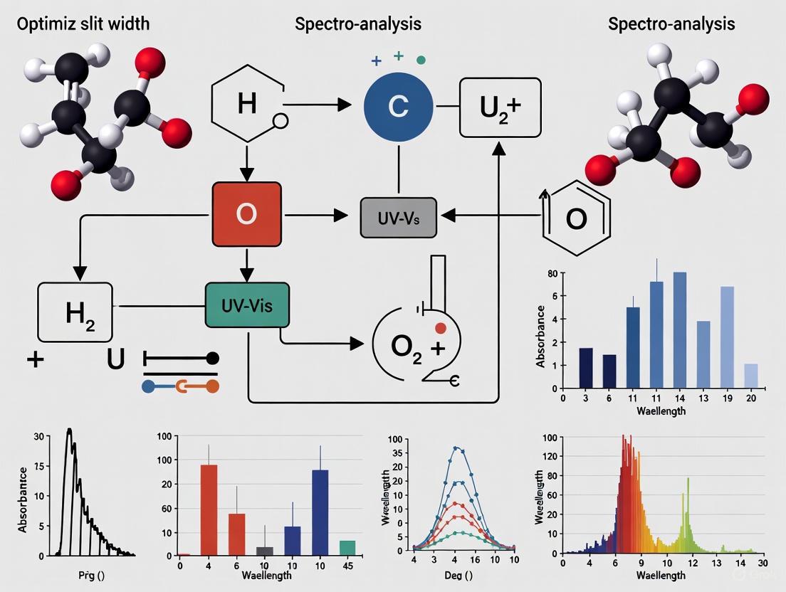
For pharmaceutical scientists, mastering these parameters is non-negotiable. The accuracy of absorbance measurements for drug compounds like sofosbuvir, simeprevir, and ledipasvir is critically dependent on the relationship between the instrument's spectral bandwidth and the sample's natural bandwidth—the inherent width of the sample's absorption band at half its maximum height [1] [4]. Optimal fidelity is achieved when the spectral bandwidth is 1/10 or less of the natural bandwidth, a condition that keeps measurement errors at or below 0.5% [2] [4]. The following diagram illustrates the core relationship between slit width, spectral bandwidth, and the resulting analytical performance, which forms the basis for the optimization strategies discussed in this guide.
Key Definitions and Instrumental Differences
Understanding the distinction between spectrometer types is crucial for correctly interpreting specifications and limitations.
Monochromator-Based Spectrophotometers
These instruments utilize a single detector and a monochromator containing an adjustable slit and a dispersive element (like a diffraction grating) to isolate individual wavelengths sequentially [1].
- Definition of Spectral Bandwidth: The width of the wavelength of light at half the maximum intensity of the light leaving the monochromator [1].
- Role of Slit Width: The slit width is the primary adjustable parameter determining the spectral bandwidth. It represents a critical trade-off: a narrower slit provides higher spectral resolution but reduces light throughput, potentially increasing noise and requiring longer measurement times [1] [3].
Diode-Array Spectrophotometers
These instruments use an array of detectors (diodes), each simultaneously responsible for detecting a specific, narrow wavelength range [1].
- Definition of Spectral Bandwidth: The wavelength range that each diode in the array can detect simultaneously [1].
- Role of Slit Width: While a slit is still present to control the amount of light entering the system, the spectral bandwidth is largely a fixed property determined by the physical design and spacing of the diode array itself [1]. This offers less user flexibility but enables extremely rapid data acquisition.
Table 1: Comparison of Spectrophotometer Types in Pharmaceutical Analysis
| Feature | Monochromator-Based | Diode-Array |
|---|---|---|
| Spectral Bandwidth | User-adjustable via slit width [1] | Fixed by diode array design [1] |
| Measurement Speed | Sequential wavelength measurement (slower) [1] | Simultaneous wavelength measurement (faster) [1] |
| Resolution Control | High degree of user control [1] | Limited user control [1] |
| Typical Pharmaceutical Use | High-resolution analysis for method development and validation | High-throughput analysis, reaction monitoring, and quality control |
The Scientist's Toolkit: Essential Materials for UV-Vis Analysis
Table 2: Key Research Reagent Solutions and Materials for Pharmaceutical UV-Vis
| Item | Function/Description | Example from Antiviral Drug Analysis [5] |
|---|---|---|
| Reference Standards | High-purity compounds used for calibration and method validation. | Sofosbuvir (99.25%), Simeprevir (98.96%), Ledipasvir (99.75%) from a regulatory authority. |
| HPLC-Grade Solvent | A high-purity solvent with minimal UV absorbance in the working range to prepare samples and blanks. | Ethanol (HPLC grade) used to dissolve drug standards and formulations. |
| Quartz Cuvettes | Cells that hold liquid samples. Quartz is essential for UV range measurements due to its high transmittance of UV light [6]. | 10 mm matched quartz cells for consistent path length. |
| Validated Software | Software for instrument control, data acquisition, and advanced chemometric analysis. | UV Probe software (v2.43) for acquisition; MATLAB for implementing CRACLS/SRACLS chemometric models [5]. |
| Vitexin2''-O-p-coumarate | Vitexin2''-O-p-coumarate, MF:C30H26O12, MW:578.5 g/mol | Chemical Reagent |
| 10α-Hydroxyepigambogic acid | 10α-Hydroxyepigambogic acid, CAS:887606-04-4, MF:C38H44O8, MW:628.7 g/mol | Chemical Reagent |
Optimizing Slit Width: A Practical Methodology
The following experimental protocol, inspired by a chemometric study on antiviral drugs, provides a framework for systematically optimizing slit width to achieve spectral fidelity [5].
1. Define Analytical Goal and Sample Characteristics:
- Identify the analytes (e.g., sofosbuvir, simeprevir, ledipasvir) and their expected concentration ranges [5].
- Acquire reference spectra for pure standards to estimate the natural bandwidth of the primary absorption peaks [1].
2. Establish the Initial Instrumental Setup:
- Use a double-beam UV-Vis spectrophotometer with an adjustable slit width [5].
- Employ a matched pair of quartz cuvettes with a 10 mm path length [5] [6].
- Prepare a stock solution of a target analyte in an appropriate solvent (e.g., 100 µg/mL in ethanol) [5].
- Set a sampling interval (data interval) that is equal to or slightly less than the spectral bandwidth you intend to test (e.g., for a 2 nm bandwidth, use a 1 or 2 nm interval) [1].
3. Execute the Slit Width Optimization Experiment:
- Prepare a series of dilutions from the stock solution to span the expected calibration range.
- Starting with the narrowest available slit width, record the absorbance spectrum of a mid-range concentration standard.
- Gradually increase the slit width in steps (e.g., 0.5 nm, 1 nm, 2 nm, 5 nm) and record the spectrum of the same standard each time.
- For each resulting spectrum, document the Signal-to-Noise Ratio (SNR) by measuring the peak height and the noise in a flat, non-absorbing region of the spectrum. Also, record the Absorbance Maxima and the Observed Peak Width at half the height (FWHM).
4. Analyze Data and Determine Optimal Slit Width:
- Plot SNR and Absorbance vs. Slit Width: This visualizes the trade-off. The optimal slit is often at the point where the SNR is high and absorbance begins to stabilize.
- Apply the 10% Rule: Calculate the ratio of the spectral bandwidth (dictated by the slit) to the natural bandwidth. The optimal setting should have a ratio of ≤ 0.1 for 99.5% accuracy [2] [4].
- Assess Resolution: If analyzing mixtures with close or overlapping peaks (like the antiviral drugs), ensure the chosen slit width allows for clear distinction between them.
Table 3: Quantitative Data from a Simulated Slit Width Optimization Experiment
| Slit Width (nm) | Spectral Bandwidth (nm) | *Measured Absorbance | Signal-to-Noise Ratio (SNR) | Observed Peak Width (FWHM, nm) |
|---|---|---|---|---|
| 0.5 | ~0.5 | 0.495 | 25:1 | 15.5 |
| 1.0 | ~1.0 | 0.499 | 100:1 | 15.8 |
| 2.0 | ~2.0 | 0.500 | 250:1 | 16.2 |
| 5.0 | ~5.0 | 0.485 | 500:1 | 18.5 |
Theoretical true absorbance is 0.500. A 5 nm slit causes a measurable decrease in peak height due to poor resolution [3].
The decision-making process for selecting the optimal slit width based on experimental goals is summarized in the workflow below.
Troubleshooting Guide and FAQs
This section addresses common issues related to slit width, bandwidth, and general instrument performance encountered in pharmaceutical research.
Frequently Asked Questions (FAQs)
Q1: How does slit width directly influence the shape and height of a spectral peak? A1: A wider slit width increases the spectral bandwidth, allowing a broader range of wavelengths to hit the detector simultaneously. This can cause absorption peaks to appear broader and their maximum height to be lower than the true value because the instrument is averaging the absorbance over a wider wavelength range [3]. This effect is critical when measuring substances with narrow natural bandwidths.
Q2: My spectrophotometer is giving inconsistent readings (drift). What should I check? A2:
- Light Source: An aging lamp (deuterium or tungsten) is a common cause. Replace the lamp if it is near or beyond its rated lifetime [7] [8].
- Warm-up Time: Ensure the instrument has stabilized for at least 20-30 minutes after powering on [7] [6].
- Calibration: Regularly perform a full baseline correction (with blank solvent) and recalibrate with certified reference standards [7] [9].
Q3: Why is my blank measurement failing, or why does the absorbance value keep fluctuating? A3:
- Cuvette Issues: Inspect the cuvette for scratches, residue, or fingerprints. Ensure it is correctly aligned in the holder and that you are using the correct type (e.g., quartz for UV) [7] [6] [8].
- Solvent Evaporation: If measuring over an extended period, solvent evaporation can change concentration and cause drift [6].
- Stray Light: Check for obstructions or debris in the light path [8].
Q4: When I try to set 0 Absorbance (100% Transmittance), the instrument displays an energy error (e.g., "L0" or "ENERGY ERROR"). What does this mean? A4: This indicates the detector is not receiving enough light. Likely causes include [8]:
- Failed Lamp: The lamp (especially the deuterium lamp for UV) may have reached the end of its life and needs replacement.
- Blocked Path: A cuvette with too high an absorbance or an object blocking the light path.
- Power Supply: In rare cases, a fault in the lamp's power supply or ignition circuit could be the cause.
Advanced Troubleshooting: Resolving Spectral Performance Issues
Table 4: Advanced Troubleshooting Guide for Spectral Fidelity
| Problem | Potential Causes | Diagnostic Steps | Solutions |
|---|---|---|---|
| Unexpected/High Noise | 1. Slit width too narrow [1].2. Lamp aging [7].3. Dirty optics or cuvette [7].4. Low source power (faulty power supply). | 1. Check noise level across a flat baseline region.2. Inspect lamp hours and energy output.3. Visually inspect and clean optics/cuvettes. | 1. Slightly increase slit width to improve SNR [1].2. Replace lamp [8].3. Clean components with recommended solvents.4. Contact technical service. |
| Poor Resolution (Cannot distinguish close peaks) | 1. Slit width too wide [1] [3].2. Incorrect sampling interval (larger than spectral bandwidth) [1].3. Instrument optical misalignment. | 1. Measure a standard with known fine structure (e.g., holmium oxide filter).2. Review and adjust data interval in method settings. | 1. Reduce slit width to the minimum practical for required SNR [1].2. Set sampling interval ≤ spectral bandwidth [1].3. Perform instrument qualification/service. |
| Photometric Inaccuracy (Absorbance too low) | 1. Spectral bandwidth too large relative to natural bandwidth (violates 10% rule) [1] [4].2. Stray light [3].3. Instrument requires calibration. | 1. Compare absorbance of a narrow-band standard at different slit widths.2. Perform a stray light test (e.g., with NiSO₄ or KCl solutions). | 1. Narrow the slit width to achieve a spectral bandwidth ≤ 10% of natural peak width [2] [4].2. Identify and eliminate source of stray light; service instrument.3. Perform full photometric accuracy calibration. |
Core Concepts: Slit Width and Its Impact on Spectrometer Performance
In a dispersive UV-Vis spectrophotometer, the slit is a critical adjustable aperture that controls the amount of light entering and exiting the monochromator. Its primary function is to govern both the spectral resolution and the intensity of light reaching the detector, creating a fundamental trade-off that researchers must manage [10] [11].
The width of the slit directly determines the spectral bandpass—the range of wavelengths that simultaneously pass through to the sample and detector. A narrower slit allows for a smaller bandpass, yielding higher spectral resolution and the ability to distinguish fine spectral details. Conversely, a wider slit increases the bandpass, which can obscure closely spaced peaks but allows significantly more light to pass through the system [10] [11].
The effect on light intensity is profound. As the slit width increases, two factors combine to boost the total light throughput:
- The total slit area increases, allowing a greater spatial fraction of the light source to enter.
- The spectral bandpass increases, allowing a broader range of wavelengths from the continuum source to pass through [10].
These factors cause the light level incident on the sample to increase with the square of the slit width. This heightened intensity directly improves the Signal-to-Noise Ratio (SNR), but the exact relationship depends on the dominant type of noise in the measurement system [10].
Table 1: How Slit Width Affects Key Spectrometer Performance Parameters
| Performance Parameter | Narrow Slit Width | Wide Slit Width |
|---|---|---|
| Spectral Resolution | Higher (Fine details are visible) | Lower (Peak broadening occurs) |
| Light Throughput | Lower | Higher (Increases with the square of the width) |
| Photon-Noise-Limited SNR | Lower | Higher (SNR ∠Slit Width) |
| Detector-Noise-Limited SNR | Lower | Higher (SNR ∠(Slit Width)²) |
| Deviation from Beer's Law | Lower (More linear calibration) | Higher (Increased non-linearity) |
Experimental Protocols for Slit Width Optimization
Protocol: Establishing the Optimal Slit Width for a Given Absorber
This methodology provides a systematic approach for determining the slit width that offers the best balance of signal quality and spectral fidelity for a specific analytical application, such as quantifying a pharmaceutical compound [10].
Key Reagent Solutions:
- Standard Solution: A pure sample of the analyte dissolved in an appropriate solvent.
- Blank Solution: The solvent used to dissolve the analyte.
- Certified Reference Materials: (e.g., Holmium Oxide filters) for wavelength calibration [12].
Procedure:
- System Preparation: Turn on the UV-Vis spectrophotometer and allow the light source (deuterium and/or tungsten lamp) to warm up for the recommended time, typically 20-30 minutes, to ensure stable output [6].
- Wavelength Calibration: Calibrate the wavelength accuracy of the instrument using a certified reference material like a Holmium Oxide filter [12].
- Initial Spectral Scan: Prepare a standard solution of the target analyte at a concentration expected in actual samples. Using a narrow slit width (e.g., 0.5 nm), record a high-resolution absorption spectrum of the standard to identify the wavelength of maximum absorption (λ_max) and observe the natural width of the absorption band [10].
- Slit Width Series: Set the instrument to the λ_max identified in the previous step. Measure the absorbance of the standard solution while systematically increasing the slit width. Record the absorbance value and any observable peak broadening at each setting.
- Signal-to-Noise Assessment: For each slit width setting, take multiple rapid readings of the blank solution at λ_max. Calculate the standard deviation of these readings, which represents the noise (N). The signal (S) is the average absorbance of the standard. Compute SNR as S/N [10].
- Data Analysis: Plot SNR versus slit width and absorbance versus slit width. The optimal slit width is typically the point just before the absorbance value begins to significantly decrease due to excessive bandpass, while the SNR has reached an acceptably high plateau [10].
Protocol: Verifying Analytical Curve Linearity at Different Slit Widths
This experiment assesses whether the chosen slit width is suitable for quantitative analysis by testing the linearity of the Beer-Lambert law calibration curve [10] [12].
Procedure:
- Standard Preparation: Prepare a series of standard solutions of the analyte across the desired concentration range.
- Measurement: Using the slit width selected from Protocol 2.1, measure the absorbance of each standard solution at λ_max.
- Calibration Curve: Plot absorbance versus concentration and perform a linear regression analysis.
- Linearity Evaluation: Examine the R² value and the residuals of the fit. A sufficiently wide slit may cause negative deviations from linearity, especially at higher absorbances, due to the polychromaticity effect [10]. If non-linearity is observed (e.g., R² < 0.999), consider using a narrower slit width and repeat the validation, accepting a potential reduction in SNR for improved quantitative accuracy.
Table 2: Troubleshooting Guide for Slit Width-Related Issues in Pharmaceutical Analysis
| Problem | Potential Cause | Solution |
|---|---|---|
| Low Signal or Noisy Baseline | Slit width is too narrow for the application, or detector noise is dominant. | Gradually increase the slit width until an acceptable SNR is achieved. Ensure the light source is warmed up [6] [8]. |
| Poor Resolution of Overlapping Peaks | Slit width is too wide, causing spectral bandpass to be larger than the separation between peaks. | Reduce the slit width to improve resolution, even if it results in a noisier signal that may require averaging multiple scans [11] [13]. |
| Deviation from Beer's Law (Non-linearity) | Excessive spectral bandpass from a wide slit introduces polychromatic light, violating a core assumption of the Beer-Lambert law [10]. | Narrow the slit width. Ensure the spectral bandpass is ≤ 1/10th the full width at half maximum (FWHM) of the absorption peak [10]. |
| Inaccurate Absorbance Readings | Stray light, which can be exacerbated by very wide slit settings, is reaching the detector [12]. | Clean the instrument's optics, use high-quality cuvettes, and avoid using slit widths that push the instrument beyond its design limits. Perform a stray light test [13]. |
FAQs on Slit Width Optimization
Q1: What is the single most important rule for choosing a slit width? There is no universal rule, as the optimal setting depends on your analytical goal. For recording an accurate absorption spectrum (e.g., for identity testing), prioritize resolution by using a narrow slit. For quantitative analysis where high SNR is critical for low-concentration detection, a wider slit is often preferable, provided it does not introduce significant non-linearity in the calibration curve [10].
Q2: How does the choice of slit width impact the analysis of pharmaceutical formulations? Excipients and degradation products in formulations can create complex, overlapping spectra. A narrow slit width is essential to resolve the analyte's peak from these interferences. Furthermore, for dissolution testing or assaying high-potency drugs where samples are highly diluted, a wider slit can enhance SNR and lower the limit of detection [10] [12].
Q3: My readings are noisy, but when I widen the slit, the absorbance of my standard changes. What should I do? A change in absorbance with slit width indicates your spectral bandpass is too wide relative to your analyte's absorption band, leading to a deviation from the Beer-Lambert law. You have reached a practical limit. Instead of widening the slit further, improve SNR by other means, such as increasing the source integration time, using a cuvette with a longer pathlength, or concentrating your sample [10] [12].
Q4: How often should I check or calibrate the slit width mechanism on my instrument? While the slit width setting itself doesn't typically require user calibration, the overall performance of the spectrophotometer should be verified regularly. Follow a quality control procedure that includes checks for wavelength accuracy, absorbance accuracy, and stray light using certified reference materials. Significant drifts in these parameters could indicate issues with the optical path, including the slits [12] [8].
Decision Pathways and Workflows
The following diagram illustrates the logical decision process for optimizing slit width based on analytical objectives:
The Scientist's Toolkit: Essential Materials for UV-Vis Spectroscopic Analysis
Table 3: Key Research Reagents and Materials for Pharmaceutical UV-Vis Analysis
| Item | Function / Explanation |
|---|---|
| Quartz Cuvettes | Provide high transmission of UV and visible light. Essential for accurate absorbance measurements below ~350 nm where plastic or glass absorbs strongly [6]. |
| Certified Reference Materials (CRMs) | Holmium Oxide solution or filters are used for wavelength calibration. Neutral density filters or potassium dichromate solutions can be used for absorbance accuracy verification [12]. |
| High-Purity Solvents | Spectroscopic-grade solvents (e.g., methanol, acetonitrile, water) with low UV absorbance are crucial to minimize background noise and avoid masking the analyte's signal [12] [13]. |
| Neutral Density Filters | Used to attenuate light source intensity without shifting its spectral composition. Helpful in diagnostics and for verifying detector linearity [14]. |
| Syringe Filters (0.45 µm or 0.2 µm) | Used to filter sample solutions immediately before analysis to remove particulates or micro-bubbles that cause light scattering, a common source of error and noise [12] [13]. |
| Antidepressant agent 1 | Antidepressant agent 1, MF:C16H19BrN2, MW:319.24 g/mol |
| Galanthamine hydrobromide | Galanthamine hydrobromide, CAS:69353-21-5, MF:C17H22BrNO3, MW:368.3 g/mol |
Theoretical Foundations: How Slit Width Influences Key Analytical Parameters
The Fundamental Relationship Between Slit Width and Spectrophotometer Performance
In UV-Vis spectrophotometry, the slit width is a crucial mechanical or software-controlled parameter that directly governs the bandwidth of light reaching the sample. This width represents a fundamental compromise in analytical performance, creating an inverse relationship between signal-to-noise ratio (sensitivity) and spectral resolution.
- Wider Slits allow more light to pass through the system, which increases the signal intensity at the detector and improves the signal-to-noise ratio. This enhancement typically leads to improved sensitivity and lower limits of detection (LOD). However, this comes at the cost of spectral resolution, as wider slits can cause adjacent spectral peaks to merge, potentially reducing the linear dynamic range at higher concentrations due to the Beer-Lambert law deviation [15].
- Narrower Slits provide higher spectral resolution by permitting a narrower band of wavelengths to interact with the sample. This is critical for distinguishing between analytes with close absorbance maxima or for obtaining fine spectral details for qualitative identification. The trade-off is reduced light throughput, which can increase baseline noise, potentially raising the LOD and limiting the lower end of quantification (LOQ) [15].
Table 1: Primary Effects of Slit Width Adjustment on Analytical Figures of Merit
| Analytical Figure of Merit | Effect of Wider Slit | Effect of Narrower Slit |
|---|---|---|
| Signal-to-Noise Ratio | Increases | Decreases |
| Spectral Resolution | Decreases | Increases |
| Limit of Detection (LOD) | Generally Lowers | May Increase |
| Limit of Quantification (LOQ) | Generally Lowers | May Increase |
| Linearity Range | May narrow at high concentrations | Can be preserved or improved |
Interaction with Data Sampling and Signal Processing
The effect of slit width is further modulated by instrument settings for data acquisition. A faster data sampling rate captures more data points across a chromatographic or spectral peak, improving its modeling and reproducibility. However, this can also capture more high-frequency noise. The time constant (or response time) acts as an electronic filter to smooth this noise. Finding the optimal balance is key; excessively fast sampling with inadequate filtering can degrade the signal-to-noise ratio, indirectly impacting the perceived LOD and LOQ [15].
Troubleshooting Guide: Slit Width Optimization FAQs
This section addresses common practical challenges faced by researchers when configuring slit width for pharmaceutical applications.
FAQ 1: How should I systematically adjust the slit width to lower the detection limit for a trace impurity?
Challenge: The current method lacks the sensitivity to detect and quantify a low-concentration degradation product.
Solution: Implement a systematic optimization protocol.
- Baseline Measurement: Begin with the current slit width setting and record the baseline noise over a region where no analyte elutes.
- Incremental Widening: Gradually increase the slit width in small steps. At each step, inject a standard at a concentration near the expected LOD.
- Signal-to-Noise Calculation: For each injection, calculate the signal-to-noise (S/N) ratio by dividing the analyte peak height by the amplitude of the baseline noise.
- Identify Optimum: The optimal slit width for the lowest LOD is the point at which the S/N ratio is maximized. Further widening may decrease resolution without meaningful S/N gain [15].
Precaution: Continuously monitor the resolution between the impurity peak and the main API peak. A wider slit might cause co-elution, leading to inaccurate quantification.
FAQ 2: Why did my calibration curve become non-linear after I changed the slit width to improve sensitivity?
Challenge: A wider slit setting, intended to improve LOD, has caused deviation from the Beer-Lambert law at higher concentrations.
Root Cause: Excessively wide slit widths can introduce stray light or cause a phenomenon known as "bandwidth error." If the slit's spectral bandwidth becomes a significant fraction of the natural width of the analyte's absorption band, the relationship between absorbance and concentration ceases to be linear, particularly at higher absorbances [16] [15].
Solution:
- Re-optimize: Slightly narrow the slit width until linearity is restored across your required concentration range. The ideal width provides sufficient sensitivity while maintaining a correlation coefficient (R²) of ≥ 0.999 for the calibration curve.
- Validate the Method: After selecting the new slit width, perform a full validation of the method's linearity, as per ICH guidelines, to ensure it meets the criteria for your intended application [17] [18].
FAQ 3: How do I balance slit width with data sampling rate to get the best peak shape and lowest LOQ in USP/UHPLC methods?
Challenge: Modern high-efficiency separations with UHPLC produce very narrow peaks, requiring careful instrument settings to prevent data loss and maintain low LOQs.
Solution: Integrate the optimization of slit width with data acquisition parameters.
- Set Sampling Rate First: Ensure your detector's data sampling rate is fast enough to capture at least 20-25 data points across the narrowest peak of interest. This is critical for accurate peak integration and quantification [15].
- Adjust Slit Width for Sensitivity: Widen the slit to improve S/N, as previously described.
- Apply Time Constant/Filtering: If a faster sampling rate introduces excessive high-frequency noise, use the instrument's time constant (or digital filtering) to smooth the baseline. The goal is to reduce noise without distorting the peak shape. A slightly wider slit can sometimes allow for the use of a lighter filter, preserving peak fidelity [15].
Table 2: Troubleshooting Guide for Common Slit Width-Related Issues
| Problem | Potential Root Cause | Corrective Action |
|---|---|---|
| High Baseline Noise | Slit width too narrow for the application. | Gradually increase slit width while monitoring the signal-to-noise ratio of a low-level standard. |
| Poor Resolution of Co-eluting Peaks | Slit width is too wide, reducing spectral resolution. | Narrow the slit width to improve peak separation; consider alternative wavelengths for greater specificity. |
| Loss of Linearity at High Concentrations | Bandwidth error or stray light from an excessively wide slit. | Slightly narrow the slit width and re-establish the calibration curve. |
| Poor Reproducibility of Integration | Inconsistent peak shape due to suboptimal slit width/data rate combination. | Optimize slit width for a stable baseline, then ensure a sufficiently high data sampling rate (>20 pts/peak). |
Experimental Protocols for Method Development and Validation
Protocol for Establishing the Linear Range and Determining LOD/LOQ
This protocol provides a step-by-step guide for validating the key figures of merit during method development, incorporating slit width as a critical variable.
Principle: A series of standard solutions are analyzed to construct a calibration curve. The linearity, sensitivity, and precision of the method are evaluated statistically. The standard deviation of the response and the slope of the calibration curve are used to determine LOD and LOQ [17] [19].
Materials & Reagents:
- API Reference Standard: Certified material of high and known purity.
- Appropriate Solvent: HPLC-grade or spectrophotometric-grade solvent that does not interfere with the analysis [16].
- Volumetric Flasks: Class A, for accurate preparation of standard solutions.
- UV-Vis Spectrophotometer or HPLC-UV System: Calibrated and with controllable slit width.
Procedure:
- Stock Solution Preparation: Accurately weigh and dissolve the API reference standard to prepare a stock solution of known concentration (e.g., 1000 μg/mL).
- Working Standard Dilutions: Dilute the stock solution serially to prepare at least 5-6 standard solutions covering the expected working range (e.g., from 50% to 150% of the target assay concentration).
- System Optimization: Set the initial slit width based on instrument recommendations or prior knowledge. Analyze the standards.
- Analysis: Measure the absorbance (or peak area) for each standard solution. Perform replicate measurements (n=3) to assess precision.
- Data Analysis:
- Linearity: Plot the mean response (y-axis) against concentration (x-axis). Calculate the regression equation (y = mx + c) and the correlation coefficient (R²). A value of ≥ 0.999 is typically expected for quantitative assays.
- LOD and LOQ Calculation: Calculate using the formulas based on the standard deviation of the response (σ) and the slope (S) of the calibration curve.
- Iterative Optimization: Repeat steps 3-5 with different slit widths to find the setting that provides the best combination of linearity (R²), low LOD/LOQ, and acceptable signal-to-noise.
Case Study: Simultaneous Determination of Atorvastatin and Aspirin
A published study on the development of a first-derivative synchronous fluorometric method for Atorvastatin (ATO) and Aspirin (ASP) exemplifies the rigorous validation of figures of merit, principles that are directly transferable to slit width optimization in UV-Vis.
Methodology:
- Technique: First-derivative synchronous fluorescence spectroscopy (FDSFS) was used to resolve the spectral overlap of the two drugs.
- Optimization: Parameters influencing fluorescence intensity, including spectral scanning speed and data interval (conceptually analogous to sampling rate in UV-Vis), were optimized for enhanced sensitivity and selectivity [17].
Validation Results:
- Linearity: Excellent linearity was demonstrated over defined ranges (0.4–6 μg/mL for ATO and 1–10 μg/mL for ASP) with high correlation coefficients.
- Sensitivity: The method achieved low LOD (0.03 μg/mL for ATO, 0.342 μg/mL for ASP) and LOQ (0.248 μg/mL for ATO, 0.714 μg/mL for ASP), confirming its suitability for trace analysis [17].
This case study underscores that systematic optimization of instrumental parameters is fundamental to achieving robust analytical figures of merit for pharmaceutical assays.
The Scientist's Toolkit: Essential Research Reagent Solutions
The following table lists key materials and reagents critical for successfully conducting experiments related to method development and validation as discussed in this guide.
Table 3: Key Research Reagent Solutions for Pharmaceutical UV-Vis Analysis
| Reagent/Material | Function/Application | Critical Quality Attributes |
|---|---|---|
| Certified Reference Standards | Used for accurate preparation of calibration standards to establish method linearity, LOD, and LOQ [17] [19]. | High purity (>98%), certified concentration, and stability. |
| HPLC-Grade Solvents | Used for dissolving samples and standards, and as mobile phase components. Minimizes background UV absorption and interference [16]. | Low UV cutoff, high purity, free of particulate matter. |
| Holmium Oxide Filter | A wavelength verification standard for calibrating the spectrophotometer, ensuring accuracy of the absorbance maxima used for quantification [16]. | NIST-traceable certification. |
| Quartz Cuvettes | Sample holders for UV-Vis measurement. Quartz is transparent across the UV and visible range. | Matched path length, clear optical surfaces without scratches. |
| Buffer Components | Used to prepare mobile phases or sample solutions at controlled pH, which can affect analyte stability and spectral properties [17]. | Analytical grade, low UV absorbance. |
| Monomethyl lithospermate | Monomethyl lithospermate, MF:C28H24O12, MW:552.5 g/mol | Chemical Reagent |
| Cyclopentolate Hydrochloride | Cyclopentolate Hydrochloride, CAS:60452-44-0, MF:C17H26ClNO3, MW:327.8 g/mol | Chemical Reagent |
Ultraviolet-Visible (UV-Vis) spectroscopy is an indispensable analytical technique in pharmaceutical research and drug development. This method measures how molecules absorb light across the ultraviolet and visible regions of the electromagnetic spectrum (typically 100-900 nm), providing critical insights into molecular structure, concentration, purity, and functional groups. The resulting spectrum reveals electronic transitions within a molecule, enabling researchers to identify compounds, quantify active pharmaceutical ingredients (APIs), and assess sample quality. Within this field, the choice of spectrophotometer configuration—single-beam, double-beam, or array detector system—profoundly impacts data quality, analytical throughput, and method robustness. Furthermore, instrumental parameters such as slit width require careful optimization to balance spectral resolution and signal-to-noise ratio, a consideration particularly crucial for regulatory-compliant pharmaceutical applications where data integrity is paramount.
Comparative Analysis of Spectrophotometer Configurations
The core configurations of UV-Vis spectrophotometers differ fundamentally in their optical designs, each presenting distinct advantages and limitations for pharmaceutical analysis.
Single-Beam Systems
Operating Principle: Single-beam instruments utilize a single light path that passes sequentially through the reference and sample. The instrument first measures the intensity of the incident light (Iâ‚€) with the reference blank in place, then replaces the blank with the sample to measure the transmitted light (I). Absorbance is calculated as A = log(Iâ‚€/I).
Characteristics and Pharmaceutical Applications:
- Advantages: Simpler optical design often translates to lower cost, higher light throughput, and potentially higher sensitivity due to fewer optical components. This can be beneficial for detecting low-concentration impurities.
- Limitations: Susceptible to source drift because any fluctuation in the lamp intensity between the Iâ‚€ and I measurements directly affects the calculated absorbance. This requires regular recalibration and stable power conditions.
- Typical Pharmaceutical Use: Best suited for quantitative single-wavelength assays (e.g., content uniformity testing, dissolution profiling at a fixed wavelength) where high throughput and cost-effectiveness are prioritized, and environmental conditions are well-controlled.
Double-Beam Systems
Operating Principle: Double-beam instruments employ a beam splitter or chopper to create two synchronous light paths: one through the sample and another through a reference blank. The detector measures both beams almost simultaneously, continuously rationing the intensities to calculate absorbance in real-time.
Characteristics and Pharmaceutical Applications:
- Advantages: Excellent stability and compensation for source drift, fluctuations in power supply, and electronic noise. This enhances long-term measurement reliability and is ideal for kinetic studies and spectral scanning.
- Limitations: More complex optics, typically higher cost, and reduced light throughput compared to single-beam systems due to the beam-splitting mechanism.
- Typical Pharmaceutical Use: Ideal for applications requiring high stability, such as full spectral scans for identity testing, kinetic reaction monitoring (e.g., enzyme activity assays), and any analysis where environmental fluctuations may be a concern.
Array Detector (Diode Array) Systems
Operating Principle: Instead of a monochromator before the sample, these systems use a polychromatic light source that passes through the sample first. The transmitted light is then dispersed onto a fixed array of hundreds of individual photodiodes, allowing for simultaneous detection of all wavelengths.
Characteristics and Pharmaceutical Applications:
- Advantages: Extremely fast acquisition (complete spectra in milliseconds), enabling real-time monitoring of fast reactions (e.g., stopped-flow kinetics) and HPLC detection. Offers high reproducibility as there are no moving mechanical parts in the monochromator.
- Limitations: Generally has lower resolution compared to high-quality scanning monochromators and a typically lower dynamic range. The light source's spectral output can also affect the signal-to-noise ratio across the wavelength range.
- Typical Pharmaceutical Use: Perfect for hyphenated techniques like HPLC-DAD (Diode Array Detection) for peak purity assessment, monitoring rapid reaction kinetics, and quality control environments requiring very high throughput.
Table 1: Quantitative Comparison of UV-Vis Spectrophotometer Systems
| Feature | Single-Beam | Double-Beam | Array Detector |
|---|---|---|---|
| Measurement Speed | Slow (sequential) | Moderate (rapid alternation) | Very Fast (simultaneous) |
| Stability / Drift Correction | Poor | Excellent | Good |
| Wavelength Selection | Sequential scanning | Sequential scanning | Simultaneous all wavelengths |
| Mechanical Complexity | Low | High | Moderate |
| Typical Cost | Low | High | Moderate to High |
| Best for Pharmaceutical Applications | Routine quantitative analysis at fixed wavelength | High-precision scanning, kinetics, R&D | HPLC detection, fast kinetics, process monitoring |
The Critical Role of Slit Width Optimization
The slit width is a crucial yet often overlooked parameter in UV-Vis spectroscopy. It controls the bandwidth of light that passes through the sample and enters the detector, directly influencing two key, competing spectral properties: resolution and signal-to-noise ratio.
How Slit Width Works: A narrower slit allows a smaller, more monochromatic band of light to pass, resulting in higher spectral resolution. This means you can distinguish between two closely spaced absorption peaks. Conversely, a wider slit permits a broader band of light to pass, which delivers more light energy to the detector, thereby increasing the signal-to-noise ratio and reducing spectral noise.
The Trade-Off: The relationship between slit width and data quality is a fundamental trade-off. Excessively narrow slits can lead to excessive noise, making it difficult to discern true absorption peaks. On the other hand, overly wide slits can cause a loss of spectral detail, including broader peaks and lower peak heights, as the instrument can no longer resolve fine spectral features. In extreme cases, a slit width equal to or larger than the natural width of an absorption band (Full Width at Half Maximum, or FWHM) will lead to inaccurate, lower absorbance measurements and poorly resolved bands [3].
Table 2: Effect of Slit Width on Spectral Data
| Slit Width Setting | Spectral Resolution | Signal-to-Noise Ratio | Peak Shape & Height | Ideal Application Context |
|---|---|---|---|---|
| Narrow (e.g., 1 nm) | High (sharp peaks) | Low (more noise) | Accurate height and fine structure preserved | Identifying sharp peaks, analyzing complex mixtures with overlapping bands |
| Wide (e.g., 5 nm) | Low (broad peaks) | High (less noise) | Peaks are broader and may have lower apparent height | Quantitative analysis of a single, well-resolved peak, analyzing low-absorbance samples |
Optimization for Pharmaceutical Applications: For most quantitative pharmaceutical analyses (e.g., API concentration determination via Beer-Lambert Law), a primary goal is a high signal-to-noise ratio. Therefore, the slit width should be opened as wide as possible without causing a significant loss in the definition of the absorbance peak being measured. A practical protocol is to perform a slit-width scan on a standard solution: start with a narrow slit and gradually increase the width while monitoring the peak shape and the baseline noise. Choose the widest slit width where the peak height and width remain constant, ensuring robust and reproducible quantitative results.
Troubleshooting Guide: FAQs on Instrument-Specific Issues
Q: My spectrophotometer is giving very noisy or unstable absorbance readings, particularly at higher absorbance values (above 1.0). What could be the cause?
A: Noisy data can stem from several instrument-specific and methodological factors.
- Instrument Check: For single-beam systems, ensure the lamp has warmed up sufficiently (typically 20-30 minutes for tungsten halogen or arc lamps) to stabilize [6]. Verify that the instrument is set to an appropriate integration time or scan speed; slowing down the measurement can improve the signal-to-noise ratio.
- Methodology Check: Adhere to the ideal absorbance range. Absorbance readings often become unstable and non-linear above 1.0 due to instrumental limitations like stray light [20] [21]. If your sample is too concentrated, dilute it to bring the absorbance at λmax between 0.1 and 1.0 for reliable quantitative analysis [21]. Ensure your cuvettes are clean and free of scratches, and that the sample is fully dissolved and free of particles that could scatter light.
Q: Why won't my spectrometer calibrate, or why does it show an error like 'Calibration Failed'?
A: Calibration failures are often related to fundamental setup issues.
- Procedure: Remember that you must calibrate the spectrometer (using an appropriate blank solvent) every time you use it in Absorbance or Transmittance mode [20].
- Blank Issues: Ensure the blank cuvette is perfectly clean and made of the correct material (e.g., quartz for UV work). The blank must be compatible with the solvent and not absorb in the wavelength range you are using. Confirm that the blank is properly positioned in the beam path.
- Instrument Health: A failing or cold light source can prevent successful calibration. Allow the lamp to warm up and listen for any unusual sounds. In severe cases, the lamp may need replacement.
Q: I am seeing unexpected peaks in my spectrum. How should I investigate this?
A: Unexpected peaks are most frequently related to sample or cuvette contamination.
- Contamination Investigation: Thoroughly clean your cuvettes using a recommended protocol. Handle cuvettes only with gloves to avoid fingerprint oils [6]. Check your solvents and reagents for purity, as contaminants or degraded solvents can introduce new chromophores.
- Solvent Interference: Verify that your solvent does not absorb significantly in the wavelength range you are scanning. For example, acetone absorbs strongly in the UV region and can obscure sample absorption below ~330 nm [21].
Q: What is the impact of resolution (slit width) on my spectral results for a pharmaceutical compound?
A: As detailed in Section 3, the slit width has a direct impact.
- Too Wide: A slit width set too large will broaden your absorption peaks and can decrease the measured peak height. This lack of resolution can hide the fine structure of a peak and make it impossible to distinguish between two closely eluting compounds in a mixture, which is critical for peak purity assessment [3].
- Too Narrow: A very narrow slit provides high resolution but reduces the light energy reaching the detector, resulting in a noisier spectrum that can mask low-intensity features.
- Action: Always justify and document the slit width used in your standard operating procedures (SOPs) for regulatory purposes.
The Scientist's Toolkit: Essential Research Reagent Solutions
Table 3: Key Materials and Their Functions in Pharmaceutical UV-Vis Analysis
| Item | Function & Importance | Technical Notes |
|---|---|---|
| Quartz Cuvettes | Sample holder for liquid analysis. Quartz is essential for UV range measurements (below ~350 nm) as glass and plastic absorb UV light. | Reusable; handle with gloves; ensure pathlength (e.g., 1 cm) is appropriate for concentration [20] [6]. |
| Optically Matched Cuvettes | A matched pair for double-beam instruments. | Critical for accurate blank subtraction; eliminates errors from slight differences in cuvette transmission [21]. |
| High-Purity Solvents | To dissolve the analyte and serve as the blank matrix. | Must be "spectroscopic grade" to ensure low UV absorption and avoid introducing contaminant peaks [21]. |
| Standard Reference Materials | Certified reference materials of the target analyte. | Used for validation of the method, calibration curve creation, and verifying instrument performance [21]. |
| Cuvette Cleaning Kit | Solutions and tools for proper cuvette cleaning. | Prevents cross-contamination between samples; essential for data integrity. |
| Phorbol 12,13-Dibutyrate | Phorbol 12,13-Dibutyrate, CAS:61557-88-8, MF:C28H40O8, MW:504.6 g/mol | Chemical Reagent |
| 2-(Morpholin-4-yl)ethane-1-sulfonamide | 2-(Morpholin-4-yl)ethane-1-sulfonamide, MF:C6H14N2O3S, MW:194.25 g/mol | Chemical Reagent |
Experimental Workflow and Decision Logic
The following diagram illustrates the logical workflow for selecting and utilizing a UV-Vis system for pharmaceutical analysis, incorporating key considerations like slit width optimization and troubleshooting.
Diagram 1: UV-Vis Analysis Workflow
Strategic Slit Width Selection for Chemometric and Multicomponent Analysis
Technical FAQ: Resolving Spectral Overlap
Question: What are the primary spectrophotometric methods for resolving severely overlapping spectra of co-administered antiviral drugs like Remdesivir (RDV) and Moxifloxacin (MFX)?
Several sophisticated mathematical spectrophotometric techniques have been successfully developed and validated to resolve the significant spectral overlap between RDV and MFX without requiring preliminary separation. These methods enable accurate, simultaneous quantification in pharmaceutical dosage forms and spiked human plasma [22].
Key Methodologies and Their Parameters:
Table 1: Spectrophotometric Methods for Resolving RDV and MFX Spectral Overlap
| Method Name | Underlying Principle | Key Analytical Wavelengths | Measured Parameter |
|---|---|---|---|
| Ratio Derivative (1DD) [22] | Derivative of the ratio spectrum of the mixture using a standard divisor. | RDV: 250 nm, MFX: 290 nm | Amplitude of the first derivative of the ratio spectrum |
| Ratio Difference (RD) [22] | Difference in amplitudes at two carefully selected wavelengths in the ratio spectrum. | RDV: 247 nm & 262 nm, MFX: 299 nm & 313 nm | Difference in peak amplitudes (ΔP) |
| Mean Centering (MC) [22] | Mathematical transformation of the ratio spectrum to a mean-centered form. | RDV: 247 nm, MFX: 299 nm | Mean-centered value |
| Area Under the Curve (AUC) [22] | Calculation of the area under the zero-order spectrum for two selected wavelength ranges. | Ranges: 243–248 nm & 290–300 nm | Area under the curve (AUC) |
| Absorbance Subtraction (AS) [23] | Uses an absorbance factor and the isoabsorptive point to mathematically separate contributions. | 229 nm (λiso) & 360 nm | Absorbance at isoabsorptive point |
Question: How does spectral bandwidth (slit width) impact the accuracy of these multicomponent analyses?
The spectral bandwidth of a spectrophotometer, determined by its physical slit width, is a critical parameter that directly affects resolution and accuracy [24].
- Narrower Bandwidth (Smaller Slit Width): Provides higher spectral resolution, which is essential for accurately distinguishing between the closely overlapping peaks of drugs like RDV and MFX. This leads to more precise results when applying mathematical techniques like ratio derivative or ratio difference [24].
- Wider Bandwidth (Larger Slit Width): Allows more light to reach the detector, which can be beneficial for signal-to-noise ratio in some cases, but at a cost. It can lower resolution and accuracy, particularly for samples with sharp, overlapping absorption peaks, by effectively "averaging" the absorbance over a wider range of wavelengths [24].
For the quantitative determination of overlapping drugs, the instrument's spectral bandwidth should be optimized and kept below the width of the spectral peaks of the analytes to ensure accurate measurement of their true extinction coefficients [24].
Experimental Protocol: Implementing the Ratio Difference Method
This protocol provides a step-by-step guide for the simultaneous determination of Remdesivir (RDV) and Moxifloxacin (MFX) using the Ratio Difference method, as derived from the literature [22].
Research Reagent Solutions
Table 2: Essential Materials and Reagents
| Item | Specification / Function |
|---|---|
| Remdesivir (RDV) & Moxifloxacin (MFX) Standards | High purity (e.g., 99.15% and 99.45% respectively) for calibration [22]. |
| Methanol | HPLC/spectroscopic grade, used as the solvent [22] [23]. |
| Volumetric Flasks | Class A, for precise preparation of standard and sample solutions. |
| UV-Vis Spectrophotometer | Equipped with 10 mm quartz cells and software for spectral manipulation (e.g., Shimadzu UV-1800) [22] [23]. |
| Ultrasonic Bath | To ensure complete dissolution of standards and samples. |
Step-by-Step Procedure:
Preparation of Standard Stock Solutions: Accurately weigh and dissolve 100 mg each of RDV and MFX pure powders separately in 70 mL methanol. Sonicate for 15 minutes and dilute to 100 mL with methanol to obtain 1000 µg/mL stock solutions. Further dilute these stocks with methanol to prepare 100 µg/mL working standard solutions [22].
Construction of Calibration Curves:
- Into a series of 10 mL volumetric flasks, transfer aliquots of RDV working standard solution to produce final concentrations of 1–15 µg/mL.
- Similarly, prepare a separate series for MFX with final concentrations of 1–10 µg/mL.
- Dilute all flasks to the mark with methanol.
- Record the zero-order absorption spectra (200–400 nm) of all solutions against a methanol blank [22].
Optimization of Divisor Concentration:
- To select the optimal concentration for the divisor spectrum, test different concentrations of one drug (e.g., 6 µg/mL MFX) by dividing the spectrum of the other drug (RDV) by them. The divisor that produces a clean ratio spectrum with minimal noise is selected [22].
Processing of Ratio Spectra and Data Analysis:
- Using the instrument software, divide the stored zero-order spectra of all RDV standard solutions by the spectrum of the optimized MFX divisor (e.g., 6 µg/mL). This generates the ratio spectra for RDV.
- Similarly, divide the spectra of all MFX standard solutions by the spectrum of the optimized RDV divisor (e.g., 8 µg/mL) [22].
- For RDV, measure the amplitudes of its ratio spectrum at 247 nm and 262 nm.
- For MFX, measure the amplitudes of its ratio spectrum at 299 nm and 313 nm.
- For each drug, plot the difference in amplitudes (ΔP) between the two selected wavelengths against its corresponding concentration to establish the calibration curve and regression equation [22].
Analysis of Laboratory-Prepared Mixtures or Samples:
- Prepare mixtures of RDV and MFX in known ratios in 10 mL volumetric flasks and dilute to volume with methanol.
- Record the zero-order spectrum of the mixture.
- Process the mixture spectrum using the exact same divisors and wavelengths established in the calibration steps.
- Use the regression equations to calculate the concentration of each drug in the mixture from the measured ΔP values [22].
The logical workflow for the entire experiment, from setup to analysis, is summarized below.
Troubleshooting Guide: Common Issues and Solutions
Problem: Poor reproducibility and high noise in ratio spectra.
- Potential Cause 1: Inconsistent or non-optimized divisor concentration.
- Potential Cause 2: Excessive spectral bandwidth (slit width) leading to poor resolution.
- Solution: Reduce the spectrophotometer's slit width to achieve a narrower spectral bandwidth. This improves the resolution of fine spectral features, which is critical for mathematical methods [24].
- Potential Cause 3: Stray light within the instrument, especially critical at high absorbance values.
- Solution: Ensure the instrument is well-maintained. Use appropriate pathlengths or dilute samples to keep absorbance readings within the validated linear range of the method and instrument [24].
Problem: Inaccurate quantification of one drug in the presence of the other.
- Potential Cause 1: Wavelength selection not optimal for the specific method.
- Solution: Re-investigate the ratio spectra to confirm the selected wavelength pairs (for Ratio Difference) or the single wavelength (for Mean Centering) show a linear and significant change with concentration while being insensitive to the other component [22].
- Potential Cause 2: Significant matrix interference from excipients or plasma components.
- Solution: The proposed methods have been applied to spiked human plasma. If interference is suspected, ensure the sample preparation procedure (e.g., protein precipitation, dilution) is followed precisely as validated. The use of multiple mathematical techniques (e.g., Ratio Difference and Mean Centering) can serve as a cross-check [22].
Why is slit width so critical in UV-Vis method development?
Slit width directly controls the bandwidth of light reaching your sample. This creates a fundamental trade-off: a narrower slit provides better spectral resolution, allowing you to distinguish between closely spaced absorbance peaks, while a wider slit increases light throughput, improving the signal-to-noise ratio and detector sensitivity for more reliable quantification of low-concentration analytes [16]. Optimizing this balance is essential for developing a robust analytical method, particularly for complex pharmaceutical mixtures.
Troubleshooting Guide: Slit Width and Experimental Design FAQs
Q1: How do I systematically determine the optimal slit width for a new method? A1: Follow this structured protocol:
- Initial Setup: Use a standard solution of your target analyte at a mid-range concentration. Set the spectrophotometer to its default or median slit width setting [16].
- Spectral Scan: Perform a full spectral scan (e.g., 200-400 nm) of the standard to identify its maximum absorbance wavelength (λmax) [16].
- Iterative Testing: At the identified λmax, sequentially measure the absorbance of the standard using a range of slit widths, from the narrowest to the widest available on your instrument.
- Data Analysis: Plot the recorded absorbance against the slit width.
- Optimization Rule: Select the widest slit width that does not result in a significant decrease in measured absorbance. This point ensures maximum signal intensity without sacrificing the spectral detail required for accurate quantification [16].
Q2: My peaks are broad and poorly resolved. Could slit width be the cause? A2: Yes, this is a classic symptom of an inappropriately wide slit width. An overly wide slit passes a broader range of wavelengths, which can cause adjacent peaks to merge and reduce your method's ability to distinguish individual components in a mixture [16].
- Solution: Progressively narrow the slit width and observe the effect on your chromatogram. You should see peaks become sharper and better separated. Be aware that an excessively narrow slit will lead to a noisier baseline; the goal is to find a width that provides the best compromise between resolution and signal stability [16].
Q3: How does slit width interact with other factors in an Experimental Design (DoE)? A3: In a DoE, slit width is a critical instrumental factor that can interact with chemical and sample preparation variables. A robust method requires testing slit width in combination with other parameters.
Table: Key Factor Interactions for DoE
| Factor | Interaction with Slit Width | DoE Consideration |
|---|---|---|
| Analyte Concentration | Low concentrations may require wider slits for a detectable signal. | Treat as a critical response variable to model. |
| Sample Purity/Spectral Overlap | Complex mixtures with overlapping peaks need narrower slits for resolution. | A key constraint that defines the required resolution goal. |
| Solvent Type | Can influence baseline noise and spectral profile. | Include as a categorical factor in the design. |
| pH / Mobile Phase | Can shift λmax, altering the optimal slit condition. | Test slit width at different pH/phase levels to find a robust setting [5]. |
Q4: I've optimized the slit width, but now my baseline is too noisy. What should I do? A4: This indicates the slit may be too narrow, severely limiting light energy to the detector [16].
- Primary Action: Slightly widen the slit width until the baseline stabilizes to an acceptable level, even if it involves a minor loss in resolution.
- Supplementary Strategy: If possible, increase the concentration of your sample or use a cuvette with a longer path length to enhance the signal without adjusting the slit.
Q5: How can I use a factorial design to model the effect of slit width? A5: A screening design, such as a Two-Level Full or Fractional Factorial, is highly effective.
- Define Factors and Ranges: Select slit width (e.g., 1 nm vs. 2 nm), analyte concentration, and pH as your key factors.
- Set Responses: Define your measurable outputs, such as peak absorbance, signal-to-noise ratio, and peak width at half height (a measure of resolution).
- Run Experiments: Execute the experimental runs prescribed by the design matrix.
- Statistical Analysis: Use the results to build a model that quantifies how slit width and other factors individually and interactively affect the responses. This model will pinpoint the optimal, robust settings for your method [5].
Experimental Protocol: A DoE-Based Approach for Slit Width Optimization
This protocol provides a step-by-step methodology for integrating slit width optimization into a systematic experimental design.
1. Goal Definition To determine the optimal instrument slit width and its interactions with sample concentration for the quantification of [Active Pharmaceutical Ingredient] in [Matrix, e.g., tablet formulation] using UV-Vis spectrophotometry, ensuring maximum signal-to-noise ratio and acceptable spectral resolution.
2. Materials and Equipment
- UV-Vis Spectrophotometer (e.g., Shimadzu UV-1800 or equivalent) with adjustable slit width [5].
- Matched quartz cuvettes (10 mm path length) [16].
- High-purity reference standard of the analyte.
- HPLC-grade or spectrophotometric-grade solvent (e.g., ethanol) [5].
- Volumetric flasks, pipettes, and analytical balance.
- Statistical software for DoE analysis (e.g., Design Expert, MATLAB) [5].
3. Experimental Workflow
4. Step-by-Step Procedure
Step 1: Define Factors and Ranges. Based on initial scouting runs, select the factors and their levels for the DoE. A typical two-factor design is shown below:
Table: Two-Factor Experimental Design for Slit Width Optimization
| Factor | Low Level (-1) | High Level (+1) |
|---|---|---|
| Slit Width | 1.0 nm | 2.0 nm |
| Analyte Concentration | 10 μg/mL | 30 μg/mL |
Step 2: Prepare Stock and Working Standards.
- Accurately weigh and dissolve the reference standard to prepare a primary stock solution (e.g., 100 μg/mL) in the selected solvent [5].
- Serially dilute the stock solution to prepare working standards covering the concentration range defined in your DoE.
Step 3: Execute the DoE Runs.
- Using the standard solutions and the instrument settings (slit width) specified in your DoE run table, measure the absorbance at the predetermined λmax.
- For each run, also record the baseline signal over a short period (e.g., 60 seconds) to calculate the signal-to-noise ratio.
- Randomize the run order to minimize the impact of systematic error.
Step 4: Data Analysis and Modeling.
- Input the responses (Absorbance, S/N Ratio) into your statistical software.
- Fit the data to a model (e.g., a linear or quadratic model) and analyze the Variance (ANOVA) to identify significant factors and interactions.
- Use the software's optimization function to find the parameter settings (slit width and concentration) that jointly maximize absorbance and S/N ratio.
Step 5: Validation.
- Confirm the model's predictive power by preparing a new validation standard at the predicted optimal conditions and measuring its absorbance and S/N. The results should agree closely with the model's prediction.
The Scientist's Toolkit: Essential Research Reagents and Materials
Table: Key Materials for UV-Vis Method Development and Slit Width Optimization
| Item | Function / Purpose | Critical Notes |
|---|---|---|
| Quartz Cuvettes | Holds sample for light absorption measurement. | Required for UV range below ~300 nm; ensure cleanliness and matched path length (e.g., 10 mm) for accuracy [16]. |
| Spectrophotometric-Grade Solvent | Dissolves analyte and serves as blank/reference. | High purity is essential to minimize background absorbance from impurities [16]. |
| Certified Reference Material (CRM) | Provides a known standard for instrument verification and calibration. | Used to validate method accuracy by comparing measured vs. known absorbance values [16]. |
| Holmium Oxide Filter | Validates wavelength accuracy of the spectrophotometer. | A critical tool for periodic instrument performance qualification [16]. |
| Pennogenin 3-O-beta-chacotrioside | Pennogenin 3-O-beta-chacotrioside, CAS:65607-37-6, MF:C45H72O17, MW:885.0 g/mol | Chemical Reagent |
| Sofosbuvir impurity F | Sofosbuvir impurity F, MF:C34H45FN4O13P2, MW:798.7 g/mol | Chemical Reagent |
Enhancing Machine Learning and Chemometric Models through Controlled Spectral Data
Troubleshooting Guides
Guide 1: Resolving Poor Model Performance Due to Suboptimal Spectral Resolution
Problem: Your machine learning model (e.g., PCA, PLS, SVM) is performing poorly, showing low classification accuracy or poor quantification in pharmaceutical analysis. This often manifests as an inability to distinguish between similar compounds or inconsistent results across different instruments.
Root Cause: The spectral data used to train the model was likely acquired with a non-optimal slit width, leading to incorrect resolution of spectral features [3] [1]. A slit width that is too wide can decrease spectral resolution, causing peaks to broaden and appear less intense, which directly alters the quantitative data the model learns from [3].
Solution: Optimize the Slit Width
- Understand Your Instrument: First, determine if your spectrophotometer is monochromator-based (slit width is often adjustable) or a diode-array system (spectral bandwidth is typically fixed) [1]. This guide primarily applies to monochromator-based systems.
- Determine the Natural Bandwidth: Find the full width at half maximum (FWHM) of the sharpest absorption peak of your active pharmaceutical ingredient (API). The natural bandwidth is the width of this absorption band at half of its maximum intensity [1].
- Apply the 0.1 Rule: For highly accurate absorbance measurements (99.5% or better), set the spectral bandwidth (determined by the slit width) to be one-tenth or less of the natural bandwidth of your sample's absorption peak [1].
- Balance with Signal-to-Noise (SNR): A narrower slit provides higher resolution but reduces the amount of light reaching the detector, which can increase noise [3] [1]. A wider slit increases light throughput and SNR but at the cost of resolution. You must find a balance where resolution is sufficient without the SNR degrading model performance.
- Set Sampling Interval: For digital data analysis, set the sampling interval (the wavelength step between data points) to be approximately equal to or slightly less than the spectral bandwidth to ensure proper digital resolution [1].
Verification: After optimization, collect spectra of a standard with known sharp peaks. The measured peak shapes and intensities should be consistent and reproducible. Retrain your model with data collected using this optimized slit width.
Guide 2: Addressing Inconsistent Results Between Different Instruments
Problem: A chemometric model developed on one UV-Vis instrument fails to produce accurate results when used with data from another instrument, even for the same pharmaceutical sample.
Root Cause: A primary reason for this inconsistency is that the two instruments were using different slit widths or have different fixed spectral bandwidths, leading to variations in the generated spectral profiles [1].
Solution: Standardize the Spectral Bandwidth
- Audit Instrument Specifications: Create a table for all instruments involved in method development and deployment. Document the instrument type (monochromator or diode-array) and its spectral bandwidth specification or available slit width settings [1].
- Align Bandwidths: For monochromator-based systems, adjust the slit widths to match the spectral bandwidth as closely as possible. If one instrument has a fixed, wider bandwidth, you may need to accept a lower ultimate resolution or develop a separate model calibrated for that specific instrument's characteristics.
- Use Certified Reference Materials (CRMs): Validate and correct for instrument-related variances by regularly running CRMs with known absorbance values and characteristic peaks [16]. The model can be adjusted if a consistent deviation is observed.
The following workflow outlines the core process for optimizing slit width to ensure high-quality spectral data for machine learning models:
Guide 3: Managing High Noise Levels in Spectral Data for Sensitive Analysis
Problem: Spectral data is too noisy, obscuring weak absorption signals and reducing the predictive power of chemometric models, particularly for low-concentration analytes.
Root Cause: The slit width may be set too narrow, severely limiting the light energy reaching the detector and thus lowering the signal-to-noise ratio (SNR) [3].
Solution: Optimize for Signal-to-Noise Ratio
- Widen the Slit: Systematically increase the slit width. A larger slit width allows more light to pass, which increases the signal intensity and improves the SNR [3] [1].
- Assess the Trade-off: Monitor the effect on spectral resolution. Widen the slit only until the noise is reduced to an acceptable level without causing significant broadening of your API's critical absorption peaks [3].
- Alternative Noise Reduction: If widening the slit degrades resolution unacceptably, consider:
Frequently Asked Questions (FAQs)
Q1: What is the difference between spectral bandwidth and slit width? A: Slit width is the physical width of the entrance slit to the monochromator, which controls the physical size of the light beam. Spectral bandwidth (SBW), often determined by the slit width and optical dispersion, is the width of the wavelength of light at half its maximum intensity (FWHM) exiting the monochromator. It is a measure of the instrument's resolution at a given setting [1].
Q2: How does slit width directly influence my machine learning model's accuracy? A: The slit width controls the resolution and the signal-to-noise ratio of your spectral data [3]. ML models learn from features within this data. Incorrect slit settings can:
- Suppress Peak Intensity: An overly wide slit can cause peaks to appear shorter and broader than they truly are, providing incorrect quantitative information to the model [3].
- Reduce Feature Separation: Overlapping peaks from excipients and APIs may not be resolved, preventing the model from identifying unique spectral fingerprints [3].
- Introduce Noise: An overly narrow slit can increase noise, obscuring subtle spectral features critical for accurate prediction [3].
Q3: My instrument is a diode-array spectrophotometer. Can I adjust its slit width? A: Typically, no. In diode-array instruments, the spectral bandwidth is a fixed property determined by the physical design and spacing of the diodes in the detector array. Users have less flexibility to adjust the resolution compared to monochromator-based systems [1].
Q4: For a general method in pharmaceutical analysis, what is a good starting point for slit width? A: While the optimal setting depends on your specific API's natural bandwidth, a common and safe starting point for many small-molecule drugs is a spectral bandwidth of 1-2 nm. However, this must be rigorously optimized and validated for your specific application, following the principles of the 0.1 rule [1].
Key Experimental Parameters for Slit Width Optimization
The table below summarizes the effects of slit width adjustments and provides guidance for method development.
Table 1: Slit Width Optimization Guide for Pharmaceutical Applications
| Slit Width Setting | Spectral Bandwidth | Impact on Resolution | Impact on Signal-to-Noise | Recommended Use Case |
|---|---|---|---|---|
| Narrow | Small | High: Can distinguish closely spaced peaks. | Low: Reduced light leads to more noise. [3] | Analysis of APIs with very sharp, distinct absorption bands. |
| Wide | Large | Low: Peaks broaden and merge; apparent peak height decreases. [3] | High: More light leads to a stronger, cleaner signal. [3] | Measuring low-concentration analytes or when analyzing broad, featureless peaks. |
| Optimized | Matched to sample (per 0.1 rule) | Sufficient for accurate quantification. | Adequate for reliable detection. | General-purpose pharmaceutical analysis; ensures data integrity for chemometric models. [1] |
The Scientist's Toolkit: Essential Research Reagents & Materials
Table 2: Key Reagents and Materials for Spectral Method Development
| Item | Function in Experiment | Critical Consideration |
|---|---|---|
| Holmium Oxide Filter | Verifies wavelength accuracy during instrument calibration to ensure peaks appear at their true known positions. [16] | Use certified reference materials. Essential for method validation and transfer. |
| Potassium Chloride (KCl) Solution | Used for stray light calibration in the UV range, ensuring low stray light levels for accurate high-absorbance measurements. [16] | Use high-purity, spectrophotometric-grade chemicals. |
| Certified Reference Materials (CRMs) | Validates the accuracy of both the instrument and the analytical method. Serves as a ground truth for machine learning models. [16] | Must have traceable certificates and cover the absorbance range of interest. |
| Quartz Cuvettes (10 mm path length) | Holds liquid samples for analysis. | Must be pristine; scratches or residues scatter light. Use for UV range measurements. [16] |
| Methyl protogracillin | Methyl protogracillin, MF:C52H86O23, MW:1079.2 g/mol | Chemical Reagent |
| Gemcitabine-O-Si(di-iso)-O-Mc | Gemcitabine-O-Si(di-iso)-O-Mc, MF:C24H36F2N4O7Si, MW:558.6 g/mol | Chemical Reagent |
FAQs: Troubleshooting Green UV-Vis Methods
FAQ 1: How can I reduce the volume of solvent consumed in my UV-Vis sample preparation?
- A: Several strategies can significantly reduce solvent use:
- Micro-volume Cuvettes: Use cuvettes with a smaller path length (e.g., 1 mm instead of 10 mm). This allows for accurate measurement of highly concentrated samples with a much smaller sample volume [6].
- Optimize Sample Concentration: Avoid using overly concentrated samples that require excessive dilution. For very high concentrations, a smaller path length cuvette can be used instead of dilution to bring the absorbance into the ideal range [6].
- Scale-Down Methods: When developing new methods, consider if the analysis can be miniaturized using microplates or specialized micro-volume accessories that require only a few microliters of sample.
FAQ 2: My baseline is unstable during a gradient HPLC-UV run, leading to poor quantification. How can I stabilize it using green principles?
- A: Baseline drift during gradient elution is often due to changing mobile phase composition and its refractive index. You can stabilize it without using additional solvents by:
- Setting a Reference Wavelength: Utilize the reference wavelength function on your diode array detector. Set the reference wavelength to a point where your analytes do not absorb (ensure absorbance is negligible, e.g., <0.1 mAu) but close to your signal wavelength. This compensates for fluctuations in lamp intensity and mobile phase background absorbance [15].
- Optimize Slit Width: A wider slit width allows more light to reach the detector, improving signal-to-noise ratio and baseline stability. However, this slightly reduces spectral resolution. Find a balance that provides a stable baseline without compromising necessary spectral detail [15].
FAQ 3: I am getting unexpected peaks in my UV-Vis spectrum. What are the most common causes related to sample preparation?
- A: Unexpected peaks are frequently traced back to sample and container handling:
- Contaminated Cuvettes/Solvents: Ensure all cuvettes and glassware are meticulously clean. Residue from previous runs or impurities in solvents can introduce spurious signals. Always use high-purity solvents [6].
- Sample Contamination: The sample itself can become contaminated during preparation, for example, from dirty spatulas, impure solvents, or containers [6].
- Inappropriate Cuvette Material: Using plastic disposable cuvettes with incompatible organic solvents can dissolve the plastic, creating contaminants and unexpected peaks. For broadest compatibility, use quartz cuvettes [6].
FAQ 4: How can I make my analytical workflow more sustainable without compromising data quality?
- A: A powerful approach is to recycle the mobile phase and consolidate analyses.
- Multi-Analyte Methods: Develop a single HPLC-UV method that can analyze multiple active pharmaceutical ingredients (APIs) from different dosage forms produced by the same manufacturer. This dramatically reduces the time, cost, and solvent waste associated with running separate analyses for each product [25].
- Mobile Phase Recycling: In isocratic HPLC methods, the mobile phase can be collected and re-used for subsequent runs, leading to a drastic reduction in solvent consumption and waste generation [25].
Troubleshooting Guide: Common Issues and Green Solutions
The following table outlines specific problems, their potential causes, and corrective actions aligned with green chemistry principles.
| Problem | Potential Cause | Green Troubleshooting Action |
|---|---|---|
| High absorbance/noise at low wavelengths | Stray light, impure solvents, or contaminated cuvettes [6]. | Use high-purity solvents (e.g., HPLC-grade). Ensure cuvettes are clean and use a solvent blank for background correction. |
| Signal is too weak (low absorbance) | Sample concentration is too low or path length is insufficient [6]. | Concentrate the sample using evaporation (under nitrogen stream to minimize solvent release) or use a cuvette with a longer path length instead of preparing a new sample. |
| Poor chromatographic peak shape in HPLC-UV | Large flow cell volume causing band broadening, especially with UHPLC [15]. | Use a flow cell with a path length and volume appropriate for your column and peak dimensions. A "light-pipe" design can offer a high path length with low volume. |
| Large solvent waste from HPLC purification | Traditional method uses a single mobile phase for a single compound isolation. | Implement mobile phase recycling where feasible, especially in isocratic mode, to drastically cut solvent consumption and waste [25]. |
| Low extraction efficiency for natural products | Use of inefficient, high-volume traditional extraction methods [26]. | Switch to green extraction techniques like Ultrasound-Assisted Extraction (UAE) or Microwave-Assisted Extraction (MAE), which often use less solvent and energy while improving yield [26]. |
Experimental Protocol: A Green Workflow for Multi-Pharmaceutical Analysis by RP-HPLC-UV
This protocol is adapted from a published method for the simultaneous analysis of Piracetam, Ketoprofen, and Omeprazole, demonstrating a significant reduction in solvent consumption [25].
Methodology
- Objective: To simultaneously separate and quantify multiple drugs from different pharmaceutical formulations in a single HPLC run to minimize solvent use, time, and cost.
- Principle: A single gradient RP-HPLC method is developed to resolve a mixture of drugs. Dosage forms from the same manufacturer are combined into a single sample solution for injection.
Materials and Reagents
- Drugs: Piracetam (PIR), Ketoprofen (KET), Omeprazole (OME) pure standards.
- Pharmaceutical Formulations: e.g., tablets/capsules containing the target APIs.
- Solvents: Methanol and Water (HPLC grade).
- Equipment: HPLC system with Diode Array Detector (DAD), autosampler, and C18 column (e.g., Zorbax Eclipse Plus C-18, 250 mm × 4.6 mm, 5 µm).
Step-by-Step Procedure
Standard Solution Preparation:
- Prepare individual stock solutions (1 mg/mL) of PIR, KET, and OME in methanol.
- Prepare mixed working standard solutions by diluting the stock solutions with methanol to concentrations within the calibration range (e.g., 5–25 µg/mL for PIR and KET; 3–20 µg/mL for OME).
Sample Solution Preparation (from Formulations):
- Accurately weigh and mix powdered tablets/capsules equivalent to 25 mg of each API.
- Sonicate the mixture with 25 mL of methanol to extract the drugs.
- Filter the solution to obtain a stock solution of 1 mg/mL of each drug.
- Dilute this stock solution with methanol to obtain a working solution with a concentration of approximately 0.1 mg/mL for each drug.
Chromatographic Conditions:
- Column: C18 (250 mm × 4.6 mm, 5 µm)
- Mobile Phase: Methanol and Water (Gradient elution: from 50% to 85% methanol over 10 minutes).
- Flow Rate: 1.5 mL/min
- Detection: DAD (PIR at 220 nm; KET and OME at 270 nm)
- Injection Volume: 20 µL
- Column Temperature: Ambient
Analysis:
- Inject the mixed working standard solutions to establish calibration curves.
- Inject the prepared sample solution from the formulations.
- Identify the drugs by their retention times and quantify them using the established calibration curves.
Green Metrics and Advantages
- Solvent Reduction: Analyzing three drugs in one run instead of three separate analyses reduces mobile phase consumption by approximately two-thirds.
- Waste Minimization: Corresponding reduction in hazardous solvent waste.
- Efficiency: Saves time and labor for sample preparation and analysis.
The Scientist's Toolkit: Essential Reagents and Materials
The following table lists key items for implementing green UV-based analytical methods.
| Item | Function/Application | Green Consideration |
|---|---|---|
| Reusable Quartz Cuvettes | For UV-Vis spectrophotometry measurements in various solvents. | Reusable and durable, reducing waste from disposable plastic cuvettes [6]. |
| Bio-based Solvents (e.g., Bio-ethanol, Ethyl Lactate) | Extraction and dissolution in sample preparation. | Derived from renewable resources (e.g., sugarcane, corn), offering a sustainable alternative to petroleum-based solvents [27]. |
| Deep Eutectic Solvents (DES) | Used as green extraction media for bioactive compounds from agri-food waste. | Low toxicity, biodegradable, and can be synthesized from cheap, natural precursors [27]. |
| C18 Analytical Column | The stationary phase for reverse-phase chromatographic separation of non-polar to moderately polar compounds. | Enables the development of fast, efficient methods that use less solvent, especially when coupled with UHPLC systems. |
| Water (HPLC Grade) | The primary solvent in reversed-phase HPLC mobile phases. | Non-toxic, inexpensive, and the greenest possible solvent choice [27]. |
| N-Boc-N-bis(PEG2-acid) | N-Boc-N-bis(PEG2-acid), MF:C19H35NO10, MW:437.5 g/mol | Chemical Reagent |
| m-PEG3-Sulfone-PEG3-azide | m-PEG3-Sulfone-PEG3-azide, MF:C15H31N3O8S, MW:413.5 g/mol | Chemical Reagent |
Workflow and Signaling Diagrams
Green UV-Method Development Workflow
Multi-Drug HPLC Analysis & Solvent Recycling
Troubleshooting Slit Width Errors: Calibration, Precision, and Corrective Strategies
Identifying and Correcting Slit Width Precision and Centering Errors
FAQ: Slit Width in UV-Vis Spectrophotometry
1. What is the slit width on a spectrophotometer, and why is it critical for pharmaceutical analysis? The slit width is the narrow opening that controls the band of wavelengths reaching your sample. It is directly related to the instrument's bandwidth [28]. In pharmaceutical UV-Vis applications, an incorrect slit width can compromise data integrity by reducing the resolution of absorption peaks, leading to inaccurate concentration measurements of active pharmaceutical ingredients (APIs) and potentially masking spectral impurities [28] [29].
2. How can I tell if my spectrophotometer's slit width is incorrectly set or malfunctioning? The primary symptom is a loss of spectral resolution. You may observe:
- Broadened or flattened absorption peaks, making it difficult to distinguish between closely spaced peaks [28].
- Inaccurate absorbance readings, especially on the slopes of absorption bands [28].
- Failure to resolve fine structural details in your sample's spectrum.
3. What is the relationship between slit width and stray light? A wider slit width allows more light to pass, which can improve the signal-to-noise ratio for very dim samples. However, it also increases the bandwidth, which can reduce the ability to resolve fine spectral details and may increase the apparent level of stray light—light of wavelengths outside the intended bandpass [28]. Stray light is a significant source of photometric error, particularly at high absorbance values.
4. What is the recommended protocol for verifying and setting the correct slit width? The optimal slit width balances the need for sufficient light throughput with the required spectral resolution.
- Consult the method: Follow any specific slit width requirements defined in your analytical pharmacopeia or standard operating procedure.
- Start with a narrow slit: Begin with the instrument's narrowest slit width and perform a preliminary scan of your sample.
- Widen progressively: Gradually increase the slit width while monitoring the signal-to-noise ratio and the resolution of key absorption peaks. Stop widening when further increases begin to broaden the peaks noticeably.
- Use a reference material: For a formal check, you can use a sample with known, sharp absorption peaks. A decrease in the recorded peak height or a shift in the peak's wavelength position as you widen the slit indicates that the bandwidth is becoming too large for accurate measurement [28].
Troubleshooting Guide: Slit Width and Centering Errors
This guide provides a systematic approach to diagnosing and resolving issues related to slit width.
Problem: Suspected Incorrect Slit Width or Misalignment
Step 1: Initial Symptom Assessment Check if your data shows the signs listed in FAQ #2. Also, verify that the slit width setting on the instrument's software matches the physical hardware setting (if adjustable).
Step 2: Perform a Resolution Check Objective: To verify the instrument's ability to distinguish between closely spaced wavelengths. Protocol:
- Obtain a standard solution with a sharp, well-defined absorption peak (e.g., a holmium oxide filter or a solution of toluene in hexane) [28].
- Set the spectrophotometer to its narrowest slit width.
- Scan the standard across its absorption maximum.
- Measure the Full Width at Half Maximum (FWHM) of the peak. This is the width of the absorption peak at half of its height and is a direct measure of the effective bandwidth.
- Compare the measured FWHM to the slit width value specified by the instrument manufacturer. A significant discrepancy may indicate a mechanical problem with the slit mechanism or misalignment [28].
Step 3: Investigate for Stray Light Objective: To determine if excessive stray light, potentially exacerbated by a wide slit, is affecting measurements. Protocol:
- Use a specialized cutoff filter or a concentrated solution of a known standard like potassium dichromate, which blocks all light below a certain wavelength [28] [29].
- Set the spectrophotometer to a wavelength where the filter or solution has near-zero transmittance.
- Measure the absorbance. A reading of less than 3 Absorbance Units (AU) suggests a significant level of stray light is reaching the detector, and your data at high absorbances may be unreliable [28].
The following diagram illustrates the logical workflow for troubleshooting these issues:
Quantitative Data on Spectrophotometric Errors
The following table summarizes key parameters and the impact of their errors, based on inter-laboratory studies. These figures highlight the critical importance of proper instrument calibration and setup, including slit width management, in pharmaceutical research [28].
Table 1: Summary of Photometric Error Observations from Comparative Studies
| Solution Tested | Wavelength (nm) | Absorbance (A) | Coefficient of Variation in Absorbance (ΔA/A) | Impact of Error |
|---|---|---|---|---|
| Acid Potassium Dichromate | 240 nm | 1.262 | 2.8% | Significant error at high absorbance, often linked to stray light. |
| Acid Potassium Dichromate | 366 nm | 0.855 | 5.8% | Demonstrates measurable variance even at mid-range absorbance. |
| Alkaline Potassium Chromate | 300 nm | 0.151 | 15.1% | High relative error at low absorbance, often due to baseline or wavelength inaccuracy. |
Table 2: Common Spectrophotometer Errors and Mitigation Strategies
| Error Type | Primary Effect | Recommended Correction Protocol |
|---|---|---|
| Incorrect Slit Width / Bandwidth | Reduced spectral resolution; inaccurate absorbance on spectral slopes [28]. | Use the narrowest slit that provides an acceptable signal-to-noise ratio. Verify with a standard with sharp peaks. |
| Wavelength Inaccuracy | Shifts in absorption maxima; incorrect compound identification [28] [29]. | Calibrate regularly using holmium oxide filters or emission lines from deuterium/mercury lamps [28]. |
| Stray Light | Non-linear photometric response; lowered apparent absorbance, especially above 1.0 AU [28]. | Use cutoff filters to test and quantify stray light at critical wavelengths. Ensure monochromator is in good condition. |
| Poor Photometric Linearity | Concentration measurements are not proportional to absorbance [28]. | Perform a linearity check using certified absorbance standards across the intended working range. |
The Scientist's Toolkit: Key Research Reagent Solutions
For reliable UV-Vis results in pharmaceutical development, consistent use of high-quality standards and materials is non-negotiable.
Table 3: Essential Materials for Spectrophotometer Performance Verification
| Item | Function / Rationale | Example in Practice |
|---|---|---|
| Holmium Oxide Filter (or Solution) | A primary standard for wavelength accuracy verification due to its sharp, well-characterized absorption peaks [28]. | Scan the filter and confirm that the observed peak maxima fall within the accepted tolerance of the certified wavelengths (e.g., at 360.8 nm, 418.5 nm, etc.). |
| Neutral Density Filters / Potassium Dichromate | Certified for photometric scale accuracy and linearity checks. Also used for stray light testing [28] [29]. | Measure the absorbance of the filter or solution at its specified wavelength and compare against the certified value to check for photometric error. |
| Stray Light Cutoff Filters | Specialized filters to quantify stray light by providing zero transmittance below a specific wavelength [28]. | Measure the "absorbance" at a wavelength where the filter blocks all light. Any signal detected indicates the presence of stray light. |
| Certified Reference Materials (CRMs) | Materials with a certified absorbance value at a given wavelength. Used for the ultimate verification of method accuracy [28]. | Used during method validation and periodic instrument qualification to ensure the entire analytical system is performing within specification. |
| Azido-PEG15-t-butyl ester | Azido-PEG15-t-butyl ester, MF:C37H73N3O17, MW:832.0 g/mol | Chemical Reagent |
Understanding the Artifacts: Wavelength Offset and Gear Backlash
In pharmaceutical UV-Vis spectroscopy, achieving precise and reproducible results is critical for compliance and product quality. Two subtle but significant instrumental artifacts can jeopardize this: wavelength offset and gear backlash.
Wavelength Offset: This is an error where the wavelength reported by the instrument's monochromator does not align with the actual wavelength of light passing through the sample. It is often caused by imperfections in the monochromator's gear drive mechanism over time [30]. Even a small offset can lead to inaccurate absorbance measurements and false conclusions about drug concentration or purity.
Gear Backlash: Sometimes called "play" or "slop," this is the intentional clearance between gear teeth that prevents binding but creates a dead zone [31] [32]. In a spectrofluorometer, backlash in the gears that control the monochromator grating can cause a discrepancy between the intended and actual wavelength, especially when the scan direction changes [30]. This can manifest as non-reproducible spectra or shifted peaks.
Both issues are particularly critical when optimizing slit widths. A narrower slit width improves resolution but reduces signal, making the measurement more sensitive to any wavelength inaccuracies. Furthermore, the motorized slit control mechanisms themselves can be a source of backlash, leading to inconsistent spectral bandwidth and jeopardizing method validation [30].
Troubleshooting Guide: Identification and Resolution
FAQ 1: How can I identify and correct for wavelength offset in my UV-Vis spectrophotometer?
Identification Protocol: The most reliable method for identifying wavelength offset uses a certified wavelength standard, such as a holmium oxide or didymium filter [16] [30].
- Preparation: Place the calibration filter in the sample holder.
- Instrument Setup: Perform a baseline correction with an empty compartment.
- Spectral Scan: Scan the filter across its specified range using a slow scan speed and a narrow slit width to maximize resolution.
- Peak Identification: Identify the observed peak maxima in the resulting spectrum.
- Offset Calculation: Compare these observed peaks to the certified reference wavelengths. The difference is your instrument's wavelength offset [30].
Resolution Protocol:
- Service Calibration: Use the instrument's built-in calibration function to input the measured offset, allowing the software to compensate automatically.
- Manual Overscanning: If automatic calibration is unavailable, you can mitigate the effects of gear hysteresis by consistently overscanning [30]. Before starting a scan from, for example, 600 nm, initiate the scan a few nanometers higher (e.g., 605 nm) and in the same direction (downward). This ensures the gears are engaged before data collection begins, improving repeatability.
FAQ 2: What steps can I take to mitigate the effects of gear backlash on my spectral measurements?
Gear backlash primarily affects reproducibility when scan directions change. The key mitigation strategy involves standardizing your scanning procedure.
- Unidirectional Scanning: Always run analytical scans in the same direction (e.g., from higher wavelength to lower wavelength, or vice versa) [30]. This ensures the gear train is consistently loaded against one side of the mechanical clearance, eliminating the variable introduced by the dead zone.
- Consistent Overscanning: As with correcting wavelength offset, incorporate an overscan into your method. By starting the data collection after the gears have already engaged in the chosen direction, you avoid the inconsistent positioning that occurs at the reversal point [30].
- Software Compensation: Some advanced motion controllers feature built-in backlash estimation and compensation functions [31]. While more common in servo systems for mechanical positioning, the principle is similar—the system learns the amount of play and adds an extra command to overcome it during direction changes.
FAQ 3: How do slit width adjustments interact with these instrumental artifacts?
The choice of slit width directly influences the system's tolerance for these artifacts. The following table summarizes the trade-offs:
Table 1: The Interplay Between Slit Width and Instrumental Artifacts
| Slit Width Setting | Impact on Resolution & Signal | Interaction with Artifacts |
|---|---|---|
| Narrow Slit | Improves spectral resolution but reduces signal intensity [16]. | Increases sensitivity to wavelength offset (a small error has a larger impact on a sharp peak) and may exacerbate the effects of backlash due to lower signal-to-noise. |
| Wide Slit | Increases signal intensity but reduces spectral resolution [16]. | Can mask the effects of small wavelength offsets and backlash because broader peaks are less sensitive to small positional shifts. |
Best Practice: When performing identity testing or working with sharp spectral features, use a narrow slit but be extra vigilant in calibrating for wavelength offset and controlling for backlash [30] [33].
The Scientist's Toolkit: Essential Materials for Reliable Calibration
Having the right tools is essential for diagnosing and mitigating the artifacts discussed. Below is a table of key reagents and materials for your quality control toolkit.
Table 2: Essential Research Reagents and Materials for Calibration
| Item Name | Function/Brief Explanation | Example Application in Pharma QA/QC |
|---|---|---|
| Holmium Oxide Filter | A certified wavelength standard with sharp, known absorption peaks [16] [30]. | Verifying wavelength accuracy of the spectrophotometer during instrument qualification (IQ/OQ/PQ) as per FDA 21 CFR Part 211 [33]. |
| Spectrophotometric-Grade Solvents | High-purity solvents (e.g., HPLC-grade) minimize UV absorption background noise [16] [33]. | Preparing sample and blank solutions for accurate baseline correction in API concentration assays [33]. |
| Matched Quartz Cuvettes | Cuvettes with identical pathlengths and high UV transmission ensure measurement uniformity [16]. | Ensuring consistent absorbance readings in dissolution testing and content uniformity studies [33]. |
| Neutral Density Filters | Filters that attenuate light uniformly across a range of wavelengths. | Checking the photometric linearity of the detector, a key parameter in ICH Q2(R1) method validation [33]. |
| Stray Light Calibration Solution | A solution like potassium chloride (KCl) that blocks all light at specific wavelengths [16]. | Calibrating the instrument to eliminate stray light artifacts, crucial for accurate high-absorbance sample measurement. |
Experimental Protocol for Combined Artifact Mitigation
This integrated protocol provides a detailed methodology for a system suitability check that addresses both wavelength offset and backlash.
Objective: To verify the wavelength accuracy and reproducibility of a UV-Vis spectrophotometer, ensuring its fitness for use in pharmaceutical analysis.
Workflow Overview:
The following diagram outlines the key steps in the artifact mitigation protocol.
Materials:
Step-by-Step Methodology:
- System Preparation: Power on the spectrophotometer and allow it to warm up for the time specified by the manufacturer to ensure stable output.
- Baseline Correction: Perform a baseline correction with an empty sample compartment.
- Install Standard: Carefully place the holmium oxide filter in the sample holder.
- Parameter Setup:
- Set the spectrophotometer to scan mode.
- Set a slow scan speed to improve data quality.
- Set a narrow slit width (e.g., 1 nm) to maximize resolution and sensitivity to offset [30].
- Note the scan direction (e.g., decreasing wavelength).
- Execute Scan with Overscan: Program the instrument to start scanning from 5 nm above your desired starting range (e.g., start at 605 nm for a 600-400 nm scan) and proceed in the chosen decreasing direction through the entire certified range of the filter (e.g., down to 400 nm) [30]. This overscanning mitigates gear backlash.
- Data Collection: Record the absorption spectrum.
- Peak Analysis: Identify the observed wavelengths of the peak maxima for key absorption bands of holmium oxide.
- Offset Calculation: Compare the measured peak wavelengths to the certified values provided with the filter. Calculate the average and maximum offset.
- Acceptance Criteria: Determine if the wavelength offset falls within the acceptance criteria defined for your analytical method (e.g., ±0.5 nm as a common threshold).
- Resolution: If the offset is outside the acceptance limit, perform the instrument's internal calibration procedure as per the user manual. Then, repeat steps 4-8 to verify the correction.
- Reproducibility Check: To confirm backlash is controlled, repeat the scan once more using the same unidirectional parameters. The peak maxima should be reproducible within a very tight tolerance (e.g., ±0.1 nm).
By integrating this protocol into regular instrument qualification, researchers can ensure their UV-Vis data is reliable, reproducible, and compliant with regulatory standards for pharmaceutical analysis [33].
Within the framework of research focused on optimizing slit width for pharmaceutical UV-Vis applications, the reliability of spectroscopic data is paramount. This technical support center provides targeted troubleshooting guides and detailed protocols to help researchers systematically evaluate their instrumentation, ensuring the accuracy and validity of their experimental results in drug development.
Troubleshooting Guides
Common UV-Vis Instrumentation Issues and Solutions
| Problem Category | Specific Symptom | Potential Cause | Recommended Solution |
|---|---|---|---|
| Sample & Preparation | Unexpected peaks in spectrum [6] | Contaminated sample or cuvette | Thoroughly wash cuvettes with compatible solvents; handle only with gloved hands. [6] |
| Absorbance too high (>1.0) or low [34] [6] | Sample concentration too high or low | Dilute concentrated samples; use a cuvette with a shorter path length for highly absorbing samples. [6] | |
| Instrument Function | Noisy or unstable data [34] [35] | Instrument not warmed up; dark noise | Allow light source (e.g., tungsten lamp) to warm up for ~20 minutes before use. [6] Ensure integration time is appropriate; dark noise is more significant at longer integration times. [35] |
| "Calibration Failed" or "Could Not Collect Values" error [34] | Software or connection issue | Use LabQuest App v2.8.8/v3.0.5, Logger Pro 3.16.1, or newer. Connect via USB directly to the computer. [34] | |
| Low signal with optical fibers [6] | Damaged or attenuated fibers | Check cable attenuation at the measurement wavelength; replace damaged fibers ensuring they are the same length. [6] | |
| Methodology & Setup | Nonlinear absorbance above 1.0 [34] | Exceeding reliable detection range | Ensure absorbance readings for samples are between 0.1 and 1.0 AU. [34] |
| Poor resolution of sharp peaks [35] | Instrument's spectral bandwidth is too wide | The spectrometer's resolution may be insufficient. A resolution of 2.5 nm may not distinguish peaks less than 2.5 nm apart. [35] | |
| Stray light interference [35] | Imperfect gratings; light leaks; damaged cuvettes | Use spectrometers with holographic gratings. Ensure cuvettes are correct and undamaged; check spectrometer seals. [35] |
Systematic Instrument Evaluation Protocol
This methodology provides a step-by-step guide for evaluating key performance parameters of a UV-Vis spectrophotometer.
Workflow for Instrument Evaluation
Wavelength Precision Verification
Principle: This test ensures the spectrometer accurately reports the wavelength of light being measured. [35]
Procedure:
- Use a holmium oxide or didymium glass filter, which has known, sharp absorption peaks.
- Place the filter in the sample holder and acquire an absorbance spectrum across the appropriate range.
- Record the measured wavelength of characteristic peaks (e.g., holmium oxide peaks at 241.5 nm, 287.5 nm, 361.0 nm, etc.).
- Calculate the deviation from the certified values. The acceptance criterion is typically within ±1 nm.
Detector Linearity Assessment
Principle: This test verifies that the instrument's response is proportional to the concentration of the analyte. [34]
Procedure:
- Prepare a series of standard solutions of a stable analyte (e.g., potassium dichromate in acidic solution) at known concentrations.
- Calibrate the spectrometer with the appropriate solvent blank.
- Measure the absorbance of each standard at its wavelength of maximum absorption (λmax).
- Plot absorbance versus concentration. The data should yield a straight line.
- Calculate the correlation coefficient (R²) for the linear regression. An R² value of ≥ 0.999 is typically expected for a linear detector.
Stray Light Test
Principle: Stray light is light detected that is outside the intended wavelength band, which can cause falsely low absorbance readings. [35]
Procedure:
- Use a solution that completely absorbs light at a specific wavelength (e.g., a potassium chloride solution cutoff filter for ~200 nm).
- Measure the absorbance of this solution at the wavelength where it blocks all light.
- Any signal measured by the detector at this wavelength is due to stray light. A stray light level of < 0.2% is generally acceptable. [35]
Baseline Noise and Stability
Principle: This measures the inherent electronic noise of the detector system. [35]
Procedure:
- With a matched pair of cuvettes containing the same solvent (blank), scan the baseline over a specific wavelength range (e.g., from 500 nm to 600 nm) for a set period.
- The peak-to-peak noise or root-mean-square (RMS) noise over this flat region is calculated. The instrument specification (e.g., < 50 counts at 50 ms integration time) should not be exceeded. [35]
Frequently Asked Questions (FAQs)
Q: My spectrometer won't calibrate and is giving very noisy data. What should I check first? A: Follow this systematic check: First, ensure the instrument is connected to AC power, turned on, and the lamp indicator LED is green. Wait 20 minutes after powering on for the lamp to stabilize. [34] [6] Second, check your software is up to date. Third, confirm you are calibrating in absorbance mode with the appropriate solvent in a clean, compatible cuvette. [34]
Q: Why are my absorbance readings unstable or non-linear at values above 1.0? A: This is expected behavior. For most instruments, the reliable range for accurate absorbance measurements is between 0.1 and 1.0 absorbance units. [34] Readings above 1.0 can become non-linear due to detector limitations or stray light. Dilute your sample to bring its absorbance into the optimal range. [6]
Q: I am finding unexpected peaks in my spectrum. What is the most likely cause? A: Unanticipated peaks are most often a sample preparation issue. Check for contamination of your sample, solvent, or cuvette. [6] Ensure you are using the correct type of cuvette (e.g., quartz for UV measurements) and that it is perfectly clean, without any fingerprints or residues. [6]
Q: How does the Quality by Design (QbD) framework relate to instrument qualification? A: QbD is a systematic approach to development that emphasizes product and process understanding. In a pharmaceutical context, a critical process parameter (CPP) like slit width in UV-Vis analysis must be controlled to ensure critical quality attributes (CQAs) of the data. [36] Robust instrument qualification, as outlined in this guide, provides the foundational data integrity required for a successful QbD-based methodology.
The Scientist's Toolkit: Essential Research Materials
| Item | Function | Application Note |
|---|---|---|
| Quartz Cuvettes | Sample holder for liquid samples. | Essential for UV range measurements due to high transmission of UV and visible light. [6] |
| Holmium Oxide Filter | Wavelength standard for calibration. | Used with a defined protocol to verify the wavelength precision of the spectrometer. [35] |
| Potassium Dichromate | Standard reference material for detector linearity. | Dissolved in perchloric acid for creating a series of standards for linearity plots. [35] |
| Potassium Chloride | Stray light reference material. | Used in solution to create a cutoff filter for testing stray light in the UV region. [35] |
| Optical Fibers (SMA) | Guides light in modular setups. | Ensure compatible connectors for a tight seal; check for damage or attenuation if signal is low. [6] |
Technical Support Center
Troubleshooting Guides
FAQ 1: What is stray light and what are its primary causes in a UV-Vis spectrophotometer?
Answer: Stray light is defined as detected light of wavelengths outside the intended measurement bandwidth. It is "false" light that reaches the detector and does not originate from the selected wavelength band, leading to significant measurement distortion [37].
The primary causes of stray light are instrumental and include [37] [38]:
- Scattering at the optical diffraction grating: Imperfections in the grooved grating can scatter light.
- Higher orders of diffraction: The diffraction grating can produce unintended higher-order wavelengths.
- Inter-reflections: Stray light can be generated from reflections between optical components like mirrors, the detector, grating, and the entrance slit.
- Diffuse reflection: Light can scatter from optically imperfect surfaces within the instrument.
- Imperfect monochromator design: This includes inadequate blocking of the 0th order from the diffraction grating.
- Light leaks: Extraneous ambient light can enter the instrument through leaks at the cell compartment or other mechanical boundaries [38].
FAQ 2: How does stray light manifest in my absorption data, and what are the quantitative errors I might expect?
Answer: Stray light causes two key observable issues in absorption data: peak distortion and apparent negative deviations from the Beer-Lambert law, especially at high absorbance values [38]. It reduces observed peak height and causes photometric inaccuracy.
The quantitative impact is severe. The following table summarizes the relationship between stray light levels and photometric error:
Table 1: Photometric Error Introduced by Stray Light
| Absorbance (A) | Transmittance (%) | 0.1% Stray Light | 1.0% Stray Light |
|---|---|---|---|
| 1.0 | 10.0 | -5% error | -33% error |
| 2.0 | 1.0 | -17% error | -67% error |
| 3.0 | 0.1 | -50% error | -91% error |
Note: A stray light level of just 0.1% is sufficient to prevent accurate absorption measurements. The error becomes disproportionately larger as the sample's true absorbance increases [38].
FAQ 3: How does slit width relate to stray light and resolution in pharmaceutical analysis?
Answer: Slit width control is a critical optimization parameter with direct trade-offs. A wider slit width allows more light to reach the sample, which improves the signal-to-noise ratio. However, it also worsens the optical bandwidth (resolution), potentially resolving fine spectral features, and can increase the level of stray light. Conversely, a narrower slit improves resolution and reduces stray light but at the sacrifice of a lower signal and a higher noise level [38].
For pharmaceutical applications, where detecting small spectral shifts or working with high-absorbance samples is common, a narrower slit is often preferred to minimize stray light and ensure spectral fidelity, provided the sample concentration is sufficient to maintain a good signal.
FAQ 4: What experimental methods can I use to characterize and correct for stray light in my system?
Answer: Several methods exist to characterize and mitigate stray light.
Characterization with Cut-On Filters: A standard method involves using high-density liquid or solid-cut-on filters (e.g., Schott GG475 or OG515). The filter is placed in the light path, which blocks all light below its cutoff wavelength. Any signal detected below this cutoff is attributed to stray light. This measurement is best viewed on a logarithmic scale to easily identify the stray light level [37].
Methodology: Stray Light Characterization with a Cut-On Filter
- Objective: To quantify the stray light level of a spectrophotometer across its wavelength range.
- Materials:
- UV-Vis spectrophotometer
- Appropriate cut-on filters (e.g., GG435 for UV range, OG515 for visible range)
- A powerful, broadband light source (e.g., halogen lamp)
- Procedure:
- Place the cut-on filter between the light source and the detector.
- Perform a baseline correction with the filter in place.
- Scan the wavelength range of interest (e.g., 200 nm to the cutoff wavelength of the filter).
- The measured signal in the blocked region (e.g., below 475 nm for a GG475 filter) represents the instrument's stray light at those wavelengths [37].
- Analysis: The stray light level is reported as a percentage of the signal intensity at a reference wavelength (usually where the source intensity is high).
Mathematical Correction (Stray Light Matrix): High-end spectrometers can be characterized using a tunable laser or an Optical Parametric Oscillator (OPO) to determine the Line Spread Function (LSF) at each wavelength. These LSFs form a Signal Distribution Function (SDF) matrix, which is stored in the instrument. During a measurement, the software uses this matrix to mathematically correct the acquired spectrum, reducing stray light by about one to two orders of magnitude [39] [37].
FAQ 5: What are the best practices for minimizing stray light and background absorption during sample preparation and measurement?
Answer: Adhering to rigorous sample handling and instrumental practices is essential.
Minimizing Stray Light:
- Use appropriate cuvettes: For UV measurements below 300 nm, use quartz cuvettes, as glass and plastic absorb strongly in this region and can become sources of stray light [40].
- Ensure compartment is light-tight: Check for and seal any light leaks in the sample compartment.
- Use high-quality optics: Blazed holographic diffraction gratings typically produce less stray light than ruled gratings [40].
- Keep optics clean: Dust and residues on cuvettes or optical components can scatter light.
- Select appropriate slit width: Use the narrowest slit width compatible with an acceptable signal-to-noise ratio [38].
Addressing Background Absorption (Complex Matrices):
- Use a matched reference solution: The reference (blank) must mimic the sample matrix as closely as possible but without the analyte. For a drug in a buffered polymer solution, the reference should be the same buffered polymer solution [41] [40].
- Employ chemometric techniques: For complex mixtures with severe spectral overlap (e.g., multiple drugs), use multivariate calibration models like Augmented Classical Least Squares (ACLS). These models can mathematically resolve the contributions of individual components from a single overlapped spectrum [5].
- Ensure absorbance is within dynamic range: Keep the absorbance of your sample below 1.0 (preferably below 0.7) to avoid non-linearity and errors exacerbated by stray light. Dilute the sample or use a shorter path length cuvette if necessary [40].
The Scientist's Toolkit: Research Reagent Solutions
Table 2: Essential Materials for Stray Light and Matrix Optimization Experiments
| Item | Function & Application |
|---|---|
| Quartz Cuvettes (1 cm path) | Standard sample holders transparent down to ~200 nm; essential for UV measurements to avoid absorption from glass/plastic [40]. |
| High-Density Cut-On Filters | Used for empirical stray light characterization. Filters like Schott GG435 or OG515 block specific wavelength regions to quantify stray light [37]. |
| Blazed Holographic Grating | A high-quality dispersive element within the monochromator that minimizes stray light generation compared to ruled gratings [40]. |
| 3D-Printed Cuvvette Cover | A custom accessory with a narrow slit to define a precise optical path; used in advanced diffusion studies to measure local concentration [41]. |
| Chemometric Software | Software capable of running multivariate models (e.g., ACLS, PLS) to resolve individual analyte spectra in complex, overlapping mixtures [5]. |
Experimental Protocols
Protocol 1: Determining Diffusion Coefficients in Complex Media using a Modified UV-Vis Method
Background: This protocol, adapted from current research, details a method to accurately determine the diffusion coefficient of small molecules and proteins in various dissolution media relevant to pharmaceutical development [41].
Workflow Diagram:
Methodology:
- Cuvette Modification: Attach a custom 3D-printed cover with a narrow, open horizontal slit to a standard quartz cuvette. This slit ensures the incident UV light only probes a specific, localized region within the cuvette [41].
- Sample Loading: Carefully layer the dissolution medium (or solvent) on top of a more dense, concentrated solution of the drug or protein at the bottom of the cuvette to create an initial sharp interface.
- Data Acquisition: Position the cuvette so the light beam passes through the slit. Monitor the absorbance at a specific wavelength as a function of time. As molecules diffuse from the bottom zone into the slit region, the absorbance will change.
- Data Analysis: Calculate the diffusion coefficient (D) based on Fick's laws of diffusion. This can be done using both analytical solutions and numerical approaches that model the concentration change over time in the defined geometry [41].
Protocol 2: Chemometric-Assisted Quantification of a Ternary Drug Mixture
Background: This protocol uses chemometric models to simultaneously quantify three drugs (e.g., Sofosbuvir, Simeprevir, Ledipasvir) in a mixture from a single UV spectrum, overcoming severe spectral overlap [5].
Workflow Diagram:
Methodology:
- Experimental Design: Generate a calibration set of 25-30 samples using a factorial design (e.g., 5-level partial factorial). The concentrations of the three analytes should vary systematically across the samples to span the expected concentration space [5].
- Spectra Acquisition: Prepare each standard mixture in ethanol or a suitable solvent. Acquire the full UV spectrum (e.g., from 200 to 400 nm) for each standard mixture using a 1 cm quartz cuvette [5].
- Model Development & Training: Import the spectral data and known concentrations into chemometric software (e.g., MATLAB). Develop and train two augmented least-squares models:
- CRACLS (Concentration Residual Augmented Classical Least Squares)
- SRACLS (Spectral Residual Augmented Classical Least Squares) Compare their predictive performance using metrics like Relative Root Mean Square Error of Prediction (RRMSEP). Research suggests SRACLS may offer superior performance with lower complexity [5].
- Validation & Prediction: Validate the optimized model using an independent set of samples (e.g., 20 samples from a central composite design). Once validated, the model can be used to predict the concentration of all three drugs in an unknown sample based solely on its UV absorption spectrum [5].
Validation and Green Metric Assessment: Benchmarking Against HPLC and Compendial Methods
### Frequently Asked Questions (FAQs)
Q1: What is the purpose of a Recovery study during method validation? Recovery studies are conducted to demonstrate the accuracy of an analytical method. They determine the ability of the method to correctly measure the analyte of interest when a known amount is added to a sample. This is typically expressed as a percentage and helps confirm that the method is free from interference from the sample matrix [42].
Q2: How is the Precision of an analytical method determined? Precision, which measures the reproducibility of method results, is evaluated at multiple levels:
- Repeatability: Determined by performing six replicate analyses of the same sample solution [42].
- Inter-day & Intra-day Precision: The method's performance is assessed over three different days and multiple times within the same day, respectively. Results are reported as the percentage Relative Standard Deviation (%RSD) [42].
Q3: What does Robustness testing entail? Robustness evaluates the reliability of an analytical method when small, deliberate changes are made to its operational parameters. It proves that the method remains effective under normal variations that might occur in a laboratory. For a UV-Vis method, this could include testing the impact of small changes in sonication time, wavelength of measurement, or the concentration of a reference solution [42].
Q4: How are the Limits of Detection (LOD) and Quantification (LOQ) calculated? LOD and LOQ, which define the sensitivity of a method, are calculated using the standard deviation of the response (σ) and the slope of the calibration curve (b). The formulas used are:
- LOD = (3.3 × σ) / b
- LOQ = (10 × σ) / b [42]
Q5: What is the importance of an Analytical Target Profile (ATP) in a QbD framework? In Analytical Quality by Design (AQbD), the ATP is a predefined objective that outlines the required quality of the analytical data. It summarizes the performance requirements a method must meet, such as accuracy and precision, for a specific quality attribute. The ATP guides the entire method development and validation process [43].
### Troubleshooting Guides
Problem: Low Recovery Rates in Standard Addition Experiments
- Potential Cause 1: Incomplete extraction of the analyte from the sample matrix.
- Solution: Optimize the solvent used for extraction and increase the sonication time, ensuring the process is validated [42].
- Potential Cause 2: Chemical instability of the analyte or its interaction with excipients.
- Solution: Review the chemical stability of the analyte under the sample preparation conditions and consider using stabilizers if necessary.
Problem: High Variability (Poor Precision) in Replicate Measurements
- Potential Cause 1: Inconsistent sample preparation, such as variations in sonication, filtration, or dilution.
- Solution: Standardize and strictly control all sample preparation steps. Use calibrated pipettes and volumetric flasks [42].
- Potential Cause 2: Instrumental fluctuations or environmental factors.
- Solution: Ensure the spectrophotometer is properly qualified and calibrated. Control laboratory environmental conditions (e.g., temperature).
Problem: Method Fails Robustness Testing for a Specific Parameter
- Potential Cause: The method's performance is unacceptably sensitive to a normal variation in an operational parameter.
- Solution: The method's procedure should be updated to more tightly control the critical parameter that caused the failure. For example, if the method is sensitive to wavelength, the standard operating procedure (SOP) should specify a narrower tolerance for wavelength calibration [42].
### Experimental Protocols & Data Presentation
The following table summarizes the key validation parameters and their target acceptance criteria based on ICH guidelines, which can be applied to UV-Vis methods for pharmaceutical analysis [42].
Table 1: Key Method Validation Parameters and Acceptance Criteria
| Validation Parameter | Protocol Summary | Target Acceptance Criteria |
|---|---|---|
| Accuracy (Recovery) | Analyze the sample at three different concentration levels (50%, 100%, 150%) with three determinations each. | Recovery percentages should be close to 100% with low %RSD. |
| Precision (Repeatability) | Perform six independent analyses of a homogeneous sample. | %RSD should typically be ≤ 2.0%. |
| Intermediate Precision | Analyze the same sample on different days, by different analysts, or using different instruments. | No significant statistical difference between results. |
| Linearity | Prepare and analyze at least five concentrations of the analyte across the specified range. | Correlation coefficient (r) ≥ 0.999. |
| Robustness | Deliberately vary parameters like wavelength (±2 nm) and extraction time (±5 min). | The method should remain unaffected by small variations. |
Protocol: Conducting a Recovery Study using the Standard Addition Method
- Prepare the Sample Solution: Accurately weigh and prepare a solution from the tablet powder or formulation matrix, as per the analytical method [42].
- Spike the Sample: Take three aliquots of the sample solution. Add known amounts of a pure standard analyte solution to these aliquots to achieve concentrations equivalent to 50%, 100%, and 150% of the nominal assay concentration.
- Analyze: Process and analyze each spiked sample solution according to the validated analytical procedure.
- Calculate Recovery: For each level, calculate the percentage recovery using the formula:
- % Recovery = (Measured Concentration / Theoretical Concentration) × 100
Table 2: Example Recovery Study Data for a UV-Vis Assay
| Spike Level | Theoretical Concentration (µg/mL) | Measured Concentration (µg/mL) | % Recovery |
|---|---|---|---|
| 50% | 6.0 | 5.95 | 99.2 |
| 50% | 6.0 | 6.02 | 100.3 |
| 50% | 6.0 | 5.98 | 99.7 |
| Mean Recovery at 50% | 99.7% | ||
| 100% | 12.0 | 11.89 | 99.1 |
| 100% | 12.0 | 12.10 | 100.8 |
| 100% | 12.0 | 12.05 | 100.4 |
| Mean Recovery at 100% | 100.1% | ||
| 150% | 18.0 | 17.92 | 99.6 |
| 150% | 18.0 | 18.15 | 100.8 |
| 150% | 18.0 | 18.05 | 100.3 |
| Mean Recovery at 150% | 100.2% | ||
| Overall Mean Recovery | 100.0% |
### The Scientist's Toolkit: Research Reagent Solutions
Table 3: Essential Materials for Pharmaceutical UV-Vis Method Development and Validation
| Item | Function | Example from Literature |
|---|---|---|
| Active Pharmaceutical Ingredient (API) Standard | Serves as the primary reference material for method calibration and quantification. | Piroxicam pure drug substance [43]. |
| Polymer Carrier/Excipient | Forms the bulk of the formulation; its interaction with the API must be understood. | Kollidon VA64, used in hot melt extrusion [43]. |
| Spectroscopic Grade Solvent | Used to prepare standard and sample solutions; high purity is critical to avoid interference. | Methanol, used in the development of a method for Drotaverine and Etoricoxib [42]. |
| Certified Reference Material | A substance with one or more properties that are sufficiently homogeneous and well-established to be used for instrument calibration. | Not explicitly named, but functions as the calibrated API standard. |
| Process Analytical Technology (PAT) Probe | An in-line sensor that enables real-time monitoring of Critical Quality Attributes (CQAs) during manufacturing. | In-line UV-Vis spectrophotometer with optical fiber probes installed in an extruder die [43]. |
### Method Validation Workflow and Troubleshooting
The following diagrams, created using the specified color palette, illustrate the logical workflow for method validation and a structured approach to troubleshooting.
Diagram Title: Method Validation Workflow
Diagram Title: Analytical Method Troubleshooting Path
Technical Support Center: Troubleshooting Guides and FAQs
Frequently Asked Questions (FAQs)
Q1: My HPLC peaks are tailing. What could be the cause and how can I fix it?
A: Peak tailing is a common issue often caused by secondary interactions between analyte molecules and active sites (e.g., residual silanol groups) on the stationary phase, particularly for basic compounds. Column overloading or a physical issue like a void at the column inlet can also be responsible [44] [45].
- Solutions:
- Reduce Sample Load: Dilute your sample or reduce the injection volume [45].
- Modify Mobile Phase: For basic compounds, consider using a mobile phase with a pH below 3 to ensure full protonation of silanol groups, if the column allows it [44].
- Change Stationary Phase: Switch to a more inert column, such as an end-capped one, designed to minimize silanol interactions [44] [45].
- Check for Physical Damage: If all peaks are tailing, suspect a physical problem like a column void or blocked frit. Examine the inlet frit and consider flushing or replacing the column [45].
Q2: I am seeing ghost peaks in my HPLC chromatogram. Where are they coming from?
A: Ghost peaks, or unexpected signals, typically arise from contamination or carryover [45].
- Solutions:
- Run a Blank: Inject a solvent-only blank to confirm the peaks are not from your sample.
- Clean the Autosampler: Perform a thorough cleaning of the autosampler and injection needle to eliminate carryover from previous injections [45].
- Check Mobile Phase and Solvents: Use fresh, high-purity mobile phase and check solvent bottles for contamination [45].
- Maintain the Column: A contaminated or degrading column can also cause ghost peaks. Replace or clean the column if suspected [45].
Q3: When should I choose a UV-chemometric method over a traditional HPLC method for my analysis?
A: The choice depends on your analytical needs and sample complexity.
- Choose UV-Chemometric Methods when:
- Choose HPLC Methods when:
Q4: My UV-chemometric model is providing poor predictions. How can I improve its performance?
A: Poor model performance can stem from inadequate calibration design or unaccounted for spectral noise.
- Solutions:
- Review Calibration Design: Ensure your calibration set covers an appropriate concentration range for all analytes and uses a robust design like Central Composite Design (CCD) to capture potential interactions [48] [47].
- Use Interval-Based Techniques: Employ algorithms like Backward Interval PLS (biPLS) to identify and use only the most relevant spectral regions, which enhances model robustness and prediction accuracy by excluding noisy or non-informative wavelengths [47].
- Validate the Model: Use a separate, external validation set to test the model's predictive ability, not just the data it was built on [47].
Advanced Troubleshooting Guide
Problem: Retention Time Shifts in HPLC
- Causes:
- Solutions:
Problem: Low Resolution in HPLC
- Causes:
- Solutions:
Problem: Noisy or Drifting Baseline in HPLC
- Causes:
- Solutions:
Experimental Protocols & Comparative Data
Detailed Methodologies
Protocol 1: Development of an RP-HPLC Method for Dexibuprofen [49]
- Instrumentation: Shimadzu LC system with SPD M-10AVP photo diode array detector, Rheodyne injector, and Princeton SPHER C18 column (25 cm × 4.6 mm, 5 µm).
- Chromatographic Conditions:
- Mobile Phase: Acetonitrile and 0.5% triethylamine (pH adjusted to 7.5 with orthophosphoric acid) in a ratio of 30:70 (v/v).
- Flow Rate: 1.0 mL/min.
- Detection: 222 nm.
- Injection Volume: 50 µL.
- Sample Preparation: Tablets were powdered and extracted with a methanol-water mixture. Ibuprofen was used as an internal standard.
Protocol 2: Chemometric-Assisted UV Method for a Quaternary Formulation [46]
- Instrumentation: Shimadzu double beam UV spectrophotometer (Model UV 1800) with 1.0 cm quartz cells.
- Analytical Procedure:
- Calibration Set: A five-level, five-factor experimental design was used to prepare 25 mixtures containing Paracetamol, Diphenhydramine, Caffeine, and Phenylephrine within specified concentration ranges.
- Spectral Acquisition: UV spectra of all mixtures were recorded over the range of 200-400 nm.
- Data Processing: The spectral data were processed using multivariate calibration techniques, specifically Partial Least Squares (PLS) and Principal Component Regression (PCR), to build models for the simultaneous quantification of all four drugs.
Protocol 3: Chemometric Method for Five-Component Mixture [47]
- Instrumentation: Shimadzu 1650 UV-PC spectrophotometer.
- Analytical Procedure:
- Calibration Design: A five-factor, five-level experimental design created 25 mixtures of Miconazole, Lidocaine, and excipients/impurities.
- Model Development: Absorption spectra (200–400 nm) were recorded. Calibration models were built using PLS, PCR, and backward interval PLS (biPLS) with 18 of the mixtures.
- Validation: The remaining 7 mixtures were used for external validation. The biPLS model, which selects optimal spectral intervals, was found to be the most efficient.
Comparative Performance Data
Table 1: Comparison of Analytical Figures of Merit for UV-Chemometric and HPLC Methods
| Analytical Parameter | UV-Chemometric Methods | HPLC Methods | References & Context |
|---|---|---|---|
| Linear Range | 2–14 mg Lâ»Â¹ (for THEO, MKST, LORA) | 10–60 µg mLâ»Â¹ (for Dexibuprofen) | [49] [48] |
| Accuracy (Mean Recovery %) | 98-102% (for various drug mixtures) | 100.01 - 102.28% (for Dexibuprofen) | [49] [46] [47] |
| Precision (% R.S.D.) | < 1.5% (for PCR/PLS models) | < 1.0% (Repeatability, HPLC) | [49] [46] [47] |
| Key Advantages | Rapid analysis; Minimal sample preparation; Low solvent consumption & cost; Green alternative. | High specificity; Superior resolution; Well-established and widely accepted. | [49] [46] [47] |
| Primary Limitations | Requires specialized software & statistical knowledge; Limited sensitivity for trace analysis. | Higher solvent consumption; Longer analysis time; More costly instrumentation & maintenance. | [49] [46] |
Signaling Pathways and Workflows
The Scientist's Toolkit: Essential Research Reagent Solutions
Table 2: Key Reagents and Materials for HPLC and UV-Chemometric Analysis
| Item | Function / Application | Example from Literature |
|---|---|---|
| C18 Reversed-Phase Column | The most common stationary phase for separating non-polar to moderately polar analytes. | Princeton SPHER C18 (250 x 4.6 mm, 5 µm) [49]. |
| Triethylamine (TEA) | A mobile phase additive used to mask residual silanol groups on silica-based columns, reducing peak tailing for basic compounds. | Used at 0.5% (v/v) in mobile phase for Dexibuprofen analysis [49] [44]. |
| HPLC-Grade Acetonitrile | A common organic modifier in reversed-phase HPLC mobile phases, affecting elution strength and selectivity. | Used in mobile phase with TEA and phosphate buffer [49]. |
| Chemometric Software | Software for building and applying multivariate calibration models (e.g., PLS, PCR) to spectral data. | MATLAB with PLS Toolbox and iToolbox [47]. |
| Central Composite Design (CCD) | An experimental design used to create an optimal calibration set with a minimal number of samples, covering a wide concentration range for all components. | Used to design the concentration levels of calibration mixtures for UV-chemometric models [48] [47]. |
Comprehensive Greenness Assessment Using AGREE, Complex GAPI, and RGB12 Metrics
Within the rigorous field of pharmaceutical analysis, the principles of Green Analytical Chemistry (GAC) are increasingly vital for developing sustainable and environmentally responsible methods. This is particularly true for techniques like UV-Vis spectrophotometry, widely used in drug development for its efficiency and cost-effectiveness. The core objective of GAC is to mitigate the adverse effects of analytical activities on human health and the environment [51]. To translate this objective into measurable outcomes, several specialized metrics have been developed. This technical support center focuses on three prominent tools—AGREE, Complex GAPI, and RGB12—providing a practical framework for researchers and scientists to understand, implement, and troubleshoot these assessments within their workflows, especially when optimizing methods like slit width configuration in UV-Vis applications to enhance both performance and greenness.
Understanding the Key Greenness Assessment Metrics
A fundamental understanding of the individual assessment tools is a prerequisite for their successful application. The following section details the principles, applications, and outputs of the AGREE, Complex GAPI, and RGB12 metrics.
The AGREE Metric
The Analytical GREEnness (AGREE) metric is a comprehensive calculator-based tool that provides a visual and quantitative output of an analytical method's environmental impact. It evaluates a method against the 12 core principles of GAC, assigning a score between 0 and 1 for each principle. These scores are aggregated into a final overall score, represented on a circular pictogram where a darker green color and a higher score (closer to 1) indicate a greener method [51]. This intuitive visual output allows for the quick and effective communication of a method's sustainability profile.
The Complex GAPI Metric
The Green Analytical Procedure Index (GAPI) is a well-established graphical tool that offers a detailed qualitative assessment of the environmental impact of each stage of an analytical process. The ComplexGAPI variant extends this functionality to multi-component analysis, making it highly relevant for modern pharmaceutical formulations that often contain multiple active ingredients [51] [52]. It uses a pentagram symbol divided into several sections, each representing a different step in the analytical procedure (e.g., sample collection, preservation, transportation, preparation, and final analysis). Each section is colored based on the ecological impact of that step, providing a clear at-a-glance overview of where the method excels and where it has potential environmental drawbacks.
The RGB 12 Algorithm
The RGB (Red-Green-Blue) 12 algorithm is a newer, innovative model that combines the assessment of a method's greenness with its analytical validity, a combined evaluation sometimes referred to as "whiteness assessment" [52]. This tool uses a RGB color model to integrate scores for greenness (environmental impact), practicality (e.g., cost, time), and analytical performance (e.g., accuracy, linearity). The resulting "whiteness" profile provides a more holistic, multi-dimensional view of the method's overall quality and sustainability, aligning with the evolving concept of White Analytical Chemistry [52].
Table 1: Comparison of Key Greenness Assessment Metrics
| Metric Name | Type of Output | Key Parameters Assessed | Primary Use Case |
|---|---|---|---|
| AGREE | Quantitative (score 0-1) & Pictogram | All 12 Principles of GAC [51] | Overall environmental impact score for a single method. |
| Complex GAPI | Qualitative & Graphical | Multiple steps in analytical procedure [51] | Detailed, step-by-step impact visualization for multi-analyte methods. |
| RGB 12 Model | Quantitative & Combined Profile | Greenness, Practicality, Analytical Performance [52] | Holistic "whiteness" assessment balancing sustainability with method quality. |
Troubleshooting Common Assessment Issues (FAQs)
Successfully applying these metrics can present challenges. This section addresses common questions and problems encountered during the greenness evaluation process.
FAQ 1: My AGREE score is low, but my method seems efficient. Which principles are most commonly overlooked?
A low AGREE score often stems from issues related to principles #8 (multianalyte or multimethod capability), #9 (energy consumption), and #12 (operator safety). Many methods are developed for a single analyte, consume significant energy through lengthy procedures or high-temperature analysis, or use hazardous reagents that require special safety precautions. To improve your score, consider developing methods that can analyze multiple components simultaneously, optimize procedures to reduce instrument run-time, and replace toxic solvents with safer alternatives (e.g., ethanol-water mixtures) [53].
FAQ 2: When using Complex GAPI for my HPLC-UV method, the "Sample Preparation" section remains red. How can I address this?
A red designation in "Sample Preparation" within Complex GAPI typically indicates a problematic, non-green step. This is frequently caused by the use of large volumes of hazardous organic solvents for extraction or dilution. To transition this section to yellow or green, explore the following strategies:
- Miniaturization: Scale down the extraction volume.
- Solvent Replacement: Substitute toxic solvents (e.g., acetonitrile, chloroform) with greener alternatives. Water-ethanol mixtures are excellent for many pharmaceutical compounds [53].
- Simplification: Eliminate unnecessary derivation or complex extraction steps. Direct analysis, where possible, is ideal.
FAQ 3: The RGB model gives my method a low "whiteness" score due to poor practicality. What factors affect this?
The practicality score in the RGB model evaluates factors beyond pure analytical performance and environmental impact. A low score here can be due to:
- High Cost: Using expensive, specialized reagents or equipment.
- Long Analysis Time: Methods that are too slow for high-throughput labs.
- Technical Complexity: Procedures that require significant operator skill or training.
- Waste Generation: High volumes of waste that require specialized disposal. Improving practicality might involve automating steps to save time, using more common and cheaper reagents, or simplifying the workflow without sacrificing accuracy [52].
The Scientist's Toolkit: Essential Research Reagents and Materials
Selecting the right materials is fundamental to developing a green analytical method. The table below lists key items and their functions, with a focus on sustainable practices.
Table 2: Research Reagent Solutions for Green Pharmaceutical Analysis
| Item/Category | Function in Analysis | Green Considerations & Alternatives |
|---|---|---|
| Solvents (e.g., Ethanol, Water) | Dissolving samples and standards, serving as mobile phase. | Prioritize non-toxic, biodegradable options. Ethanol-water mixtures are a premier green choice for UV-Vis, replacing hazardous solvents like acetonitrile or methanol where possible [53]. |
| Quartz Cuvettes | Holding liquid samples for spectrophotometric measurement. | Opt for reusable quartz cuvettes over disposable plastic ones to minimize solid waste. Ensure they are cleaned properly to extend lifespan [54] [6]. |
| HPLC Columns (C18) | Separating analyte mixtures in liquid chromatography. | Select columns that allow for faster separations (reducing solvent consumption) or are compatible with greener mobile phases (e.g., pure water or ethanol). |
| Reference Standards | Calibration and method validation. | Source accurately weighed and certified materials to avoid repeated experiments and waste generated from failed analyses. |
| Green Assessment Software/Tools | Calculating AGREE, GAPI, and RGB scores. | Utilize freely available software like the AGREE calculator to systematically evaluate and improve your method's sustainability [51]. |
Experimental Protocol: A Case Study in Green UV-Vis Method Development
The following workflow, based on a published study, illustrates how green metrics are integrated into the development of a UV-Vis method for a pharmaceutical combination product.
Background: Simultaneous quantification of Meloxicam (MEL) and Rizatriptan (RIZ) in newly approved fixed-dose combination tablets [53].
Aim: To develop a sustainable, green UV-Vis spectrophotometric method using chemometric modeling for resolution of overlapping spectra.
Step-by-Step Methodology:
- Sample Preparation:
- Prepare stock solutions of MEL and RIZ using a binary solvent system of water:ethanol (1:1, v/v). This solvent is chosen for its low toxicity and environmental impact [53].
- Dilute aliquots of these stock solutions to prepare calibration standards and validation samples.
- Instrumental Setup & Data Collection:
- Use a double-beam UV-Vis spectrophotometer with 1.0 cm quartz cuvettes.
- Configure the instrument with a 1.0 nm data bandwidth and a 0.5 nm sampling interval.
- Record absorption spectra of all standard and sample solutions over an appropriate wavelength range (e.g., 200-400 nm).
- Chemometric Analysis & Optimization:
- The Fedorov algorithm is applied to select the most informative calibration and validation samples from the full dataset, optimizing the experimental design and minimizing the number of required measurements [53].
- Develop multivariate calibration models (e.g., Partial Least Squares - PLS) to resolve the overlapping spectra of MEL and RIZ and quantify their concentrations.
- Greenness Assessment:
- AGREE: Input parameters such as the use of a green solvent, minimal energy consumption (~0.1 kWh per sample), and negligible waste generation into the AGREE calculator to obtain a pictogram and score [53].
- Complex GAPI: Construct the GAPI diagram by evaluating each step, from sample collection (non-destructive) and preparation (green solvent) to the instrumental analysis itself.
- RGB 12 Model: Calculate scores for the method's greenness, its analytical performance (accuracy, precision), and its practicality (cost, time) to generate a unified whiteness profile [52].
Workflow and Signaling Pathways
The following diagram illustrates the logical workflow and decision points involved in developing and assessing an analytical method using the described green metrics, from initial setup to final validation.
The diagram above shows the iterative process of green method development. After initial method development and data collection, the three greenness assessments are conducted in parallel. Their results are integrated to decide if the method is sufficiently green and white. If not, the process returns to the optimization stage, creating a feedback loop for continuous improvement until a validated green method is achieved.
FAQs: Addressing Common Challenges in GTI Analysis
FAQ 1: What are the key regulatory limits for Genotoxic Impurities (GTIs), and how do they influence method sensitivity requirements?
According to ICH M7 guidelines, the general Threshold of Toxicological Concern (TTC) for genotoxic impurities is 1.5 μg per day [55] [56]. For a drug with a 1-gram daily dose, this translates to a control level of 1.5 parts per million (ppm) [56]. This level is several hundred times lower than the 500 ppm control level for ordinary impurities per ICH Q3 guidance, necessitating exceptionally sensitive and robust analytical methods [56]. The required sensitivity, often in the nanogram per milliliter (ng/mL) range, must be demonstrated during method validation [55].
FAQ 2: My API sample has a complex matrix. How can I reduce matrix interference to achieve the required sensitivity for GTIs?
Matrix interference from the Active Pharmaceutical Ingredient (API) is a common challenge in trace analysis. Several techniques can enhance sensitivity:
- Headspace GC-MS: For volatile analytes, headspace injection minimizes the introduction of non-volatile API into the GC system, thus reducing potential contamination and interference [56].
- Liquid-Liquid Extraction (LLE): Small, neutral GTI molecules can often be extracted from an ionizable API matrix using organic solvents like n-hexane, thereby concentrating the analyte and reducing the matrix [56].
- Solid-Phase Microextraction (SPME): This solvent-free technique uses a coated fiber to extract analytes and can be optimized for selective GTI enrichment [56].
- Two-Dimensional GC (2D-GC): This technique can divert the majority of the matrix to waste after the first dimension, allowing only the fraction containing the GTIs to pass to the detector, significantly reducing background noise [56].
FAQ 3: Which analytical technique should I select for my specific GTI?
The choice of technique depends heavily on the physicochemical properties of the GTI and the required sensitivity. The following table outlines common strategies:
Table 1: Analytical Technique Selection for Genotoxic Impurities
| GTI Property | Recommended Technique | Key Considerations |
|---|---|---|
| Volatile and thermally stable (e.g., alkyl mesylates, alkyl halides) | GC-MS (Direct Injection or Headspace) | Headspace is preferred if the analyte has sufficient vapor pressure to avoid inlet contamination [56]. |
| Non-volatile or thermally labile (e.g., sulfonic acids, nitrosamines) | LC-MS/MS (e.g., UPLC-MS/MS) | Provides high sensitivity and specificity. Multiple Reaction Monitoring (MRM) mode is highly selective for trace-level quantitation [55] [57]. |
| Lacks a strong UV chromophore | MS Detection (coupled with LC or GC) | Essential for achieving the low detection limits required, as UV detection may lack the necessary sensitivity [55] [56]. |
FAQ 4: What are the critical validation parameters for a GTI method, and what are typical target values?
Methods must be validated per ICH Q2(R1) guidelines. Key parameters and typical targets for a UPLC-MS/MS method are summarized below:
Table 2: Key Validation Parameters and Exemplary Data for a UPLC-MS/MS GTI Method [55]
| Validation Parameter | Exemplary Performance Data for Sulfonate Ester PGIs |
|---|---|
| Linearity | Correlation coefficient (R) > 0.9900 over a specified range (e.g., LOQ to 15 ppm) |
| Limit of Quantification (LOQ) | 0.15 to 0.39 ng/mL (for an API sample concentration of 0.5 mg/mL) |
| Accuracy (Recovery) | 94.9% to 115.5% |
| Precision | RSD (%) at LOQ level typically < 1.1% to 2.5% |
Troubleshooting Guides for GTI Analysis
LC-MS/MS Troubleshooting
Liquid Chromatography-Mass Spectrometry is a powerful technique for GTI analysis, but users may encounter several issues.
Table 3: Troubleshooting Common LC-MS/MS Problems in GTI Analysis
| Problem | Potential Causes | Recommended Solutions |
|---|---|---|
| Low Sensitivity | - Incorrect mobile phase composition- Air bubbles in system- Contaminated guard column/column- Needle blockage- Detector time constant too large | - Prepare fresh mobile phase [50]- Degas mobile phase and purge the system [50]- Replace guard column/column [50]- Flush or replace the needle [50]- Decrease detector time constant [50] |
| Poor Peak Shape (Tailing) | - Active sites on the column- Prolonged analyte retention- Blocked column frit- Incorrect mobile phase pH | - Change to a different column chemistry [50]- Modify mobile phase composition [50]- Reverse-flush column or replace it [50]- Adjust pH and prepare new mobile phase [50] |
| Retention Time Drift | - Poor temperature control- Incorrect mobile phase composition- Poor column equilibration- Change in flow rate | - Use a thermostat column oven [50]- Prepare fresh mobile phase and check mixer function [50]- Increase column equilibration time [50]- Reset flow rate and check with a flow meter [50] |
| High Background Noise | - Contaminated detector cell- Detector lamp failure- Mobile phase contamination- System leak | - Clean the detector flow cell [50]- Replace the lamp [50]- Use high-purity, fresh mobile phase [50]- Check and tighten all fittings [50] |
General HPLC Troubleshooting
For methods using HPLC with UV detection, the following issues are common.
Table 4: Troubleshooting Common HPLC Problems
| Problem | Potential Causes | Recommended Solutions |
|---|---|---|
| Broad Peaks | - Low column temperature- Too low flow rate- Column overloading- Guard column/column contamination | - Increase column temperature [50]- Increase flow rate within method limits [50]- Decrease injection volume or dilute sample [50]- Replace guard column/column [50] |
| Extra/Ghost Peaks | - Sample contamination- Carryover from previous injection- Degradation of mobile phase | - Flush system; use guard column; filter sample [50]- Increase wash cycle/flush needle with strong solvent [50]- Prepare fresh mobile phase [50] |
| Pressure Fluctuations/High Pressure | - Column blockage- Blocked in-line filter- Mobile phase precipitation | - Backflush or replace column [50]- Replace in-line filter [50]- Flush system with a strong solvent and prepare fresh mobile phase [50] |
The Scientist's Toolkit: Essential Reagents and Materials
Successful GTI analysis relies on the selection of appropriate, high-purity materials.
Table 5: Key Research Reagent Solutions for GTI Analysis
| Item | Function/Explanation | Application Example |
|---|---|---|
| UPLC-MS/MS Grade Acetonitrile | A high-purity diluent that provides good solubility for many APIs and GTIs, minimizes matrix effects, and offers consistent recovery rates [55]. | Used as the sample diluent and mobile phase component for analyzing sulfonate esters in a novel P2Y12 receptor antagonist API [55]. |
| UPLC HSS T3 C18 Column | A reversed-phase column designed for better retention of polar compounds and improved peak shapes, which is critical for separating complex mixtures [55]. | Provided optimum separation for four potential genotoxic sulfonate esters where other columns failed [55]. |
| High-Purity Water (e.g., 18 MΩ·cm) | Essential for preparing mobile phases and standards to minimize background noise and contamination that can interfere with trace-level detection [55] [56]. | Used in the mobile phase for gradient elution in UPLC-MS/MS methods [55]. |
| GTI Reference Standards | Highly purified chemical compounds used for method development, calibration, and identification to ensure accuracy and regulatory compliance. | 4-nitrobenzenesulfonic acid and its ester analogs were used as standards to develop a quantitative method [55]. |
| Ammonium Acetate / Formic Acid | Common mobile phase additives that help control pH and improve ionization efficiency in the mass spectrometer, enhancing sensitivity and signal stability. | Evaluated during method development to optimize chromatographic separation and MS response [55]. |
Experimental Protocols and Workflows
Detailed Protocol: UPLC-MS/MS Analysis of Sulfonate Ester PGIs
This protocol is adapted from a validated method for the determination of four potential genotoxic sulfonate esters in an active pharmaceutical ingredient [55].
1. Instrumentation and Conditions:
- Chromatography System: Ultra-Performance Liquid Chromatography (UPLC) system.
- Column: Waters ACQUITY UPLC HSS T3 C18 (2.1 mm × 100 mm, 1.8 µm).
- Mobile Phase: A) Water, B) Acetonitrile. Use a gradient elution program.
- Flow Rate: 0.3 mL/min.
- Column Temperature: 30 °C.
- Injection Volume: As optimized (typically 1-10 µL).
- Mass Spectrometer: Tandem Mass Spectrometer (MS/MS).
- Ionization Mode: Electrospray Ionization (ESI), Negative mode.
- Detection Mode: Multiple Reaction Monitoring (MRM).
2. Sample and Standard Preparation:
- Diluent: Use acetonitrile as the diluent for both the API sample and standards.
- Stock Standard Solution: Accurately weigh and dissolve each PGI reference standard in acetonitrile to prepare individual stock solutions.
- Working Standard Solutions: Dilute the stock solutions with acetonitrile to prepare a series of working standard solutions for constructing the calibration curve, covering the range from the LOQ to the specification limit (e.g., 15 ppm).
- Test Sample Solution: Prepare the API sample at a concentration of 0.5 mg/mL in acetonitrile.
3. Analysis Procedure:
- Equilibrate the UPLC column with the initial mobile phase composition for a sufficient time (e.g., 10-20 column volumes).
- Inject the working standard solutions to establish the calibration curve.
- Inject the test sample solution.
- The identification of PGIs is confirmed by comparing the retention times and MRM transitions of the peaks in the sample to those of the standard.
4. System Suitability:
- Before analysis, ensure the system meets predefined criteria, such as retention time reproducibility, peak shape, and signal-to-noise ratio at the LOQ level.
Workflow: Decision Pathway for GTI Method Development
The following diagram illustrates a logical workflow for developing an analytical method for genotoxic impurities, integrating considerations from the search results.
Conclusion
Optimizing slit width is a critical, yet often overlooked, parameter that directly dictates the success of pharmaceutical UV-Vis applications. A strategically chosen slit width enables the development of robust, sensitive, and environmentally sustainable analytical methods, as evidenced by its pivotal role in advanced chemometric assays for complex drug mixtures. This synergy between instrumental optimization and green chemistry principles, validated by modern sustainability metrics, paves the way for more efficient and eco-friendly quality control processes. Future directions should focus on the integration of automated slit width selection with machine learning algorithms and the expansion of these optimized, green methods to a wider range of biologics and complex therapeutic agents, ultimately accelerating drug development while upholding the highest standards of analytical quality and environmental responsibility.
References
- 1. What Is Spectral Bandwidth within UV-Vis Spectroscopy? [https://knowledge.cphnano.com/en/pages/what-is-spectral-bandwidth-within-uv-vis-spectroscopy]
- 2. Principles of UV/vis spectroscopy (7) Bandwidth [https://www.jasco-global.com/principle/principles-of-uv-vis-spectroscopy-7-bandwidth/]
- 3. How does resolution (slit width) influence spectral peak ... [https://www.ssi.shimadzu.com/service-support/faq/uv-vis/resolution-and-stray-light/2/index.html]
- 4. Spectral Bandwidth - an overview [https://www.sciencedirect.com/topics/engineering/spectral-bandwidth]
- 5. An eco-friendly chemometrics assisted UV spectrophotometric ... [https://pmc.ncbi.nlm.nih.gov/articles/PMC12128377/]
- 6. UV-Vis Spectroscopy Troubleshooting Made Easy [https://www.ossila.com/pages/troubleshooting-uv-vis-spectroscopy]
- 7. Spectrophotometer Selection and Troubleshooting [https://sperdirect.com/blogs/news/spectrophotometer-selection-and-troubleshooting-a-practical-guide?srsltid=AfmBOorsXBYT-p0X0XSQeOSUOkLAaVqK-ESmlZI4wgLm82JVSsfCcbY3]
- 8. 20 Common Problems for Spectrophotometers [https://www.hinotek.com/20-common-problems-and-solutions-for-uv-vis-spectrophotometers/]
- 9. UV-VIS Spectrophotometer Troubleshooting and FAQs [https://www.vernier.com/til/3044?srsltid=AfmBOoodt3bkklIZ8yuvKNsCAFFN6Zt1bMpNfKdw38z5KJxrOzycFH6q]
- 10. Effect of Slit Width on Signal-to-Noise Ratio in Absorption ... [https://terpconnect.umd.edu/~toh/models/AbsSlitWidth.html]
- 11. Understanding Spectral Resolution & Bandpass in ... [https://www.edinst.com/resource/understanding-bandpass-and-resolution-in-photoluminescence-spectroscopy/]
- 12. UV-VIS Spectroscopy Not Working? Common Limitations ... [https://www.pgeneral.com/service-maintenance-and-repair/uv-vis-spectroscopy-not-working-common-limitations-and-how-to-overcome-them/]
- 13. Common Challenges in UV-Vis Spectroscopy and How to ... [https://www.herald-dispatch.com/sponsored/common-challenges-in-uv-vis-spectroscopy-and-how-to-overcome-them/article_8d766c72-da65-11ef-a1f7-4ff0689d93a1.html]
- 14. Real-Time UV/VIS Spectroscopy to Observe Photocatalytic ... [https://www.mdpi.com/2073-4344/13/4/683]
- 15. The LCGC Blog: So Just How Well Set-Up is Your UV ... [https://www.chromatographyonline.com/view/lcgc-blog-so-just-how-well-set-your-uv-detector]
- 16. Improve UV Visible Spectrophotometer Measurement ... [https://www.metashcorp.com/How-to-Improve-the-Measurement-Accuracy-of-UV-Visible-Spectrophotometer.html]
- 17. Ecofriendly first-derivative synchronous fluorometric ... [https://www.nature.com/articles/s41598-025-99718-x]
- 18. High Performance Thin Layer Chromatography (HPTLC ... [https://www.mdpi.com/1424-8247/17/8/1002]
- 19. Analytical Performance and Validation of a Reliable Method ... [https://pmc.ncbi.nlm.nih.gov/articles/PMC8708685/]
- 20. UV-VIS Spectrophotometer Troubleshooting and FAQs [https://www.vernier.com/til/3044?srsltid=AfmBOoqogk8_LBo4yEmtUBCIJ8UGIyXkQ3m9u2ZBB_yDkgkfWCBWnQYX]
- 21. How to Interpret UV-Vis Spectroscopy Results [https://www.azooptics.com/Article.aspx?ArticleID=2768]
- 22. Selective six spectrophotometric methods for determination ... [https://pmc.ncbi.nlm.nih.gov/articles/PMC12372283/]
- 23. Ecofriendly spectrophotometric methods for simultaneous ... [https://www.nature.com/articles/s41598-024-83049-4]
- 24. Ultraviolet–visible spectroscopy [https://en.wikipedia.org/wiki/Ultraviolet%E2%80%93visible_spectroscopy]
- 25. A new green approach for the reduction of consumed solvents ... [https://pubs.rsc.org/en/content/articlehtml/2022/ra/d2ra02395d]
- 26. A review on extraction, isolation, characterization of ... [https://www.frontiersin.org/journals/chemistry/articles/10.3389/fchem.2025.1669737/full]
- 27. Green solvents and their role in modern sample ... [https://link.springer.com/article/10.1007/s44371-025-00325-6]
- 28. Errors in Spectrophotometry and Calibration Procedures to ... [https://pmc.ncbi.nlm.nih.gov/articles/PMC5293527/]
- 29. Top 10 Mistakes to Avoid in UV-Vis Spectrophotometry [https://labindia-analytical.com/Blog_detail/view/Top-10-Mistakes-to-Avoid-in-UV-Vis-Spectrophotometry?srsltid=AfmBOopP1Y08zOAvSaZDMU1RjMDZd14DNug_r3VPfRD-rCfelCUAw6nW]
- 30. Challenges of Spectrofluorometry, Part 2: Instrument- ... [https://www.spectroscopyonline.com/view/challenges-of-spectrofluorometry-part-2-instrument-specific-concerns]
- 31. Understanding and Mitigating Backlash in Mechanical ... [https://www.valin.com/resources/blog/understanding-and-mitigating-backlash-mechanical-systems]
- 32. 4 Reasons Worm Gears Have Backlash (And Why It's Not a ... [https://okdor.com/reasons-worm-gears-backlash-not-problem/]
- 33. Spectroscopic Methods in Pharma QA/QC: UV-Vis, IR, and ... [https://www.labmanager.com/spectroscopic-methods-in-pharma-qa-qc-uv-vis-ir-and-nmr-applications-34005]
- 34. UV-VIS Spectrophotometer Troubleshooting and FAQs [https://www.vernier.com/til/3044?srsltid=AfmBOopsK_YU2zzYC8hbKm_3K-w6jS9hZJy_aBH1ctnq1i0uAMr_uqLL]
- 35. Sources of Error in UV-Vis Spectroscopy [https://www.ossila.com/pages/sources-of-error-uv-vis-spectroscopy]
- 36. Quality by Design (QbD) based process optimisation to ... [https://pmc.ncbi.nlm.nih.gov/articles/PMC6211725/]
- 37. Stray Light Causes and Reduction Gigahertz-Optik [https://www.gigahertz-optik.com/en-us/service-and-support/knowledge-base/stray-light-suppression-correction-in-spectroradiometers/]
- 38. Stray Light - an overview [https://www.sciencedirect.com/topics/engineering/stray-light]
- 39. Stray Light Correction [https://www.instrumentsystems.com/en/products/calibration-standards-services/stray-light-correction]
- 40. UV-Vis Spectroscopy: Principle, Strengths and Limitations ... [https://www.technologynetworks.com/analysis/articles/uv-vis-spectroscopy-principle-strengths-and-limitations-and-applications-349865]
- 41. Application of a UV-vis spectrometer to investigate the ... [https://www.sciencedirect.com/science/article/pii/S0022354924004131]
- 42. Development and validation of UV-Visible ... [https://pmc.ncbi.nlm.nih.gov/articles/PMC3658078/]
- 43. Development and Validation of an in-line API ... [https://pmc.ncbi.nlm.nih.gov/articles/PMC7076712/]
- 44. Advanced Guide to HPLC Troubleshooting: Solve common ... [https://pharmacores.com/advanced-guide-to-hplc-troubleshooting/]
- 45. LC Troubleshooting Essentials: A Guide to Common ... [https://www.chromatographyonline.com/view/lc-troubleshooting-essentials-a-guide-to-common-problems-and-solutions-for-peak-tailing-ghost-peaks-and-pressure-spikes]
- 46. Chemometric assisted UV spectrophotometric and RP ... [https://www.sciencedirect.com/science/article/abs/pii/S1386142520307800]
- 47. Chemometric-assisted UV spectrophotometric methods for ... [https://bmcchem.biomedcentral.com/articles/10.1186/s13065-025-01447-9]
- 48. UV-Vis Spectrophotometry and Multivariate Calibration ... [https://pmc.ncbi.nlm.nih.gov/articles/PMC5149025/]
- 49. Development and Validation of a HPLC and an UV ... [https://pmc.ncbi.nlm.nih.gov/articles/PMC3263711/]
- 50. HPLC Troubleshooting Guide [https://scioninstruments.com/us/blog/hplc-troubleshooting-guide-2/]
- 51. Green analytical chemistry metrics for evaluating the ... [https://www.sciencedirect.com/science/article/pii/S2095177924001102]
- 52. AGREE, Complex MoGAPI, and RGB 12-model assessments [https://pmc.ncbi.nlm.nih.gov/articles/PMC11568669/]
- 53. Sustainable UV–spectrophotometric analysis of meloxicam ... [https://www.sciencedirect.com/science/article/abs/pii/S0026265X25033892]
- 54. UV-VIS Spectrophotometer Troubleshooting and FAQs [https://www.vernier.com/til/3044?srsltid=AfmBOoouVzGiaI_Gt5brlNYm9oq7pVrjoHwaWmjX7pQGjaAq1fhx1qTz]
- 55. Quantitative Determination of Four Potential Genotoxic ... [https://pmc.ncbi.nlm.nih.gov/articles/PMC9268482/]
- 56. Analytical Technologies for Genotoxic Impurities in ... [https://www.chromatographyonline.com/view/analytical-technologies-genotoxic-impurities-pharmaceutical-compounds]
- 57. Development and validation of a targeted LC-MS ... [https://www.sciencedirect.com/science/article/pii/S0731708524004990]