A Practical Guide to Validating Spectral Flow Cytometry Panels for Robust Clinical and Research Applications
This article provides a comprehensive roadmap for researchers, scientists, and drug development professionals to design, optimize, and validate high-parameter spectral flow cytometry panels.
A Practical Guide to Validating Spectral Flow Cytometry Panels for Robust Clinical and Research Applications
Abstract
This article provides a comprehensive roadmap for researchers, scientists, and drug development professionals to design, optimize, and validate high-parameter spectral flow cytometry panels. Covering foundational principles to advanced clinical implementation, it addresses core challenges like spectral unmixing, autofluorescence management, and panel optimization. The content synthesizes the latest methodological frameworks, troubleshooting strategies, and validation protocols essential for achieving reproducible, high-quality data in complex applications such as immunophenotyping, minimal residual disease (MRD) detection, and immunotherapy monitoring.
Spectral Flow Cytometry Fundamentals: Principles, Advantages, and Core Concepts for Panel Validation
The evolution from conventional bandpass filtering to full-spectrum detection represents a fundamental technological shift that is revolutionizing analytical capabilities across multiple scientific disciplines. In fields ranging from biomedical research to wireless communications, this transition enables researchers to capture and analyze complete spectral information rather than isolated data fragments. Spectral flow cytometry stands as a prime example of this transformation, overcoming the severe limitations of conventional systems that rely on optical filters to separate fluorescent signals into discrete channels [1]. This methodological advancement is not merely an incremental improvement but rather a complete reimagining of detection philosophy—moving from targeted signal isolation to comprehensive spectral capture and computational deconvolution.
The core distinction between these technologies lies in their fundamental approach to signal processing. Conventional bandpass flow cytometry operates on a "one detector–one fluorophore" principle, where dichroic mirrors and bandpass filters separate light into narrow wavelength ranges that approximate the emission peaks of known fluorophores [1]. This approach physically constrains the number of parameters that can be simultaneously measured and requires complex optical systems with numerous filters and detectors. In contrast, full-spectrum detection captures the entire emission spectrum of all fluorophores using an array of highly sensitive detectors, then employs sophisticated computational algorithms to "unmix" the overlapping signals based on their unique spectral fingerprints [1] [2]. This fundamental shift from hardware-based separation to software-based resolution represents the core of the technological transformation.
Technical Comparison: Bandpass versus Full-Spectrum Architectures
Fundamental Operational Principles
The architectural differences between conventional and spectral flow cytometers create dramatically different operational capabilities and limitations. Conventional flow cytometers require extensive optical filter systems that physically direct specific wavelength ranges to dedicated detectors. A typical instrument registering signals from 12 fluorophores contains 12-14 independent detectors and more than 40 optical filters, with the complexity and cost increasing significantly as parameters are added [1]. This hardware-intensive approach fundamentally limits multiplexing capability due to the physical constraints of the optical path and the inevitable spectral overlap between fluorophores that must be compensated mathematically.
Spectral flow cytometers replace this complex filter-based architecture with a prism or diffraction grating that scatters emitted light across an array of detectors—typically 32-64 channels—that capture the full fluorescence spectrum from each cell [1]. This simplified optical design actually reduces manufacturing costs while simultaneously expanding capabilities. The critical innovation lies not in the hardware alone but in the subsequent spectral unmixing algorithms that mathematically separate the contributions of individual fluorophores from the composite signal [2]. This approach leverages the complete spectral signature of each fluorophore, including subtle differences in emission shape that conventional systems discard, enabling resolution of dyes with highly overlapping emission spectra that would be impossible to distinguish with conventional bandpass filters.
Performance Advantages of Full-Spectrum Detection
Table 1: Key Performance Metrics Comparison Between Conventional and Spectral Flow Cytometry
| Performance Metric | Conventional Flow Cytometry | Spectral Flow Cytometry |
|---|---|---|
| Maximum Parameters | Typically 10-20 markers [1] | 40+ markers demonstrated [1] [3] |
| Detection System | Optical filters + PMTs* [1] | Prism/grating + detector array [1] |
| Spectral Resolution | Limited to filter bandwidth (20-50 nm) [1] | Full spectrum capture with 2-10 nm resolution |
| Autofluorescence Handling | Difficult to separate from specific signal | Mathematical subtraction using reference spectrum [2] |
| Panel Design Flexibility | Requires minimal spectral overlap | Enables use of spectrally similar fluorophores [1] |
| Hardware Complexity | High (40+ filters for 12-color system) [1] | Reduced (no complex filter configuration) [1] |
*PMTs: Photomultiplier Tubes
The performance advantages of full-spectrum detection extend beyond simply increasing the number of measurable parameters. Perhaps the most significant benefit is the superior signal-to-noise ratio achieved through computational separation of cellular autofluorescence from specific fluorescence signals [2]. Cellular autofluorescence, which presents a particular challenge when analyzing inherently autofluorescent cell types like macrophages or dendritic cells, has a distinct spectral signature that can be characterized and subtracted during the unmixing process. This capability dramatically improves detection sensitivity for low-abundance markers that might otherwise be masked by background noise.
Additionally, the full-spectrum approach provides unprecedented flexibility in panel design. Where conventional cytometry requires careful selection of fluorophores with minimal spectral overlap, spectral cytometry enables researchers to utilize dyes with highly similar emission maxima, provided their full spectral profiles are distinguishable [1]. This significantly expands the effective palette of usable fluorophores and simplifies the panel design process. The technology also demonstrates particular strength in resolving complex cellular populations, as evidenced by a 28-color spectral platform that successfully identified rare memory γδ T cells and exhausted T cell subsets in clinical trial samples [3].
Experimental Validation: Implementing High-Parameter Spectral Panels
Protocol for Spectral Panel Development and Optimization
The implementation of full-spectrum detection systems requires specialized experimental protocols that differ significantly from conventional approaches. A representative methodology for developing and validating high-parameter spectral panels involves multiple critical stages, as demonstrated in a 30-color lymphocyte immunophenotyping panel tailored for a 3-laser spectral flow cytometer [4]. The process begins with antibody titration to determine optimal staining concentrations, where antibodies are serially diluted (typically from 1:12.5 to 1:200) and tested against cell samples to identify the concentration that provides the best signal-to-noise ratio without excessive background [4].
Following titration, researchers perform stain index calculation for each marker to quantitatively assess performance across different antibody lots and instrument configurations. The actual staining protocol involves plating approximately 2×10⁶ cells in 96-well U-bottom plates, centrifugation, and incubation with antibody mixtures precisely optimized for spectral compatibility [4]. A critical step unique to spectral cytometry is the creation of single-stain controls for every fluorophore in the panel, which are essential for building the spectral reference library used during unmixing [4]. After staining, cells are fixed with specialized buffer systems (e.g., eBioscience Foxp3/Transcription Factor Staining Buffer Set) to preserve signal integrity [4]. Data acquisition on spectral cytometers is followed by compensation using reference spectra rather than traditional compensation matrices, where the unique spectral fingerprint of each fluorophore is used to computationally separate overlapping signals.
Key Reagents and Research Solutions
Table 2: Essential Research Reagents for Spectral Flow Cytometry Panel Validation
| Reagent/Material | Function | Application Example |
|---|---|---|
| Fluorophore-conjugated Antibodies | Specific detection of cellular markers | Spark, Vio, eFluor dyes [1] |
| Viability Dyes | Exclusion of dead cells | Ghost Dye v450 [4] |
| Cell Fixation Buffers | Preservation of sample integrity | eBioscience Foxp3/Transcription Factor Staining Buffer Set [4] |
| Cell Processing Media | Sample preparation and maintenance | RPMI with HI-FCS [4] |
| Reference Control Particles | Instrument calibration and standardization | Single-stain controls for spectral unmixing [4] |
| Cryopreservation Media | Long-term sample storage | 90% HI-FCS with 10% DMSO [4] |
The successful implementation of spectral cytometry panels depends on specialized reagents and materials optimized for full-spectrum detection. The expanding repertoire of commercially available fluorophores represents a critical enabling resource, with recent introductions including Spark, Spark PLUS, Vio, and eFluor series dyes that provide the spectral diversity needed for high-parameter panels [1]. These fluorophores span multiple laser excitation lines and emission ranges, allowing researchers to construct comprehensive panels targeting dozens of cellular markers simultaneously.
Sample preparation reagents play an equally crucial role in ensuring data quality. Viability dyes such as Ghost Dye v450 are essential for excluding dead cells that exhibit nonspecific staining and increased autofluorescence [4]. Specialized cell fixation and permeabilization buffers enable intracellular staining for markers like transcription factors (FOXP3) and proliferation antigens (Ki-67), expanding the biological questions that can be addressed [3] [4]. The availability of optimized cell processing media containing specific serum formulations and additives maintains cell viability and surface antigen integrity throughout the staining procedure, reducing technical variability, particularly when working with precious clinical samples [4].
Data Analysis: Computational Approaches for Spectral Resolution
Spectral Unmixing Algorithms and Workflows
The computational transformation of raw spectral data into biologically meaningful information represents the intellectual core of full-spectrum detection. The process begins with the acquisition of full emission spectra from each cell, captured as intensity values across dozens of detection channels [2]. These composite spectra represent linear combinations of the fluorescence from all dyes present in the panel plus cellular autofluorescence. The fundamental computational task, known as spectral unmixing, involves solving a linear equation system to determine the relative contribution of each component to the total signal.
The mathematical foundation of spectral unmixing relies on a reference spectrum library containing the characteristic emission profile of each fluorophore used in the panel [2]. These reference spectra are typically obtained from single-stain controls or compensation particles. Once the library is established, the unmixing algorithm calculates the combination of reference spectra that most closely matches the measured signal from each cell, typically using least-squares approximation or similar mathematical approaches. This process simultaneously accounts for and subtracts cellular autofluorescence by including its characteristic spectrum in the reference library [2]. The output consists of purified values for each fluorescent marker, free from spectral overlap artifacts that plague conventional cytometry.
High-Dimensional Data Analysis Techniques
Following spectral unmixing, researchers employ sophisticated computational analysis pipelines to extract biological insights from the high-dimensional data. Traditional manual gating approaches, which sequentially select cell populations using two-dimensional plots, become impractical and statistically inadequate for datasets with 20+ parameters [2]. Instead, computational flow cytometry leverages dimensionality reduction algorithms such as t-SNE (t-Distributed Stochastic Neighbor Embedding) and UMAP (Uniform Manifold Approximation and Projection) to visualize the high-dimensional data in two dimensions while preserving its intrinsic structure [2].
Complementing these visualization approaches, automated clustering algorithms including FlowSOM and PhenoGraph objectively identify cell populations based on multidimensional similarity without requiring pre-defined gates [2]. These unsupervised methods reveal novel cellular subsets that might be overlooked by traditional analysis and provide a comprehensive view of population heterogeneity. The integration of artificial intelligence and machine learning further enhances analytical capabilities, enabling pattern recognition, automated population identification, and quality control [5]. These computational advances transform spectral flow cytometry from a data collection method to a discovery platform, with several studies reporting the identification of previously unrecognized immune cell subsets in conditions like systemic lupus erythematosus and rheumatoid arthritis using these approaches [3] [4].
Comparative Performance Data and Applications
Quantitative Assessment of Technological Capabilities
Rigorous experimental comparisons demonstrate the superior performance of full-spectrum detection across multiple metrics critical for biomedical research. In side-by-side evaluations, spectral cytometry systems consistently outperform conventional platforms in parameter capacity, sensitivity, and resolution. Current spectral instruments from leading manufacturers support configurations with 40-45 colors, a significant advancement over the 10-20 parameters typically achievable with conventional cytometers [1]. This expanded multiplexing capability directly translates to more comprehensive cellular profiling, as evidenced by a 28-color spectral platform that simultaneously measured T cell, B cell, and NK cell populations along with activation, proliferation, regulatory, and exhaustion markers in a single tube [3].
The analytical advantages extend beyond mere parameter count to fundamental improvements in data quality. Studies directly comparing identical panels run on conventional and spectral systems report enhanced signal separation and reduced background fluorescence with spectral detection, particularly for dim populations and markers with significant spectral overlap [2]. The ability to mathematically separate and subtract autofluorescence provides dramatic improvements for samples with inherent background fluorescence, such as tissue-derived immune cells or genetically encoded fluorescent reporters. This sensitivity advantage proves particularly valuable in clinical applications where sample quantity is limited and marker expression may be subtle, such as monitoring minimal residual disease or identifying rare circulating tumor cells.
Table 3: Instrument Specifications for Commercial Spectral Flow Cytometers
| Instrument Model | Laser Configuration | Detection Channels | Maximum Parameters |
|---|---|---|---|
| Cytek Aurora | 5 lasers (355/405/488/561/640 nm) [1] | 64 fluorescent channels [1] | Up to 40 colors [1] |
| Sony ID7000 | Up to 7 lasers (320-808 nm) [1] | 184 fluorescent channels [1] | 44+ colors [1] |
| Agilent NovoCyte Opteon | Up to 5 lasers (349/405/488/561/637 nm) [1] | 73 fluorescent channels [1] | Up to 45 colors [1] |
| BD FACSymphony A5 SE | 5 lasers (355/405/488/561/637 nm) [1] | 48 fluorescent channels [1] | Up to 40 colors [1] |
Applications in Biomedical Research and Drug Development
The technological advantages of full-spectrum detection have enabled transformative applications across biomedical research, particularly in immunology and drug development. In immuno-oncology, spectral cytometry has become indispensable for comprehensive immune monitoring in clinical trials, providing detailed characterization of immune cell subsets in patients receiving checkpoint inhibitors, CAR-T therapies, and other immunomodulatory agents [3] [5]. The 28-color spectral platform developed by Navigate BioPharma, for instance, has been actively deployed in blood cancer trials for over two years, demonstrating reliable performance in capturing disease-relevant immune phenotypes including exhausted T cells and rare memory γδ T cells [3].
In autoimmune disease research, spectral cytometry enables simultaneous assessment of multiple immune pathways dysregulated in conditions like rheumatoid arthritis and systemic lupus erythematosus. Researchers at the Australian National University utilized a 30-color spectral panel to establish comprehensive immunophenotyping of lymphocyte and monocyte subsets in healthy individuals, creating a critical baseline for identifying pathological deviations in autoimmune disorders [4]. This approach provides unprecedented resolution of immune dysregulation patterns, potentially enabling patient stratification and personalized treatment approaches. The technology also shows growing importance in infectious disease research, particularly for understanding complex immune responses to pathogens and vaccines, where simultaneous measurement of multiple immune parameters provides a systems-level view of host-pathogen interactions.
The transition from conventional bandpass to full-spectrum detection represents more than a technical improvement—it constitutes a fundamental paradigm shift in how researchers measure and analyze complex biological systems. By capturing complete spectral information and applying sophisticated computational analysis, spectral flow cytometry has dramatically expanded the analytical capabilities available to researchers and clinicians. The technology's ability to simultaneously measure dozens of parameters with improved sensitivity and resolution has already accelerated discovery in immunology, oncology, and drug development.
Future directions for full-spectrum technologies include continued expansion of parameter capacity, increased integration with artificial intelligence for automated analysis, and broader adoption in clinical diagnostics [5]. As the repertoire of commercially available fluorescent dyes continues to grow and computational methods become more sophisticated, the boundaries of what can be measured in single cells will continue to expand. The standardization of high-parameter panels and development of validated analytical pipelines will further facilitate the transition of these technologies from research tools to clinical diagnostics, ultimately enabling more precise characterization of disease states and treatment responses. The era of full-spectrum detection has fundamentally transformed our approach to cellular analysis, providing an unprecedentedly comprehensive view of biological complexity that promises to drive scientific discovery and clinical advancement for years to come.
Spectral flow cytometry represents a significant technological evolution from conventional flow cytometry, primarily by addressing its fundamental limitation: spectral overlap. In conventional systems, this overlap is managed through complex optical filters and post-acquisition compensation. In contrast, spectral cytometry captures the full emission spectrum of every fluorophore, transforming this challenge into a powerful asset for high-dimensional analysis [6] [7]. This shift is redefining single-cell analysis across immunology, oncology, and drug development. The adoption of spectral flow cytometry is driven by three core advantages: its ability to dramatically enhance multiplexing capabilities, its unique proficiency in managing cellular autofluorescence, and its simplified optical architecture. This guide objectively compares the performance of spectral flow cytometry to conventional alternatives and provides supporting experimental data and methodologies relevant to the validation of spectral flow cytometry panels.
Comparative Analysis: Spectral vs. Conventional Flow Cytometry
The table below summarizes the key performance differences between spectral and conventional flow cytometry, based on current commercial technologies and published applications.
Table 1: Performance Comparison of Spectral and Conventional Flow Cytometry
| Feature | Spectral Flow Cytometry | Conventional Flow Cytometry |
|---|---|---|
| Maximum Parameters | 40+ colors [6] [8] [9] | ~15-30 colors [6] [9] [10] |
| Detection Method | Captures full emission spectrum across multiple detectors [6] [11] | Measures a discrete bandwidth per fluorochrome with a single detector [6] [12] |
| Signal Resolution | Spectral unmixing [6] [9] | Compensation [6] [12] |
| Autofluorescence Handling | Yes, can be extracted as a separate signal [6] [8] [9] | Limited, primarily through calculation tools [9] |
| Fluorophore Flexibility | High; enables use of dyes with highly similar emissions [6] [11] | Limited by laser and filter configuration [6] |
| Optical Pathway | Simplified; uses prisms/gratings without complex filter arrays [11] [7] | Complex; relies on intricate arrangements of dichroic mirrors and bandpass filters [7] |
Advantage 1: Enhanced Multiplexing Capability
Mechanism and Experimental Evidence
The capacity of spectral flow cytometry to resolve over 40 markers in a single tube stems from its fundamental operational principle: it treats the entire emission spectrum as a unique "fingerprint" for each fluorophore [10]. Unlike conventional cytometry that uses bandpass filters to select only the peak emission, spectral instruments employ a grating or prism to spread the emitted light from each cell across a linear array of detectors [11]. This allows the instrument to capture subtle spectral differences across the entire range of wavelengths, even for fluorophores with nearly identical peak emissions [6].
This capability has been robustly validated in high-parameter immunophenotyping panels. For instance, a cited 40-color panel on the BD FACSymphony A5 SE Cell Analyzer was designed to characterize diverse immune cell populations in human peripheral blood [6]. Similarly, a 42-marker panel for deep phenotyping of hematopoietic cells has been reported [9]. In clinical diagnostics, this translates to highly comprehensive single-tube assays. A key application is in Measurable Residual Disease (MRD) detection. For example, a 24-color spectral panel for Acute Myeloid Leukemia (AML) achieved a sensitivity below 0.02%, while a 23-color panel for B-cell Acute Lymphoblastic Leukemia (B-ALL) enabled the detection of critical CD19-negative leukemic clones, a known challenge in conventional flow [13].
Protocol: Validating a High-Parameter Spectral Panel
- Panel Design: Utilize online spectral viewers (e.g., FluoroFinder) to select fluorophores with minimal overlap in their major peaks, even if their emission maxima are close [11] [14]. Assign bright fluorophores (e.g., PE, APC) to low-abundance markers and dimmer fluorophores to highly expressed markers [11] [12].
- Control Preparation: For spectral unmixing, prepare single-stain controls. The use of biological cells (e.g., unstained or compensation beads/cells stained with each antibody-fluorophore conjugate) is highly recommended over artificial beads, as cells account for background autofluorescence that can interfere with unmixing [14].
- Data Acquisition and Unmixing: Acquire data from all controls and the fully stained sample. The instrument's software will use a mathematical algorithm, typically based on linear least squares unmixing, to deconvolute the composite spectrum from each cell into the contribution of each individual fluorophore [15] [7].
- Validation with Known Samples: Test the panel on well-characterized samples (e.g., healthy donor PBMCs) to verify that known cell populations are correctly identified and resolved. Check for spreading error in the unmixed data, which can manifest as increased background in negative populations [11] [14].
Advantage 2: Advanced Autofluorescence Management
Mechanism and Experimental Evidence
Cellular autofluorescence, caused by intrinsic fluorophores like NAD(P)H and flavins, is a significant source of background noise in flow cytometry, particularly in tissues like lung or with certain cell types such as macrophages [9] [7]. Conventional cytometry struggles to address this, often leading to reduced sensitivity for dimly expressed markers.
Spectral flow cytometry manages this by treating autofluorescence as a separate, measurable component. During the unmixing process, the algorithm uses the spectral signature of autofluorescence—obtained from an unstained control sample—and subtracts it from the total signal of stained cells [6] [8] [13]. This process effectively "cleans" the data, improving the resolution and signal-to-noise ratio. As noted in a recent review, this extraction "enhances cell characterization and minimizes background noise in comparison to CFC [conventional flow cytometry]" [13].
The practical impact is profound in complex tissues. A study on asthma lung samples highlighted how spectral cytometry's handling of autofluorescence helped identify specific cellular subsets associated with impaired lung function, which were previously obscured [9]. This capability is crucial for detecting scarce targets or resolving populations with subtle phenotypic differences.
Protocol: Autofluorescence Extraction
- Prepare an Unstained Control: Include a sample of the cells or tissue under investigation that has undergone the same preparation and fixation steps but has not been stained with any fluorescent antibodies.
- Acquire Unstained Signature: Run the unstained control on the spectral cytometer to capture the specific autofluorescence spectral profile of that sample type.
- Incorporate in Unmixing: In the unmixing software, load the autofluorescence spectrum (from the unstained control) as one of the reference files alongside the single-stain controls for the fluorophores.
- Analyze Stained Sample: When analyzing the fully stained sample, the software will computationally separate and subtract the autofluorescence component, leaving a more accurate representation of the specific antibody-derived fluorescence.
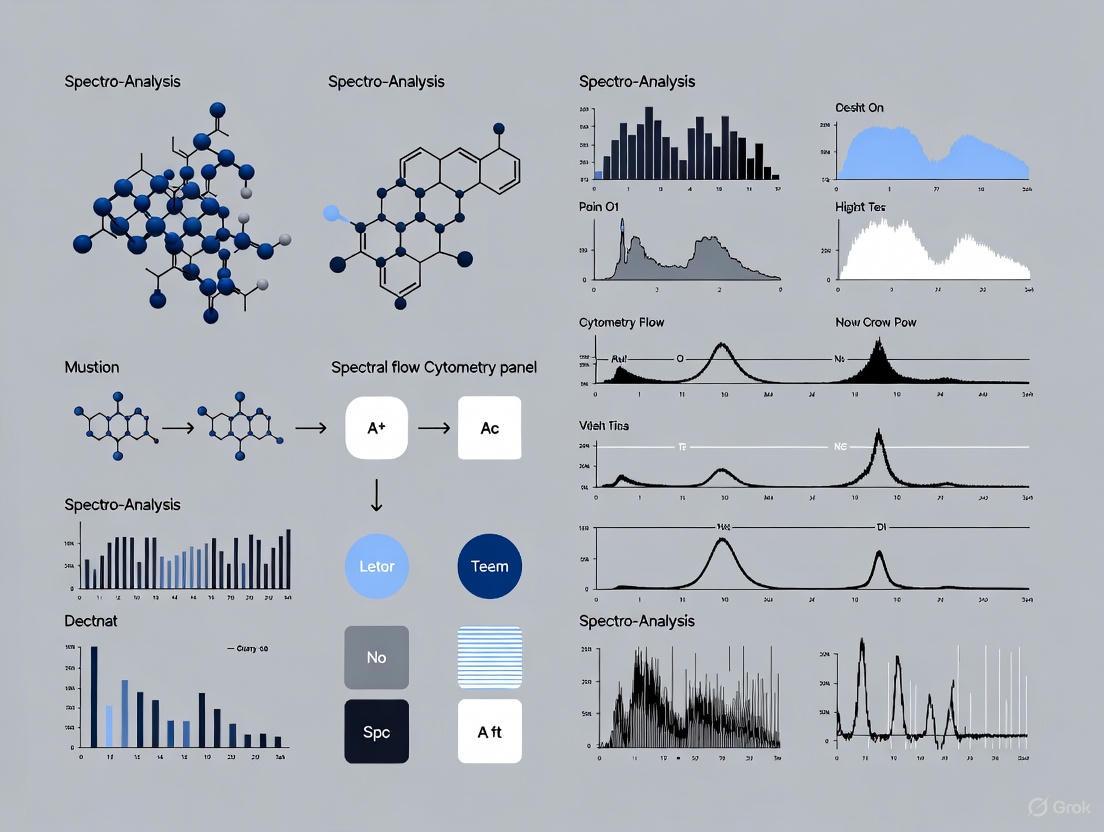
Diagram 1: Autofluorescence unmixing workflow.
Advantage 3: Simplified Optics and Workflow
Mechanism and Experimental Evidence
The optical design of a spectral cytometer is inherently more straightforward than that of a high-parameter conventional cytometer. Conventional systems require a complex and custom-engineered "forest of filters"—a precise arrangement of dichroic mirrors and bandpass filters to direct specific wavelengths of light to dedicated detectors [7]. This setup is physically rigid; changing a panel often requires manually changing filters, a process prone to error and misalignment.
In contrast, spectral cytometers replace this complex filter array with a fixed optical path consisting of a grating or prism that spreads the full emission light onto a consistent array of detectors [11] [7]. There are no filters to change when designing a new panel. This simplifies the instrument's hardware and makes panel configuration a purely computational task, managed through the software that defines the reference spectra for unmixing [6]. This leads to greater robustness and reduced downtime.
From a workflow perspective, this simplification translates to increased flexibility and faster panel design. Researchers can theoretically test an infinite combination of fluorophores in silico using spectral viewers without any hardware modifications [10] [11]. Furthermore, the process of "compensation" is replaced by automated spectral unmixing, which, while still requiring single-color controls, is a more integrated and less error-prone process, especially as the number of parameters increases [10] [7].
Diagram 2: Optical pathway comparison.
The Scientist's Toolkit: Key Reagent Solutions
Successful implementation of spectral flow cytometry relies on the appropriate selection of reagents and tools. The following table details essential items for panel design and validation.
Table 2: Key Research Reagent Solutions for Spectral Flow Cytometry
| Item | Function | Example Application/Note |
|---|---|---|
| Spectral Cytometer | Instrument platform for high-parameter full-spectrum acquisition. | Examples include Cytek Aurora, BD FACSymphony A5 SE, and Thermo Fisher Attune Xenith [8] [6] [10]. |
| Spectral Panel Builder | Online tool for visualizing fluorophore spectra and optimizing panel combinations for a specific instrument. | Critical for minimizing spreading error during panel design [11] [14]. |
| Single-Stain Controls | Biological cells or beads stained with individual antibody-fluorophore conjugates. | Provide reference spectra for the unmixing algorithm [6] [14]. |
| Calibration Beads | Standardized fluorescent particles. | Used for instrument normalization and quality control to ensure day-to-day reproducibility [14]. |
| Fluorophore Families | Dyes with distinct spectral properties to fill "gaps" in the spectrum. | Includes BD Horizon RealYellow/RealBlue, BioLegend's APC/Fire dyes, and Biotium's CF dyes [6] [11]. |
| Validated Antibody Panels | Pre-optimized multicolor panels. | Resources like the OMIP (Optimized Multicolor Immunofluorescence Panel) series provide a strong starting point [14]. |
Validation of flow cytometry panels is a critical process that ensures data generated in life sciences research is reliable, reproducible, and fit for its intended purpose. The validation requirements vary significantly across clinical, preclinical, and basic research settings, reflecting their distinct regulatory frameworks and operational objectives. In clinical diagnostics, flow cytometry is indispensable for the diagnosis and classification of hematological malignancies, with its application governed by stringent standards to ensure patient safety and diagnostic accuracy [16] [17]. In contrast, preclinical research, particularly in drug development and biomarker discovery for contract research organizations (CROs) and pharmaceutical companies, requires robust validation to support regulatory submissions and therapeutic development [13] [18]. Basic research environments typically maintain more flexible validation protocols focused on exploratory biological discovery.
The emergence of spectral flow cytometry has revolutionized single-cell analysis by enabling high-dimensional cellular phenotyping with unprecedented depth and precision [13] [19]. Unlike conventional flow cytometry, which captures fluorescence through discrete optical filters, spectral cytometry captures the full emission spectrum of each fluorochrome, allowing for more precise signal unmixing and simultaneous analysis of significantly more parameters—often exceeding 40 markers in a single tube [13] [20]. This technological advancement introduces new considerations for panel validation, including spectral unmixing algorithms, reference spectrum validation, and increased data complexity management.
Comparative Analysis of Validation Requirements
The stringency of validation protocols escalates from basic research to clinical diagnostics, reflecting the critical application of results in patient care. The table below summarizes key validation parameters across different settings.
Table 1: Validation Requirements Across Different Settings
| Validation Parameter | Basic Research | Preclinical/Drug Development | Clinical Diagnostics |
|---|---|---|---|
| Panel Design & Optimization | Fluorochrome brightness, spillover spread, antigen density matching [13] | Standardized protocols, reproducibility across sites [18] [16] | Adherence to established diagnostic panels (e.g., WHO, Euroflow) [16] |
| Instrument Validation | Daily QC (CS&T); laser power, fluidics, optical alignment [16] | Cross-instrument standardization, rigorous QC schedules [21] [18] | Full operational qualification, adherence to regulatory standards (e.g., CLIA) [22] [16] |
| Assay Precision | Limited replication, technical replicates for key populations | Intra-assay, inter-assay, inter-operator variability assessment [18] | Defined precision metrics, repeatability and reproducibility testing [22] [16] |
| Assay Accuracy | Often verified with biological controls or known cell lines | Comparison to validated methods, spike-recovery experiments [18] | Correlation with gold-standard methods (e.g., cytomorphology, genetics) [16] |
| Sensitivity | Population-dependent, focus on resolution from background | Quantitative for rare cells (e.g., CAR-T, MRD), limits of detection [18] [23] | Defined clinical sensitivity; MRD detection as low as 0.001-0.02% [13] [16] |
| Specificity | Gating strategy based on biological knowledge | Comprehensive gating strategy validation, inclusion of controls [18] | Validated gating strategies, minimal false-positive/negative rates [16] |
| Sample Stability | Often process immediately or defined empirically | Staining stability, sample aging cutoffs established [18] | Rigorously defined sample handling and processing timelines [16] |
| Data Management & Standardization | Laboratory-specific protocols | Standardized SOPs across all labs, data transfer agreements [18] | Full traceability, audit trails, compliance with data integrity regulations |
Key Differences and Their Implications
Clinical diagnostic settings demand the most rigorous validation, where flow cytometry results directly impact patient management decisions. Laboratories must adhere to standardized panels aligned with the World Health Organization (WHO) classification of haematolymphoid tumors and Bethesda guidelines [16]. For applications like minimal residual disease (MRD) monitoring, validated assays must demonstrate exceptional sensitivity, capable of detecting one malignant cell among 10,000 to 100,000 normal cells (sensitivity of 0.01% to 0.001%) [13] [16]. The recent integration of artificial intelligence with multi-parameter flow cytometry has further enhanced diagnostic accuracy, with one model for myelodysplastic syndromes achieving a sensitivity of 91.8% and specificity of 92.5% [17].
Preclinical and drug development environments require validation that ensures consistency across multiple sites and over time. The focus extends beyond accurate cell population identification to include pharmacodynamic biomarkers and receptor occupancy assays that inform drug efficacy and dosing decisions [18]. Standardization becomes paramount, necessitating consistent sample preparation, processing, instrument configuration, and data analysis protocols across all participating laboratories [18] [16]. For CROs and pharmaceutical companies utilizing spectral flow cytometry, additional validation steps include verifying the performance of high-parameter panels (often 30+ colors) and ensuring reproducibility in archived and cryopreserved specimens [13].
Basic research applications maintain greater flexibility, with validation often focused on ensuring that experimental results are biologically meaningful rather than compliant with regulatory standards. Researchers must still perform appropriate instrument quality control and validate that their panels can adequately resolve the cell populations of interest, but the process is typically less documented and more iterative.
Experimental Protocols for Validation
Validation of a Spectral Flow Cytometry Panel for MRD Detection
The protocol below outlines a comprehensive validation approach for a spectral flow cytometry panel designed to detect MRD in acute myeloid leukemia (AML), adaptable to other hematological malignancies.
Table 2: Key Research Reagent Solutions for Spectral Flow Cytometry Validation
| Reagent Category | Specific Examples | Function in Validation |
|---|---|---|
| Instrument QC Beads | CS&T Beads, Rainbow Calibration Particles | Verify laser power, fluidic stability, optical alignment, and daily performance [16] |
| Viability Dyes | Fixable Viability Stains (e.g., Zombie dye) | Distinguish live/dead cells; critical for accurate immunophenotyping [13] |
| Reference Control Cells | Healthy donor PBMCs, Cell lines (e.g., HL-60) | Establish reference spectra, assess inter-assay precision, validate gating strategies [16] |
| Standardized Antibody Panels | EuroFlow panels, Dry reagent formats (e.g., BD Horizon) | Ensure reproducibility and standardization across laboratories [21] [16] |
| Compensation Beads | Antibody Capture Beads | Generate single-color controls for spectral unmixing matrix calculation [13] |
| Automated Analysis Software | Machine learning algorithms, Clinical decision support software | Reduce technician analysis time, improve standardization, enable automated population identification [21] [17] |
Sample Preparation Protocol:
- Sample Type: Bone marrow aspirates or peripheral blood collected in EDTA or heparin tubes [16].
- Cell Processing: Isolate mononuclear cells using density gradient centrifugation (Ficoll-Paque) or lyse whole blood with ammonium chloride solution [16].
- Cell Counting: Determine cell concentration and viability using automated cell counters or flow cytometry with viability dyes.
- Staining Procedure: Aliquot 1-5×10^6 cells per tube. Add Fc receptor blocking agent to reduce non-specific binding. Incubate with pre-titrated antibody cocktail for 30 minutes at 4°C in the dark. Wash cells with PBS containing 1% BSA. If including intracellular markers, fix and permeabilize cells according to manufacturer protocols before intracellular staining [16].
- Data Acquisition: Acquire data on spectral flow cytometer (e.g., Cytek Aurora, BD FACSDiscover) following instrument-specific setup procedures. Collect a minimum of 1-5×10^6 events per sample to ensure adequate sensitivity for rare cell detection [13].
Validation Experiments:
- Precision Assessment: Perform intra-assay precision testing by running the same sample 10 times in one session. Determine inter-assay precision by testing the same sample across 5 different days. Calculate coefficients of variation (CV%) for population percentages and median fluorescence intensities (MFIs), with acceptable CVs typically <15-20% for clinical assays [22].
- Linearity and Sensitivity: Prepare serial dilutions of positive cells (e.g., leukemia cell lines) in negative matrix (normal bone marrow). Assess the limit of detection (LOD) as the lowest concentration where positive cells are consistently detected, and limit of quantification (LOQ) where CV is <20% [13] [16].
- Accuracy Comparison: Compare results with alternative methods such as next-generation sequencing (NGS) or quantitative PCR. For AML MRD, a 24-color spectral panel demonstrated sensitivity below 0.02% with strong correlation to genetic methods [13].
- Reference Range Establishment: Test samples from healthy donors (n≥20) to establish normal reference ranges for all populations included in the panel.
- Interfering Substances: Test potential interfering substances including hemolyzed, lipemic, and icteric samples to assess their impact on assay performance.
Protocol for Cross-Platform Comparison Studies
With multiple spectral flow cytometry platforms available (e.g., Cytek Aurora, BD FACSDiscover, ID7000), cross-platform comparison is essential for method transfers or multi-center trials.
Experimental Design:
- Sample Selection: Include a range of sample types (normal PBMCs, patient samples, cell lines) representing various expression levels [22].
- Instrumentation: Test identical stained samples on different cytometers (conventional and spectral) within a defined timeframe [22].
- Standardization: Use the same sample preparation protocol, antibody lots, and software analysis versions across all instruments.
Data Analysis:
- Correlation Assessment: Calculate correlation coefficients (R²) for median fluorescence intensity (MFI) and population percentages across platforms [22].
- Concordance Evaluation: Determine concordance for critical outcomes such as serostatus in autoimmune disease testing or MRD positivity in leukemia [22].
- Precision Comparison: Compare intra-assay and inter-assay precision across platforms, with spectral cytometers often demonstrating improved repeatability (e.g., 4.6% CV on ID7000 vs. 6.8% on conventional Fortessa) [22].
Technological Considerations for Spectral Flow Cytometry Validation
Unique Aspects of Spectral Panel Validation
Spectral flow cytometry introduces distinct validation requirements beyond conventional flow cytometry. Spectral unmixing represents the most significant difference, requiring verification that reference spectra for each fluorochrome are properly characterized and that the unmixing algorithms correctly resolve signals from highly overlapping fluorochromes [13] [20]. Unlike conventional compensation, which uses simple matrix calculations, spectral unmixing employs complex algorithms that analyze the entire emission spectrum, necessitating rigorous validation with single-stained controls [13].
Autofluorescence handling presents another unique consideration. Spectral cytometry can characterize and extract autofluorescence signals using the same unmixing algorithms applied to fluorochromes [13]. While this enhances resolution by minimizing background noise, improper handling can introduce unmixing errors and false-positive events [13]. Validation must confirm that autofluorescence extraction improves population resolution without distorting data.
The high-parameter nature of spectral panels (often 30+ colors) creates additional validation complexity. The stain index (a measure of signal-to-noise ratio) becomes highly dependent on instrument-specific configurations [13]. Validating such panels requires assessing not only individual marker performance but also potential interactions between numerous reagents and their collective impact on population resolution.
Emerging Technologies and Their Validation Implications
Several emerging technologies are reshaping the validation landscape for flow cytometry. Mass cytometry (CyTOF) utilizes metal-labeled antibodies detected by time-of-flight mass spectrometry, eliminating spectral overlap concerns but requiring validation of metal标签 stability and instrument tuning [17]. Imaging mass flow cytometry combines high-parameter phenotyping with morphological information, necessitating validation of both fluorescence and imaging components [17].
Artificial intelligence and machine learning are increasingly integrated into flow cytometry data analysis. These tools can identify complex patterns in high-dimensional data beyond human capability, but require rigorous validation of training datasets and algorithm performance [21] [17]. For clinical use, these systems must demonstrate consistent performance across diverse patient populations and sample types.
The validation requirements for spectral flow cytometry panels exist on a continuum, with complexity escalating from basic research to clinical diagnostics. While all settings share fundamental validation principles—including instrument qualification, assay precision assessment, and sensitivity verification—the stringency, documentation, and regulatory compliance requirements differ substantially. The core differentiator lies in the application of results: basic research prioritizes biological discovery, preclinical studies focus on generating robust data for regulatory submissions, and clinical diagnostics demand unwavering accuracy for patient care decisions.
The transition to spectral flow cytometry has enhanced our analytical capabilities through improved multiplexing, superior resolution of complex populations, and reduced sample consumption. However, these advantages introduce new validation considerations, particularly regarding spectral unmixing performance, autofluorescence management, and high-dimensional data analysis. As flow cytometry continues to evolve with integrations of artificial intelligence, mass cytometry, and imaging technologies, the validation frameworks must similarly advance to ensure that technological progress translates to scientifically valid and clinically useful applications.
Researchers and laboratory directors should implement validation protocols aligned with their specific application environment while maintaining awareness of requirements from more stringent settings. This approach not only ensures data quality appropriate for immediate needs but also facilitates the translation of research findings along the development pipeline, ultimately accelerating the application of scientific discoveries to improved human health.
Strategic Panel Design and Staining Protocols for Reliable Spectral Assays
In the rapidly evolving field of flow cytometry, the design of analytical panels is no longer a one-size-fits-all process. The concept of "fit-for-purpose" assay design has emerged as a critical principle, emphasizing that each flow cytometry panel should be meticulously crafted to reflect the specific scientific questions, clinical contexts, and intended use of the data being generated [24]. This approach is particularly crucial in drug development and clinical research, where reliable cellular data informs critical decisions about therapeutic efficacy and safety.
The fundamental premise of fit-for-purpose design begins with a clear scientific question, which then dictates every subsequent decision in panel development—from biomarker selection to instrument choice and validation strategy [24]. As noted by Cerba Research, "each assay should be molded to reflect the design of the study and the needs of the investigators, of which no two are exactly alike" [24]. This philosophy ensures that resources are allocated efficiently while generating data of sufficient quality and reliability for its intended application, whether for exploratory research or pivotal clinical decision-making.
Foundational Principles of Flow Cytometry Panel Design
The Strategic Foundation: Defining Purpose and Context of Use
The initial phase of panel design requires rigorous upfront planning to establish the context of use (COU), which fundamentally dictates all subsequent technical decisions [25] [26]. As emphasized in biomarker method development workshops, without a clear understanding of the intended use of data, it is not possible to properly validate an assay for its purpose [25]. Key considerations at this stage include:
- Regulatory requirements: Determining whether the assay supports exploratory research, secondary endpoints, or primary clinical endpoints significantly impacts the validation stringency [25] [26].
- Sample constraints: Assessing available sample volumes, particularly with precious clinical samples or longitudinal studies with limited collection volumes [24] [27].
- Data application: Defining how results will inform decisions—from early mechanistic understanding to dose selection or diagnostic application [25].
This foundational step ensures the panel design aligns with the broader experimental or clinical objectives before addressing technical implementation.
Technology Selection: Conventional vs. Spectral Flow Cytometry
A critical decision in modern panel design involves selecting between conventional and spectral flow cytometry technologies, each with distinct advantages for different applications.
Table 1: Comparison of Conventional and Spectral Flow Cytometry Technologies
| Feature | Conventional Flow Cytometry | Spectral Flow Cytometry |
|---|---|---|
| Core Principle | Compensation-based detection | Full-spectrum fingerprinting with spectral unmixing [28] |
| Best Applications | Well-refined biomarker panels with strict regulatory requirements [24] | Exploratory panels, high-parameter profiling [24] [29] |
| Parameter Capacity | Limited by filter-based detection | Higher multiplexing capability [29] [26] |
| Implementation Complexity | Standardized workflows, easier validation [24] | Evolving standardization, more complex validation [24] |
| Data Quality | Proven performance for established panels [24] | Enhanced detection sensitivity and resolution [29] |
| Clinical Adoption | Widely established in clinical labs [29] | Emerging with validation studies underway [29] [26] |
This technology decision fundamentally influences all subsequent panel design choices, particularly regarding fluorophore selection and panel complexity.
Implementing Fit-for-Purpose Panel Design: A Strategic Framework
Biomarker Selection and Fluorochrome Pairing Strategy
The core of panel design involves strategic pairing of biomarkers with appropriate fluorochromes based on antigen density and expression patterns. The guiding principle is to match low-expression biomarkers with high-brightness fluorochromes, while highly expressed markers can be detected with lower brightness labels [24] [28]. This approach ensures optimal resolution of critical populations.
Resources such as Optimized Multicolor Immunofluorescence Panels (OMIPs) provide valuable starting points for panel design, offering previously optimized panels for specific cell types or research questions [28]. Additionally, panel design tools like EasyPanel and Fluorofinder can assist in initial fluorochrome selection, though these automated suggestions often require empirical refinement based on actual performance [24].
Antibody Validation and Titration Protocols
Antibody validation represents a critical component of robust panel development. Current best practices recommend using monoclonal or recombinant antibodies to improve reproducibility, with careful attention to vendor validation data [28]. The International Working Group for Antibody Validation (IWGAV) has proposed guidelines emphasizing application-specific validation to ensure antibodies are specific, selective, sensitive, and reproducible [28].
A key step in optimization involves antibody titration to determine the optimal concentration that provides the best separation between positive and negative populations. The stain index (SI) is commonly used for this assessment, calculated as: SI = (MFIpositive - MFInegative) / (2 × rSD_negative) where MFI represents median fluorescence intensity and rSD is the robust standard deviation [29]. This quantitative approach ensures each reagent is used at its optimal concentration before full panel integration.
Experimental Validation: From Design to Implementation
The following workflow diagram illustrates the comprehensive process of fit-for-purpose assay design and validation:
Case Studies in Fit-for-Purpose Design and Validation
Clinical Application: Minimal Residual Disease Detection in B-ALL
A recent comparative study demonstrates the practical implementation of fit-for-purpose design in clinical diagnostics. Researchers developed and validated a 24-color spectral flow cytometry panel for detecting minimal residual disease (MRD) in B-cell acute lymphoblastic leukemia (B-ALL) and compared its performance against conventional 8-color panels [29].
Table 2: Performance Comparison in B-ALL MRD Detection
| Parameter | Conventional 8-Color Panel | Spectral 24-Color Panel |
|---|---|---|
| Number of Markers | 8 | 24 |
| Backbone Markers | CD19, CD45, CD34, CD10, CD20 | Same backbone plus additional characterization markers [29] |
| Aberrant Markers | Limited to 3 additional markers | Expanded to include CD9, CD123, CD66c, CD73, CD304, and others [29] |
| Sample Requirement | Standard volume | Equivalent volume with more information extracted |
| Detection Sensitivity | Established for MRD | Potentially enhanced through improved resolution [29] |
| Validation Approach | Clinical laboratory standards | Comprehensive validation following internal SOPs under ISO15189 accreditation [29] |
This study highlights how fit-for-purpose design expanded the analytical capabilities while maintaining reliability for critical clinical applications. The integration of all conventional markers into the expanded spectral panel provided continuity with existing clinical practice while enhancing characterization power [29].
Preclinical Application: Murine Immunology Studies
In preclinical research, fit-for-purpose principles address unique constraints such as limited sample volumes. A recent study developed specialized panels for comprehensive immune monitoring in mice using only 50 μL of peripheral blood per panel, enabling longitudinal assessments previously challenging due to blood volume limitations [27].
The researchers designed three complementary panels (myeloid, lymphoid, and intracellular) with careful attention to antibody titration, spectral overlap considerations, and antigen density matching to fluorochrome brightness [27]. This approach allowed comprehensive immune profiling while respecting the practical constraints of murine studies, demonstrating how fit-for-purpose design directly addresses specific research limitations.
Advanced Implementation: Harmonization and Standardization
In regulated environments, organizations like GSK have implemented harmonized high-parameter spectral flow cytometry panels (26-37 markers) as off-the-shelf solutions for most clinical trial needs [26]. This approach maintains consistency across studies while allowing for asset-specific customization through "drop-in" marker slots [26].
Additionally, adoption of standards like the Clinical Data Interchange Standards Consortium (CDISC) Cell Phenotyping (CP) domain enables improved data standardization and reporting [26]. This ensures that complex high-parameter data remains interpretable and reusable across studies and organizations, addressing a critical challenge in modern flow cytometry applications.
Fit-for-purpose assay design represents a fundamental shift from standardized approaches to context-driven panel development in flow cytometry. By aligning panel strategy with specific scientific and clinical questions from the outset, researchers can optimize resources while generating reliable, actionable data. The continuing evolution of spectral flow cytometry, combined with rigorous validation frameworks and standardized reporting, promises to enhance our ability to extract meaningful biological insights from complex cellular systems across both preclinical and clinical applications. As the field advances, the principles of fit-for-purpose design will remain essential for ensuring that technological capabilities translate to scientifically valid and clinically relevant outcomes.
The advancement of spectral flow cytometry has revolutionized multiparameter panel design, enabling researchers to simultaneously analyze dozens of cellular markers within a single tube. This technological leap forward demands increasingly sophisticated strategies for fluorochrome selection and biomarker pairing to ensure optimal panel performance. The fundamental challenge lies in balancing three critical factors: fluorochrome brightness, spectral spillover, and antigen density—a triad that directly determines the sensitivity, specificity, and reliability of flow cytometry data [30] [31].
In clinical diagnostics and drug development, proper panel design becomes paramount, particularly for applications like measurable residual disease (MRD) detection in acute lymphoblastic leukemia, where sensitivity below 0.001% may be required [29] [31]. Spectral flow cytometry provides distinct advantages over conventional systems by capturing the full emission spectrum of each fluorochrome, enabling more precise signal unmixing and facilitating the use of fluorochromes with highly overlapping emission profiles [31]. However, this capability does not eliminate the need for strategic fluorochrome selection; it merely changes the optimization parameters. This guide examines the key considerations, experimental validation methodologies, and practical implementation strategies for designing robust spectral flow cytometry panels that meet the rigorous demands of contemporary clinical and research applications.
Core Principles of Fluorochrome Selection
The Brightness-Antigen Density Hierarchy
The cornerstone of effective panel design lies in matching fluorochrome brightness with the expression level of target antigens. This principle ensures sufficient signal resolution while minimizing background noise and spillover effects.
High-Antigen Density Markers: These abundantly expressed cellular proteins (e.g., CD45, CD4) generate strong signals even with dimmer fluorochromes. Pairing them with overly bright fluorophores can cause excessive spillover into other detectors and potentially saturate signals, reducing resolution and complicating data analysis [30].
Low-Antigen Density Markers: Weakly expressed antigens (e.g., certain cytokine receptors, transcription factors) require the brightest available fluorochromes to achieve adequate separation from negative populations. Failing to do so may result in undetectable signals or poor population resolution [30].
Moderate-Antigen Density Markers: These occupy the middle ground and should be paired with fluorochromes of intermediate brightness to maintain balanced panel performance [30].
Table 1: Fluorochrome Brightness Classification for Common Dyes
| Brightness Category | Example Fluorochromes | Recommended Application |
|---|---|---|
| Very Bright | PE, APC, Brilliant Violet 421 | Low-density antigens, critical differentiation markers |
| Bright | PE-Cy7, APC-Cy7, Brilliant Violet 510 | Medium-to-low density antigens |
| Intermediate | FITC, PerCP-Cy5.5, Brilliant Violet 605 | Medium-density antigens |
| Dim | Pacific Blue, Alexa Fluor 488 | High-density antigens, backbone markers |
Managing Spectral Spillover and Spillover-Spreading Matrix
Spectral spillover occurs when a fluorochrome's emission is detected in channels designated for other fluorochromes [32]. In spectral flow cytometry, this phenomenon is quantified using a spillover-spreading matrix (SSM), which represents the degree to which each fluorochrome spills into other detection channels [32]. The SSM values indicate signal spread measured in standard deviations and provide crucial guidance for assessing fluorochrome compatibility.
Strategies to minimize spillover impacts include:
- Avoid high-spillover combinations for co-expressed markers, particularly those with SSM values exceeding 10 [32]
- Leverage spectral unmixing algorithms that utilize full emission spectra rather than single-peak measurements [31]
- Employ similarity and complexity indices during panel design to assess overall spectral compatibility [32]
The complexity index represents the cumulative similarity of all fluorochromes within a panel, while the similarity index quantifies spectral overlap between specific fluorochrome pairs [32]. Optimal panel design maintains a complexity index below 0.95 for all fluorochrome combinations [29].
Advanced Considerations for Tandem Fluorochromes
Tandem dyes, created by coupling a donor fluorochrome (e.g., PE, APC) to an acceptor molecule, present special considerations in panel design. While they expand the usable spectrum, tandems introduce potential instability due to:
- Variable FRET efficiency (Förster Resonance Energy Transfer) between donor and acceptor molecules [32]
- Susceptibility to photobleaching and batch-to-batch variability [32]
- Temperature and solvent sensitivity that can alter emission profiles [32]
Ideal tandems would feature 100% donor quenching, discrete single-laser excitation, and high photostability, though such "perfect" tandems do not yet exist [32]. When using tandems, researchers should prioritize recent manufacturing lots, protect samples from light exposure, and validate performance with compensation beads [33] [32].
Experimental Validation Methodologies
Antibody Titration and Stain Index Calculation
Precise antibody titration forms the foundation of reproducible panel performance. The optimal antibody concentration maximizes the stain index (SI), which quantifies the separation between positive and negative populations [29]. The standard formula for calculating stain index is:
SI = (MFIpos - MFIneg) / (2 × rSDneg)
Where MFIpos is the median fluorescence intensity of the positive population, MFIneg is the median fluorescence intensity of the negative population, and rSDneg is the robust standard deviation of the negative population [29].
The titration protocol involves:
- Preparing a series of antibody dilutions (typically 6-point titration) [29]
- Staining control samples with each dilution
- Calculating the stain index for each concentration
- Selecting the lowest antibody volume that achieves the highest stain index [29]
This approach conserves reagents while ensuring optimal signal-to-noise ratio. For a 24-color B-ALL panel described in recent literature, this method reduced antibody consumption to 2.5-5μL per test for most markers [29].
Single-Stain Controls and Unmixing Validation
Accurate spectral unmixing requires high-quality single-stain controls to establish reference spectra for each fluorochrome. Two primary approaches exist for control preparation:
UltraComp eBeads Spectral Unmixing Beads: These specialized beads contain both positive (antibody-binding) and negative populations, providing consistent reference signals with low background noise [33]. They are compatible with multiple species (human, mouse, rat, hamster, rabbit) and excel in spectral flow cytometry applications [33].
Biological Controls: Cells with known antigen expression patterns can serve as controls but introduce more variability due to biological heterogeneity and autofluorescence [33].
Recent comparisons demonstrate that UltraComp eBeads Spectral Unmixing Beads provide superior unmixing performance compared to traditional compensation beads, particularly for tandem dyes and red-emitting fluorophores [33]. This results in better alignment between positive and negative populations during unmixing [33].
Table 2: Single-Stain Control Options for Spectral Flow Cytometry
| Control Type | Advantages | Limitations | Recommended Use |
|---|---|---|---|
| UltraComp eBeads Spectral | Low background, consistent signal, species flexible | Does not reflect biological staining | Primary choice for spectral unmixing |
| UltraComp eBeads Plus | Good performance, broad laser compatibility | Less optimal for spectral applications | Conventional flow cytometry |
| Cellular Controls | Biological relevance, includes autofluorescence | Variable, limited availability | When biological matrix is essential |
Panel Complexity Assessment and Optimization
Before experimental validation, in silico panel assessment using specialized software tools can predict potential issues and guide optimization. Key assessment parameters include:
Similarity Index: Measures spectral overlap between fluorochrome pairs on a scale from 0 (completely dissimilar) to 1 (identical spectra). Pairs with similarity indices >0.8 should be avoided for co-expressed markers [32].
Complexity Index: Evaluates the overall spectral complexity of the entire panel, with lower values indicating better fluorochrome compatibility [29] [32].
Modern panel design platforms like FluoroFinder's IntelliPanel and Cytek Cloud enable researchers to simulate panel performance before purchasing reagents [29] [32]. These tools calculate similarity and complexity indices, allowing iterative refinement of fluorochrome combinations to minimize spectral conflicts [29] [32].
Diagram 1: Panel design and validation workflow.
Spectral vs Conventional Flow Cytometry: Performance Comparison
Technical Capabilities and Limitations
Spectral flow cytometry represents a paradigm shift from conventional flow cytometry, with distinct advantages for high-parameter panel design:
- Detection Method: Conventional systems measure only peak emissions, while spectral cytometers capture full emission spectra [31]
- Multiplexing Capacity: Conventional systems typically max at 8-12 colors, while spectral systems routinely accommodate 30+ parameters [29] [31]
- Spillover Compensation: Conventional relies on mathematical compensation, while spectral employs unmixing algorithms [32] [31]
- Autofluorescence Handling: Conventional treats autofluorescence as background noise, while spectral can identify and subtract it [31]
These technical differences translate to practical advantages in resolution and sensitivity. Spectral cytometry demonstrates particular strength in detecting low-abundance markers and resolving complex populations, such as distinguishing leukemic blasts from normal progenitors in MRD detection [29] [31].
Application-Specific Performance Data
Recent validation studies directly comparing both technologies demonstrate the measurable benefits of spectral flow cytometry in clinical applications:
Table 3: Performance Comparison in Clinical Applications
| Application | Conventional Flow | Spectral Flow | Clinical Impact |
|---|---|---|---|
| B-ALL MRD Detection | 8-color multi-tube approach | 24-color single-tube panel [29] | Reduced sample consumption, improved efficiency |
| Sensitivity | 0.01% typical | <0.001% demonstrated [31] | Enhanced detection of minimal residual disease |
| Antigen-Loss Detection | Requires additional tubes | CD19-negative clones detected in single tube [31] | Identification of treatment-resistant variants |
| Immune Monitoring | Limited phenotyping depth | 35+ parameters in cryopreserved samples [31] | Comprehensive biomarker discovery |
In B-cell acute lymphoblastic leukemia (B-ALL) monitoring, spectral flow cytometry enabled the design of a 24-color single-tube panel that incorporated all markers previously distributed across multiple conventional tubes [29]. This consolidation improved workflow efficiency while maintaining correlation with conventional approaches and enhancing resolution of maturation states [29].
For CAR-T cell therapy monitoring, spectral panels have identified critical predictive biomarkers, including PD-1+ CD8+ CAR-T subsets in lymphoma responders and CCR7+ early-memory cells in CLL patients with favorable outcomes [31]. These findings highlight the clinical value of comprehensive immunophenotyping made possible by spectral technology.
Implementation in Clinical and Research Settings
Practical Workflow Integration
Implementing spectral flow cytometry requires careful consideration of workflow adaptations:
Sample Processing: Spectral cytometry remains compatible with standard processing protocols, including bulk erythrocyte lysis and Ficoll separation [29] [28]. For intracellular staining, fixation and permeabilization steps follow established methodologies [28].
Instrument Setup: Quality control procedures mirror conventional systems, with daily performance tracking using calibration particles [29]. However, spectral systems require validation of full spectral profiles rather than single-detector intensities [31].
Data Acquisition: The fundamental acquisition process remains similar, though spectral systems typically require lower sample volumes due to increased information content per cell [29] [31].
Analysis Workflow: While basic gating strategies (doublet discrimination, viability, lineage identification) remain consistent, subsequent analysis leverages spectral unmixing rather than compensation [29] [32].
Diagram 2: Spectral flow cytometry clinical workflow.
Essential Research Reagent Solutions
Successful implementation of spectral flow cytometry panels requires specific reagent systems optimized for full-spectrum detection:
Table 4: Essential Research Reagents for Spectral Flow Cytometry
| Reagent Category | Specific Products | Application Function | Spectral Compatibility |
|---|---|---|---|
| Spectral Unmixing Beads | UltraComp eBeads Spectral Unmixing Beads [33] | Establish reference spectra for unmixing | Excellent - specifically designed for spectral |
| Viability Dyes | LIVE/DEAD Fixable Stains [33] | Distinguish live/dead cells | Compatible with amine-reactive beads |
| Bright Fluorochromes | Brilliant Violet 421, Brilliant Ultra Violet 500 [29] | Detect low-abundance antigens | High resolution with minimal spillover |
| Tandem Dyes | PE-Cy7, APC-Cy7, Brilliant Violet 785 [29] | Expand panel multiplexing | Require careful validation due to instability |
| Reference Controls | Frozen normal PBMCs, cell lines [29] | Panel performance tracking | Essential for longitudinal studies |
Strategic fluorochrome selection and biomarker pairing represent both an art and science in spectral flow cytometry panel design. The fundamental principle of matching fluorochrome brightness with antigen expression levels remains paramount, while new considerations around spectral similarity and complexity indices have emerged as critical design parameters. Through systematic antibody titration, appropriate control selection, and iterative panel optimization, researchers can develop high-performance spectral panels that push the boundaries of multiplexing while maintaining analytical sensitivity.
The transition from conventional to spectral flow cytometry enables unprecedented depth in cellular characterization, particularly valuable in clinical diagnostics and therapeutic monitoring where comprehensive immunophenotyping provides actionable insights. As spectral technology continues to evolve, adherence to these fundamental principles of fluorochrome selection will ensure that researchers maximize the potential of this powerful technology while generating robust, reproducible data that advances both basic research and clinical care.
The adoption of spectral flow cytometry in clinical diagnostics and drug development has highlighted a critical dependency on robust sample handling protocols. Pre-analytical variables introduced during sample collection, stabilization, and storage directly determine the quality and reproducibility of high-dimensional immunophenotyping data, especially in the context of multi-site clinical trials. The validation of any spectral flow cytometry panel is therefore inextricably linked to the rigorous standardization of these pre-analytical steps. This guide objectively compares the performance of current methods for stabilizing whole blood, cryopreserved samples, and low-volume specimens, providing experimental data to inform protocol selection for clinical research.
Comparative Analysis of Sample Stabilization Methods
The choice between fresh, stabilized, and cryopreserved samples involves trade-offs between marker stability, logistical feasibility, and data fidelity. The table below summarizes key performance characteristics of different sample types based on recent studies.
Table 1: Performance Comparison of Sample Handling Methods for Flow Cytometry
| Sample Type | Key Advantages | Key Limitations | Ideal Use Cases | Reported Stability |
|---|---|---|---|---|
| Fresh Whole Blood | - Maximum marker integrity- No fixative-induced epitope damage | - Very short processing window (<48 hrs for many RO assays) [34]- Logistically challenging for multi-site trials | - Single-site studies with local flow core- Receptor occupancy assays requiring high sensitivity [34] | 24-48 hours for most markers; granulocyte scatter degrades within 24 hours [35] |
| Chemically Stabilized Whole Blood | - Extended stability (up to 120 days for some markers) [34]- Enables batch analysis at central labs [34] | - Variable impact on different markers [36]- May constrain dynamic range [34]- Increased site burden for processing [34] | - Global clinical trials with shipment to central labs [34]- Longitudinal studies requiring batch analysis | Prot1: Better preserved B cells; variable FoxP3 [36]Streck: More consistent FoxP3, albeit at lower frequencies [36] |
| Cryopreserved PBMCs | - Maximum logistical flexibility- Enables long-term storage for batch analysis | - Potential loss of some cell populations during processing- May adversely impact some assays (e.g., RO) [34] | - Large retrospective studies- Biobanking for future analysis | Cryopreservation of lysed whole blood maintains stable frequencies for most immune populations [36] |
| Low-Volume Samples | - Enables longitudinal studies in volume-limited models (e.g., mice) [27] | - Requires extensive antibody titration to maximize signal-to-noise [27]- Limited replicates per sample | - Pediatric studies- Preclinical murine studies [27] | Murine panels validated using only 50 µL of peripheral blood per panel [27] |
Whole Blood Stabilization for Centralized Testing
Chemical stabilization is a key solution for multi-site trials requiring shipment to a central laboratory. A receptor occupancy (RO) case study demonstrated that fresh whole blood required testing within 48 hours of collection, making single-lab testing infeasible for a global trial [34]. While PBMC isolation was tested, it adversely impacted RO data. Instead, evaluation of fixatives identified Smart Tube Proteomic fixative, which produced results correlating with fresh whole blood data and provided a 120-day stability window, allowing for long-term storage and batch analysis [34].
However, stabilizers are not universally compatible. The same study found that Cyto-Chex BCT tubes were not compatible with all markers in their panel [34]. Another independent evaluation confirmed that performance varies by marker, finding that Prot1 stabilizer better preserved B cells but showed variability in FoxP3 detection, while Streck offered more consistent FoxP3 staining, albeit at lower frequencies [36].
Cryopreservation as a Practical Alternative
Cryopreservation offers a practical balance between sample viability and logistical flexibility. Champions Oncology found that cryopreservation of lysed whole blood maintains stable frequencies and marker intensities for most immune populations, making it a viable alternative to Ficoll-isolated PBMCs [36]. This method is particularly valuable for creating biobanks for future, as-yet-undefined analyses.
Strategies for Low-Volume Samples
Volume limitation presents a distinct challenge, particularly in murine and pediatric studies. To address this, researchers have developed specialized panels that comprehensively analyze immune populations using minimal blood. One study designed two novel flow cytometry panels for murine studies that require only 50 µL of peripheral blood per panel, enabling longitudinal monitoring without exceeding ethical blood volume limits [27]. Critical to this success was the systematic titration of all antibodies to determine the concentration that maximizes the stain index, thus optimizing the signal from small cell numbers [27].
Standardizing Multi-Site Analysis
Beyond sample preservation, consistent instrument performance across testing sites is paramount for data comparability. Traditional approaches of shipping reagents from the same lot to multiple sites are inefficient and do not fully address inter-site variability [37].
A novel approach using lyophilized (freeze-dried) beads stained with a set of CD4 antibodies in various fluorochromes has demonstrated success. These beads, stable at room temperature for 18 months, were shipped once to five global sites for instrument alignment [37]. This method allowed sites to create a stable baseline, detect and correct instrument fluctuations, and standardize over 10 assays across all locations [37]. The results showed long-term precision, with less variability in Mean Fluorescence Intensity (MFI) and over 80% of readouts exhibiting a coefficient of variation (CV) of less than 30% when comparing reference and receiving laboratories [37].
Table 2: Essential Research Reagent Solutions for Sample Handling & Standardization
| Reagent / Solution | Primary Function | Application Context | Key Considerations |
|---|---|---|---|
| Smart Tube Proteomic Fixative | Chemical stabilization of whole blood samples | Extends stability for RO assays and complex immunophenotyping; enables batch analysis [34] | Validate for all markers; may constrain dynamic range; requires custom collection kits [34] |
| Cyto-Chex BCT Tubes | Blood collection tubes with anticoagulant and cell preservative | Phenotyping assays requiring extended stability [35] | Not compatible with all markers; requires validation for specific panels [34] |
| Lyophilized CompBeads | Instrument standardization and performance tracking | Multi-site instrument alignment; longitudinal monitoring of cytometer stability [37] | 18-month room temperature stability; reduces shipping needs for global trials [37] |
| FOXP3/Transcription Factor Staining Buffer Set | Intracellular staining for transcription factors and cytotoxic proteins | Detailed immune profiling of fixed/permeabilized cells (e.g., murine intracellular panels) [27] | Optimized for nuclear targets; requires specific fixation/permeabilization steps [27] |
| RBC Lysis/Fixation Buffer | Red blood cell lysis with simultaneous cell fixation | Sample preparation for surface marker staining in whole blood [27] | Combines lysis and fixation in a single step; preserves light scatter properties [27] |
Experimental Protocols for Validation
Sample Stability Assessment Protocol
A standardized process for determining specimen stability is crucial for assay robustness. The workflow should begin by defining assay parameters and stability requirements, then proceed through a structured evaluation [35].
- Define Assay Parameters: Determine the required stability based on the assay's intended use and logistical needs (e.g., required shipping duration) [35].
- Select Specimen Type and Collection Method: Choose appropriate anticoagulants (e.g., EDTA, Sodium Heparin) or preservative tubes based on the target markers [35].
- Establish Acceptance Criteria: Use assay precision and relative percent change between fresh and stored specimens as guides. Examine light scatter properties and population histograms for degradation [35].
- Evaluate Shipping Conditions: Test the impact of simulated transit with temperature-buffering agents. Consider temperature tracking for critical variables [35].
Figure 1: Sample stability assessment workflow. This structured process ensures specimen stability meets assay performance requirements throughout the collection, storage, and shipping chain [35].
Antibody Titration for Low-Volume Panels
For low-volume panels where reagent conservation is critical, precise antibody titration is essential. The following protocol, adapted from a murine panel validation study, ensures optimal signal-to-noise ratio [29] [27].
- Preparation: Resuspend cells in RPMI for 2 hours to allow surface marker recovery. Prepare an unstained control and a series of single-stained tubes with decreasing amounts of each antibody (e.g., 5 × 10⁻¹ dilutions, creating a 6-point titration) [29].
- Data Acquisition: Run all tubes on the cytometer using standardized instrument settings.
- Stain Index Calculation: For each antibody dilution, calculate the Stain Index (SI) using the formula:
( \text{SI} = \frac{\text{MFI}{\text{pos}} - \text{MFI}{\text{neg}}}{2 \times \text{rSD}_{\text{neg}}} )
where
MFI_posis the median fluorescent intensity of the positive population,MFI_negis the median fluorescent intensity of the negative population, andrSD_negis the robust standard deviation of the negative population [29]. - Optimal Volume Selection: Identify the antibody volume that provides the highest stain index, indicating the best separation between positive and negative populations. This volume is typically the lowest that achieves this maximum SI, conserving reagent without sacrificing performance [29] [27].
Multi-Site Instrument Alignment Protocol
Ensuring consistent instrument performance across multiple laboratories is achievable with a standardized alignment protocol [37].
- Preparation of Lyophilized Beads: Stain polystyrene microbeads (e.g., BD CompBeads) with antibodies conjugated to the full range of fluorochromes used in the panel. Lyophilize the stained beads to ensure room-temperature stability [37].
- Initial Shipment and Setup: Ship the lyophilized beads once to all participating sites. Due to their 18-month stability, no repeat shipments are required [37].
- Baseline Establishment and Harmonization: At each site, resuspend the beads and run them on the local instrument. Use the results to adjust target values and create a standardized baseline for all instruments [37].
- Ongoing Monitoring: Use the beads regularly (e.g., monthly) to monitor instrument performance, detect fluctuations, and apply corrections to maintain alignment across sites. Fluorochromes showing underperformance should be replaced with more stable alternatives [37].
Figure 2: Multi-site instrument alignment workflow. Using lyophilized beads in a single shipment enables harmonization of multiple flow cytometric assays across global laboratories, ensuring data consistency [37].
The successful validation and deployment of spectral flow cytometry panels in clinical research hinge on the rigorous control of pre-analytical variables. No single sample handling method is universally superior; the choice between fresh, chemically stabilized, or cryopreserved samples must be guided by the specific assay requirements, marker sensitivities, and logistical constraints of the study. Similarly, low-volume workflows and multi-site standardization demand dedicated protocols for antibody titration and instrument alignment. By adopting the standardized comparisons and experimental frameworks outlined in this guide, researchers can make evidence-based decisions that enhance data reproducibility, thereby strengthening the conclusions drawn from clinical trials and preclinical studies utilizing spectral flow cytometry.
Spectral flow cytometry represents a transformative advancement over conventional flow cytometry, enabling unprecedented resolution in cellular analysis. Unlike conventional systems that use optical filters to measure a narrow band of wavelengths for each fluorochrome, spectral cytometry captures the full emission spectrum of every fluorophore across a wide range of wavelengths using detector arrays [1] [31]. This fundamental technological difference allows researchers to resolve complex cellular phenotypes with dramatically increased parameter capacity—routinely analyzing 30-50 markers simultaneously from a single sample [1] [38] [39]. The ability to characterize immune cell populations at this depth is revolutionizing applications in minimal residual disease (MRD) detection, CAR-T cell therapy monitoring, and comprehensive immunophenotyping for both research and clinical diagnostics [31] [17].
The clinical and research implications of this technology are substantial. Spectral cytometry conserves precious patient samples, particularly critical for limited-volume samples like bone marrow aspirates or pediatric biopsies [31]. Additionally, the technology's enhanced resolution improves detection sensitivity for rare cell populations, a crucial requirement for MRD detection where sensitivity thresholds of 0.001% (10⁻⁵) are often necessary for accurate risk stratification [40]. As we examine specific validated applications, these inherent advantages of spectral cytometry will become increasingly evident across multiple disease contexts.
Spectral Flow Cytometry in MRD Detection
Technology and Workflow
Minimal residual disease detection represents one of the most technically demanding applications in flow cytometry, requiring the identification of rare leukemic cells among millions of normal cells. Spectral cytometry has significantly advanced this field through enhanced sensitivity and comprehensive immunophenotyping. The workflow for MRD detection involves a meticulously optimized process from sample collection to data interpretation, with each step critical for achieving the required sensitivity and specificity [40].
Table 1: Key Steps in Spectral Flow Cytometry MRD Detection
| Step | Description | Technical Considerations |
|---|---|---|
| Sample Preparation | Bone marrow aspirate collection and processing | Avoidance of hemodilution; maintenance of cell viability [40] |
| Panel Design | Selection of antibody-fluorochrome combinations | Inclusion of backbone (e.g., CD45, CD34) and disease-specific markers; fluorochrome brightness matching [40] |
| Staining & Acquisition | Cell staining and data collection on spectral cytometer | Acquisition of ≥4 million events for sensitivity of 0.001% [40] |
| Data Analysis | Identification of aberrant cell populations | Use of leukemia-associated immunophenotypes (LAIPs); automated algorithms or expert gating [40] |
The following workflow diagram outlines the standardized process for MRD detection using spectral flow cytometry:
Validated MRD Panels and Performance
Spectral flow cytometry panels for MRD detection have been rigorously validated across multiple hematologic malignancies with demonstrated clinical utility. These high-parameter panels enable comprehensive detection of residual disease cells while identifying phenotypic shifts that may occur following treatment.
Table 2: Validated Spectral MRD Panels Across Hematologic Malignancies
| Disease | Panel Size | Key Markers | Sensitivity | Clinical Utility |
|---|---|---|---|---|
| B-ALL [31] | 23-color | CD19, CD22, CD24, CD81, CD79a | <0.001% | Detection of CD19-negative escape variants |
| AML [31] | 24-color | CD45, CD34, CD117, CD123, CD33 | <0.02% | Improved resolution of maturation states |
| Multiple Myeloma [40] | Not specified | CD38, CD138, CD56, CD19, κ/λ | 10⁻⁵ | Risk stratification post-treatment |
In B-cell acute lymphoblastic leukemia (B-ALL), a validated 23-color spectral panel successfully identifies CD19-negative leukemic clones, a critical challenge following CD19-targeted therapies like CAR-T cells [31]. For acute myeloid leukemia (AML), a 24-color panel demonstrated sensitivity below 0.02% while preserving marker correlation and improving the resolution of maturation states [31]. These advanced panels incorporate surrogate lineage markers and abnormal expression patterns that enable reliable detection of residual disease even when therapeutic pressure induces antigen loss or modulation.
Advanced Applications in CAR-T Cell Therapy Monitoring
Cellular Kinetics and Treatment Response
Spectral flow cytometry has become indispensable for monitoring CAR-T cell therapies, providing comprehensive data on both persistence of the therapeutic product and characteristics of the tumor microenvironment. Research has established clear correlations between CAR-T cellular kinetics and treatment outcomes. In a study of 50 pediatric B-ALL patients, the median peak CAR-T cell expansion (Cmax) was 30,860 copies/μg DNA, with time-to-peak expansion averaging 10.5 days post-infusion [41]. Patients achieving MRD-negative complete remission exhibited significantly higher Cmax and area under the curve (AUC0-28d) values compared to non-responders (p=0.017 and p=0.029, respectively) [41].
The relationship between CAR-T cellular kinetics and treatment outcomes can be visualized as follows:
Phenotypic Characterization of CAR-T Products
High-dimensional spectral cytometry enables deep phenotypic profiling of CAR-T infusion products, identifying subsets correlated with efficacy and toxicity. Distinct cellular phenotypes in infusion products have been associated with clinical outcomes: enrichment of PD-1+ CD8+ CAR-T subsets in responders with lymphoma, and CCR7+ early-memory cells with low CD39 expression predicting favorable outcomes in chronic lymphocytic leukemia [31]. These findings highlight the value of multiparametric cytometry in delineating response and resistance mechanisms in cellular immunotherapy.
A standardized 28-color spectral flow cytometry panel for comprehensive immunophenotyping in clinical trials incorporates markers for activation (HLA-DR, CD38), proliferation (Ki-67), regulatory function (FOXP3), and exhaustion (PD-1) [3]. This panel has been successfully deployed in clinical trials for over two years, demonstrating reliable performance on patient samples and capturing disease-relevant immune phenotypes in autoimmune conditions like systemic lupus erythematosus and rheumatoid arthritis [3].
Deep Immunophenotyping Panels for Research and Clinical Applications
Comprehensive Immune Profiling Panels
The expanded parameter capacity of spectral flow cytometry has enabled the development of highly sophisticated immunophenotyping panels that provide unprecedented resolution of immune cell subsets. These panels allow researchers to move beyond basic lineage mapping to detailed functional characterization within a single tube.
Table 3: Validated High-Parameter Spectral Immunophenotyping Panels
| Panel Focus | Panel Size | Cell Populations Identified | Key Markers | Application Context |
|---|---|---|---|---|
| Lymphocyte & Monocyte [4] | 30-color | >50 lymphocyte and monocyte populations | CD45, CD3, CD19, CD4, CD8, CD16, CD56, HLA-DR, CD38 | Healthy reference ranges |
| NK Cell Profiling [38] | 37-color | NK cell subsets, tissue residency, exhaustion | Differentiation, maturation, tissue residency, checkpoint molecules | Liver cancer and periphery |
| Immune Monitoring [39] | 42-color | 22+ immune cell populations, effector phenotypes | Exhaustion, metabolic, immunomodulatory receptors | ME/CFS, long COVID |
The 42-color panel for deep immunophenotyping in myalgic encephalomyelitis/chronic fatigue syndrome (ME/CFS) enables simultaneous analysis of main cell subsets in peripheral blood mononuclear cells, including surface receptors linked to immunomodulatory, metabolic, or effector function [39]. This approach facilitates comprehensive characterization of immune cell subsets to help decipher their potential roles in disease pathogenesis, particularly for conditions with complex immune dysregulation like ME/CFS and long COVID [39].
Panel Design and Optimization Workflow
Developing validated high-parameter panels requires systematic optimization to ensure reproducible performance and data quality. The process involves multiple iterative stages from initial conception to final validation.
The development of a 30-color lymphocyte immunophenotyping panel highlights the importance of rigorous optimization and careful analysis to minimize non-biological variability arising from factors like day-to-day fluorophore variation and environmental conditions [4]. This meticulous approach is essential for producing high-resolution data with minimal error, allowing for reliable comparisons across experimental batches and establishing robust reference ranges for clinical application.
Essential Research Reagent Solutions
The implementation of validated spectral flow cytometry panels requires specific reagent systems carefully optimized for high-parameter applications. The table below details essential research reagents and their functions in advanced spectral cytometry applications.
Table 4: Essential Research Reagent Solutions for Spectral Flow Cytometry
| Reagent Category | Specific Examples | Function in Experimental Design |
|---|---|---|
| Fluorochromes | Spark, Spark PLUS, Vio, eFluor series [1] | Signal generation with minimal spillover; expanded panel complexity |
| Viability Dyes | Ghost Dye v450 [4] | Exclusion of dead cells; improved data quality |
| Cell Stimulation | anti-CD3/CD28 dynabeads [41] | T-cell activation for functional assays |
| Cell Preservation | Cryopreservation media (HI-FCS + DMSO) [4] | Sample integrity for batch analysis |
| Staining Buffers | Foxp3/Transcription Factor Staining Buffer Set [4] | Intracellular marker detection |
| Cell Separation | Ficoll-Paque, Leucosep tubes [4] | PBMC isolation from whole blood |
Spectral flow cytometry has fundamentally expanded the capabilities of single-cell analysis, enabling validated panels that support advanced applications in MRD detection, CAR-T therapy monitoring, and deep immunophenotyping. The technology's capacity to resolve 40-50 parameters simultaneously addresses critical challenges in modern immunology and oncology research, particularly for characterizing complex cellular phenotypes and rare cell populations. The validated panels discussed herein demonstrate robust performance across diverse clinical and research contexts, providing frameworks that can be adapted to specific experimental needs. As spectral technology continues to evolve alongside computational analytics and artificial intelligence, these panels will undoubtedly refine further, offering even greater insights into disease mechanisms and treatment responses.
Solving Common Spectral Challenges: Spillover, Autofluorescence, and Panel Optimization
Titration and Concentration Optimization for Optimal Signal-to-Noise Ratio
In the context of validating spectral flow cytometry panels, titration of antibody reagents serves as a fundamental prerequisite for achieving high-quality, reproducible data. Titration is the systematic process of determining the antibody concentration that optimally resolves a positive signal from background noise, characterized by saturation of all antigen-binding sites with minimal antibody excess [42]. This process is not merely a preliminary optimization step but a critical component in the broader thesis of assay validation, directly impacting the accuracy, reliability, and interpretability of multiparametric experimental data.
The necessity for rigorous titration stems from the core principles of immunoassay dynamics. Antibodies bind to specific epitopes, forming immune complexes whose stability depends on factors including affinity (binding strength for a single epitope), avidity (overall binding strength considering multiple interactions), and reaction equilibrium [42]. When antibody concentration is too low, signals become weak and unreliable, leading to underestimation of cell populations expressing the target marker. Conversely, antibody excess promotes non-specific binding to low-affinity off-target sites, increases background noise, causes detector overloading, and wastes precious reagents [42] [43]. In spectral flow cytometry, where the complexity of multicolor panels amplifies the potential for spectral overlap and data interpretation challenges, precise titration becomes even more crucial for maintaining high signal-to-noise ratios across all detection channels.
Theoretical Foundation: Principles of Signal-to-Noise Optimization
The Binding Dynamics of Antibodies
Monoclonal antibodies used in flow cytometry are glycoproteins that specifically bind to unique epitopes on target antigens. Their binding efficacy is governed by several factors: the association and dissociation dynamics of the immune complex, the dissociation constant, binding reversibility, and whether the reaction reaches equilibrium [42]. The antigen binding fragment (F(ab')2) is responsible for target recognition, while the crystallizable fragment (Fc) can mediate unwanted binding to Fc receptors on certain cell types, such as monocytes, necessitating the use of Fc receptor blocking agents in some protocols [42].
The distinction between affinity and avidity is particularly relevant for titration. Affinity refers to the strength of a single binding site interaction, while avidity describes the cumulative binding strength of multiple interactions. For instance, IgM antibodies typically exhibit low affinity but high avidity due to their multivalent structure with ten antigen-binding sites [42]. Understanding these binding characteristics informs the titration process, as high-avidity antibodies may require different optimal concentrations than high-affinity antibodies to achieve the same signal-to-noise ratio.
Signal-to-Noise Ratio in Flow Cytometer Optics
The signal-to-noise ratio (SNR) in flow cytometry depends on the entire instrumental system—from lasers and optical components to detection electronics—as well as experimental factors including sample preparation and staining protocols [44].
Key optical components affecting SNR include:
- Lasers: Their output power stability and beam quality (M² factor) significantly influence signal strength and noise levels. Time-dependent power variations (peak-to-peak and root mean square noise) contribute directly to background noise, with more sensitive applications (e.g., detecting small particles) requiring lower noise thresholds [44].
- Optical Filters: Bandpass filters must be carefully matched to specific fluorophore emission spectra to minimize spectral overlap and crosstalk between detection channels. Proper filter selection prevents double-counting of emissions from different fluorophores with overlapping spectra [44].
- Dichroic Mirrors and Lenses: These components route specific wavelength ranges to appropriate detectors. Their quality and specifications affect the efficiency of light transmission and rejection of unwanted wavelengths [44].
Beyond the instrument itself, biological and reagent-based noise sources include cellular autofluorescence, non-specific antibody binding, and spectral spillover. Titration directly addresses the component of noise arising from non-specific antibody binding, making it an essential tool for comprehensive SNR optimization.
Comparative Experimental Data: Titration Performance Across Conditions
The following table summarizes quantitative data on titration outcomes from various experimental conditions, highlighting how optimal concentrations and resulting signal-to-noise metrics vary with specific parameters.
Table 1: Titration Performance Across Experimental Conditions
| Experimental Variable | Impact on Optimal Titer | Signal-to-Noise Metric | Performance Outcome |
|---|---|---|---|
| Different Antibody Clones (e.g., CD85d clones 287219 vs. 42D1) [42] | Clone-specific binding affinity and specificity profiles | Staining Index (SI) | Clone 42D1 showed no cross-reactivity with other CD85 family members, enabling better specific signal resolution |
| Instrument Configuration (Spectral vs. Conventional) [42] | Varies with detector sensitivity and optical configuration | Resolution Index | Higher multiplexing (40-color panel) required protocol modifications to address steric hindrance |
| Sample Preparation Method (Fixing, digestion) [42] | CD25 clone BC96 more sensitive to fixation than clones 4E3 or M-A251 | Median Fluorescence Intensity (MFI) | Fixation-sensitive clones showed reduced signal intensity with suboptimal titer |
| Cell Type (e.g., monocytes with Fc receptors) [42] | Requires Fc receptor blocking prior to staining | Signal-to-Background Ratio | Blocking improved SI by reducing non-specific binding |
| Reagent Lot Variability [42] | Different batches may have different binding strengths | % Positive Cells, MFI | Consistent lot-to-lot performance required re-titration |
The table above demonstrates that optimal antibody concentration is not an intrinsic property of the reagent but depends on the specific experimental context. For example, some antibody clones exhibit differential sensitivity to fixation steps commonly used in staining protocols, necessitating titration under actual experimental conditions [42]. Similarly, the presence of Fc receptors on certain cell types like monocytes can dramatically increase background noise unless addressed with appropriate blocking protocols [42].
Another critical finding from comparative studies is that vendor-recommended concentrations often differ from experimentally determined optimal titers. In one documented case, the experimentally identified optimal concentration was approximately half the vendor's recommendation, resulting in both improved staining quality and significant cost savings [43].
Methodologies: Protocols for Titration Optimization
Standard Antibody Titration Protocol
The following detailed protocol adapts and expands upon methodologies from published sources [42]:
Materials Required:
- Flow staining buffer (e.g., 1× PBS or commercial staining buffers)
- V-bottom 96-well plates
- Multichannel pipette (15-300 μL range)
- Centrifuge with plate adapters
- Paper towels
Antibody Dilution Preparation:
- Determine the antibody stock concentration from the product sheet or certificate of analysis.
- Calculate volumes needed for the first dilution. For antibodies quantified by mass (mg/mL), begin dilutions at 1000 ng/test in a final volume of 200 μL. For antibodies provided as μL/test, start at double the recommended volume in 300 μL final volume.
- Prepare an 8-12 point titration series in the 96-well plate. Add 150 μL of stain buffer to all wells except the first.
- Create the first (highest) antibody concentration in the first well, then perform 2-fold serial dilutions: mix well contents and transfer 150 μL to the next well, repeating across the plate. Discard 150 μL from the final well.
Cell Staining Procedure:
- Prepare cells (e.g., PBMCs) in staining buffer at 2 × 10^6 cells/mL. Ensure sufficient cell numbers for all titration points.
- Add 100 μL of cell suspension to each antibody dilution well (final volume typically 250 μL).
- Incubate for 20 minutes at room temperature in the dark (or according to specific staining protocol requirements).
- Centrifuge at 400× g for 5 minutes, decant supernatant, and blot on paper towels.
- Resuspend in 200 μL staining buffer and repeat the wash steps twice.
- Store plates at 4°C in the dark until acquisition on the flow cytometer.
For rare cell populations or low-abundance markers, increasing the initial cell concentration or acquiring more events may be necessary to achieve statistically significant results [42].
Data Analysis and Staining Index Calculation
After data acquisition, analysis follows a standardized approach to determine the optimal antibody concentration:
- Identify positive and negative cell populations for the marker of interest.
- For each antibody dilution, record the median fluorescence intensity (MFI) of both positive (Medpos) and negative (Medneg) populations.
- Calculate the Staining Index (SI) using the formula: SI = (Medpos - Medneg) / (2 × SDneg) where SDneg is the standard deviation of the negative population [43]. Some implementations use (84%neg - Medneg) in the denominator, representing the right side of the negative curve [43].
- Plot SI values against antibody concentration to generate a titration curve.
- Identify the optimal concentration as the point where SI is maximized, typically at the plateau region of the curve where slight variations cause minimal SI changes [43].
Table 2: Research Reagent Solutions for Titration Experiments
| Reagent Category | Specific Examples | Function in Titration |
|---|---|---|
| Staining Buffers | Flow Staining Buffer (Tonbo), PBS with BSA | Provide optimal pH and protein content to maintain antibody stability and reduce non-specific binding |
| Blocking Reagents | Fc Receptor Blocking Agents, Monoblock | Prevent antibody binding through Fc regions, reducing background noise |
| Viability Stains | Fixable Viability Stains (FVS), Propidium Iodide, 7-AAD | Distinguish live from dead cells during analysis; should be titrated separately [45] |
| Cell Preparation Tubes | BD Vacutainer CPT Mononuclear Cell Preparation Tubes | Standardize PBMC isolation workflow for consistent cell preparation [45] |
| Reference Controls | Isotype Controls, FMO (Fluorescence Minus One) Controls | Establish background signals and gating boundaries for accurate positive population identification |
| Absolute Counting Tools | BD Trucount Tubes | Enable precise cell enumeration for concentration standardization |
The experimental workflow for titration involves sequential steps from preparation to data analysis, with multiple critical decision points affecting outcome quality:
Diagram 1: Antibody Titration Experimental Workflow
Validation in Spectral Flow Cytometry Applications
Integration with Panel Validation
In spectral flow cytometry, where highly multiparametric panels (e.g., 40-color configurations) are increasingly common, antibody titration represents the initial step in comprehensive panel validation [42] [44]. The complexity of these advanced panels introduces unique challenges, including steric hindrance between antibodies targeting proximal epitopes and increased potential for inter-reagent interactions [42]. These factors necessitate that titration be performed not only for individual antibodies but also in the context of the full panel, as optimal concentrations determined in isolation may require adjustment when multiple reagents are combined.
The spectral unmixing algorithms fundamental to spectral flow cytometry performance are particularly sensitive to signal intensity variations and background noise. Properly titrated antibodies contribute cleaner spectral signatures, enhancing the accuracy of the unmixing process and resulting in higher quality data. Post-titration, additional validation steps should include assessing spillover spreading matrix (SSM) metrics and evaluating population resolution across all parameters to confirm that titration has effectively optimized the entire panel [42].
Applications in Clinical Assay Development
The principles of antibody titration find critical application in clinical flow cytometry, particularly in diagnostic and minimal residual disease (MRD) monitoring panels. For example, in multiple myeloma monitoring, validated flow cytometry panels require precise titration to accurately detect and phenotype malignant plasma cells based on surface marker profiles (e.g., CD19-CD56+BCMA+) [46]. Similarly, simplified MRD assays (MRDLite) for acute lymphoblastic leukemia have been successfully implemented in resource-constrained settings using limited antibody panels, where proper titration was essential for maintaining clinical-grade sensitivity with fewer reagents [47].
Clinical validation of flow cytometry panels demands demonstration of robust performance characteristics including intra- and inter-assay precision, sensitivity, and sample stability. As documented in multiple myeloma panel validation, properly titrated antibodies showed consistent performance meeting acceptance criteria across different temperature conditions and time points [46]. For clinical laboratories, adherence to established guidelines (e.g., CLSI H62) for assay validation is mandatory, with titration forming a foundational component of the validation process [48].
Titration and concentration optimization represent a critical foundation for ensuring optimal signal-to-noise ratio in spectral flow cytometry applications. Through systematic evaluation of serial antibody dilutions and calculation of the Staining Index, researchers can identify reagent concentrations that maximize specific binding while minimizing background noise and non-specific binding. This process is not a one-time exercise but should be repeated whenever critical experimental conditions change, including alterations in sample type, cell processing methods, instrument configuration, or reagent lots.
The comparative data presented demonstrates that optimal titers vary significantly based on experimental context, underscoring the limitation of relying solely on vendor recommendations. When properly executed as part of a comprehensive validation workflow, titration enhances data quality, improves reproducibility, reduces reagent consumption and cost, and ultimately strengthens the scientific conclusions drawn from flow cytometry experiments. For researchers developing and validating spectral flow cytometry panels, rigorous titration protocols provide the necessary foundation for achieving the high-quality, reliable data essential for both basic research and clinical applications.
Strategies for Managing and Algorithmically Subtracting Autofluorescence
Spectral flow cytometry has revolutionized high-parameter single-cell analysis by providing powerful tools to manage a pervasive challenge: cellular autofluorescence. This background signal, originating from intracellular molecules like flavins and NADPH, can obscure dim markers, reduce resolution, and compromise data interpretation [49]. Effective autofluorescence management is now a critical component of robust assay development and validation for research and drug development. This guide compares the leading algorithmic and methodological strategies for its subtraction.
Theoretical Foundation: Why Autofluorescence Matters
Autofluorescence (AF) is not merely background noise; it is a measurable, intrinsic fluorescent property of cells that contributes significantly to the total signal detected. In sensitivity theory, its impact is mathematically described alongside instrument quantum efficiency (Q) and background (B) [49]. The coefficient of variation (CV) of a measurement, which affects population resolution, is influenced by the photon noise from autofluorescence. This relationship can be expressed as:
CV²total = [QF + QA + QB] / [QF + QA]² + ΣCV²i
Where F represents the number of fluorescence molecules, A represents autofluorescence (expressed as fluorochrome-equivalent molecules), and B represents background [49]. This equation shows that as autofluorescence (A) increases, the measurement error increases, thereby diminishing the resolution of dim fluorescent signals. The primary goal of AF subtraction is to minimize the contribution of 'A' to this equation, thus enhancing the signal-to-noise ratio and enabling the detection of low-abundance markers [49] [31].
Comparative Analysis of Autofluorescence Subtraction Methods
The following table summarizes the core strategies for AF management, their principles, and their performance characteristics.
Table 1: Comparison of Autofluorescence Subtraction Methodologies
| Method | Core Principle | Ease of Use | Accuracy & Specificity | Best Use Case |
|---|---|---|---|---|
| FSC/SSC Gating [50] | Software uses a scatter gate on unstained cells to derive a single, median AF spectrum for the entire dataset. | High | Low; applies a generalized signature to all cells. | Homogeneous samples (e.g., human PBMCs or purified lymphocytes). |
| Treating AF as a Fluorophore [51] [50] | A specific AF signature from a defined population (e.g., macrophages) is added to the spectral library as a separate "fluorophore" and unmixed. | Moderate | High for the specific isolated signature. | Panels targeting highly autofluorescent, well-defined subsets within a heterogeneous sample. |
| AF Explorer Tools [50] | An advanced tool that allows gating on multiple cell populations within an unstained control to extract several distinct AF signatures simultaneously. | Low (Complex) | Very High; accounts for cell-specific AF variations. | Complex tissues (e.g., lung, skin) with multiple cell types possessing unique AF fingerprints. |
| Full-Spectrum Unmixing with AF Extraction [49] [31] | The instrument's native unmixing algorithm identifies and mathematically separates the unique spectral signature of AF from the total signal during data processing. | High (Integrated) | High; leverages the full spectral profile for clean separation. | Standard workflow for most spectral experiments; foundational to all other methods. |
| AutoSpill Algorithm [51] | Uses robust linear regression and iterative refinement on single-color controls to calculate a spillover matrix that includes autofluorescence as an "endogenous dye." | Moderate (Software-based) | High; automates and improves the accuracy of spillover compensation, including AF. | High-parameter panels (>20 colors) where traditional compensation struggles; compatible with common flow cytometry software. |
Experimental data underscores the critical importance of these methods. One study demonstrated that AF extraction via full-spectrum unmixing was essential for unmasking dim signals and accurately delineating the expression of low-abundance markers [49]. This process minimizes inconsistencies arising from variations in intrinsic cell autofluorescence, which is crucial for phenotype discovery projects.
Experimental Protocols for Autofluorescence Subtraction
Protocol 1: Basic AF Subtraction via Full-Spectrum Unmixing
This is the foundational protocol for most spectral cytometers (e.g., Cytek Aurora, Sony ID7000).
- Sample Preparation: Include a well-prepared, unstained control sample from the same tissue or cell source as your experimental samples. This control must be processed identically (e.g., same fixation, permeabilization) to accurately represent the AF signature [52] [50].
- Data Acquisition: Acquire the unstained control sample on your spectral flow cytometer.
- Spectral Unmixing Setup: In the instrument's software (e.g., SpectroFlo), the unstained control is typically designated as the source for AF extraction.
- Application of AF Signature: The software uses the full emission spectrum from the unstained control to create a reference AF signature. During unmixing of experimental samples, this signature is mathematically extracted from the total signal, creating a new "AF" channel and clearing other fluorescence channels of this background [31] [50].
Protocol 2: Advanced Multi-Population AF Profiling with AF Explorer
For complex tissues containing cell types with vastly different AF properties (e.g., macrophages vs. lymphocytes).
- Acquire a Representative Unstained Sample: Use a sample that contains all the cell populations of interest.
- Enter the AF Explorer Tool: Load the unstained sample and open the AF exploration module in your analysis software [50].
- Identify and Gate AF-Rich Populations: Use a combination of FSC/SSC and fluorescence channels known to capture high AF (e.g., channels detecting ~510-570 nm or UV-excited ranges) to create gates for distinct populations (e.g., "Lymphocyte AF," "Monocyte AF," "Macrophage AF") [50].
- Extract and Vet Signatures: Command the software to extract the median spectral signature from each gated population. Visually inspect the normalized spectra for differences. A similarity index threshold (e.g., 0.9) can be used to vet which signatures are distinct enough to warrant separate unmixing [50].
- Apply Multiple AF References: Save the vetted AF signatures as a group and apply this multi-AF reference during the unmixing of your full experimental dataset.
Protocol 3: AF Compensation using the AutoSpill Framework
A software-agnostic method for improved spillover and AF calculation.
- Prepare Single-Color Controls: Follow standard practices for creating single-stained controls for every fluorochrome in your panel. Include an unstained control.
- Automated Gating: Run the AutoSpill algorithm, which begins with a robust tessellation-based gating to identify the single-cell population in each control, eliminating debris and aggregates automatically [51].
- Spillover Matrix Calculation: AutoSpill uses robust linear regression to calculate the initial spillover coefficients between all detection channels, analyzing the linear relationship between the primary and secondary channels on a single-cell level. This method does not require well-defined positive and negative populations [51].
- Iterative Refinement and AF Integration: The algorithm iteratively refines the spillover matrix to minimize compensation error. Critically, it processes the autofluorescence from the unstained control as an endogenous dye, integrating its spectrum into the final spillover matrix [51].
- Application: Use the final AutoSpill-generated compensation matrix in your preferred flow cytometry analysis software.
Instrument-Specific Implementation and Workflow
The following diagram illustrates the decision-making workflow for selecting and applying the appropriate AF subtraction strategy on a spectral cytometer.
The Scientist's Toolkit: Essential Reagents and Materials
Table 2: Key Research Reagent Solutions for Autofluorescence Management
| Item | Function in AF Management |
|---|---|
| Viability Dye [53] | Critical for excluding dead cells, which often have higher and more variable autofluorescence, thereby improving the consistency of the AF signature from live cells. |
| Ultra-compromised/Activation-controlled Beads | Used for instrument calibration and quality control, ensuring stable performance for detecting dim signals amidst AF. |
| Pre-defined Spectral Panels (e.g., Spectral CompLymphocyte) [53] | Off-the-shelf, validated high-parameter panels that have been designed with AF management in mind, reducing optimization time. |
| Validated Monoclonal/Recombinant Antibodies [52] | Conjugated to bright fluorophores, they provide strong, specific signals that maximize the stain index and help overcome background AF. |
| Tissue Dissociation Kits [52] | Generate high-quality single-cell suspensions from tissues, minimizing cell debris and dead cells that contribute to spurious AF signals. |
| DNase I [52] | Reduces cell clumping and DNA-release-induced artifacts during tissue processing, leading to cleaner samples and more consistent AF profiles. |
| Cryopreservation Media [52] | Allows for batch testing of samples. Note that cryopreservation and thawing can alter cell viability and AF, requiring standardized protocols. |
The strategic subtraction of autofluorescence is a cornerstone of rigorous spectral flow cytometry panel validation. While full-spectrum unmixing provides a powerful foundational tool, the choice between basic scatter-gating, treating AF as a fluorophore, using advanced explorer tools, or implementing the AutoSpill algorithm depends on sample complexity and experimental goals. For clinical applications like MRD detection or immunotherapy monitoring, where resolution of dim populations is paramount, advanced multi-population AF profiling is often indispensable [31] [53]. By systematically implementing these protocols, researchers and drug developers can unlock the full potential of high-parameter cytometry, ensuring the highest data quality for biomarker discovery and therapeutic development.
The advancement of spectral flow cytometry has enabled the development of ultra-high-parameter panels exceeding 50 colors, transforming our ability to perform deep immunophenotyping at single-cell resolution [54] [1]. However, this increased complexity introduces significant technical challenges, primarily through unmixing-dependent spreading (UDS) and spillover spreading error (SSE), which progressively deteriorate the signal-to-noise ratio in unmixed data [54] [55]. These spectral conflicts manifest as a mathematical consequence of spectral overlap among fluorochromes, leading to increased variance in unmixed signals that compromises data resolution and biological discovery [54] [56].
The iterative refinement of spectral panels is not merely advantageous but essential for generating publication-quality data from high-dimensional experiments. Unlike conventional flow cytometry, where spillover compensation follows established pathways, spectral cytometry requires a proactive approach to panel design that anticipates and mitigates UDS before sample acquisition [54]. This guide provides a systematic framework for identifying, quantifying, and resolving spectral conflicts through computational prediction and empirical validation, enabling researchers to maintain biological resolution while expanding panel complexity.
Understanding Spectral Spreading Errors: Definitions and Mechanisms
Unmixing-Dependent Spreading (UDS) vs. Spillover Spreading Error (SSE)
Spectral spreading errors manifest in two primary forms, each with distinct characteristics and underlying mechanisms:
Unmixing-Dependent Spreading (UDS): A mathematical phenomenon occurring in spectral panels containing fluorochrome combinations with significant spectral overlap, leading to progressive deterioration of unmixed signal-to-noise ratios irrespective of actual expression levels [54]. UDS results from spectral collinearity where the unmixing algorithm struggles to distinguish between highly similar emission spectra, causing inflation of variance across multiple parameters simultaneously [54].
Spillover Spreading Error (SSE): Also known as the "Trumpet Effect," SSE occurs when photonic shot noise from one fluorochrome's emission leads to increased variance in another spectrally overlapping fluorochrome's unmixed signal [54] [56]. SSE affects specific detector-fluorochrome relationships and is directly proportional to signal strength, creating characteristic trumpet-shaped distributions in bi-exponential plots [55] [56].
The key distinction lies in their scope and manifestation: UDS produces global resolution loss across multiple parameters due to mathematical limitations in unmixing, while SSE creates specific interference patterns between fluorochrome pairs [54].
Mathematical Foundations of Spectral Spreading
The mathematical basis for spectral spreading errors stems from the ordinary least squares (OLS) algorithm used for spectral unmixing. When fluorochromes exhibit high spectral similarity (quantified as cosine similarity), the spectral matrix becomes ill-conditioned, with a high condition number indicating numerical instability during matrix inversion [54]. This collinearity introduces uncertainty that propagates through the unmixing process, inflating the variance of populations that should theoretically be negative [54] [55].
The fundamental relationship between signal intensity and spreading error cannot be overstated. As noted by cytometry experts, "spread is directly proportional to signal strength" [55]. This relationship explains why excessively bright staining can paradoxically reduce resolution in high-parameter panels, as the increased signal amplifies spreading into neighboring detectors.
Table 1: Comparative Characteristics of Spectral Spreading Errors
| Characteristic | Unmixing-Dependent Spreading (UDS) | Spillover Spreading Error (SSE) |
|---|---|---|
| Scope | Global panel-wide effect | Specific to fluorochrome pairs |
| Primary Cause | Spectral collinearity | Photonic shot noise |
| Dependence on Signal | Indirect via unmixing mathematics | Directly proportional |
| Diagnostic Metrics | Hotspot Matrix, SIF Ratio | Spillover Spreading Matrix (SSM) |
| Resolution Strategy | Fluorochrome substitution | Signal intensity management |
Diagnostic Metrics and Tools for Identifying Spectral Conflicts
Computational Prediction Metrics
Strategic panel design employs computational metrics that predict spectral conflicts before experimental validation:
Cosine Similarity (CS): Measures the angular separation between two fluorochromes' spectral signatures, with values approaching 1.0 indicating high similarity and potential unmixing difficulties [54]. While useful for pairwise comparisons, CS cannot specify which specific fluorochromes cause UDS in a full panel context [54].
Condition Number (CN): Assesses the overall "complexity" or numerical stability of a spectral panel by calculating the ratio of largest to smallest singular values in the spectral matrix [54]. Higher CN values (>30) indicate ill-conditioned matrices prone to UDS, but provide limited guidance for targeted optimization [54].
Hotspot Matrix and Spreading Inflation Factors (SIFs): Novel metrics that overcome limitations of CS and CN by identifying specific fluorochrome combinations responsible for UDS [54]. The Hotspot Matrix is derived from the inverse of the similarity matrix, with diagonal values representing SIFs that quantify each fluorochrome's contribution to spreading [54]. The "SIF ratio" compares a fluorochrome's full-panel impact to its single-color baseline, enabling targeted troubleshooting [54].
Experimental Validation Methods
Computational predictions require empirical validation using well-designed experimental controls:
Spillover Spreading Matrix (SSM): Quantifies the magnitude of spreading error between all fluorochrome pairs in a panel, measured using single-stain controls [54] [57]. The SSM provides a comprehensive overview of spreading relationships but requires access to actual FCS data and computational resources for calculation [54].
Fluorescence-Minus-One (FMO) Controls: Essential for establishing gating boundaries in the presence of spreading error, particularly for markers with continuous expression or low expression levels [57] [56]. FMO controls reveal how spreading error affects resolution for specific marker combinations.
Unmixing Spreading Error (USE) Quantification: A specialized metric comparing the robust standard deviation (rSD) of an unstained population when unmixed with a full-panel matrix versus a single-color matrix [54]. USE values >1.5 indicate significant spreading error requiring remediation.
Diagram 1: Iterative Panel Refinement Workflow for Identifying and Resolving Spectral Conflicts
Experimental Protocols for Quantifying Spreading Error
Protocol 1: Calculating the Spillover Spreading Matrix (SSM)
The SSM provides a comprehensive assessment of spreading relationships in an established panel [54] [57]:
Prepare single-stain controls: Stain separate aliquots of cells or compensation beads with each individual fluorochrome-conjugated antibody used in the panel.
Acquire spectral data: Collect a sufficient number of events (≥10,000) for each single-stain control on the spectral cytometer using the same acquisition settings as experimental samples.
Generate the SSM: Process the raw spectral data using computational tools (Python, R, or FlowJo) to calculate the spreading matrix according to the established method [55]. The matrix elements represent the increase in rSD (robust standard deviation) of the unstained population in detector j when fluorochrome i is positive.
Interpret results: Identify fluorochrome pairs with SSM values >5% of the primary signal intensity, as these combinations likely require optimization through the iterative refinement process.
Protocol 2: Hotspot Matrix Analysis for UDS Prediction
This computational method predicts UDS before running samples [54]:
Compile spectral signatures: Obtain normalized emission spectra for all fluorochromes in the proposed panel, either from public databases (spectrum viewers) or by measuring single-stain controls.
Construct the similarity matrix: Calculate the cosine similarity between all fluorochrome pairs to create the panel similarity matrix (S).
Compute the Hotspot Matrix: Calculate the inverse of the similarity matrix (S⁻¹), then take the square root of the absolute value of each element: H = √|S⁻¹|.
Identify problematic fluorochromes: The diagonal elements of H represent Spreading Inflation Factors (SIFs) for each fluorochrome. Calculate the SIF ratio (full-panel SIF divided by single-color SIF) to identify fluorochromes disproportionately contributing to UDS.
Table 2: Quantitative Metrics for Spectral Conflict Resolution
| Metric | Calculation Method | Interpretation | Threshold for Action |
|---|---|---|---|
| Cosine Similarity | CS = (A·B)/(∥A∥∥B∥) of spectral signatures | Pairwise spectral overlap | >0.95 indicates high conflict potential |
| Condition Number | CN = σmax/σmin from SVD of spectral matrix | Overall panel complexity | >30 suggests significant UDS risk |
| SIF Ratio | SIFfullpanel/SIFsinglecolor | Individual fluorochrome's UDS contribution | >2.0 indicates problematic fluorochrome |
| USE Metric | rSDfullpanel/rSDsinglecolor | Empirical spreading measurement | >1.5 requires optimization |
| SSM Value | rSDpositive - rSDnegative | Specific pairwise spreading | >5% of signal intensity |
Strategic Approaches for Iterative Panel Refinement
Fluorochrome Substitution and Brightness Matching
The most direct approach to resolving spectral conflicts involves strategic fluorochrome substitution based on diagnostic metrics:
Replace high-SIF fluorochromes: Identify fluorochromes with elevated Spreading Inflation Factor ratios and substitute with alternatives exhibiting lower spectral similarity to other panel members [54]. For example, replacing PE-Fire700 with a spectrally distinct red laser-excited fluorochrome if the Hotspot Matrix identifies it as a UDS hotspot.
Match fluorochrome brightness to antigen density: Assign bright fluorochromes (e.g., PE, BV421) to low-abundance antigens, while reserving dimmer fluorochromes for highly expressed markers [58] [56]. This strategy ensures sufficient signal for resolution without generating excessive spreading from overexpression.
Prioritize low-spread fluorochromes for gating markers: Select fluorochromes with narrow emission spectra and minimal secondary peaks (e.g., BV421, Alexa Fluor 488) for lineage markers used in sequential gating strategies [55]. This minimizes spreading error in fundamental population identification.
Signal Management and Sub-panel Strategies
When fluorochrome substitution is limited by reagent availability, alternative strategies can mitigate spreading:
Optimize antibody titration: Reduce staining intensity to the minimum necessary for clear population resolution, as "spread is directly proportional to signal strength" [55] [56]. Overtitration increases spreading without improving resolution.
Implement "sawtooth" design principles: Separate spectrally conflicting fluorochromes onto different cell types within heterogeneous samples [55]. For example, assign fluorochrome A to a T-cell specific marker and fluorochrome B to a B-cell marker when A and B exhibit significant spectral overlap.
Utilize dump channels for exclusion markers: Combine multiple exclusion markers into a single channel using the same fluorochrome, conserving spectral space while maintaining exclusion capability [56].
Diagram 2: Strategic Resolution Pathways for Spectral Conflicts
The Scientist's Toolkit: Essential Research Reagent Solutions
Table 3: Essential Resources for Spectral Panel Refinement
| Resource Category | Specific Tools | Application in Panel Refinement |
|---|---|---|
| Spectral Viewers | Molecular Probes Fluorescence SpectraViewer | Visualization of excitation/emission spectra and spectral overlap [58] |
| Panel Design Tools | Fluorofinder, EasyPanel, Flow Cytometry Panel Builder | Automated fluorochrome selection based on instrument configuration [56] |
| Reference Panels | OMIP (Optimized Multicolor Immunophenotyping Panel) publications | Validated panel designs for specific biological questions [56] |
| Validation Controls | Compensation beads, Single-stain controls | Empirical measurement of spreading error [54] [56] |
| Analysis Software | FlowJo, FCS Express, Cytobank | SSM calculation and spreading error quantification [59] [55] |
| Public Data Repositories | FlowRepository, Human Protein Atlas | Marker expression data and reference datasets [59] [56] |
Iterative panel refinement represents a fundamental shift in approach to high-parameter spectral flow cytometry, moving from reactive troubleshooting to proactive conflict prediction and resolution. By integrating computational metrics like the Hotspot Matrix with empirical validation through SSM and FMO controls, researchers can systematically identify and resolve spectral conflicts before they compromise data quality [54] [55].
The successful implementation of this iterative refinement process requires understanding both the mathematical principles underlying spectral unmixing and the practical strategies for fluorochrome management. Through strategic fluorochrome substitution, signal intensity optimization, and thoughtful panel architecture employing sawtooth principles, researchers can overcome the inherent limitations of spectral overlap to develop robust 40+ color panels capable of delivering high-resolution data [54] [55] [56].
As spectral cytometry continues to evolve with increasing panel complexity, these iterative refinement methodologies will become increasingly essential for extracting biologically meaningful information from high-dimensional single-cell data, ultimately advancing our understanding of immune function in health and disease.
Within clinical diagnostic laboratories and pharmaceutical research, flow cytometry is a cornerstone technology for detailed immunophenotyping. The transition toward spectral flow cytometry (SFC), which captures the full emission spectrum of fluorochromes, is enabling deeper cell characterization with panels of over 40 markers [31]. A critical pre-analytical factor influencing the success of these high-dimensional assays is sample integrity. The choice between using fresh whole blood or fixed/cryopreserved whole blood directly impacts the reliability of data used for critical applications like measurable residual disease (MRD) detection and immune monitoring [60] [61].
This case study objectively compares the performance of fixed and fresh whole blood samples within the context of validating a spectral flow cytometry panel. We summarize experimental data on sample quality, provide detailed protocols for both workflows, and offer recommendations to guide researchers in selecting the appropriate sample type for their clinical or research objectives.
Comparative Performance Data: Fixed vs. Fresh Whole Blood
The selection of a sample preservation method involves trade-offs between logistical practicality and data fidelity. The tables below summarize key performance characteristics based on comparative studies.
Table 1: Qualitative and Logistical Comparison
| Characteristic | Fresh Whole Blood | Fixed/Cryopreserved Whole Blood |
|---|---|---|
| Sample Processing | Stained immediately or within 2-8 hours of collection [62] | Requires processing for fixation and freezing; then thawing before staining [60] |
| Workflow Flexibility | Low; requires immediate access to flow cytometer | High; enables batch analysis and shipping [60] |
| Marker Stability | Optimal for labile surface and intracellular markers | Potential for epitope alteration or loss, especially with over-fixing [61] |
| Key Advantages | Gold standard for immunophenotyping; minimal antigen alteration [63] | Facilitates multi-center trials; allows for repeat or add-on assays |
| Key Limitations | Logistically challenging; not suitable for long-term studies | Some markers (e.g., CD27, CXCR3, CCR6) may be undetectable with certain fixatives [60] |
Table 2: Quantitative Comparison of Cell Recovery and Data Fidelity
| Parameter | Fresh Whole Blood | Fixed/Cryopreserved Whole Blood | Notes & Experimental Context |
|---|---|---|---|
| Cell Viability | High (Benchmark) | Variable; decreased compared to fresh [61] | Cryopreservation and thawing alter cell viability. |
| Granulocyte Stability | Stable | Unstable if not fixed promptly; degranulation and clumping occur [61] | Fixation should occur within 2 hours of blood draw. |
| Impact on Immune Cell Frequencies | Reference standard | Some cell frequencies vary compared to fresh [60] | DMSO-based cryopreservation methods show the closest similarity to fresh data [60]. |
| Suitability for Rare Population Detection | Excellent | Possible, but dependent on method and markers | Fixative methods can prevent detection of markers critical for identifying certain B and T cell subsets [60]. |
| Post-Staining Stability | Limited (<24 hours) | High (for mass cytometry) [61] | Spectral flow shares the limited post-stain stability of conventional flow cytometry. |
Experimental Protocols for Sample Processing
Protocol A: Staining and Analysis of Fresh Whole Blood
The following protocol is adapted for processing fresh whole blood for spectral flow cytometry, minimizing pre-analytical manipulation [63].
Materials:
- Blood Collection Tubes: Heparin or EDTA tubes.
- Flow Cytometry Staining Buffer: Phosphate-buffered saline (PBS) with 1-2% fetal bovine serum.
- Antibody Cocktail: Titrated, fluorochrome-conjugated antibodies.
- RBC Lysis Buffer: 1X ammonium chloride-based solution.
- Optional: Fixation buffer (e.g., 1-4% paraformaldehyde).
Procedure:
- Collection & Aliquoting: Collect whole blood into anticoagulant tubes. Within 2 hours of collection, aliquot 100 μL of unlysed whole blood into a 12 x 75 mm tube [62].
- Antibody Staining: Add a pre-mixed antibody cocktail directly to the 100 μL blood aliquot. Vortex gently and incubate for 30 minutes at 2–8°C (or as optimized), protected from light [63].
- Red Blood Cell Lysis: Add 2 mL of room-temperature 1X RBC Lysis Buffer to the tube. Invert gently to mix and incubate for 10-15 minutes in the dark.
- Washing: Centrifuge at 500 x g for 5 minutes. Carefully decant the supernatant. Resuspend the cell pellet in 2-3 mL of staining buffer and repeat the centrifugation step.
- Resuspension & Acquisition: Resuspend the final cell pellet in 300-500 μL of staining buffer. Filter cells through a 0.45 μM cell strainer if necessary. Acquire on a spectral flow cytometer promptly [52].
Protocol B: Cryopreservation, Thawing, and Staining of Whole Blood
This protocol is based on a DMSO-based cryopreservation method, which has been shown to produce results most similar to fresh samples [60].
Materials:
- Freezing Media: RPMI 1640 medium supplemented with 20% DMSO, resulting in a final concentration of 10% DMSO.
- Cryovials.
- Controlled-Rate Freezer or Mr. Frosty for gradual cooling.
- -80°C or Liquid Nitrogen storage.
Procedure:
- Freezing:
- Mix one volume of freshly collected whole blood with one volume of freezing media (20% DMSO) by slow, dropwise addition while gently swirling. The final DMSO concentration will be 10% [60].
- Aliquot the mixture into cryovials.
- Place cryovials in a controlled-rate freezer or an isopropanol-based freezing container (e.g., "Mr. Frosty") at -80°C for 24 hours to ensure a cooling rate of approximately -1°C/minute.
- Transfer vials to long-term storage at -80°C or in liquid nitrogen vapor.
- Thawing:
- Rapidly thaw cryovials in a 37°C water bath until just a small ice crystal remains.
- Immediately transfer the cell suspension to a tube containing 10 mL of pre-warmed RPMI 1640 medium or PBS.
- Washing & Staining:
- Centrifuge at 500 x g for 5 minutes. Discard the supernatant to remove residual DMSO.
- Resuspend the cell pellet in staining buffer and perform a cell count.
- Proceed with the antibody staining and RBC lysis steps as outlined in Protocol A.
The following workflow diagram illustrates the key decision points and steps for processing fresh and fixed/cryopreserved whole blood samples.
The Scientist's Toolkit: Essential Research Reagent Solutions
Successful implementation of the protocols above requires carefully selected reagents. The following table details key materials and their critical functions in the workflow.
Table 3: Essential Reagents for Whole Blood Flow Cytometry
| Reagent / Material | Function / Purpose | Key Considerations |
|---|---|---|
| Heparin/EDTA Tubes | Prevents blood coagulation during and after collection. | Choice of anticoagulant can affect some cell markers and subsequent staining. |
| DMSO (Cell Culture Grade) | Cryoprotectant that prevents ice crystal formation, preserving cell viability during freezing. | Must be used at optimal concentrations (e.g., 10%) and removed via washing after thawing [60]. |
| RPMI 1640 Medium | A balanced salt solution used as a base for cryopreservation media and for washing cells. | Provides nutrients and maintains osmotic balance during processing steps. |
| RBC Lysis Buffer | Lyses red blood cells without significantly damaging nucleated leukocytes. | Ammonium chloride-based buffers are common. Lysis after surface staining reduces background and improves gating [63]. |
| Fluorochrome-Conjugated Antibodies | Tag specific cell surface or intracellular markers for detection. | Critical to titrate antibodies and assign bright fluorochromes to low-abundance antigens [29] [62]. |
| Flow Cytometry Staining Buffer | A protein-supplemented buffer used to wash and resuspend cells. | Reduces non-specific antibody binding and helps maintain cell stability. |
Discussion & Concluding Recommendations
The data indicates that fresh whole blood remains the gold standard for spectral flow cytometry, providing the most reliable and comprehensive immunophenotyping profile, and is essential for detecting labile markers [60] [62]. However, the logistical advantages of fixed and cryopreserved samples are substantial for multi-center trials or when batch analysis is required.
The choice between them should be guided by the study's primary objective:
- For high-sensitivity MRD detection or discovery-phase biomarker research where data fidelity is paramount, fresh blood processing is strongly recommended.
- For large-scale longitudinal studies or immune monitoring in clinical trials where sample logistics are a constraint, a validated DMSO-based cryopreservation method is a viable alternative, provided its limitations for specific markers are acknowledged and accounted for [60].
Ultimately, the sample processing protocol must be rigorously validated in the context of the specific spectral panel and biological questions being asked. As spectral cytometry continues to evolve, standardizing these pre-analytical workflows will be crucial for generating robust, reproducible, and clinically meaningful data.
Establishing Assay Robustness: Precision, Sensitivity, and Comparative Analysis
The adoption of spectral flow cytometry in clinical and research laboratories represents a significant advancement for high-dimensional single-cell analysis. This technology's ability to profile more than 40 parameters simultaneously enables unprecedented deep immunophenotyping for hematologic malignancies, immunological disorders, and drug development [31]. However, the increased complexity of spectral panels introduces substantial validation challenges. Establishing robust metrics for precision, sensitivity, and specificity is paramount to generating reliable, reproducible data that can withstand scientific and regulatory scrutiny. This guide objectively compares validation approaches for spectral flow cytometry against conventional methods and emerging technologies, providing researchers with a framework for rigorous assay validation.
Core Validation Metrics for Flow Cytometry Assays
Validation of flow cytometry assays requires a comprehensive approach assessing multiple analytical performance characteristics. The table below summarizes the key metrics, definitions, and performance targets essential for demonstrating assay robustness.
Table 1: Essential Validation Metrics for Flow Cytometry Assays
| Validation Metric | Definition | Typical Performance Target | Application Example |
|---|---|---|---|
| Intra-Assay Precision | Agreement between replicate measurements within the same run [64] | Coefficient of variation (CV) <10% for abundant populations; CV 30-35% acceptable for rare events [64] | Replicate staining of PBMC samples from a single donor acquired in one sequence |
| Inter-Assay Precision | Agreement between measurements across different runs, days, or operators [64] | CV <10-25% for most cell populations; higher CV may be acceptable for low-frequency subsets [64] | Same donor PBMCs stained and acquired daily over one week to assess day-to-day variability |
| Limit of Blank (LOB) | The highest apparent analyte concentration in a blank sample [64] | Determined experimentally using isotype controls or negative matrix [64] | Measuring signal in CXCR5-unstained samples for Tfh cell detection [64] |
| Limit of Detection (LOD) | The lowest analyte concentration reliably differentiated from the LOB [65] [64] | Varies by assay and target population; typically 0.01-0.1% for rare cell detection [65] | Detecting mast cells in canine lymph nodes at minimal concentrations [65] |
| Lower Limit of Quantification (LLOQ) | The lowest analyte concentration that can be quantitatively measured with acceptable precision and accuracy [65] [64] | LLoQ of 0.1% demonstrated for mast cell quantification in veterinary samples [65] | Precise enumeration of follicular helper T cells (Tfh) in peripheral blood [64] |
| Specificity | Ability to exclusively measure the intended analyte [66] | Strong correlation (r > 0.9) with reference method for lymphocyte subsets [66] | Accurate classification of CD4+ helper T cells and CD8+ cytotoxic T cells by AI algorithms [66] |
| Carryover | Transfer of analyte from a high-concentration sample to subsequent samples [64] | Negligible carryover (<0.1%) demonstrated in validated assays [64] | Alternating high-level and low-level Tfh samples in acquisition sequence [64] |
Experimental Protocols for Key Validation Experiments
Protocol 1: Assessing Intra- and Inter-Assay Precision
This protocol evaluates both within-run and between-run variability, essential for establishing assay reproducibility.
- Sample Preparation: Collect peripheral blood samples (N=3-6) in appropriate anticoagulant [64]. For rare cell analysis, consider enriching target populations (e.g., CD4+ T cell isolation for Tfh cell analysis) to improve assessment reliability [64].
- Staining Procedure: Stain replicates following standardized SOPs. For intra-assay precision, prepare three replicates of each sample for analysis in a single run. For inter-assay precision, prepare three replicates of each sample across four different runs performed on separate days [64].
- Data Acquisition: Acquire data on a spectral cytometer, ensuring consistent instrument settings across all runs. Collect a minimum of 100,000 events for lymph node and bone marrow samples, and 500,000 events for peripheral blood to ensure sufficient statistical power for rare population detection [65].
- Data Analysis: Calculate the percentage or absolute count of the target cell population. Intra-assay precision is expressed as the percent coefficient of variation (%CV) for replicates within a single run. Inter-assay precision is expressed as the mean %CV across all runs for each sample [64].
- Interpretation: For most cell populations, a CV <10% is considered excellent, while CVs up to 25-35% may be acceptable for rare cell populations comprising less than 0.1% of total cells [64].
Protocol 2: Determining LOB, LOD, and LLOQ
This protocol establishes the fundamental sensitivity parameters of a flow cytometry assay.
- LOB Determination: Use a minimum of 10 blank replicates. Blank samples consist of the biological matrix (e.g., peripheral blood) stained with appropriate isotype controls or omitting critical antibodies [64]. Acquire and analyze blanks exactly like test samples.
- LOD Calculation: The LOD is typically calculated as LOB + 1.645 × (Standard Deviation of low-level sample). Alternatively, it can be determined by progressively diluting a positive sample into a negative matrix until the signal is no longer distinguishable from background [65].
- LLOQ Establishment: The LLOQ is the lowest concentration that can be measured with acceptable precision (CV ≤20-35%) and accuracy (relative difference ±20%). Prepare samples with known low concentrations of the target analyte, such as spiking cultured mast cells (NI-1 cell line) into canine peripheral blood at concentrations ranging from 0.1% to 10% [65]. Analyze multiple replicates to determine at which concentration the CV and accuracy meet acceptance criteria.
- Verification: For clinical assays, verify that the established LLOQ provides sufficient sensitivity for its intended use, such as detecting MRD in acute lymphoblastic leukemia with sensitivity below 0.01% [31].
Comparative Analysis of Flow Cytometry Technologies
Understanding the performance characteristics of different cytometry platforms is crucial for method selection and validation strategy.
Table 2: Technology Comparison for High-Parameter Cell Analysis
| Feature | Spectral Flow Cytometry (SFC) | Conventional Flow Cytometry | Mass Cytometry (MC) |
|---|---|---|---|
| Multiplexing Capacity | High (≥40 parameters) [31] [67] | Moderate (Typically 6-12 colors) [16] | Very High (≥50 parameters) [67] |
| Sensitivity | High (≈40 molecules/cell) [67] | Moderate | Lower (≈400-500 molecules/cell) [67] |
| Analysis Speed | High (≈20,000 events/second) [67] | High (Thousands of cells/second) [16] | Low (≈300 events/second) [67] |
| Autofluorescence Handling | Can be extracted via unmixing algorithms [68] [31] | Challenging, contributes to background | Not applicable [67] |
| Throughput for Clinical Labs | Suitable for fast parallel analysis of multiple patient samples [67] | Well-established for clinical workflows | Less suitable due to slow acquisition and required batch processing [67] |
| Key Validation Consideration | Managing spectral overlap, panel design complexity, autofluorescence subtraction [31] | Compensation, reagent variability, manual gating consistency [16] [69] | Cell recovery rates, signal sensitivity, acquisition time variability [67] |
The Scientist's Toolkit: Essential Research Reagent Solutions
Successful validation requires high-quality reagents and materials. The following table details essential solutions for spectral flow cytometry validation.
Table 3: Key Research Reagent Solutions for Validation Workflows
| Reagent/Material | Function in Validation | Application Example |
|---|---|---|
| Viability Dye (e.g., 7AAD) | Distinguishes live from dead cells to ensure analysis of intact cells and prevent false positives [65] | Used in canine mast cell detection to gate on viable cells only [65] |
| Isotype Controls | Establish background fluorescence and define positive/negative populations for LOB determination [64] | Served as negative control for defining Tfh cell populations in precision studies [64] |
| Stabilization Buffer (e.g., TransFix) | Preserves cell surface epitopes and prevents degradation for stability testing [64] | Enabled stability assessment of Tfh cells in peripheral blood for up to 48 hours [64] |
| Reference Standard Cells | Provide consistent positive signal for inter-assay precision and longitudinal monitoring [69] | Fluorescence quantitation beads used to calibrate instruments and assess gating consistency [69] |
| Cell Isolation Kits | Enrich rare cell populations to facilitate LLOQ and precision studies [64] | CD4+ T cell isolation increased Tfh cell proportion for more reliable LLOQ establishment [64] |
| Validated Antibody Panels | Ensure consistent staining performance for multiparametric analysis [16] [31] | Standardized panels for AML and B-ALL MRD detection enable cross-lab comparability [16] [31] |
| Automated Gating Software | Reduce analysis variability and subjectivity in validation studies [69] [66] | BD ElastiGate and DeepFlow AI demonstrated high concordance (F1 scores >0.9) with manual analysis [69] [66] |
Visualizing the Validation Workflow
The following diagram outlines a structured workflow for validating a spectral flow cytometry assay, incorporating the key metrics and experiments described in this guide.
Spectral Flow Cytometry Assay Validation Workflow
The validation of spectral flow cytometry panels demands a systematic approach grounded in well-defined metrics for precision, sensitivity, and specificity. As demonstrated through comparative data, spectral technology offers significant advantages in multiplexing capacity while introducing unique validation considerations for spectral unmixing and autofluorescence handling. By implementing the protocols and metrics outlined in this guide—including precision targets with CVs <10% for abundant populations, established LLOQs of 0.1% for rare events, and verification of specificity through correlation with reference methods—researchers can ensure their assays generate reliable, reproducible data. The continued evolution of standardized validation frameworks, complemented by AI-driven analysis and automated gating tools, will further enhance the robustness of spectral flow cytometry applications across research and clinical diagnostics.
The validation of any new technology requires rigorous benchmarking against established methods. For spectral flow cytometry, demonstrating a high correlation with conventional flow cytometry and molecular techniques is fundamental to its adoption in research and clinical diagnostics. This guide objectively compares the performance of spectral flow cytometry with these gold standards, providing a synthesis of experimental data that underscores its capabilities and limitations in high-dimensional single-cell analysis.
Performance Correlation with Conventional Flow Cytometry
High Concordance in Major Immune Populations
Studies directly comparing spectral flow cytometry to conventional flow cytometry (CFC) have demonstrated strong agreement in the identification and quantification of major immune cell subsets.
A pivotal study assessed a 33-color antibody panel on both mass cytometry (CyTOF) and full spectral flow cytometry platforms. The research revealed an overall high concordance in the quantification of major immune cell populations between the two technologies when using a semi-automated clustering approach. Both manual and automated cluster comparisons showed strong correlation, with minor disagreements observed primarily in the quantification and assignment of rare cell subpopulations [70]. This suggests that for most core immunophenotyping applications, spectral flow cytometry can reliably reproduce data generated by other high-parameter technologies.
Advantages in Resolution and Panel Size
While correlation is high, spectral flow cytometry offers distinct advantages that extend beyond what conventional flow cytometry can achieve, particularly in panel complexity and resolution.
Unlike conventional flow cytometry, which collects only a discrete portion of each fluorochrome's emission spectrum, spectral flow cytometry captures the full emission spectrum for each fluorochrome across multiple laser lines. This fundamental difference enables spectral flow cytometry to resolve highly similar fluorochromes and support panel sizes of 40+ colors, significantly beyond the ~28-color capability of conventional systems [6]. This expanded multiplexing capacity allows researchers to investigate complex biological questions in a single tube that would previously require multiple tubes with conventional instrumentation.
Table 1: Key Technical Comparisons Between Spectral and Conventional Flow Cytometry
| Feature | Spectral Flow Cytometry | Conventional Flow Cytometry |
|---|---|---|
| Detector:Fluorochrome Ratio | More detectors than fluorochromes | 1:1 |
| Signal Separation Method | Spectral unmixing | Compensation |
| Autofluorescence Extraction | Yes | No |
| Resolution of Highly Similar Fluorochromes | Yes | No |
| Multi-color Panel Capability | 40+ colors | ~28 colors |
| Workflow | Similar sample prep, staining, and acquisition | Standard sample prep, staining, and acquisition |
Correlation with Molecular Techniques and Clinical Standards
Minimal Residual Disease (MRD) Detection
The clinical application of spectral flow cytometry is particularly evident in the detection of minimal residual disease (MRD), where it demonstrates strong correlation with established molecular methods and often provides enhanced capabilities.
In hematologic malignancies, spectral flow cytometry has been validated for high-resolution MRD detection with sensitivities comparable to molecular techniques. For instance, in B-cell acute lymphoblastic leukemia (B-ALL), a 23-color spectral panel demonstrated capabilities for detecting CD19-negative leukemic clones—a significant challenge following CD19-targeted therapies [13]. Similarly, in acute myeloid leukemia (AML), a validated 24-color spectral flow cytometry panel achieved sensitivity below 0.02% while preserving marker correlation and improving resolution of maturation states [13].
For multiple myeloma, spectral panels incorporating EuroFlow NGF markers in a single-tube assay have demonstrated sensitivity of approximately 10⁻⁶, providing clear distinction between clonal and normal plasma cells while supporting standardized MRD endpoints [13]. This performance aligns with molecular standards while offering the additional advantage of multidimensional phenotypic information.
Table 2: Spectral Flow Cytometry Performance in MRD Detection Across Hematologic Malignancies
| Disease | SFC Panel Design | Achieved Sensitivity | Key Advantages |
|---|---|---|---|
| B-ALL | 23-color panel | ~10⁻⁵ | Enhanced detection of dim antigens; improved separation of leukemic and normal phenotypes |
| AML | 24-color single-tube assay | <0.02% | High-resolution profiling of blast maturation; single-tube format |
| CLL and Lymphoma | Panels with CD5, CD19, CD20, CD23, CD79b, ROR1 | As low as 0.005% | Increased specificity; improved detection of rare malignant clones |
| Multiple Myeloma | EuroFlow NGF markers in single-tube assay | ~10⁻⁶ | Clear distinction of clonal vs. normal plasma cells; supports standardized MRD endpoints |
Correlation with Mass Cytometry
Mass cytometry (CyTOF) represents another high-dimensional single-cell proteomics technology that has served as a benchmark for spectral flow cytometry validation.
A comprehensive comparative assessment of mass cytometry and full spectral flow cytometry using an identical 33-color antibody panel on samples from four healthy individuals revealed highly overlapping results [70]. The study substantiated that the choice of technology is not the primary factor for successful biological assessment of cell profiles but must be considered within the broader design framework of clinical studies, including sample availability, required throughput, and need for cell sorting.
Experimental Protocols for Benchmarking Studies
Sample Preparation and Staining for Correlation Studies
Proper sample preparation is critical for valid benchmarking comparisons between spectral flow cytometry and other technologies.
The process begins with creating a single-cell suspension appropriate for the sample type (non-adherent cells, adherent cells, or tissues) [52]. For peripheral blood mononuclear cells (PBMCs), common protocols involve extraction from heparinized blood using density gradient centrifugation with Ficoll-Paque PLUS [71]. Cells are typically frozen in RPMI 1640 medium supplemented with 10% DMSO and 20% heat-inactivated fetal calf serum, then stored in liquid nitrogen until use to enable batch analysis across multiple platforms [71].
For staining procedures, a standardized protocol should be applied across comparison platforms. This includes:
- Cell counting and resuspension (e.g., 2 million cells per sample in FACS buffer)
- Fc receptor blocking with Human TruStain FcX or similar reagents
- Sequential antibody staining with washes between steps
- Viability staining to exclude dead cells
- Intracellular staining following fixation and permeabilization when needed [71]
Instrument Calibration and Quality Control
Accurate benchmarking requires rigorous instrument calibration and quality control measures to ensure data comparability.
The use of standardized reference particles and calibration protocols is essential. One methodology employs a suite of measurements including:
- LED test equipment for signal calibration
- Multilevel, multidye particle sets from commercial suppliers
- Calibrated dye scales using dried beads preloaded with dye-conjugated antibody
- Background evaluation using small unlabeled particles [72]
For quantitative sensitivity comparisons, researchers have adopted the 2+2SD limit of resolution metric, which defines the amount of dye signal added to the background distribution that would yield a positive distribution whose point 2SDs below its mean equals the point 2 background SDs above the background mean [72]. This approach provides a more meaningful and intuitive metric than background alone for evaluating instrument capability.
Spectral Flow Cytometry Benchmarking Workflow: This diagram illustrates the key steps for validating spectral flow cytometry against conventional flow cytometry and molecular techniques, highlighting critical sub-processes at each stage.
Data Analysis and Integration
Preparing spectral flow cytometry datasets for comparative analysis requires specific computational workflows to ensure valid comparisons.
An R-based pipeline has been developed for proper preparation of spectral flow cytometry data for high-dimensional analysis, incorporating:
- Quality control and pre-gating of spectral flow cytometry data
- Data cleaning and transformation
- Batch effect correction using quality control samples
- Subsampling for computational efficiency
- Clustering and data integration [71]
This workflow enables researchers to objectively compare cell population frequencies and marker expression levels between spectral flow cytometry and other technologies, ensuring that observed differences reflect true biological variation rather than technical artifacts.
Essential Research Reagent Solutions
The successful implementation of spectral flow cytometry benchmarking requires carefully selected reagents and materials. The following table details key solutions for these comparative studies.
Table 3: Essential Research Reagent Solutions for Spectral Flow Cytometry Benchmarking
| Reagent/Material | Function | Examples/Specifications |
|---|---|---|
| Viability Dyes | Exclusion of dead cells | Fixable viability stains (e.g., Zombie dyes) compatible with fixation protocols |
| Reference Particles | Instrument calibration and quality control | Multilevel, multidye bead sets (e.g., Spherotech, Thermo Fisher); Unlabeled particles for background (e.g., Duke Standards 1.011 μm) |
| Calibrated Dye Beads | Fluorescence quantification and standardization | Dried beads preloaded with dye-conjugated antibody (e.g., FACSuite FC Beads) with assigned brightness values |
| Antibody Validation Tools | Confirmation of antibody specificity | CRISPR/Cas9-mediated knockout cell lines; siRNA knockdown models; Isotype controls |
| Standardized Buffer Systems | Consistent staining conditions | FACS buffer (PBS with 0.5% BSA and 0.05% sodium azide); Brilliant Stain Buffer Plus for polymer dye stability |
| Single-Stain Controls | Reference spectra generation and unmixing validation | UltraComp eBeads; Single-stain PBMCs with known expression patterns |
Spectral flow cytometry demonstrates strong correlation with both conventional flow cytometry and molecular techniques across diverse applications, from basic research to clinical diagnostics. The experimental data summarized in this guide confirms that spectral flow cytometry maintains concordance with established methods while offering expanded multiplexing capabilities, enhanced resolution of complex populations, and sensitivity appropriate for demanding applications like MRD detection. Proper experimental design, including standardized sample processing, instrument calibration, and analytical workflows, is essential for valid benchmarking studies. As the technology continues to evolve, spectral flow cytometry is positioned to become an increasingly central platform in both research and clinical laboratories, complementing and extending the capabilities of existing gold standard methods.
The transition of spectral flow cytometry from a research tool to a clinical platform necessitates robust validation frameworks to ensure diagnostic accuracy, reliability, and regulatory compliance. Spectral flow cytometry's ability to perform high-dimensional cellular analysis represents a significant advancement over conventional flow cytometry, enabling deep immunophenotyping crucial for diagnostics in hematologic malignancies, immunological disorders, and therapy monitoring [31]. However, this increased power introduces complexity in assay design, performance verification, and adherence to regulatory standards. Clinical validation is the comprehensive process of establishing that a diagnostic test is analytically valid, clinically valid, and clinically useful for a specific intended use. For laboratory-developed tests (LDTs) using spectral flow cytometry, this process must demonstrate rigorous performance characteristics while navigating evolving regulatory landscapes, including the recent vacating of the U.S. Food and Drug Administration's (FDA) final rule that had applied medical device regulations to LDTs [73] [74]. This guide objectively compares validation frameworks and performance data for spectral versus conventional flow cytometry, providing clinical researchers and scientists with the experimental protocols and evidence base needed for successful diagnostic assay implementation.
Regulatory Landscape for Diagnostic Assays
The regulatory environment for diagnostic assays, particularly LDTs, is dynamic. In the United States, the FDA and the Centers for Medicare & Medicaid Services (CMS) play distinct but sometimes overlapping roles. The CMS is primarily responsible for implementing the Clinical Laboratory Improvement Amendments of 1988 (CLIA), which oversees laboratory quality and analytical validity [75]. The FDA's authority over medical devices, including in vitro diagnostics (IVDs), is based on the Federal Food, Drug, and Cosmetic Act. Recently, the FDA issued a final rule that would have explicitly included LDTs under its IVD regulations, but this rule was vacated by a federal district court in March 2025, and the FDA subsequently rescinded it in September 2025 [73] [74]. This action reverts to the previous framework where LDTs generally operate under CLIA oversight without direct FDA regulation as medical devices, though the agency has asserted its authority in this space for decades [75].
This regulatory context is critical for clinical laboratories developing spectral flow cytometry assays. The validation requirements under CLIA focus on a test's analytical validity—its accuracy, precision, analytical sensitivity, and specificity—before patient results can be reported [75]. For a spectral flow cytometry LDT, this means laboratories must design and execute extensive validation studies to prove the assay reliably measures what it claims to measure, even in the absence of direct FDA pre-market review.
Recent CLIA Updates Impacting Test Validation
Recent updates to CLIA regulations, which went into full effect in 2025, strengthen standards for proficiency testing (PT) and personnel qualifications [76]. These changes aim to enhance the quality of testing services. Key updates include:
- Stricter Proficiency Testing Criteria: Hemoglobin A1c is now a regulated analyte, with CMS setting a performance range of ±8% and the College of American Pathologists (CAP) using a ±6% accuracy threshold for evaluating results. Laboratories must take corrective actions for results outside these limits [76].
- Revised Personnel Qualifications: Nursing degrees no longer automatically qualify as equivalent to biological science degrees for high-complexity testing. However, new equivalency pathways allow nursing graduates to qualify through specific coursework. "Grandfathering" provisions allow personnel who met qualifications before December 28, 2024, to continue under prior criteria [76].
- Enhanced Technical Consultant (TC) Qualifications: New TCs must now have a degree in a chemical, biological, or clinical laboratory science, with options for those with an associate's degree and extensive experience. TCs are responsible for ensuring proper competency assessments and overseeing test performance [76].
These updates underscore the importance of rigorous internal validation and quality control for all clinical assays, including those based on spectral flow cytometry.
Fundamental Principles of Assay Validation for Flow Cytometry
Clinical validation of any flow cytometry assay, whether conventional or spectral, requires establishing several key performance characteristics. The process involves a fit-for-purpose approach, where the assay design is molded to reflect the specific study and clinical question [24]. The foundational principles include:
- Clear Scientific and Clinical Objective: The validation process begins with a precise definition of the assay's intended use and the specific cell populations and biomarkers it must detect [24]. This determines the required level of analytical sensitivity and specificity.
- Analytical Validation: This core component involves experimentally establishing the assay's performance specifications, including its accuracy, precision (repeatability and reproducibility), analytical sensitivity (detection limit), analytical specificity (including interference testing), and reportable range [22] [4].
- Robustness and Stability: The assay must be demonstrated to perform reliably under normal variations in experimental conditions, such as minor changes in reagent incubation times or temperature [4].
For spectral flow cytometry, these general principles are applied with a particular focus on managing the complexities of high-parameter panels, spectral unmixing, and autofluorescence extraction.
Comparative Performance: Spectral vs. Conventional Flow Cytometry
Spectral flow cytometry fundamentally differs from conventional flow cytometry in its detection system. Conventional cytometers use dichroic mirrors and bandpass filters to direct a narrow band of light, approximating a fluorochrome's emission peak, to a dedicated detector—a "one detector–one fluorophore" approach [1]. In contrast, spectral cytometers capture the full emission spectrum of every fluorophore using a prism or diffraction grating and an array of highly sensitive detectors [1] [31]. This fundamental difference underlies the distinct performance characteristics and validation considerations for each technology.
The table below summarizes a direct comparison of key performance metrics based on published validation studies.
Table 1: Performance Comparison Between Conventional and Spectral Flow Cytometry
| Performance Metric | Conventional Flow Cytometry | Spectral Flow Cytometry | Supporting Evidence |
|---|---|---|---|
| Maximum Parameters in Single Panel | Typically ~10-20 [1] | Up to 40-50 [1] [31] | Instrument specifications (Aurora, ID7000, FACSymphony) [1] |
| Sensitivity (Detection Limit) | Standard | Enhanced for low-abundance targets; Enables MRD detection <0.02% in AML [31] | 24-color AML MRD panel achieved sensitivity <0.02% [31] |
| Signal Resolution | Limited by spectral overlap; requires compensation | High; resolves dyes with similar peak emissions via full-spectrum unmixing [31] | Ability to use fluorophores with overlapping spectra [1] |
| Autofluorescence Handling | Can obscure signal, increasing background | Can be characterized and subtracted via unmixing algorithms [31] | AF extraction enhances cell characterization and minimizes background noise [31] |
| Reproducibility (Inter-assay Precision) | Good on standardized platforms | High; demonstrated in multi-instrument studies | MOG-IgG CBA showed high concordance (κ=1) across cytometers [22] |
| Sample Throughput & Volume | Requires more sample for large panels | Higher data per tube; ideal for low-volume samples (e.g., bone marrow, pediatric) [31] [24] | Critical for bone marrow aspirates, pediatric biopsies [31] |
Experimental Data from Diagnostic Assay Validation
A compelling example of diagnostic validation comes from a study comparing a live cell-based assay (CBA) for detecting myelin oligodendrocyte glycoprotein immunoglobulin G (MOG-IgG), a key biomarker for MOG antibody-associated disease. The study compared performance across three conventional (Fortessa, BDLSRII, Gallios) and two spectral cytometers (Aurora, ID7000) [22].
Table 2: Validation Metrics from MOG-IgG Cell-Based Assay Study [22]
| Cytometer | Type | MFI Detection Range (vs. Fortessa) | Intra-Assay Precision (CV%) | Inter-Assay Precision (CV%) | Serostatus Concordance |
|---|---|---|---|---|---|
| Fortessa | Conventional | Baseline (1-fold) | 6.8% | 15.7% | κ = 1 |
| BD LSRII | Conventional | Not specified | Not specified | Not specified | κ = 1 |
| Gallios | Conventional | Not specified | Not specified | Not specified | κ = 1 |
| Aurora | Spectral | 12-fold higher (p=0.0001) | Not specified | Not specified | κ = 1 |
| ID7000 | Spectral | 4.75-fold higher (p=0.04) | 4.6% | 15.7% | κ = 1 |
The study concluded that while spectral cytometers offered a significantly higher median fluorescence intensity (MFI) detection range—attributed to the technology itself rather than the laboratory environment—all platforms demonstrated excellent reproducibility and perfect concordance in final diagnostic serostatus (κ=1) [22]. This highlights that both conventional and spectral platforms can be validated for diagnostic use, but spectral technology provides enhanced signal detection.
Experimental Protocols for Key Validation Experiments
Protocol 1: Antibody Titration and Panel Optimization
A critical step in validating any flow cytometry panel is the titration of antibody-fluorochrome conjugates to determine the optimal concentration that provides the best signal-to-noise ratio.
Detailed Methodology [4]:
- Cell Preparation: Plate 2.5 x 10^5 peripheral blood mononuclear cells (PBMCs) into wells of a 96-well U-bottom plate. Centrifuge for 5 minutes at 400g and decant the supernatant.
- Antibody Dilution: Prepare a series of antibody dilutions in FACS wash (e.g., create a 1:12.5 dilution, then perform serial 2-fold dilutions up to 1:200).
- Staining: Add 25 µL of each antibody dilution to the cell pellets. Mix and incubate for 30 minutes on ice.
- Viability Staining: Add a viability dye, such as Ghost Dye v450 (pre-titrated), and incubate for 20 minutes at room temperature.
- Fixation: Fix cells using a commercial fixative (e.g., Foxp3/Transcription Factor Staining Buffer Set) or 1% paraformaldehyde for 20 minutes at room temperature.
- Acquisition and Analysis: Acquire data on the spectral cytometer. The optimal antibody concentration is identified as the point where the stain index (a measure of resolution) peaks, balancing specific signal against background and minimizing spillover.
Protocol 2: Assessing Assay Precision (Repeatability and Reproducibility)
Precision measures the assay's consistency over time and across operators/instruments.
Detailed Methodology [22]:
- Sample Selection: Use stabilized control samples or freshly prepared PBMCs from healthy donors. For inter-assay precision, include patient samples positive for the marker of interest.
- Repeatability (Intra-Assay Precision): Run the same sample in multiple replicates (e.g., n=5-10) within a single run by the same operator on the same instrument. Calculate the coefficient of variation (CV%) for the key fluorescence parameters and population percentages.
- Reproducibility (Inter-Assay Precision): Run the same sample across different days (e.g., 5-10 separate runs), ideally by different operators. This can be extended to include different instruments of the same model, or even different models as in the MOG-IgG study [22].
- Data Analysis: Calculate the CV% for each key parameter across all runs. Acceptance criteria are context-dependent, but CVs <10-15% for intra-assay and <15-20% for inter-assay are often targeted for clinical assays.
Visualizing the Clinical Validation Workflow
The following diagram illustrates the key stages in the clinical validation pathway for a spectral flow cytometry assay, integrating both technical and regulatory requirements.
The Scientist's Toolkit: Essential Reagents and Materials
Successful validation and deployment of a clinical spectral flow cytometry assay depend on a suite of high-quality, well-characterized reagents and materials.
Table 3: Essential Research Reagent Solutions for Spectral Flow Cytometry
| Item | Function | Considerations for Clinical Validation |
|---|---|---|
| Validated Antibodies | Specific binding to cellular targets. | Prioritize clones from vendors with consistent large-batch production and regulatory support files (e.g., CERTO) [24]. |
| Bright & Stable Fluorophores | Signal generation for detection. | Use dyes with unique spectral profiles (e.g., StarBright Dyes, Spark PLUS) to maximize panel resolution [1] [77]. |
| Viability Dyes | Exclusion of dead cells to reduce background. | Critical for assay accuracy. Must be titrated and validated (e.g., Ghost Dye v450) [4]. |
| Cell Preparation Reagents | Isolation and preservation of target cells (e.g., PBMCs). | Standardize protocols (e.g., Ficoll-Paque separation) and freezing media to minimize pre-analytical variability [4]. |
| Instrument Calibration Beads | Daily quality control and performance tracking. | Essential for ensuring instrument stability and data reproducibility over time. |
| Reference Control Cells | Positive and negative assay controls. | Required for daily assay runs and for determining precision and reportable range during validation. |
| Panel Design Software | In silico tool for fluorophore selection and spillover assessment. | Platforms like FluoroFinder and EasyPanel help optimize panel design before wet-lab testing [24]. |
The clinical validation of spectral flow cytometry assays requires a meticulous, multi-faceted framework that integrates the technology's advanced capabilities with stringent regulatory and analytical standards. As the regulatory landscape for LDTs continues to evolve, the fundamental requirement for rigorous analytical validation remains constant. The high-dimensional data generated by spectral flow cytometry offers unparalleled opportunities for deep immunophenotyping in diagnostics, minimal residual disease detection, and therapy monitoring. By adhering to structured validation protocols—encompassing precise antibody titration, comprehensive precision and sensitivity studies, and robust quality control—researchers and clinical laboratories can successfully translate these powerful analytical tools into reliable, regulated diagnostic assays. This ensures that the full potential of spectral flow cytometry is realized in improving patient care and treatment outcomes.
Spectral flow cytometry represents a significant technological advancement over conventional flow cytometry by capturing the full emission spectrum of fluorochromes, rather than isolating specific wavelengths through optical filters [1] [13]. This enables researchers to resolve complex cellular phenotypes with unprecedented precision through high-dimensional analysis, dramatically increasing multiplexing capabilities to over 40 parameters in a single tube [78]. However, this enhanced capability introduces substantial challenges for experimental reproducibility, particularly when data generation occurs across different instrument platforms or utilizes different reagent lots [31].
The fundamental principle of spectral flow cytometry relies on creating a reference spectrum for each fluorochrome used in a panel [1]. During analysis, the detected signal from each cell is unmixed using these reference spectra to determine the contribution of each fluorophore [13]. This process creates inherent dependencies on both instrument calibration and reagent consistency. When reference spectra vary between instruments or fluorochrome characteristics shift between reagent lots, the unmixing algorithms produce different results, compromising data comparability and experimental reproducibility [31].
For researchers validating spectral flow cytometry panels, particularly in regulated environments like drug development, addressing these sources of variability is paramount [13]. This guide examines current technologies, experimental approaches, and standardization protocols that support harmonization across platforms and reagent lots, enabling reliable deployment of high-parameter panels in multi-center studies and longitudinal research.
Comparative Analysis of Spectral Flow Cytometry Platforms
Modern spectral flow cytometers from leading manufacturers employ distinct optical designs and unmixing algorithms, each with implications for data harmonization. The table below compares key specifications of current-generation systems.
Table 1: Comparison of Current Spectral Flow Cytometry Platforms
| Manufacturer & Model | Laser Configuration | Detection System | Number of Fluorescence Detectors | Key Harmonization Features |
|---|---|---|---|---|
| Cytek Aurora Evo [79] [80] | Up to 5 lasers (355, 405, 488, 561, 640 nm) | Full Spectrum Profiling (FSP) with detector array | 64 (FSC/2 SSC + 64F) | Instrument standardization for consistent data across systems and time; Built-in small particle detection |
| BD FACSDiscover A8 [80] | 5 lasers (349, 405, 488, 561, 637 nm) | BD SpectralFX Technology with APD array | 78 spectral + 6 imaging | AI-optimized optics; System-aware unmixing algorithm; LED-based QC |
| Beckman Coulter CytoFLEX Mosaic [80] | Configurable with module | Modular spectral detection | Up to 88 detection channels | Dual-mode (conventional/spectral); Grow-as-you-need design |
| Thermo Fisher Attune Xenith [80] | 6 lasers (349-781 nm) | Acoustic-assisted hydrodynamic focusing | 51 fluorescent detectors | Real-time pressure monitoring; Automated clog recovery |
Technology-Specific Harmonization Approaches
Full Spectrum Profiling (FSP) technology, utilized in Cytek platforms, employs a standardized optical path and calibration system to ensure reference spectra remain consistent between instruments [79]. This approach facilitates data harmonization through instrument standardization, allowing researchers to establish uniform settings across multiple systems [79]. Recent implementations include automated startup, shutdown, and maintenance procedures to reduce operational variability [80].
BD SpectralFX Technology incorporates a system-aware unmixing algorithm that dynamically adjusts for instrument-specific characteristics during data acquisition [80]. This approach uses AI-optimized optics and accounts for detector noise and signal brightness variations on a per-cell basis, potentially reducing inter-instrument variability [80]. The platform's integrated LED-based quality control system provides consistent calibration metrics [80].
Modular systems like Beckman Coulter's CytoFLEX Mosaic offer a different approach, allowing laboratories to implement identical spectral detection modules across multiple instruments [80]. This "grow-as-you-need" strategy can enhance reproducibility by maintaining consistent optical configurations across a facility's flow cytometry infrastructure.
Experimental Protocols for Cross-Platform Validation
Validating spectral flow cytometry panels across different platforms requires systematic assessment of both instrument performance and reagent consistency. The following protocols provide frameworks for establishing reproducible measurements.
Reference Standard Testing for Instrument Performance
Purpose: To verify that different spectral flow cytometers produce equivalent measurements for the same samples.
Materials:
- Commercially available calibration beads (e.g., SPHERO Calibration Particles) [81]
- Stable control cell lines with defined expression patterns
- Standardized staining protocol materials
Procedure:
- Daily QC Procedure: Run calibration beads on all instruments to verify detector sensitivity and optical alignment [80].
- Cross-Platform Comparison: Stain aliquots of control cells using identical protocols and reagents. Split samples across all test instruments.
- Data Acquisition: Use standardized instrument settings (laser power, voltage) where possible. Acquire minimum of 10,000 events per sample.
- Analysis: Compare population resolution, median fluorescence intensity (MFI), and coefficient of variation (CV) for key markers.
Table 2: Key Metrics for Cross-Platform Validation
| Validation Metric | Target Performance | Calculation Method |
|---|---|---|
| MFI Ratio | ≤1.2-fold difference between platforms | (MFI Platform A / MFI Platform B) |
| Stain Index | ≥20% of reference platform | (MFIpositive - MFInegative) / (2 × SDnegative) |
| Detection Sensitivity | ≤0.01% for rare populations | Percentage of target population detected in spiked samples |
| CV Agreement | ≤15% difference | (CV Platform A - CV Platform B) |
Reagent Lot-to-Lot Validation Protocol
Purpose: To assess consistency between different lots of fluorescently conjugated antibodies.
Materials:
- Current and new lots of fluorescently conjugated antibodies
- Cell samples for staining
- Reference standardization beads
Procedure:
- Titration Testing: Perform antibody titrations with both current and new lots to determine optimal concentrations.
- Parallel Staining: Stain split cell samples using both antibody lots at their determined optimal concentrations.
- Spectral Profile Assessment: Acquire single-stain controls on spectral cytometers to detect differences in fluorescence spectra.
- Data Analysis: Compare staining intensity, resolution, and background for each marker.
Experimental data from recent studies demonstrates that proper validation can identify significant lot-to-lot variations in brightness (up to 30% differences) and optimal concentration (up to 2-fold differences) for certain fluorochrome-antibody combinations [13].
Visualization of Harmonization Workflow
The following diagram illustrates a comprehensive workflow for validating spectral flow cytometry panels across platforms and reagent lots:
Research Reagent Solutions for Harmonization
Implementing robust harmonization protocols requires specific reagents and materials. The table below details essential components for ensuring reproducibility.
Table 3: Essential Research Reagents and Materials for Harmonization Studies
| Reagent/Material | Function | Implementation in Harmonization |
|---|---|---|
| Calibration Beads [81] | Instrument performance verification | Daily QC tracking; Cross-platform standardization |
| Reference Standard Cells | Biological controls for staining | Assessment of lot-to-lot reagent variability; Cross-platform comparison |
| Single-Stain Controls | Spectral signature reference generation | Critical for establishing reference spectra on each instrument |
| Viability Dyes | Exclusion of dead cells | Reduces non-specific binding variability between experiments |
| Standardized Buffer Systems | Cell staining environment | Minimizes technical variation from staining conditions |
| Antibody Stabilizers | Reagent preservation | Maintains consistent antibody performance across lots |
Data Analysis and Computational Approaches
Computational methods play an increasingly important role in harmonizing spectral flow cytometry data. Several approaches can address platform-specific variations:
Reference-Based Normalization utilizes shared biological controls across platforms to identify and correct systematic technical variations [13]. This method applies scaling factors derived from control samples to experimental data, effectively aligning measurements from different systems.
Automated Gating Algorithms reduce operator-dependent variability in population identification [82]. Tools like OMIQ and FlowJo offer both traditional and machine learning-based gating approaches that can be standardized across laboratories [82].
Data Standardization Platforms such as Cytobank and Dotmatics Luma provide cloud-based environments for centralizing analysis protocols [82]. These platforms enable research teams to implement identical analytical workflows, reducing interpretation variability between operators and sites.
Recent implementations incorporating machine learning with spectral flow cytometry demonstrate potential for further standardization. For example, one study showed that integrating high-parameter spectral data with machine learning-based clinical decision support simplified data analysis and reduced technician workload in leukemia and lymphoma diagnosis [78].
As spectral flow cytometry continues to evolve, with recent advancements including integrated imaging capabilities and expanded particle detection [80], the need for robust harmonization strategies becomes increasingly critical. Successful implementation of reproducible spectral panels requires systematic approaches addressing both instrument and reagent variability.
The most effective harmonization strategies combine technical standardization of instrument configurations, systematic validation of reagent performance, and computational alignment of data outputs. As the field progresses toward more integrated analysis platforms and automated workflows, these foundational practices will support the translation of high-dimensional spectral profiling into reliable biomarkers for clinical development and therapeutic monitoring.
For research teams implementing spectral cytometry across multiple sites, establishing standardized validation protocols using the approaches outlined in this guide provides a pathway to generating comparable, high-quality data that maximizes the technology's potential for deep immunophenotyping and complex cellular analysis.
Conclusion
The successful validation of spectral flow cytometry panels is paramount for unlocking its full potential in both research and clinical diagnostics. By adhering to a structured framework that encompasses strategic panel design, rigorous troubleshooting, and comprehensive validation, researchers can generate highly reliable, high-dimensional data. The future of spectral cytometry is intrinsically linked to the development of standardized validation workflows, the integration of AI-driven unmixing and analysis, and its expanding role in guiding personalized medicine through applications like therapy monitoring and ultra-sensitive MRD detection. As the technology continues to mature and become more accessible, robustly validated spectral panels will become the cornerstone of deep cellular analysis.
References
- 1. Spectral Flow Cytometry: The Current State and Future of ... [https://pmc.ncbi.nlm.nih.gov/articles/PMC12193525/]
- 2. Advances in Multiparametric Flow Cytometry and Data ... [https://www.labx.com/resources/advances-in-multiparametric-flow-cytometry-and-data-analysis-software/5522]
- 3. ICCS 2025 [https://cytometry.org/public-admin/mobile/show_presentation.php?abstractno=154]
- 4. Advanced immunophenotyping of lymphocyte and ... [https://www.frontiersin.org/journals/immunology/articles/10.3389/fimmu.2025.1577206/full]
- 5. The future direction and applications of flow cytometry [https://www.ddw-online.com/the-future-direction-and-applications-of-flow-cytometry-34291-202504/]
- 6. Spectral Flow Cytometry [https://www.bdbiosciences.com/en-us/learn/applications/multicolor-flow-cytometry/spectral-flow-cytometry]
- 7. Spectral flow cytometry: Fundamentals and future impact [https://www.sciencedirect.com/science/article/abs/pii/S0091679X24000530]
- 8. Spectral Flow Cytometry: Understanding Its Advantages [https://kcasbio.com/blogs/spectral-flow-understanding-the-advantages/]
- 9. Why Labs Should Use Spectral Flow Cytometry [https://cellcarta.com/science-hub/why-labs-should-use-spectral-flow-cytometry/]
- 10. 5 Reasons to Try Spectral for Both Low and High ... [https://www.thermofisher.com/blog/life-in-the-lab/5-reasons-to-try-spectral-for-both-low-and-high-parameter-flow-cytometry/]
- 11. Considerations for Spectral Flow Cytometry Panel Design [https://fluorofinder.com/newsletter-considerations-for-spectral-flow-cytometry-panel-design/]
- 12. Conventional Flow Cytometry VS. Spectral Flow Cytometry [https://www.novusbio.com/antibody-news/conventional-flow-cytometry-vs-spectral-flow-cytometry?srsltid=AfmBOorJURvf_0aUWysRm2izHjkCuh9X0eyTcgLCCDVFtnsJcTlfUFx2]
- 13. Beyond the Limits: How Is Spectral Flow Cytometry ... [https://pmc.ncbi.nlm.nih.gov/articles/PMC12248732/]
- 14. A practitioner's view of spectral flow cytometry - PMC [https://pmc.ncbi.nlm.nih.gov/articles/PMC10991079/]
- 15. Advantages of full spectrum flow cytometry - PMC [https://pmc.ncbi.nlm.nih.gov/articles/PMC3592788/]
- 16. An Update on Flow Cytometry Analysis of Hematological ... [https://pmc.ncbi.nlm.nih.gov/articles/PMC12190783/]
- 17. Twenty years' development and application of flow cytometry [https://link.springer.com/article/10.1007/s12519-025-00949-9]
- 18. The Power of Flow Cytometry in Biomarker-Driven Clinical ... [https://www.precisionformedicine.com/blog/harnessing-flow-cytometry-biomarker-driven-clinical-trials/]
- 19. How Is Spectral Flow Cytometry Reshaping the Clinical ... [https://pubmed.ncbi.nlm.nih.gov/40643518/]
- 20. Transformation in Cell Analysis: From Conventional to ... [https://rsisinternational.org/journals/ijrsi/articles/transformation-in-cell-analysis-from-conventional-to-spectral-flow-cytometry/]
- 21. Corporate Workshops [https://www.cytometry.org/2025/program-corporate-workshop.php]
- 22. Live MOG-IgG cell-based assay: Comparison across flow ... [https://www.sciencedirect.com/science/article/pii/S0165572825002413]
- 23. Flow Matters Flow Cytometry Blog [https://www.cerbaresearch.com/insights/blog/flow-matters/]
- 24. Blog Post - Principles of Clinical Flow Assay Design [https://www.cerbaresearch.com/article/blog-post-principles-of-clinical-flow-assay-design/]
- 25. Best practices for the development and fit-for-purpose ... [https://aapsopen.springeropen.com/articles/10.1186/s41120-021-00050-1]
- 26. applying spectral flow cytometry to clinical sample testing [https://www.bioanalysis-zone.com/end-to-end-innovation-in-a-regulated-environment-applying-spectral-flow-cytometry-to-clinical-sample-testing/]
- 27. Design and validation of novel flow cytometry panels to ... [https://www.frontiersin.org/journals/immunology/articles/10.3389/fimmu.2024.1432816/full]
- 28. Flow Cytometry Experimental Process—Spectral versus ... [https://www.thermofisher.com/us/en/home/life-science/cell-analysis/flow-cytometry/flow-cytometry-learning-center/flow-cytometry-resource-library/flow-cytometry-methods/spectral-flow-cytometry-experimental-process.html]
- 29. Validation of a Spectral Flow Cytometry Single-Tube Panel ... [https://pmc.ncbi.nlm.nih.gov/articles/PMC11592797/]
- 30. Fluorochrome Selection | Cytometry [https://cytometry.mlsascp.com/fluorochrome-selection.html]
- 31. Beyond the Limits: How Is Spectral Flow Cytometry ... [https://www.mdpi.com/2073-4409/14/13/997]
- 32. Spectral Spillover in Flow Cytometry [https://fluorofinder.com/spectral-spillover/]
- 33. Flow Cytometry Compensation Beads [https://www.thermofisher.com/jm/en/home/life-science/cell-analysis/flow-cytometry/flow-cytometry-calibration/flow-cytometry-compensation-tools.html.html]
- 34. Moving from Exploratory to Clinical Flow Cytometry Assays [https://www.precisionformedicine.com/blog/moving-from-exploratory-to-clinical-flow-cytometry-assays-in-clinical-development/]
- 35. Specimen Stability Considerations During Flow Cytometry ... [https://blog.td2inc.com/specimen-stability-considerations]
- 36. Flow cytometry | Cancer Drug Screening | PDX [https://www.championsoncology.com/2025cyto-evaluating-the-limitations-of-current-stabilization-and-cryopreservation-methods-for-flow-cytometric-analysis]
- 37. Ensure Consistent Flow Cytometry & Optimize Clinical Trials [https://cellcarta.com/science-hub/consistent-flow-cytometry-readouts/]
- 38. A 41-marker 37-color full spectrum flow cytometry panel ... [https://www.frontiersin.org/journals/immunology/articles/10.3389/fimmu.2025.1609732/full]
- 39. Deep Immunophenotyping in ME/CFS Using Spectral Flow ... [https://www.s4me.info/threads/deep-immunophenotyping-in-me-cfs-using-spectral-flow-cytometry-2025-gibson-brooks-et-al.44152/]
- 40. The Power of Flow Cytometry in MRD Detection in Oncology [https://www.cerbaresearch.com/article/blog-post-finding-a-needle-in-a-haystack-the-power-of-flow-cytometry-in-mrd-detection-in-oncology/]
- 41. Effective MRD clearance and long-term survival with CD19 ... [https://www.frontiersin.org/journals/immunology/articles/10.3389/fimmu.2025.1672509/full]
- 42. The Power of Reagent Titration in Flow Cytometry - PMC [https://pmc.ncbi.nlm.nih.gov/articles/PMC11506663/]
- 43. Importance of Antibody Titration in Flow cytometry [https://bitesizebio.com/22374/importance-of-antibody-titration-in-flow-cytometry/]
- 44. Flow Cytometry Part 3: Enhancing Signal-to-Noise Ratio [https://avantierinc.com/resources/application-note/improving-signal-to-noise-ratio-in-flow-cytometer-optics/]
- 45. 7 Tips for Optimizing Your Flow Cytometry Experiments [https://www.bdbiosciences.com/en-us/learn/science-thought-leadership/blogs/optimizing-tips-flow-cytometry-experiments]
- 46. Validated Flow Cytometry for Monitoring Multiple Myeloma [https://cellcarta.com/science-hub/multiple-myeloma-flow-cytometry-panel-validated-for-clinical-monitoring-of-patients/]
- 47. Enhancing diagnostics through flow cytometry [https://www.frontiersin.org/journals/oncology/articles/10.3389/fonc.2025.1604295/full]
- 48. Ad hoc antibody modification of a validated flow cytometric ... [https://pubmed.ncbi.nlm.nih.gov/40952067/]
- 49. Unlocking autofluorescence in the era of full spectrum ... [https://pmc.ncbi.nlm.nih.gov/articles/PMC9519814/]
- 50. Five ways to extract autofluorescence (and store it) [https://www.colibri-cytometry.com/post/five-ways-to-extract-autofluorescence-and-store-it]
- 51. AutoSpill is a principled framework that simplifies the ... [https://www.nature.com/articles/s41467-021-23126-8]
- 52. Flow Cytometry Experimental Process—Spectral versus ... [https://www.thermofisher.com/us/en/home/life-science/cell-analysis/flow-cytometry/flow-cytometry-learning-center/flow-cytometry-resource-library/flow-cytometry-methods/spectral-flow-cytometry-experimental-process.html/1000]
- 53. Unlocking the Power of Spectral Flow: High Parameter ... [https://oncology.labcorp.com/unlocking-power-spectral-flow-high-parameter-immunophenotypic-analysis-preclinical-and-clinical]
- 54. Measurement and prediction of unmixing-dependent ... [https://pmc.ncbi.nlm.nih.gov/articles/PMC12139922/]
- 55. Panel Design: Dealing with spread [https://www.colibri-cytometry.com/post/panel-design-dealing-with-spread]
- 56. Guide to Flow Cytometry Panel Building [https://www.ptglab.com/news/blog/guide-to-flow-cytometry-panel-building/?srsltid=AfmBOooEVDMRQd6qj3t4uyeyb57l3QmI5khLBL8wCMQGY0pTBTjmRCTT]
- 57. A user's guide to multicolor flow cytometry panels for ... [https://www.sciencedirect.com/science/article/pii/S0003269721001111]
- 58. Flow Cytometry Panel Design: The Basics [https://www.thermofisher.com/us/en/home/references/newsletters-and-journals/bioprobes-journal-of-cell-biology-applications/bioprobes-71/bioprobes-71-flow-cytometry-panel-design.html]
- 59. Spectral Flow Cytometry Data Analysis [https://www.thermofisher.com/us/en/home/life-science/cell-analysis/flow-cytometry/flow-cytometry-learning-center/flow-cytometry-resource-library/flow-cytometry-methods/spectral-flow-cytometry-data-analysis.html]
- 60. Comparison of Whole Blood Cryopreservation Methods for ... [https://pmc.ncbi.nlm.nih.gov/articles/PMC9103885/]
- 61. Spectral Flow and Mass Cytometry: Select the Right Platform [https://cellcarta.com/science-hub/spectral-flow-and-mass-cytometry/]
- 62. Standardisation of flow cytometry for whole blood ... [https://journals.plos.org/plosone/article?id=10.1371/journal.pone.0217163]
- 63. Whole Blood Staining Protocol for Flow Cytometry Analysis [https://www.thermofisher.com/us/en/home/life-science/cell-analysis/cell-analysis-learning-center/immunology-at-work/immunology-protocols/whole-blood-staining-protocol-flow-cytometry.html]
- 64. Validation of High-sensitivity Flow Cytometry for Reliable ... [https://pmc.ncbi.nlm.nih.gov/articles/PMC10345173/]
- 65. Analytical validation of a flow cytometric method for the ... [https://www.frontiersin.org/journals/veterinary-science/articles/10.3389/fvets.2025.1542460/full]
- 66. Validation of Artificial Intelligence (AI)-Assisted Flow ... [https://www.mdpi.com/2075-4418/14/4/420]
- 67. Performance of spectral flow cytometry and mass ... [https://www.frontiersin.org/journals/immunology/articles/10.3389/fimmu.2023.1191992/full]
- 68. Deep metabolic profiling of immune cells by spectral flow ... [https://www.sciencedirect.com/science/article/pii/S2589004225011551]
- 69. Automation of flow cytometry data analysis with elastic ... [https://www.nature.com/articles/s41598-025-99118-1]
- 70. Comparative assessment of cytometry by time-of-flight and ... [https://www.sciencedirect.com/science/article/pii/S0022175924000267]
- 71. How to Prepare Spectral Flow Cytometry Datasets for High ... [https://www.frontiersin.org/journals/immunology/articles/10.3389/fimmu.2021.768113/full]
- 72. Methodology for Evaluating and Comparing Flow Cytometers [https://pmc.ncbi.nlm.nih.gov/articles/PMC7901711/]
- 73. FDA vacates final rule regulating lab-developed tests as ... [https://www.aha.org/news/headline/2025-09-18-fda-vacates-final-rule-regulating-lab-developed-tests-medical-devices]
- 74. Laboratory Developed Tests [https://www.fda.gov/medical-devices/in-vitro-diagnostics/laboratory-developed-tests]
- 75. Laboratory-developed tests and in vitro diagnostics [https://pmc.ncbi.nlm.nih.gov/articles/PMC12086060/]
- 76. 2025 Changes to Point-of-Care Testing Regulatory ... [https://myadlm.org/education/point-of-care-testing-professional-certification/the-point/articles/2025/may]
- 77. Bio-Rad Expands Range of StarBright Dyes to Enhance ... [https://www.clinicallab.com/bio-rad-expands-range-of-starbright-dyes-to-enhance-panel-design-capabilities-for-spectral-flow-cytometry-28401]
- 78. Cytek Biosciences Brings Full Spectrum Cell Analysis to ... [https://cytekbio.com/blogs/news/cytek-biosciences-brings-full-spectrum-cell-analysis-to-leading-clinical-global-conferences-iccs-and-escca]
- 79. News Release Details [https://investors.cytekbio.com/news-releases/news-release-details/cytekr-biosciences-setting-new-standard-full-spectrum-flow/]
- 80. New & Emerging Spectral Flow Cytometry Instrumentation [https://fluorofinder.com/new-and-emerging-spectral-flow-cytometry-instrumentation/]
- 81. Buyers' Guide: Flow Cytometers [https://www.labcompare.com/10-Featured-Articles/130931-Buyers-Guide-Flow-Cytometers/]
- 82. How to choose the best flow cytometry analysis software? [https://www.graphpad.com/guides/best-flow-cytometry-analysis-software]