Antibody Titration for Spectral Flow Cytometry: A Complete Protocol for High-Parameter Panel Optimization
This article provides a comprehensive guide for researchers and drug development professionals on optimizing antibody titration specifically for spectral flow cytometry.
Antibody Titration for Spectral Flow Cytometry: A Complete Protocol for High-Parameter Panel Optimization
Abstract
This article provides a comprehensive guide for researchers and drug development professionals on optimizing antibody titration specifically for spectral flow cytometry. It covers the foundational principles distinguishing spectral from conventional cytometry, detailed step-by-step staining and titration protocols, advanced troubleshooting for high-parameter panels, and rigorous validation strategies for clinical and preclinical applications. By synthesizing current best practices, this guide aims to empower scientists to achieve superior signal-to-noise ratios, maximize panel resolution, and ensure reproducible, high-quality data in complex immunophenotyping experiments.
Why Titration is Critical for Spectral Flow Cytometry Success
The Principle of Spectral Unmixing and Its Impact on Antibody Concentration
Spectral flow cytometry represents a significant evolution from conventional flow cytometry by capturing the full emission spectrum of every fluorophore across a wide range of wavelengths, rather than measuring fluorescence through discrete optical filters. This fundamental difference enables researchers to simultaneously analyze dozens of cellular markers with unprecedented resolution. The core principle underlying this technology is spectral unmixing, a computational process that deconvolutes the complex, overlapping fluorescence signals from multiple fluorophores into their individual components based on their unique spectral signatures [1].
The relationship between spectral unmixing and antibody concentration is critically important for experimental success. As panel complexity increases with more fluorochromes, the potential for spectral overlap and spreading error escalates, requiring precise optimization of antibody amounts. Excessive antibody concentrations can exacerbate spectral spreading and compromise data quality, while insufficient concentrations may yield weak signals unable to distinguish from background. Thus, understanding spectral unmixing principles is essential for determining optimal antibody concentrations that maximize signal-to-noise ratios in high-parameter panels [1].
Theoretical Foundation of Spectral Unmixing
Fundamental Principles
Spectral unmixing operates on the mathematical principle that the total fluorescence signal measured at each detector represents a linear combination of the contributions from all fluorophores present in the sample. This relationship can be expressed as:
IMG = M × F
Where IMG represents the acquired mixed images (fluorescence measurements across all detectors), F contains the unmixed images (pure signals from individual fluorophores), and M is the mixing matrix that defines the spectral signature of each fluorophore [2].
The unmixing process requires prior knowledge of the spectral profile ("reference spectrum") for each fluorophore used in the panel. These reference spectra are typically obtained from control samples stained with single antibodies and serve as the basis for calculating the contribution of each fluorophore to the overall signal in multicolor samples [1]. Advanced implementations like the PICASSO (Process of ultra-multiplexed Imaging of biomoleCules viA the unmixing of the Signals of Spectrally Overlapping fluorophores) algorithm can minimize mutual information between channels through iterative subtraction, effectively separating signals from highly overlapping fluorophores without reference measurements in certain applications [2].
Comparison of Conventional vs. Spectral Flow Cytometry
Table 1: Key Differences Between Conventional and Spectral Flow Cytometry
| Characteristic | Conventional Flow Cytometry | Spectral Flow Cytometry |
|---|---|---|
| Detection System | Optical filters (dichroic mirrors, bandpass filters) | Prism or diffraction grating with detector array |
| Signal Detection | "One detector–one fluorophore" approach | Full spectrum measurement across multiple detectors |
| Spectral Resolution | Limited to filter bandwidth (20-50 nm) | High-resolution across full spectrum |
| Multiplexing Capacity | Typically up to 20 parameters | 40+ parameters simultaneously |
| Fluorophore Requirements | Requires minimal spectral overlap | Can distinguish fluorophores with highly overlapping spectra |
| Optical Complexity | Complex system of 40+ optical filters | Simplified optics without extensive filter systems |
| Data Analysis | Compensation for spectral overlap | Spectral unmixing based on full reference spectra |
The fundamental difference in detection systems enables spectral cytometers to measure the entire fluorescence emission spectrum (350-850 nm) using an array of highly sensitive detectors (typically 32-64 channels), compared to conventional systems that rely on optical filters to direct specific wavelength ranges to individual detectors [1]. This comprehensive spectral capture allows for more precise discrimination between fluorophores with similar emission peaks but distinct spectral shapes.
Spectral Unmixing and Antibody Concentration Optimization
Impact of Antibody Concentration on Unmixing Efficiency
Antibody concentration directly influences spectral unmixing performance through its effect on signal intensity and background fluorescence. At excessive concentrations, antibodies can cause non-specific binding and increase background fluorescence, which introduces noise into the spectral unmixing algorithm and reduces its accuracy. Conversely, insufficient antibody concentrations yield weak specific signals that may fall below the detection threshold or become indistinguishable from autofluorescence [3].
The relationship between antibody concentration and unmixing efficiency is particularly crucial when using tandem dyes, which consist of a fluorophore donor coupled to a fluorophore acceptor. These complexes can exhibit batch-to-batch variability in their fluorescence emission spectra due to differences in the dye conjugation chemistry. Proper antibody titration ensures consistent fluorescence intensity ratios across detection channels, which is essential for accurate spectral unmixing [1].
Spectral Spreading and Spillover Spreading Coefficient
In spectral flow cytometry, the phenomenon analogous to "spillover" in conventional cytometry is termed spectral spreading or spreading error. This occurs when the fluorescence from one fluorophore is detected in channels primarily assigned to other fluorophores. The extent of spectral spreading increases with:
- Fluorophore density per cell
- Degree of spectral overlap between fluorophores
- Brightness of the fluorophores
- Antibody concentration
Higher antibody concentrations can lead to increased spectral spreading, which complicates the unmixing process and may require mathematical correction. The spillover spreading coefficient (SSC) quantifies this effect and is used to evaluate panel performance, with lower values indicating better separation between signals [1].
Table 2: Effects of Improper Antibody Concentration on Spectral Unmixing
| Antibody Concentration | Impact on Signal Quality | Effect on Spectral Unmixing |
|---|---|---|
| Excessive Concentration | Increased non-specific binding; Higher background fluorescence | Introduces noise; Reduces unmixing accuracy; Increases spectral spreading |
| Optimal Concentration | Strong specific signal; Minimal background | Maximizes unmixing efficiency; Clear separation of signals |
| Insufficient Concentration | Weak specific signal; Poor signal-to-noise ratio | Compromises detection of low-abundance targets; Increases unmixing errors |
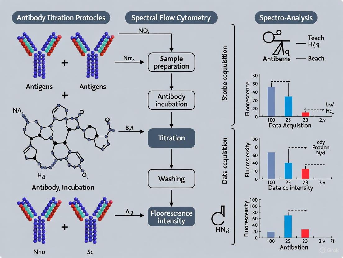
Antibody Titration Protocol for Spectral Flow Cytometry
Sample Preparation and Staining Procedure
Materials Required:
- Cell suspension (relevant cell type at 1×10⁶ cells/100 μL)
- Antibody of interest and isotype control
- Staining buffer (PBS with protein stabilizer)
- FC receptor blocking solution
- Flow cytometry tubes
- Centrifuge capable of 400×g at 4°C
- Ice bucket or refrigerated centrifuge
Procedure:
Cell Preparation
Antibody Serial Dilution
- Prepare a stock antibody solution at the manufacturer's recommended concentration (typically 300 μg/mL in PBS) [3].
- Centrifuge the antibody stock for 10 minutes at 15,000×g, 4°C to remove aggregates [3].
- Perform serial dilutions in six tubes:
- Tube 1: 10 μL antibody + 20 μL PBS = 30 μL
- Subsequent tubes: 10 μL of previous dilution + 20 μL PBS = 30 μL [3]
- Aliquot 50 μL of cell suspension into flow cytometry tubes [4].
- Add 10 μL of each antibody dilution to separate cell aliquots [3].
- Include controls: cells alone (autofluorescence), and isotype control [3].
Staining and Washing
- Gently mix and incubate for 15-45 minutes on ice in the dark [3].
- Add 2 mL cold washing buffer (PBS + 2% BSA), gently mix, and centrifuge for 5 minutes at 200-400×g, 4°C [3] [4].
- Aspirate and discard supernatant. Gently vortex to resuspend cell pellet.
- Repeat wash step once [3].
- Resuspend final pellet in 100-300 μL protein-free, cold PBS [3] [4].
Data Acquisition and Analysis
Acquire data on spectral flow cytometer using the same instrument settings that will be used for final experiments.
Calculate Stain Index (SI) for each antibody dilution using the formula:
- SI = (MFI of positive population - MFI of negative population) / (2 × standard deviation of negative population) [4]
Identify optimal antibody concentration as the dilution that yields the highest stain index [4].
Validate unmixing efficiency by comparing the spectral signature of titrated antibodies with reference spectra in the spectral viewer of your analysis software.
Spectral Antibody Titration Workflow
Advanced Applications and Integration with Novel Technologies
Integration with Cellular Interaction Mapping
The Interact-omics framework represents an advanced application of spectral flow cytometry that enables mapping of physical cell-cell interactions (PICs) across all immune cell types. This method relies on high-parameter spectral cytometry (24-plex panels or more) to accurately discriminate between single cells and interacting cell complexes based on scatter properties and co-expression of mutually exclusive lineage markers [5].
For such complex applications, antibody titration becomes even more critical as under-staining may fail to detect rare cell populations and their interactions, while over-staining can increase spectral spreading beyond the unmixing algorithm's correction capacity. The implementation of FSC ratio-based classification alongside carefully titrated antibody panels enables precise identification of interacting cell partners in heterogeneous samples [5].
Artificial Intelligence in Antibody and Unmixing Development
Artificial intelligence is transforming both antibody design and spectral unmixing algorithms. AI platforms can now design antibodies de novo with optimized binding characteristics, stability, and developability profiles [6]. These computational approaches can predict antibody properties that influence spectral performance, including:
- Binding affinity and its effect on fluorophore density
- Epitope specificity and its impact on staining patterns
- Cross-reactivity potential that may affect background signal
Machine learning approaches are also being applied to improve spectral unmixing algorithms, particularly for handling complex staining patterns and reducing unmixing errors in high-parameter panels [7]. The integration of AI-powered antibody design with optimized titration protocols represents the future of high-performance spectral flow cytometry.
Essential Reagents and Research Solutions
Table 3: Research Reagent Solutions for Spectral Flow Cytometry
| Reagent Category | Specific Examples | Function in Spectral Experiments |
|---|---|---|
| Fluorophores | Spark, Spark PLUS dyes [1] | High-resolution spectral separation with minimal spreading error |
| Cell Preparation | Staining buffer with protein stabilizer [4] | Maintain cell viability and reduce non-specific antibody binding |
| Blocking Reagents | FC receptor block; True-Stain Monocyte Blocker [4] | Minimize non-specific antibody binding through Fc receptors |
| Fixation Reagents | PBS + 2% paraformaldehyde [3] | Preserve cellular morphology and antibody binding for delayed acquisition |
| Viability Dyes | Propidium Iodide [3] | Distinguish live from dead cells to improve unmixing accuracy |
| Reference Standards | Compensation beads or single-stained controls [1] | Generate reference spectra for spectral unmixing algorithms |
Spectral unmixing and antibody concentration optimization are fundamentally interconnected processes in high-parameter flow cytometry. The principle of spectral unmixing relies on capturing complete fluorescence emission spectra and computationally separating overlapping signals based on reference profiles. The accuracy of this process is highly dependent on proper antibody titration, which ensures optimal signal-to-noise ratios while minimizing spectral spreading.
As spectral cytometry continues to evolve toward even higher parameter panels, the precise optimization of antibody concentrations becomes increasingly critical for data quality. Integration of these fundamental practices with emerging technologies, including artificial intelligence and cellular interaction mapping, will further expand the capabilities of spectral flow cytometry in both basic research and drug development applications.
Antibody titration is a critical step in flow cytometry assay development, ensuring optimal staining while minimizing background and non-specific binding. While the core principle of determining the optimal antibody-to-cell ratio remains consistent, the distinct signal detection and unmixing methodologies of spectral and conventional flow cytometry introduce significant differences in titration protocols and data interpretation. This application note delineates these key differences, providing detailed, standardized protocols for antibody titration within each system. Emphasizing the context of high-parameter panels, we outline how spectral cytometry's full-spectrum acquisition and unmixing algorithms allow for greater fluorophore flexibility and more complex panel design. The guidance herein is intended to empower researchers in immunology and drug development to achieve robust, reproducible, and high-resolution data from their flow cytometry experiments.
In multiparameter flow cytometry, the accuracy of cell population identification hinges on a high signal-to-noise ratio. Antibody titration is the foundational process for achieving this, balancing saturated target binding with minimized non-specific signal [8]. Conventional flow cytometry relies on optical filters and compensation to correct for fluorescence spillover, a process where the signal from one fluorophore is detected in another's channel [9]. This system constrains panel design, as fluorophores with significant spectral overlap can drastically increase spreading error, compromising the resolution of dimly expressed antigens [8] [10].
Spectral flow cytometry represents a paradigm shift, capturing the full emission spectrum of every fluorophore using detector arrays and employing unmixing algorithms to resolve individual signals [1] [9]. This capability allows for the use of fluorophores with highly overlapping emission peaks, provided their full spectral signatures are unique [1]. Consequently, the objectives and analytical considerations for antibody titration in spectral systems extend beyond those of conventional cytometry, directly enabling the 40+ parameter panels that are reshaping deep immunophenotyping in clinical and pharmaceutical research [10].
Fundamental Technological Divergences
The approach to antibody titration is fundamentally shaped by the underlying technology of the flow cytometer. The core differences in signal detection and processing between conventional and spectral systems are summarized in Table 1.
Table 1: Core Technological Comparison Influencing Titration Practices
| Feature | Conventional Flow Cytometry | Spectral Flow Cytometry |
|---|---|---|
| Detection Principle | "One detector–one fluorophore"; measures signal near emission peak [1]. | Collects full emission spectrum across a wide wavelength range for every fluorophore [1] [9]. |
| Spillover Correction | Compensation: mathematically subtracts spillover signal post-acquisition [9]. | Unmixing: identifies fluorophores based on unique spectral signatures using reference controls [9]. |
| Optical Setup | Complex system of dichroic mirrors and band-pass filters for each detector [1]. | Prism or diffraction grating to scatter light onto a detector array; optically simpler [1]. |
| Fluorophore Flexibility | Limited by the number of available detectors and filter configurations [1]. | High; limited primarily by the uniqueness of the fluorophore's spectral signature [1] [9]. |
| Autofluorescence Handling | Typically treated as background noise that reduces signal resolution [10]. | Can be characterized and "unmixed" as a separate signal, effectively subtracting it [10] [9]. |
The following diagram illustrates the fundamental difference in how these two technologies process fluorescent light from a sample.
Key Differences in Antibody Titration
The technological divergence leads to distinct practical considerations for antibody titration, particularly as panels increase in complexity.
Primary Objectives of Titration
- In Conventional Cytometry, the primary goal is to find the antibody concentration that provides the best Stain Index (SI), which balances the separation between positive and negative populations (ΔMFI) with the spread of the negative population (2SD). A key focus is minimizing spreading error caused by compensation, which can obscure dim populations [8].
- In Spectral Cytometry, while the SI remains important, titration also aims to generate a high-quality, cell-type-specific reference control for the unmixing algorithm. The accuracy of unmixing for all other samples depends on the purity of these reference spectra [9].
Impact of Spillover and Panel Complexity
- In Conventional Cytometry, spillover is a major constraint. Adding a fluorophore with significant spillover into a detector used by another marker increases the spread of the negative population for that second marker, potentially masking dim positive signals. Titration must therefore be performed in the context of the full panel to assess these interactions [8].
- In Spectral Cytometry, "spillover" is managed computationally via unmixing. The limitation is not spillover per se, but the uniqueness of the full spectral signature. Fluorophores with nearly identical peak emissions but different off-peak profiles can be distinguished [9]. Titration can often be performed for individual antibodies, though full-panel validation is still recommended.
The Role of Controls
Both methodologies require rigorous controls, but their application differs.
- For Conventional Cytometry, Fluorescence Minus One (FMO) controls are critical for setting gates correctly, especially for dim markers where compensation spread can cause false positives [8].
- For Spectral Cytometry, single-stained controls are paramount. They are not just for compensation but are used to build the spectral library for unmixing. These controls must be exceptionally clean and ideally performed on the same cell type as the experimental sample (e.g., CD4+ T cells for a T-cell marker) to account for cell-specific autofluorescence [10] [9].
Detailed Experimental Protocols
The following protocols provide a step-by-step guide for antibody titration in both conventional and spectral flow cytometry.
Protocol for Conventional Flow Cytometry Titration
Principle: Identify the antibody concentration that yields the maximal Stain Index, indicating optimal separation between positive and negative cell populations.
Materials:
- Cell suspension (e.g., PBMCs or a relevant cell line)
- Titration range of antibody (e.g., 0.06 µg/mL to 4.0 µg/mL, in 2-4-fold dilutions)
- Flow cytometry staining buffer
- Optional: FMO control
Procedure:
- Prepare Cells: Aliquot a sufficient number of cells (e.g., 2.0 x 10^5 per tube) for each antibody dilution and an unstained control.
- Serially Dilute Antibody: Prepare the predetermined range of antibody concentrations in staining buffer.
- Stain Cells: Add each antibody dilution to its respective cell pellet. Vortex gently and incubate for 20-30 minutes in the dark at 4°C.
- Wash and Resuspend: Wash cells twice with staining buffer, then resuspend in a fixed volume of buffer for acquisition.
- Acquire Data: Run samples on a conventional flow cytometer. Record the Mean Fluorescence Intensity (MFI) of both the positive and negative cell populations for the target antigen.
Data Analysis:
- For each dilution, calculate the Stain Index (SI) using the formula: ( \text{SI} = (\text{MFI}{\text{positive}} - \text{MFI}{\text{negative}}) / (2 \times \text{SD}_{\text{negative}}) )
- Plot the SI against the antibody concentration. The optimal concentration is at the plateau of the SI curve, before the SI plateaus or begins to decrease while the MFI of the negative population remains low.
Protocol for Spectral Flow Cytometry Titration
Principle: Identify the antibody concentration that provides a robust signal for unmixing while ensuring the generated reference spectrum is clean and specific.
Materials:
- Cell suspension (ideally positive for the target antigen)
- Titration range of antibody
- Flow cytometry staining buffer
- Critical: Ultra-compensation beads or antigen-negative cells for single-stain control validation
Procedure:
- Cell Preparation and Staining: Follow steps 1-4 from the conventional protocol.
- Acquire Single-Stain Controls: Run each titration point. The instrument software will use this data to build a reference spectrum for that fluorophore-antibody conjugate at each concentration.
- Validate Control Purity: Inspect the single-stain control data. The fluorescence should be detected primarily in its intended channel with minimal signal in off-target channels. A "clean" control is essential for accurate unmixing.
Data Analysis:
- Analyze in the Context of the Full Panel: Stain a sample with the full, multi-color panel at each antibody concentration. Use the spectral library generated from the single-stain controls to unmix the data.
- Evaluate Resolution: The optimal antibody concentration is one that, after unmixing, provides clear separation of positive and negative populations. Assess this by viewing the data in 2D plots for the marker of interest.
- Leverage Software Tools: Utilize spectral viewer tools in the instrument software (e.g., SpectroFlo, Sony Spectrum Analyzer) to visually inspect the quality and uniqueness of the reference spectrum generated at each concentration [1].
The workflow for the spectral cytometry protocol, emphasizing the critical role of single-stain controls, is detailed below.
The Scientist's Toolkit: Essential Reagents & Materials
Successful implementation of these protocols requires specific reagents and controls. Key materials are listed in Table 2.
Table 2: Essential Research Reagents and Materials for Titration
| Reagent / Material | Function | Application Notes |
|---|---|---|
| UltraComp Compensation Beads | Provide a uniform, autofluorescence-free particle for generating high-quality single-stain controls. | Crucial for building a clean initial spectral library in spectral cytometry and for compensation in conventional cytometry. |
| CD16/CD32 Fc Block | Binds to Fc receptors on cells, preventing non-specific antibody binding and reducing background. | Should be used prior to antibody staining, especially for immune cells like monocytes and macrophages. |
| Viability Dye (e.g., Zombie UV) | Distinguishes live from dead cells; dead cells bind antibodies non-specifically, increasing background. | Essential for excluding false-positive events. Must be titrated and added prior to fixation/permeabilization. |
| Brilliant Stain Buffer | Mitigates hydrophobic interactions between certain polymer-based "Brilliant" dyes, preventing aggregation and fluorescence energy transfer. | Critical for panels using multiple BV and BY dyes in conventional cytometry; often recommended in spectral panels. |
| Pre-titrated Antibody Panels (OMIPs) | Published, validated panels (Published OMIPs in Cytometry A) provide an excellent starting point for panel design. | Saves time and resources; the provided titration and panel layout can be adapted to specific research needs [8]. |
Antibody titration is not a one-size-fits-all procedure. The transition from conventional to spectral flow cytometry necessitates a refined approach that aligns with the underlying detection technology. While conventional cytometry titration focuses on maximizing the stain index and managing spillover-induced spreading error, spectral cytometry titration emphasizes the generation of pristine reference spectra for accurate computational unmixing. Adherence to the detailed protocols and considerations outlined in this document will enable researchers to effectively leverage the high-parameter capabilities of spectral cytometry. This ensures the generation of robust, high-fidelity data crucial for advanced applications in clinical diagnostics, deep immunophenotyping, and drug discovery.
In spectral flow cytometry, achieving optimal signal detection requires precise quantification of fluorescence resolution through key metrics like stain index and separation index. These measurements provide critical insights into fluorophore brightness and population discrimination, enabling researchers to develop robust antibody titration protocols that maximize detection sensitivity while minimizing background interference. This application note details the theoretical foundations, calculation methodologies, and practical implementation of these indices within antibody titration workflows for spectral flow cytometry research, providing drug development professionals with standardized approaches for assay optimization.
Signal resolution metrics are fundamental to optimizing fluorescence detection in spectral flow cytometry, particularly when developing antibody titration protocols for high-parameter panels. The stain index and separation index provide quantitative measurements that capture both the signal intensity and spread of fluorescence distributions, enabling researchers to make informed decisions about fluorophore selection and antibody concentration optimization [11] [12]. These metrics are especially valuable in spectral flow cytometry where the simultaneous detection of multiple fluorophores requires careful balancing of signal-to-noise ratios across all channels.
The stain index specifically measures the relative brightness of fluorophores on a given cytometer, accounting for instrument-specific variables that affect fluorescence detection [11]. Meanwhile, the separation index provides a refined metric for evaluating the relationship between positive and negative populations, with particular emphasis on the right-hand slope of the negative distribution to minimize error in fluorescence distribution assessment [12]. For researchers developing antibody titration protocols, these indices serve as objective criteria for determining optimal staining concentrations that maximize resolution while conserving reagents.
Theoretical Foundations and Calculations
Stain Index: Definition and Formula
The stain index represents a normalized measure of fluorophore brightness that accounts for both the signal intensity and the spread of the negative population. This metric is formally defined as the difference between the mean fluorescence intensity of the positive and negative populations, divided by two times the standard deviation of the negative population [11] [13]. The calculation can be expressed as:
Table 1: Stain Index Calculation Formula
| Metric | Calculation Formula | Components |
|---|---|---|
| Stain Index | ( \text{SI} = \frac{\text{MFI}{\text{positive}} - \text{MFI}{\text{negative}}}{2 \times \text{SD}_{\text{negative}}} ) | Where MFIpositive = Mean Fluorescence Intensity of positive population, MFInegative = Mean Fluorescence Intensity of negative population, SDnegative = Standard Deviation of negative population [11] |
Some flow cytometry software packages utilize median values rather than mean fluorescence intensity, calculating the stain index as the difference between the median of positive and negative populations divided by twice the standard deviation of the negative population [13]. This variation offers robustness against outliers in the fluorescence distribution.
Separation Index: Definition and Distinctive Features
The separation index provides a complementary metric that specifically weights the right-hand slope of the negative population more heavily to minimize error in the negative fluorescence distribution [12]. While similar in concept to the stain index, this distinctive approach makes the separation index particularly valuable for evaluating population discrimination where the negative distribution may exhibit skewness.
Table 2: Comparison of Signal Resolution Metrics
| Metric | Primary Application | Key Differentiating Factor | Interpretation |
|---|---|---|---|
| Stain Index | Fluorophore brightness comparison [11] | Normalizes signal difference by spread of negative population | Higher values indicate brighter fluorophores [14] |
| Separation Index | Population discrimination assessment [12] | Emphasizes right-hand slope of negative distribution | Higher values indicate better population separation [12] |
Relationship Between Metrics and Experimental Optimization
The relationship between stain index, separation index, and experimental parameters follows a predictable workflow that researchers can leverage during assay development. The following diagram illustrates how these metrics inform decision-making throughout the optimization process:
Signal Optimization Workflow
Practical Applications in Experimental Design
Fluorophore Selection and Panel Design
The strategic application of stain index values enables researchers to make informed decisions during fluorophore selection and panel design. By calculating the stain index for various fluorophores on their specific instrument, researchers can create a brightness ranking that guides optimal fluorophore-antigen pairing [11]. This approach is particularly valuable when working with antigens of varying expression levels, as it facilitates the matching of bright fluorophores with weakly expressed markers and dimmer fluorophores with highly expressed markers [11] [8].
For spectral flow cytometry panels, the stain index provides critical information for minimizing spreading error while maintaining detection sensitivity. When designing multicolor panels, researchers should prioritize fluorophores with higher stain indices for:
- Low-density antigens or markers with continuous expression patterns [12]
- Tertiary antigens that are either expressed at low levels or uncharacterized [12]
- Co-expressed antigens that require fluorophores with distinct spectral signatures [12]
Antibody Titration Optimization
Stain index values are particularly valuable for evaluating antibody titrations to identify optimal staining concentrations. By plotting stain index against antibody concentration, researchers can visualize the relationship between reagent usage and signal resolution, identifying the point of diminishing returns where additional antibody does not substantially improve the stain index [11] [14]. This approach facilitates evidence-based titration rather than reliance on manufacturer recommendations that may not account for instrument-specific or application-specific variables.
The integration of separation index calculations into titration protocols provides additional confidence in population discrimination, especially for markers with minimal separation between positive and negative populations [12]. This dual-metric approach ensures that titration decisions account for both signal brightness and population resolution, critical factors in experimental reproducibility.
Essential Research Reagent Solutions
The implementation of robust stain index and separation index protocols requires specific reagents designed to minimize non-specific binding and preserve fluorescence signals. The following table details essential materials for these optimization workflows:
Table 3: Key Research Reagents for Signal Optimization
| Reagent | Function | Application Notes |
|---|---|---|
| Normal Sera (Mouse, Rat) | Blocks Fc receptor-mediated non-specific binding [15] | Use serum from same species as staining antibodies; critical for hematopoietic cells [15] |
| Brilliant Stain Buffer | Prevents dye-dye interactions between polymer fluorophores [15] | Essential for panels containing SIRIGEN "Brilliant" or "Super Bright" dyes [15] |
| Tandem Stabilizer | Maintains integrity of tandem dye conjugates [15] | Reduces breakdown of tandem dyes that causes erroneous signals [15] |
| Antibody Capture Beads | Generate single stain controls for compensation [12] | Verify spectral signatures match cell-bound antibodies in spectral flow cytometry [12] |
| FACS Buffer | Base medium for antibody dilutions and washes [15] | Typically contains 2% FBS in PBS; compatible with most fluorophores [15] |
Comprehensive Experimental Protocol
Surface Staining with Integrated Blocking Protocol
This optimized protocol for surface staining incorporates blocking steps to minimize non-specific interactions, thereby improving stain index values through enhanced signal-to-noise ratios [15]:
Preparation of Blocking Solution
- Combine 300μl mouse serum, 300μl rat serum, 1μl tandem stabilizer, and 389μl FACS buffer to create 1ml of blocking solution [15]
- Include serum from any host species used for staining antibodies in the panel
Cell Staining Procedure
- Dispense cells into V-bottom 96-well plates and centrifuge at 300×g for 5 minutes [15]
- Resuspend cell pellets in 20μl blocking solution and incubate for 15 minutes at room temperature in the dark [15]
- Prepare surface staining master mix containing tandem stabilizer (1:1000), Brilliant Stain Buffer (up to 30% v/v), and titrated antibodies in FACS buffer [15]
- Add 100μl staining mix to each sample, mix by pipetting, and incubate for 1 hour at room temperature in the dark [15]
- Wash with 120μl FACS buffer, centrifuge at 300×g for 5 minutes, and discard supernatant [15]
- Repeat wash with 200μl FACS buffer [15]
- Resuspend samples in FACS buffer containing tandem stabilizer (1:1000 dilution) [15]
Data Acquisition and Analysis
- Acquire samples on spectral flow cytometer using established instrument settings [16]
- For stain index calculation, identify positive and negative populations for each fluorophore
- Record mean or median fluorescence intensity of positive and negative populations
- Calculate standard deviation of negative population
- Compute stain index using formula in Table 1 [11] [13]
Stain Index Calculation for Antibody Titration
This specialized protocol employs stain index calculations to determine optimal antibody concentrations for spectral flow cytometry:
Titration Series Setup
Data Analysis and Interpretation
- For each antibody concentration, calculate the stain index as described in Section 5.1
- Plot stain index values against antibody concentration to visualize the relationship
- Identify the concentration point where stain index plateaus (saturating titer) [12]
- Select the antibody concentration that provides ≥90% of maximum stain index for routine use
- Validate selected concentration using separation index calculations to ensure population discrimination [12]
Implementation in Spectral Flow Cytometry
Spectral flow cytometry presents both opportunities and challenges for stain index applications. The unmixing algorithms used in spectral cytometry can identify and remove autofluorescence, potentially improving stain index values compared to conventional flow cytometry [12]. However, researchers must ensure that single stain controls share the exact same spectral signature and are processed identically to experimental samples to generate accurate stain index calculations [12].
For complex multicolor panels, the stain index provides a standardized approach to evaluate fluorophore performance across multiple laser lines and detection channels. When designing spectral panels, researchers should calculate stain indices for all fluorophores under consideration using their specific instrument configuration, as the complexity index of the panel directly impacts the practical resolution achievable for each marker [12]. This instrument-specific approach is particularly valuable when incorporating new fluorophore chemistries or when working with complex sample types that exhibit high autofluorescence.
The Role of Antibody Kinetics and Binding Saturation in Titration
The process of antibody titration is a critical foundational step in the development of robust and reproducible flow cytometry assays, particularly as the field advances with spectral cytometry technology. Antibody kinetics—encompassing the dynamics of antibody binding, including affinity (the strength of a single binding interaction) and avidity (the overall strength of multiple simultaneous interactions)—directly influences the outcome of staining reactions [17]. The primary goal of titration is to identify the optimal antibody concentration that achieves binding saturation of all available antigen targets while minimizing non-specific background, thereby providing the highest possible resolution between positive and negative cell populations [17] [18].
Understanding the underlying principles of antibody binding is essential for assay optimization. Antibodies are glycoproteins produced by B cells, consisting of two heavy and two light chains forming a Y-shaped structure. The variable regions at the ends of the "Y" are critical for specific antigen binding [17]. IgM antibodies, with their pentameric structure containing 10 antigen binding sites, typically exhibit low affinity but high avidity. In contrast, IgG antibodies have two binding sites and generally possess higher affinity [17]. The titration process systematically evaluates serial dilutions of a fluorescently conjugated antibody on cells expressing the target antigen to determine the concentration that provides the optimal signal-to-noise ratio, calculated using the stain index [17] [4]. This optimization is crucial for reliable and reproducible results, as it must be performed for each sample type, reagent clone and lot, and specific staining protocol [17].
Theoretical Principles of Antibody Binding
Key Kinetic Parameters
The interaction between an antibody and its target antigen is governed by several fundamental parameters that collectively determine staining quality and must be considered during titration. Affinity refers to the strength of a single antigen-binding site interacting with its epitope, representing the equilibrium between associated and dissociated states [17]. Avidity, in contrast, describes the overall binding strength resulting from the sum of multiple interactions between an antibody molecule and a complex antigen. This distinction explains why IgM, despite lower individual binding site affinity, can demonstrate high avidity due to its multivalent structure with 10 antigen binding sites [17].
The concept of binding saturation is central to effective titration. It represents the point at which all available antigen binding sites are occupied by antibodies, beyond which additional antibody does not increase the specific signal but may elevate background noise through non-specific binding [17] [18]. Achieving optimal saturation is reflected in the stain index (SI), a quantitative metric calculated as (MFIpositive - MFInegative) / (2 × standard deviation_negative), where MFI represents the mean fluorescence intensity [4]. The optimal titer is identified at the dilution that yields the highest stain index, indicating superior separation between positive and negative populations [18].
Consequences of Improper Titration
Under-Titration: At antibody concentrations that are too low, the signal becomes too weak for accurate determination of antigen expression, particularly for low-abundance targets [17]. This leads to suboptimal data resolution, high measurement variability, and potential underestimation of cell population frequencies [17] [18].
Over-Titration: Excessive antibody concentrations promote non-specific binding through several mechanisms, including Fc receptor interactions and hydrophobic non-immune binding [17] [18]. This results in increased background fluorescence, reduced signal-to-noise ratio, inefficient reagent use, and potential detector overloading with signal off-scale and increased spillover spreading [17].
Table 1: Quantitative Stain Index Analysis for Titer Determination
| Antibody Dilution | MFI Positive | MFI Negative | rSD Negative | Stain Index | Resolution Assessment |
|---|---|---|---|---|---|
| 1:50 | 45,200 | 1,850 | 185 | 117.2 | Excessive background |
| 1:100 | 44,850 | 950 | 98 | 224.0 | Optimal separation |
| 1:200 | 44,100 | 650 | 72 | 301.4 | Recommended titer |
| 1:400 | 42,900 | 520 | 65 | 326.2 | Optimal stain index |
| 1:800 | 39,500 | 480 | 68 | 286.8 | Good resolution |
| 1:1600 | 32,100 | 450 | 72 | 219.8 | Suboptimal for low expressers |
| 1:3200 | 21,500 | 430 | 75 | 140.7 | Poor resolution |
Comprehensive Titration Protocol for Spectral Flow Cytometry
Reagent and Cell Preparation
The following protocol provides a detailed methodology for determining the optimal working concentration for fluorescently conjugated antibodies in spectral flow cytometry applications [17] [3] [4].
Cell Preparation: Harvest fresh cells of the relevant type (e.g., PBMCs) in staining buffer. The cell type used for titration must match the experimental system, as staining characteristics can vary significantly between tissues [4]. Resuspend cells at a concentration of 5-10 × 10^6 cells/mL in staining buffer [3]. For procedures involving intracellular staining, include fixation and permeabilization steps at this stage using appropriate commercial buffer systems [19].
Fc Receptor Blocking: To minimize non-specific binding through Fc receptors, add Fc blocking reagent (e.g., Human TruStain FcX) and incubate for 5-10 minutes at room temperature before antibody addition [19] [4]. This step is particularly crucial for immune cells such as monocytes and macrophages which express high levels of Fc receptors [18].
Antibody Dilution Series: Prepare a stock antibody solution at the manufacturer's recommended starting concentration. Create an 8-12 point serial dilution series in a 96-well V-bottom plate using staining buffer, with each dilution prepared in a final volume of 150-300 μL [17]. Two-fold serial dilutions are typically sufficient to identify the optimal concentration range.
Table 2: Essential Research Reagents for Antibody Titration
| Reagent Category | Specific Examples | Function in Titration Protocol |
|---|---|---|
| Staining Buffer | PBS + 0.5% BSA + 0.05% azide | Maintains cell viability and reduces non-specific binding |
| Fc Blocking Reagents | Human TruStain FcX, purified IgG | Blocks Fc receptor-mediated non-specific antibody binding |
| Viability Dyes | Propidium Iodide, Fixable Viability Dyes | Distinguishes live/dead cells; must be titrated first |
| Fixation Reagents | Paraformaldehyde (2-4%) | Preserves cellular integrity and staining post-processing |
| Permeabilization Buffers | Commercial buffer sets (e.g., FoxP3 kit) | Enables intracellular antigen staining |
| Reference Beads | UltraComp eBeads | Used for generating consistent single-stain controls |
Staining Procedure and Data Acquisition
Staining Incubation: Aliquot 50-100 μL of cell suspension (containing 0.5-1 × 10^6 cells) into each well of the titration plate [3] [4]. Add equal volumes of each antibody dilution to corresponding wells, mix gently by pipetting, and incubate for 20-45 minutes in the dark at room temperature or on ice, depending on antibody sensitivity [3] [4].
Washing and Fixation: Following incubation, add 2-3 mL of cold washing buffer to each tube, centrifuge at 200-400 × g for 5 minutes at 4°C, and carefully decant the supernatant [3]. Repeat this washing step twice to ensure removal of unbound antibody [3]. Resuspend the final cell pellet in 100-300 μL of protein-free PBS or staining buffer, with or without paraformaldehyde fixation (1-4%) depending on experimental requirements [3].
Flow Cytometry Acquisition: Acquire samples on a spectral flow cytometer using consistent instrument settings across all samples [19]. Include appropriate controls: unstained cells (to determine autofluorescence), and negative biological controls (cells known not to express the target antigen) [18]. For spectral cytometry, high-quality single-stain controls are essential for generating the spectral unmixing matrix [18] [19].
Data Analysis and Optimal Titer Determination
Stain Index Calculation and Interpretation
Following data acquisition, analyze the fluorescence intensity for both positive and negative cell populations at each antibody dilution. Calculate the stain index (SI) for each dilution using the formula: SI = (MFIpositive - MFInegative) / (2 × rSD_negative), where rSD represents the robust standard deviation of the negative population [4]. Plot the stain index values against the antibody concentration to identify the peak value, which represents the optimal titer [18].
The optimal antibody concentration is typically identified at or near the saturation point, where further increases in antibody concentration do not substantially increase the specific signal (MFIpositive) but may increase background (MFInegative) [17] [18]. This point represents the ideal balance between maximal specific binding and minimal non-specific background. For critical applications, particularly when staining for low-abundance antigens, Fluorescence Minus One (FMO) controls should be used to confirm proper gate placement and distinguish true positive signals from background spreading error [18].
Integration with Spectral Flow Cytometry Controls
The titration process must be integrated with appropriate control strategies specific to spectral flow cytometry. Single-stain controls, prepared using either beads or cells, are essential for generating the spectral unmixing matrix [18] [19]. These controls should be acquired using the same antibody lots and instrument settings as the experimental samples [19]. For multicolor panels, titrate each antibody individually before assessing potential interactions in combination, as steric hindrance or unexpected interactions may require further optimization of concentrations when antibodies are used together [17] [18].
Applications in Advanced Assay Development
High-Parameter Panel Optimization
The principles of antibody kinetics and titration find critical application in the development of high-parameter spectral flow cytometry panels. As panel complexity increases, individual antibody titrations become essential for minimizing spectral overlap and maximizing population resolution [19]. Recent advancements have demonstrated successful implementation of panels exceeding 30 colors, with each antibody requiring individual titration under conditions matching the final experimental setup [19]. This meticulous optimization enables simultaneous investigation of numerous cellular parameters while maintaining data quality and reproducibility.
Specialized Applications
Beyond standard immunophenotyping, optimized titration protocols enable advanced applications including the detection of low-abundance targets, intracellular signaling proteins, and cytokine production [19]. In pharmaceutical development, properly titrated assays support critical evaluations such as antibody affinity maturation studies [20] and minimal residual disease (MRD) detection in hematological malignancies [21]. The bead-based multiplex assay platform for simultaneous detection of IgM, IgG, and IgA antibodies against SARS-CoV-2 spike RBD demonstrates how titration principles can be scaled for high-throughput applications, enabling analysis of 624 serum samples within 2 hours [22].
Antibody titration grounded in the principles of antibody kinetics and binding saturation represents a fundamental requirement for generating high-quality, reproducible data in spectral flow cytometry. The systematic determination of optimal antibody concentrations through stain index calculation ensures maximal resolution of target populations while minimizing background and non-specific binding. As flow cytometry continues to advance with increasingly complex multicolor panels and applications in both basic research and clinical diagnostics, rigorous titration protocols remain essential for assay validation and standardization across laboratories. By adhering to the detailed methodologies outlined in this document, researchers can establish robust titration practices that enhance data quality and support reliable scientific conclusions in drug development and biological research.
Antibody titration is a fundamental yet critical step in optimizing spectral flow cytometry experiments. Proper titration ensures that fluorescent signals accurately represent antigen expression levels, maximizing resolution while minimizing background noise and non-specific binding. Within the broader context of developing robust antibody titration protocols for spectral flow cytometry research, understanding the direct consequences of improper titration is paramount. This application note details how suboptimal antibody concentrations compromise data quality by increasing background noise and diminishing resolution, ultimately threatening experimental reproducibility and reliability. We provide a quantitative framework for evaluating these effects and detailed protocols for effective titration.
The Direct Link Between Titration and Data Quality
Defining the Problem: How Improper Titration Introduces Noise
In spectral flow cytometry, the signal-to-noise ratio is the primary determinant of a panel's resolution. Improper titration directly degrades this ratio through two main mechanisms:
- Excessive Antibody Concentration: Using an antibody concentration that is too high leads to non-specific binding, where antibodies bind to off-target epitopes. This increases the background fluorescence of the negative population, obscuring the true positive signal [23]. This effect is a significant source of reagent noise [24].
- Insufficient Antibody Concentration: An overly dilute antibody fails to saturate all target antigens, resulting in a diminished positive signal. This reduces the separation between positive and negative populations, making it difficult to distinguish dimly expressed markers and identify rare cell subsets [25].
The relationship between antibody concentration and the resulting stain index, a key metric for resolution, is not linear. There is a distinct optimal point where the stain index is maximized; deviating from this point in either direction reduces the quality of the separation [26].
Quantifying the Impact: From Stain Index to Population Resolution
The stain index (SI) is a crucial quantitative measure for evaluating the effectiveness of an antibody stain and the consequence of its titration. It is calculated using the formula:
SI = (MFIpositive - MFInegative) / (2 × SD_negative)
Where:
- MFI_positive is the median fluorescence intensity of the positive population.
- MFI_negative is the median fluorescence intensity of the negative population.
- SD_negative is the standard deviation of the negative population [26].
A higher stain index indicates better separation between positive and negative cells. The following table summarizes the measurable impacts of improper titration on this and other key parameters:
Table 1: Quantitative and Qualitative Impacts of Improper Antibody Titration
| Parameter | Impact of Under-Titration | Impact of Over-Titration |
|---|---|---|
| Stain Index | Decreased due to lower MFI_positive | Decreased due to higher MFInegative and increased SDnegative |
| Background Noise (MFI_negative) | Minimal change | Significant increase due to non-specific binding |
| Signal Strength (MFI_positive) | Significantly reduced | Slight increase or plateau, insufficient to offset noise |
| Population Resolution | Poor separation, loss of dim populations | Poor separation, increased spread of negative population |
| Data Reproducibility | High well-to-well variability | High well-to-well variability |
| Experimental Cost | Lower per sample | Significantly higher, wasteful of reagents |
The progression from optimal to poor titration directly impacts the visualization and gating of cell populations, as illustrated in the following workflow:
Figure 1. Logical workflow diagram illustrating the consequences of improper antibody titration on data resolution in flow cytometry.
Experimental Protocols for Effective Titration
Comprehensive Antibody Titration Protocol
This protocol is designed to systematically identify the optimal working concentration for an antibody, maximizing the stain index and minimizing background.
Materials:
- Antibody to be titrated
- Cell sample (≥ 5 × 10^6 cells) expressing the target antigen at a representative level
- Staining buffer (e.g., PBS with 1-5% FBS)
- Flow cytometry tubes or a 96-well plate
- Spectral flow cytometer
Procedure:
- Prepare a single-cell suspension and determine the total cell count and viability.
- Prepare antibody dilutions. Create a series of at least 5-6 doubling dilutions of the antibody in staining buffer. A typical series may include 1:25, 1:50, 1:100, 1:200, 1:400, and 1:800 dilutions of the stock concentration.
- Aliquot cells. Distribute a sufficient number of cells (e.g., 2.5 × 10^5 to 5 × 10^5) into each tube or well for every dilution point, including an unstained control.
- Stain cells. Add the corresponding antibody dilution to each cell aliquot. Mix gently and incubate for 20-30 minutes in the dark at 4°C.
- Wash cells. Add 2-3 mL of staining buffer to each tube, centrifuge, and carefully decant the supernatant. For plate-based protocols, use a plate centrifuge.
- Resuspend cells. Resuspend the cell pellets in a fixed volume of staining buffer for acquisition.
- Acquire data. Run all samples on the spectral flow cytometer, ensuring instrument settings (laser power, detector voltages) are kept constant throughout the acquisition.
Data Analysis:
- For each dilution, identify the positive and negative cell populations.
- Record the MFI of both the positive and negative populations.
- Calculate the robust standard deviation (rSD) of the negative population.
- Calculate the Stain Index (SI) for each dilution using the formula: SI = (MFIpositive - MFInegative) / (2 × rSD_negative).
- Plot the Stain Index against the antibody dilution or volume. The optimal dilution is the point that yields the highest stain index, providing the best separation with the least background [26].
Validating Titration in a Polychromatic Panel
Once individual antibodies are titrated, their performance must be verified within the full polychromatic panel to account for interactions like spectral overlap and potential new sources of background.
Procedure:
- Incorporate titrated antibodies into the full spectral panel.
- Stain cells following the established panel protocol.
- Compare the resolution of key populations to the resolution observed in single-color titrations.
- Check for spreading error (increased variance in negative populations) in all channels, which can be exacerbated by over-titration in a multicolor setting [23].
- If resolution is compromised, adjust the concentration of the problematic antibody slightly. The optimal concentration in a panel may sometimes differ from the optimal single-color concentration.
The Scientist's Toolkit: Essential Reagents and Materials
Table 2: Key Research Reagent Solutions for Antibody Titration
| Item | Function/Benefit | Application Note |
|---|---|---|
| Recombinant Antibodies | Engineered for high specificity; lack Fc region to minimize binding to Fc receptors, reducing non-specific background [23] [27]. | Ideal for difficult targets or highly autofluorescent cells. |
| High-Quality Tandem Dyes | Bright fluorophores for detecting low-abundance antigens. | Exhibit lot-to-lot variation; use the same lot for titration and experiments [28]. |
| Cell Staining Buffer | Provides optimal pH and protein content to maintain antibody stability and cell viability during staining. | Buffers with BSA or FBS help block non-specific binding. |
| Compensation Beads / Cells | Used for generating single-color controls for spectral unmixing. | For critical fluorophores, using cells for controls is preferred over beads for a more accurate spectral signature [28]. |
| Viability Dye | Distinguishes live cells from dead cells; dead cells are highly autofluorescent and cause non-specific antibody binding. | Essential for eliminating false positives. |
| Fc Receptor Blocking Solution | Blocks non-specific binding of antibodies to Fc receptors on immune cells. | Crucial for staining immune cells from blood, spleen, or other tissues [27]. |
Proper antibody titration is not merely a recommended best practice but a fundamental requirement for generating high-quality, reproducible data in spectral flow cytometry. As demonstrated, improper titration directly and measurably increases background noise and diminishes resolution, undermining the powerful multiplexing capabilities of spectral technology. By adopting the rigorous, quantitative titration protocols outlined in this note—centered on the calculation of the stain index—researchers can systematically optimize their panels, ensure accurate identification of cell populations, and build a solid foundation for reliable scientific discovery.
A Step-by-Step Guide to Spectral Flow Titration and Staining Protocols
Within the framework of antibody titration protocols for spectral flow cytometry, the strategic selection of cells, buffers, and blocking reagents is a critical pre-analytical phase that fundamentally determines the success of high-throughput immune monitoring in clinical trials and drug development [29]. Robust assays are paramount for monitoring patient immune responses, and the integrity of these assays begins long before data acquisition on instruments like the Sony ID7000 spectral cell analyzer [29]. The spectral flow cytometry workflow, while sharing fundamental principles with conventional flow cytometry, imposes unique requirements for sample preparation to fully leverage its capabilities in resolving complex multicolor panels exceeding 40 parameters [9]. This application note provides detailed methodologies and structured data to guide researchers in making informed decisions at this crucial planning stage, ensuring that subsequent antibody titration and validation steps are built upon a solid, reproducible foundation.
Cell Selection and Preparation
Defining the Cellular Material
The choice of cellular starting material directly impacts the resolution and reliability of spectral flow cytometry data. For clinical research correlative studies, Peripheral Blood Mononuclear Cells (PBMCs) are a frequently used sample type, requiring optimized in-plate staining protocols for high-throughput analysis [29]. The principles outlined, however, apply broadly to various cell types.
A key consideration is ensuring a single-cell suspension of high viability. Cells should be processed to minimize aggregation and debris, which can cause instrument clogging and data artifacts. For tissues like mouse spleens, this involves mechanical dissociation and, if necessary, enzymatic digestion at 37°C using specialized dissociation media [30]. Subsequent steps must focus on preserving cell integrity and antigenicity.
Pre-Enrichment for Complex or Rare Populations
For rare cell types or complex samples, using FACS (Fluorescence-Activated Cell Sorting) alone can be time-consuming, expensive, and detrimental to cell viability [30]. Pre-enrichment via immunomagnetic negative or positive selection is a highly effective strategy to overcome this.
Case Study: Isolating Rare Innate Lymphoid Cells (ILCs)
- Challenge: ILCs are exceptionally rare, comprising <0.1% of CD45+ leukocytes in human peripheral blood, making isolation by FACS alone inefficient [30].
- Solution: Pre-enrichment using an immunomagnetic kit (e.g., EasySep Human Pan-ILC Enrichment Kit).
- Result: The pre-FACS frequency of ILCs increased from 0.1% to 27%. This reduced the required FACS time from an estimated 3,200 minutes to just 12 minutes to process 2 x 10^9 cells, while achieving a final purity of 99% for Lin⁻CD127⁺ cells [30].
This pre-enrichment step drastically improves efficiency, reduces sort time, and yields viable, functional cells ready for downstream staining and analysis.
A Hierarchical Gating Strategy for Target Population Identification
Once cells are prepared and acquired on the cytometer, a sequential gating strategy is essential to isolate the live, single cells of interest for analysis. The following workflow outlines this standard process.
Diagram 1: Sequential gating workflow for precise cell population isolation.
The workflow begins by excluding debris using a plot of Forward Scatter-Area (FSC-A) versus Side Scatter-Area (SSC-A), drawing a gate (P1) around the main cell population [31] [32]. The next critical step is doublet exclusion by plotting FSC-A against FSC-Width (FSC-W); single cells form a diagonal linear cluster, while doublets deviate from this pattern and are excluded (Gate P2) [31] [32]. Subsequently, non-viable cells are excluded by gating on cells negative for a viability dye like propidium iodide or 7-AAD (Gate P3) [33] [32]. When working with heterogeneous samples like whole blood or splenocytes, gating on CD45-positive cells helps isolate leukocytes and exclude residual red blood cells or other contaminants (Gate P4) [31] [30]. Finally, the refined population is analyzed using fluorescence markers to define the target phenotype, such as CD3⁺CD4⁺ T cells, with gates set using appropriate negative controls like FMO controls [31] [32].
Buffer Systems and Staining Conditions
The Critical Role of Buffer Consistency
The buffer system used throughout staining, panel preparation, and experimental sample acquisition must be identical to maintain consistency. Any variation in the buffer can affect both the sample cells and the antibody-binding characteristics, leading to non-specific binding and compromised data [34]. A standard flow buffer consists of Phosphate-Buffered Saline (PBS) supplemented with 2% bovine serum albumin (BSA) or fetal bovine serum (FBS), and sometimes a preservative like 0.02% sodium azide [35].
For intracellular staining, a fixation and permeabilization buffer system is required. These are often used as dedicated kits, with a fixation buffer (commonly containing formaldehyde) to cross-link and preserve cells, followed by a permeabilization buffer (often containing saponin or Triton) to dissolve membranes and allow antibodies access to intracellular targets [35].
Optimizing Staining Conditions
The specific staining protocol is highly dependent on the experimental question and cellular targets. Key considerations include:
- Staining Volume: A common starting point is 100 µL of staining buffer per 1 x 10^6 cells [35].
- Antibody Incubation: Typically performed at room temperature for 30 minutes, protected from light [35].
- Fixation: If required, fixation is usually done before permeabilization and intracellular staining. Note that fixation can alter fluorescence intensity and autofluorescence, which must be accounted for [34].
Blocking Reagents to Minimize Background
Overcoming Non-Specific Binding
A common cause of non-specific background staining is the binding of the Fc portion of antibodies to Fc receptors expressed on many immune cell types, such as macrophages and monocytes [34]. Several reagents can be used to block this interaction:
- Purified Immunoglobulin G (IgG) or Serum: A source of non-specific immunoglobulin that saturates Fc receptors, preventing subsequent binding by staining antibodies [34].
- Commercial Fc Blocking Reagents: Specifically formulated reagents that bind to Fc receptors with high affinity [34].
- Cell-Type Specific Blocking Reagents: Specialized reagents are available for cell types known for challenging non-specific binding, such as macrophages and monocytes [34].
The Scientist's Toolkit: Essential Research Reagent Solutions
Table 1: Key reagents for cell preparation, blocking, and staining in spectral flow cytometry.
| Item | Function & Rationale | Example Applications |
|---|---|---|
| Immunomagnetic Enrichment Kits | Pre-enriches rare cells (e.g., DCs, ILCs) via negative/positive selection, drastically reducing FACS time and improving purity [30]. | Isolation of rare innate lymphoid cells (ILCs) or dendritic cell (DC) subsets from complex tissues [30]. |
| Fixable Viability Dyes | Distinguishes live from dead cells; fixable dyes allow subsequent fixation steps without signal loss [35]. | Essential pre-stain step for excluding dead cells, which cause autofluorescence and non-specific antibody binding [33]. |
| Fc Receptor Blocking Reagent | Blocks non-specific antibody binding via Fc receptors on immune cells, improving signal resolution [34]. | Critical for staining immune cells like monocytes, macrophages, and B cells that express high levels of Fc receptors [34]. |
| Flow Cytometry Staining Buffer | Provides consistent ionic and protein environment for antibody staining; typically PBS with 2% BSA/FBS [35]. | Used for all antibody dilution and washing steps to maintain consistent staining conditions and minimize non-specific binding [34]. |
| Fixation/Permeabilization Kit | Enables intracellular/intranuclear staining by fixing cell structures and dissolving membranes [35]. | Staining of cytokines (e.g., IFNγ), transcription factors, or other intracellular antigens [35]. |
Integrated Protocols for Cell Staining
Protocol: Cell Surface Antigen Staining
This protocol is tailored for high-throughput analysis of PBMCs in clinical research, forming the basis for subsequent antibody titration [29].
- Prepare Cell Suspension: Create a single-cell suspension of PBMCs. Adjust cell density to 10 x 10^6 cells per mL in PBS or flow buffer [35].
- Viability Staining (Optional but Recommended): Resuspend cells in PBS and stain with a fixable viability dye according to the product protocol. This allows for the exclusion of dead cells during analysis [35].
- Fc Blocking: Incubate cells with an Fc blocking reagent or purified IgG for 10-15 minutes on ice to reduce non-specific binding [34].
- Cell Surface Staining:
- Aliquot 100 µL of cell suspension (containing 1 x 10^6 cells) into each well of a U-bottom or V-bottom plate.
- Add titrated antibodies directly to the cell suspension. Mix by gentle pipetting.
- Incubate for 30 minutes at room temperature (or on ice if antibody internalization is a concern), protected from light.
- Wash Cells: Add 1-2 mL of flow buffer to each well. Pellet cells by centrifugation at 350 x g for 5 minutes. Carefully decant the supernatant.
- Fixation (Optional): If the cells need to be fixed for biosafety reasons or analyzed later, resuspend in a fixation buffer (e.g., 1-4% formaldehyde) and incubate for 20 minutes at room temperature [35]. Wash once with flow buffer.
- Data Acquisition: Resuspend cells in an appropriate volume of flow buffer (e.g., 200-500 µL) and acquire data on a spectral flow cytometer [29].
Protocol: Intracellular Antigen Staining
This protocol extends the cell surface staining procedure to detect intracellular targets, a common requirement in immunophenotyping.
- Complete Cell Surface Staining: Perform Steps 1-5 of the Cell Surface Antigen Staining protocol. Do not fix the cells yet if intracellular staining is to follow.
- Fixation: After the final wash from surface staining, resuspend the cell pellet in 100 µL of fixation buffer. Incubate at room temperature for 20 minutes, protected from light [35].
- Wash: Add 1 mL of PBS. Pellet cells by centrifugation at 350 x g for 5 minutes. Decant the supernatant.
- Permeabilization and Intracellular Staining:
- Add 100 µL of permeabilization buffer to the cell pellet. Vortex gently to resuspend.
- Add the directly conjugated primary antibodies for intracellular targets to the tube. Incubate at room temperature for 30 minutes, protected from light.
- Wash: Add 1 mL of flow buffer. Pellet cells by centrifugation at 350 x g for 5 minutes. Decant the supernatant. Repeat this wash step once more.
- Data Acquisition: Resuspend cells in 500 µL of flow buffer and acquire data on a spectral flow cytometer [35].
Technical and Biological Controls for Validation
Implementing Essential Controls
The use of proper controls is non-negotiable for validating the staining panel and accurately interpreting results. These controls are distinct from the biological controls (e.g., healthy vs. treated samples) and are essential for the technical validation of the flow cytometry data itself [34].
Table 2: Essential technical controls for spectral flow cytometry validation.
| Control Type | Composition | Purpose & Application |
|---|---|---|
| Unstained Cells | Cells processed without any fluorescent reagents. | Determines cellular autofluorescence, which can be mathematically extracted during unmixing. Use for every cell type and treatment [34]. |
| Single Stain Controls | Cells or beads stained with only one antibody-conjugate from the panel. | Required to generate the reference spectral signature for each fluorophore, which is used by the unmixing algorithm [34]. |
| Fluorescence Minus One (FMO) | Cells stained with all antibodies in the panel except one. | Determines background "spread" into the omitted channel. Critical for accurate gate placement, especially for low-abundance antigens [34] [31]. |
| Isotype Controls | Cells stained with an antibody matched to the specific antibody's isotype but with irrelevant specificity. | Assesses non-specific antibody binding. Must be matched for species, immunoglobulin class, subclass, fluorophore, and concentration [34] [33]. |
| Biological Controls | Known positive/negative samples (e.g., stimulated cells, knockout cells). | Validates the biological specificity of the assay and helps define what "positive" and "negative" populations look like [34]. |
The Experimental Pathway from Sample to Data
The entire process, from initial sample selection to final data acquisition, is a multi-stage pathway where strategic planning at each step ensures robust and reproducible results. The following diagram synthesizes the key stages and decision points outlined in this application note.
Diagram 2: Strategic experimental pathway for spectral flow cytometry.
This integrated pathway begins with Sample Selection, such as PBMCs from clinical trials [29]. The next stage is Cell Preparation, creating a single-cell suspension of high viability [30]. A critical decision point is Pre-Enrichment, where researchers determine if immunomagnetic selection is needed to isolate rare cells like dendritic cells or ILCs, dramatically improving efficiency [30]. The Staining Plan is then formulated, deciding between surface staining only or a more complex intracellular/extracellular combination [35]. The Blocking & Staining stage executes the protocols with careful attention to buffer consistency and the use of blocking reagents [34]. The Inclusion of Controls is mandatory, with FMO and single-stain controls being particularly crucial for multicolor spectral panels [34] [31]. The pathway culminates in Data Acquisition on a spectral flow cytometer, where the full emission spectrum of every fluorophore is captured for subsequent unmixing [9].
In high-parameter spectral flow cytometry, the quality of data is profoundly influenced by the specificity of antibody binding. Non-specific interactions, primarily through Fc receptors and unstable tandem dyes, can compromise assay sensitivity and lead to biological misinterpretation [15]. The strategic incorporation of Fc blocking reagents and tandem dye stabilizers into buffer formulations is therefore not merely an optional step, but a fundamental requirement for achieving a high signal-to-noise ratio [36] [37]. This application note details optimized buffer formulations and protocols, framed within the critical context of antibody titration for spectral flow cytometry, to guide researchers and drug development professionals in enhancing the reproducibility and accuracy of their assays.
Strategic Planning and Reagent Selection
The effectiveness of a blocking strategy is contingent on a careful consideration of the experimental panel. Key factors must be evaluated during the planning phase to ensure the chosen reagents are compatible with the assay goals.
A primary consideration is the host species of the conjugated antibodies. For optimal blocking of Fc receptor-mediated binding, it is recommended to use normal sera from the same species as the primary antibodies [15]. Furthermore, the fluorophore composition of the panel dictates the need for specific buffer additives. Panels utilizing SIRIGEN "Brilliant" polymer dyes require Brilliant Stain Buffer to mitigate dye-dye interactions, whereas those containing certain NovaFluors may need specialized blockers [15]. Finally, the inclusion of tandem dyes (e.g., PE-Cy7, APC-Cy7) necessitates the use of a tandem stabilizer to prevent dye degradation and the consequent erroneous signal misassignment [15] [37].
The Scientist's Toolkit: Essential Research Reagents
The following table catalogues key reagents essential for implementing the optimized protocols described in this note.
Table 1: Essential Reagents for Optimized Flow Cytometry Buffers
| Reagent Category | Product Name | Function | Key Applications |
|---|---|---|---|
| Fc Blocking Reagent | Purified Anti-Mouse CD16/32 Antibody [36] | Blocks mouse low-affinity Fcγ receptors (CD16/32) | Reducing non-specific antibody binding in mouse immune cells |
| Fc Blocking Reagent | Purified Anti-Human CD16 Antibody [36] | Blocks human Fcγ receptor (CD16) | Reducing non-specific antibody binding in human immune cells |
| Polymer Dye Buffer | Brilliant Stain Buffer [15] [38] | Reduces non-specific interactions among polymer dyes | Panels containing BV, BUV, or SB dyes |
| Tandem Stabilizer | Tandem Stabilizer (BioLegend #421802) [15] [37] | Prevents degradation of tandem dye structures | Panels containing PE-Cy7, APC-Cy7, and other tandem dyes |
| Cell Staining Buffer | Cell Staining Buffer [36] | Base buffer for antibody dilution and washing | General immunofluorescent staining |
| Fixation/Permeabilization Buffer | Foxp3/Transcription Factor Stating Kit [36] [39] | Fixes cells and permeabilizes membranes for intracellular staining | Staining intranuclear antigens like FoxP3 |
Quantitative Buffer Formulations
Precise formulation is critical for buffer performance. The following tables provide detailed recipes for preparing blocking and staining solutions.
Primary Blocking and Staining Buffer Formulations
Table 2: Formulation for Primary Blocking Solution [15]
| Reagent | Dilution Factor | Volume for 1 mL Mix | Final Concentration/Note |
|---|---|---|---|
| Mouse Serum | 3.3 | 300 µL | Provides immunoglobulins for blocking |
| Rat Serum | 3.3 | 300 µL | Provides immunoglobulins for blocking |
| Tandem Stabilizer | 1000 | 1 µL | 1:1000 dilution |
| Sodium Azide (10%) | 100 | 10 µL | 0.1%; optional for short-term use |
| FACS Buffer | Remaining Volume | 389 µL | PBS without Ca²⁺/Mg²⁺ + 0.1-1% BSA |
Table 3: Formulation for Surface Staining Master Mix [15]
| Reagent | Dilution Factor | Volume for 1 mL Mix | Final Concentration/Note |
|---|---|---|---|
| Tandem Stabilizer | 1000 | 1 µL | 1:1000 dilution |
| Brilliant Stain Buffer | 3.3 | 300 µL | Up to 30% (v/v) of total mix |
| Antibody 1 | As Appropriate | X µL | Based on titration data |
| Antibody 2 | As Appropriate | X µL | Based on titration data |
| FACS Buffer | Remaining Volume | Y µL | To final volume |
Supporting Buffer Recipes
FACS Buffer Recipe: The base FACS buffer can be prepared as Phosphate-Buffered Saline (PBS) without calcium and magnesium, supplemented with 0.1-1% Bovine Serum Albumin (BSA) or 1-10% Fetal Bovine Serum (FBS). The addition of 0.1-1% sodium azide acts as a preservative, and 0.5-5mM EDTA can be included to prevent cell aggregation [36] [37].
Experimental Protocols
Basic Protocol 1: Surface Staining with Integrated Blocking
This protocol provides an optimized workflow for reducing non-specific interactions during surface antigen staining [15].
Diagram 1: Surface staining workflow. Key steps incorporating Fc block and stabilizers are highlighted.
Materials:
- Mouse Serum (e.g., Thermo Fisher, cat. no. 10410)
- Rat Serum (e.g., Thermo Fisher, cat. no. 10710C)
- Tandem Stabilizer (e.g., BioLegend, cat. no. 421802)
- Brilliant Stain Buffer (e.g., Thermo Fisher, cat. no. 00‐4409‐75) or BD Horizon Brilliant Stain Buffer Plus (BD Biosciences, cat. no. 566385)
- FACS Buffer
- Sterilin 96-well V-bottom plates
Procedure:
- Prepare Blocking Solution: Formulate the blocking solution as specified in Table 2.
- Cell Preparation: Dispense cells into a V-bottom 96-well plate. Centrifuge for 5 minutes at 300 × g and carefully decant the supernatant.
- Fc Blocking: Resuspend the cell pellet thoroughly in 20 µL of the prepared blocking solution. Incubate for 15 minutes at room temperature in the dark.
- Prepare Staining Mix: While blocking, prepare the surface antibody master mix according to Table 3.
- Antibody Staining: Add 100 µL of the surface staining master mix directly to the cells (without washing out the block). Mix gently by pipetting. Incubate for 1 hour at room temperature in the dark.
- Washing: Wash the cells by adding 120 µL of FACS buffer, centrifuging (300 × g, 5 min), and discarding the supernatant. Repeat this wash with a larger volume of 200 µL FACS buffer.
- Post-stabilization and Acquisition: Resuspend the final cell pellet in FACS buffer containing tandem stabilizer at a 1:1000 dilution. Acquire data on a spectral cytometer.
Basic Protocol 2: Intracellular Staining with Additional Blocking
For intracellular targets, permeabilization exposes a vast array of new epitopes, making an additional blocking step critical for reducing background [15] [39].
Procedure:
- Complete Surface Staining: First, finish Basic Protocol 1, including the final wash step.
- Fix and Permeabilize: Following the manufacturer's instructions, fix and permeabilize the cells using a validated buffer set (e.g., Foxp3/Transcription Factor Staining Kit) [36].
- Intracellular Blocking: After permeabilization, resuspend the cells in an intracellular blocking solution. This can be the same formulation used for surface blocking (Table 2) or a commercially available intracellular blocking reagent. Incubate for 15-30 minutes at room temperature.
- Intracellular Staining: Prepare a master mix for intracellular antibodies in a permeabilization wash buffer. Add this mix to the cells and incubate as required.
- Wash and Acquire: Wash the cells twice with permeabilization wash buffer, then resuspend in FACS buffer with tandem stabilizer for acquisition.
Integration with Antibody Titration in Spectral Flow Cytometry
The effectiveness of optimized buffers is fully realized only when antibodies are used at their optimal concentration. Antibody titration is, therefore, a non-negotiable prerequisite [40] [41].
Titration should be performed using the same finalized buffer conditions (including Fc block and stabilizers) as the actual experiment. The optimal antibody concentration is determined by calculating the Staining Index (SI). The formula for SI is: SI = (MFIpositive - MFInegative) / (2 × SD_negative), where MFI is the median fluorescence intensity and SD is the standard deviation of the negative population [41] [40]. The dilution that yields the highest SI provides the best separation between positive and negative cells, maximizing the signal-to-noise ratio that the blocking strategies aim to improve.
For complex panels, combinatorial titration methods can save time and reagents without compromising data quality [41]. This approach involves titrating groups of antibodies together, which is particularly efficient for establishing large spectral panels.
The integration of Fc blocking reagents and tandem stabilizers into flow cytometry buffer systems is a critical step in assay optimization for high-dimensional spectral cytometry. Fc receptor blockade directly targets a major pathway of non-specific cellular binding [15] [40], while tandem stabilizers counter the inherent instability of popular tandem dyes, preventing spectral unmixing errors and data misinterpretation [15] [37] [42].
The protocols and formulations presented here provide a robust, generalizable foundation. However, researchers must remember that final optimization is panel- and sample-dependent. The choice between serum and purified Fc block, the exact concentration of tandem stabilizer, and the duration of blocking may require fine-tuning. As spectral flow cytometry continues to reshape the clinical landscape [10], the demand for robust, reproducible, and high-sensitivity assays will only grow. Adherence to these detailed protocols for buffer formulation, grounded in a rigorous antibody titration framework, will empower researchers and drug developers to generate data of the highest quality, ultimately accelerating discovery and diagnostic accuracy.
Antibody titration is a critical foundation for achieving reliable, reproducible results in spectral flow cytometry. This process determines the optimal concentration of a fluorochrome-conjugated antibody that provides the best separation between positive signal and background noise [40]. Proper titration ensures maximal signal-to-noise ratio, minimizes non-specific binding, and is essential for the accuracy of high-parameter panels [43] [17]. This application note provides detailed methodologies for establishing robust titration protocols within spectral flow cytometry workflows, framed specifically for drug development and biomedical research applications.
The Scientist's Toolkit: Essential Research Reagents
The following reagents and materials are fundamental to executing a successful antibody titration experiment in spectral flow cytometry.
Table 1: Essential Reagents and Materials for Antibody Titration
| Item | Function/Description |
|---|---|
| Phosphate-Buffered Saline (PBS) | Isotonic buffer for washing and resuspending cells [44] [45]. |
| Flow Cytometry Staining Buffer | Buffer (often containing protein like FCS) to maintain cell viability and reduce non-specific binding [44] [45]. |
| V-bottom 96-well Plates | Ideal plate format for efficient serial dilution preparation and low-volume cell staining [43] [46]. |
| Antibody of Interest | The fluorochrome-conjugated antibody to be titrated. |
| Fresh Cellular Sample | Cells expressing the target antigen (e.g., PBMCs). Must match the experimental sample type [43]. |
| Viability Dye | A dye (e.g., Ghost Dye, SYTOX) to exclude dead cells, which cause non-specific binding [45] [46]. |
| Fc Receptor Blocking Reagent | Reagent (e.g., human IgG, serum) to block non-specific binding via Fc receptors, crucial for immune cells [40] [45]. |
| Multichannel Pipette | For accurate and reproducible liquid handling during serial dilution preparation [43] [17]. |
| Centrifuge with Plate Adapters | For pelleting cells during wash steps [43]. |
| Fixative | Solution (e.g., 1-4% PFA) to preserve cells for delayed acquisition if needed [45] [46]. |
Establishing the Dilution Series
A well-designed dilution series is crucial for identifying the optimal antibody concentration.
Series Preparation
The recommended practice is to perform an 8- to 12-point titration using two-fold serial dilutions [43] [17]. This range provides sufficient data points to accurately plot a concentration-response curve and identify the saturation point.
Table 2: Exemplary Two-Fold Serial Dilution Scheme in a 96-Well Plate
| Well Number | 1 | 2 | 3 | 4 | 5 | 6 | 7 | 8 |
|---|---|---|---|---|---|---|---|---|
| Relative Antibody Concentration | 1X (Neat/Starting) | 1:2 | 1:4 | 1:8 | 1:16 | 1:32 | 1:64 | 1:128 |
Protocol Steps:
- Calculate Starting Concentration: For antibodies with concentration in mg/mL, a common starting point is 1000 ng/test. For those specified in µL/test, begin at double the manufacturer's recommended volume [17].
- Prepare First Dilution: Create the first, highest concentration of antibody in staining buffer in the first well of the plate.
- Perform Serial Dilution: Add staining buffer to the subsequent wells. Using a multichannel pipette, transfer 150 µL from the first well to the second well, mix thoroughly, and then continue this process down the line, ensuring consistent mixing between each transfer. Discard the excess volume from the final well [43] [17].
- Store Dilutions: Protect the plate from light until the cells are ready for staining.
Cell Staining for Titration
The staining protocol during titration must mirror the final experimental conditions.
Diagram 1: Titration staining workflow.
Protocol Steps:
- Cell Preparation: Use a fresh or properly thawed cellular sample, such as PBMCs. Resuspend cells at a concentration of 2 × 10^6 cells/mL in staining buffer. Aliquot 100 µL of cell suspension (containing 200,000 cells) into each well of the titration plate [43] [17]. For markers with rare or low expression, consider increasing the cell number per well [17].
- Staining: Add the pre-prepared antibody dilutions to the cells, pipetting to mix. The final staining volume is typically 250 µL [17].
- Incubation: Incubate the plate for 20-30 minutes at room temperature in the dark. Adhere to the specific incubation conditions (time, temperature) planned for the final experiment [44] [46].
- Washing: Centrifuge the plate at ~400-500 × g for 5 minutes. Decant the supernatant by flicking the plate and blotting on a paper towel. Resuspend the pellet in 200 µL of staining buffer and repeat the wash step a second time [17].
- Post-staining: After the final wash, resuspend cells in a suitable volume of staining buffer or fixative (e.g., 1% PFA) for acquisition on the spectral flow cytometer [46].
Quantifying Key Parameters for Optimal Titer
After data acquisition, specific calculations are used to determine the optimal antibody concentration objectively. Two primary methods are widely recommended.
Stain Index (SI)
The Stain Index is a robust metric that accounts for both the separation between positive and negative populations and the spread of the negative population [40] [41]. It is calculated as follows:
[SI = \frac{MFI{positive} - MFI{negative}}{2 \times SD_{negative}}]
Where MFI is the median fluorescence intensity and SD is the standard deviation of the negative population [41].
Signal-to-Noise Ratio (SNR)
The Signal-to-Noise Ratio is a simpler calculation that provides the fold-difference between the positive and negative signals [41].
[SNR = \frac{MFI{positive}}{MFI{negative}}]
Table 3: Comparison of Key Titration Calculation Methods
| Parameter | Formula | Interpretation |
|---|---|---|
| Stain Index (SI) | (MFIpositive - MFInegative) / (2 × SD_negative) | Higher values indicate better separation. The optimal titer is at the peak SI [40] [41]. |
| Signal-to-Noise Ratio (SNR) | MFIpositive / MFInegative | Higher values indicate a stronger signal relative to background. The optimal titer is at the peak SNR [41]. |
For both calculations, the optimal antibody titer is identified as the concentration that yields the highest SI or SNR value before the metric plateaus or begins to decrease [40]. This point represents the best balance between specific signal and non-specific background binding.
Critical Considerations for Robust Assay Design
- Sample Specificity: Titration must be performed on the specific sample type (e.g., whole blood, PBMCs, tissue) and cell type used in the final experiment, as autofluorescence and antigen density can vary [40] [43].
- Reagent and Protocol Consistency: Any change in antibody clone, fluorochrome, lot number, or staining protocol (e.g., fixation, permeabilization) necessitates re-titration [43] [41] [17].
- Integration with Broader Panel: The concentration determined from individual titrations may require fine-tuning when antibodies are combined into a full panel due to effects like steric hindrance or background spreading [40] [41]. Fluorescence Minus One (FMO) controls are essential for final gate placement [40].
Application Notes
This protocol outlines an optimized in-plate staining procedure for Peripheral Blood Mononuclear Cells (PBMCs) from clinical trials, designed for high-throughput spectral flow cytometry analysis. Framed within a broader thesis on antibody titration, this method emphasizes reagent conservation, minimal sample handling, and the generation of high-quality, reproducible data essential for precise immunophenotyping and drug mechanism studies. The in-plate approach reduces cell loss and variability, which is critical when working with precious clinical samples and titrating expensive antibody cocktails.
Experimental Protocol: In-Plate Staining
2.1 Principle PBMCs are stained directly in the wells of a 96-well U-bottom plate. The procedure involves a viability dye staining step, followed by a surface marker antibody incubation, fixation, and finally, resuspension in a stabilizing buffer for acquisition on a spectral flow cytometer.
2.2 Materials and Reagents
- Biological Sample: Cryopreserved PBMCs from human clinical donors.
- Consumables: 96-well U-bottom microplate (non-treated, polypropylene), sterile reagent reservoirs, multichannel pipettes, adhesive plate seals.
- Buffers:
- FACS Buffer: PBS (Ca2+/Mg2+-free), 2% Fetal Bovine Serum (FBS), 1 mM EDTA.
- Fixation Buffer: 1.5% Formaldehyde in PBS or a commercial cell fixation buffer.
- Cell Staining Buffer: Commercial cell staining buffer (e.g., from BioLegend or Tonbo Biosciences) for antibody dilutions.
2.3 Step-by-Step Procedure
Thaw and Wash PBMCs:
- Rapidly thaw cryopreserved PBMCs in a 37°C water bath.
- Transfer cell suspension to a tube containing pre-warmed complete culture medium (e.g., RPMI-1640 + 10% FBS).
- Centrifuge at 300 × g for 5 minutes. Aspirate supernatant.
- Resuspend cell pellet in complete medium and count using an automated cell counter or hemocytometer. Adjust cell concentration to 5-10 × 10^6 cells/mL in FACS Buffer.
Plate Cells:
- Transfer 100 µL of cell suspension (0.5-1 × 10^6 cells) to each well of the 96-well U-bottom plate.
- Centrifuge the plate at 400 × g for 3 minutes. Decant the supernatant by a quick inversion of the plate over a sink. Blot residual liquid on clean absorbent paper.
Viability Staining:
- Resuspend the cell pellet in 100 µL of a viability dye (e.g., Zombie NIR, Fixable Viability Dye eFluor 780) diluted in PBS according to the manufacturer's titration.
- Incubate for 15-20 minutes at room temperature (RT), protected from light.
- Add 100 µL of FACS Buffer to each well. Centrifuge at 400 × g for 3 minutes and decant the supernatant.
Fc Receptor Blocking (Optional but Recommended):
- Resuspend cells in 20-50 µL of human Fc receptor blocking solution (e.g., Human TruStain FcX) diluted in FACS Buffer.
- Incubate for 10 minutes at RT.
Surface Antibody Staining:
- Directly add titrated antibody cocktail: Without washing, add the pre-mixed, titrated surface antibody cocktail in a volume of 20-50 µL directly to the well. The final concentration of each antibody should be based on prior titration experiments.
- Gently pipette mix. Incubate for 30 minutes at 4°C, protected from light.
- Add 150 µL of FACS Buffer to each well. Centrifuge and decant the supernatant. Repeat this wash step twice.
Fixation:
- Resuspend the cell pellet in 100-200 µL of Fixation Buffer.
- Incubate for 15-20 minutes at RT, protected from light.
- Centrifuge and decant the supernatant. The fixed cells can be stored at 4°C in the dark for up to 24-48 hours before acquisition.
Data Acquisition:
- Resuspend the fixed cell pellet in an appropriate volume of FACS Buffer or a specific spectral flow cytometry acquisition buffer.
- Acquire data on a spectral flow cytometer, ensuring that the system is calibrated with appropriate compensation beads or has an updated spectral unmixing matrix.
Data Presentation: Antibody Titration Results
The following table summarizes example data from a titration experiment for a conjugated antibody, demonstrating the selection of the optimal concentration based on the Staining Index.
Table 1: Titration of a CD3-BV421 Antibody on Human PBMCs
| Antibody Conc. (µg/mL) | Median Fluorescence Intensity (MFI) | Staining Index* | % of Positive Cells |
|---|---|---|---|
| 0.125 | 1,205 | 18.5 | 74.2% |
| 0.25 | 8,540 | 45.2 | 75.1% |
| 0.5 | 25,100 | 58.9 | 75.8% |
| 1.0 | 28,450 | 55.1 | 75.5% |
| 2.0 | 29,100 | 52.3 | 75.3% |
| 4.0 | 29,800 | 51.8 | 75.0% |
*Staining Index = (MFI_positive - MFI_negative) / (2 × SD_negative), where SD is the standard deviation of the negative population. The concentration yielding the highest Staining Index (0.5 µg/mL) is considered optimal.
The Scientist's Toolkit
Table 2: Essential Research Reagent Solutions
| Item | Function/Benefit |
|---|---|
| 96-Well U-Bottom Plate | Facilitates high-throughput processing, cell pelleting, and supernatant removal with minimal loss. |
| Cryopreserved Human PBMCs | Standardized, clinically relevant starting material for immunology and drug discovery research. |
| Zombie NIR Viability Dye | A fixable viability dye excited by the red laser (633-647 nm) with emission in the NIR, ideal for multiplexing. |
| Human TruStain FcX | Blocks non-specific antibody binding to Fc receptors, reducing background and improving signal-to-noise. |
| Cell Staining Buffer | A proprietary, protein-based buffer that minimizes non-specific staining and maintains cell viability. |
| Spectral Flow Cytometer | Enables high-parameter panel design by unmixing overlapping fluorophore spectra, crucial for deep immunophenotyping. |
Visualization
Diagram 1: In-Plate Staining Workflow
Diagram 2: Antibody Titration Logic Flow
Within the broader context of optimizing antibody titration protocols for spectral flow cytometry research, the calculation of the Stain Index (SI) represents a critical, quantitative step for assay validation. This Application Note details the methodology for data acquisition and analysis specifically for the purpose of calculating the Stain Index across a series of antibody dilutions. The precise determination of SI allows researchers to identify the optimal antibody concentration that maximizes the signal-to-noise ratio, a cornerstone for generating reproducible and high-quality data in immunophenotyping and drug development studies [4] [43]. By following this standardized protocol, scientists can systematically enhance the resolution of their flow cytometry assays, ensuring that positive populations are clearly distinguishable from background staining.
The Critical Role of Stain Index in Titration
Antibody titration is a fundamental process in flow cytometry assay development, aimed at identifying the reagent concentration that provides the best separation between a positive signal and the background [40]. Using an antibody concentration that is too low results in weak staining intensity, impairing the detection of cells with low antigen density. Conversely, using an excessive concentration of antibody increases non-specific binding, elevates background fluorescence, leads to wasteful reagent use, and can increase spillover spreading in multicolor panels [43] [40].
The Stain Index is a simple, robust metric that quantifies the separation between positive and negative cell populations. Unlike the Signal-to-Noise Ratio (SNR), which is a simple ratio, the SI incorporates the variance of the negative population, providing a more reliable measure of population resolution that accounts for spread [4] [41]. Selecting the antibody dilution that yields the highest SI ensures that the assay is both sensitive and economical, providing the clearest possible data for downstream analysis [43].
Methodology for Stain Index Calculation
Calculation Formula
The Stain Index is calculated using the following formula, which requires the Mean Fluorescence Intensity (MFI) of both the positive and negative populations, as well as the spread (standard deviation) of the negative population.
Stain Index (SI) Calculation Workflow: The diagram illustrates the data inputs required from flow cytometry analysis and the final calculation to determine the Stain Index for a given antibody dilution.
The formula is [4]: SI = (MFI Pos – MFI Neg) / (2 × rSD Neg) Where:
- MFI Pos is the Mean Fluorescence Intensity of the positive cell population.
- MFI Neg is the Mean Fluorescence Intensity of the negative cell population.
- rSD Neg is the robust Standard Deviation (or the standard deviation) of the negative population.
Data Acquisition for Stain Index
To calculate the SI for each dilution, data must be acquired from cells stained with a series of antibody dilutions. The following table outlines the key parameters for setting up this experiment.
Table 1: Experimental Setup for Antibody Titration Data Acquisition
| Parameter | Specification | Rationale |
|---|---|---|
| Cell Type | Use the same cell type as the final experiment (e.g., PBMCs, lung cells) [4]. | Antigen density and background can vary significantly between tissues. |
| Cell Number | 1 million cells per titration tube is a common starting point [4]. | Ensures a sufficient number of events for robust statistical analysis of both positive and negative populations. |
| Dilution Series | Prepare an 8-12 point serial dilution with 2-fold increments [43]. | Adequately samples the concentration-response curve to identify the saturation point. |
| Controls | Include an unstained control and a FMO control if needed. | The unstained control helps define autofluorescence and negative populations [40]. |
| Acquisition | Acquire a sufficient number of events for the positive population, especially for rare cells. | For low-frequency populations, increasing the acquired event count is necessary [43]. |
Step-by-Step Experimental Protocol
Antibody Dilution Preparation
This protocol follows a serial dilution method in a 96-well plate to minimize pipetting error and avoid stock contamination [4] [43].
- Determine Starting Concentration: Begin with the manufacturer's recommended concentration (e.g., 4 µL/test) or a calculated value (e.g., 1000 ng/test) [43].
- Prepare First Dilution: Create the first (highest) antibody dilution in the first column of a V-bottom 96-well plate in a final volume of 150-300 µL of staining buffer.
- Perform Serial Dilutions:
- Add 150 µL of stain buffer to the remaining wells in the dilution series.
- Using a multichannel pipette, mix the antibody solution in the first column and transfer 150 µL to the second column. Mix thoroughly.
- Continue this transfer and mixing process across the plate, creating a 2-fold serial dilution series.
- Discard 150 µL from the final well to maintain consistent volume [43].
Cell Staining and Acquisition
The following steps are adapted from optimized in-plate staining protocols for high-throughput analysis [47].
- Harvest and Aliquot Cells: Resuspend cells in staining buffer at a concentration of 2 × 10^6 cells/mL. Aliquot 100 µL (containing 200,000 cells) into each well of the titration plate [43]. Centrifuge the plate (300 × g for 5 minutes) and decant the supernatant.
- Blocking: Resuspend the cell pellet in 20 µL of a blocking solution (e.g., containing Fc block and serum) to reduce non-specific binding [4] [15]. Incubate for 15 minutes at room temperature in the dark.
- Staining: Add 100 µL of the pre-prepared antibody dilutions from Section 4.1 to their corresponding cell pellets. Mix gently by pipetting. Incubate for 20-60 minutes at room temperature in the dark [4] [47].
- Washing: Add 120-200 µL of cold staining buffer to each well. Centrifuge (300 × g for 5 minutes) and decant the supernatant. Repeat this wash step once more [4].
- Resuspension and Acquisition: Resuspend the stained cells in 200-300 µL of staining buffer or fixation buffer. Acquire data on a flow cytometer, ensuring to collect enough events for robust statistical analysis [4].
Data Analysis and Interpretation
Quantitative Analysis of Titration Data
After data acquisition, calculate the Stain Index for each antibody dilution. The optimal titer is identified as the concentration that yields the highest SI value [4] [41]. The following table simulates the analysis for a hypothetical antibody.
Table 2: Example Stain Index Calculation for a Serial Antibody Titration
| Dilution Factor | Antibody per Test (µL) | MFI Positive | MFI Negative | rSD Negative | Stain Index (SI) |
|---|---|---|---|---|---|
| 1:10 | 4.0 | 45,000 | 800 | 120 | 184.2 |
| 1:20 | 2.0 | 42,500 | 550 | 95 | 220.8 |
| 1:40 | 1.0 | 38,000 | 450 | 90 | 208.3 |
| 1:80 | 0.5 | 25,000 | 350 | 85 | 144.9 |
| 1:160 | 0.25 | 12,000 | 300 | 80 | 73.1 |
| 1:320 | 0.125 | 5,000 | 280 | 75 | 31.5 |
| Unstained | 0.0 | - | 250 | 70 | - |
Note: The highest Stain Index is achieved at a 1:20 dilution (2.0 µL/test), indicating the optimal concentration for this antibody.
Interpreting the Results
- Identifying the Optimal Titer: In the example above, the 1:20 dilution provides the best separation between positive and negative cells. While the 1:10 dilution has a higher MFI for the positive population, its SI is lower, indicating poorer resolution due to a broader negative population or higher background [43].
- Saturation Point: The point of saturation is where adding more antibody does not significantly increase the MFI of the positive population but can increase the background. The SI often peaks at or just above the saturation point [40].
- Recording for Future Use: The optimal antibody concentration (in µL/test or µg/test) and its corresponding lot number should be recorded for future experiments. It is critical to re-titrate when using a new antibody lot or changing key staining conditions [4] [41].
The Scientist's Toolkit: Essential Reagent Solutions
Table 3: Key Reagents for Antibody Titration and Stain Index Protocols
| Reagent / Solution | Function / Purpose | Example Products |
|---|---|---|
| Staining Buffer | Provides a physiological environment for staining; often contains protein (e.g., BSA) to reduce non-specific binding. | Flow Staining Buffer (Tonbo), FACS Buffer [43] [47] |
| Fc Receptor Blocking Reagent | Binds to Fc receptors on cells, preventing non-specific antibody binding via the Fc portion, thereby reducing background. | Human TruStain FcX (BioLegend), Purified IgG, Normal Serum [15] [40] [47] |
| Viability Dye | Distinguishes live from dead cells; dead cells exhibit high non-specific antibody binding and must be excluded from analysis. | Zombie NIR Fixable Viability Kit (BioLegend) [47] |
| Brilliant Stain Buffer | Contains additives that prevent aggregation and stabilize dye-dye interactions for polymer-based "Brilliant" dyes, minimizing off-target signal. | Brilliant Stain Buffer / Plus (BD Biosciences) [15] [47] |
| Tandem Dye Stabilizer | Protects susceptible tandem dyes from degradation, which can cause false-positive signals in the donor fluorophore's channel. | Tandem Stabilizer (BioLegend) [15] |
| Compensation Beads | Used to generate single-stain controls for accurate compensation or unmixing in multicolor panels. | UltraComp eBeads Plus (Thermo Fisher) [47] |
Calculating the Stain Index for each dilution in an antibody titration is a non-negotiable practice for rigorous spectral flow cytometry research. This objective metric removes subjectivity from the process of selecting an optimal antibody concentration, directly leading to improved data quality, reagent economy, and assay reproducibility. By integrating this quantitative analysis into a standardized titration protocol, as detailed in this Application Note, researchers and drug development professionals can ensure their immunophenotyping data is both reliable and sensitive, forming a solid foundation for robust scientific conclusions.
Integrating Titrated Antibodies into a Final High-Parameter Panel
In the age of big data and deep immunophenotyping, spectral flow cytometry has emerged as a powerful tool that significantly increases the number of markers analyzed in a single panel, enabling more in-depth studies of cell populations [1]. Unlike conventional flow cytometry, which uses a "one detector–one fluorophore" approach, spectral cytometers collect the entire emission spectrum of each fluorophore over a wide range of wavelengths using an array of highly sensitive detectors [1]. This technological advancement allows researchers to resolve complex multicolor panels with 40 or more parameters [48].
The process of antibody titration—finding the optimal concentration that provides the best separation between positive and negative cell populations—takes on critical importance in these high-parameter assays [17]. Proper titration ensures reliable and reproducible results while minimizing background signal and spillover spreading [49]. For spectral flow cytometry, titration is particularly valuable as it enables researchers to leverage the full potential of spectral unmixing algorithms by providing clean, well-resolved reference spectra [1] [48]. When integrating titrated antibodies into a final high-parameter panel, researchers must consider numerous factors including antigen density, fluorophore brightness, spectral overlap, and the complex interactions between multiple antibodies in a single staining mixture [17].
This application note provides a comprehensive framework for integrating titrated antibodies into final high-parameter panels for spectral flow cytometry, with specific protocols and guidelines tailored to the needs of researchers and drug development professionals working in advanced immunophenotyping and translational research.
Theoretical Foundation: Scientific Principles of Antibody Titration
Fundamental Concepts of Antibody-Antigen Binding
Antibodies, or immunoglobulins, are glycoproteins produced by B cells that bind to specific antigens through structural complementarity [17]. Each antibody consists of four polypeptides (two heavy and two light chains) forming a Y-shaped molecule with constant and variable regions. The variable regions are critical for specific antigen binding, with affinity representing the strength of the immune complex binding and avidity describing the overall binding strength influenced by factors such as affinity, binding site arrangement, and antigen multivalency [17].
In flow cytometry, monoclonal antibodies (mAbs) specifically bind to a unique epitope on the target antigen [17]. The binding reaction depends on the concentration and configuration of both components, the dynamics of association and dissociation, the dissociation constant, binding reversibility, and whether equilibrium is reached. Factors such as molecular orientation, protein folding, charge, glycosylation, temperature, and target accessibility can all influence this binding interaction [17].
Impact of Antibody Concentration on Signal Resolution
The primary goal of titration is to identify the antibody concentration that provides the highest signal-to-noise ratio, resulting in optimal separation between positive and negative populations [17]. When antibody concentration is too low, the signal may be too weak to accurately detect, leading to suboptimal data resolution, high variability across measurements, and potential underestimation of cell populations expressing the target marker [17].
Conversely, excess antibody can lead to several issues:
- Non-specific binding and increased background signal
- Resource wastage and increased experimental costs
- Detector overloading with signal off-scale
- Enhanced spillover spreading into other detection channels [17] [49]
Table 1: Comparison of Antibody Concentration Effects in Flow Cytometry
| Parameter | Insufficient Antibody | Optimal Concentration | Excess Antibody |
|---|---|---|---|
| Signal Strength | Weak, potentially undetectable | Strong specific signal | Saturated, potentially off-scale |
| Background | Low, but positive population may be missed | Minimal non-specific binding | High due to non-specific binding |
| Population Resolution | Poor separation between positive and negative | Clear distinction between populations | Reduced separation due to high background |
| Spillover Spreading | Minimal | Managed effectively | Significantly increased |
| Data Reproducibility | Low due to weak signal | High and consistent | Variable due to saturation effects |
The stain index (SI) provides a quantitative measure of relative fluorophore brightness and is calculated using the formula: SI = (Meanₚₒₛᵢₜᵢᵥₑ − Meanₙₑᵍₐₜᵢᵥₑ) ÷ (2 × SDₙₑᵍₐₜᵢᵥₑ) [49]. During titration, researchers should identify both the separating concentration (providing good distinction between labeled and unlabeled cells) and the saturating concentration (where antibody has saturated all available antigens) [49]. For most applications, the separating concentration is preferred as it reduces spreading error and conserves antibody, while saturating concentrations may be necessary only for detecting low-abundance antigens [49].
Technical Protocols: Antibody Titration Methodologies
Traditional Individual Antibody Titration
Best practices in flow cytometry dictate that each new lot of antibody should be titrated on the sample of interest [50]. The following protocol provides a standardized approach for individual antibody titration:
Materials Required:
- Flow staining buffer
- Phosphate-buffered saline (PBS)
- V-bottom 96-well plates
- Multichannel pipette (15-300 μL range)
- Centrifuge with plate adapters
- Paper towels [17]
Protocol Steps:
Antibody Dilution Preparation:
- Determine the antibody stock concentration from the product sheet or certificate of analysis
- For antibodies provided as mg/mL, start dilutions at 1000 ng/test in a final volume of 200 μL
- For antibodies provided as μL/test, start dilutions at double the recommended volume in a final volume of 300 μL
- Prepare an 8-12 point titration series in a 96-well plate using 2-fold serial dilutions
- Add 150 μL of stain buffer to all wells except the first
- Perform serial dilutions by transferring 150 μL from the first well to subsequent wells, mixing 5 times before each transfer
- Remove 150 μL from the final well to maintain consistent volumes [17]
Cell Preparation and Staining:
- Use PBMCs or relevant cell type resuspended in staining buffer at 2 × 10⁶ cells/mL
- Add 100 μL of cell suspension to each titration well (final volume 250 μL)
- Incubate for 20 minutes at room temperature in the dark
- Centrifuge at 400× g for 5 minutes, decant supernatant, and blot on paper towels
- Resuspend in 200 μL of staining buffer and repeat wash steps twice
- Store plate at 4°C in the dark until acquisition [17]
Data Analysis and Optimal Concentration Determination:
- Acquire data on flow cytometer
- Calculate the stain index for each dilution
- Plot stain index against antibody concentration
- Identify the concentration that provides the optimal stain index while minimizing spillover spreading [49]
Combinatorial Titration Approach for High-Parameter Panels
With the expansion of high-parameter panels (30+ colors), traditional individual titration quickly becomes impractical due to the unmanageable number of individual titrations required [50]. A combinatorial approach offers significant time and cost savings while maintaining data quality.
Combinatorial Titration Protocol:
Antibody Grouping:
- Group antibodies based on antigen abundance and cellular distribution
- Include representatives from different brightness categories
- Ensure each group contains antibodies that will not interfere with each other's binding
Matrix Design:
- Create a titration matrix that tests multiple antibodies simultaneously
- Use a fractional factorial design to reduce the number of conditions
- Include control wells with individual antibodies to confirm performance
Validation:
- Compare combinatorial results with historical individual titration data
- Verify population resolution in full-panel context
- Adjust concentrations based on observed interactions in the mixture
This approach has been demonstrated to effectively identify optimal antibody concentrations without compromising data quality, resulting in considerable time and cost savings for high-parameter cytometry experiments [50].
Integration Strategies: From Titrated Antibodies to Functional Panels
Fluorophore Selection and Allocation
The integration of titrated antibodies into a final panel requires careful consideration of fluorophore properties and their alignment with target characteristics:
Table 2: Fluorophore Selection Guidelines for High-Parameter Panels
| Target Characteristic | Recommended Fluorophore Type | Rationale | Examples |
|---|---|---|---|
| Low-Abundance Antigens | Bright fluorophores (PE, APC) | Maximize detection sensitivity | PE, APC, Brilliant Violet 421 |
| Highly Expressed Antigens | Dim fluorophores | Prevent detector saturation | FITC, PerCP-Cy5.5 |
| Co-expressed Markers | Spectrally distinct fluorophores | Enable clear population discrimination | Combinations with minimal spectral overlap |
| Markers on Separate Populations | Spectrally similar fluorophores | Efficient use of detection channels | Tandem dyes with similar emission profiles |
| Rare Cell Populations | Brightest available fluorophores | Enhance detection of small populations | PE, Spark PLUS dyes |
When building high-parameter panels, several key principles should guide fluorophore allocation:
- Use bright fluorophores with antibodies for low-abundance targets and dimmer fluorophores for highly expressed antigens [25]
- Minimize spectral overlap between fluorophores to reduce spillover [49]
- Use spectrally distinct fluorophores for detection of coexpressed markers [49]
- Reserve spectrally similar fluorophores for different cell subpopulations that will be gated separately [49]
Panel Assembly and Validation
After determining optimal antibody concentrations through titration, the assembly of the final panel requires systematic validation:
Staining Index Verification:
- Confirm that each antibody maintains adequate stain index in the full panel context
- Check for resolution degradation compared to individual titrations
Spillover Spreading Assessment:
- Evaluate the spillover spread matrix (SSM) for the complete panel
- Identify problematic combinations that require adjustment
- Consider alternative fluorophores for markers with excessive spreading
Reference Control Preparation:
- Prepare single-stain controls for spectral unmixing using the same cell type as experimental samples
- Ensure positive populations in controls are at least as bright as experimental samples
- Include unstained controls for autofluorescence assessment [48]
Specificity Controls:
- Include fluorescence minus one (FMO) controls for proper gate placement
- Incorporate viability dyes to exclude dead cells [49]
- Use biological controls (known positive and negative samples)
Advanced Applications: Spectral Flow Cytometry Considerations
Leveraging Spectral Unmixing
Spectral flow cytometry fundamentally changes how fluorescent signals are detected and analyzed. Instead of the "one detector–one fluorophore" approach used in conventional cytometry, spectral systems capture the full emission spectrum of each fluorophore using detector arrays [1]. This approach provides several advantages for high-parameter panels:
Enhanced Fluor Discrimination:
- Distinguish fluorophores with highly overlapping emission spectra
- Resolve complex mixtures of similar fluorophores
- Utilize the entire spectral signature rather than peak emissions alone
Autofluorescence Extraction:
- Identify and subtract cellular autofluorescence during unmixing
- Improve detection sensitivity for dim markers
- Better resolution of positive populations from background [48]
Reduced Compensation Issues:
- Minimize the need for traditional compensation calculations
- Use reference controls for spectral unmixing instead
- Separate overlapping fluorescent signals mathematically [48]
Special Considerations for Spectral Panel Design
When building high-parameter panels for spectral cytometers, several unique factors must be considered:
Reference Control Quality:
- Use the same cell type for single-stain controls as experimental samples
- Ensure bright, specific staining for reference spectra
- Avoid excessively bright staining that may cause detector saturation
Fluorophore Selection:
- Choose fluorophores with distinct spectral profiles rather than just emission peaks
- Consider the entire spectral shape when assessing potential overlaps
- Utilize spectral viewing tools to visualize potential conflicts
Validation Approaches:
- Verify unmixing quality for each fluorophore in the panel
- Check for residual signals in unrelated channels
- Confirm that autofluorescence is properly accounted for in the unmixing algorithm
Essential Reagents and Materials
Table 3: Research Reagent Solutions for Antibody Titration and Panel Validation
| Reagent/Material | Function | Application Notes |
|---|---|---|
| Flow Staining Buffer | Provides optimal staining environment with protein stabilizers | Use throughout staining procedure; maintain consistent composition |
| V-Bottom 96-Well Plates | Facilitates efficient staining and washing | Enables parallel processing of multiple titration conditions |
| Viability Dyes | Distinguishes live from dead cells | Critical for excluding dead cells that show nonspecific antibody binding |
| Compensation Beads | Provide consistent positive and negative populations | Alternative to cells for compensation controls; ensure lot-to-lot consistency |
| Fc Receptor Blocking Reagent | Reduces nonspecific antibody binding | Particularly important for myeloid cells and other Fc receptor-expressing populations |
| Reference Standard Cells | Enable instrument performance tracking | Use for daily quality control and longitudinal monitoring |
| Antibody Stabilizers | Maintain antibody integrity during storage | Prevent aggregation and degradation of conjugated antibodies |
The integration of properly titrated antibodies into high-parameter spectral flow cytometry panels is essential for obtaining reliable, reproducible data in complex immunophenotyping experiments. The process requires systematic optimization from initial titration through final panel validation, with careful attention to antibody concentration, fluorophore selection, and spectral characteristics. By following the protocols and guidelines outlined in this document, researchers can leverage the full potential of spectral flow cytometry for advanced applications in immunology, oncology, and drug development.
The combinatorial titration approach addresses the practical challenges of managing 30+ color panels while maintaining data quality. Furthermore, the unique capabilities of spectral cytometry—including full-spectrum detection and mathematical unmixing—provide enhanced flexibility in panel design while improving resolution and sensitivity. As flow cytometry continues to evolve toward higher parameter configurations, these titration and integration strategies will become increasingly important for extracting meaningful biological insights from complex cellular systems.
Solving Common Titration and Staining Problems in Spectral Panels
Addressing High Background and Non-Specific Staining
High background and non-specific staining are significant challenges in flow cytometry that can obscure true positive signals and compromise data interpretation. These artifacts arise from multiple sources, including biological factors like autofluorescence and Fc receptor binding, technical issues such as suboptimal antibody concentrations, and experimental conditions that promote undesired antibody interactions. Within spectral flow cytometry research, where multiparameter panels are standard, addressing these issues through rigorous optimization is essential for achieving clear resolution of cell populations. This application note provides a detailed framework for identifying the sources of high background and implementing targeted protocols, with a core focus on antibody titration, to enhance data quality and reliability.
Accurate identification of the root causes is the first step in mitigating high background. The primary sources can be categorized as follows.
Biological and Cellular Factors
- Autofluorescence: Caused by endogenous fluorescent molecules within cells, such as flavins and NADPH. It is more pronounced in fixed cells, certain cell types (e.g., macrophages), and can be minimized by using fresh samples and choosing fluorochromes with emissions in wavelengths where autofluorescence is lower [51] [52].
- Dead Cells: Necrotic and apoptotic cells are notoriously "sticky" and bind antibodies non-specifically, primarily due to the exposed DNA and disrupted cell membranes. Incorporating a viability dye, such as propidium iodide or a fixable viability stain, is critical for identifying and excluding these cells during analysis [51] [53].
- Fc Receptor Binding: Fc receptors on immune cells (e.g., monocytes, macrophages, dendritic cells) can bind the Fc portion of antibodies, leading to non-specific staining. This is not species-specific, as Fc receptors from one species can often bind antibodies from another [51].
Antibody and Fluorochrome-Related Factors
- Non-Specific Antibody Binding: Using an excessive concentration of antibody is a common cause of background, as surplus antibodies can bind to low-affinity, off-target epitopes [51] [54].
- Fluorochrome-Specific Interactions: Certain fluorochromes can bind directly to cellular receptors.
- Cyanine Dyes (e.g., Cy5, PE-Cy5, APC-Cy7): Can bind to Fc-gamma-RI (CD64) on cells like monocytes [51].
- Phycoerythrin (PE): Can bind to mouse Fc-gamma-RII/III (CD16/32) and is recognized as a specific antigen by a small subset of B and T cells [51].
- FITC: As a charged molecule, it can cause non-specific binding to cytoplasmic elements during intracellular staining, especially if the fluorochrome-to-protein (F/P) ratio is high [51].
Table 1: Major Sources and Characteristics of Background Staining
| Source Category | Specific Cause | Manifestation | Primary Cell Types Affected |
|---|---|---|---|
| Cellular Factors | Autofluorescence | Broad spectrum fluorescence across channels | Fixed cells, macrophages, epithelial cells |
| Dead Cells | High, non-specific antibody binding | All cell types undergoing apoptosis/necrosis | |
| Fc Receptor Binding | Staining with multiple, unrelated antibodies | Monocytes, macrophages, dendritic cells | |
| Reagent Factors | Antibody Over-titration | High background on negative population | All cell types |
| Fluorochrome Chemistry (e.g., Cy dyes, FITC) | Specific, non-antibody-mediated binding | Monocytes (Cyanines), Intracellular targets (FITC) |
Core Principles for Reducing Background
A proactive experimental design is fundamental to minimizing background. The following principles should be foundational to any flow cytometry protocol.
Fc Receptor Blocking
Blocking Fc receptors prior to antibody staining is crucial for experiments involving immune cells. Effective strategies include:
- Commercial Fc Block: Using antibodies like clone 2.4G2 (for mouse CD16/32) or specific blocking reagents for human cells [51].
- Unconjugated Antibody: Saturating Fc receptors with an unconjugated antibody from the same species and isotype as the staining antibodies [51].
- Protein Block: Including serum (e.g., FBS) or BSA (e.g., 2%) in wash and staining buffers to cover non-specific protein-binding sites [51].
Optimization of Wash Steps and Buffers
- Increased Washes: Performing multiple wash steps (typically two) with cold buffer after staining helps remove unbound antibody [3] [52].
- Appropriate Buffer Formulation: Using protein-based buffers (PBS with 1-2% BSA or serum) reduces non-specific interactions. For intracellular staining, permeabilization buffers must be maintained to prevent membrane reformation [3] [52].
Titration of All Reagents
Antibody titration is the most critical step for optimizing the signal-to-noise ratio. Using a vendor's recommended "per test" concentration can lead to excessive antibody and high background, as these are often optimized for ideal conditions or specific cell types like PBMCs [53] [54]. A proper titration identifies the concentration that delivers the brightest specific signal with the lowest background, often saving reagents and improving data [54].
Experimental Protocol: Antibody Titration
The following protocol is adapted for directly conjugated antibodies and can be performed in the context of a multicolor panel.
Reagent Preparation
- Staining Buffer: Phosphate Buffered Saline (PBS) containing 2% Bovine Serum Albumin (BSA) or fetal bovine serum (FCS). Keep cold [3] [53].
- Fc Block: Species-specific Fc blocking reagent.
- Viability Dye: A fixable viability dye is recommended.
- Antibody Dilutions: Reconstitute the antibody according to the manufacturer's instructions. Prepare a series of 2-fold serial dilutions in staining buffer. A typical series includes 6 dilutions (e.g., 1:50, 1:100, 1:200, 1:400, 1:800, 1:1600) [41] [54]. Using a higher starting concentration than the vendor's recommendation is advisable.
Staining Procedure
- Cell Preparation: Harvest and wash the cells of interest. For titration, it is essential to use the same cell type as in the final experiment (e.g., lung cells, not just easier-to-obtain spleen cells) [4]. Resuspend the cells at a concentration of 5-10 x 10^6 cells/ml in cold staining buffer [3].
- Aliquoting: Dispense 50 µl of cell suspension (containing 0.25-0.5 x 10^6 cells) into a series of 12 x 75 mm tubes [3].
- Fc Blocking and Viability Staining: Add Fc block (e.g., 5 µl per tube) and the viability dye according to their respective protocols. Incubate for 10 minutes on ice or at room temperature as optimized [4].
- Antibody Staining: Add 10-50 µl of each antibody dilution to the corresponding tube. Include three critical controls:
- Incubation: Mix gently and incubate for 20-45 minutes on ice, protected from light [3] [4].
- Washing: Add 2 ml of cold washing buffer to each tube, mix gently, and centrifuge at 200-400 x g for 5 minutes at 4°C. Aspirate the supernatant carefully. Repeat this wash step once more [3] [4].
- Fixation (Optional): If required, resuspend the cell pellet in 100-300 µl of cold PBS-based fixative (e.g., 2% paraformaldehyde). Fixed samples can be stored refrigerated and protected from light for up to 10 days [3].
- Data Acquisition: Acquire data on a flow cytometer, ensuring to collect a sufficient number of events (e.g., >5,000 for positive populations) for robust statistical analysis [52].
Diagram 1: Antibody Titration Workflow
Data Analysis and Optimal Concentration Determination
After data acquisition, identify the positive and negative cell populations for the antigen of interest.
- Record Fluorescence Values: For each antibody dilution tube, record the Median Fluorescence Intensity (MFI) of both the positive (MFIpos) and negative (MFIneg) populations. Also, record the standard deviation (rSD) of the negative population [4].
- Calculate the Staining Index (SI): The SI is a robust metric that accounts for the separation between positive and negative populations and the spread of the negative population. A higher SI indicates a better signal-to-noise ratio. [SI = \frac{MFI{pos} - MFI{neg}}{2 \times rSD{neg}}] Alternative SI formulas also exist, such as ( (MFI{pos} - MFI{neg}) / (84\%{neg} - Med_{neg}) ) [54].
- Identify Optimal Concentration: Plot the calculated SI values against the antibody concentration. The optimal concentration is the one that yields the highest SI [4] [54]. This point typically represents the best balance between strong positive signal and minimal background.
Table 2: Example Titration Data and Staining Index Calculation
| Antibody Dilution | MFI (Positive) | MFI (Negative) | rSD (Negative) | Staining Index (SI) |
|---|---|---|---|---|
| 1:50 | 18,500 | 1,200 | 450 | 19.2 |
| 1:100 | 15,000 | 650 | 220 | 32.7 |
| 1:200 | 12,100 | 450 | 150 | 38.8 |
| 1:400 | 8,900 | 350 | 130 | 32.9 |
| 1:800 | 5,200 | 300 | 120 | 20.4 |
| 1:1600 | 2,800 | 280 | 115 | 11.0 |
Diagram 2: Staining Index Calculation
The Scientist's Toolkit: Essential Reagents and Materials
Table 3: Key Research Reagent Solutions for Background Reduction
| Reagent / Material | Function / Purpose | Example Products / Components |
|---|---|---|
| Fc Blocking Reagent | Blocks Fc receptors to prevent non-specific antibody binding. | Anti-mouse CD16/32 (e.g., clone 2.4G2), Human Fc Block [4] [51]. |
| Fixable Viability Dye | Distinguishes and allows for the exclusion of dead cells during analysis. | Zombie dyes, LIVE/DEAD Fixable Stains [53]. |
| Staining Buffer (with Protein) | Provides proteins to block non-specific binding sites on cells and tubes. | PBS with 2% BSA or FBS [3] [51]. |
| Biotin Blocking System | For intracellular staining with biotinylated antibodies; blocks endogenous biotin. | Unconjugated Streptavidin [51]. |
| Antibody Capture Beads | Used for setting compensation controls independently of cellular samples. | Compensation Beads [52]. |
| DNAse I | Degrades extracellular DNA released by dead cells, reducing cell clumping. | Recombinant DNAse I [53]. |
Advanced Applications: Indirect Staining and Special Cases
While direct staining is preferred, indirect staining (using a labeled secondary antibody) is necessary when no conjugated primary antibody is available. This method requires additional optimization to control background.
Key Considerations for Indirect Staining
- Secondary Antibody Selection: Choose a secondary antibody raised against the host species of the primary antibody. It should be cross-absorbed against the species of the cells being stained to minimize cross-reactivity [55].
- Titration of Both Antibodies: Both the primary unconjugated and secondary antibodies must be titrated to find optimal concentrations [55].
- Blocking: In addition to Fc block, consider using normal serum from the host species of the secondary antibody to block non-specific sites [55].
- Use of F(ab')₂ Fragments: Using F(ab')₂ fragments of secondary antibodies eliminates the Fc portion, thereby preventing binding to Fc receptors and reducing background [55] [51].
Concluding Remarks
Mitigating high background and non-specific staining is not a single step but a comprehensive strategy integral to robust spectral flow cytometry research. The process begins with understanding the cellular and reagent-based sources of noise and is decisively addressed through systematic antibody titration. By adopting the protocols and principles outlined here—particularly the use of Fc receptor blocking, viability dyes, and the calculation of the Staining Index—researchers and drug development professionals can significantly enhance the resolution and reliability of their data, ensuring that positive signals are clearly distinguished from experimental artifacts.
Optimizing Signal for Low-Abundance and Intracellular Targets
The accurate detection of low-abundance intracellular targets, such as transcription factors, phosphorylated signaling proteins, and cytokines, represents a significant challenge in spectral flow cytometry. These targets exist at critically low copy numbers and are shielded by cellular membranes, requiring sophisticated methodological approaches to distinguish specific signal from background noise. For researchers and drug development professionals, optimizing these detection assays is paramount for investigating intracellular signaling pathways, transcriptional regulation, and functional immune responses in both health and disease [56] [57]. The foundation of any robust high-dimensional assay begins with rigorous antibody titration and validation, which is especially critical for spectral flow cytometry where the complexity of panels can introduce unique interference issues [16] [29] [15]. This application note details advanced protocols and reagent strategies to enhance signal detection for the most challenging intracellular targets, framed within the essential context of antibody titration for spectral research.
Amplification Strategies for Enhanced Detection
Overcoming the limited sensitivity of conventional cytometry for low-copy-number targets requires signal amplification. The table below summarizes and compares two primary amplification approaches.
Table 1: Signal Amplification Strategies for Low-Abundance Targets
| Strategy | Mechanism | Amplification Factor | Key Applications | Considerations |
|---|---|---|---|---|
| Amplification by Cyclic Extension (ACE) [58] | DNA polymers extended from target-bound antibodies provide multiple sites for metal-tagged detector binding. | >500-fold | Mass cytometry, Imaging Mass Cytometry (IMC), low-abundance proteins and PTMs. | Requires specialized DNA-conjugated antibodies; involves crosslinking and silica-free tubing. |
| Indirect Staining [59] | Fluorophore-conjugated secondary antibodies bind to primary antibodies, enabling multiple fluorophores per target. | Variable (No specific fold given) | Detecting lowly expressed targets when directly conjugated antibodies provide insufficient signal. | Can increase background; requires careful control and host species selection for multiplexing. |
Protocol: Amplification by Cyclic Extension (ACE) for Mass Cytometry
The ACE method is a novel technique that fundamentally enhances signal intensity in mass cytometry.
- Step 1: Antibody Conjugation. Conjugate target-specific primary antibodies with DNA oligonucleotide "extender" sequences [58].
- Step 2: Cell Staining. Stain fixed and permeabilized cells with the DNA-conjugated antibodies using standard protocols.
- Step 3: Cyclic Amplification. Subject the cells to a series of amplification steps where the DNA extenders are elongated. This process creates numerous binding sites on the DNA polymer [58].
- Step 4: Detector Binding. Introduce metal-isotope-tagged detector oligonucleotides that bind specifically to the amplified DNA extensions.
- Step 5: Crosslinking. Stabilize the DNA-antibody complex using a crosslinking step to prevent denaturation during the high-temperature vaporization step in the mass cytometer [58].
- Step 6: Data Acquisition. Acquire data on a mass cytometer, ensuring the sample introduction tubing is made of non-DNA binding plastic (e.g., plastic) to prevent adhesion [58].
Optimized Staining Protocols for Intracellular Targets
The detection of intracellular targets hinges on effective cell preparation, fixation, and permeabilization to allow antibody access while preserving epitope integrity and minimizing background.
Protocol: Combined Surface and Intracellular Antigen Staining
This protocol is adapted for high-parameter spectral flow cytometry and allows for simultaneous immunophenotyping and analysis of intracellular signaling or transcription factors [56] [15].
Step 1: Surface Antigen Staining.
- Prepare a single-cell suspension (0.5–1 x 10⁶ cells/mL) in a cold suspension buffer (e.g., PBS with 5-10% FCS) [45].
- Critical: Perform a live/dead stain using a fixable viability dye compatible with subsequent fixation steps. Incubate in the dark at 4°C, then wash [45] [57].
- Blocking: Resuspend the cell pellet in a blocking solution. For high-parameter panels, use a solution containing normal sera from the host species of your antibodies (e.g., rat and mouse serum) and a tandem dye stabilizer to reduce non-specific binding and dye interactions [15].
- Surface Staining: Add a pre-titrated master mix of fluorophore-conjugated antibodies against surface markers. Incubate for 1 hour at room temperature in the dark. Wash the cells twice with FACS buffer [15].
Step 2: Fixation and Permeabilization.
- Fixation: Resuspend the cell pellet in a fixative. For most intracellular targets, 1-4% paraformaldehyde (PFA) for 15-20 minutes on ice is recommended. Methanol (90%, 10 min at -20°C) can be used as it simultaneously fixes and permeabilizes but can destroy some epitopes and denature protein-based fluorophores like PE and APC [45] [57].
- Wash cells twice with suspension buffer [45].
- Permeabilization: Resuspend the fixed cell pellet in a detergent solution. The choice of detergent is critical:
- Mild detergents (Saponin, Tween-20, 0.2-0.5%): Suitable for cytoplasmic antigens and the cytoplasmic face of the plasma membrane. Saponin is reversible and must be included in all subsequent staining buffers [45] [57].
- Harsh detergents (Triton X-100, NP-40, 0.1-1%): Required for nuclear antigen staining, as they partially dissolve the nuclear membrane. Incubate for 10-15 minutes at room temperature [45]. Note: Acetone fixation also permeabilizes cells, making a separate permeabilization step unnecessary [45].
Step 3: Intracellular Antigen Staining.
- Blocking: After permeabilization, perform an additional blocking step to reduce high background from exposed intracellular epitopes. Use a buffer containing serum and tandem stabilizer [15].
- Intracellular Staining: Add a pre-titrated master mix of antibodies against intracellular targets. Assign bright fluorophores to low-abundance targets. If methanol was used for fixation, ensure your fluorophores are compatible. Incubate for 1 hour at room temperature in the dark [57].
- Wash the cells twice with permeabilization buffer (if using saponin) or standard FACS buffer [45] [15].
- Resuspend the final cell pellet in FACS buffer containing tandem stabilizer and acquire data on a spectral flow cytometer, such as the Sony ID7000 [16] [15].
The following workflow diagram illustrates the key decision points in this sequential staining process.
The Scientist's Toolkit: Essential Reagent Solutions
Successful intracellular staining relies on a carefully selected set of reagents. The following table details key materials and their specific functions in optimizing signal and reducing noise.
Table 2: Key Research Reagent Solutions for Intracellular Flow Cytometry
| Reagent / Solution | Function / Purpose | Application Notes |
|---|---|---|
| Fc Receptor Blocking Buffer [15] | Reduces non-specific antibody binding via Fc receptors on immune cells. | Use a mixture of normal sera (e.g., rat, mouse) or purified IgG. Critical for high-parameter panels. |
| Brilliant Stain Buffer [15] | Prevents dye-dye interactions between conjugated antibodies, reducing off-target signals. | Essential for panels containing polymer dyes (e.g., Brilliant Violet). Contains PEG. |
| Tandem Dye Stabilizer [15] | Minimizes degradation of tandem fluorophores, preserving signal integrity and preventing misassignment. | Should be included in the final resuspension buffer and can be added to staining mixes. |
| Fixable Viability Dyes [57] | Distinguishes live from dead cells; fixable versions remain stable after permeabilization. | Must be used instead of DNA-binding dyes (e.g., PI, 7-AAD) for intracellular staining. |
| Saponin-based Permeabilization Buffer [45] [57] | Reversibly permeabilizes the plasma membrane, allowing antibody access to cytoplasmic targets. | Must be included in all intracellular antibody incubation and wash steps. |
| Triton X-100-based Permeabilization Buffer [45] | Harsh detergent that permeabilizes all cellular membranes, including the nuclear envelope. | Required for staining nuclear antigens (e.g., transcription factors, phospho-STATs). |
Antibody Titration and Validation
A cornerstone of assay optimization, antibody titration is non-negotiable for achieving the optimal signal-to-noise ratio, particularly for low-abundance targets where background can easily obscure a weak positive signal [16] [41]. The "best" concentration is not the one that gives the brightest signal, but the one that provides the greatest discrimination between positive and negative populations.
Titration Methods:
- Staining Index (SI) Method: This robust method calculates the value (Median Fluorescence Intensity (MFI) of positive population - MFI of negative population) / (2 × Standard Deviation of the negative population). The optimal antibody dilution is the one that yields the highest SI [41].
- Serial Titration & Combinatorial Titration: For multicolor panels, serial titration establishes the concentration of critical markers first. Combinatorial titration, where multiple antibodies are titrated simultaneously in a single tube, can save significant time and reagents without compromising data quality [41].
Validation and Controls:
- Critical Controls: Always include unstimulated controls (for phospho-proteins), fluorescence minus one (FMO) controls for gating, and isotype controls or biological negative populations to define background [56].
- Specificity Validation: Adhere to validation hallmarks, using genetic or other strategies to confirm antibody specificity, especially for intracellular targets like transcription factors or phospho-epitopes [57].
- Lot-to-Lot Variability: Re-titrate antibodies when a new lot is received or if any part of the staining protocol is changed [41].
The diagram below summarizes the logic of the antibody titration and validation workflow.
In the context of spectral flow cytometry, particularly for high-parameter panels essential in drug development research, the integrity of fluorescent signals is paramount. Tandem dye degradation and dye-dye interactions represent two significant challenges that can severely compromise data quality by increasing background noise, creating spectral spillover artifacts, and leading to biological misinterpretation [15] [60]. These issues become increasingly problematic in large panels where the spectral signatures of dozens of fluorophores must be unmixed accurately. Understanding and mitigating these fluorophore-specific issues is therefore a critical component of assay validation, directly impacting the reliability of immunophenotyping data in clinical trial research [16].
This application note details the mechanisms behind these phenomena and provides optimized protocols to manage them, framed within the broader context of antibody titration and panel design for spectral flow cytometry.
Understanding the Mechanisms
Tandem Dye Degradation
Tandem dyes, such as PE-Cy7 and APC-Cy7, consist of two covalently linked fluorophores: a donor molecule (e.g., PE or APC) and an acceptor molecule (e.g., Cy7). They function through Fluorescence Resonance Energy Transfer (FRET), where the excited donor molecule transfers energy to the acceptor, which then emits light at its characteristic wavelength [60]. A breakdown of the covalent bond between the donor and acceptor molecules leads to tandem dye degradation. This uncoupling results in a loss of emission from the acceptor molecule and an increased emission from the donor molecule [15] [60]. For example, degradation of PE-Cy7 in a panel that also includes a PE-conjugated antibody will cause false-positive signals in the PE channel [60].
Dye-Dye Interactions
Dye-dye interactions, distinct from spectral overlap, are non-specific physicochemical interactions between fluorophore molecules themselves. These interactions are particularly prevalent among certain dye families, such as Brilliant Violet dyes, NovaFluors, and Qdots [15]. When multiple reagents from these families are used simultaneously, these interactions can lead to heterologous energy transfer or aggregation, manifesting as correlated emission patterns and skewed representation of signals across multiple markers [15]. This can create artifactual populations and compromise the resolution of high-parameter data.
The following diagram illustrates the mechanisms of these two issues and the primary strategies to mitigate them.
Quantitative Data and Contributing Factors
The factors contributing to tandem dye degradation and dye-dye interactions are multifaceted. The following table summarizes the primary causes, their mechanisms, and consequences for data quality.
Table 1: Factors Contributing to Tandem Dye Degradation and Dye-Dye Interactions
| Factor | Mechanism of Action | Impact on Signal & Data Quality |
|---|---|---|
| Photobleaching [60] | Breaks covalent bond in tandem dyes via repeated/ intense light exposure. | Reduced FRET efficiency; increased donor emission and decreased acceptor emission. |
| Improper Storage [60] | Freezing denatures donor fluorophore (e.g., PE, APC) in tandem conjugates. | Permanent loss of fluorescence signal for the tandem dye. |
| Cell Metabolism [60] | Active cellular processes in unfixed cells degrade the tandem dye bond. | Cell-type specific degradation (e.g., higher on monocytes); uncoupling and erroneous signal. |
| Fixation/ Permeabilization [15] [60] | Harsh chemicals and detergents disrupt the tandem dye covalent bond. | Tandem dye breakdown, leading to signal misassignment. |
| Dye-Dye Aggregation [15] | Non-specific interactions between certain dye families (e.g., Brilliant Violet dyes). | Energy transfer between non-bound antibodies; increased background and correlated artifacts. |
The Scientist's Toolkit: Essential Reagents for Mitigation
Successfully managing fluorophore-specific issues requires a set of key reagents, each with a defined function. The selection of these reagents should be guided by the specific fluorophores used in the panel.
Table 2: Key Research Reagent Solutions
| Reagent | Primary Function | Application Note |
|---|---|---|
| Tandem Stabilizer [15] [61] | Stabilizes the covalent bond in tandem dyes, reducing uncoupling. | Add to staining buffer and sample resuspension buffer. Essential for human cell staining. |
| Brilliant Stain Buffer (BSB) [15] [61] | Blocks non-specific dye-dye interactions between polymer ("Brilliant") dyes. | Use at up to 30% (v/v) in staining mix. Titrate down (to 1/4 or 1/2) as it is mildly fluorescent. |
| Normal Serum [15] [40] | Blocks Fc receptor-mediated, non-specific antibody binding to cells. | Use serum from the host species of your antibodies (e.g., rat serum for rat antibodies). |
| Fc Blocking Reagent [40] | Purified antibody that specifically binds to and blocks Fc receptors. | An alternative to normal serum for specific Fc receptor blockade. |
| CellBlox [15] | Commercial buffer designed to reduce non-specific binding of NovaFluor dyes. | Required for panels containing NovaFluors; optimization may be needed. |
Integrated Experimental Protocols
The following protocols are designed to be integrated into a standard antibody titration and staining workflow for spectral flow cytometry.
Basic Protocol: Comprehensive Staining with Integrated Blocking
This protocol provides an optimized, general-use approach for surface staining that mitigates both Fc-mediated binding and fluorophore-specific issues [15].
Materials
- Cells (e.g., PBMCs)
- Mouse Serum (e.g., Thermo Fisher, cat. no. 10410)
- Rat Serum (e.g., Thermo Fisher, cat. no. 10710C)
- Tandem Stabilizer (e.g., BioLegend, cat. no. 421802)
- Brilliant Stain Buffer (BSB) (e.g., Thermo Fisher, cat. no. 00‐4409‐75) or BD Horizon Brilliant Stain Buffer Plus
- FACS Buffer (PBS with 0.5-2% BSA or FBS)
- Titrated antibody panel
- V-bottom 96-well plates
Procedure
- Prepare Blocking Solution: Create a mix containing mouse serum, rat serum, and Tandem Stabilizer in FACS buffer. A recommended starting formulation is 300 µl mouse serum, 300 µl rat serum, and 1 µl Tandem Stabilizer, brought to a final volume of 1 mL with FACS buffer [15].
- Wash and Aliquot Cells: Centrifuge cells in a V-bottom 96-well plate at 300 × g for 5 minutes and discard the supernatant.
- Block Cells: Resuspend the cell pellet in 20 µL of the prepared blocking solution. Incubate for 15 minutes at room temperature in the dark.
- Prepare Staining Master Mix: Create a master mix containing the titrated antibodies, Tandem Stabilizer (at 1:1000), and Brilliant Stain Buffer (at up to 30% v/v, or titrated to a lower concentration) in FACS buffer [15] [61].
- Stain Cells: Add 100 µL of the staining master mix directly to the pre-blocked cells (without washing). Mix thoroughly by pipetting.
- Incubate: incubate for 1 hour at room temperature in the dark. For some tandems, incubation on ice (4°C) can reduce cell-mediated degradation [60].
- Wash Cells: Add 120 µL of FACS buffer, centrifuge, and discard the supernatant. Repeat with a larger volume (200 µL) to ensure complete removal of unbound antibody.
- Resuspend for Acquisition: Resuspend the final cell pellet in FACS buffer containing Tandem Stabilizer at a 1:1000 dilution. Acquire data on a spectral flow cytometer.
Specialized Protocol: Adaptations for Fixed-Cell Staining and Tandems
Staining after fixation can be a highly effective strategy to mitigate tandem dye degradation, as fixation ablates cell metabolism that contributes to uncoupling [61].
Procedure
- Surface Stain: Perform surface staining following Basic Protocol 1, but omit tandem dyes from this step.
- Fix Cells: After the final wash, resuspend cells in a suitable fixative (e.g., 1-4% paraformaldehyde in PBS) and incubate for 20 minutes at room temperature. CAUTION: Use a fume hood and appropriate PPE.
- Wash: Centrifuge and wash cells twice with FACS buffer to remove residual fixative.
- Stain with Tandem Dyes: Resuspend the fixed cell pellet in a staining mix containing the tandem dye-conjugated antibodies, diluted in FACS buffer. Incubate for 30-60 minutes at room temperature in the dark.
- Wash and Acquire: Wash cells twice with FACS buffer, resuspend in buffer, and acquire data.
Note: This protocol is primarily for surface markers. Tandem dyes are generally not recommended for intracellular staining due to their large size and increased susceptibility to degradation from permeabilization agents [60].
Workflow Visualization for Experimental Planning
The following diagram integrates the protocols and mitigation strategies into a comprehensive workflow for managing fluorophore issues, from panel design to data acquisition.
Managing tandem dye degradation and dye-dye interactions is not a single-step fix but a critical philosophy embedded throughout the experimental process—from panel conception to data collection. By understanding the mechanisms, employing the right reagent tools, and adhering to optimized protocols, researchers can significantly enhance the sensitivity, specificity, and overall quality of their high-parameter spectral flow cytometry data. This rigorous approach is essential for generating reliable and reproducible results in drug development and advanced immunophenotyping.
Correcting for Cellular Autofluorescence in Data Analysis
Cellular autofluorescence is the inherent emission of light by biological molecules within cells when excited by a laser, a phenomenon that becomes a significant source of background noise in flow cytometry. This background is particularly problematic for the detection of low-abundance markers, as it diminishes the signal-to-noise ratio and can obscure the true fluorescence signal from antibodies and probes [62]. In the context of spectral flow cytometry and antibody titration protocols, uncontrolled autofluorescence can lead to inaccurate estimations of optimal staining concentrations, resulting in either under-stained populations (if concentrations are too low) or excessive spillover (if concentrations are too high). The impact of autofluorescence is not uniform; it varies by cell type and state. For instance, larger, more complex cells like neutrophils and macrophages typically exhibit higher autofluorescence, primarily in the green and red regions of the spectrum, which can drastically reduce the detection sensitivity for dim markers on these populations [63] [62]. Therefore, correcting for autofluorescence is not merely a data cleaning step but a critical procedure for ensuring the validity of antibody titration and the reliability of high-dimensional phenotyping.
The mathematical foundation of autofluorescence's impact on measurement precision is described by sensitivity theory in cytometry. The total measured signal is a combination of the specific antibody-associated fluorescence (F), autofluorescence (A), and background noise (B), all translated by the instrument's quantum efficiency (Q). The associated coefficient of variation (CV) of the measurement, which affects population resolution, can be represented as:
CV_total² = [Q(F + A + B)] / [Q(F + A)]² + ΣCV_i² [62].
This equation demonstrates that autofluorescence (A) directly contributes to the Poisson noise of the measurement. As the value of A increases, the CV_total increases, thereby reducing the resolution between dimly stained and negative populations. For antibody titration, the goal is to find the concentration that maximizes the separation between positive and negative cells, a calculation fundamentally distorted if the "negative" population's signal is significantly elevated by autofluorescence. Correcting for this effect is thus essential for accurate staining index calculation and subsequent panel design [64] [62].
Autofluorescence Correction Through Spectral Unmixing
The Principle of Full-Spectrum Detection
Spectral flow cytometry corrects for autofluorescence by leveraging a fundamental difference in how data is collected. Unlike conventional cytometers, which use optical filters to direct a narrow band of light to each detector, spectral cytometers capture the full emission spectrum of every particle across a wide range of wavelengths using a prism or diffraction grating and an array of detectors [1]. This detailed spectral capture enables the creation of a unique fluorescence fingerprint for every fluorophore and, critically, for the autofluorescence itself [64]. During the unmixing process, which works on principles similar to compensation but with higher dimensionality, the algorithm references these pre-defined fingerprints to deconvolute the complex signal from a stained cell into its individual components [63] [1]. This process can mathematically isolate and subtract the autofluorescence component from the total signal, revealing the pure, specific fluorescence of the antibody-associated fluorophores [64] [62].
Autofluorescence Extraction in Practice
The practical implementation involves adding the autofluorescence signature as a separate "reference" during the unmixing step. This signature can be obtained from unstained control cells or, more effectively, can be isolated from the stained sample itself using advanced bioinformatics. For example, highly autofluorescent cell subsets within the stained sample can be identified through dimensionality reduction algorithms like t-SNE or UMAP based on their unique spectral profiles. The pure autofluorescence spectra from these subsets are then extracted and used for unmixing, ensuring the reference is context-specific [62]. Figure 1 below illustrates the key steps of this integrated workflow.
Figure 1: Workflow for autofluorescence extraction and correction in spectral flow cytometry.
The effect of this correction is profound. As demonstrated in one study, the extraction of autofluorescence via full-spectrum unmixing was key to unmasking dim signals and accurately delineating the expression and subset distribution of low-abundance markers, which is a central goal in immunophenotype discovery projects [62]. An example is shown in Figure 2, where the removal of autofluorescence reveals a clear positive population that was previously indistinguishable from the negative continuum.
Practical Protocol for Autofluorescence Correction
Sample Preparation and Data Acquisition
- Prepare Control Samples: In addition to your fully stained sample and FMO controls, include an unstained control comprising the same cell type as your experimental sample. This control is crucial for defining the baseline autofluorescence signature [62].
- Titrate Antibodies: Following standard antibody titration protocols, use serial dilutions of each conjugated antibody to determine the optimal concentration. The goal is to identify the dilution that provides the best staining index for each marker. This step is performed prior to the full panel stain and is critical for minimizing spillover and maximizing signal resolution [64].
- Acquire Spectral Data: Acquire data for the unstained control, single-stain controls (for unmixing), and the fully stained experimental samples on a spectral flow cytometer. Ensure the instrument settings (e.g., laser power, detector gains) are identical for all samples to maintain consistency in the spectral profiles [64].
Data Analysis and Autofluorescence Unmixing
- Create a Spectral Library: Using the single-stain controls, generate a reference library containing the unique spectral fingerprint for every fluorophore in your panel [64].
- Extract the Autofluorescence Signature:
- Method A (From Unstained Control): Use the unstained control sample to create an autofluorescence reference spectrum [62].
- Method B (From Stained Sample): For a more refined approach, within your stained sample, use clustering or dimensionality reduction on the raw spectral data to identify and gate on highly autofluorescent cell subsets (e.g., macrophages, granulocytes). Extract the average spectral profile from these cells to use as your autofluorescence reference [62].
- Perform Unmixing with AF Extraction: In your spectral analysis software (e.g., SpectroFlo, Cytek SpectroFlo), add the extracted autofluorescence signature as an additional reference in your unmixing library. Then, apply the full unmixing algorithm to your experimental FCS files. The software will mathematically separate the autofluorescence signal from the specific fluorophore signals for each event [64] [62].
- Validate Results: Compare the data before and after autofluorescence extraction. Successful correction is indicated by a clear reduction in background fluorescence and improved resolution of dim populations, often visualized as a clearer separation between negative and positive clusters on a plot [62].
The Scientist's Toolkit: Essential Reagents and Materials
Table 1: Key research reagent solutions for autofluorescence correction and spectral flow cytometry.
| Item | Function & Relevance to Autofluorescence Correction |
|---|---|
| Spectral Flow Cytometer (e.g., Cytek Aurora, Sony ID7000) | Instrument capable of capturing the full emission spectrum, which is a prerequisite for spectral unmixing and autofluorescence extraction [1] [64]. |
| Viability Dye (e.g., Fixable Viability Stain) | Distinguishes live cells from dead cells, as the latter often exhibit elevated and highly variable autofluorescence, which can confound analysis [64]. |
| Ultra-Pure Antibody Conjugates | Titrated antibodies with bright, spectrally distinct fluorophores are essential. Matching dim markers to bright dyes helps overcome the signal-to-noise challenge posed by autofluorescence [64] [62]. |
| Single-Stain Control Particles (e.g., capture beads or cells) | Used to build the instrument's spectral library, which is the reference set for deconvolving signals, including autofluorescence, during unmixing [64]. |
| Software for Spectral Unmixing (e.g., SpectroFlo, FACS Diva) | Performs the critical computational step of separating the composite signal into its individual components, including the specific subtraction of the autofluorescence signal [1] [64]. |
Quantitative Assessment of Fluorophore Performance
When designing panels, especially for detecting markers on highly autofluorescent cells, it is critical to select fluorophores based on empirical performance metrics. The staining index (SI) is a key quantitative measure for this purpose. It is calculated as SI = (Median_Positive - Median_Negative) / (2 * SD_Negative) and reflects the resolution power of a fluorophore-antibody combination [64]. A higher SI indicates better separation from the negative population. The table below, based on data from a 3-laser spectral flow analyzer, ranks a selection of fluorophores by their typical SI to guide panel design.
Table 2: Staining indices of selected fluorophores to guide panel design for dim marker detection. Data is representative and should be empirically determined for each instrument [64].
| Fluorophore | Laser | Approximate Staining Index (SI) Rank | Suitability for Dim Markers on Autofluorescent Cells |
|---|---|---|---|
| Spark NIR 685 | Red | 1 (Highest) | Excellent |
| Super Bright 780 | Violet | 2 | Excellent |
| PE | Yellow-Green | 3 | Very Good |
| APC | Red | 4 | Very Good |
| Spark Blue 574 | Violet | 5 | Good |
| Brilliant Violet 605 | Violet | 6 | Good |
| Alexa Fluor 488 | Blue | 7 | Moderate |
| FITC | Blue | 8 (Lower) | Less Ideal |
Integrating robust autofluorescence correction protocols into spectral flow cytometry workflows is indispensable for modern immunophenotyping and drug development research. By leveraging full-spectrum detection and mathematical unmixing, researchers can effectively eliminate a major source of biological noise, thereby unmasking dim but biologically critical signals. This capability is not an endpoint but a foundation that enhances every subsequent analytical step, from accurate antibody titration and optimal panel design to the precise discovery and characterization of novel cell populations. As the field moves toward increasingly complex high-parameter panels, the ability to correct for cellular autofluorescence will remain a cornerstone of generating reliable, high-fidelity data.
Resolving Weak or Saturated Fluorescence Signals
In spectral flow cytometry, achieving optimal signal resolution is a fundamental prerequisite for generating high-quality, reproducible data. Weak or saturated fluorescence signals represent two sides of the same coin, significantly compromising data integrity and leading to either loss of critical biological information or introduction of analytical artifacts. The expansion of high-parameter panels in both research and clinical settings has intensified these challenges, as they involve complex fluorophore combinations with overlapping spectral signatures [65]. Within the broader context of antibody titration protocols, understanding and addressing signal intensity issues becomes paramount for assay validation, particularly in pharmaceutical development and clinical trial research where data accuracy directly impacts therapeutic decision-making [16].
The fundamental principles of spectral flow cytometry differ from conventional approaches through its capacity to capture the full emission spectrum of each fluorophore, enabling more precise signal unmixing even between dyes with highly overlapping peak emissions [65] [66]. However, this advanced capability introduces unique considerations for signal optimization that extend beyond traditional compensation. Recent methodological advances have highlighted the interconnected nature of signal preservation, non-specific binding mitigation, and proper reagent handling as essential components for resolving signal intensity challenges in high-parameter assays [15].
Understanding Signal Pathology: Mechanisms Behind Suboptimal Fluorescence
Root Causes of Weak Signals
Weak fluorescence signals typically stem from multiple interrelated factors that reduce the signal-to-noise ratio. Insufficient antibody binding represents a primary cause, often resulting from inadequate titration, improper staining conditions, or epitope damage during sample processing. Fluorophore degradation constitutes another significant factor, particularly for tandem dyes that are susceptible to breakdown into constituent fluorophores, leading to erroneous signals and reduced target detection [15]. Excessive non-specific binding creates elevated background noise that can mask legitimate signals, while suboptimal instrument configuration, including detector voltage settings that are too low, further diminishes signal resolution [67].
The complex interplay between these factors necessitates a systematic diagnostic approach. As noted in recent optimization protocols, "Blocking these non-specific interactions can enhance the signal to noise ratio, improving sensitivity, provided that the blocking reagents are used appropriately and with attention to the potential for introducing new undesirable effects" [15]. This highlights the delicate balance required in troubleshooting weak signals, where solutions must be applied judiciously to avoid introducing new artifacts.
Mechanisms Driving Signal Saturation
Signal saturation occurs when fluorescence intensity exceeds the detector's dynamic range, resulting in loss of quantitative data and potential spreading errors that affect neighboring channels. Excessive antibody concentration represents the most common cause, typically resulting from inadequate titration or failure to account for antigen density on target cells. Instrument gain settings that are too high will similarly compress the measurable intensity range, while fluorophore brightness mismatched to antigen abundance creates inherent saturation risk, particularly with high-density antigens combined with brilliant polymer dyes [38].
The recent development of uncertainty quantification frameworks for flow cytometry has revealed that "traditional solutions [for parameter estimation]却因简化噪声模型而导致参数估计不稳定" (traditional solutions have led to unstable parameter estimation due to simplified noise models) [67]. This underscores the need for more sophisticated approaches to signal optimization that account for the complex, multi-parameter nature of modern spectral flow cytometry.
Quantitative Framework: Signal Resolution Metrics and Parameters
Table 1: Critical Parameters for Signal Resolution in Spectral Flow Cytometry
| Parameter | Optimal Range | Impact on Weak Signals | Impact on Saturated Signals |
|---|---|---|---|
| Antibody Titration | 0.5–5 µg/mL (vendor-dependent) | Primary optimization lever | Primary optimization lever |
| Detector Voltage | Instrument-specific linear range | Increases signal but also noise | Must be reduced to resolve saturation |
| Signal-to-Noise Ratio | >3:1 for detection | Direct measure of resolution | Less relevant due to clipping |
| Stain Index | >5 for good separation | Improves with optimized titration | Affected by spreading error |
| Spreading Error | Minimize with panel design | Exacerbated by low signal | Significantly increases with saturation |
Table 2: Troubleshooting Guide for Signal Pathology
| Symptom | Possible Causes | Immediate Actions | Long-term Solutions |
|---|---|---|---|
| Weak signals across multiple channels | Insufficient laser power, detector voltage too low, expired reagents | Check instrument settings, use fresh aliquots | Implement reagent QC, regular instrument validation |
| Single channel weak signal | Antibody degradation, improper titration, epitope damage | Titrate antibody, check staining protocol | Optimize panel design with brighter alternatives |
| Global saturation | Detector voltage too high, excessive PMT gain | Reduce voltage settings immediately | Establish voltage optimization protocol |
| Selected channel saturation | Antibody concentration too high, bright fluorophore on high-density antigen | Dilute antibody, reduce staining volume | Re-titrate with target cells, switch to less bright dye |
The quantitative relationship between signal intensity and measurement uncertainty has been formally addressed in recent metrology studies, with researchers developing "全局数据分析策略" (global data analysis strategies) that "不仅获得稳定的Q/B估计值, 更能量化其他噪声源的相对影响" (not only obtain stable Q/B estimates but also quantify the relative impact of other noise sources) [67]. This represents a significant advancement beyond traditional optimization approaches that often relied on simplified noise models.
Experimental Protocols: Systematic Approaches for Signal Optimization
Comprehensive Antibody Titration Protocol
Purpose: To determine the optimal antibody concentration that maximizes signal-to-noise ratio while avoiding saturation, specifically tailored for spectral flow cytometry applications.
Materials:
- Antibody of interest (multiple aliquots at original concentration)
- Cell sample with known antigen expression (positive and negative populations)
- Staining buffer (PBS with 2-5% FBS)
- Brilliant Stain Buffer or similar for dye interaction mitigation [38]
- Spectral flow cytometer with properly calibrated detectors
Procedure:
- Prepare a single-cell suspension at 1×10^7 cells/mL in staining buffer.
- Create a 2-fold serial dilution of the antibody across 8-12 tubes, covering a range from 1:50 to 1:1600 of the manufacturer's recommended concentration.
- Aliquot cells (100 µL per tube) and add the antibody dilutions.
- Incubate for 30 minutes in the dark at 4°C (adjust time based on antibody affinity).
- Wash cells twice with 2 mL staining buffer, centrifuging at 400×g for 5 minutes.
- Resuspend in 200-300 µL staining buffer for acquisition.
- Acquire data using standardized instrument settings across all samples.
- Analyze stain index using the formula: (Median Positive - Median Negative) / (2 × SD Negative)
Interpretation: Select the concentration that provides the highest stain index without causing saturation in the positive population. For critical applications, verify with actual experimental samples as expression levels may differ from validation cells [38].
Integrated Blocking Protocol for Non-Specific Signal Reduction
Purpose: To minimize non-specific binding through comprehensive Fc receptor blocking and dye interaction mitigation, specifically designed for high-parameter spectral panels.
Materials:
- Mouse serum (Thermo Fisher, cat. no. 10410 or equivalent)
- Rat serum (Thermo Fisher, cat. no. 10710C or equivalent)
- Tandem stabilizer (BioLegend, cat. no. 421802)
- Brilliant Stain Buffer (BD Biosciences, cat. no. 566385 or equivalent)
- Fc receptor blocking buffer (optional supplement)
Procedure:
- Prepare a master blocking solution containing:
- 300 µL mouse serum
- 300 µL rat serum
- 1 µL tandem stabilizer
- 10 µL 10% sodium azide (optional for short-term assays)
- 389 µL FACS buffer
- Dispense cells into V-bottom 96-well plates (1-5×10^5 cells per well).
- Centrifuge at 300×g for 5 minutes and remove supernatant.
- Resuspend cells in 20 µL blocking solution per well.
- Incubate 15 minutes at room temperature in the dark.
- Prepare surface staining master mix containing antibodies, tandem stabilizer (1:1000), and Brilliant Stain Buffer (up to 30% v/v).
- Add 100 µL staining mix to each sample, mix by pipetting.
- Incubate 60 minutes at room temperature in the dark.
- Wash twice with 200 µL FACS buffer, centrifuging at 300×g for 5 minutes between washes.
- Resuspend in FACS buffer with tandem stabilizer (1:1000) for acquisition [15].
Technical Notes: For intracellular staining, additional blocking after permeabilization is recommended due to exposure of more epitopes. The composition of blocking reagents should match the host species of staining antibodies for optimal results.
Signal Acquisition Optimization Protocol
Purpose: To establish detector settings that maximize dynamic range while maintaining linearity across all channels.
Procedure:
- Begin with instrument manufacturer's recommended baseline settings.
- Using unstained and fully stained control samples, adjust voltage for each detector to place negative populations in the first decade of logarithmic scale.
- Verify that the brightest signals do not exceed the maximum detection threshold (avoid right-axis saturation).
- Utilize reference beads with known fluorescence intensity to standardize settings across experiments.
- For spectral cytometers, validate that unmixing algorithms perform optimally across the established dynamic range.
- Document all settings for reproducibility, noting any deviations from standard protocols.
The Scientist's Toolkit: Essential Reagents for Signal Optimization
Table 3: Research Reagent Solutions for Signal Resolution
| Reagent Category | Specific Examples | Primary Function | Application Notes |
|---|---|---|---|
| Fc Blocking Reagents | Mouse serum, Rat serum, Human FcR Blocking Buffer | Reduce non-specific antibody binding through Fc receptors | Match serum species to antibody host; critical for high-parameter panels |
| Dye Stabilizers | Tandem Stabilizer, Brilliant Stain Buffer | Prevent tandem dye degradation and dye-dye interactions | Essential for panels containing SIRIGEN "Brilliant" or similar polymer dyes |
| Viability Dyes | Fixable Viability Stains, DNA binding dyes (7-AAD) | Exclude dead cells to reduce non-specific binding | Choose spectra non-overlapping with panel fluorophores |
| Reference Standards | Calibration beads, Negative/positive control cells | Instrument performance verification and standardization | Enable cross-experiment and cross-platform comparability |
| Permeabilization Reagents | Saponin, Triton X-100, Commercial kits | Enable intracellular antibody access while preserving epitopes | Optimization required for different cellular locales |
Workflow Integration: Strategic Implementation for Robust Assays
Signal Resolution Decision Pathway
The integration of these optimization strategies into a cohesive workflow represents the culmination of recent advances in spectral cytometry. The demonstrated utility of high-parameter spectral flow in monitoring treatment efficacy, tracking cellular kinetics, and evaluating measurable residual disease underscores the critical importance of robust signal resolution [65]. Furthermore, the adaptation of these protocols for high-throughput clinical applications, such as the described "in-plate staining for high-throughput spectral flow cytometry of PBMCs," highlights their translational relevance in pharmaceutical development and clinical trial research [16].
Concluding Remarks: From Signal Optimization to Biological Insight
The resolution of weak or saturated fluorescence signals in spectral flow cytometry extends beyond technical troubleshooting to represent a fundamental component of rigorous assay validation. When framed within the broader context of antibody titration protocols, these optimization strategies enable researchers to extract maximum biological insight from high-dimensional datasets while maintaining data integrity. The implementation of systematic blocking protocols, comprehensive antibody titration, and strategic panel design collectively addresses the root causes of signal pathology rather than merely treating symptoms.
For research and drug development professionals, these protocols provide a standardized framework applicable across diverse experimental contexts—from basic immune profiling to sophisticated clinical trial correlative studies. The ongoing evolution of spectral technologies, including enhanced unmixing algorithms and improved detector sensitivity, will continue to refine these approaches [65]. However, the fundamental principles of careful reagent validation, systematic protocol optimization, and comprehensive signal resolution will remain essential for generating reliable, reproducible data that advances both scientific understanding and therapeutic development.
Validating Your Panel and Comparing Spectral Performance
Within spectral flow cytometry, the power to resolve complex immune phenotypes hinges on the precise measurement of multiple fluorescent signals simultaneously. While antibody titration establishes the optimal concentration for clear signal detection, the integrity of the resulting high-dimensional data is fundamentally secured by rigorous technical controls. These controls—unstained cells, single-stain controls, and fluorescence minus one (FMO) controls—form an interdependent system that enables accurate instrument setup, spectral unmixing, and data interpretation. Framed within the critical context of antibody titration protocols, this application note details the methodologies and applications of these essential controls, providing researchers and drug development professionals with the protocols necessary for generating reproducible and reliable spectral flow cytometry data.
The Scientist's Toolkit: Essential Reagents and Materials
The following table catalogues key reagents and materials essential for the preparation of samples and controls in spectral flow cytometry.
Table 1: Key Research Reagent Solutions for Technical Controls
| Item | Function in Protocol |
|---|---|
| Purified IgG or Serum | Used prior to staining to block Fc receptors, thereby reducing non-specific antibody binding [40]. |
| Fc Blocking Reagent | A specific reagent that binds to Fc receptors on immune cells to prevent non-specific binding of antibodies [40]. |
| Cell Blocking Reagents | Specially formulated reagents to prevent non-specific binding, particularly critical when working with macrophages and monocytes [40]. |
| Antibody-Capture Beads | Can be used as an alternative to cells for generating high-quality single-stain controls to create the spectral unmixing matrix [40]. |
| Isotype Control Antibodies | Matched to the species, class, subclass, and fluorophore of the primary antibody; help demonstrate non-specific background from sources like Fc receptor binding [40]. |
| Permeabilization (Perm) & Blocking Buffer | A buffer system used for intracellular staining to permeabilize cells and block non-specific antibody binding sites [68]. |
| Quenching Buffer | A solution used to reduce tissue autofluorescence, which can be a significant source of background signal [68]. |
Understanding and Implementing Technical Controls
Technical controls are indispensable for adjusting detector settings, generating the unmixing matrix, and establishing the boundaries for positive signal expression. The following sections provide a detailed breakdown of each control type.
Unstained Control
Purpose and Workflow Integration: The unstained control consists of cells that have undergone the same preparation and processing as the experimental sample but have not been exposed to any fluorescent reagents. Its primary function is to quantify cellular autofluorescence, a property that varies by cell type and can be altered by treatments, activation, or fixation [40]. In spectral flow cytometry, the unmixing algorithm uses the unstained control to isolate and mathematically subtract the spectral contribution of autofluorescence from the signals of the experimental samples, thereby significantly improving signal resolution [40]. A properly matched unstained control is required for every cell type and treatment condition in an experiment.
Experimental Protocol:
- Cell Preparation: Take an aliquot of the cell suspension being used in the experiment.
- Processing: Subject this aliquot to the exact same procedures as the test samples, including fixation, permeabilization (if used), and washing steps.
- Staining: Omit all fluorescent antibodies, viability dyes, and other fluorescent probes.
- Acquisition: Resuspend the cells in the appropriate buffer and run them on the spectral cytometer alongside the stained samples and other controls. Use this sample to set the baseline for forward and side scatter parameters and to inform the autofluorescence profile.
Single-Stain Controls
Purpose and Workflow Integration: A complete set of single-stain controls is the cornerstone of any successful multiparameter spectral flow cytometry experiment. Unlike conventional flow cytometry where they are used for compensation, in spectral flow cytometry, they are used to build a reference library of spectral signatures for each fluorophore [40]. The unmixing algorithm utilizes this library to determine the unique contribution of each fluorophore to the total signal measured for each cell. These controls can be generated using either cells or antibody-capture beads, stained with only one antibody-fluorophore conjugate at a time [40].
Experimental Protocol:
- Preparation: For each fluorophore conjugate used in the full panel, prepare one tube.
- Staining: To each tube, add cells or beads and a single antibody-fluorophore conjugate. The concentration of the antibody should be the optimally titrated concentration determined prior to the experiment.
- Four Critical Rules for Single-Stain Controls [40]:
- The control must be stained with only one antibody-fluorophore conjugate.
- The fluorophore must be identical to the one used in the full panel.
- The cell type or beads should be identical to those used in the experiment where possible.
- The controls must be acquired using the same instrument settings as the full panel samples.
- Acquisition: Run the complete set of single-stain controls to build the spectral unmixing matrix.
FMO Controls
Purpose and Workflow Integration: Fluorescence Minus One (FMO) controls are samples stained with all the antibodies in the multiparameter panel except one. They are superior to unstained or single-stain controls for setting gates because they account for the fluorescent spillover spread from all other fluorophores in the panel into the channel of interest [40]. This is especially critical for identifying populations with low antigen expression or when a continuum of expression makes distinguishing positive from negative cells challenging.
Experimental Protocol:
- Panel Design: For a panel with 'n' colors, you will need 'n' FMO controls.
- Control Preparation: For each FMO control, prepare a master mix containing all antibodies from the full panel except one. Each control omits a different antibody.
- Staining: Stain cell samples with each of the FMO master mixes.
- Gating: During analysis, use the FMO control (e.g., FMO-IFNγ) to establish the positive/negative boundary for the omitted parameter (e.g., IFNγ) in the fully stained sample [40]. This ensures that the background signal due to spreading is not misinterpreted as a true positive population.
Table 2: Summary of Essential Technical Controls
| Control Type | Primary Purpose | Key Application in Analysis |
|---|---|---|
| Unstained Cells | Determine cellular autofluorescence [40]. | Baseline setting and autofluorescence subtraction during unmixing [40]. |
| Single-Stain Controls | Generate a reference library for each fluorophore's spectral signature [40]. | Create the matrix for spectral unmixing [40]. |
| FMO Controls | Account for spillover spread from all other fluorophores in the panel into a specific channel [40]. | Accurate gate placement for dimly expressed or continuously expressed antigens [40]. |
| Isotype Controls | Assess non-specific background staining from Fc receptor binding, matched to the primary antibody [40]. | Context for non-specific binding, though not a standalone method for setting positive gates [40]. |
Visualizing the Workflow of Technical Controls
The following diagram illustrates the logical relationship and workflow for implementing these essential technical controls in a spectral flow cytometry experiment.
The journey from a well-titrated antibody panel to publication-ready data is paved with rigorous technical validation. Unstained, single-stain, and FMO controls are not optional extras but are fundamental components of the spectral flow cytometry workflow. They provide the necessary foundation for the instrument's unmixing algorithms to function correctly and empower the researcher to draw accurate biological conclusions from complex, high-parameter data. By integrating these controls into every spectral cytometry experiment, researchers and drug developers ensure the reliability, reproducibility, and integrity of their findings, ultimately accelerating the pace of scientific discovery and therapeutic innovation.
Assessing Panel Resolution and Spreading Error Post-Titration
In spectral flow cytometry, the successful titration of antibodies is a critical first step, but it does not guarantee optimal panel performance. Following titration, a systematic assessment of panel resolution and spreading error is essential for ensuring the quality and biological accuracy of high-dimensional data. Spreading error, or spillover-spreading error, is an inherent property of all flow cytometers that becomes visible after compensation or spectral unmixing and is characterized by increased variability in the data [69]. This error is exacerbated in large, complex panels and can significantly diminish the resolution of co-expressed markers if not properly managed post-titration [69]. The process of panel validation is therefore iterative, requiring careful design, validation, and analysis that accounts for these factors to achieve reliable identification of cellular populations [70].
Theoretical Foundation: Spreading Error in Spectral Cytometry
Distinction Between Spill and Spread
In conventional flow cytometry, spillover refers to the phenomenon where a fluorophore's emission is detected in a channel assigned to another fluorophore. This can be corrected through compensation. In contrast, spreading error (spillover-spreading error) is the increased variance or "spread" of data that remains after compensation or spectral unmixing has been applied [69]. This manifests visually as the characteristic "trumpet" or "umbrella" pattern on bi-axial plots. While present in all flow cytometry, its impact is magnified in spectral cytometry, particularly with large panels, due to the principles governing its behavior [69].
Principles of Spreading Error
The magnitude of spreading error is governed by three key rules [69]:
- More fluorophores per detector: Panels with a higher number of fluorophores will exhibit greater overall spreading error.
- Greater spillover: Fluorophores that spill significantly into other detectors will cause more spreading error in those channels.
- Higher fluorescence intensity: The intensity of a fluorophore is directly proportional to the amount of spreading error it causes.
These principles inform the core strategies for panel design and assessment post-titration. The most valuable tool for this purpose is the spread matrix provided by instrument manufacturers, which predicts the pairwise spreading error between all fluorophores in a panel [69].
Post-Titration Assessment Protocol
A rigorous assessment protocol is necessary to identify and resolve issues related to spreading error after antibody concentrations have been optimized.
Pilot Experiment and Data Acquisition
Execute the fully stained panel and all necessary controls using the titrated antibody concentrations. Acquire data on the spectral cytometer following established instrument guidelines and the staining protocol defined during titration [70]. It is critical to exclude cellular aggregates and dead cells during acquisition, as they can increase background and create false-positive events [70].
Data Evaluation Workflow
The evaluation process involves a series of steps to check for problems and identify their root causes.
Single Stain Control Validation
After unmixing, single stain controls must be visually evaluated. Plot the single stain control for a given fluorophore (on the x-axis) against every other fluorophore in the panel (on the y-axis) [70]. In a well-unmixed control, the median fluorescence of the positive and negative populations should be equal across all other channels. Misalignment of these medians indicates a problem with the unmixing, potentially due to contaminations or an suboptimal control [70].
Resolution Comparison: Single vs. Full Stain
A key test for spreading error is to compare the staining pattern of a single stain control to the same fluorophore within the fully stained sample. This is typically done by overlaying histogram plots. A loss of resolution in the full panel—seen as a broader negative population or a dimmer positive population—is frequently attributed to spreading error from other fluorophores in the panel, non-optimal fluorophore selection, or sample preparation issues [70].
Full Stained Sample and FMO Controls
Visually inspect all combinations of plots in the full experiment, paying particular attention to pairs of fluorophores with highly similar spectra [70]. Fluorescence Minus One (FMO) controls are essential for assessing the impact of spreading error on population resolution. An FMO control contains all antibodies in the panel except one, providing a reference for the background signal and spread when gating on the omitted marker [70]. Significant differences between the FMO and the fully stained sample indicate that spreading error is impairing the resolution of the marker of interest.
Table 1: Key Quantitative Metrics for Post-Titration Assessment
| Metric | Calculation | Interpretation | Optimal Value |
|---|---|---|---|
| Staining Index (SI) [54] | (MFI_positive - MFI_negative) / (2 × SD_negative) |
Measures the ability to distinguish positive from negative cells. Higher SI is better. | The peak value from the titration curve, balancing signal and background [54]. |
| Spread Matrix Value [69] | N/A (Instrument-generated) | Quantifies the spreading error from one fluorophore into another's detector. | Lower values are better. Red/High values require careful placement [69]. |
| Median Fluorescence Intensity (MFI) Shift [70] | MFI_single_stain - MFI_full_stain |
Indicates loss of resolution for a marker in the full panel. | Minimal difference between single stain and full stain. |
Panel Optimization Strategies to Manage Spreading Error
When assessment reveals problematic spreading error, the following strategies should be employed to optimize the panel.
Strategic Fluorophore Assignment
The core of managing spreading error lies in the intelligent pairing of fluorophores and antigens [69].
- Assign dim fluorophores to highly expressed markers: This limits the intensity-driven spreading error they cause.
- Assign bright fluorophores to low-expression or rare markers: This ensures sufficient signal without excessive spreading error.
- Avoid similar fluorophores for co-expressed markers: For markers that are co-expressed on the same cells, choose fluorophore pairs with minimal spread into each other's channels. Fluorophores with high spectral similarity (e.g., APC and AF647) should be assigned to markers expressed on mutually exclusive cell populations [69].
- Manage problematic fluorophores: Fluorophores that cause a lot of spreading error (many red boxes in a spread matrix row) should be assigned to a dump channel, a low-abundance marker, or a marker on a cell subset that does not co-express other panel markers. Fluorophores that receive a lot of spreading error (many red boxes in a column) are best paired with markers having an on/off staining pattern to facilitate population resolution [69].
Troubleshooting and Refinement
If resolution losses are observed after combining all reagents, several aspects should be re-examined [70]:
- Review sample preparation: Staining order, timing, and washing steps can impact results.
- Test antibody cocktails: Pre-mixed antibody cocktails may cause unexpected interactions and should be validated.
- Re-titrate in complex panels: The optimal concentration identified for a single antibody may need adjustment in the context of a multicolor panel due to antibody competition or other interactions [41].
Table 2: Panel Optimization Strategies for Managing Spreading Error
| Strategy | Principle Addressed | Specific Action | Expected Outcome |
|---|---|---|---|
| Fluorophore-Antigen Matching [69] [25] | Intensity-driven error | Pair bright fluorophores with low-expression antigens and dim fluorophores with high-expression antigens. | Maximized signal for dim markers while minimizing spreading error from bright markers. |
| Separation of Co-expressed Markers [69] | Spillover-driven error | Assign fluorophores with high spectral similarity to markers on mutually exclusive cell populations. | Prevents spreading error from obscuring double-positive populations. |
| Use of Dump Channels [69] | Intensity-driven error | Assign problematic fluorophores (high spread) to antibodies used for excluding unwanted cell lineages. | Removes the source of spreading error from the cells of interest. |
| FMO Controls [70] | N/A | Use to set accurate gates and identify which fluorophore is causing resolution loss for a specific marker. | Enables precise gating and pinpoints problematic fluorophore pairs for replacement. |
The Scientist's Toolkit: Essential Reagents and Materials
Table 3: Key Research Reagent Solutions for Panel Validation
| Item | Function | Application Notes |
|---|---|---|
| Titrated Antibody Conjugates | Specific detection of cellular markers. | Use at the optimized concentration determined by titration. Critical for minimizing spillover and background [54]. |
| Viability Stain | Live/Dead discrimination. | Allows exclusion of dead cells, which non-specifically bind antibodies and increase background, improving data quality [70]. |
| Compensation Beads / Single Stain Controls | Generation of reference spectra for unmixing. | Must be brighter than any sample and ideally use the same cell type as the experiment for accurate unmixing [70] [25]. |
| FMO Controls | Assessment of spreading error impact on gating. | Contains all antibodies except one; essential for setting gates and diagnosing resolution issues in complex panels [70]. |
| UltraComp eBeads / Similar Beads | Setup and tracking of cytometer performance. | Used for instrument quality control and compensation in conventional flow cytometry. |
| Spread Matrix | Panel design tool. | Instrument-specific matrix predicting pairwise spreading error; the primary tool for designing panels to minimize error [69]. |
Minimal residual disease (MRD) detection and biomarker monitoring represent critical frontiers in clinical oncology, enabling the precise assessment of treatment efficacy and early identification of disease recurrence. The integration of advanced technologies such as spectral flow cytometry and circulating tumor DNA (ctDNA) analysis has significantly enhanced the sensitivity and specificity of these monitoring approaches, facilitating more personalized treatment strategies and improved patient outcomes [71] [10]. These methodologies are particularly transformative in hematologic malignancies and solid tumors, where they provide crucial insights into tumor heterogeneity and dynamic response to therapy.
The clinical utility of MRD detection is well-established in numerous studies. For instance, in colorectal cancer, the presence of ctDNA after therapy and during surveillance has been associated with a 24- and 37-fold increased risk of recurrence, respectively [72]. Similarly, in acute myeloid leukemia, spectral flow cytometry panels have demonstrated detection sensitivities below 0.02%, allowing for high-resolution quantification of leukemic burden critical for risk assessment and treatment guidance [10]. The evolution from conventional to spectral flow cytometry has been particularly instrumental in advancing these applications, enabling unprecedented deep phenotyping and more precise cell characterization through full-spectrum fluorescence detection [1] [10].
Current Technological Landscape
Key Platforms and Detection Modalities
The field of MRD detection and biomarker monitoring utilizes several complementary technological approaches, each with distinct advantages and applications. The table below summarizes the primary platforms and their clinical implementation:
Table 1: Comparison of Major MRD Detection and Biomarker Monitoring Platforms
| Technology | Key Features | Detection Sensitivity | Primary Clinical Applications | Representative Examples |
|---|---|---|---|---|
| Spectral Flow Cytometry | Full-spectrum fluorescence detection, high-dimensional analysis (up to 40+ parameters) [10] | <0.02% (AML) [10], <0.001% (B-ALL with surrogate markers) [10] | Hematologic malignancies, immunological disorders, MRD detection [10] | Cytek Aurora, Sony ID7000, BD FACSymphony A5 SE [1] |
| Tumor-informed ctDNA Analysis | Patient-specific variant tracking, whole-genome sequencing | <1 part per million (next-gen) [72] | Solid tumors (colorectal cancer), recurrence monitoring [72] | Exact Sciences Oncodetect, next-generation MRD test with MAESTRO technology [72] |
| Digital Biomarkers | Continuous monitoring via wearables, real-world data collection | Varies by application | Oncology clinical trials, neurological disorders, patient-reported outcomes [73] | Wearable devices for heart rate variability, smartphone-based cognitive assessments [73] |
| Multi-omics Approaches | Integration of genomics, proteomics, metabolomics | Enhanced through data integration | Comprehensive biomarker profiling, systems biology [71] [74] | Next-generation sequencing, mass spectrometry, liquid chromatography [71] |
Clinical Validation Data
Recent clinical studies have generated robust validation data supporting the implementation of these technologies in standard care. The Beta-CORRECT clinical validation study, a subset of the GALAXY cohort, demonstrated that Exact Sciences' Oncodetect test accurately predicts recurrence in stage II-IV colorectal cancer, confirming the test's role in supporting treatment and surveillance decisions [72]. Those with ctDNA-positive results after therapy showed a 24-fold increased risk of recurrence, rising to a 37-fold increased risk during surveillance periods [72].
In hematological malignancies, spectral flow cytometry has enabled the development of comprehensive panels that overcome previous limitations. For B-cell acute lymphoblastic leukemia (B-ALL), researchers have developed 23-color panels that identify CD19-negative leukemic clones—a critical challenge following CD19-targeted therapies [10]. These panels achieve sensitivities below 0.001% by incorporating surrogate B-lineage markers such as CD22, CD24, and CD81, along with lineage-specific markers like CD79a [10].
Antibody Titration Protocols for Spectral Flow Cytometry
Foundations of Antibody Titration
Antibody titration represents a fundamental step in panel development for spectral flow cytometry, directly impacting assay sensitivity, specificity, and reproducibility. Proper titration optimizes the signal-to-noise ratio by ensuring antibodies are used at concentrations that maximize the separation between positive and negative populations while minimizing non-specific binding and the spread of the negative population [75]. In the context of MRD detection, where resolving rare cell populations is critical, optimal antibody titration becomes even more crucial for achieving the required sensitivity and specificity.
The stain index (SI) serves as the primary metric for evaluating titration results, calculated as follows: SI = (Median Fluorescence Positive - Median Fluorescence Negative) / (rSD Negative × 2) [75]. This quantitative approach allows researchers to objectively compare different antibody concentrations and select the optimal titer that provides the best population resolution while conserving reagents.
Combinatorial Titration Methodology
Traditional antibody titration approaches, which test each antibody individually, become prohibitively time-consuming and resource-intensive for high-parameter panels. A recent innovation—combinatorial antibody titration—addresses this limitation by enabling simultaneous evaluation of multiple antibodies in a single tube [75].
Table 2: Combinatorial Titration Implementation Strategy
| Step | Procedure | Considerations | Expected Outcome |
|---|---|---|---|
| Antibody Grouping | Cluster 4-5 antibodies with non-overlapping fluorophores [75] | Select fluorophores from different laser lines (e.g., BUV395, AF532, BV421) [75] | Minimal spectral overlap enabling accurate stain index calculation |
| Titration Scheme | Prepare serial dilutions for each antibody group | Include a full staining control and unstained control | Concentration series covering typical working range |
| Sample Processing | Stain patient samples or appropriate cell lines | Use consistent cell numbers and staining conditions | Reproducible staining across conditions |
| Data Acquisition | Run samples on spectral cytometer | Standardize instrument settings across runs | Consistent data for comparative analysis |
| Analysis | Calculate stain index for each antibody-concentration combination | Use FlowJo or FCS Express titration plugins [75] | Identification of optimal concentration for each antibody |
This methodology significantly reduces the experimental burden. For a 25-marker panel, combinatorial titration requires approximately 50 samples compared to the 150 samples needed for individual titrations, making comprehensive panel optimization feasible in resource-limited settings [75].
Validation of Titration Results
Following combinatorial titration, verification experiments should be performed to confirm that the selected antibody concentrations perform as expected in the full panel configuration. This includes:
- Panel Balance Assessment: Ensure low-abundance markers retain sufficient signal intensity while high-abundance markers do not oversaturate detection channels.
- Sensitivity Verification: Test the optimized panel against samples with known low-frequency populations to confirm detection sensitivity meets MRD requirements.
- Reproducibility Evaluation: Assess inter-assay variability using the same sample across multiple runs to establish performance consistency.
The implementation of rigorous antibody titration protocols directly addresses the complexity inherent in spectral panel design, particularly managing fluorochrome compatibility, optimizing signal-to-noise ratios, and minimizing spectral overlap [10]. These factors are essential for successful clinical deployment of MRD assays.
Experimental Workflows
Integrated MRD Detection Pathway
The following diagram illustrates the comprehensive workflow for MRD detection, integrating both spectral flow cytometry and ctDNA analysis approaches:
Diagram 1: Integrated MRD Detection Workflow
This integrated pathway highlights the complementary nature of cellular (flow cytometry) and molecular (ctDNA) approaches to MRD detection. The workflow begins with appropriate sample collection, followed by processing to isolate the relevant analytes (cells or plasma). The choice between spectral flow cytometry and ctDNA analysis depends on clinical context, available resources, and specific monitoring requirements. Both pathways converge at the interpretation stage, where results inform clinical decision-making regarding treatment continuation, modification, or surveillance intensity.
Antibody Titration Optimization Workflow
The following diagram details the experimental workflow for optimizing antibody titrations in spectral flow cytometry:
Diagram 2: Antibody Titration Optimization Workflow
This workflow emphasizes the systematic approach required for successful antibody titration in high-parameter spectral flow cytometry panels. The process begins with thoughtful panel design that considers fluorophore brightness and antigen expression levels. The combinatorial titration approach significantly streamlines the optimization process while maintaining rigor. Critical to this workflow is the final validation step, where the fully optimized panel is tested against relevant biological samples to ensure performance in real-world scenarios.
Research Reagent Solutions
Successful implementation of MRD detection and biomarker monitoring assays requires carefully selected reagents and materials. The following table details essential research reagent solutions for these applications:
Table 3: Essential Research Reagent Solutions for MRD Detection and Biomarker Monitoring
| Reagent Category | Specific Examples | Function and Application | Technical Considerations |
|---|---|---|---|
| Fluorophore-Conjugated Antibodies | Spark, Spark PLUS dyes [1]; Vio dyes [1]; Brilliant Violet [1] | Cell surface and intracellular marker detection | Titration required [75]; spectral compatibility essential [10] |
| Viability Dyes | Fixable viability stains (e.g., Zombie dyes, LIVE/DEAD kits) | Exclusion of dead cells to reduce non-specific binding | Must be compatible with fixation/permeabilization if used |
| Cell Processing Reagents | Ficoll density gradient media; RBC lysis buffers; enzymatic dissociation kits | Sample preparation for analysis; PBMC isolation [16] | Processing time critical for preservation of epitopes [10] |
| Nuclear Acid Extraction Kits | ctDNA extraction kits; DNA/RNA co-purification systems | Isolation of circulating tumor DNA for molecular analysis | Input volume and yield critical for low-frequency variant detection [72] |
| Sequencing Library Preparation | Hybridization capture panels; amplicon-based NGS kits | Target enrichment for tumor-informed or fixed-panel MRD assays | Molecular barcoding essential for error correction [72] |
| Quality Control Materials | Calibration beads; compensation beads; reference control cells | Instrument calibration and assay standardization | Daily QC essential for reproducible results [10] |
| Data Analysis Software | Spectral unmixing algorithms; population clustering tools | High-dimensional data analysis and visualization | Automated analysis pipelines enhance reproducibility [10] |
Discussion and Future Perspectives
The field of MRD detection and biomarker monitoring continues to evolve rapidly, driven by technological innovations and growing clinical validation. Several emerging trends are poised to further transform this landscape in the coming years.
Artificial intelligence and machine learning are increasingly integrated into biomarker analysis, enabling more sophisticated predictive models that can forecast disease progression and treatment responses based on complex biomarker profiles [71] [74]. These computational approaches facilitate the automated analysis of complex datasets, significantly reducing the time required for biomarker discovery and validation while enhancing pattern recognition in high-dimensional data [74].
Liquid biopsy technologies represent another area of rapid advancement, with next-generation approaches achieving unprecedented sensitivity. Exact Sciences' upcoming MRD test, leveraging the Broad Institute's MAESTRO technology and scheduled for launch in 2026, demonstrates the direction of innovation with capabilities to track up to 5,000 patient-specific variants and detect ctDNA levels below 1 part per million [72]. Such advancements will further establish liquid biopsies as standard tools for real-time monitoring of disease progression and treatment responses across multiple disease areas.
Multi-omics approaches are also gaining momentum, with researchers increasingly leveraging integrated data from genomics, proteomics, metabolomics, and transcriptomics to achieve a holistic understanding of disease mechanisms [71] [74]. This comprehensive profiling enables the identification of complex biomarker signatures that more accurately reflect disease complexity, facilitating improved diagnostic accuracy and treatment personalization.
The regulatory landscape is simultaneously adapting to support these innovations, with agencies expected to implement more streamlined approval processes for biomarkers validated through large-scale studies and real-world evidence [74]. Collaborative efforts among industry stakeholders, academia, and regulatory bodies are promoting standardized protocols for biomarker validation, enhancing reproducibility and reliability across studies [74].
As these technological advances continue, the focus is increasingly shifting toward patient-centric approaches that incorporate patient-reported outcomes into biomarker studies and engage diverse populations to enhance the relevance and applicability of biomarkers across different demographics [74]. This comprehensive evolution in MRD detection and biomarker monitoring promises to further personalize medicine, ultimately improving patient outcomes through more precise and timely interventions.
The detection of Minimal Residual Disease (MRD) is a critical prognostic factor in Acute Myeloid Leukemia (AML), guiding post-remission treatment strategies and improving patient outcomes [76]. The advent of full-spectrum flow cytometry has enabled the development of high-parameter panels that profoundly enhance the sensitivity and depth of MRD monitoring. A 24-color panel for AML-MRD detection represents a significant technological advancement, allowing for the detailed characterization of leukemic cells and their microenvironment within a single tube [76] [10].
The performance of such complex panels is critically dependent on rigorous antibody titration. Proper titration ensures optimal signal-to-noise ratio, minimizes spectral spillover, and is fundamental for achieving the high resolution required to distinguish rare MRD populations from normal bone marrow cells [26]. This case study details the experimental protocol and results of antibody titration for a 24-color AML-MRD panel, providing a framework for robust assay development.
Experimental Protocol
Panel Design and Antibody Selection
The 24-color AML-MRD panel was designed to comprehensively profile myeloid primitive cells and identify aberrant immunophenotypes [76]. The selected markers and their intended functions are summarized in Table 1.
Table 1: Research Reagent Solutions for the 24-Color AML-MRD Panel
| Marker | Primary Function | Marker Category |
|---|---|---|
| CD34, CD117, CD45, HLA-DR | Gatekeeping and identification of primitive cells | Gating Antibodies |
| CD13, CD33, CD371 | Pan-myeloid profiling | Pan-myeloid Markers |
| CD15, CD64, CD11b, CD11c, CD14 | Myeloid lineage differentiation | Developmental Markers |
| CD7, CD56, CD19, CD2, CD4 | Detection of lineage infidelity | LAIP-associated Antigens |
| CD123, CD200, CD38, CD96, CD71, CD9, CD36 | Identification of aberrant expression patterns | MRD/Targeted Markers |
Antibody Titration Workflow
The titration process was conducted systematically to determine the optimal antibody volume that delivers the highest stain index (SI), indicating the best separation between positive and negative cell populations [26].
Key Experimental Steps:
- Initial Dilution Preparation: Antibody dilutions were prepared at 3.0, 2.0, 1.0, 0.5, and 0.25 times the manufacturer's recommended volume [76].
- Cell Staining: For each antibody, distinct cell samples known to express the target antigen strongly were selected. Cells were stained with the different antibody dilutions. A Brilliant Stain Buffer was added to mitigate fluorochrome polymer formation [76].
- Data Acquisition: Stained cells were acquired on a spectral flow cytometer.
- Stain Index Calculation: The SI was calculated for each dilution using the formula: SI = (Median Fluorescence Intensity of Positive Population - Median Fluorescence Intensity of Negative Population) / (2 × Robust Standard Deviation of the Negative Population) [26].
- Optimal Volume Selection: The antibody volume yielding the highest SI was selected for the final panel. If a clear optimum was not identified, the highest value closest to the appropriate amount was chosen for further fine-tuning [76].
Results and Data Analysis
Titration Data and Optimal Volumes
The titration process generated quantitative data for each antibody, guiding the selection of the final volume for the 24-color cocktail. A representative example of the SI calculation for one antibody is provided in Figure 1, and the summarized results for a subset of key antibodies are presented in Table 2.
Table 2: Representative Antibody Titration Data and Optimal Volumes
| Marker | Clone | Fluorochrome | Tested Volumes (µL) | Stain Index at 1.0µL | Selected Optimal Volume (µL) |
|---|---|---|---|---|---|
| CD34 | 8G12 | PerCP-Cy5.5 | 3.0, 2.0, 1.0, 0.5, 0.25 | 48.5 | 1.0 |
| CD117 | 104D2 | Super Bright 600 | 3.0, 2.0, 1.0, 0.5, 0.25 | 52.1 | 1.0 |
| CD123 | 6H6 | APC | 3.0, 2.0, 1.0, 0.5, 0.25 | 25.7 | 2.0 |
| CD200 | OX-108 | BV711 | 3.0, 2.0, 1.0, 0.5, 0.25 | 18.3 | 2.0 |
| CD96 | NK92.39 | PE | 3.0, 2.0, 1.0, 0.5, 0.25 | 15.9 | 2.0 |
| CD45 | HI30 | BUV395 | 3.0, 2.0, 1.0, 0.5, 0.25 | 89.4 | 0.5 |
Impact on Assay Performance
The implementation of a fully titrated 24-color panel conferred significant advantages for AML-MRD detection [76]:
- Enhanced Sensitivity: The panel achieved a high sensitivity for MRD detection, crucial for monitoring treatment response. The optimized staining ensured that rare leukemic cells (down to 0.02% or lower) could be reliably distinguished from normal progenitors [10].
- Comprehensive Cell Subset Identification: Beyond MRD detection, the titrated panel enabled fine clustering of bone marrow cells. Populations such as mast cells, basophils, and dendritic cells were more clearly resolved, providing a richer dataset for immune monitoring [76].
- Reliable Identification of Immune Checkpoints: Proper titration of antibodies against immune checkpoints like CD96 and CD200 was essential. The study confirmed that these markers were differentially expressed in AML-MRD samples compared to normal bone marrow, revealing their potential role in immune regulation and as therapeutic targets [76].
Discussion
This case study underscores that rigorous antibody titration is not merely a preliminary step but a foundational requirement for the success of high-parameter spectral flow cytometry panels. The process directly impacts key assay metrics, including population resolution, sensitivity, and data quality [26] [10]. The systematic approach described—using serial dilutions and the stain index for quantification—provides a reproducible framework that can be adapted for other complex panel designs.
The functional outcome of this optimized 24-color panel is a powerful tool for clinical research. It allows for the simultaneous assessment of MRD and the immune context within a single tube, which is particularly valuable for precious samples with low cell numbers, such as bone marrow aspirates [76] [10]. Furthermore, the inclusion of immune checkpoint markers like CD96 and CD200 opens new avenues for investigating the immunosuppressive microenvironment in AML and designing combined therapeutic strategies [76].
In conclusion, as flow cytometry continues to evolve towards higher dimensionality, the principles of careful reagent validation and titration remain paramount. Adherence to these protocols ensures that the resulting data are robust, reliable, and capable of driving meaningful insights in leukemia research and drug development.
Antibody titration is a foundational step in flow cytometry assay development, serving as a critical determinant of data quality and resolution. In the context of spectral flow cytometry, which enables unprecedented deep phenotyping through high-dimensional analysis, proper reagent optimization becomes even more crucial for achieving reliable and reproducible results [65]. This application note provides detailed protocols and benchmarking strategies for researchers, scientists, and drug development professionals seeking to quantitatively assess how antibody titration impacts data quality in spectral flow cytometry applications.
The fundamental principle of antibody titration involves identifying the reagent concentration that provides optimal signal-to-noise ratio by saturating all binding sites while minimizing antibody excess [43]. When antibody concentration is too low, signals become weak and difficult to distinguish from background, leading to suboptimal resolution and potential underestimation of cell populations expressing specific markers. Conversely, antibody excess can cause non-specific binding, reagent wastage, and detector overloading, which increases spillover spreading and compromises data integrity [43]. Through systematic titration and quality assessment, researchers can establish optimized protocols that maximize population resolution while minimizing technical variability, thereby ensuring that subsequent biological interpretations are based on high-quality data.
The Impact of Antibody Titration on Spectral Flow Cytometry Data Quality
Spectral flow cytometry represents a significant advancement over conventional flow cytometry by capturing the full fluorescence emission spectrum for each fluorochrome, enabling more precise signal unmixing and simultaneous analysis of a greater number of parameters [65]. This enhanced capability, however, places greater demands on reagent optimization, as suboptimal antibody concentrations can profoundly affect data quality in high-dimensional panels.
The selection of optimal antibody dilution directly influences key data quality metrics. Proper titration enhances the separation between positive and negative populations, improves resolution of dimly expressed markers, reduces spillover spreading, and minimizes background signal [43]. These improvements are particularly important in spectral flow cytometry, where the unmixing algorithms rely on distinct spectral signatures to properly assign signals to their correct channels. When antibodies are used at inappropriate concentrations, the resulting data may exhibit compressed dynamic range, increased background noise, and impaired ability to resolve closely related cell populations [65] [43].
For clinical applications such as minimal residual disease (MRD) detection in hematologic malignancies, proper titration becomes critical for achieving the necessary sensitivity and specificity. Studies have demonstrated that optimized titration in spectral flow cytometry panels can achieve sensitivities below 0.02% for acute myeloid leukemia (AML) and below 0.001% for acute B-lymphoblastic leukemia (B-ALL) [65]. These performance characteristics highlight how rigorous titration protocols directly translate to enhanced diagnostic capability in both research and clinical settings.
Experimental Protocols
Antibody Titration Protocol for Spectral Flow Cytometry
This protocol provides a standardized approach for determining optimal antibody concentrations in spectral flow cytometry applications, adapted from established methodologies [3] [43].
Materials and Reagents
- Cells: Peripheral blood mononuclear cells (PBMCs) or other relevant cell type expressing the target antigen
- Antibody: The antibody to be titrated, with known stock concentration
- Staining Buffer: Phosphate-buffered saline (PBS) with 2% bovine serum albumin (BSA)
- Blocking Solution: Normal serum from the same species as the antibody host species [15]
- Equipment: V-bottom 96-well plates, multichannel pipette, centrifuge with plate adapters, spectral flow cytometer
Step-by-Step Procedure
Cell Preparation:
Antibody Dilution Series Preparation:
- Determine the antibody stock concentration from the certificate of analysis.
- For antibodies provided in mg/mL, begin dilutions at 1000 ng/test in a final volume of 200 μL [43].
- Prepare an 8-12 point two-fold serial dilution series in a 96-well V-bottom plate:
- Add 150 μL of staining buffer to all wells except the first.
- Prepare the highest antibody concentration in the first well.
- Perform serial dilutions by transferring 150 μL from the first well to the second, mixing thoroughly, and continuing through the dilution series.
- Discard 150 μL from the final well to maintain consistent volumes.
Staining Procedure:
- Aliquot 100 μL of cell suspension (containing 2 × 10^5 cells) to each well of the titration series.
- Incubate for 30-45 minutes on ice in the dark [3].
- Add 200 μL of cold washing buffer to each well and centrifuge at 300 × g for 5 minutes at 4°C.
- Aspirate supernatants and repeat the wash step.
- Resuspend cells in 200-300 μL of staining buffer or fixative for acquisition.
Data Acquisition:
- Acquire data on a spectral flow cytometer following manufacturer's recommendations.
- Collect a sufficient number of events to ensure statistical robustness, particularly for rare populations.
- Include appropriate controls: unstained cells, fluorescence minus one (FMO) controls, and isotype controls if applicable.
Data Quality Assessment Protocol
Comprehensive data quality assessment should be performed both pre- and post-optimization to quantitatively benchmark improvements. The following protocol outlines key assessment methodologies [77] [78].
Visual Inspection Methods
Histogram Analysis:
- Overlay histograms of the target marker across the titration series.
- Identify the dilution that provides optimal separation between positive and negative populations.
- Note the mean fluorescence intensity (MFI) and peak resolution.
Two-Dimensional Plots:
- Create contour plots or pseudocolor plots of the target marker versus side scatter or a lineage marker.
- Assess population resolution and identify non-specific binding patterns.
Empirical Cumulative Distribution Function (ECDF) Plots:
- Generate ECDF plots for each dilution to compare distribution patterns [77].
- Look for deviations from expected distribution shapes that might indicate staining issues.
Quantitative Assessment Metrics
Signal-to-Noise Ratio Calculation:
- Calculate the signal-to-noise ratio for each dilution using the formula:
- Stain Index = (MFIpositive - MFInegative) / (2 × SD_negative) [43]
- Plot the stain index against antibody concentration to identify the optimal titer.
- Calculate the signal-to-noise ratio for each dilution using the formula:
Spillover Spreading Assessment:
- Evaluate the impact of antibody concentration on spillover spreading in other detection channels.
- Use the spillover spreading matrix available in spectral flow cytometry software.
Population Resolution Metrics:
- Calculate the separation power between positive and negative populations using statistical distance measures such as Fisher's discriminant ratio or Bhattacharyya distance.
Benchmarking Data Quality: Pre- vs. Post-Optimization
Comprehensive benchmarking requires the systematic comparison of multiple data quality metrics before and after titration optimization. The following tables summarize key performance indicators that should be evaluated.
Table 1: Quantitative Metrics for Benchmarking Antibody Titration Effects
| Quality Metric | Pre-Optimization | Post-Optimization | Measurement Method |
|---|---|---|---|
| Stain Index | Baseline | Improvement ≥ 25% | (MFIpositive - MFInegative) / (2 × SD_negative) [43] |
| % CV of Positive Population | Typically > 25% | Typically < 15% | (SD / MFI) × 100 |
| Spillover Spreading Coefficient | Variable, often elevated | Minimized | Spectral unmixing residual analysis [65] |
| Population Resolution Score | Suboptimal | Maximized | Statistical distance between positive and negative populations |
| Background MFI | Potentially elevated | Minimized | MFI of negative population |
Table 2: Impact of Titration on Panel Performance in Multicolor Experiments
| Parameter | Suboptimal Concentration | Optimized Concentration | Impact on Data Quality |
|---|---|---|---|
| Detection of Dim Markers | Compromised sensitivity | Enhanced resolution | Improved identification of low-abundance populations [43] |
| Spillover Spreading | Increased | Minimized | More accurate unmixing and population identification [65] |
| Reproducibility | Higher well-to-well variability | Improved consistency | More reliable results across experiments |
| Data Resolution | Compressed dynamic range | Optimal dynamic range | Better separation of closely related populations |
| Background Signal | Potentially elevated | Minimized | Cleaner data with reduced noise |
Visualizing the Titration Optimization Workflow
The following diagram illustrates the comprehensive workflow for antibody titration and data quality benchmarking in spectral flow cytometry:
Antibody Titration and Quality Assessment Workflow
Essential Reagents and Research Solutions
The following table outlines key reagents and materials required for implementing robust antibody titration protocols in spectral flow cytometry:
Table 3: Essential Research Reagent Solutions for Antibody Titration
| Reagent/Material | Function/Purpose | Example Products/Formats |
|---|---|---|
| Flow Staining Buffer | Provides optimal pH and protein background for antibody binding | Tonbo Flow Staining Buffer, PBS + 2% BSA [43] |
| Fc Receptor Blocking Solution | Reduces non-specific antibody binding via Fc receptors | Purified anti-FcR antibodies, normal serum [15] |
| V-bottom 96-well Plates | Enables high-throughput titration series setup | Sterilin clear microtiter plates [15] |
| Viability Dyes | Distinguishes live/dead cells to improve data quality | Propidium iodide, 7-AAD, fixable viability dyes [79] |
| Brilliant Stain Buffer | Prevents dye-dye interactions in polymer-based fluorophores | BD Horizon Brilliant Stain Buffer [15] |
| Tandem Stabilizer | Maintains integrity of tandem fluorophores | BioLegend Tandem Stabilizer [15] |
| Compensation Controls | Enables accurate spectral unmixing | Capture beads, single-stained cells [78] |
| Reference Control Cells | Provides consistent positive and negative populations | PBMCs, cell lines with known expression patterns [43] |
Systematic antibody titration followed by rigorous data quality assessment is fundamental to achieving optimal performance in spectral flow cytometry applications. The protocols and benchmarking strategies outlined in this application note provide researchers with a standardized approach to quantitatively compare data quality pre- and post-optimization. By implementing these methodologies, scientists and drug development professionals can ensure that their spectral flow cytometry data exhibits maximum resolution, minimal background, and optimal spillover characteristics, thereby supporting robust biological conclusions and reliable diagnostic applications.
The integration of proper titration practices within high-dimensional panel development represents a critical investment in data quality that pays dividends throughout the research pipeline. As spectral flow cytometry continues to evolve, enabling increasingly complex cellular analyses, the foundational principles of reagent optimization remain essential for harnessing the full potential of this powerful technology [65]. Through consistent application of these protocols and comprehensive benchmarking of optimization outcomes, the flow cytometry community can advance both basic research and clinical applications with heightened confidence in data quality and reproducibility.
Conclusion
Antibody titration is not merely a recommended step but a fundamental requirement for unlocking the full potential of spectral flow cytometry. A meticulously titrated panel is the cornerstone of high-quality, reproducible high-parameter data, enabling precise deep immunophenotyping in critical areas like minimal residual disease detection, immunotherapy monitoring, and biomarker discovery. As spectral cytometry continues to evolve with more powerful instruments and complex panels, the principles of rigorous titration and validation outlined here will remain essential for translating technological capability into reliable biological and clinical insight, ultimately paving the way for more personalized and effective therapeutic interventions.
References
- 1. Spectral Flow Cytometry: The Current State and Future of ... [https://pmc.ncbi.nlm.nih.gov/articles/PMC12193525/]
- 2. PICASSO allows ultra-multiplexed fluorescence imaging of ... [https://www.nature.com/articles/s41467-022-30168-z]
- 3. Antibody Titration - Protocols - Flow Cytometry [https://sites.medschool.ucsd.edu/research/moores/shared-resources/flow-cytometry/protocols/Pages/antibody-titration.aspx]
- 4. Titration of Antibody Protocol | Flow Cytometry Core | ECU [https://medicine.ecu.edu/flow-core/titration-of-antibody-protocol/]
- 5. Ultra-high-scale cytometry-based cellular interaction ... [https://www.nature.com/articles/s41592-025-02744-w]
- 6. Designed by AI: the future of antibody drugs [https://pharmaceutical-journal.com/article/feature/designed-by-ai-the-future-of-antibody-drugs]
- 7. Best practices for machine learning in antibody discovery ... [https://www.sciencedirect.com/science/article/pii/S1359644624001508]
- 8. 7 Advanced Flow Cytometry Data Analysis Tips For Multi ... [https://expertcytometry.com/flow-cytometry-data-analysis-tips-for-multi-color-experiments/]
- 9. Spectral Flow Cytometry Fundamentals [https://www.thermofisher.com/us/en/home/life-science/cell-analysis/flow-cytometry/flow-cytometry-learning-center/flow-cytometry-resource-library/flow-cytometry-methods/spectral-flow-cytometry-fundamentals.html]
- 10. Beyond the Limits: How Is Spectral Flow Cytometry ... [https://www.mdpi.com/2073-4409/14/13/997]
- 11. Stain Index for Flow Cytometry - Explained [https://fluorofinder.com/stain-index-flow-cytometry/]
- 12. Spectral Flow Cytometry Glossary of Terms [https://www.thermofisher.com/us/en/home/life-science/cell-analysis/flow-cytometry/flow-cytometry-learning-center/flow-cytometry-resource-library/flow-cytometry-methods/spectral-flow-cytometry-glossary.html]
- 13. What is “Stain Index” and how do I calculate it with FCS ... [https://denovosoftware.com/faq/kb-what-is-stain-index-and-how-do-i-calculate-it-with-fcs-express/]
- 14. Newsletter: Staining Index [https://fluorofinder.com/newsletter-staining-index/]
- 15. Optimization of the Blocking and Signal Preservation ... [https://pmc.ncbi.nlm.nih.gov/articles/PMC12462741/]
- 16. Protocol for in-plate staining of clinical peripheral blood ... [https://www.sciencedirect.com/science/article/pii/S2666166725001819]
- 17. The Power of Reagent Titration in Flow Cytometry - PMC [https://pmc.ncbi.nlm.nih.gov/articles/PMC11506663/]
- 18. Panel... | Thermo Fisher Scientific - AM Spectral Flow Cytometry [https://www.thermofisher.com/am/en/home/life-science/cell-analysis/flow-cytometry/flow-cytometry-learning-center/flow-cytometry-resource-library/flow-cytometry-methods/spectral-flow-cytometry-panel-controls-sample-preparation.html]
- 19. Frontiers | How to Prepare Spectral Datasets for High... Flow Cytometry [https://www.frontiersin.org/journals/immunology/articles/10.3389/fimmu.2021.768113/full]
- 20. Flow Cytometry-Based Rapid Assay for Antigen Specific ... [https://www.mdpi.com/1422-0067/26/8/3664]
- 21. An Update on Flow Cytometry Analysis of Hematological ... [https://pmc.ncbi.nlm.nih.gov/articles/PMC12190783/]
- 22. A Novel Flow Cytometry Array for High Throughput ... [https://pmc.ncbi.nlm.nih.gov/articles/PMC12567774/]
- 23. On Brightness | Cytometry and Antibody Technology [https://voices.uchicago.edu/ucflow/2021/05/27/on-brightness/]
- 24. Noise | Cytometry [https://cytometry.mlsascp.com/noise.html]
- 25. Designing a multicolour flow protocol | Abcam cytometry [https://www.abcam.com/en-us/technical-resources/guides/flow-cytometry-guide/designing-a-multicolor-protocol]
- 26. Validation of a Spectral Flow Cytometry Single-Tube Panel ... [https://pmc.ncbi.nlm.nih.gov/articles/PMC11592797/]
- 27. Flow Cytometry Experimental Process—Spectral versus ... [https://www.thermofisher.com/us/en/home/life-science/cell-analysis/flow-cytometry/flow-cytometry-learning-center/flow-cytometry-resource-library/flow-cytometry-methods/spectral-flow-cytometry-experimental-process.html/1000]
- 28. Spectral Unmixing in Flow Cytometry: 7 Top Tips for Success [https://bitesizebio.com/70438/spectral-unmixing-in-flow-cytometry/]
- 29. for in-plate staining of clinical peripheral blood mononuclear... Protocol [https://pubmed.ncbi.nlm.nih.gov/40347474/]
- 30. Cell Enrichment Prior To FACS | Reduce Cell Sorting Time [https://www.stemcell.com/pre-enrichment-of-cells-prior-to-facs-sorting-improves-efficiency.html]
- 31. Considerations for Flow Cytometry Gating | FACS Analysis [https://www.stemcell.com/considerations-for-facs-gating.html]
- 32. Comprehensive Guide to Gating Strategies in Flow Cytometry [https://www.bosterbio.com/blog/post/comprehensive-guide-to-gating-strategies-in-flow-cytometry?srsltid=AfmBOoqrtZsb17SNi-qMVkQ_vKzcm0tUVe889oELn5JywhH89xUH2Hws]
- 33. Flow Cytometry Reagents [https://www.bio-techne.com/applications/flow-cytometry]
- 34. Panel... | Thermo Fisher Scientific - RU Spectral Flow Cytometry [https://www.thermofisher.com/ru/ru/home/life-science/cell-analysis/flow-cytometry/flow-cytometry-learning-center/flow-cytometry-resource-library/flow-cytometry-methods/spectral-flow-cytometry-panel-controls-sample-preparation.html]
- 35. : Intracellular Protocol Staining for Antibody - Biotium Flow Cytometry [https://biotium.com/tech-tips-protocols/protocol-intracellular-antibody-staining-for-flow-cytometry/]
- 36. Common Flow Cytometry Buffers [https://www.fn-test.com/news/product-news/common-flow-cytometry-buffers/]
- 37. 5 Recipes for Flow Cytometry Buffers [https://fluorofinder.com/flow-cytometry-buffers/]
- 38. 7 Tips for Optimizing Your Flow Cytometry Experiments [https://www.bdbiosciences.com/en-us/learn/science-thought-leadership/blogs/optimizing-tips-flow-cytometry-experiments]
- 39. Comparison of Different Fix/Permeabilization Buffers for use in ... [https://blog.td2inc.com/comparison-of-different-fix-permeabilization-buffers]
- 40. Spectral Flow Cytometry Panel Controls and Sample ... [https://www.thermofisher.com/us/en/home/life-science/cell-analysis/flow-cytometry/flow-cytometry-learning-center/flow-cytometry-resource-library/flow-cytometry-methods/spectral-flow-cytometry-panel-controls-sample-preparation.html]
- 41. Flow Cytometry Antibody Titration Protocol [https://www.fn-test.com/news/product-news/flow-cytometry-antibody-titration-protocol/]
- 42. Stabilizing master mixes [https://www.colibri-cytometry.com/post/stabilizing-master-mixes]
- 43. The Power of Reagent Titration in Flow Cytometry [https://www.mdpi.com/2073-4409/13/20/1677]
- 44. Whole Blood Staining Protocol for Flow Cytometry Analysis [https://www.thermofisher.com/us/en/home/life-science/cell-analysis/cell-analysis-learning-center/immunology-at-work/immunology-protocols/whole-blood-staining-protocol-flow-cytometry.html]
- 45. Flow Cytometry Protocol | Abcam [https://www.abcam.com/en-us/technical-resources/protocols/flow-cytometry-for-intracellular-and-extracellular-targets]
- 46. Advanced immunophenotyping of lymphocyte and ... [https://www.frontiersin.org/journals/immunology/articles/10.3389/fimmu.2025.1577206/full]
- 47. Protocol for in-plate staining of clinical peripheral blood ... [https://pmc.ncbi.nlm.nih.gov/articles/PMC12139499/]
- 48. Spectral Flow Cytometry: Understanding Its Advantages [https://kcasbio.com/blogs/spectral-flow-understanding-the-advantages/]
- 49. Best Practices for Multiparametric Flow Cytometry [https://www.thermofisher.com/us/en/home/references/newsletters-and-journals/bioprobes-journal-of-cell-biology-applications/bioprobes-79/best-practices-multiparametric-flow-cytometry.html]
- 50. Combinatorial antibody titrations for high-parameter flow ... [https://pubmed.ncbi.nlm.nih.gov/38317641/]
- 51. Flow Cytometry (FACS) Recommendations for Background ... [https://www.sinobiological.com/category/fcm-facs-background-control]
- 52. Flow Cytometry Troubleshooting Guide [https://www.bio-techne.com/applications/flow-cytometry/flow-cytometry-troubleshooting-guide]
- 53. 6 Tips for Improving Sample Staining for Flow Cytometry [https://www.technologynetworks.com/diagnostics/how-to-guides/6-tips-for-improving-sample-staining-for-flow-cytometry-htg-304760]
- 54. Importance of Antibody Titration in Flow cytometry [https://bitesizebio.com/22374/importance-of-antibody-titration-in-flow-cytometry/]
- 55. Indirect Optimizing for Staining ... - KCAS Bio Flow Cytometry [https://kcasbio.com/blogs/optimizing-indirect-staining-for-flow-cytometry-applications/]
- 56. Flow Cytometric Methods for the Detection of Intracellular ... [https://pmc.ncbi.nlm.nih.gov/articles/PMC7762542/]
- 57. Staining Strategies for Intracellular Flow Cytometry [https://fluorofinder.com/staining-strategies-for-intracellular-flow-cytometry/]
- 58. A New Approach Improves Signal Detection in Mass Cytometry [https://www.the-scientist.com/a-new-approach-improves-signal-detection-in-mass-cytometry-72273]
- 59. Direct vs. Indirect Staining for Flow Cytometry [https://www.fortislife.com/resources/antibody-resources/direct-vs-indirect-staining-for-flow-cytometry]
- 60. FAQs - Tandem Dye Conjugated Antibodies [https://www.novusbio.com/support/tandem-dye-conjugated-antibodies-faqs?srsltid=AfmBOooGGg3uV_FQG-kv2YzZXUCPB77SMwwcaYmwzqnwUU3TjIMxHS8P]
- 61. Optimisation of blocking during flow cytometry [https://www.listonlab.uk/blog/2025/9/25/optimisation-of-blocking-during-flow-cytometry.html]
- 62. Unlocking autofluorescence in the era of full spectrum analysis: Implications for immunophenotype discovery projects - PMC [https://www.ncbi.nlm.nih.gov/pmc/articles/PMC9519814/]
- 63. Blog Post - Seeing in Full Color – Selecting the Right ... [https://www.cerbaresearch.com/article/blog-post-seeing-in-full-color-selecting-the-right-reagents-and-instrumentation-for-your-flow-cytometry-assay/]
- 64. A Guide to Spectral Flow Cytometry Fluorophore Selection [https://www.thermofisher.com/us/en/home/life-science/cell-analysis/flow-cytometry/flow-cytometry-learning-center/flow-cytometry-resource-library/flow-cytometry-methods/spectral-flow-cytometry-fluorophore-selection.html]
- 65. Beyond the Limits: How Is Spectral Flow Cytometry ... [https://pmc.ncbi.nlm.nih.gov/articles/PMC12248732/]
- 66. Spectral Flow Cytometry: The Current State and Future of ... [https://pubmed.ncbi.nlm.nih.gov/40565376/]
- 67. 流式细胞术荧光信号不确定度量化研究:超越检测效率(Q)与 ... [https://www.ebiotrade.com/newsf/2025-8/20250824001107089.htm]
- 68. 使用Histoflow 流式细胞术生成和分析高参数组织学图像 [https://www.jove.com/cn/t/66889/generating-analyzing-high-parameter-histology-images-with-histoflow]
- 69. How to Design Aurora Panels Around Spreading Error [https://voices.uchicago.edu/ucflow/2020/03/04/understanding-the-trumpet-effect-how-to-design-aurora-panels-around-spreading-error/]
- 70. Spectral Flow Cytometry Panel Evaluation [https://www.thermofisher.com/us/en/home/life-science/cell-analysis/flow-cytometry/flow-cytometry-learning-center/flow-cytometry-resource-library/flow-cytometry-methods/spectral-flow-cytometry-panel-evaluation.html]
- 71. Applications and challenges of biomarker-based predictive ... [https://pmc.ncbi.nlm.nih.gov/articles/PMC12399543/]
- 72. Exact Sciences Announces Expanded Clinical Validation ... [https://www.exactsciences.com/newsroom/press-releases/exact-sciences-announces-expanded-clinical-validation-of-the-oncodetect-test]
- 73. Unlocking the Power of Digital Biomarkers in Clinical Trials [https://acrpnet.org/2025/08/19/unlocking-the-power-of-digital-biomarkers-in-clinical-trials]
- 74. Challenges and Future Trends in Biomarker Analysis for ... [https://blog.crownbio.com/challenges-and-future-trends-in-biomarker-analysis-for-clinical-research-whats-ahead-in-2025]
- 75. Combinatorial Antibody Titrations - UChicago Voices [https://voices.uchicago.edu/ucflow/2024/02/27/combinatorial-antibody-titrations/]
- 76. Twenty-four-color full spectrum flow cytometry panel for ... [https://pmc.ncbi.nlm.nih.gov/articles/PMC10390751/]
- 77. Data Quality Assessment of Ungated Flow Cytometry Data in ... [https://pmc.ncbi.nlm.nih.gov/articles/PMC2768034/]
- 78. Data Quality Assessment | Cytometry [https://cytometry.mlsascp.com/data-quality-assessment.html]
- 79. Data analysis in flow cytometry [https://www.abcam.com/en-us/technical-resources/guides/flow-cytometry-guide/data-analysis-in-flow-cytometry]