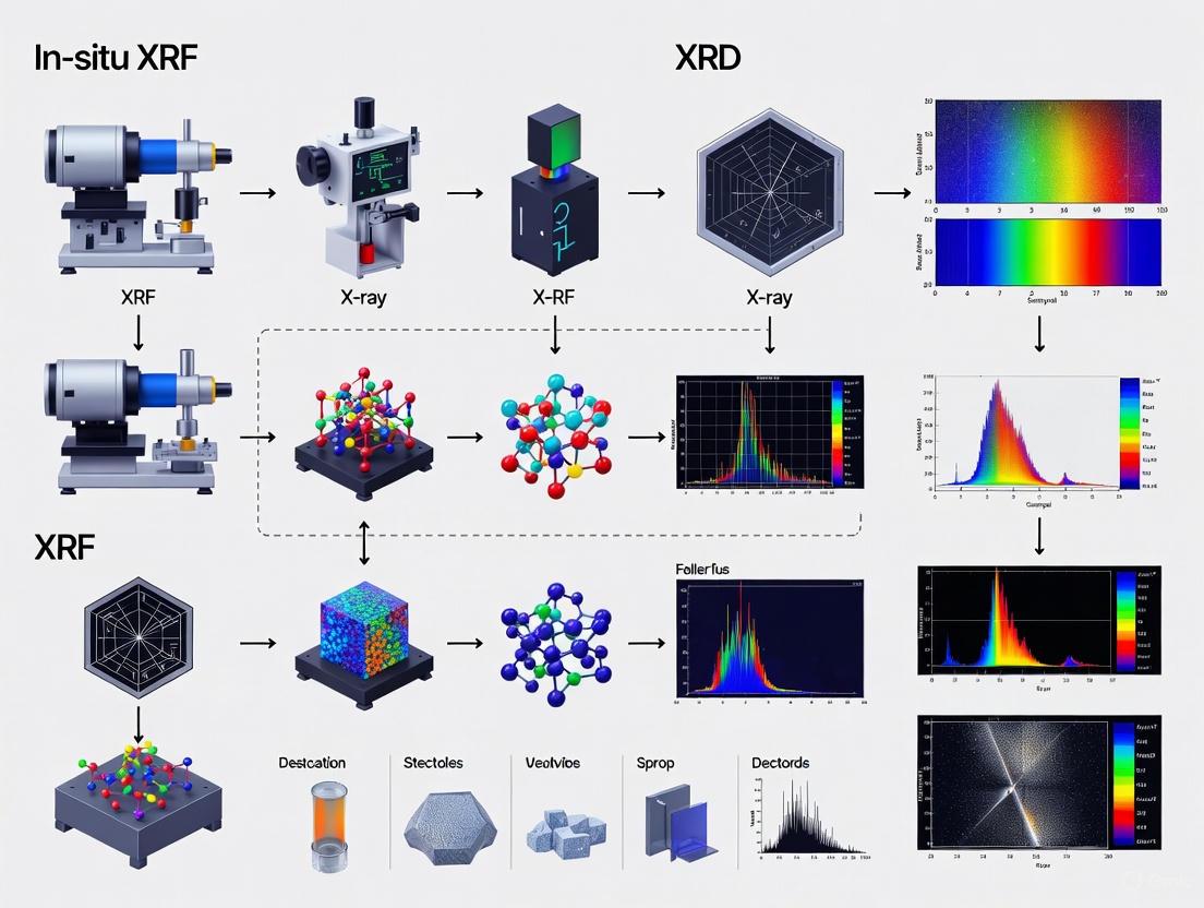
In-Situ XRF and XRD for Field Mineralogical Characterization: A Complete Guide for Research and Development
This article provides a comprehensive overview of the principles, applications, and best practices for using combined in-situ X-ray Fluorescence (XRF) and X-ray Diffraction (XRD) analysis for field-based mineralogical characterization.
In-Situ XRF and XRD for Field Mineralogical Characterization: A Complete Guide for Research and Development
Abstract
This article provides a comprehensive overview of the principles, applications, and best practices for using combined in-situ X-ray Fluorescence (XRF) and X-ray Diffraction (XRD) analysis for field-based mineralogical characterization. Tailored for researchers, scientists, and drug development professionals, it explores the powerful synergy of these non-destructive techniques for obtaining real-time elemental and structural data directly on-site. The content covers foundational concepts, methodological workflows, advanced troubleshooting, and validation protocols, offering a complete framework for implementing these techniques to enhance decision-making in fields ranging from geology and mining to pharmaceutical development and materials science.
Understanding In-Situ XRF and XRD: Core Principles and Synergistic Power
In the field of modern material characterization, particularly for field mineralogical research, the ability to analyze samples under real-world conditions has become paramount. In-situ analysis refers to the measurement of a material in its native environment or state without removing it for ex-situ laboratory examination. In the context of X-ray techniques, this means analyzing samples within their natural setting—whether in a catalytic reactor, a geological formation, or an archaeological artifact—under conditions that may involve specific temperatures, pressures, or atmospheric compositions. A more advanced concept, operando analysis, represents a subset of in-situ studies where characterization is performed not only in the reactive environment but simultaneously while monitoring the functional performance or activity of the material. The term "operando" specifically implies that spectroscopic or diffraction data is collected concurrently with catalytic activity data or other performance metrics, providing direct correlation between structural/chemical changes and material function [1] [2].
The fundamental distinction between these approaches lies in their operational context. Traditional ex-situ analysis requires moving samples from their operational environment to a laboratory setting, potentially altering their structure and chemistry during transfer. In-situ analysis preserves the material's environment during measurement, while operando analysis adds the crucial dimension of simultaneous functional monitoring. This evolution in analytical philosophy has been particularly transformative for heterogeneous catalyst research, mineralogical studies, and cultural heritage conservation, where the active state of materials often differs dramatically from their resting state [1]. For field mineralogical characterization using X-ray fluorescence (XRF) and X-ray diffraction (XRD), these approaches enable researchers to understand mineral behavior under genuine geological conditions rather than idealized laboratory environments.
Technical Foundations of In-Situ XRF and XRD
X-ray Fluorescence (XRF) and X-ray Diffraction (XRD) represent complementary techniques in material characterization, each providing distinct but related information about a sample. XRF is primarily an elemental analysis technique that determines the chemical composition of a material. When a material is bombarded with high-energy X-rays, its atoms become excited and emit secondary (fluorescent) X-rays with energies characteristic of the specific elements present. This allows for qualitative and quantitative elemental analysis without compromising sample integrity. XRF can be further divided into two major branches: wavelength dispersive (WDXRF) and energy dispersive X-ray fluorescence (EDXRF), which differ in how the X-ray spectrum is dispersed and detected [3].
In contrast, XRD is a structural analysis technique used to determine the crystallographic structure of materials. When X-rays interact with a crystalline material, they are diffracted by the lattice planes of the crystal according to Bragg's Law. The resulting diffraction pattern provides information about the material's crystal structure, phase composition, lattice parameters, and crystallinity. While XRF identifies which elements are present, XRD reveals how those elements are arranged—distinguishing between different polymorphs of the same chemical composition and quantifying phase mixtures [3].
The synergy between these techniques becomes particularly powerful in in-situ and operando analysis. Simultaneous XRD/XRF systems have been developed that provide complementary structural and elemental data from the same sample volume under identical conditions. Such systems typically feature a position-sensitive detector for XRD and a silicon-drift detector (SDD) for XRF, allowing non-invasive, in-situ analysis of complex materials [4]. This combination is especially valuable for field mineralogical characterization where sample transportation may be impractical or undesirable, and where understanding both composition and structure under native conditions provides crucial insights into mineral formation, stability, and reactivity.
Table 1: Fundamental Differences Between XRF and XRD Techniques
| Aspect | XRF (X-ray Fluorescence) | XRD (X-ray Diffraction) |
|---|---|---|
| Primary Purpose | Elemental composition analysis | Crystallographic structure analysis |
| Underlying Principle | Detects secondary X-rays emitted by excited atoms | Measures diffraction of X-rays by crystal lattices |
| Type of Information | Qualitative and quantitative elemental composition | Qualitative and quantitative phase composition, crystal structure |
| Sample Requirements | Works for both crystalline and amorphous materials | Primarily for crystalline materials |
| Typical Output | Spectrum with peaks for each element's characteristic X-rays | Diffractogram of intensity vs. 2θ angle |
| Common Field Applications | Mining, environmental analysis, cultural heritage | Geology, materials science, pharmaceuticals |
Applications in Field Mineralogy and Materials Research
Planetary Exploration and Extreme Environments
The CheMin instrument aboard NASA's Mars Science Laboratory Curiosity rover represents a landmark achievement in in-situ XRD/XRF analysis for field mineralogy. As the first XRD instrument flown in space, CheMin operates in transmission geometry with a Co X-ray source and utilizes an energy-sensitive charge-coupled device (CCD) to collect both diffracted and fluoresced photons. This design allows simultaneous structural identification of minerals through XRD and complementary elemental analysis via XRF, though the XRF data remains qualitative. The instrument has identified diverse habitable environments in Martian geological history through mineralogical analysis of depositional environments, subsurface/diagenetic environments, and conditions that may destroy evidence of habitability [5].
Technological advances have led to the next-generation CheMinX instrument, which replaces the CCD with hybrid pixel detectors and incorporates improved focusing optics. These enhancements dramatically decrease analysis time from 22 hours to just 15 minutes while improving angular resolution from 0.30° to 0.18° 2θ. The addition of a silicon-drift detector (SDD) enables quantification of major, minor, and some trace elements via XRF, addressing the qualitative limitations of the original CheMin. This advanced simultaneous XRD/XRF capability in a field-deployable instrument makes it ideal for Discovery-class life-detection missions, including the recommended Mars Life Explorer mission [5].
Cultural Heritage and Archaeological Science
In-situ XRF has become an indispensable tool for analyzing pigments in architectural heritage, where the irreplaceable nature of ancient structures demands non-destructive research methods. Studies of traditional architectural paints in the Rebgong region of Qinghai, China, utilized XRF to identify chromophores in historical pigments: red pigments contained Fe, S, and Pb (suggesting iron red and lead red); yellow pigments showed Pb and Fe; green samples exhibited Cu; white pigments contained Ca and Mg; while blue samples showed Al and S correlated with ultramarine blue [6].
Similarly, analysis of the cliff sculptures in the Qionglai Grottoes combined XRF with XRD to comprehensively characterize the pigment systems. The red pigment contained Fe and Pb, identified as hematite (Fe₂O₃) and lead tetroxide (Pb₃O₄); the green pigment contained Cu, Pb, Ca, Fe, and Na, identified as copper arsenite and tricopper potassium sodium oxide sulfate; while white and blue pigments were identified as gypsum and lazurite respectively [6]. These applications demonstrate how in-situ analysis preserves the integrity of priceless cultural artifacts while providing essential data for conservation and historical interpretation.
Catalysis and Materials Synthesis
In-situ XRD has proven particularly valuable for studying oxide and metal oxide catalysts during their "lifetime"—under synthesis, activation, operation, and deactivation conditions. Traditional ex-situ approaches to catalyst characterization suffer from the significant disadvantage that studies are performed under conditions far from the working conditions of the catalytic process, which typically proceeds at elevated temperatures and pressures in the presence of gas/liquid reagents. The catalyst state can change dramatically under reaction conditions due to interactions with reagents and the effects of temperature and pressure [1].
One of the first in-situ XRD studies of catalysts investigated iron-containing catalysts for ammonia synthesis in 1951, establishing the methodology for studying materials under reactive conditions. Modern applications include investigating processes of loss and uptake of oxygen in oxide catalysts, phase transformations during oxidation/reduction, and deactivation processes. These studies provide invaluable information about the active component of catalysts under reaction conditions and their interactions with supports [1].
Table 2: Representative Applications of In-Situ XRF and XRD Analysis
| Application Field | Specific Example | Technique Used | Key Information Obtained |
|---|---|---|---|
| Planetary Science | CheMin instrument on Mars Curiosity rover | Simultaneous XRD/XRF | Mineral identification (clay minerals, sulfates) and elemental composition of Martian rocks and soils |
| Cultural Heritage | Pigment analysis in architectural heritage | Portable XRF, sometimes combined with XRD | Elemental composition of historical pigments for conservation and provenance studies |
| Heterogeneous Catalysis | Oxide and metal oxide catalysts | In-situ XRD | Phase transformations, active state structure, deactivation mechanisms under working conditions |
| Geology & Mining | Mineral exploration and analysis | Portable XRF, field XRD | Rapid elemental and mineralogical analysis for resource assessment and process control |
| Materials Science | Crystal plasticity studies | Synchrotron-based XRD techniques | Evolution of lattice rotation and elastic strain in polycrystalline materials under stress |
Experimental Protocols for In-Situ Analysis
Protocol for Simultaneous XRD/XRF Analysis of Mineral Samples
The following protocol outlines a standardized approach for simultaneous XRD/XRF analysis, adapted from methodologies successfully deployed in both laboratory and field settings [4]:
Sample Preparation:
- For powdered samples (soils, crushed rocks), homogenize the material by grinding to a consistent particle size (<45 μm recommended to minimize micro-absorption effects and ensure reproducible peak intensities).
- For irregular solid samples (rock surfaces, artifacts), ensure a relatively flat analysis area when possible, though modern portable systems can accommodate some topography.
- Load powdered samples into appropriate holders designed for simultaneous XRD/XRF analysis, taking care to minimize preferred orientation effects that can affect diffraction intensity ratios.
- For field analysis of immovable objects, position the instrument probe perpendicular to the analysis surface at the prescribed working distance.
Instrument Setup:
- Configure the XRD component with a curved position-sensitive detector (CPS) with 120° 2θ range in reflection mode asymmetric geometry.
- Implement an SDD detector for simultaneous XRF measurements positioned optimally for fluorescence collection.
- Select X-ray source parameters based on sample type: Cu Kα radiation (λ = 1.5418 Å) is common for laboratory systems, while Co sources may be preferred for iron-rich samples to minimize fluorescence.
- Set appropriate generator settings (e.g., 40 mA, 40 kV) and define scanning parameters (typically 3° to 70° 2θ for XRD with step size of 0.0167° and scan speed of 2°/min).
Data Collection:
- Acquire XRD and XRF data simultaneously to ensure identical sample conditions for both measurements.
- For time-resolved studies, implement sequential acquisitions with appropriate time intervals to monitor dynamic processes.
- Maintain constant temperature and humidity conditions (e.g., 25 ± 3 °C, 60% RH) when possible to minimize environmental effects on the sample.
Data Analysis:
- Process XRD data using Rietveld refinement (for crystalline phases with known structures), full pattern summation (for complex mixtures including clay minerals), or reference intensity ratio methods (for simpler mixtures).
- Analyze XRF spectra using fundamental parameters or empirical calibration methods to convert peak intensities to elemental concentrations.
- Correlate structural information from XRD with compositional data from XRF to comprehensively characterize the sample.
Simultaneous XRD/XRF Analysis Workflow
Protocol for In-Situ Catalyst Studies Under Reactive Conditions
This protocol specializes in monitoring catalysts during operation, combining methodology from heterogeneous catalysis research [1] with advanced sample environment design [2]:
Reaction Cell Design:
- Utilize specialized reaction chambers that allow control of temperature (up to 1000°C), pressure (vacuum to elevated pressures), and gas atmosphere.
- Incorporate X-ray transparent windows (e.g., Kapton, beryllium) to permit X-ray transmission while maintaining controlled environments.
- Integrate gas delivery systems for introducing reactive mixtures and mass spectrometry capabilities for simultaneous activity measurement (operando mode).
- Ensure thermal homogeneity across the sample through appropriate heating design and temperature monitoring.
Sample Preparation:
- For powdered catalysts, prepare thin uniform beds to minimize X-ray absorption and path length effects.
- For supported catalysts, ensure representative sampling of the catalytic material.
- Implement appropriate sample thickness to balance signal intensity with transmission characteristics.
Operando Measurement:
- Establish steady-state catalytic conditions before beginning measurements.
- Simultaneously collect XRD patterns (for structural information) and gas composition data (for catalytic activity).
- Implement rapid data collection protocols to capture transient states and intermediates.
- Utilize synchrotron radiation sources when high time-resolution or exceptional signal-to-noise is required.
Data Interpretation:
- Correlate structural changes (phase transformations, lattice parameter variations) with catalytic performance metrics.
- Apply Rietveld refinement to extract quantitative phase composition and structural parameters.
- Identify structure-activity relationships through multivariate analysis of structural and performance data.
Essential Research Tools and Reagent Solutions
Table 3: Essential Research Reagent Solutions for In-Situ XRF/XRD Analysis
| Reagent/Material | Function/Application | Technical Specifications | Notes on Usage |
|---|---|---|---|
| Certified Reference Materials (CRMs) | Calibration and validation of both XRF and XRD measurements | Matrix-matched to samples of interest with certified composition | Essential for quantitative analysis; wide range available for different industries and matrices |
| Microfluidic Reaction Cells | Controlled sample environments for in-situ studies | X-ray transparent windows (Kapton, Be); precise T/P control | Enable time-resolved studies of reactions and phase transformations |
| Rietveld Refinement Software | Quantitative phase analysis from XRD patterns | Programs: HighScore, TOPAS, GSAS, BGMN, Maud | Requires crystal structure models; capable of quantifying complex mixtures |
| Full Pattern Summation Software | Quantitative analysis without structural models | Programs: FULLPAT, ROCKJOCK | Particularly effective for clay-containing samples and disordered phases |
| Portable XRD/XRF Systems | Field-deployable analysis | Simultaneous data collection; cartridge-based sample handling | Represented by systems like CheMinX; enable mineralogical analysis in remote locations |
| Synchrotron Beam Access | High-resolution time-resolved studies | High brightness; rapid data collection; various specialized beamlines | Enables studies of rapid processes and weakly scattering materials |
Comparative Analysis of Quantitative Methodologies
The quantitative analysis of XRD data can be approached through several methodologies, each with distinct advantages and limitations. A systematic comparison of three primary methods—Reference Intensity Ratio (RIR), Rietveld, and Full Pattern Summation (FPS)—reveals important considerations for method selection in mineralogical analysis [7].
The Rietveld method represents the most sophisticated approach, performing refinement between observed and calculated patterns through least-squares regression based on a crystal structure database. This method provides exceptional accuracy for quantifying complicated non-clay samples and can extract detailed structural parameters including lattice constants, atomic positions, crystallite size, and preferred orientation. However, most conventional Rietveld software struggles with phases having disordered or unknown crystal structures, limiting its application for certain clay minerals and amorphous materials [7].
The Full Pattern Summation (FPS) method operates on the principle that the observed diffraction pattern equals the sum of signals from individual component phases. This method utilizes reference libraries of pure diffraction patterns rather than structural models, making it particularly effective for analyzing sediments and clay-mineral-containing samples where structures may be imperfect or partially disordered. Comparative studies indicate FPS has wider applicability for complex geological samples, though it requires comprehensive reference libraries [7].
The Reference Intensity Ratio (RIR) method, also known as the 'matrix flushing' method, relies on the intensity of individual diffraction peaks correlated with mineral content through predetermined RIR values. While this approach represents a handy technique requiring minimal computational resources, it generally provides lower analytical accuracy compared to full-pattern methods and may struggle with complex mixtures featuring peak overlap [7].
For samples free from clay minerals, all three methods demonstrate comparable accuracy. However, significant differences emerge for clay-containing samples, where the FPS method generally outperforms the others. The detection limits for XRD analysis typically range between 0.1-2 wt% depending on the specific mineral phase and matrix composition, with analytical uncertainty following a relationship where reliable quantification should generally fall within ±50X⁻⁰·⁵ wt% at the 95% confidence level (where X is concentration) [7].
Quantitative XRD Method Selection Guide
In-situ and operando analysis using XRF and XRD has fundamentally transformed materials characterization across diverse fields from planetary science to cultural heritage conservation. The ability to probe materials under realistic conditions—rather than idealized ex-situ environments—has revealed critical insights into material behavior, functionality, and degradation pathways. For field mineralogical characterization specifically, the development of simultaneous XRD/XRF instruments like CheMinX represents a paradigm shift, enabling comprehensive structural and compositional analysis in remote locations with minimal sample preparation.
The continuing evolution of these techniques points toward several promising directions: further miniaturization of instrumentation for enhanced field deployment, increased temporal resolution for capturing transient states, improved data analysis algorithms for handling complex mixtures, and more sophisticated sample environments for simulating extreme conditions. As these capabilities advance, in-situ and operando analysis will continue to bridge the gap between laboratory characterization and real-world material performance, providing increasingly accurate understanding of mineralogical systems in their native contexts.
X-ray Fluorescence (XRF) is an analytical technique that uses the interaction of X-rays with a material to determine its elemental composition. Suitable for solids, liquids, and powders, XRF is widely recognized for being non-destructive, requiring no or minimal sample preparation, and providing rapid, reliable results for elements ranging from beryllium (Be) to americium (Am) [8] [9]. It is a vital tool in numerous fields, including geology, metallurgy, environmental science, archaeology, and pharmaceuticals, where it performs both qualitative and quantitative analysis with detection limits from 100% down to sub-ppm levels [10] [8]. The fundamental principle underpinning XRF is that the emitted fluorescent X-rays possess energies characteristic of the specific elements present in the sample, thereby providing an elemental "fingerprint" [8].
In the context of field mineralogical characterization research, the portability and robustness of modern XRF instruments make them indispensable. As highlighted in research on New-Caledonian Ni-rich harzburgite, there is a strong industrial demand for on-site real-time analyses that provide decision-making support for field exploration, material sorting, and quality control [11]. When coupled with X-ray Diffraction (XRD), which identifies mineral phases, XRF provides the complementary chemical data necessary for a comprehensive understanding of geological samples directly in the field [11]. This combination offers a powerful, integrated approach to raw materials characterization.
The Fundamental Principles of XRF
The process of X-ray fluorescence occurs at the atomic level and can be understood through a relatively straightforward, sequential process. When a primary X-ray beam with sufficient energy strikes an atom within the sample, it interacts with the inner-shell electrons, ejecting one from its orbital [10] [8]. This creates a void or "hole" in the inner shell, resulting in an unstable, high-energy atomic configuration [8]. To restore stability, an electron from an outer, higher-energy shell fills the hole. The excess energy from this electron transition is released almost instantaneously in the form of a secondary, fluorescent X-ray [10].
The energy of this emitted fluorescent X-ray is precisely equivalent to the difference in energy between the two electron shells involved in the transition [8]. Crucially, this energy difference is a unique characteristic of the specific atomic element, meaning that the energy of the emitted X-ray identifies the element present, while the intensity of the emission is related to its concentration [10] [8]. It is important to note that, because the process involves inner-shell electrons, which are not engaged in chemical bonding, the XRF spectrum of an element is largely independent of its chemical form (e.g., lead metal versus lead oxide) [12].
Key Transitions and Spectral Lines
Atoms consist of multiple electron shells (K, L, M, etc.), leading to several possible electron transitions and, consequently, multiple characteristic X-ray peaks for a single element. The most common transitions observed in XRF spectra are from the K and L series [12].
- K-Lines: Result from a hole in the innermost K-shell being filled by an electron from the L- or M-shell. These are labeled Kα and Kβ. Kα lines are typically approximately twice the intensity of Kβ lines. K lines are the primary lines observed for low to medium atomic number elements (e.g., from sodium to cadmium) [12].
- L-Lines: Result from a hole in the L-shell being filled by an electron from the M- or N-shell. These lines are more complex and are the primary lines observed for high atomic number elements (e.g., barium to uranium), as the energy required to eject a K-shell electron becomes impractically high for portable instruments [12].
The relationship between the atomic number (Z) and the energy of the characteristic X-ray lines is described by Moseley's Law, which shows that the energy of the emitted X-ray is proportional to Z², forming the foundational basis for elemental identification [12].
Instrumentation and Methodology
XRF instrumentation primarily falls into two categories, Energy Dispersive XRF (ED-XRF) and Wavelength Dispersive XRF (WD-XRF), each with distinct advantages and operational principles.
Energy Dispersive XRF (ED-XRF) is characterized by its simplicity and speed. In an ED-XRF system, all emitted fluorescent X-rays from the sample are collected by a semiconductor detector (such as a Si-PIN or Silicon Drift Detector (SDD)) simultaneously [10] [8]. The detector directly measures the energy of each incoming X-ray photon, and a multi-channel analyzer sorts and counts these photons by energy to produce a spectrum [10]. Modern SDDs offer both excellent energy resolution and high count rate capabilities, which are essential for resolving closely spaced peaks and for rapid analysis [10]. ED-XRF covers elements from sodium (Na) to uranium (U) and is the technology most commonly used in portable and benchtop instruments [8].
Wavelength Dispersive XRF (WD-XRF) offers superior spectral resolution. Instead of measuring energy directly, WD-XRF uses analyzing crystals to physically separate X-rays based on their wavelengths (which are inversely related to energy) through diffraction [8]. A detector is positioned at specific angles to count X-rays of specific wavelengths. This method can detect lighter elements, down to beryllium (Be), and provides significantly lower background and better detection limits for trace elements [8] [9]. However, the system is more complex, typically requires higher power X-ray tubes, and is slower (or more expensive if multiple simultaneous detectors are used), making it predominantly a laboratory-based technique [8].
Table 1: Comparison of ED-XRF and WD-XRF Techniques
| Feature | Energy Dispersive XRF (ED-XRF) | Wavelength Dispersive XRF (WD-XRF) |
|---|---|---|
| Working Principle | Direct energy measurement by a semiconductor detector [8] | Diffraction of X-rays by crystals to separate wavelengths [8] |
| Typical Resolution | 150 eV to 300 eV [8] | 5 eV to 20 eV [8] |
| Elemental Range | Sodium (Na) to Uranium (U) [8] | Beryllium (Be) to Uranium (U) [8] |
| Analysis Speed | Very fast; entire spectrum acquired simultaneously [8] | Slower; sequential or limited simultaneous measurement [8] |
| Key Advantage | Simplicity, speed, portability, lower cost [8] [9] | High resolution, superior trace element detection, lighter element analysis [8] [9] |
The Scientist's Toolkit: Essential Components for XRF Analysis
A functional XRF system, whether ED or WD, relies on several key components. For researchers, especially those working in field applications, understanding these components is critical for method selection and data interpretation.
Table 2: Key Research Reagent Solutions and Instrument Components
| Component / Reagent | Function / Description | Application Notes |
|---|---|---|
| X-Ray Tube | Generates the primary X-rays that excite the sample. Key parameters include anode material (e.g., Rh, Pd, Co), window type, and power [10]. | Anode material selection is critical for optimizing sensitivity for specific elements [10]. |
| Semiconductor Detector (SDD) | Measures the energy of incoming fluorescent X-rays. The core of an ED-XRF system [10] [8]. | Silicon Drift Detectors (SDDs) offer the best combination of high resolution and high count rate capability [10]. |
| Analyzing Crystals | Used in WD-XRF to diffract and separate X-rays by wavelength [8]. | Different crystals are used for different element ranges to optimize performance. |
| Flux / Binder (e.g., Li₂B₄O₇) | A chemical flux used to create fused beads from powdered samples, eliminating mineralogical and particle size effects [10] [9]. | Essential for accurate major element analysis in geological samples; high dilution can preclude trace element analysis [9]. |
| Press & Die Set | Equipment used to prepare pressed powder pellets from sample powders, often with a binding agent [10]. | A common and rapid preparation method for solids and powders that improves surface homogeneity. |
| Certified Reference Materials (CRMs) | Well-characterized standards with known elemental compositions, used for instrument calibration and quantification [13]. | Crucial for achieving accurate quantitative results; must be matrix-matched to the samples for best accuracy. |
Experimental Protocols for XRF Analysis
The accuracy of XRF analysis is highly dependent on proper experimental protocol, from sample preparation to data quantification.
Sample Preparation Protocol
While XRF is often considered non-destructive, the quality of the surface analyzed is paramount, especially for light elements where the information depth is very shallow (on the order of micrometers) [10] [9]. The chosen preparation method depends on the sample type and the required analytical precision.
- No Preparation: Suitable for quick, non-destructive screening of homogeneous materials like metal alloys or finished goods. The surface should be clean and flat [10].
- Surface Cleaning/Polishing: For solid samples like metals or glasses, surface preparation by machining, polishing, or cleaning to remove oxides or coatings is necessary to ensure a representative, flat surface for analysis [10].
- Pressed Powder Pellets: This is a common method for powders and granulates.
- Procedure: The sample is finely crushed and ground to a consistent particle size (typically <75 µm). The powder is then mixed with a binder (e.g., cellulose, wax) to add cohesion and pressed in a die at high pressure (e.g., 10-25 tons) to form a solid, flat pellet [10] [9].
- Application: Ideal for the analysis of trace elements in geological materials, as it avoids the high dilution of fused beads [9].
- Fused Beads: This is the gold standard for achieving the highest accuracy for major elements, as it eliminates mineralogical and particle size effects.
- Procedure: A finely powdered sample is mixed with a flux (e.g., lithium tetraborate or metaborate) in a specific ratio (e.g., 1:10 sample-to-flux). The mixture is fused in a platinum crucible at high temperatures (1000-1200°C) until it forms a homogeneous glass bead [10] [9].
- Application: Essential for accurate major and minor element analysis in complex geological samples and ceramics [9].
Protocol for Quantification of XRF Data
Quantification translates raw XRF intensity counts into elemental concentrations, correcting for matrix effects where the presence of one element can affect the measured intensity of another (e.g., through absorption or enhancement) [10] [13].
- Spectrum Acquisition: Collect fluorescence spectra from the sample under defined conditions (tube voltage, current, measurement time). Longer measurement times improve counting statistics, yielding better precision and lower detection limits [10].
- Spectral Fitting/Deconvolution: Use specialized software (e.g., MAPS) to fit the raw spectra. This step is critical for accurately separating overlapping peaks from different elements and accounting for the background signal, which includes scattered radiation (Bremsstrahlung, Rayleigh, and Compton peaks) [13] [12].
- Calibration with Standards: Measure well-characterized, matrix-matched certified reference materials (CRMs) under identical conditions. The software establishes a relationship between the measured intensities and the known concentrations of elements in the CRMs [13].
- Apply a Quantification Model: Use the calibration data to correct for matrix effects in the unknown samples. Common models include:
- Empirical Calibration: Uses a suite of CRMs to create a calibration curve for each element. It is highly accurate if the standards closely match the samples [10].
- Fundamental Parameters (FP): A mathematical method that uses theoretical models of X-ray generation and absorption to correct for matrix effects. It is powerful when perfect matrix-matched standards are not available [10].
Application in Field Mineralogy: Combined XRF-XRD Analysis
The integration of XRF with XRD represents a significant advancement for field-based mineralogical characterization. While XRF provides the elemental composition, XRD identifies the specific mineral phases present, even those made of light elements, which XRF may struggle to detect [11]. This combined approach was successfully demonstrated in the analysis of a Ni-rich serpentinized harzburgite from New Caledonia using a transportable instrument (ID2B) [11].
The study concluded that combined XRF-XRD analysis provided quantitative chemical and mineralogical data in real-time that was consistent with laboratory-based reference methods (SEM-EDS, EPMA, traditional XRF, and XRD) [11]. This synergy allows researchers to not only know what elements are present but also to understand their mineralogical hosts—a critical distinction for exploration and geometallurgy. For instance, it helps identify penalizing or valuable minerals and provides insights into crystallographic texture and structural disorganization, such as turbostratism in phyllosilicates, which is highly relevant for understanding ore genesis and processing behavior [11].
Table 3: Complementary Data from Combined XRF-XRD Analysis
| Aspect | XRF Contribution | XRD Contribution | Combined Insight |
|---|---|---|---|
| Chemical Composition | Provides quantitative data on elemental concentrations from Be/U [11] [9]. | Limited direct chemical data. | Links specific elements to their host minerals. |
| Mineralogy | Cannot distinguish isocompositional phases or different oxides [9]. | Identifies and quantifies specific mineral phases [11]. | Enables discrimination of lithologies and ore types based on both chemistry and mineralogy [11]. |
| Crystallography | Provides no information on long-range order or crystal structure. | Reveals structural details, preferred orientation, and disorder (e.g., turbostratism) [11]. | Provides a more complete picture of the sample's geological history and processing properties [11]. |
X-ray diffraction (XRD) is a powerful, non-destructive analytical technique that provides unparalleled insights into the atomic and molecular structure of crystalline materials by measuring the diffraction pattern produced when X-rays interact with a crystal lattice [14]. The fundamental phenomenon was discovered in the early 20th century, with W.L. Bragg formulating the essential mathematical relationship now known as Bragg's Law in 1913 [14]. When X-rays encounter a crystalline solid, most scatter in random directions and interfere destructively, canceling each other out. However, in specific directions determined by the crystal's internal structure, X-rays scatter constructively and reinforce one another, producing a measurable diffraction pattern that serves as a unique fingerprint for material identification and structural analysis [15] [14].
The versatility of XRD extends across numerous scientific disciplines and industrial applications. In pharmaceutical development, XRD characterizes active compounds and verifies polymorphic forms to ensure drug efficacy and safety [15]. In materials science, it enables the development of advanced batteries and electronics by determining structure-property relationships [15] [14]. For geological field research, particularly in the context of in-situ characterization, XRD combined with X-ray fluorescence (XRF) provides rapid mineralogical and geochemical data for exploration and process control [11]. The technique's ability to determine crystal structure, identify unknown phases, measure lattice parameters, analyze crystal defects, and quantify phase mixtures makes it indispensable for researchers and scientists across multiple fields [14] [16].
Theoretical Foundations of XRD
The Physical Principle of X-Ray Diffraction
X-ray diffraction occurs due to the wave nature of X-rays, which are electromagnetic radiation with wavelengths (typically 0.1-10 nm) comparable to the spacing between atoms in crystal structures [14]. When monochromatic X-rays strike a crystalline sample, they interact with the electrons around atoms, causing the electrons to oscillate and generate secondary X-rays with the same frequency in a process known as X-ray scattering [16]. In a crystalline material, where atoms are arranged in a periodic, three-dimensional structure, these scattered spherical waves have fixed phase relationships that result in constructive interference in specific directions and destructive interference in others [15] [16].
The constructive interference occurs when the path difference between X-rays scattered from parallel crystal planes equals an integer multiple of the X-ray wavelength. This condition produces detectable diffraction peaks that reveal the geometric arrangement of atoms within the crystal [15] [14]. For amorphous materials, which lack long-range atomic order, XRD produces only broad, diffuse scattering patterns without sharp peaks, while crystalline materials with regular atomic arrangements yield distinctive patterns with sharp, well-defined peaks [16].
Bragg's Law
The fundamental equation governing X-ray diffraction is Bragg's Law, formulated by William Lawrence Bragg in 1913 [14]. This relationship describes the precise conditions necessary for constructive interference to occur and can be expressed mathematically as:
nλ = 2d sinθ
Where:
- n = order of diffraction (an integer: 1, 2, 3...)
- λ = wavelength of the X-ray radiation
- d = interplanar spacing between parallel crystal planes
- θ = angle between the incident X-ray beam and the crystal plane
This deceptively simple equation forms the cornerstone of XRD analysis [16]. It reveals that by measuring the angle θ at which diffraction occurs for X-rays of known wavelength λ, researchers can calculate the distance d between atomic planes in the crystal structure [15] [17]. This interplanar spacing serves as a unique identifier for crystalline phases, much like a fingerprint distinguishes individuals [14].
Bragg's Law was historically crucial in determining the double-helix structure of DNA. Rosalind Franklin's XRD work, particularly her analysis of "Photo 51," provided quantitative data including the 3.4 Å spacing between consecutive base pairs, the 34 Å helical repeat distance, and the 20 Å helix diameter, which collectively enabled Watson and Crick to propose their revolutionary model of DNA structure [14].
The Scherrer Equation
While Bragg's Law enables the calculation of interplanar spacings from diffraction peak positions, the Scherrer Equation relates diffraction peak broadening to crystallite size, providing the theoretical basis for measuring crystallite dimensions in polycrystalline materials [16]:
D = Kλ / (B cosθ)
Where:
- D = average thickness of crystallites perpendicular to the crystal plane
- K = Scherrer constant (typically 0.89)
- λ = X-ray wavelength
- B = full width at half maximum (FWHM) of the diffraction peak in radians
- θ = Bragg angle (half of the 2θ peak position)
This relationship demonstrates that smaller crystallites produce broader diffraction peaks, while larger crystallites yield sharper peaks [16]. The Scherrer Equation thus enables researchers to determine the average crystallite size in nanoscale materials, an essential parameter in fields ranging from pharmaceutical development to advanced materials synthesis [16].
XRD Instrumentation and Methodology
X-Ray Diffractometer Components
A modern X-ray diffractometer consists of several essential components that work in coordination to produce high-quality diffraction data [14] [16]:
X-ray source: Generates monochromatic X-rays through electron bombardment of a metal target, with copper (Cu Kα, λ = 1.5418 Å) being the most common target material for general applications [14] [16]. The X-ray tube typically operates at high voltage (30-60 kV) and current (10-50 mA) to produce sufficient intensity [14].
Incident beam optics: Conditions the X-ray beam using various optical elements including Soller slits for controlling beam divergence, monochromators for wavelength selection, and focusing mirrors for beam concentration [14].
Sample stage: Holds the specimen and allows precise positioning and rotation during measurement, providing accurate angular positioning that may include environmental controls for specialized applications [14].
Detector system: Modern diffractometers employ position-sensitive detectors (PSDs) or area detectors that simultaneously collect data over a range of angles, significantly reducing measurement time while maintaining high resolution [14].
Goniometer: A precision mechanical system that controls the angular relationships between the X-ray source, sample, and detector, achieving angular accuracy better than 0.001° in modern instruments [14].
The instrument operates by directing X-rays at the sample while rotating both the sample and detector according to θ-2θ geometry, ensuring the detector captures diffracted beams at the correct angle for constructive interference [14]. This configuration maintains the proper geometric relationships throughout the measurement process.
XRD Measurement Approaches
XRD techniques are broadly categorized based on sample morphology and crystal size [15]:
Single Crystal XRD
- Requires a crystal large enough for analysis (typically microscopic in size)
- Solves complete crystal structure ranging from simple inorganic solids to complex macromolecules
- Produces a pattern of defined, isolated peaks on the detector
- Provides maximum structural information, including atomic positions and bond lengths
Powder XRD (XRPD)
- Used when samples do not form crystals large enough for single-crystal analysis
- Analyzes microcrystalline powders with randomly oriented crystallites
- Produces concentric diffraction rings known as Debye rings
- Simpler and faster than single-crystal methods but provides less structural information
- Ideal for phase identification, purity analysis, and materials characterization
The following diagram illustrates the fundamental workflow of the XRD process and the distinct patterns generated by different sample types:
Experimental Protocol for Powder XRD Analysis
Sample Preparation Protocol
- Particle Size Reduction: Grind the sample to an appropriate particle size (<10 μm) using a mortar and pestle or mechanical grinder to ensure a homogeneous powder and minimize preferred orientation effects [18].
- Sample Mounting: Load the powdered sample into a specimen holder, ensuring a flat, level surface. For standard holders, use the back-loading technique to minimize preferred orientation [11].
- Surface Smoothing: Use a glass slide or blade to create a smooth, flat surface flush with the holder edge to ensure consistent diffraction geometry [11].
- Minimal Preparation Samples: For field applications with limited preparation capabilities, analyze saw-cut or rock surfaces provided they are sufficiently flat, acknowledging that matrix and preferred orientation effects may influence results [11].
Instrument Measurement Protocol
- X-ray Source Selection: Select an appropriate X-ray target based on sample composition. Cu Kα radiation (λ = 1.5418 Å) is suitable for most samples except those containing Cu or Fe, for which Co or Cr targets are preferred to minimize fluorescence [16].
- Measurement Parameters:
- Voltage/Current: Typically 40 kV/40 mA for Cu target systems [18]
- Scan Range: 2° to 90° for qualitative analysis; 2° to 150° for quantitative analysis and Rietveld refinement [16]
- Scan Speed: 1° to 8°/min for qualitative analysis; 0.001° to 1°/min for quantitative calculations [16]
- Step Size: 0.01° to 0.02° for high-resolution data collection [18]
- Measurement Mode: Use continuous scan for qualitative analysis and step scan for quantitative analysis, lattice parameter calculation, and Rietveld refinement [16].
Data Collection and Analysis Protocol
- Phase Identification:
- Collect diffraction pattern and measure 2θ positions of all detectable peaks
- Calculate d-spacings using Bragg's Law
- Compare with standard reference patterns from databases (PDF-4+, ICSD, COD)
- Identify phases based on peak positions and relative intensities [16]
- Quantitative Analysis (if required):
- Perform Rietveld refinement for full-pattern quantitative analysis
- Use internal standard or reference intensity ratio methods for conventional quantification [18]
- Crystallite Size Determination:
- Measure full width at half maximum (FWHM) of representative peaks
- Apply Scherrer equation to calculate average crystallite size [16]
XRD Data Interpretation and Analysis
Understanding XRD Patterns
An XRD pattern displays diffraction intensity as a function of diffraction angle (2θ), where each peak corresponds to a specific set of parallel crystal planes characterized by Miller indices (hkl) [14]. This diffraction pattern serves as a unique fingerprint for each crystalline phase, enabling identification and quantitative analysis [14]. The characteristics of XRD peaks provide comprehensive structural information about the material being analyzed [14] [17]:
Peak Position: The angular position (2θ) directly relates to the d-spacing (interplanar spacing) through Bragg's law. Peak positions determine lattice parameters, identify phases, and detect structural changes due to composition, temperature, or pressure variations [14].
Peak Intensity: The height or integrated area of diffraction peaks indicates the atomic arrangement within the crystal structure and the relative abundance of different phases. Intensity ratios provide information about preferred orientation effects and enable quantitative phase analysis [14] [17].
Peak Width: The breadth of diffraction peaks reveals crystal quality, including crystallite size and microstrain effects. Narrow peaks indicate large, well-formed crystals with minimal strain, while broad peaks suggest small crystallites or high levels of structural disorder [17] [16].
Peak Shape: The detailed shape of diffraction peaks provides insights into crystal defects, stacking faults, and other structural imperfections. Asymmetric peak shapes often indicate compositional gradients or structural distortions [14].
Phase Identification and Quantitative Analysis
Phase identification through XRD relies on comparing the measured diffraction pattern with reference patterns from established databases such as the Powder Diffraction File (PDF), Inorganic Crystal Structure Database (ICSD), or Crystallography Open Database (COD) [16]. Modern analysis software facilitates this process through automated search-match algorithms that identify potential phase matches based on peak positions, intensities, and profiles [18].
For quantitative analysis, the Rietveld refinement method has become the standard approach, offering significant advantages over traditional methods [18]. This full-pattern analysis technique:
- Quantifies all crystalline phases simultaneously without requiring individual standard curves
- Accounts for and corrects instrumental aberrations, preferred orientation, and sample displacement effects
- Handles complex multi-phase mixtures with overlapping diffraction peaks
- Provides precision of 1-2 wt.% for major phases in well-prepared samples
- Does not require external standards or calibration curves when crystal structures are known
The following table summarizes the key applications of XRD analysis in materials characterization:
Table 1: Key Applications of XRD Analysis in Materials Characterization
| Application | Analytical Approach | Key Information Obtained | Typical Precision |
|---|---|---|---|
| Phase Identification | Comparison of d-spacings and intensities with reference databases | Crystalline phase composition, polymorphism, impurity detection | Qualitative to semi-quantitative |
| Quantitative Phase Analysis | Rietveld refinement, reference intensity ratio (RIR) methods | Weight percentages of crystalline phases in mixtures | 1-5 wt.% depending on phase and sample preparation |
| Crystallite Size Determination | Scherrer equation analysis of peak broadening | Average crystallite size in nanoscale materials | ±10-20% for crystallites <100 nm |
| Lattice Parameter Determination | Precise peak position measurement using internal standards | Unit cell dimensions, solid solution composition, thermal expansion | ±0.001 Å for well-crystallized materials |
| Crystallinity Determination | Comparison of integrated intensities of crystalline and amorphous regions | Degree of crystallinity in semi-crystalline materials | ±2-5% with proper calibration |
| Residual Stress Analysis | Precise measurement of lattice strain through peak shifts | Macro and micro-stresses in engineered components | ±50 MPa for typical metallic materials |
Advanced XRD Analysis Techniques
Beyond basic phase identification, XRD supports numerous advanced analytical techniques:
In Situ and Non-Ambient XRD
- Temperature-dependent studies (cryogenic to >1500°C) for thermal expansion, phase transitions, and reaction pathways
- Environmental chambers for studying materials under controlled atmospheres or gas exposure
- Time-resolved studies for monitoring crystallization, decomposition, or chemical reactions
Texture and Preferred Orientation Analysis
- Pole figure measurement for quantifying crystallographic preferred orientation
- Orientation distribution function (ODF) calculation for complete texture characterization
- Critical for understanding anisotropic properties in polycrystalline materials
Pair Distribution Function (PDF) Analysis
- Total scattering measurements for nanocrystalline and amorphous materials
- Local structure determination beyond long-range periodicity
- Reveals short and medium-range order in disordered materials
XRD in Combined Analysis and Field Applications
Complementary XRF-XRD Analysis
The combination of X-ray fluorescence (XRF) and X-ray diffraction (XRD) in a single analytical approach creates a powerful methodology for complete materials characterization, particularly valuable for field mineralogical studies [11]. These two techniques provide complementary information:
XRF Analysis Strengths
- Provides elemental composition regardless of crystalline state
- Detects all elements except the lightest (typically Z > 11 for portable systems)
- Offers rapid quantitative analysis for major and trace elements
- Less affected by sample preparation and particle statistics
XRD Analysis Strengths
- Identifies and quantifies specific mineral phases and compounds
- Detects light elements that are challenging for XRF
- Determines crystal structure, polymorphism, and crystallinity
- Reveals structural disorder and amorphous content
The synergy between these techniques provides the missing link between elemental and phase analyses, enabling researchers to not only determine what elements are present but also how they are arranged structurally and what compounds they form [11]. For example, in geological applications, XRF might detect calcium and silicon, while XRD would distinguish whether these elements are present as calcite (CaCO₃) and quartz (SiO₂) or combined in minerals like wollastonite (CaSiO₃) [11].
Field Deployment and On-Site Analysis
Recent advances in instrument miniaturization have made combined XRF-XRD analysis feasible for field deployment, addressing the scientific community's need for on-site real-time analyses that provide decision-making support for field exploration, material sorting, and process monitoring [11]. Modern portable systems offer significant advantages for field mineralogical characterization:
- Rapid Analysis: Combined XRF-XRD measurements completed in <30 minutes per sample [11]
- Minimal Sample Preparation: Ability to analyze powders, saw cuts, and rock surfaces with little or no preparation [11]
- Real-Time Decision Support: Immediate geochemical and mineralogical data for exploration targeting and resource assessment
- Cost Efficiency: Reduced costs associated with sample shipping, preparation, and laboratory analysis
The integration of these techniques in field instrumentation has been demonstrated in challenging environments such as the analysis of Ni-rich serpentinized harzburgite in New Caledonia, where combined XRF-XRD analysis successfully provided quantitative chemical and mineralogical data comparable to laboratory instruments while operating directly in the field [11].
Case Study: Analysis of Direct Reduced Iron (DRI)
The application of XRD to industrial process control is effectively demonstrated in the analysis of Direct Reduced Iron (DRI), where XRD provides critical parameters for quality control and process optimization [18]. In this application:
Analytical Challenge
- DRI contains multiple iron phases: metallic iron (Fe), wuestite (FeO), magnetite (Fe₃O₄), hematite (Fe₂O₃), cohenite (Fe₃C), and fayalite (Fe₂SiO₄)
- Traditional wet chemistry methods are time-consuming and labor-intensive
- Process control requires rapid feedback on metallization efficiency and carbon content
XRD Methodology
- Configuration: Cobalt X-ray tube with iron beta filter to minimize fluorescence
- Measurement Time: 6 minutes per sample using high-speed detector
- Sample Preparation: Milling followed by pressing into steel ring holders
- Analysis: Rietveld refinement with reference database PDF-4+
Results and Process Parameters XRD quantification enables calculation of critical process parameters:
- Metallic iron (Femet) from iron and cohenite content
- Total iron (Fetot) from all iron-containing phases
- Metallization (Metn) as percentage ratio of Femet to Fetot
- Total carbon (Ctot) from cohenite content
The strong correlation between XRD results and traditional wet chemistry methods (systematic difference of only 2% for Femet content) demonstrates the reliability of XRD for process control while providing additional mineralogical information about the reduction process efficiency and optimal raw material mixture [18].
The Scientist's Toolkit: Essential Materials for XRD Analysis
Table 2: Essential Research Reagent Solutions and Materials for XRD Analysis
| Category | Item | Specification/Function | Application Notes |
|---|---|---|---|
| X-Ray Sources | Copper X-ray Tube | Kα radiation (λ = 1.5418 Å), 30-60 kV operation | General purpose analysis for most samples [14] [16] |
| Cobalt X-ray Tube | Kα radiation (λ = 1.7902 Å) | Preferred for Fe-containing samples to reduce fluorescence [18] | |
| Sample Preparation | Sample Holders | Steel rings, glass slides, or capillaries | Mounting powdered samples with flat surface [11] [14] |
| Grinding Equipment | Mortar and pestle, mechanical grinder | Particle size reduction to <10 μm for homogeneous powder [18] | |
| Presses | Hydraulic or manual powder presses | Creating consolidated powder pellets with uniform density [18] | |
| Reference Materials | Silicon Powder Standard | NIST SRM 640e or equivalent | Instrument alignment and peak position calibration |
| Corundum (α-Al₂O₃) | NIST SRM 676a or equivalent | Quantitative analysis calibration and internal standard | |
| Analytical Databases | PDF-4+ | ICDD database of powder patterns | Primary reference for phase identification [18] |
| ICSD | Inorganic Crystal Structure Database | Crystal structure data for Rietveld refinement [11] | |
| COD | Crystallography Open Database | Open-access crystal structure database [11] | |
| Software Tools | HighScore Plus | Comprehensive XRD data analysis | Phase identification, Rietveld refinement, quantification [18] |
| JADE | XRD pattern processing and analysis | Peak fitting, residual stress, crystallite size analysis | |
| FullProf Suite | Rietveld refinement and pattern matching | Advanced structural analysis including magnetic structures |
X-ray diffraction remains one of the most powerful and versatile techniques for crystalline materials characterization, with applications spanning pharmaceutical development, materials science, geology, and industrial process control. The fundamental principles of XRD, rooted in Bragg's Law and the constructive interference of X-rays, provide unparalleled insights into atomic-scale structure through the measurement of diffraction patterns.
The integration of XRD with complementary techniques like XRF creates a comprehensive analytical approach particularly valuable for field applications, where rapid, on-site analysis enables real-time decision making for exploration and process control. As instrumentation continues to advance toward more portable and automated systems, the applications of combined XRF-XRD analysis are expanding, making sophisticated materials characterization accessible in field settings without sacrificing analytical precision.
For researchers engaged in field mineralogical characterization, XRD provides not only phase identification but also quantitative information about crystallite size, strain, preferred orientation, and structural disorder—essential parameters for understanding material properties and behavior. The continued development of portable instruments, enhanced detectors, and sophisticated analysis software ensures that XRD will remain a cornerstone technique for unlocking the structural secrets of crystalline materials across scientific disciplines and industrial applications.
The demand for real-time, on-site analytical capabilities in fields such as mineral exploration, pharmaceuticals, and environmental monitoring has driven the development of integrated instrumental approaches [11]. Combined X-ray Fluorescence (XRF) and X-ray Diffraction (XRD) analysis represents a transformative methodological advancement that bridges elemental composition with crystallographic structural information in a single analytical workflow. This synergy provides a more complete characterization of materials than either technique can deliver independently [19].
XRF and XRD are complementary techniques that extract different information from samples through X-ray interactions. XRF determines elemental composition by measuring the fluorescent X-rays emitted from a sample when excited by a primary X-ray source, providing data on what elements are present and their concentrations [20]. In contrast, XRD reveals the crystalline structure of materials by measuring the diffraction pattern produced when X-rays interact with the periodic arrangements of atoms in crystal lattices, providing information about phases, crystal structure, and polymorphism [21]. When deployed together in a combined instrumentation platform, these techniques offer researchers a powerful tool for comprehensive material characterization, enabling informed decision-making directly in the field [11].
Fundamental Principles and Technical Synergies
Core Principles of XRF and XRD
XRF spectroscopy operates on the principle of exciting atoms in a sample using high-energy X-rays, causing the ejection of inner-shell electrons. As outer-shell electrons fill these vacancies, they emit secondary (fluorescent) X-rays with energies characteristic of specific elements. This emission allows for both qualitative identification and quantitative analysis of elemental composition, typically for elements ranging from sodium (Na) to uranium (U), with modern systems capable of detecting elements down to sub-parts per million (ppm) levels [21].
XRD is based on Bragg's Law, which describes the conditions under which X-rays are diffracted by crystal lattice planes. When a crystalline sample is irradiated with X-rays, the regularly spaced atoms act as scattering centers, producing constructive interference at specific angles. The resulting diffraction pattern serves as a unique fingerprint for crystalline phases present in the material, enabling identification, quantification, and structural characterization [20].
Complementary Analytical Capabilities
The fundamental synergy between XRF and XRD stems from their complementary strengths and limitations, creating a comprehensive analytical picture that neither technique can provide alone.
- Elemental vs. Phase Information: XRF provides total elemental composition but cannot distinguish between different chemical forms or polymorphs of the same element. XRD identifies and quantifies crystalline phases but offers limited elemental sensitivity, particularly for trace components [19].
- Crystalline and Amorphous Materials: XRF analyzes both crystalline and amorphous materials with similar efficacy, while XRD primarily characterizes crystalline components, with limited capability for amorphous content quantification [20].
- Detection Limits and Sensitivity: XRF excels at detecting trace elements at ppm levels, whereas XRD typically has higher detection limits (generally 0.1-1% for crystalline phases) but provides detailed structural information about major and minor phases [21] [22].
- Polymorphism Distinction: XRD can differentiate between polymorphs (different crystal structures with identical chemical composition), while XRF cannot distinguish between these forms [21].
Comparative Technique Characteristics
Table 1: Fundamental Characteristics of XRF and XRD Techniques
| Characteristic | XRF (X-ray Fluorescence) | XRD (X-ray Diffraction) |
|---|---|---|
| Primary Information | Elemental composition | Crystalline structure and phase identification |
| Detection Principle | Measures fluorescent X-rays emitted by excited atoms | Measures diffracted X-rays from crystal lattices |
| Polymorphism Detection | Cannot distinguish between polymorphs | Can differentiate different polymorphic forms |
| Detection Limits | Sub-ppm for trace elements | Typically 0.1-1% for crystalline phases |
| Sample Requirements | Crystalline and amorphous materials | Primarily crystalline materials |
| Primary Output | Elemental spectrum | Diffractogram (intensity vs. 2θ angle) |
| Quantitative Output | Elemental concentrations (e.g., mg/g of Fe) | Phase composition (e.g., 30% Fe₂O₃, 50% amorphous) |
Instrumentation and Methodological Approaches
Combined Instrumentation Platforms
Recent technological advances have led to the development of integrated XRF-XRD systems that merge both analytical capabilities into a single instrument. The ID2B instrument, developed within the SOLSA project, represents one such innovation, performing fast (under 30 minutes) combined X-ray data acquisition on-site [11]. This configuration collects diffracted and fluorescent X-ray signatures quasi-simultaneously from the same sample volume, ensuring data consistency and eliminating positional uncertainties [11].
These integrated systems typically feature:
- A single X-ray source with optimized excitation characteristics for both techniques
- Dual detection systems capable of measuring both fluorescent and diffracted X-rays
- A modular sample holder accommodating various sample forms (powders, saw cuts, raw rocks)
- Unified software interface for simultaneous data acquisition and analysis
- Compact, transportable designs suitable for field deployment [11] [19]
The benefits of such integrated systems include simplified workflow with only one sample introduction, unified data analysis interface, merged elemental and phase results in a single analysis bulletin, minimized laboratory footprint, and reduced infrastructure requirements [19].
Sample Preparation Methodologies
Proper sample preparation is critical for obtaining accurate and reproducible results from combined XRF-XRD analysis. The optimal approach varies based on sample type, analytical requirements, and operational context (field vs. laboratory).
Table 2: Sample Preparation Methods for Combined XRF-XRD Analysis
| Sample Type | Preparation Method | Advantages | Limitations |
|---|---|---|---|
| Powdered Samples | Successive crushing and grinding to homogeneous fine powder; may be pressed into pellets | Minimizes preferred orientation effects; improves quantitative accuracy | Time-consuming; requires additional equipment; challenging in field conditions |
| As-Sawn Samples | Cutting to create flat surfaces with minimal preparation | Rapid analysis; suitable for field deployment; preserves original texture | Subject to matrix and preferred orientation effects; potential surface roughness artifacts |
| In-Situ Measurements | No preparation; direct measurement on natural surfaces | Maximum field applicability; non-destructive; preserves spatial context | Highest potential for matrix effects and surface irregularities |
For powdered samples, the preparation protocol involves sequential crushing using jaw crushers followed by grinding in planetary ball mills or vibratory disc mills to achieve particle sizes below 50-100 μm. For clay-rich materials, spray-drying may be employed to minimize preferred orientation effects [11]. For field applications with minimal preparation, saw-cut surfaces should be as flat as possible, with surface irregularities maintained below 100 μm to ensure analytical quality [11].
Experimental Protocols for Combined Analysis
Protocol 1: Mineralogical Characterization of Ni-Rich Harzburgite
This protocol outlines the methodology for combined XRF-XRD analysis of geological samples, specifically adapted from the analysis of New-Caledonian Ni-rich harzburgite [11].
Materials and Equipment
- Sample Material: Serpentinized harzburgite sample (HI0) with strong serpentinization index [11]
- Reference Materials: Certified reference materials matching sample matrix [11]
- Sample Preparation Equipment: Jaw crusher, vibratory disc mill, hydraulic press (for laboratory analysis)
- Analytical Instruments: Combined XRF-XRD instrument (e.g., ID2B system) with Rh X-ray tube [11]
- Software: Integrated analysis software with quantitative analysis capabilities [11]
Procedure
Sample Preparation:
- For laboratory analysis: Crush sample using jaw crusher, then grind using vibratory disc mill to achieve particle size <100 μm. Prepare pressed powder pellets using hydraulic press at 10-20 tons.
- For field analysis: Cut representative sample slice using diamond saw to create flat surface with irregularities <100 μm. Clean surface with compressed air.
Instrument Setup:
- Mount sample in modular sample holder appropriate for sample type.
- Configure X-ray source parameters: Rh tube at 30-50 kV and 30-50 mA depending on sample characteristics.
- Set detection systems for simultaneous XRF and XRD data acquisition.
- Align sample position to ensure identical analyzed volume for both techniques.
Data Acquisition:
- Collect XRF spectral data across appropriate energy range (typically 0-40 keV).
- Acquire XRD pattern across angular range of 5-70° 2θ with step size of 0.02°.
- Maintain total analysis time under 30 minutes per sample.
- For trace phase detection, utilize slower scan speeds to enhance signal-to-noise ratio [22].
Data Processing:
- Process XRF spectra using fundamental parameters method with matrix-specific calibrations.
- Analyze XRD patterns using Rietveld refinement method with reference patterns from crystallographic databases (ICSD, COD) [11].
- Correlate elemental data from XRF with phase identification from XRD to validate mineralogical assignments.
Quality Control:
- Analyze certified reference materials with each batch of samples.
- Monitor instrument stability using control charts for key elemental and phase concentrations.
- Verify analytical accuracy through comparison with laboratory reference methods (SEM-EDS, EPMA) [11].
Expected Results
Application of this protocol to Ni-rich harzburgite should yield:
- Quantitative elemental composition including Mg, Si, Fe, Ni, Cr, and other relevant elements
- Identification of major mineral phases: olivine, orthopyroxene (enstatite), chromite, and serpentine
- Determination of serpentinization degree through serpentine content quantification
- Correlation between nickel content and specific mineral hosts [11]
Protocol 2: Pharmaceutical Polymorph Characterization
This protocol adapts combined XRF-XRD analysis for pharmaceutical applications, particularly focusing on polymorph identification and active pharmaceutical ingredient (API) characterization.
Materials and Equipment
- Sample Material: Pharmaceutical formulations, excipients, and API compounds
- Reference Standards: Certified polymorphic forms of APIs
- Sample Preparation Equipment: Mortar and pestle, hydraulic press for powder pellets
- Analytical Instruments: Benchtop combined XRF-XRD system with Cu X-ray tube
- Software: Phase identification software with pharmaceutical pattern libraries
Procedure
Sample Preparation:
- Gently grind tablet or powder formulation using mortar and pestle to achieve homogeneous mixture.
- Prepare flat powder surface in sample holder, minimizing preferred orientation effects.
- For temperature-dependent polymorph studies, utilize non-ambient sample stage.
Instrument Configuration:
- Configure X-ray source with Cu tube optimized for pharmaceutical compounds (typically 40 kV, 40 mA).
- Set XRD angular range to 5-40° 2θ with fine step size (0.01-0.02°) for precise polymorph discrimination.
- Program XRF detection for elements of interest (e.g., catalysts, metal impurities).
Data Acquisition:
- Collect simultaneous XRF and XRD data with extended counting times for minor polymorph detection.
- For stability studies, implement temperature ramping protocols with continuous data collection.
- Utilize high-resolution detector settings to maximize signal-to-noise ratio for trace polymorphs.
Data Analysis:
- Identify polymorphic forms by comparing XRD patterns to reference databases.
- Quantify phase composition using Rietveld refinement.
- Correlate elemental impurities detected by XRF with specific polymorphic forms.
- Determine amorphous content using internal standard methods.
Method Validation:
- Establish detection limits for minor polymorphs through standard addition methods.
- Validate quantitative results against reference methods (DSC, TGA, NMR).
- Document method robustness through system suitability testing.
Expected Results
Application of this protocol should yield:
- Positive identification of API polymorphic forms
- Quantification of polymorph ratios in final formulations
- Detection of elemental impurities in pharmaceutical products
- Correlation between processing conditions and polymorphic outcomes
- Stability assessment under various temperature and humidity conditions
Analytical Workflow and Data Integration
The power of combined XRF-XRD analysis emerges through systematic integration of data from both techniques. The following workflow diagram illustrates the logical relationship between analytical steps and how information from each technique complements the other.
Diagram 1: Combined XRF-XRD analytical workflow showing parallel data streams that converge to provide comprehensive material characterization. The workflow highlights how elemental information from XRF complements structural information from XRD to yield complete mineralogical understanding.
This integrated workflow enables researchers to:
- Validate mineralogical assignments through elemental consistency checks
- Identify non-crystalline components by comparing elemental and phase balances
- Detect trace phases that might be overlooked by XRD alone through elemental anomalies
- Refine crystallographic models using elemental constraints from XRF data
- Generate comprehensive reports that include both chemical and structural information
Essential Research Reagent Solutions
Successful implementation of combined XRF-XRD analysis requires specific materials, instruments, and software solutions. The following table details essential components for establishing this analytical capability.
Table 3: Essential Research Reagent Solutions for Combined XRF-XRD Analysis
| Category | Item | Specification/Function | Application Notes |
|---|---|---|---|
| Reference Materials | Certified Reference Materials (CRMs) | Matrix-matched standards for calibration and quality control | Essential for quantitative analysis; should match sample composition and particle size distribution |
| Sample Preparation | Hydraulic Press | 10-50 ton capacity for producing pressed powder pellets | Improves analytical precision for powdered samples |
| Vibratory Disc Mill | Particle size reduction to <100 μm | Homogenizes samples and reduces particle size effects | |
| Instrumentation | Combined XRF-XRD System | Integrated instrument with single X-ray source and dual detection | Enables simultaneous data acquisition from same sample volume |
| Modular Sample Holder | Accommodates powders, pellets, and irregular samples | Provides flexibility for different sample types and preparation levels | |
| Software | Quantitative Analysis Software | Fundamental parameters method for XRF quantification | Enables standardless analysis with reasonable accuracy |
| Rietveld Refinement Software | Quantitative phase analysis from XRD patterns | Essential for accurate quantification of complex mineral mixtures | |
| Crystallographic Databases | ICSD, COD reference patterns for phase identification | Critical for reliable phase identification; requires regular updating | |
| Field Equipment | Portable Sample Preparation Kit | Crushers, splitters, and saws for field deployment | Enables representative sampling and minimal preparation in remote locations |
Applications Across Industries
The synergy of combined XRF-XRD analysis finds application across diverse scientific and industrial fields, each benefiting from the complementary nature of elemental and structural information.
Mining and Geological Exploration
In mineral exploration and mining, combined analysis provides rapid on-site characterization of ores and geological materials, enabling real-time decision making. Key applications include:
- Ore Grade Control: Simultaneous determination of elemental composition and mineral hosts for metals of interest, allowing for optimized processing strategies [11] [23].
- Lithological Discrimination: Identification of rock types through both geochemical and mineralogical fingerprints, enhancing geological mapping accuracy [11].
- Weathering and Alteration Studies: Tracking mineralogical changes coupled with elemental mobility during weathering processes, particularly valuable in laterite and supergene enrichment studies [11].
- Drilling Target Generation: Rapid analysis of drill cores and cuttings to identify mineralized intervals and guide exploration programs [23].
Field deployment of portable XRF analyzers in mining has demonstrated significant economic benefits, with one ASX-listed explorer projecting savings of $2.75 million over three years by using handheld XRF for analyzing 100,000 samples compared to laboratory assay costs [23].
Pharmaceutical Industry
In pharmaceutical development and quality control, combined XRF-XRD analysis addresses critical challenges related to drug formulation and regulatory compliance:
- Polymorph Screening: Identification and quantification of different crystalline forms of active pharmaceutical ingredients (APIs), which can significantly impact drug efficacy, stability, and bioavailability [21].
- Formulation Analysis: Determination of both elemental composition (excipients, catalysts) and crystalline structure in final drug products [24].
- Regulatory Compliance: Detection and quantification of elemental impurities in accordance with regulatory guidelines (USP <232>, ICH Q3D) [24].
- Process Optimization: Monitoring of phase transformations during manufacturing processes, enabling quality by design approaches [19].
The pharmaceutical industry segment represents a significant growth area for XRF technologies, with the market valued at approximately $944 billion in 2019 and continuing to expand [24].
Materials Science and Industrial Applications
Combined XRF-XRD analysis provides comprehensive characterization capabilities for advanced materials and industrial products:
- Cement and Clinker Analysis: Determination of free lime content, clinker phases, and elemental composition for quality control in cement manufacturing [19].
- Metallurgical Products: Characterization of sinters, direct reduced iron (DRI), and alloys with simultaneous chemical and phase information [19].
- Ceramics and Refractories: Identification of crystalline phases coupled with elemental analysis of raw materials and finished products [25].
- Environmental Monitoring: Analysis of contaminated soils, sediments, and particulates with both elemental quantification (heavy metals) and mineralogical identification (asbestos, silica polymorphs) [25].
Archaeological and Cultural Heritage
The non-destructive nature of combined XRF-XRD analysis makes it particularly valuable for investigating precious artifacts and works of art:
- Artifact Provenancing: Elemental and mineralogical characterization of ceramics, stones, and pigments to determine geographical origins and manufacturing techniques [25].
- Authentication Studies: Identification of anachronistic materials or manufacturing methods through combined chemical and structural analysis [25].
- Conservation Science: Assessment of degradation products and selection of appropriate conservation materials based on comprehensive material characterization [25].
Quantitative Data and Performance Metrics
The analytical performance of combined XRF-XRD systems can be evaluated through specific metrics that demonstrate their capabilities for various applications.
Table 4: Performance Metrics for Combined XRF-XRD Analysis
| Performance Parameter | XRF Capability | XRD Capability | Combined Advantage |
|---|---|---|---|
| Detection Limits | Sub-ppm to ppm for trace elements | 0.1-1.0% for crystalline phases | Comprehensive characterization from trace elements to major phases |
| Analysis Time | 1-5 minutes per sample | 10-30 minutes per sample | Simultaneous data acquisition reduces total analysis time |
| Accuracy | 1-5% relative for major elements | 1-10% relative for phase quantification | Cross-validation between techniques improves overall reliability |
| Precision | 0.1-2% RSD for major elements | 1-5% RSD for phase quantification | Consistent sampling volume enhances data correlation |
| Sample Throughput | High (数十 samples per day) | Moderate (10-20 samples per day) | Optimized workflow maintains throughput while maximizing information |
| Trace Phase Detection | Elemental detection at ppm levels | Structural identification at >0.1% levels | Elemental anomalies guide trace phase identification |
The global market for XRF instrumentation reflects growing adoption of these techniques, with the market valued at $2.5 billion in 2023 and projected to reach $3.2 billion by 2028, representing a compound annual growth rate (CAGR) of approximately 4% [26]. Specific segments show even stronger growth, with the total reflection XRF (TXRF) spectrometer market expected to grow at 8.4% CAGR from 2025-2029 [24].
Combined XRF-XRD analysis represents a significant advancement in material characterization, effectively bridging the gap between elemental composition and structural information. The synergy between these techniques provides researchers across multiple disciplines with a comprehensive analytical tool that delivers insights neither technique could provide alone. From field geology to pharmaceutical development, this integrated approach enables more informed decision-making, reduces analytical costs, and accelerates research timelines.
As instrumentation continues to evolve toward more compact, field-deployable systems, the applications of combined XRF-XRD analysis are expected to expand further. Future developments will likely focus on enhanced automation, improved data integration algorithms, and more sophisticated pattern recognition capabilities, making this powerful analytical synergy accessible to an even broader range of scientific and industrial applications.
Field-deployable X-ray fluorescence (XRF) and X-ray diffraction (XRD) analyzers have transformed mineralogical characterization by moving laboratory-grade analysis directly to the sample source. This paradigm shift enables researchers and industry professionals to obtain real-time geochemical and mineralogical data, fundamentally enhancing decision-making processes and operational efficiency in fields such as mining, geology, and metallurgy [11] [27]. The core advantage of this in-situ approach lies in the synergistic combination of two complementary techniques: portable XRF (pXRF) provides rapid elemental composition data, while portable XRD (pXRD) delivers precise mineralogical and structural phase identification [28] [11]. This application note details the quantitative benefits, provides validated experimental protocols, and illustrates the integrated workflows that make field deployment a superior approach for modern mineralogical research.
Quantitative Advantages of Field Deployment
The transition from traditional laboratory analysis to on-site characterization with portable instruments offers significant, measurable benefits. These advantages are primarily realized through dramatic reductions in analysis time and substantial cost savings.
Operational Efficiency and Cost Analysis
Table 1: Comparative Analysis: Traditional Laboratory vs. Field-Based Deployment
| Metric | Traditional Laboratory Analysis | Field-Based XRF/XRD Analysis | Reference |
|---|---|---|---|
| Analysis Turnaround Time | Days to weeks | Seconds to minutes (<30 min for combined XRD-XRF) [11] | [23] [11] |
| Sample Throughput | Limited by preparation, shipping, and queue times | High; one company tested 100,000 samples over 3 years [23] | [23] |
| Projected Cost Savings | N/A (Baseline) | ~$2.75 million over 3 years (for 100,000 samples) [23] | [23] |
| Sample Preparation | Extensive (crushing, grinding, pressing) | Minimal to none; possible on unprepared samples [28] [11] | [28] [11] |
| Informed Decision-Making | Delayed, based on historical data | Real-time, enabling immediate actions [27] | [27] |
Instrument Performance and Ruggedness
Field instruments are engineered for challenging environments, ensuring data quality and instrument longevity, which directly contributes to lower total cost of ownership.
Table 2: Performance Specifications of Portable Field Instruments
| Parameter | Typical Specification | Impact on Field Deployment |
|---|---|---|
| Drop Test Rating | MIL-STD-810G (4-foot drop) [29] | Reduces repair costs and downtime from accidental impacts. |
| Environmental Rating | IP55/IP54 (dust and water resistant) [27] | Allows operation in adverse conditions like rain or dusty mine sites. |
| Operation Temperature | Up to 50°C (122°F) [27] | Ensures reliable performance in hot environments without cooling. |
| X-ray Tube Warranty | Up to 5 years [29] | Predictable service costs and reduced risk of major capital expense. |
Experimental Protocols for Field Analysis
This section provides detailed methodologies for conducting combined XRF-XRD analysis in the field, from sample handling to data integration.
Sample Selection and Preparation
Protocol 1: Minimal-Prep Analysis for Rapid Screening
- Objective: To obtain quick chemical and mineralogical data with no sample preparation.
- Procedure:
- Select a representative sample piece with a relatively flat surface (≥5 mm in diameter).
- Clean the surface with a brush or compressed air to remove loose debris.
- Place the sample directly in the instrument's measurement chamber or against the instrument nozzle.
- Proceed with simultaneous XRF-XRD data acquisition as outlined in Protocol 3.
- Applications: Initial face scanning in mines, rapid lithology identification, and analysis of cultural heritage objects where damage is unacceptable [28] [11].
Protocol 2: Powder Preparation for Quantitative Accuracy
- Objective: To achieve high-quality, quantitative-grade results comparable to laboratory data.
- Procedure:
- Crushing: Reduce the rock sample to small fragments (<5 mm) using a jaw crusher or hydraulic press.
- Grinding: Use a ring-and-puck mill or ball mill to pulverize the fragments to a fine powder (~75 µm or finer).
- Homogenization: Mix the powder thoroughly to ensure a representative composition.
- Presentation: For XRD, pack ~15 mg of powder into a dedicated sample holder [27]. For XRF, the powder can be analyzed in a cup with a polypropylene film or pressed into a pellet.
- Applications: Grade control, resource estimation, and metallurgical testwork where high data accuracy is critical [11] [27].
Instrument Setup and Data Acquisition
- Protocol 3: Simultaneous Combined XRF-XRD Analysis
- Principle: Leveraging a single instrument to collect diffracted and fluorescent X-rays from the same sample volume quasi-simultaneously, providing a direct link between elemental and phase analyses [11].
- Setup: Utilize a transportable combined XRF-XRD instrument (e.g., ID2B type configuration used in the SOLSA project) with a modular sample holder [11].
- Acquisition Parameters:
- Voltage/Current: Adjust based on target elements and minerals. For iron ore, 7 kV is effective, while for limestone (calcite/dolomite), 4 kV is suitable [28].
- Measurement Time: Typically less than 30 minutes per sample for combined analysis [11].
- Geometry: For handheld XRD, a back-reflection energy-dispersive XRD (EDXRD) geometry is often used, as it is largely insensitive to sample morphology [28].
- Data Output: A single dataset containing both elemental peaks (XRF) and mineral diffraction peaks (XRD) for integrated analysis.
Data Integration and Interpretation Workflow
The power of combined analysis is fully realized when XRF and XRD data are interpreted together.
Figure 1: Logical workflow for integrating XRF and XRD data in field analysis.
The Scientist's Toolkit: Essential Research Reagents and Materials
Table 3: Key Equipment and Consumables for Field Deployment
| Item | Function | Example/Specification |
|---|---|---|
| Handheld XRF Analyzer | Provides on-site elemental analysis for a wide range of elements. | Vanta series analyzers (e.g., Vanta Max for mining); MIL-STD-810G drop-tested, IP55 rated [27]. |
| Portable XRD Analyzer | Provides quantitative mineralogy in the field. | TERRA II (portable) or BTX III (benchtop); requires only 15 mg of sample, battery-operated [27]. |
| Combined XRF-XRD Instrument | Allows for simultaneous acquisition of chemical and mineralogical data from the same sample volume. | ID2B-type instrument [11]. |
| Sample Preparation Kit | For producing powdered samples for high-accuracy quantitative analysis. | Jaw crusher, ring mill/puck mill, powder presses, sample holders. |
| Reference Materials | Crucial for calibrating instruments and validating analytical results on-site. | Certified ore standards relevant to the specific deposit being studied. |
The deployment of portable XRF and XRD technologies in the field provides an undeniable advantage over traditional laboratory-dependent workflows. The documented capabilities for real-time decision support—encompassing grade control, ore boundary definition, and process optimization—directly translate into significant operational cost reductions and enhanced productivity [23] [27]. The experimental protocols and integrated workflow presented herein provide a reliable framework for researchers and industry professionals to implement these powerful techniques, thereby unlocking new potentials in field-based mineralogical characterization.
The analysis of material composition and structure directly at the point of interest—a concept known as in-situ characterization—has transformed fields ranging from archaeological science to pharmaceutical development. For researchers conducting field mineralogical characterization, this evolution is epitomized by the migration of X-ray fluorescence (XRF) and X-ray diffraction (XRD) technologies from fixed laboratory benchtops to portable field units. Where scientists were once constrained to collecting samples for later laboratory analysis, they can now perform real-time, on-site elemental and phase identification with instruments that fit in a backpack. This paradigm shift enables rapid decision-making in geological surveying, mineral exploration, and archaeological site investigation, fundamentally changing fieldwork methodologies. This application note details this instrumental evolution through quantitative performance comparisons and provides structured experimental protocols for deploying portable XRF and XRD in field research.
Technological Evolution: Performance Comparison
The transition from laboratory to field instrumentation involves deliberate trade-offs between analytical performance, portability, and operational flexibility. The following tables quantify these evolutionary stages across key parameters for XRF and XRD systems.
Table 1: Evolution of XRF Instrumentation from Laboratory to Field Applications
| Parameter | Traditional Lab Benchtop | Portable Field Unit | Implications for Field Research |
|---|---|---|---|
| Analytical Range | Full elemental range (Na-U) with vacuum/helium purge for light elements | Typically Mg-U in air; light elements (Na-Si) may require optimized conditions | Field analysis of major rock-forming elements (Mg, Al, Si) can be challenging; good for heavier elements |
| Detection Limits | Low ppm for most elements | Varies (ppm to %), highly matrix-dependent | Sufficient for many mineral exploration and characterization tasks |
| Excitation Source | High-power (kW range) Rh tube standard | Lower-power (W range) Rh, Ag, or W tubes | Reduced power is compensated by source-to-detector proximity in portable units |
| Sample Throughput | High, with automated sample changers | Rapid single-site measurement (seconds-minutes) | Enables high-density spatial mapping in the field |
| Key Applications | High-precision quantitation, reference analysis | Screening, spatial mapping, in-situ artifact analysis | Revolutionized field screening and archaeological analysis like rock art characterization [30] |
Table 2: Evolution of XRD Instrumentation from Laboratory to Field Applications
| Parameter | Traditional Lab Benchtop | Portable/Benchtop Field Unit | Implications for Field Research |
|---|---|---|---|
| Phase Identification | Comprehensive; identifies and quantifies all crystalline phases | Targeted; identifies major phases; can struggle with complex mixtures or low concentrations | Excellent for primary mineral identification in the field; complex clay analysis may still require lab confirmation |
| Measurement Geometry | Bragg-Brentano with moving parts common [31] | Position-Sensitive Detector (PSD) for real-time, motionless pattern acquisition [31] | Eliminates moving parts, enhancing robustness for field use and enabling faster measurements |
| Angular Range (2θ) | Wide range (e.g., 0-160°) | Possibly limited range depending on design | Sufficient for most common mineral phases |
| Sample Throughput | Minutes to hours per pattern | Seconds to minutes per pattern [31] | Enables rapid assessment of mineralogy at multiple field sites |
| Key Applications | Complete structural and phase analysis, polymorph identification [31] | Rapid QA/QC, field mineral identification, on-site polymorph screening [31] | Ideal for real-time mineralogical decision-making, as seen in mining and materials recycling [32] |
Experimental Protocols for Field Deployment
Protocol 1: In-Situ Multi-Point Elemental Mapping with Portable XRF
This protocol is designed for characterizing elemental distributions across a heterogeneous surface, such as a rock art panel [30] or mineral vein exposure.
1. Principle A portable XRF analyzer is used to collect a series of point measurements across a predefined grid. The elemental intensities at each point are compiled to create spatial distribution maps for key elements, enabling the identification of compositional patterns and relationships directly in the field.
2. Research Reagent Solutions & Essential Materials
Table 3: Essential Materials for Portable XRF Field Analysis
| Item | Function | Field Consideration |
|---|---|---|
| Portable XRF Analyzer | Provides excitation X-rays and detects emitted fluorescent spectra. | Choose a model with a ruggedized case and long battery life. |
| Beam Collimator | Defines the analysis area on the sample surface. | A smaller collimation (e.g., 3 mm) enhances spatial resolution for fine features. |
| Portable Docking Station | Used for data transfer, battery charging, and system diagnostics. | Essential for multi-day field campaigns. |
| Polypropylene Film | A thin, X-ray transparent barrier placed over the sample. | Protects the instrument window from abrasion and contamination. |
| Calibration Certified Reference Materials (CRMs) | Blocks or pellets with known composition. | Used for initial instrument verification and quality control checks. |
| Rigid Measurement Grid | A physical grid placed over the sample area. | Ensures consistent and reproducible measurement spacing. |
3. Procedure
- Step 1: Site Selection & Preparation. Visually identify the area of interest (e.g., a rock art panel). Clean the surface gently with a soft brush to remove loose debris. Avoid using water or chemicals that could alter the surface chemistry.
- Step 2: Grid Establishment. Secure a non-metallic measurement grid over the analysis area. Document the grid location photographically with a scale.
- Step 3: Instrument Preparation. Attach a clean polypropylene film over the instrument's measurement window. Power on the XRF analyzer and allow it to initialize. Verify instrument performance using a CRM.
- Step 4: Data Acquisition. Place the instrument's nose cone securely against the surface at the first grid point. Initiate measurement; a typical analysis time is 30-60 seconds per point (live time). Move sequentially through all grid points, ensuring consistent contact and orientation.
- Step 5: Data Processing. Transfer data to a field laptop. Use the instrument's software or dedicated graphing software to interpolate elemental intensities and generate false-color elemental maps.
Protocol 2: On-Site Mineral Phase Identification with Portable XRD
This protocol outlines the steps for the non-destructive identification of crystalline mineral phases in a field setting using a portable XRD system.
1. Principle The instrument directs a beam of X-rays onto a minimally prepared sample. The diffracted X-rays are captured by a Position Sensitive Detector (PSD), producing a characteristic pattern of peaks. The positions and intensities of these peaks serve as a fingerprint to identify the crystalline phases present by comparison with a standard database [31].
2. Research Reagent Solutions & Essential Materials
Table 4: Essential Materials for Portable XRD Field Analysis
| Item | Function | Field Consideration |
|---|---|---|
| Portable XRD Analyzer | Directs X-rays at the sample and detects the diffracted pattern. | Systems with a PSD are preferred for their speed and lack of moving parts [31]. |
| Sample Holder/Cup | Holds the sample in a reproducible geometry for analysis. | Multiple disposable cups are useful for analyzing different samples. |
| Sample Preparation Kit | Tools for obtaining and lightly preparing a sample. | Includes a rock hammer, spatula, mortar/pestle, and a fine sieve. |
| Powder Standard (e.g., Si) | A reference material with a known, precise diffraction pattern. | Used to verify the angular calibration and resolution of the instrument. |
| International Centre for Diffraction Data (ICDD) PDF Database | A digital library of reference powder patterns. | Loaded on a field laptop for immediate phase identification. |
3. Procedure
- Step 1: Sample Collection. For bulk analysis, obtain a representative sample using a rock hammer. A small amount (∼0.5 g) is sufficient.
- Step 2: Sample Preparation. Gently crush the sample with a mortar and pestle to a fine powder (<50 μm). This ensures a random orientation of crystallites, which is critical for a quantitative diffraction pattern. If necessary, use a sieve to homogenize the particle size.
- Step 3: Sample Loading. Pack the powdered sample into a sample holder, ensuring a flat, level surface is presented to the X-ray beam.
- Step 4: Instrument Setup & Calibration. Power on the portable XRD. If available, run a measurement on a silicon powder standard to confirm system alignment and performance.
- Step 5: Data Acquisition. Place the sample holder into the instrument's measurement chamber. Initiate the measurement sequence. Acquisition times for a usable pattern can be as short as a few minutes [31] to several minutes, depending on the required data quality.
- Step 6: Phase Identification. The acquired diffraction pattern is automatically compared against the onboard or laptop-based ICDD database. The software suggests potential mineral matches based on the peak positions and intensities. The researcher must interpret these results in the geological context.
Integrated Workflow for Comprehensive Mineralogical Analysis
For the most complete field characterization, XRF and XRD analyses should be performed on the same sample. The following workflow diagram illustrates how these techniques synergize to provide a robust, in-situ mineralogical assessment.
The evolution of XRF and XRD from laboratory benchtops to portable field units has fundamentally empowered researchers in mineralogy, archaeology, and materials science. This shift enables a powerful, integrated analytical approach where elemental composition from XRF and crystalline phase identification from XRD are obtained rapidly and directly at the source. While laboratory instruments remain essential for the highest precision and most complex analyses, the capability to make informed, on-site decisions based on real-time data dramatically accelerates research cycles and opens new possibilities for in-situ characterization. By following the detailed protocols and understanding the performance characteristics outlined in this application note, scientists can confidently deploy these advanced tools to uncover deeper insights in their field research.
Methodologies and Real-World Applications: From Sample Preparation to Data Acquisition
Effective Sample Preparation Techniques for Solid and Powdered Materials
Sample preparation is a foundational step in analytical chemistry, particularly for techniques like X-ray Fluorescence (XRF) and X-ray Diffraction (XRD). For researchers conducting field mineralogical characterization, the challenge lies in balancing the rigorous requirements of analytical precision with the practical constraints of in-situ analysis. Effective preparation transforms raw samples into representative, homogenous specimens suitable for analysis, directly determining the accuracy, reproducibility, and sensitivity of the results [33]. Within the context of a broader thesis on in-situ XRF and XRD, this document outlines detailed protocols and application notes to guide researchers in preparing solid and powdered geological materials.
Core Principles of Sample Preparation
The goal of sample preparation is to produce a specimen that is representative of the bulk material and conforms to the physical requirements of the analytical technique. The following principles are paramount:
- Representativity: The analyzed specimen must accurately reflect the entire bulk material in both composition and structure [34] [35]. This begins with correct macro-sampling from the field site and continues through every subsequent division and processing step.
- Homogeneity: The sample must be uniform in composition and particle size at the micro-scale to ensure that the X-ray beam interacts with a consistent matrix, preventing intensity fluctuations and erroneous concentrations [36] [37].
- Surface Integrity: For XRF, the analysis occurs at shallow depths, particularly for light elements (e.g., a few micrometers for sodium) [35]. The surface must be flat and smooth to minimize scattering and geometric errors.
- Contamination Control: The introduction of foreign materials during preparation must be avoided. This involves using clean tools, gloves, and containers, and being aware that grinding media and binders can contaminate samples [38] [36].
- Minimizing Preparation Artifacts: Certain techniques can introduce errors, such as preferred orientation in XRD (where non-random crystal alignment distorts peak intensities) and mineralogical/particle size effects in XRF (where different minerals with the same element yield different intensities) [36] [35].
Sample Preparation Protocols
Protocol 1: Powder Preparation for Pressed Pellet XRF Analysis
This protocol is designed for preparing powdered geological samples for quantitative XRF analysis via pressed pellets, balancing accuracy with practicality for field-laboratory settings.
Materials and Equipment
- Jaw Crusher: For initial coarse crushing of rock samples.
- Disk Mill or Ball Mill: For fine grinding to achieve the required particle fineness.
- Mortar and Pestle (Agate or WC): For manual grinding or homogenizing small quantities.
- Laboratory Oven: For drying samples at 100°C.
- Hydraulic Pellet Press: Capable of applying 15-40 tons of force.
- XRF Pellet Die Set (32 mm or 40 mm diameter), with optional aluminum cups or support rings.
- Microbalance (± 0.0001 g accuracy).
- Powder Binder: e.g., Cellulose wax, Boric acid, or X-ray powder.
Step-by-Step Procedure
- Drying: Place the bulk sample in an oven at 100°C for 2 hours to remove adsorbed moisture, which can affect weight-based calculations and analytical results [39].
- Coarse Crushing: Use a jaw crusher to reduce large rock pieces to a gravel-sized consistency (approximately <5 mm).
- Fine Grinding:
- Transfer the crushed material to a disk mill or ball mill.
- Grind until a fine powder is achieved. The optimal particle size is typically less than 50 µm [39]. Note: The grinding time can be optimized by creating a "grinding curve" – plotting the intensity of an element of interest against grinding time to find the point of diminishing returns [35]. .
- Homogenization: Mix the powder thoroughly by repeatedly transferring it between containers or using a mechanical mixer to ensure a uniform composition [39].
- Mixing with Binder:
- Weigh out a representative aliquot of the powder (typically 5-10 g).
- Mix with a binder (e.g., 1 g of cellulose wax powder) at a ratio of approximately 5:1 (sample:binder). Using a binder improves pellet cohesion and stability [40].
- Pressing the Pellet:
- Assemble the die set, including an aluminum cup if required.
- Transfer the powder-binder mixture into the die cavity.
- Place the die in the hydraulic press and apply a load of 15-25 tons for geological materials, maintaining the pressure for 30-60 seconds [40]. A programmable press with a "step function" can help release trapped air for better pellet integrity.
- Pellet Ejection and Storage: Carefully eject the pellet from the die. Store the finished pellet in a dry, clean environment to prevent surface contamination or degradation before analysis.
Protocol 2: Powder Preparation for XRD Analysis
The goal for XRD is to obtain a statistically random orientation of crystallites to ensure accurate representation of peak intensities.
Materials and Equipment
- Grinding Equipment (as in Protocol 1).
- Sample Holder: Low-background, amorphous (e.g., silicon) holder or a glass slide.
- Adhesive: Double-sticky tape or a thin layer of vacuum grease.
Step-by-Step Procedure
- Grinding: Grind the sample to a fine powder, ideally to a particle size of less than 10 µm (approximately 200-mesh) [41]. Note: Grinding in a fluid (e.g., ethanol) can help reduce introducing lattice strain that can offset peak positions [41].
- Mounting (Choose one method):
- Side-Loading: Gently pack the powder into a cavity-type sample holder to minimize preferred orientation.
- Smear Mounting: Smear the powder uniformly onto a glass slide to create a flat surface.
- Adhesive Mounting: Sprinkle powder onto a double-sticky tape mounted on a glass slide.
- Surface Finishing: For packed holders, use a flat-edged blade (e.g., razor blade) to scrape excess material and create a flat, smooth surface level with the holder.
Protocol 3: Preparation of Solid Samples with Minimal Processing
This protocol is for field-deployable, in-situ analysis where minimal preparation is required, as highlighted in studies of on-site mineralogical characterization [11].
Materials and Equipment
- Rock Saw: With a diamond-edged blade.
- Grinding and Polishing Wheel: With successive grades of abrasive media (e.g., silicon carbide paper).
- Ultrasonic Cleaner.
Step-by-Step Procedure
- Sectioning: Use a rock saw to create a flat, representative surface on the rock sample. The surface should be large enough to cover the X-ray beam spot (typically several cm in diameter).
- Polishing: Progressively polish the cut surface using finer and finer abrasives to achieve a mirror-finish, flat surface. This step is crucial for reducing surface roughness effects in XRF [36].
- Cleaning: Clean the polished surface in an ultrasonic bath with a mild detergent or isopropyl alcohol to remove polishing residues and contaminants [38].
- Drying: Allow the sample to air-dry completely before analysis.
Data Presentation: Key Parameters
Table 1: Comparison of Sample Preparation Methods for XRF/XRD Analysis
| Preparation Method | Typical Application | Key Advantages | Key Limitations | Suitability for Field Use |
|---|---|---|---|---|
| Pressed Powder Pellet | Quantitative XRF of powders [37] | High homogeneity; good precision [35] | Potential for mineralogical effects; requires more equipment | Medium (requires press) |
| Fused Glass Bead | High-accuracy quantitative XRF [35] | Eliminates mineralogical & particle size effects; highest accuracy [35] | Time-consuming; requires high-temperature furnace; dilutes sample | Low |
| Minimally Prepared Solid | Qualitative/Semi-quantitative in-situ XRF/XRD [11] | Rapid; preserves textural context; minimal equipment | Subject to surface roughness & mineralogical effects; lower accuracy [11] | High |
| XRD Powder Mount | Qualitative/Quantitative XRD | Manages preferred orientation | Can still have slight preferred orientation | Medium |
Table 2: Troubleshooting Common Sample Preparation Issues
| Problem | Potential Cause | Solution |
|---|---|---|
| Pellet crumbles | Insufficient binder; low pressing force; particle size too large [40] | Increase binder concentration; increase pressing force (e.g., to 25+ tons); grind sample finer |
| XRD shows preferred orientation | Platey/elongated crystals aligning during mounting [36] | Use back-loading mounting technique; grind sample finer; use a rotational stage during analysis |
| Poor analytical reproducibility | Sample inhomogeneity; inconsistent pressing/grinding [35] | Improve homogenization; standardize grinding time and pressure; use automated press |
| Contamination peaks in spectrum | Contamination from grinding media or previous samples [36] | Use harder grinding media (e.g., WC for Fe-rich samples); clean equipment thoroughly between samples |
Experimental Workflow Visualization
Sample Preparation Workflow for XRF and XRD Analysis
The Scientist's Toolkit
Table 3: Essential Reagents and Materials for Sample Preparation
| Item | Function/Application |
|---|---|
| Hydraulic Pellet Press | Applies high pressure (15-40 tons) to compress powder and binder into a solid, cohesive pellet for XRF analysis [40]. |
| XRF Pellet Die Set | A mold, typically 32 mm or 40 mm in diameter, used to shape the powder under pressure into a pellet of the correct dimensions for the spectrometer [40]. |
| Boric Acid / Cellulose Wax | Binding agents that are mixed with powdered samples to provide structural integrity to pressed pellets, preventing them from breaking apart [40]. |
| Liquid Nitrogen | Used to embrittle elastic or temperature-sensitive samples (e.g., some polymers, plastics) before grinding, making them easier to pulverize [34]. |
| Lithium Metaborate Flux | A fluxing agent used in the fusion method to dissolve powdered samples at high temperatures (~1000°C), creating a homogeneous glass bead that eliminates mineralogical effects for high-accuracy XRF [35] [37]. |
| Low-Background Sample Holder | A sample holder made from a single crystal of silicon (amorphous to X-rays) that minimizes background scattering (noise) in XRD analysis [36]. |
Designing an Effective Workflow for Simultaneous Combined XRF-XRD Acquisition
The demand for real-time, on-site analytical capabilities in field geology and mineral exploration has catalyzed the development of integrated instrumental techniques. Simultaneous combined X-ray fluorescence (XRF) and X-ray diffraction (XRD) analysis represents a transformative methodological approach that merges elemental and structural characterization in a single acquisition step [11]. This integration provides the missing link between elemental composition and phase identification, offering a powerful solution for in-situ mineralogical characterization where traditional laboratory instruments are impractical due to logistical constraints [11]. For researchers conducting field mineralogical characterization, this synchronous approach delivers comprehensive data critical for rapid decision-making in exploration, resource assessment, and process control.
Scientific Principles and Technical Advantages
Complementary Analytical Techniques
XRF and XRD provide fundamentally different but highly complementary information about geological samples. X-ray fluorescence (XRF) spectroscopy is an elemental analysis technique that determines the chemical composition of a material by measuring the characteristic secondary X-rays emitted when atoms are excited by a primary X-ray source [20]. XRF identifies and quantifies elements in both crystalline and amorphous materials, with crystal structures having no effect on the results [21]. In contrast, X-ray diffraction (XRD) analyzes the crystallographic structure of materials by measuring the diffraction patterns produced when X-rays interact with the periodic arrangement of atoms in crystal lattices [20]. XRD provides molecular-level information, identifying specific mineral phases and their structural characteristics [21].
The fundamental synergy arises from XRF's capacity for elemental quantification and XRD's capability for phase identification. While XRF can detect elements regardless of their crystalline state, it cannot distinguish isocompositional phases with different crystal structures [11]. Conversely, XRD excels at identifying minerals but is less sensitive to chemical substitutions of elements with similar atomic numbers [11]. This complementary relationship enables a more complete material characterization than either technique could provide independently.
Technical Synergies in Simultaneous Acquisition
Simultaneous combined XRF-XRD acquisition provides significant advantages over sequential or separate analyses:
- Identical Sample Volume: Analyzing the same sample volume simultaneously eliminates uncertainties associated with sample heterogeneity [11].
- Correlated Data Interpretation: Elemental and structural data are perfectly aligned temporally and spatially, facilitating direct correlation between composition and mineralogy [11].
- Streamlined Workflow: Single acquisition reduces total analysis time and simplifies operational procedures for field deployment [11].
- Matrix Effect Mitigation: XRD data can help account for mineralogical effects that influence XRF quantification accuracy [42].
Table 1: Comparison of XRF and XRD Capabilities
| Analytical Aspect | XRF | XRD |
|---|---|---|
| Primary Information | Elemental composition | Crystalline structure |
| Detection Principle | X-ray fluorescence | X-ray diffraction |
| Sample Requirements | Crystalline & amorphous materials | Primarily crystalline materials |
| Phase Identification | Limited | Excellent |
| Elemental Quantification | Excellent | Limited |
| Polymorph Discrimination | Not possible | Excellent |
| Detection Limits | ppm to sub-ppm levels | ~1-5% for crystalline phases |
| Analysis Depth | Microns to millimeters | Microns to millimeters |
Instrumentation and System Configuration
Integrated System Components
The simultaneous combined XRF-XRD system (exemplified by the ID2B instrument developed within the SOLSA project) incorporates several key components [11]:
- X-ray Source: Dual-purpose excitation source capable of generating X-rays for both fluorescence and diffraction phenomena. Typical configurations may use Rhodium (Rh), Silver (Ag), or Palladium (Pd) targets for XRF, and Copper (Cu) or Molybdenum (Mo) for XRD [21].
- Detection System: Integrated detector array capable of measuring both fluorescent X-rays and diffracted X-rays, often employing silicon drift detectors (SDD) for XRF and scintillation counters or solid-state strip detectors for XRD [21].
- Collimation and Filtration: Adaptive optical components that optimize X-ray beam characteristics for both analytical techniques.
- Sample Chamber: Modular holder accommodating various sample formats (powders, saw cuts, intact rocks) with positioning systems ensuring optimal geometry for both measurements [11].
- Environmental Control: Configurable atmosphere options (air, helium, vacuum) to optimize measurement conditions for different elements [21].
Research Reagent Solutions and Essential Materials
Table 2: Essential Materials and Research Reagents for Combined XRF-XRD Analysis
| Item | Function | Application Notes |
|---|---|---|
| Certified Reference Materials (CRMs) | Calibration and validation | Matrix-matched to geological samples; wide elemental and mineralogical coverage [20] |
| Sample Preparation Kits | Sample processing for analysis | Mortar and pestle, sieves, hydraulic presses for powder pellets [11] |
| Collimators | X-ray beam definition | Various sizes for spatial resolution control [21] |
| X-ray Filters | Spectral optimization | Primary beam filters for background reduction [21] |
| Sample Holders | Sample presentation | Modular designs accommodating powders, pellets, and irregular specimens [11] |
| Alignment Standards | Instrument calibration | Certified materials for routine performance verification |
Experimental Protocols and Methodologies
Sample Preparation Workflow
Proper sample preparation is critical for obtaining accurate and reproducible results from combined XRF-XRD analysis. The specific protocol varies based on sample type and analytical requirements:
Powder Preparation Protocol
For highest accuracy quantitative analysis, powdered samples provide optimal results:
- Communication: Crush representative sample fragments using hardened steel or tungsten carbide jaw crusher to <2 mm particle size.
- Pulverization: Further reduce particle size using ring-and-puck mill or planetary ball mill to achieve homogeneous powder with typical particle size <75 μm [11].
- Homogenization: Mix powder thoroughly using mechanical mixer or manual rocking to ensure compositional consistency.
- Pellet Preparation:
- Option A: Create loose powder in sample cup with transparent supporting film.
- Option B: Produce pressed pellet using hydraulic press (15-30 tons pressure) with or without binding agent.
- Option C: Prepare fused bead using flux (typically lithium tetraborate) in high-temperature furnace (1000-1200°C).
Minimal-Preparation Protocol
For field applications where extensive preparation is impractical:
- Surface Preparation: Create flat analysis surface using rock saw or grinding tool. Surface roughness should be minimized to reduce topographic effects [11].
- Surface Cleaning: Remove contamination and weathering layers using air blasting, brushing, or gentle abrasion.
- Orientation Documentation: Record spatial orientation of analyzed surface for structural context.
- Mounting: Secure sample in holder using appropriate mounting materials to ensure stability during analysis.
Simultaneous Data Acquisition Protocol
The integrated acquisition sequence enables concurrent collection of XRF and XRD data:
System Initialization:
- Power up instrument and allow X-ray source to stabilize (typically 15-30 minutes).
- Verify detector cooling systems are operational.
- Initialize acquisition software and set measurement parameters.
Sample Loading:
- Place prepared sample in modular holder.
- Position sample at optimal geometry for combined analysis (typically same point for both techniques).
- Evacuate chamber or purge with helium if analyzing light elements (Z<20).
Acquisition Parameter Optimization:
- Set X-ray tube voltage and current based on target elements and minerals (typically 30-50 kV, 0.5-2.0 mA).
- Configure detector settings for both XRF and XRD acquisition.
- Define measurement duration based on analytical requirements (typically 10-30 minutes total) [11].
Simultaneous Data Collection:
- Initiate concurrent XRF and XRD acquisition sequences.
- Monitor data quality in real-time to identify potential issues.
- Complete measurement cycle with automated data saving.
Diagram 1: Simultaneous XRF-XRD acquisition workflow highlighting the synchronous measurement advantage.
Data Processing and Analysis Protocol
Following data acquisition, integrated processing extracts maximum information from the complementary datasets:
XRF Data Processing:
- Energy calibration using known calibration standards.
- Spectral deconvolution to separate overlapping peaks.
- Matrix effect correction using fundamental parameters or empirical coefficients.
- Elemental quantification based on calibration curves [20].
XRD Data Processing:
- Diffraction pattern preprocessing (smoothing, background subtraction).
- Phase identification using database matching (ICSD, COD) [43].
- Quantitative phase analysis via Rietveld refinement or reference intensity ratio methods.
- Crystallite size and strain analysis using peak broadening analysis.
Integrated Data Interpretation:
- Correlation of elemental associations with mineral identifications.
- Validation of XRF results using XRD mineralogy.
- Identification of amorphous content by comparing elemental and phase balances.
- Calculation of normative mineralogy from bulk chemistry.
Diagram 2: Integrated data processing workflow showing parallel analysis of XRF and XRD data with machine learning integration.
Advanced Applications and Performance Validation
Case Study: Ni-Rich Harzburgite Analysis
A validation study analyzing a Ni-rich serpentinized harzburgite from New Caledonia (sample HI0) demonstrated the efficacy of simultaneous XRF-XRD analysis [11]. The sample was prepared both as a powdered pellet (extensive preparation) and as-sawn surface (minimal preparation) to evaluate performance across different preparation scenarios.
Table 3: Performance Metrics for Combined XRF-XRD Analysis of Geological Samples
| Performance Metric | Powdered Sample | Minimal-Prep Sample | Analytical Technique |
|---|---|---|---|
| Analysis Time | 20-30 minutes | 20-30 minutes | Combined XRF-XRD [11] |
| Phase Detection Limit | ~1% | 1-5% | XRD [21] |
| Elemental Detection Limit | ppm to sub-ppm | ppm range | XRF [21] |
| Accuracy (vs. Lab) | High | Moderate to High | Combined Analysis [11] |
| Quantitative Precision | ±1-5% relative | ±5-15% relative | Method Dependent |
| Phase Identification Accuracy | ~100% | >90% | CNN-assisted XRD [43] |
The study revealed excellent agreement between results from the combined XRF-XRD instrument and traditional laboratory techniques (SEM-EDS, EPMA, conventional XRF, and XRD), particularly for powdered samples [11]. For minimal-preparation samples, results remained analytically valuable despite increased variability due to particle size and orientation effects.
Advanced Data Integration with Machine Learning
Recent advances incorporate machine learning algorithms to enhance data interpretation from combined XRF-XRD analysis:
- Convolutional Neural Networks (CNN): Deep-learning models trained on synthetic XRD patterns can achieve nearly 100% accuracy in phase identification, dramatically reducing analysis time compared to traditional methods [43].
- Hybrid Linear-Nonlinear Models: Advanced computational approaches like dynamically regularized stochastic configuration networks (DSCNs) combined with least squares methods can improve quantification accuracy for complex mineral assemblages [42].
- Spectral Deconvolution: Machine learning algorithms enhance separation of overlapping peaks in both XRF and XRD data, improving detection limits and quantification accuracy [42] [43].
The integration of these computational approaches with simultaneous XRF-XRD acquisition creates a powerful analytical system capable of rapid, comprehensive mineralogical characterization directly in field settings.
Simultaneous combined XRF-XRD acquisition represents a significant advancement in field mineralogical characterization, providing researchers with laboratory-grade analytical capabilities in field-deployable instrumentation. The synergistic combination of elemental and structural analysis in a single measurement cycle enables comprehensive material characterization with minimal sample preparation, addressing critical needs in mineral exploration, resource assessment, and environmental monitoring. As computational methods continue to evolve, particularly through machine learning and artificial intelligence, the value and applications of this integrated approach will expand further, solidifying its position as an essential methodology for modern geoscientific research.
In-situ X-ray Fluorescence (XRF) and X-ray Diffraction (XRD) have revolutionized field mineralogical characterization by providing immediate, on-site elemental and phase analysis. For researchers and scientists engaged in field-based studies, minimal sample preparation is paramount to maintaining the integrity of the sampling location, increasing throughput, and enabling real-time decision-making. XRF is an analytical technique used to determine the elemental composition of materials, exciting atoms and measuring the characteristic fluorescent X-rays they emit [44]. XRD is a complementary technique that identifies and quantifies the crystalline phases present in a material by measuring the diffraction pattern of X-rays interacting with the crystal lattice [45]. The core challenge lies in implementing these techniques in field settings while managing variables that traditionally require controlled laboratory environments, such as sample homogeneity, surface roughness, and particle size effects [37] [35]. This application note outlines practical strategies and protocols to overcome these challenges, enabling reliable data collection with minimal sample preparation.
Fundamental Principles and Challenges of In-Situ Analysis
Key Physical Factors Affecting Field Analysis
The accuracy of in-situ XRF and XRD analysis is governed by several physical factors that become more pronounced in field conditions. Understanding these is crucial for developing effective minimal-preparation strategies:
- Particle Size and Mineralogical Effects: Variations in particle size can significantly affect XRF results due to differential absorption and enhancement effects [35]. The mineralogical effect presents a particular challenge; for example, three polymorphs of Al₂O₃·SiO₂ (kyanite, sillimanite, and andalusite) have identical chemical composition but yield vastly different analysis totals (75% to 125%) when using pressed powder techniques without fusion, due to their different crystal structures [35].
- Effective Layer Thickness: XRF analyzes only a thin surface layer of the sample. The effective layer thickness—from which 99% of the analytical signal originates—varies by element and matrix, from as little as 4 µm for sodium to approximately 200 µm for heavier elements [35]. This underscores the critical importance of surface representation.
- Infinite Thickness: For reliable quantification, samples must have sufficient thickness to ensure that the measured X-ray intensity is independent of further thickness increases. This infinite thickness is element-specific and matrix-dependent [35].
Accuracy vs. Precision in Field Analysis
A fundamental concept for field researchers is distinguishing between accuracy and precision. Precision refers to the closeness of agreement between replicate results, while accuracy refers to the nearness of a result to the true or accepted value [35]. It is possible to have high precision but poor accuracy with certain field techniques, particularly when mineralogical or particle size effects are not properly accounted for. The Golden Rule for Accuracy in XRF Analysis states that the closer the standards are to the unknown samples in terms of mineralogy, particle homogeneity, particle size, and matrix characteristics, the more accurate the analysis will be [35].
Equipment Selection for Field Deployment
XRF Instrumentation Options
Selecting the appropriate field instrumentation is the first critical step in minimizing sample preparation. The two primary configurations for XRF analysis offer distinct advantages for field deployment:
Table 1: Comparison of XRF Spectrometer Types for Field Use
| Spectrometer Type | Technology Features | Field Advantages | Analytical Limitations |
|---|---|---|---|
| Energy-Dispersive (EDXRF) [44] | Simultaneous multi-element detection; compact solid-state design; no moving parts | Portability (including handheld designs); no external utilities required; rapid analysis; lower cost | Lower spectral resolution; higher detection limits; potential for spectral overlaps in complex samples |
| Wavelength-Dispersive (WDXRF) [44] | Monochromatic measurement via analyzing crystal; sequential element detection | Superior spectral resolution; better detection limits (especially light elements); high precision and accuracy | Larger instrument size; slower analysis; requires gases/chillers; higher cost |
Handheld XRF (HHXRF) analyzers, a form of EDXRF, are particularly suited for field applications where portability and immediate results are paramount. These devices enable on-site identification of material chemistry or alloy grade without the need to transport samples to a laboratory [44]. For field mineralogy, portable XRD analyzers are also available, providing complementary crystalline phase information crucial for complete mineralogical characterization.
The Researcher's Field Toolkit
Table 2: Essential Research Reagent Solutions and Equipment for Field Analysis
| Item/Category | Function & Application in Field Sample Preparation |
|---|---|
| Portable Grinder/Mechanical Mill [37] | Achieves uniform particle size for representative analysis; crucial for reducing mineralogical and particle size effects. |
| Portable Hydraulic Press [37] | Creates pressed powder pellets from ground samples, improving homogeneity and analytical consistency. |
| Binding Agents (Wax, Cellulose) [37] | Mixed with powders to create a solid matrix for analysis, particularly for powders difficult to press into pellets. |
| Sample Cups & X-ray Transparent Films [37] [44] | Contain loose powders or liquids during analysis; films prevent contamination and allow X-ray transmission. |
| Field Standards [35] | Matrix-matched reference materials essential for calibrating instruments and verifying accuracy in field conditions. |
| Surface Renewal Tools [35] | Files, sandpaper, or cutting tools for refreshing metal or alloy surfaces to remove oxidation or contamination. |
Minimal-Preparation Protocols for Various Sample Types
Solid Samples: Rocks, Alloys, and Bulk Materials
For solid samples that cannot be ground or altered, the primary protocol focuses on surface preparation and representative positioning:
Diagram: Field Protocol for Solid Sample Analysis
- Surface Preparation: Clean the sample surface to remove dirt, weathering rinds, or oxidation layers using a brush or abrasive paper [35]. For metals, cutting or sanding may be necessary to create a fresh, representative surface [35].
- Representative Sampling: Position the analyzer to ensure the measurement spot is representative of the bulk material. Avoid cracks, inclusions, or obvious heterogeneities unless specifically targeting them.
- Measurement Strategy: Take multiple readings from different spots on the sample to account for micro-heterogeneity. Use the instrument's built-in averaging function or calculate means and standard deviations post-measurement.
- Standardization: Utilize field standards with similar matrix composition to verify instrument calibration and correct for any matrix effects [35].
Powder Samples: Soils, Sediments, and Crushed Materials
When minimal alteration of powders is permissible, the following protocol enhances analytical quality:
- Field Homogenization: Gently mix the bulk powder sample in its collection bag to ensure uniformity before analysis [37].
- Particle Size Reduction (Optional): For highest accuracy, use a portable grinder or mechanical mill to reduce particle size to a consistent fine powder (<75 µm is often ideal) [37] [35].
- Sample Presentation:
- Loose Powder Method: Fill a sample cup with the homogenized powder, ensuring a level surface and consistent infinite thickness [37] [44].
- Pressed Pellet Method (When Press Available): Mix approximately 5 g of sample with 1 g of binder (such as wax or cellulose), and press in a portable hydraulic press at 10-20 tons to form a stable pellet [37] [35].
- Analysis: Place the prepared cup or pellet in the analyzer, ensuring good contact with the measurement window.
Special Case: Liquid Sample Analysis
Liquid samples (e.g., brines, process waters) require specific handling in field conditions:
- Filtration: Use a syringe filter (0.45 µm) to remove suspended particles that might interfere with analysis [37].
- Presentation: Transfer the filtered liquid to a specialized liquid cup with an X-ray transparent window (e.g., polypropylene or Mylar) [37].
- Analysis: Ensure the cup is properly sealed and positioned horizontally to prevent leaks. Account for the lower elemental concentrations typical in liquids by increasing counting times if necessary.
Validation and Quality Assurance in the Field
Implementing quality control measures is essential when using minimal preparation protocols:
- Field Blank Analysis: Analyze a known blank material (e.g., pure silica sand or cellulose) to monitor instrument background and potential contamination.
- Reference Material Analysis: Regularly analyze certified reference materials (CRMs) with similar matrix to the samples being tested. Document recovery rates to validate method accuracy [35].
- Duplicate Measurements: Collect and analyze duplicate samples (at least 1 in 10) to assess method precision under field conditions.
- Documentation: Record all sample conditions, preparation steps (however minimal), and instrument parameters to maintain data traceability.
Table 3: Quantitative Performance Expectations for Field XRF with Minimal Preparation
| Elemental Range | Expected Detection Limits | Typical Precision (RSD) | Key Interferences |
|---|---|---|---|
| Major Elements (>1%) [35] | 0.1 - 0.5% | 1 - 5% | Mineralogical effects, particle size heterogeneity |
| Minor Elements (0.1% - 1%) [35] | 100 - 500 ppm | 5 - 15% | Spectral overlaps, matrix absorption effects |
| Trace Elements (<0.1%) [44] | 10 - 100 ppm | 15 - 30% | Background noise, detector resolution limitations |
Successful field mineralogical characterization with minimal sample preparation requires a balanced approach that combines appropriate instrumentation, understanding of fundamental principles, and practical adaptations of laboratory techniques. While minimal preparation inevitably involves some compromise in analytical performance compared to rigorous laboratory methods (such as fusion [46]), the strategies outlined in this application note enable researchers to obtain scientifically valid data directly in the field. This facilitates rapid decision-making for exploration, environmental assessment, and process control while maintaining the contextual integrity of the samples. The protocols provided can be adapted to specific research needs, with the understanding that more extensive preparation generally yields more accurate results, but often at the cost of time and sample alteration.
The accurate and rapid identification of rock types (lithology) and the categorization of mineral ores are critical steps in mineral exploration. Traditional methods often rely on visual inspection and laboratory-based analyses, which can be subjective, time-consuming, and logistically challenging for field deployment. Within this context, in-situ field mineralogical characterization using portable X-ray fluorescence (pXRF) and portable X-ray diffraction (pXRD) has emerged as a transformative approach. These techniques provide rapid, on-site elemental and mineralogical data, enabling real-time decision-making for exploration targeting and ore sorting [11]. This application note details the protocols and benefits of using combined XRF-XRD analysis, framed within a broader thesis on advanced field-deployable analytical techniques.
XRF and XRD are complementary techniques that, when used together, provide a more complete geological picture than either can alone.
Portable X-Ray Fluorescence (pXRF) provides the elemental composition of a sample by measuring the characteristic fluorescent X-rays emitted by atoms when excited by a primary X-ray beam. It is highly effective for identifying and quantifying elements present in a sample, regardless of their crystalline state [11]. However, it cannot distinguish between different mineral phases that share the same chemical composition.
Portable X-Ray Diffraction (pXRD) determines the mineralogical composition and crystal structure of a sample by measuring the diffraction patterns produced when X-rays interact with the atomic planes of crystalline materials. This allows for the precise identification of mineral phases, even those with light elements, but is less sensitive to chemical substitutions and amorphous phases [11].
The integration of these two data streams creates a powerful tool for lithology discrimination and ore typing, effectively linking elemental abundance to specific mineral hosts.
Comparative Analysis of XRF and XRD
Table 1: Complementary strengths and limitations of pXRF and pXRD for mineral exploration.
| Feature | X-Ray Fluorescence (XRF) | X-Ray Diffraction (XRD) |
|---|---|---|
| Primary Output | Elemental composition & concentration [11] | Mineral phase identification & crystal structure [14] [11] |
| Key Strength | Quantifies penalizing or valuable elements [11] | Identifies mineral assemblages and lithology [11] |
| Phase Detection | Does not distinguish isocompositional phases (e.g., polymorphs) [11] | Uniquely identifies crystalline phases, even with light elements [11] |
| Material State | Analyzes crystalline and amorphous materials [11] | Primarily analyzes crystalline materials; less sensitive to amorphous phases [11] |
| Light Elements | Limited detection for elements lighter than Mg, Al, or Si [11] | Capable of identifying minerals containing light elements [11] |
Experimental Protocols for Combined XRF-XRD Analysis
The following protocol is adapted from a methodological approach developed for the analysis of Ni-rich laterites in New Caledonia, demonstrating a real-world application for ore typing [11].
Instrumentation and Sample Preparation
Core Instrumentation: A transportable combined XRF-XRD analyzer (e.g., the ID2B system used in the SOLSA project) is required. This instrument performs fast (typically under 30 minutes) and quasi-simultaneous data acquisition of diffracted and fluorescent X-rays from the same sample volume [11].
Sample Preparation Methods: The protocol allows for two levels of sample preparation, offering flexibility for field conditions:
Powdered Sample (High-Preparation):
- Process: Successive crushing and grinding of the rock sample to a fine, homogeneous powder.
- Purpose: Provides optimal data quality by minimizing matrix and preferred orientation effects, and is used for validation against laboratory standards [11].
- Mounting: The powder is packed into a modular sample holder that presents a flat surface to the X-ray beam.
As-Sawn Sample (Low-Preparation):
- Process: The rock core or fragment is cut with a rock saw to create a relatively flat surface.
- Purpose: Enables rapid, on-site analysis with minimal preparation, crucial for real-time decision-making during field campaigns [11].
- Consideration: Results may be subject to matrix effects and preferred crystal orientation, but the combined XRF-XRD analysis has been shown to remain effective [11].
Data Acquisition and Processing Workflow
The analytical workflow involves a tightly integrated process of data collection and interpretation, as illustrated below.
Key Steps in Data Processing:
- XRF Data Processing: The fluorescent X-ray spectra are processed to generate quantitative data on the concentration of major, minor, and trace elements. This identifies enrichment of valuable elements (e.g., Ni) and penalizing elements [11].
- XRD Data Processing: The diffraction patterns are analyzed using Rietveld refinement or similar full-profile fitting methods. This identifies and quantifies the mineral phases present (e.g., olivine, serpentine, garnierite) [11].
- Data Integration: The elemental data from XRF is directly correlated with the mineralogical data from XRD. For instance, elevated nickel concentrations (from XRF) are linked to specific Ni-bearing host minerals like garnierite (from XRD), enabling accurate ore typing and lithology discrimination [11].
The Scientist's Toolkit: Essential Research Reagents & Materials
Table 2: Key materials and reagents for field-based combined XRF-XRD analysis.
| Item / Solution | Function / Purpose |
|---|---|
| Portable Combined XRF-XRD Analyzer | Integrated instrument for simultaneous elemental and mineralogical data acquisition in the field [11]. |
| Modular Sample Holder | Accommodates samples of different shapes and preparation levels (powders, as-sawn rocks) [11]. |
| Reference Standard Materials | Certified geological standards with known elemental and mineralogical composition for instrument calibration and data validation [11]. |
| Sample Preparation Kit | Field-deployable kit including a portable crusher, mortar and pestle, and rock saw for creating powdered and as-sawn samples [11]. |
| Crystallographic Databases (e.g., ICSD, COD) | Reference databases of known mineral structures for phase identification during XRD pattern analysis [11]. |
Advanced Applications and Emerging Trends
The fusion of XRF and XRD data is a cornerstone of modern geochemical and mineralogical analysis. Emerging trends are pushing the boundaries of what is possible with these techniques.
- Hyperspectral Imaging Fusion: Research demonstrates that fusing hyperspectral imagery with other data can achieve lithology identification accuracy of over 99%. A dual-channel residual neural network can combine spectral and texture features, capturing key characteristics through spectral-spatial attention mechanisms. This approach allows for non-destructive, non-contact, and rapid identification, providing robust technical support for field investigations [47].
- XRF/XRD Tomography (XDS-CT): Advanced synchrotron-based techniques like X-ray Diffraction Scattering Computed Tomography (XDS-CT) can non-destructively provide spatially resolved chemical and structural information in three dimensions. This is revolutionizing the analysis of complex material systems by allowing cross-sectional visualization of phase distributions within a sample [48].
- Machine Learning for Data Analysis: The adoption of machine learning (ML) and artificial intelligence (AI) is fundamentally changing how XRF and XRD data are processed. ML algorithms are being used for tomographic reconstruction, data analysis, and enhancement of scattering patterns, which is particularly valuable for handling large and complex datasets [48].
In pharmaceutical development, the solid-state form of an Active Pharmaceutical Ingredient (API), particularly its degree of crystallinity, is a critical quality attribute that directly influences the stability, solubility, dissolution rate, and ultimately, the bioavailability and efficacy of the final drug product [49] [50]. APIs can exist in a variety of solid forms, including crystalline polymorphs, solvates, hydrates, and amorphous forms, each possessing distinct physical and chemical properties [51]. The quantification of crystallinity is therefore essential for ensuring consistent product performance and quality, from formulation development through manufacturing and storage.
This application note details the use of high-sensitivity X-ray powder diffractometry for the precise detection and quantification of crystalline content within substantially amorphous pharmaceuticals. The focus is on methodologies that push the limits of detection for trace crystalline materials, which can serve as indicators of potential physical instability [52]. Within the broader context of field mineralogical characterization, the techniques of X-ray diffraction (XRD) and X-ray fluorescence (XRF) offer complementary toolsets; while XRD is the gold standard for crystalline phase identification and quantification, XRF provides rapid, non-destructive elemental analysis of both crystalline and amorphous materials [45] [20] [21].
Technical Background and Relevance
The Critical Role of Crystallinity
A crystalline solid possesses a long-range, three-dimensional periodic arrangement of atoms or molecules, which produces a unique diffraction pattern with sharp, distinct peaks when analyzed by XRD. In contrast, an amorphous material lacks this long-range order and results in a diffuse, halo-like diffraction pattern [51]. Even within a predominantly amorphous formulation, the presence of trace crystallinity—as low as 0.2% w/w—can have profound implications. These small crystalline regions can act as nucleation sites, triggering rapid, large-scale crystallization of the API or excipients during storage, thereby compromising the drug's solubility and shelf-life [52].
Synchrotron XRD vs. Conventional XRD vs. XRF
The choice of analytical technique is paramount for accurate quantification.
- Conventional Laboratory XRD: Standard benchtop XRD systems, such as the Malvern Panalytical Aeris, are workhorses for polymorph identification and qualitative crystallinity assessment in quality control environments [50]. They are highly accessible but are typically limited by X-ray flux and optic configuration, resulting in higher limits of detection for crystalline phases (approximately 1% w/w) and longer data collection times [52] [49].
- Synchrotron XRD (SXRD): Utilizing high-intensity, tunable X-rays generated by a synchrotron source, SXRD overcomes the sensitivity limitations of laboratory instruments. Its high brilliance and collimation allow for rapid data acquisition (e.g., time resolutions of 40 ms) with significantly enhanced signal-to-noise ratios. This enables the detection of crystalline phases at levels as low as 0.2% w/w and the monitoring of fast solid-state reactions, such as the isothermal crystallization of amorphous sucrose [52] [49].
- X-ray Fluorescence (XRF): While XRD analyzes crystallographic structure, XRF determines elemental composition. It is non-destructive, requires minimal sample preparation, and is excellent for identifying and quantifying elements in a sample, such as catalyst residues (e.g., palladium) or heavy metal contaminants in raw materials [53] [45]. However, XRF cannot distinguish between different crystalline polymorphs of the same chemical composition [21].
Table 1: Comparison of X-ray Techniques for Pharmaceutical Analysis
| Feature | Synchrotron XRD (SXRD) | Conventional XRD | XRF |
|---|---|---|---|
| Primary Information | Crystalline structure & phase quantification | Crystalline structure & phase identification | Elemental composition |
| Detection Limit (Crystalline) | ~0.2% w/w [52] | ~1% w/w [52] | Not Applicable |
| Sample Crystallinity | Primarily crystalline | Primarily crystalline | Crystalline & amorphous |
| Key Advantage | Ultra-high sensitivity & time-resolution | Accessibility & ease of use | Rapid elemental screening |
| Throughput | Low (limited access) | Medium to High | Very High |
Quantitative Data and Experimental Insights
Key Performance Metrics
Research has demonstrated the superior capabilities of synchrotron-based techniques for quantifying low levels of crystallinity. A seminal study on the crystallization of amorphous sucrose utilized SXRD with a 2-D area detector at the European Synchrotron Radiation Facility. The study established a limit of detection (LOD) for crystalline sucrose in an amorphous matrix of 0.2% w/w, a five-fold improvement over conventional XRD [52]. This high sensitivity allows scientists to discern the very first evidence of crystallization, providing an early indicator of physical instability.
Experimental Parameters for SXRD
The following table summarizes the typical experimental parameters used in high-sensitivity SXRD studies for crystallinity quantification, as evidenced by the sucrose crystallization study and related research [52] [49].
Table 2: Exemplary Experimental Parameters for SXRD Crystallinity Quantification
| Parameter | Specification | Rationale |
|---|---|---|
| X-ray Source | Synchrotron Beamline | High flux and brilliance for sensitivity and speed. |
| Wavelength | Tunable, typically monochromatic | Select optimal wavelength for scattering power and sample interaction. |
| Detector | 2-D Charge-Coupled Device (CCD) | Rapid data acquisition with high spatial resolution. |
| Data Acquisition Time | As low as 40 ms per pattern | Enables real-time, in situ monitoring of dynamic processes. |
| Temperature Control | In situ stages (e.g., 90–160°C for sucrose) | Study phase transformations under pharmaceutically relevant conditions. |
| Data Analysis | Custom algorithm for intensity separation | Mathematically deconvolute scattering contributions from crystalline and amorphous components. |
Detailed Experimental Protocol: SXRD for Crystallinity Quantification
The following diagram illustrates the end-to-end workflow for quantifying crystallinity using synchrotron XRD, from sample preparation to data analysis.
Materials and Reagents
Table 3: Research Reagent Solutions and Essential Materials
| Item | Function / Specification |
|---|---|
| Amorphous API | The drug substance under investigation, prepared in a substantially amorphous state (e.g., via quench cooling, spray drying). |
| Capillary Tube | Thin-walled (e.g., glass or boron-rich) capillary for mounting powder samples, typically 0.5-1.0 mm diameter. |
| Synchrotron Beamline | High-brilliance source with tunable wavelength and appropriate optics (e.g., at ESRF, APS, etc.). |
| 2-D Area Detector | High-speed detector (e.g., CCD or hybrid pixel detector) for capturing diffraction rings. |
| In Situ Stage | A sample holder with precise temperature and/or humidity control for simulating processing or storage conditions. |
| Standard Reference | A physical mixture of known composition (e.g., pure crystalline API mixed with amorphous material) for calibration. |
| Data Analysis Software | Software capable of handling 2-D integration, profile fitting, and implementation of custom quantification algorithms (e.g., MATLAB, Python with SciPy, GSAS-II). |
Step-by-Step Procedure
Sample Preparation:
- Preparation of Amorphous API: Generate the amorphous form of the API using a validated method, such as quench cooling of the melt or spray drying. Confirm the absence of crystalline peaks via a preliminary XRD scan.
- Loading: Gently load the amorphous powder into a thin-walled capillary tube, taking care to avoid introducing stress or preferred orientation. Seal the capillary.
Instrument Setup:
- Beamline Configuration: Mount the capillary on the in situ stage at the synchrotron beamline. Align the sample to the center of the X-ray beam.
- Parameter Selection: Define the X-ray wavelength, beam size, and detector distance. Set the acquisition time per frame (e.g., 1-10 seconds) based on the required time resolution and signal-to-noise ratio.
In Situ Data Acquisition (Isothermal Crystallization):
- Equilibration: Ramp the temperature of the in situ stage to the desired isothermal hold temperature (e.g., 120°C) and allow the sample to equilibrate.
- Initiate Monitoring: Immediately begin collecting sequential XRD patterns with the predefined time resolution. Continue data collection until the crystallization process is complete, as indicated by the stabilization of crystalline peak intensities.
Data Processing and Analysis:
- Integration: Convert the collected series of 2-D diffraction images into 1-D intensity vs. 2θ patterns using the beamline's integration software.
- Intensity Separation: Apply a dedicated algorithm to separate the total scattering intensity, Itotal(2θ), into its crystalline, Icrystalline(2θ), and amorphous, Iamorphous(2θ), components for each time point [52]. This often involves fitting a background function to the amorphous halo and sharp peak functions to the crystalline Bragg peaks.
- Quantification: The mass fraction of the crystalline phase, Xcrystalline, can be calculated using the relationship: Xcrystalline = k × (Icrystalline / Itotal) where k is a scaling constant determined from a calibration curve constructed from physical mixtures of known crystalline content.
Technique Selection and Workflow Integration
Decision Pathway for Analysis
The choice between analytical techniques depends on the specific research question, required detection limits, and resource availability. The following logic diagram guides this selection process.
Complementary Use with XRF
In a field mineralogical context, and by extension in pharmaceutical raw material characterization, XRF serves as a powerful complementary technique. Before detailed crystallographic analysis with XRD, XRF can rapidly screen raw materials or finished products for elemental composition, confirming the presence of key elements and identifying unwanted catalysts or heavy metal contaminants [53] [45]. This combined XRD-XRF approach provides a comprehensive picture of both the elemental makeup and the crystalline structure of a material.
The precise quantification of crystallinity in drug products is non-negotiable for ensuring pharmaceutical quality and performance. While conventional XRD is indispensable for routine phase identification, synchrotron-based XRD stands as the most powerful technique for detecting trace crystallinity and studying solid-state dynamics in real-time. The protocols outlined herein provide a framework for employing these advanced techniques to achieve unparalleled sensitivity in crystallinity analysis. When integrated with the elemental screening power of XRF, the pharmaceutical scientist is equipped with a robust toolkit for comprehensive solid-state characterization, from the mine to the medicine cabinet.
The pursuit of authentic in-situ conditions in material characterization demands specialized reactor designs that allow for direct observation of processes within their native environments. In-situ analysis is defined as performing a measurement where the reaction or process takes place without moving the sample, while operando analysis extends this concept by conducting measurements while the reaction or process is actively occurring [2]. The development of microfluidic and millifluidic sample environments represents a significant advancement in this field, enabling high temporal and spatial resolution X-ray analysis across chemical and materials sciences [2].
These specialized reactors serve as critical interfaces between complex natural processes and advanced analytical techniques, particularly X-ray Fluorescence (XRF) and X-ray Diffraction (XRD). By creating environments that mimic authentic conditions while allowing penetration and measurement by X-ray beams, researchers can overcome the traditional limitations of ex-situ analysis, which often requires sample movement, preparation, and quenching that alter fundamental material properties [2]. The strategic combination of XRD and XRF technologies within these reactor systems provides complementary data streams—XRD reveals detailed structural and crystallographic information, while XRF provides elemental composition data—enabling comprehensive material characterization under dynamic conditions [19].
Fundamental Principles and Design Considerations
Core Technical Requirements
Designing reactors for authentic in-situ conditions requires addressing multiple technical challenges simultaneously. X-ray transparency stands as the primary requirement, necessitating specialized materials and geometries that minimize X-ray absorption while maintaining structural integrity under experimental conditions. Microfluidic devices with channel dimensions optimized for X-ray pathlength (approximately 1 mm for aqueous solutions with Cu Kα radiation) provide an ideal balance between sample volume and signal-to-noise ratio [2].
Flow dynamics and mixing efficiency represent another critical consideration, particularly for time-resolved studies of dynamic processes. Laminar flow characteristics in microfluidic environments enable precise control over reaction initiation and progression, while specialized mixing geometries allow for rapid homogenization of reagents. This capability is essential for capturing transient intermediates in crystallization pathways or reaction mechanisms that occur on millisecond timescales [2].
The material compatibility of reactor components must accommodate aggressive chemical environments, extreme temperatures, and high pressures encountered in authentic processes. Chemical resistance to solvents, acids, bases, and corrosive species, coupled with thermal stability across operational temperature ranges, ensures reactor integrity throughout experiments. Additionally, integration capabilities with both synchrotron and laboratory-based X-ray sources expand the accessibility of these techniques beyond large-scale facilities to routine laboratory applications [2].
Theoretical Framework
The design of in-situ reactors operates within well-established theoretical principles governing X-ray matter interactions. Bragg's Law (nλ = 2d sinθ) forms the fundamental basis for interpreting XRD data, describing the relationship between X-ray wavelength (λ), interplanar spacing in crystalline materials (d), and diffraction angle (θ) [54]. This principle enables the determination of crystal structures, phase identification, and quantification of structural parameters from diffraction patterns.
For XRF analysis, the photoelectric effect governs the emission of characteristic fluorescent X-rays when materials are excited by primary X-ray sources. Each element produces a unique set of characteristic X-rays, allowing for qualitative and quantitative elemental analysis [19]. The simultaneous application of both techniques provides complementary data streams—XRD characterizing crystalline structure and XRF determining elemental composition—creating a comprehensive analytical approach for complex material systems [19].
Table 1: Fundamental Principles Governing In-Situ X-ray Analysis
| Principle | Mathematical Expression | Analytical Information | Technique | ||
|---|---|---|---|---|---|
| Bragg's Law | nλ = 2d sinθ | Crystal structure, phase identification, lattice parameters | XRD | ||
| Photoelectric Effect | E = hν - Φ | Elemental composition, concentration | XRF | ||
| Fluorescence Yield | ω = K/(K + R) | Elemental sensitivity, detection limits | XRF | ||
| Scattering Intensity | I ∝ | F | ² | Crystal structure, atomic positions | XRD |
Advanced Reactor Configurations
Microfluidic and Millifluidic Platforms
Microfluidic and millifluidic platforms represent cutting-edge approaches to reactor design for in-situ X-ray analysis. These systems enable precise control over heat and mass transport phenomena, allowing researchers to recreate extreme conditions—high temperatures, pressures, and aggressive chemical environments—safely within the laboratory [2]. The miniaturized dimensions of these reactors (channel dimensions ≪1 mm for microfluidic, approximately 1 mm for millifluidic) provide inherent advantages for radiation damage mitigation by enabling continuous sample flow through the X-ray beam [2].
Recent innovations in this domain include the development of multi-functional analysis chips that integrate capabilities for simultaneous XRD, XRF, and other spectroscopic techniques. These platforms incorporate multiple material regions optimized for different analytical techniques—X-ray transparent windows (Kapton, glass capillaries) for beam passage, conductive elements for electrochemical studies, and thermal control elements for temperature-dependent experiments. The modular cartridge systems employed in advanced field instruments like CheMinX demonstrate how single-use cells in dispenser configurations can simplify operational complexity while maintaining analytical integrity [5].
High-pressure flow reactors have been developed specifically for studying geological and industrial catalytic processes under authentic conditions. These systems incorporate diamond windows or beryllium compartments to withstand extreme pressures while maintaining X-ray transparency, enabling studies of mineral transformations, catalyst deactivation, and battery failure mechanisms that were previously inaccessible to direct observation [2].
Field-Deployable Reactor Systems
The transition from laboratory to field-based analysis has driven the development of robust, portable reactor systems capable of operating in challenging environments. The CheMinX instrument developed for planetary exploration represents a pinnacle of this engineering approach, incorporating both XRD and XRF capabilities in a compact, low-power configuration [5]. This system replaces traditional charge-coupled devices (CCD) with hybrid pixel detectors, improving angular resolution (0.18° 2θ vs. 0.30° 2θ for previous models) while eliminating cooling requirements and reducing analysis time from 22 hours to just 15 minutes [5].
Cartridge-based sample handling systems have emerged as particularly effective for field deployment, eliminating the complexity of reusable sample cells and wheel mechanisms. These disposable cartridge systems provide fresh analysis environments for each sample, preventing cross-contamination and simplifying operational protocols—critical advantages for remote deployments where technical support is limited [5]. The integration of silicon-drift detectors (SDD) for XRF measurement in these compact systems enables quantification of major, minor, and trace elements simultaneously with structural data collection, providing comprehensive mineralogical characterization from a single instrument [5].
Table 2: Advanced Reactor Configurations for In-Situ Analysis
| Reactor Type | Channel Dimensions | Optimal Applications | Key Advantages | Material Compatibility |
|---|---|---|---|---|
| Microfluidic | < 1 mm | Rapid mixing, time-resolved studies, radiation-sensitive samples | Fast transport, minimal sample volume, high temporal resolution | Glass, Silicon, PDMS, PMMA |
| Millifluidic | ≈ 1 mm | Aqueous solutions, suspension analysis, environmental samples | Optimized X-ray pathlength, improved SNR, easier fabrication | Kapton, PEEK, PTFE, Glass |
| High-Pressure | Custom | Geological processes, catalytic reactions, supercritical fluids | Extreme condition replication, diamond windows, safety containment | Steel alloys, Diamond, Beryllium |
| Cartridge-Based | Field-specific | Planetary exploration, field mineralogy, remote operations | Single-use simplicity, no cross-contamination, operational reliability | Polymer composites, Disposable materials |
Experimental Protocols
Protocol 1: Time-Resolved Crystallization Studies Using Microfluidic Reactors
Objective: To investigate nucleation and growth pathways of mineral phases under controlled hydrodynamic conditions.
Materials and Reagents:
- Microfluidic chip (glass or silicon with Kapton windows)
- Syringe pumps (minimum 2 channels, precise flow control)
- Precursor solutions (filtered to 0.2 μm to prevent clogging)
- Calibration standards (NIST traceable for quantitative analysis)
- X-ray compatible mounting stage with alignment capabilities
Procedure:
- Chip Preparation: Clean microfluidic channels with appropriate solvents (ethanol, acetone) and dry with inert gas. Verify X-ray transparency by collecting background spectra.
- Solution Preparation: Prepare supersaturated solutions of target minerals. Filter all solutions through 0.2 μm membranes to remove particulate matter.
- System Priming: Load precursor solutions into separate syringes, connect to fluidic inputs, and prime channels while avoiding bubble formation.
- Flow Rate Calibration: Establish desired flow conditions based on Reynolds number calculations for appropriate mixing characteristics.
- X-ray Alignment: Pre-align the X-ray beam to the analysis region using alignment markers on the chip.
- Reaction Initiation: Start simultaneous flow of precursor solutions, triggering crystallization.
- Data Collection: Acquire simultaneous XRD and XRF data with time resolution appropriate to reaction kinetics (typically 1-30 second frames).
- Post-processing: Apply corrections for background scattering, capillary effects, and flow artifacts.
Critical Parameters:
- Supersaturation ratio must be carefully controlled and monitored
- Wall effects may influence nucleation kinetics and require characterization
- Radiation damage potential increases with intense sources and requires flow optimization
- Beam-induced heating can be mitigated through flow rate optimization [2]
Protocol 2: Field-Based Mineral Characterization Using Portable Reactors
Objective: To perform combined XRD/XRF analysis of mineral samples in field settings with minimal sample preparation.
Materials and Reagents:
- Portable XRD/XRF instrument (e.g., CheMinX design principles)
- Disposable sample cartridges with X-ray transparent windows
- Sample preparation tools (mortar and pestle, sieves)
- Portable computer with data collection software
- Calibration check standards for quality assurance
Procedure:
- Sample Collection: Obtain representative field samples using appropriate geological methods.
- Sample Preparation: Gently crush samples to fine powder (<50 μm particle size) using mortar and pestle. Sieve to ensure uniform particle size distribution.
- Cartridge Loading: Transfer powdered sample to disposable cartridge, ensuring uniform packing density without preferential orientation.
- Instrument Loading: Insert cartridge into instrument analysis chamber, ensuring proper seating and window alignment.
- Analysis Parameters: Set measurement time based on instrument capability and analytical requirements (typically 15-60 minutes for field instruments).
- Data Collection: Initiate simultaneous XRD/XRF data collection. Monitor instrument stability and environmental conditions.
- Quality Assessment: Verify data quality through comparison with internal standards and check for measurement artifacts.
- Data Interpretation: Use Rietveld refinement for quantitative phase analysis and fundamental parameters approach for XRF quantification.
Critical Parameters:
- Particle statistics must be adequate for representative sampling (typically >1,000 particles in beam)
- Preferred orientation effects can be minimized through proper sample preparation
- Matrix effects in XRF require careful calibration for quantitative analysis
- Environmental conditions (temperature, humidity) may require monitoring and compensation [5]
Research Reagent Solutions
Table 3: Essential Research Reagents and Materials for In-Situ XRD/XRF Studies
| Reagent/Material | Function | Application Examples | Technical Specifications |
|---|---|---|---|
| Kapton Polyimide | X-ray transparent windows | Microfluidic chips, reaction cells | High X-ray transparency, chemical resistance, thermal stability to 400°C |
| Boron-Rich Glass | Radiation shielding | Portable instrument housings | Effective X-ray attenuation, mechanical stability |
| Silicon Drift Detectors | XRF signal detection | Elemental analysis systems | High energy resolution, room temperature operation, fast response |
| Hybrid Pixel Detectors | XRD pattern collection | Structural analysis systems | No cooling requirements, high angular resolution, rapid readout |
| NIST Traceable Standards | Quantitative calibration | Method validation, quality control | Certified composition, uncertainty documentation |
| Ionic Liquid Carriers | Sample suspension media | Powder analysis in field instruments | Minimal background scattering, stable suspension properties |
| Perfluorinated Lubricants | Mechanical component protection | High-pressure reactor seals | Chemical inertness, radiation resistance |
Workflow Visualization
In-Situ XRD/XRF Experimental Workflow: This diagram outlines the comprehensive process for conducting in-situ analysis, from initial sample preparation through data interpretation, highlighting the three main phases of experimentation.
The development of advanced reactor configurations for authentic in-situ conditions represents a transformative approach in materials characterization. The integration of XRD and XRF techniques within specialized sample environments enables researchers to bridge the gap between laboratory analysis and real-world processes, providing unprecedented insights into dynamic material behavior. The continued miniaturization of these systems, coupled with enhancements in detector technology and data processing capabilities, promises to further expand the accessibility and applications of in-situ analysis across diverse scientific and industrial domains.
Future advancements in this field will likely focus on increasing the operational robustness of field-deployable systems while enhancing the temporal resolution of laboratory-based instruments. The integration of artificial intelligence and machine learning tools for real-time data analysis and experiment control represents another promising direction, potentially enabling autonomous optimization of experimental parameters during data collection [54]. As these technologies mature, the capability to perform detailed mineralogical characterization under truly authentic conditions will become increasingly routine, driving innovations in materials science, geology, and industrial process optimization.
Troubleshooting and Optimizing Analysis: Avoiding Common Pitfalls for Reliable Data
In the realm of field mineralogical characterization using in-situ X-ray fluorescence (XRF) and X-ray diffraction (XRD), the reliability of analytical data is paramount. While advancements in instrumentation have enhanced portability and data acquisition speed, the quality of results remains fundamentally dependent on sample preparation. Contamination and particle size inconsistency are two of the most pervasive and impactful errors that can compromise data integrity. This application note details the sources and consequences of these errors and provides robust, field-adaptable protocols to mitigate them, ensuring the accuracy required for high-stakes research and development.
The Critical Impact of Preparation Errors on Data Quality
The principle of "garbage in, garbage out" is acutely relevant for XRF and XRD analysis. The analytical process begins with the sample, and any introduced error at the preparation stage propagates through the entire data pipeline.
- Accuracy vs. Precision: In spectroscopic analysis, it is possible to have high precision (reproducible results) with poor accuracy (results far from the true value). This often stems from consistent but improper preparation techniques, such as the chronic introduction of a contaminant or a systematic failure to achieve a representative particle size [35].
- Fundamental XRF Principles: The analysis is highly surface-sensitive. The effective layer thickness—the depth from which most of the characteristic X-rays escape—is element-specific and can be remarkably shallow. For longer-wavelength elements like sodium (Na), the analysis depth may be only 10 micrometers or less [35] [55]. If the sample surface within this shallow depth is contaminated or composed of unrepresentative, large grains, the resulting concentration data will be inaccurate, regardless of the instrument's calibration or stability.
- Fundamental XRD Principles: XRD relies on a random orientation of a sufficient number of crystallites to produce a representative diffraction pattern [56]. Inadequate grinding leading to large particle sizes or preferred orientation—where platey or fibrous minerals align preferentially—skews intensity ratios, making phase identification difficult and quantitative analysis erroneous [56].
Table 1: Consequences of Sample Preparation Errors on Analytical Data
| Analytical Technique | Preparation Error | Primary Impact on Data | Downstream Research Risk |
|---|---|---|---|
| XRF | Contamination | Introduction of extraneous elements; alteration of matrix absorption effects. | Incorrect geochemical modeling; flawed environmental or mineralogical interpretations. |
| XRF | Particle Size Inconsistency | Altered elemental intensities due to heterogeneity and surface roughness. | Inaccurate resource estimation; invalid compositional comparisons. |
| XRD | Contamination | Appearance of foreign mineral phases in the diffraction pattern. | Misidentification of key mineral assemblages; incorrect paragenetic conclusions. |
| XRD | Particle Size Inconsistency & Preferred Orientation | Non-representative peak intensities; increased background noise. | Failure to identify minor phases; erroneous quantification of phase abundances. |
Error #1: Contamination - The Silent Saboteur
Contamination is the unintended introduction of foreign material that alters the sample's elemental or mineralogical composition. In field preparation, risks are heightened due to non-laboratory environments.
- Grinding Equipment: Cross-contamination from previous samples is a primary risk. Equipment made of hard alloys (e.g., tungsten carbide) can introduce trace elements like W, Co, and Ni into the sample [55]. Contamination also occurs when the grinding medium is softer than the sample, causing abrasive wear.
- Sample Handling and Containers: Dust, fibers, and skin cells can introduce organic and inorganic contaminants. Plastic containers may shed particles, and improper cleaning leads to sample residue carryover.
- Binders and Preparation Media: Binders like cellulose/wax mixtures or acrylics are necessary for producing pellets but can be a source of organic contamination and may contain trace inorganic elements [55].
Experimental Evidence and Impact
The impact of contamination is direct and quantifiable. For instance, a study utilizing XRF for precise mineral quantification would be severely compromised if a sample intended for low-concentration lead analysis was contaminated with calcium residue from a previous sample, potentially causing a false positive or inaccurate quantification [55]. In XRD, contaminants can produce their own diffraction patterns, leading to the misidentification of mineral phases that are not actually present in the original sample [57].
Error #2: Particle Size Inconsistency - The Hidden Heterogeneity
The Science of Particle Size
Achieving a consistent and fine particle size is not merely a procedural step; it is fundamental to ensuring the sample is representative of the bulk material.
- Representativity and Homogeneity: A powder composed of fine, uniformly-sized particles ensures that a small aliquot (such as a pressed pellet) has the same composition as the bulk sample. Large particles create heterogeneity, meaning different portions of the pellet will have different compositions [35] [55].
- XRF Analytical Depth: As previously noted, the escape depth for low-energy X-rays is minimal. A single large grain of a mineral exposed at the surface will disproportionately influence the signal for its constituent elements, skewing the results for the whole sample [35].
- XRD Diffraction Statistics: For a powder XRD pattern to be truly representative, a very large number of randomly oriented crystallites must be illuminated by the X-ray beam. A coarse grind means fewer particles are in the beam path, leading to poor "particle statistics," which manifests as spotty, non-uniform diffraction rings (in Debye-Scherrer geometry) or erratic peak intensities [56].
Quantitative Impact on Analytical Results
The relationship between particle size and analytical signal is well-documented. A demonstration involving XRD showed that a poorly ground sample produced a pattern with a low signal-to-background ratio, causing minor peaks to disappear into the noise and yielding incorrect peak intensity ratios. In contrast, a sample ground to a flour-like consistency (particles mostly below 44 µm) produced a high-quality pattern with correct intensity relationships, enabling accurate phase identification [56]. For XRF, the pressed powder technique is particularly susceptible to mineralogical effects when particle sizes are large and unrepresentative, making accurate quantification of major elements challenging [35].
Table 2: Target Particle Sizes for Field-Based Mineral Analysis
| Material Type | Target Particle Size (µm) | Grinding Technique | Rationale |
|---|---|---|---|
| Hard Ores (e.g., Quartz) | < 50 | Mechanical grinder with hardened steel or WC | Ensures liberation of minerals; avoids excessive wear on equipment. |
| Soft Minerals (e.g., Gypsum) | < 75 | Hand mortar and pestle (agate, corundum) | Prevents over-grinding and phase transformation due to heat. |
| Clay-Rich Samples | < 20 for XRD, < 50 for XRF | Gentle mechanical milling with liquid N₂ | Preserves crystal structure; avoids amorphization and preferred orientation. |
| General Sediment | < 50 | Portable mechanical grinder | Balances representativity with field practicality for both XRF and XRD. |
Protocols for Mitigating Contamination
Protocol: Cleaning and Decontamination of Field Equipment
Objective: To eliminate cross-contamination between samples during field preparation.
Materials: Grinding vessels and puck(s), pellet press die, spatulas, tweezers; Compressed air duster; Laboratory-grade wipes; Deionized water; >70% isopropanol or ethanol; Ultrasonic bath (if field power is available).
Methodology:
- Disassembly: Fully disassemble the grinder and pellet press die.
- Dry Removal: Use a dedicated brush and compressed air to remove all visible powder residue.
- Wet Cleaning: Wipe all components thoroughly with a lab wipe soaked in deionized water.
- Solvent Rinse: Follow with a wipe or rinse using ethanol/isopropanol to remove organic residues and promote rapid drying.
- Ultrasonic Clean (Recommended): For a thorough clean, place components in an ultrasonic bath filled with a mild detergent or solvent solution for 10-15 minutes. Rinse with deionized water and then ethanol.
- Drying: Air-dry completely in a dust-free environment or use compressed air.
- Validation: Process a blank (e.g., pure silica sand or binder) and analyze it to confirm the absence of contaminants from the equipment.
Protocol: Selection of Equipment Materials
Objective: To choose preparation tools that minimize the introduction of contaminant elements.
- Grinders: For silicate mineralogy, use grinding vessels and pucks made of agate or corundum (Al₂O₃). These are exceptionally hard and introduce minimal trace elements. Avoid chrome-steel for trace element work, as it introduces Fe, Cr, and Ni. Tungsten carbide is very hard but introduces W and Co, which must be accounted for in the analytical plan [56] [55].
- Binders: Use high-purity cellulose or boric acid as a backing for pressed pellets. Verify the binder's certificate of analysis for trace element levels. When using cellulose/wax binders mixed with the sample, a 20-30% proportion by mass is typical [55].
Protocols for Ensuring Particle Size Consistency
Protocol: Field-Adaptable Grinding and Verification
Objective: To achieve a consistent, homogenous powder with a target particle size of less than 50 µm.
Materials: Field-portable jaw crusher; Mechanical grinder (e.g., vibratory disc mill); Set of precision sieves (e.g., 75 µm, 50 µm); Mortar and pestle (agate or corundum); Optical hand lens (20x magnification).
Methodology:
- Primary Crushing: Reduce large rock fragments to < 1 mm using a jaw crusher.
- Representative Splitting: Use a field sample splitter (riffle splitter) to obtain a representative 50-100 g aliquot for grinding.
- Fine Grinding: Grind the aliquot in a mechanical grinder for a standardized time (e.g., 2-3 minutes). The optimal time should be pre-determined for the dominant lithology in the study area.
- Particle Size Verification:
- Sieve Test: Pass the ground powder through a 50 µm sieve. If more than 5% by weight is retained, regrind the entire sample or the retained portion.
- Tactile/Optic Test: Rub a small amount of powder between fingers. It should feel smooth with no grittiness. Under a 20x hand lens, individual grains should not be easily distinguishable [56]. The powder should have a flour-like consistency.
Protocol: Mitigating Preferred Orientation in XRD Samples
Objective: To prepare an XRD sample that provides a random distribution of crystallites.
Materials: Fine powder (< 20 µm ideal); Side-loading sample holder; Glass slide; Filter membrane.
Methodology:
- Side-Loading Technique: Instead of pressing powder into a cavity, create a shallow well in a sample holder. Sprinkle the powder into the well and carefully fill it by dragging a glass slide or razor blade across the top. This minimizes the force that causes platy minerals to align parallel to the surface.
- Spray-Drying (Lab-based follow-up): For critical quantitative work, samples can be spray-dried with an amorphous silica standard. This creates perfect spheres of powder where crystallites are randomly oriented, virtually eliminating preferred orientation.
- Rotation: During XRD analysis, use a sample spinner to rotate the specimen, averaging out any residual orientation effects [56].
The Scientist's Toolkit: Essential Reagents and Materials
Table 3: Key Research Reagent Solutions for Field Sample Preparation
| Item | Function | Application Notes |
|---|---|---|
| Agate Mortar & Pestle | Contamination-free hand grinding and mixing. | Preferred for its hardness and low trace element background. Essential for final grinding of hard minerals [56]. |
| High-Purity Cellulose Binder | Binds powder particles into a coherent pellet for XRF analysis. | Provides structural integrity without significant elemental interference. Typical proportion is 20-30% binder to sample [55]. |
| Boric Acid (H₃BO₃) | Used as a backing material for pressed powder pellets. | Creates a clean, stable base that supports the sample pellet during handling and analysis. |
| Anhydrous Ethanol | Grinding and cleaning solvent. | Facilitates grinding by reducing dust and preventing caking. Evaporates quickly, leaving no residue. Used for equipment cleaning [56]. |
| Precision Test Sieves (e.g., 50 µm) | Verification of final particle size distribution. | Critical for quality control to ensure the grinding protocol consistently meets the sub-50 µm target. |
| Tungsten Carbide (WC) Grinding Set | Efficient grinding of very hard materials. | Use with caution and document its use, as it introduces W and Co contamination. Suitable for major element analysis where these elements are not of interest. |
Meticulous sample preparation is the foundation of accurate in-situ mineralogical characterization. For researchers relying on field-based XRF and XRD data, a disciplined focus on eliminating contamination and controlling particle size is non-negotiable. By integrating the protocols and quality control measures outlined in this document—rigorous equipment cleaning, the use of appropriate materials, standardized grinding, and systematic verification—scientists can confidently generate data of the highest integrity. This ensures that subsequent geological interpretations, resource models, and developmental decisions are built upon a reliable analytical foundation.
In-situ X-ray Fluorescence (XRF) and X-ray Diffraction (XRD) analysis has revolutionized field mineralogical characterization, providing researchers with the capability to obtain real-time geochemical and mineralogical data directly on-site [11]. However, the accuracy and reliability of these measurements are fundamentally dependent on rigorous calibration and instrument verification protocols. For researchers and scientists engaged in field research, a robust understanding of these procedures is not merely a supplementary skill but a core component of generating publication-quality data. This application note details the essential methodologies for calibrating portable XRF (pXRF) and XRD instruments and verifying their measurement accuracy within the specific challenges of field-based mineralogical studies.
The Critical Role of Calibration and Verification
The transition of laboratory-grade analytical techniques to the field introduces unique challenges. Field instruments are subjected to environmental variables—such as temperature fluctuations, dust, and vibration—that can affect analytical stability [58] [59]. Furthermore, the heterogeneous nature of geological samples, including variable grain size, surface roughness, and mineral intergrowth, can significantly influence XRF and XRD signals [11] [59].
Calibration is the process of establishing a known relationship between the instrument's signal and the concentration of an element or phase. Verification is the subsequent process of confirming that the instrument continues to perform within specified tolerances over time [58]. For in-situ analysis, where decisions about sampling or mineral identification may be made in real-time, a failure in either process can lead to erroneous data, misdirected resources, and flawed scientific conclusions. A primary advantage of pXRF is its ability to provide rapid, in-situ analysis with minimal sample preparation, but its performance can vary widely depending on calibration, soil characteristics, and instrument settings [59].
Calibration Methodologies for Field Instruments
XRF Calibration Protocols
The accuracy of pXRF analysis is highly dependent on proper calibration to minimize analytical uncertainty [59]. Two primary approaches are employed, often in combination:
- Fundamental Parameters (FP) Method: This method relies on mathematical models of X-ray physics to correct for inter-element matrix effects. It is highly versatile as it can analyze a wide range of sample types without requiring a perfect match to pre-calibrated standards. However, its absolute accuracy can be lower than empirical methods without verification against known materials [60].
- Empirical Calibration Method: This method uses a set of certified reference materials (CRMs) with a known chemical composition and a matrix similar to the samples being tested. The instrument builds a calibration curve by measuring the intensity of X-ray signals from these CRMs against their known concentrations. Rousseau (2001) emphasizes that calibration using CRMs with a chemical composition ideally close to the sample's targeted composition is crucial [59].
Recommended Workflow for pXRF Calibration:
- Define the Analytical Task: Identify the target elements and their expected concentration ranges (e.g., trace Ni and Cr in serpentinized harzburgite [11]).
- Select Appropriate CRMs: Choose CRMs that closely match the mineralogy and matrix of the samples under investigation. For instance, when analyzing Ni-rich laterites, CRMs derived from ultramafic rocks are preferable [11] [59].
- Establish the Calibration Model: If using an empirical method, measure the full suite of CRMs and allow the instrument software to generate the calibration curves. For FP methods, measure CRMs to validate and potentially adjust the model.
- Verify with Blind Standards: Test the calibration using CRMs that were not included in the original calibration set to independently assess its accuracy.
XRD Calibration Protocols
Calibration for portable XRD (pXRD) focuses on accurately determining the instrument's geometry and detector response to enable precise phase identification and quantification.
- Angle Calibration: This is performed using a standard material with a well-known and stable diffraction pattern, such as NIST SRM 674b (CeO₂) or silicon powder. The measured diffraction pattern of the standard is used to correct for any zero-point error and deviations in the 2θ angle of the instrument.
- Instrument Function Calibration: This characterizes the broadening of diffraction peaks due to the instrument itself. This profile is essential for subsequent analysis, such as Rietveld refinement, which is used for quantitative phase analysis [11].
Recommended Workflow for pXRD Calibration:
- Run a Standard: Acquire a diffraction pattern from a certified standard material.
- Profile Fitting: The instrument software fits the peak positions and shapes to a model, determining the instrumental parameters.
- Save the Calibration: The derived parameters are saved and applied to all subsequent measurements. For combined XRF-XRD systems like the ID2B instrument, this ensures that both data types are collected from the same sample volume and are spatially correlated [11].
Instrument Verification Procedures
Verification ensures the instrument's performance remains stable and accurate over time. It should be performed at the start of each analysis day and after any event that might affect the instrument, such as an impact or extreme temperature change [58].
Performance Verification Tests
Most modern analyzers come with the ability to check and adjust the instrument hardware [58]. Key verification tests include:
- Instrument Check: Running an instrument check at the manufacturer-recommended intervals monitors the stability of the detector, X-ray intensity, detector resolution, and gain. The instrument will automatically make small adjustments based on this check [58].
- Measurement of a Verification Standard: A stable, homogeneous material with known composition (a verification standard) must be measured. This can be a trusted production part, but for the best results, a certified standard is recommended [58]. The measured values are then compared to the certified values to determine accuracy.
Quantitative Assessment of Verification Data
The data from verification measurements must be quantitatively assessed. Table 1 provides example accuracy and precision thresholds for pXRF analysis of soils, as established in literature.
Table 1: Exemplary Performance Criteria for pXRF Analysis of Heavy Metal(loid)s in Soils (adapted from Jenkins et al., 2025)
| Element | Acceptable Accuracy Range (Relative %) | Acceptable Precision (RSD%) | Key Considerations |
|---|---|---|---|
| Lead (Pb) | ± 10 - 15% | < 5% | Well-established for pXRF; high fluorescence yield [59]. |
| Arsenic (As) | ± 15 - 20% | < 8% | Spectral overlaps (e.g., with Pb L-lines) require robust calibration [59]. |
| Chromium (Cr) | ± 10 - 20% | < 10% | Affected by inter-element effects from Fe and Mn [59]. |
| Copper (Cu) | ± 10 - 15% | < 5% | Generally reliable performance in pXRF [59]. |
| Zinc (Zn) | ± 10 - 15% | < 5% | Generally reliable performance in pXRF [59]. |
| Nickel (Ni) | ± 15 - 20% | < 10% | Critical for Ni-laterite studies; requires matrix-matched calibration [11] [59]. |
If the results from the verification standard fall outside pre-defined tolerance limits (e.g., those in Table 1), the instrument may require re-calibration or service. Avoid running instrument checks more frequently than recommended, as this could introduce errors due to the instrument overcorrecting itself [58].
Experimental Protocols for Field Verification
Daily Verification Protocol for Combined XRF-XRD Operation
The following protocol is designed for a field campaign utilizing a combined XRF-XRD instrument for mineralogical characterization.
Aim: To verify the analytical performance of a combined XRF-XRD system before daily field operation. Materials:
- Certified reference material (CRM) for XRF, matched to field sample matrix (e.g., OREAS 100a for Ni-laterites).
- Certified XRD standard (e.g., NIST CeO₂ or Corundum plate).
- The field instrument (e.g., SOLSA ID2B or equivalent pXRF/pXRD).
- Standard sample holder/cup.
Method:
- Instrument Warm-up: Power on the instrument and allow it to stabilize for a minimum of 15 minutes in the ambient field conditions.
- Hardware Check: Execute the manufacturer's internal instrument check routine to monitor X-ray tube intensity and detector resolution [58].
- XRF Verification: a. Place the XRF CRM in the sample holder, ensuring a flat, clean surface for analysis. b. Analyze the CRM using the standard field method (e.g., 60-second beam time, two beams if available). c. Record the measured concentrations for all key elements (e.g., Ni, Fe, Mg, Si, Cr). d. Calculate the relative error for each element: [(Measured Value - Certified Value) / Certified Value] * 100%.
- XRD Verification: a. Place the XRD standard in the instrument and run a quick diffraction analysis using standard parameters. b. Check the resulting diffraction pattern for the correct peak positions (2θ angles). The deviation should be less than 0.05° 2θ. c. For quantitative systems, verify the peak intensity and shape consistency against a reference profile.
- Data Review and Action: a. Compare the calculated errors from Step 3 against the acceptance criteria (see Table 1 for XRF examples). b. If all values are within tolerance, the instrument is verified and ready for sample analysis. c. If any key element is outside tolerance, re-measure the CRM. If the failure persists, the instrument must be re-calibrated using a full set of CRMs before proceeding with sample analysis.
Workflow Diagram: Field Instrument Verification
The following diagram illustrates the logical workflow for the daily field verification protocol.
The Scientist's Toolkit: Essential Research Reagent Solutions
The following table details key materials and reagents essential for the calibration and verification of XRF and XRD instruments in the field.
Table 2: Essential Research Reagents and Materials for Field Calibration
| Item Name | Function / Purpose | Application Notes |
|---|---|---|
| Certified Reference Materials (CRMs) | To calibrate and verify the accuracy of elemental (XRF) analysis. | Must be matrix-matched to the sample type (e.g., OREAS series for mineral exploration). Shipped with a traceable certificate of analysis [58] [59]. |
| XRD Alignment Standard | To verify the correct angular calibration and instrumental broadening of the XRD system. | A material with a known, stable diffraction pattern, such as NIST SRM 674b (CeO₂) or a silicon wafer. |
| Portable Sample Preparation Kit | To improve analytical accuracy by creating a homogeneous, flat surface. | Includes a field mortar and pestle, powder press, and mylar XRF film. Essential for ex-situ analysis of powders [11] [59]. |
| Quality Control (QC) Sample | A secondary, in-house standard run regularly to monitor long-term instrument precision and drift. | Should be a homogeneous, stable material representative of the samples. Not used for the initial calibration [58]. |
In the context of in-situ mineralogical characterization, the integrity of scientific conclusions is built upon the foundation of reliable data. Rigorous calibration and systematic instrument verification are not optional best practices but fundamental requirements. By implementing the detailed protocols and workflows outlined in this application note—utilizing matrix-matched CRMs, performing daily verification checks, and quantitatively assessing performance against defined criteria—researchers can ensure the accuracy and precision of their field-based XRF and XRD measurements. This disciplined approach is crucial for generating data that is fit for purpose, whether for guiding exploration efforts, supporting resource estimation, or publishing rigorous scientific findings.
For researchers conducting field mineralogical characterization using in-situ X-ray fluorescence (XRF) and X-ray diffraction (XRD), the selection of measurement parameters is a critical determinant of data quality and scientific value. Among these parameters, acquisition time stands out as a pivotal factor that directly influences signal quality, measurement uncertainty, and the practical feasibility of field campaigns. This application note examines the fundamental relationship between acquisition time and measurement sensitivity, providing validated protocols for optimizing this parameter to achieve precise, reliable data for mineralogical analysis within the constraints of field-deployable instrumentation.
The expansion of synchrotron facilities and advances in laboratory-based instruments have enabled more sophisticated field research, yet acquisition time remains a central consideration in experimental design [61] [2]. Proper optimization balances the need for high-quality data with practical limitations on measurement duration, especially when analyzing multiple samples or large areas. For XRD, longer acquisition times reduce peak parameter uncertainties, while for XRF, they improve elemental detection limits and quantification accuracy—both essential for definitive mineral identification and characterization.
Theoretical Framework: Acquisition Time and Measurement Uncertainty
The Photon Shot Noise Limit
The theoretical foundation for acquisition time optimization rests on understanding photon shot noise, which follows Poisson statistics. In photon counting detectors, the uncertainty of intensity measurements is governed by the square root of the number of collected photons, making total photon counts directly proportional to acquisition time [62]. This relationship establishes the fundamental limit of measurement precision for both XRF and XRD techniques.
For XRD analysis, the uncertainties of angular moments—including peak position (first moment, M₁), integrated intensity (zeroth moment, M₀), and peak width (second moment, M₂)—can be theoretically predicted based on photon statistics [62]. The formulae for these uncertainties are:
- Zeroth moment (Integrated Intensity):
u(M₀) = √M₀ - First moment (Peak Position):
u(M₁) = √(M₂/M₀) - Second moment (Peak Width):
u(M₂) = √((M₄ - M₂²)/M₀)
Where M₀ represents the total photon counts, M₂ is the second moment related to peak width, and M₄ is the fourth moment [62]. These relationships demonstrate that precision in peak parameters improves with increased photon counts, which is directly achievable through longer acquisition times.
Quantitative Impact of Acquisition Time on Sensitivity
Table 1: Theoretical Uncertainties of XRD Bragg Peak Parameters vs. Photon Counts
| Total Photons (M₀) | Position Uncertainty (u(M₁)) | Width Uncertainty (u(M₂)) | Typical Acquisition Time |
|---|---|---|---|
| 10⁴ | ~1/1000 pixel | ~0.1% of peak width | ~1-10 seconds |
| 10⁵ | ~1/3000 pixel | ~0.03% of peak width | ~10-100 seconds |
| 10⁶ | ~1/10000 pixel | ~0.01% of peak width | ~1-10 minutes |
Experimental validations have demonstrated that these theoretical predictions hold across different instrumentation setups, with achieved sensitivities below 1/1000th of a detector pixel and below 1 µrad for peak position measurements [62]. This level of precision enables the detection of subtle lattice strain variations and minute phase fractions essential for comprehensive mineralogical characterization.
Experimental Protocols for Acquisition Time Optimization
Workflow for Systematic Parameter Optimization
The following workflow provides a systematic approach to acquisition time optimization for combined XRF and XRD analysis in field mineralogy research:
XRD-Specific Methodology for Bragg Peak Characterization
For XRD analysis of mineral samples, follow this protocol to determine optimal acquisition times:
Initial Setup: Collect a single diffraction frame with an estimated acquisition time based on sample characteristics and instrument flux.
Moment Analysis: Calculate the angular moments (M₀, M₁, M₂) from the diffraction frame using the definitions:
- Zeroth moment (M₀):
Σfⱼ(total integrated counts) - First moment (M₁):
Σ(xⱼfⱼ)/M₀(peak position) - Second moment (M₂):
Σ((xⱼ - M₁)²fⱼ)/M₀(peak width) [62]
- Zeroth moment (M₀):
Uncertainty Calculation: Apply the uncertainty formulae provided in Section 2.1 to determine the achievable sensitivities for your specific measurement conditions.
Iterative Optimization: Adjust acquisition time until the uncertainties of key parameters (peak position, width) meet your research requirements for mineral identification and structural analysis.
This method enables rapid determination of experimentally achieved sensitivities from a single diffraction frame, significantly streamlining the optimization process [62].
XRF-Specific Methodology for Elemental Mapping
For XRF analysis in field mineralogy, consider these specific factors when optimizing acquisition time:
Element-Specific Requirements: Determine critical detection limits for key elements in your study. Acquisition times must be extended for trace elements with higher detection limits.
Spectral Quality Assessment: Ensure sufficient counts for accurate peak deconvolution, particularly for overlapping elemental lines in complex mineral assemblages.
Diffraction Peak Interference: In heterogeneous geological samples, crystalline domains can produce diffraction peaks in XRF spectra. These require longer acquisitions and specialized processing to distinguish from fluorescence signals [63].
Multi-Detector Advantage: If using instruments with multiple detectors (e.g., Bruker Tornado M4), compare spectra across detectors to identify and correct for diffraction artifacts, which typically appear on only one detector while true XRF signals remain consistent [63].
Field Deployment Considerations
When implementing these protocols in field settings, additional practical factors influence acquisition time decisions:
- Sample Throughput Requirements: Balance analysis depth with the number of sampling points needed for representative mineralogical characterization.
- Power Availability: Field deployments may have limited power resources, constraining total measurement time.
- Environmental Stability: Varying temperature and humidity conditions may affect instrument performance, potentially requiring longer acquisitions for compensation.
- Spatial Resolution Needs: Higher spatial resolution mapping inherently requires more measurement points, often necessizing shorter acquisition times per point to maintain practical overall campaign duration.
Advanced Applications in Mineralogical Research
High-Sensitivity Mineral Phase Analysis
Advanced XRD studies of bone mineral (hydroxyapatite) demonstrate the critical importance of acquisition time optimization for precise crystallographic analysis. Research scanning full mouse femora required high-quality data to determine crystallite orientation parameters across different bone regions [61]. The protocol employed:
- Fast fly-scanning at 40 Hz acquisition rate
- Pixel size of 15×15 μm²
- Total scan time of 2-4 hours per sample
- Over 5 million data points collected
This approach enabled statistical analysis of crystallite orientation, size, and composition across multiple biological samples, revealing significant differences between trabecular and cortical bone regions [61]. Such detailed structural analysis would be impossible without carefully optimized acquisition parameters balancing spatial resolution, sample coverage, and measurement precision.
Time-Resolved Operando Studies
Recent developments in laboratory-based diffractometers have enabled operando XRD studies of dynamic processes, such as phase transitions in lithium-ion batteries under fast charge-discharge conditions [64]. These applications push the limits of time resolution, with modern instruments capable of capturing full XRD spectra within 10 seconds while maintaining data quality comparable to synchrotron radiation [64].
Key instrumental advances enabling these rapid acquisitions include:
- High-brightness sources (Ga-In alloy metal-jet X-ray source)
- Efficient optics (ellipsoidal multilayer mirrors)
- High-performance detectors (Pilatus 3R 1M with high signal-to-noise ratio)
For mineralogical research, similar approaches can monitor mineral transformation processes in real-time, such as weathering reactions, dissolution-precipitation sequences, or thermal-induced phase transitions.
The Scientist's Toolkit: Essential Research Materials
Table 2: Key Research Reagent Solutions for Field Mineralogical Analysis
| Material/Standard | Function | Application Notes |
|---|---|---|
| Certified Reference Materials (CRMs) | Calibration and validation of both XRF and XRD systems | Matrix-matched to geological materials; provide traceability to international standards [65] |
| Synthetic Calibration Standards | XRF calibration for specific elemental ranges | Bespoke fusion-based standards tailored to expected composition; essential when CRMs are unavailable [66] |
| In-House Reference Materials | Quality control and drift monitoring | Stable, well-characterized field samples; verify instrument performance between calibrations [65] |
| Microfluidic Sample Environments | Containment and manipulation of liquid samples | Enable in-situ XRD/XRF of precipitation/dissolution processes; X-ray transparent windows required [2] |
| Full-Field XRF Imaging Systems | Elemental mapping of 3D objects | Custom systems with robotic positioning; pinhole optics provide infinite depth of field for irregular samples [67] |
Acquisition time represents a fundamental parameter that directly controls measurement quality in field mineralogical characterization using XRF and XRD. By understanding the theoretical foundations of photon shot noise and implementing systematic optimization protocols, researchers can significantly enhance data quality while maintaining practical field deployment constraints. The methodologies presented herein enable researchers to make informed decisions about acquisition parameters based on specific research objectives, sample characteristics, and instrument capabilities. As detector technologies and source brilliance continue to advance, the principles of careful parameter optimization will remain essential for extracting maximum scientific value from field mineralogy studies.
In-situ X-ray Fluorescence (XRF) and X-ray Diffraction (XRD) have become indispensable tools for field mineralogical characterization, providing immediate information on the elemental and crystalline composition of materials. However, the interpretation of data from these techniques is frequently complicated by persistent spectral challenges. Spectral overlaps in XRF and matrix effects present significant obstacles to obtaining accurate quantitative results, particularly in field conditions where sample preparation is minimal and environmental variables are uncontrolled [20] [68]. Effectively addressing these challenges is paramount for enhancing the reliability of data used in critical decision-making processes across mining, environmental science, and pharmaceutical development.
This application note provides a structured framework for identifying, troubleshooting, and mitigating the effects of spectral overlaps and matrix effects. By integrating detailed experimental protocols, quantitative data summaries, and clear visual workflows, we aim to equip researchers with a practical toolkit for improving the accuracy of their in-situ analyses. The strategies outlined herein are designed to be implemented in both field and laboratory settings, ensuring that data quality remains high regardless of location.
Understanding the Fundamental Challenges
XRF Spectral Overlaps
Spectral overlaps occur when the characteristic emission lines of different elements possess nearly identical energies, causing their signals to merge in the XRF spectrum. This conflation makes it difficult to accurately determine the concentration of individual elements. The table below summarizes common spectral interferences encountered in field analysis:
Table 1: Common XRF Spectral Overlaps and Resolution Techniques
| Affected Element (Line) | Interfering Element (Line) | Approximate Energy (keV) | Recommended Resolution Technique |
|---|---|---|---|
| Cadmium (Kα) | Potassium (Kβ) | ~3.1 | Spectrum Deconvolution Software |
| Arsenic (Kα) | Lead (Lα) | ~10.5 | Use of High-Resolution Detector |
| Sulfur (Kα) | Molybdenum (Lα) | ~2.3 | Mathematical Peak Overlap Corrections |
| Silicon (Kα) | Tungsten (Mα) | ~1.7 | Empirical Calibrations |
| Vanadium (Kβ) | Chromium (Kα) | ~5.4 | Fundamental Parameters Method |
XRD Matrix Effects
In X-ray Diffraction, matrix effects refer to the influence of a sample's overall composition and physical properties on the intensity and position of diffraction peaks. These effects can lead to inaccurate phase identification and quantification. Unlike XRF, which is primarily affected by elemental composition, XRD matrix effects are predominantly related to the crystalline structure and microstructure of the sample.
Table 2: Common XRD Matrix Effects and Mitigation Strategies
| Matrix Effect Type | Impact on XRD Pattern | Common Mitigation Strategy |
|---|---|---|
| Micro-absorption | Intensities do not scale with phase abundance | Particle Size Reduction (<10 µm) [68] |
| Preferred Orientation | Abnormal peak intensities; texture effects | Sample Spinning; Back-Loading Preparation |
| Amorphous Content | Elevated background; reduced peak sharpness | Internal Standard Method (e.g., Corundum) |
| Crystallite Size & Strain | Peak broadening; shifts in peak position | Rietveld Refinement with microstructure analysis [54] |
Experimental Protocols for Challenge Mitigation
Protocol 1: Combined XRD-XRF Analysis for Comprehensive Characterization
Principle: Utilizing the complementary nature of XRD and XRF to cross-validate results and resolve ambiguities caused by matrix effects and spectral overlaps [20] [25]. XRF provides accurate elemental composition, which can inform and constrain XRD phase analysis.
Materials:
- Field-portable XRD-XRF analyzer (e.g., TERRAS or BTX II Benchtop systems)
- Sample preparation kit (puck and socket, milling equipment)
- Certified Reference Materials (CRMs) matching expected sample composition
- Portable computer with spectral analysis software (e.g., MDI Jade, HighScore)
Procedure:
- Sample Preparation: For solid samples, create a flat, representative surface. For powders, grind to <75 µm and pack uniformly into a sample cup or puck to minimize preferred orientation [68].
- Data Collection:
- Acquire XRF spectrum first to determine elemental composition. Use multiple beam conditions if available to optimize excitation for both light and heavy elements.
- Collect XRD pattern using Co or Cu Kα radiation, with 2θ range from 5° to 70°, and step size of 0.02°–0.05° depending on required resolution [54].
- Data Integration:
- Use XRF-derived elemental data as constraints in Rietveld refinement of the XRD pattern.
- Identify potential phase candidates based on the elemental composition to limit database search parameters.
- Validation: Compare quantitative results against CRMs with similar matrix composition to verify accuracy.
Protocol 2: Advanced Deconvolution of Overlapping XRF Peaks
Principle: Implementing mathematical approaches to disentangle overlapping spectral peaks, leveraging the known properties of X-ray emission lines.
Materials:
- Energy-dispersive XRF spectrometer with high-resolution silicon drift detector (SDD)
- Spectral analysis software with deconvolution capabilities (e.g., Axil, PyMca)
- Pure element standards for the overlapping peaks of interest
Procedure:
- System Calibration: Ensure the spectrometer is properly calibrated for energy using pure element standards before analysis.
- Peak Modeling:
- For each element of interest, model the peak shape and full width at half maximum (FWHM) using data from single-element standards.
- Account for all major emission lines (Kα, Kβ, Lα, Lβ) of the interfering elements.
- Spectrum Fitting:
- Apply least-squares fitting algorithms to the collected spectrum, using the predefined peak models.
- Include background modeling (e.g., polynomial or SNIP background) to account for scattered radiation.
- Iterative Refinement: Adjust peak positions and intensities iteratively until the sum of the modeled peaks matches the collected spectrum within an acceptable residual (χ² < 2).
- Validation: Analyze certified reference materials with known concentrations of the overlapping elements to validate the deconvolution accuracy.
Figure 1: XRF Spectral Deconvolution Workflow for resolving overlapping peaks using mathematical fitting approaches.
Protocol 3: Machine Learning-Assisted Phase Identification in Complex XRD Patterns
Principle: Leveraging data augmentation and convolutional neural networks (CNNs) to identify crystalline phases in complex mixtures with overlapping diffraction peaks, particularly when reference patterns are unavailable or insufficient [69].
Materials:
- XRD instrument with consistent measurement parameters
- Computational resources (GPU-enabled computer preferred)
- Access to crystallographic databases (ICSD, COD)
- Python environment with TensorFlow/PyTorch and scikit-learn
Procedure:
- Training Data Preparation:
- Extract reference XRD patterns from the Inorganic Crystal Structure Database (ICSD) for phases of interest.
- Apply physics-informed data augmentation to simulate thin-film effects, including peak intensity variations due to preferred orientation and slight peak shifts [69].
- Model Training:
- Implement a 1D Convolutional Neural Network (CNN) architecture with global average pooling.
- Train the model using augmented data, with crystal dimensionality and space group as target classes.
- Apply cross-validation to prevent overfitting, especially given the limited experimental datasets.
- Pattern Classification:
- Input unknown experimental XRD patterns into the trained model.
- Generate class activation maps (CAMs) to identify which regions of the pattern contributed most to the classification, providing interpretability for researchers [69].
- Validation: Compare ML classifications with traditional Rietveld refinement results to build confidence in the method.
Figure 2: Machine Learning Workflow for XRD phase identification, enhancing traditional methods with pattern recognition capabilities.
The Scientist's Toolkit: Essential Research Reagents and Materials
Table 3: Key Research Reagent Solutions for Spectral Challenge Mitigation
| Reagent/Material | Function | Application Context |
|---|---|---|
| Certified Reference Materials (CRMs) | Calibration and validation of analytical methods; accuracy verification | Both XRF and XRD analysis [20] |
| Microcrystalline Cellulose | Binder and diluent for powder preparation; minimal diffraction background | XRD sample preparation for difficult matrices |
| Corundum (α-Al₂O₃) Standard | Internal standard for amorphous content determination and pattern alignment | Quantitative XRD analysis [54] |
| Pure Element Standards | Peak modeling and spectrometer calibration for deconvolution algorithms | XRF spectral overlap resolution |
| Polypropylene Film | Sample support with low elemental background and minimal diffraction features | XRF analysis of light elements |
| LaB₆ Standard | Instrument broadening calibration for crystallite size analysis | XRD microstructure analysis [54] |
Successfully addressing spectral challenges in XRF and XRD analysis requires a systematic approach that combines robust experimental design, appropriate sample preparation, and advanced data analysis techniques. The protocols outlined in this document provide a framework for overcoming the persistent problems of spectral overlaps and matrix effects, which are particularly prevalent in field mineralogical characterization. By implementing these methods—ranging from combined XRD-XRF analysis to machine-learning assisted phase identification—researchers can significantly improve the accuracy and reliability of their results. The integration of these strategies enables more confident mineralogical characterization, supporting advancements in research across geological, environmental, and pharmaceutical domains.
For researchers conducting in-situ mineralogical characterization, the integrity of field data is paramount. X-ray Fluorescence (XRF) and X-ray Diffraction (XRD) provide complementary elemental and phase analysis, essential for accurate geological interpretation [21] [19]. However, in field environments, instruments are exposed to conditions that can accelerate wear and compromise data quality. A proactive maintenance regimen is not merely about instrument preservation; it is a fundamental scientific practice to ensure the consistency, reliability, and traceability of research data over long-term studies. This document outlines detailed protocols to prevent unplanned downtime and data drift in XRF and XRD instrumentation within the context of field-based geological research.
Understanding Your Instruments and Data Quality Risks
XRF and XRD, while both utilizing X-rays, provide distinct information and are susceptible to different forms of data drift.
- XRF (X-ray Fluorescence) identifies and quantifies the elemental composition of a sample, from major components down to trace parts per million (ppm) levels [21]. It is unaffected by the crystallinity of the material.
- XRD (X-ray Diffraction) identifies and analyzes the crystalline phases in a sample, providing information on mineralogy, crystal structure, and atomic arrangement [21].
The synergy of these techniques provides a complete picture of a sample's geochemistry and mineralogy, which is why they are increasingly deployed in tandem for field research [11] [19]. Data drift in these instruments can manifest as:
- Calibration Shift: Gradual changes in the instrument's response, leading to inaccurate elemental concentrations or phase quantifications.
- Decreased Resolution: Broadening of peaks in XRF spectra or XRD patterns, reducing the ability to distinguish between adjacent element peaks or closely spaced diffraction lines.
- Intensity Loss: A general reduction in X-ray signal, increasing noise and lowering the signal-to-noise ratio, which adversely affects detection limits.
The Researcher's Toolkit: Essential Maintenance Materials
Table 1: Key materials and reagents for routine XRF and XRD maintenance.
| Item | Function | Application Note |
|---|---|---|
| Soft Lint-free Cloths & Vacuums | Removes dust from instrument surfaces, filters, and internal components without introducing static or debris. | Prevents overheating and protects electronic components. Compressed air is not recommended as it can force dust deeper into the instrument [70]. |
| Isopropyl Alcohol (>70%) | Gently cleans external surfaces and sample holders to remove contaminants. | Ensures sample area cleanliness to prevent cross-contamination. |
| Personal Protective Equipment (PPE) - Gloves, Safety Glasses, Dust Mask | Protects the user from exposure to potentially hazardous samples and cleaning agents. | Essential during all maintenance procedures [71]. |
| Dedicated Power Supply (UPS) | Provides stable, uninterrupted power and protects against voltage spikes. | Power fluctuations can cause malfunctions, breakdowns, and data inaccuracies [72] [70]. |
| Certified Reference Materials (CRMs) | Validates instrument calibration and performance over time. | Use CRMs relevant to your geological matrices for periodic performance verification. |
| Appropriate Cooling Water/Fluid | Maintains optimal X-ray tube temperature. | Never use pure de-ionized water, as it is chemically aggressive. Maintain ideal inlet temperature (e.g., 20-25°C) [73]. |
Routine Maintenance Schedules and Protocols
A structured, tiered approach to maintenance is the most effective strategy for managing instruments in a field setting.
Maintenance Workflow
The following diagram illustrates the logical relationship and frequency of the core maintenance activities required to prevent downtime and data drift.
Detailed Maintenance Schedule
Table 2: Summary of routine maintenance tasks for XRF and XRD instruments.
| Frequency | Task | Detailed Protocol | Purpose & Rationale |
|---|---|---|---|
| Daily / Per-Session | Visual Inspection & Sample Area Clean | Check instrument casing, cables, and sample holder for damage. Use a soft cloth slightly dampened with isopropyl alcohol to clean the sample chamber [71]. | Prevents cross-contamination and identifies physical damage before it leads to failure. |
| Environmental Check | Verify ambient temperature is stable, ideally around 20-25°C, and not exceeding 35°C [74]. Ensure relative humidity is within manufacturer specifications. | Prevents overheating shutdowns and condensation, which can damage electronics and X-ray tubes [72] [73]. | |
| Weekly / Bi-Weekly | Filter & Vent Cleaning | Power down the instrument. Remove and clean air intake filters according to manufacturer instructions (often by vacuuming or gentle washing). Use a vacuum to remove dust from external vents [72] [74]. | Prevents overheating due to insufficient cooling, a primary cause of instrument shutdown and component stress [72] [74]. |
| Monthly / Quarterly | Performance Validation | Analyze a suite of Certified Reference Materials (CRMs) that span your analytical range. Track intensity, resolution, and accuracy against control limits. | Provides objective, early detection of data drift, allowing for recalibration before data is compromised [75]. |
| X-ray Window Inspection | Visually inspect the X-ray tube window (beryllium or similar) for punctures, wrinkles, or contamination. Do not touch the window. [73] | A compromised window can lead to tube failure and inaccurate results. | |
| Annually | Professional Service & Calibration | Schedule an on-site visit from a factory-trained technician for a comprehensive inspection, internal cleaning, and full calibration [72] [76] [75]. | Maintains peak instrument performance, provides advanced warning of component failure, and ensures safety compliance. Extends instrument lifespan [75]. |
Advanced Care for X-ray Tubes and Components
The X-ray tube is the heart of both XRF and XRD instruments and requires specific care to maximize its operational lifespan, which typically ranges from 3 to 7 years in floor-standing systems [73].
Operational Protocols for X-ray Tube Care
- Avoid Maximum Power When Possible: Operating at 80-90% of the maximum power significantly reduces the rate of physical and chemical ageing inside the tube, enhancing both stability and lifetime [73].
- Proper Use of Standby Mode: Place the instrument in standby mode during short breaks in use (a few hours). For longer periods of inactivity (over 24 hours), the tube should be switched off completely [73].
- Minimize Thermal Cycling: Frequent, rapid power cycling (ramping up and down) causes fatigue to the tube's filament. Avoid bypassing the instrument's built-in gradual ramp-up sequence [72].
- Cooling System Maintenance: For water-cooled tubes, regularly check the chiller unit. Ensure coolant levels are adequate and the chemical composition is correct. A fast degradation in intensity is often a sign of inadequate cooling [73].
- Proper Shutdown Sequence: Always switch off the X-ray tube power before stopping the cooling water flow. Switching off the water first can cause instantaneous and catastrophic tube failure. The water flow should be stopped within 1-2 minutes after power-down to prevent condensation [73].
For the field researcher, instrument maintenance is inseparable from the scientific method. The rigorous, proactive protocols outlined herein—from daily cleaning to annual professional servicing—form a robust defense against the twin threats of unplanned downtime and insidious data drift. By integrating these practices into your standard operating procedures, you safeguard not only a significant capital investment but, more importantly, the integrity and validity of your mineralogical characterization data. This ensures that your research findings remain reliable, reproducible, and defensible throughout your field campaign and beyond.
Mitigating Environmental and Operational Pitfalls in Field Deployment
Field deployment of portable X-ray fluorescence (pXRF) and X-ray diffraction (pXRD) instruments provides rapid, in-situ geochemical and mineralogical data critical for decision-making in mineral exploration and mining. However, transitioning these techniques from controlled laboratory environments to variable field conditions introduces significant environmental and operational challenges that can compromise data quality. This document outlines the primary pitfalls encountered during field deployment and provides detailed protocols to mitigate them, ensuring the collection of reliable data for mineral characterization. The complementary nature of XRF, which provides elemental composition, and XRD, which identifies mineral phases, makes their combined use particularly powerful for comprehensive field analysis [77] [11].
Environmental and Operational Pitfalls: Identification and Mitigation
Field operations are susceptible to a range of conditions that are absent in the laboratory. The table below summarizes the key challenges and recommended mitigation strategies.
Table 1: Common Field Deployment Pitfalls and Mitigation Strategies
| Pitfall Category | Specific Challenge | Impact on Data Quality | Recommended Mitigation Strategy |
|---|---|---|---|
| Sample Preparation | Lack of controlled, laboratory-grade preparation (e.g., crushing, grinding) [11]. | Introduces mineralogical and particle size effects, preferred orientation, and surface roughness, leading to inaccurate quantitation in both XRF and XRD [78]. | Implement standardized field preparation protocols; use portable crushers; employ matrix-matched standards for calibration [78]. |
| Environmental Conditions | Variable temperature, humidity, and dust exposure [11]. | Can destabilize instrument electronics, cause detector drift, and physically degrade samples and instrument components. | Use protective enclosures; allow instrument acclimatization; perform frequent re-standardization using a certified reference material. |
| Instrument Operation | Limited technical support and expertise on-site. | Incorrect operation leads to suboptimal data collection and misinterpretation of results. | Utilize integrated systems (e.g., ID2B) for simultaneous data acquisition; provide comprehensive hands-on training for field personnel [11]. |
| Data Quality & Validation | Difficulty in detecting and quantifying minor/trace phases or light elements with pXRF [11]. | Incomplete mineralogical and chemical characterization. | Employ a combined XRF-XRD approach to cross-validate data; collect duplicate samples for confirmatory laboratory analysis [11] [79]. |
Experimental Protocols for Combined XRF-XRD Field Analysis
The following protocol is adapted from the methodology validated on the SOLSA project's ID2B instrument, which allows for quasi-simultaneous XRF and XRD data acquisition from the same sample volume [11].
A transportable combined XRF-XRD analyzer is used to perform fast (typically under 30 minutes) and combined X-ray data acquisition on-site. This protocol covers two common field scenarios: prepared powders and "as-sawn" rock samples with minimal preparation.
Materials and Reagents
Table 2: Essential Research Reagent Solutions and Materials
| Item Name | Function / Application | Specifications / Notes |
|---|---|---|
| Portable Crusher/Pulverizer | Reduces particle size for representative powder analysis. | Aims to achieve particle sizes below 75 μm to minimize mineralogical and particle size effects [78]. |
| Borate Flux (Lithium/Sodium) | Sample fusion for high-accuracy XRF analysis. | Used to create homogeneous glass beads, eliminating mineralogical effects; requires a heating source (~1000°C) and Pt-Au crucibles [78]. |
| Pre-weighed Flux Vials | Accelerate and improve the accuracy of the fusion process. | Eliminates weighing errors and protects flux from humidity; essential for maintaining consistency in field conditions [78]. |
| Modular Sample Holder | Accommodates samples of different shapes with little preparation. | Must provide a flat surface for analysis; essential for analyzing saw-cuts and rock chips [11]. |
| Certified Reference Materials (CRMs) | Calibration and quality control for both XRF and XRD. | Must be matrix-matched to the samples being analyzed; used for daily instrument standardization. |
| Portable XRD System | Benchtop or transportable system for field mineralogical identification. | Ranges from simple benchtop systems to more advanced transportable models; does not require special infrastructure [77]. |
Step-by-Step Procedure
Step 1: Sample Preparation
- For Powder Analysis: If possible, sub-sample the field specimen and use a portable crusher and grinder to create a fine, homogeneous powder. For highest accuracy, prepare a fused bead using a borate flux and a portable fusion device [78].
- For Minimal-Prep Analysis: For samples that cannot be powdered (e.g., drill core, rock specimens), use a saw to create a flat surface. Mount the sample in the modular holder, ensuring the analysis surface is as level as possible [11].
Step 2: Instrument Setup and Calibration
- Set up the combined XRF-XRD instrument (e.g., ID2B) on a stable, level surface.
- Power on the instrument and allow it to acclimatize to ambient temperature and humidity for the manufacturer's recommended time.
- Perform an instrument standardization using a certified reference material (CRM) that is compositionally similar to the expected samples.
Step 3: Combined Data Acquisition
- Mount the prepared sample (powder or solid) in the instrument.
- Initiate the combined analysis sequence. The system will quasi-simultaneously collect XRF and XRD data from the same sample volume. A typical analysis time is less than 30 minutes [11].
- The XRF component will collect elemental data, while the XRD component will collect diffraction patterns for mineral identification and quantification.
Step 4: Data Processing and Validation
- Process the data using integrated software. For XRD, this may involve Rietveld refinement for quantitative phase analysis [80].
- Cross-validate the results: the elemental composition from XRF should be consistent with the mineralogical composition derived from XRD (e.g., total Ca from XRF should align with the sum of Ca-bearing phases like calcite and gypsum identified by XRD) [77] [11].
- If discrepancies are found, investigate potential causes such as amorphous content, poor crystallinity, or the presence of light elements not detected by pXRF.
Step 5: On-site Decision Making
- Use the integrated chemical and mineralogical results to make informed decisions on-site, such as guiding further sampling, identifying lithological boundaries, or performing initial ore sorting.
The workflow for this integrated field analysis is depicted below.
Performance Metrics and Data Validation
Rigorous validation is required to ensure field data is comparable to laboratory results. The following table summarizes performance data from a study comparing a transportable combined XRF-XRD analyzer (ID2B) against laboratory standards for a Ni-rich harzburgite sample [11].
Table 3: Performance Metrics of Combined XRF-XRD vs. Laboratory Methods
| Analysis Type | Parameter | Laboratory Result | Field (ID2B) Result | Deviation | Acceptance Criteria |
|---|---|---|---|---|---|
| XRF (Elemental Oxide) | SiO₂ (wt%) | 39.20 | 38.90 | -0.30 | ≤ ±0.5 wt% for major elements |
| MgO (wt%) | 40.10 | 40.50 | +0.40 | ≤ ±0.5 wt% for major elements | |
| Fe₂O₃ (wt%) | 8.50 | 8.20 | -0.30 | ≤ ±0.5 wt% for major elements | |
| NiO (wt%) | 1.05 | 1.10 | +0.05 | ≤ ±0.1 wt% for minor elements | |
| XRD (Mineral Abundance) | Serpentine (wt%) | 85 | 83 | -2 | ≤ ±5 wt% |
| Olivine (wt%) | 5 | 6 | +1 | ≤ ±5 wt% | |
| Orthopyroxene (wt%) | 7 | 8 | +1 | ≤ ±5 wt% |
The successful field deployment of pXRF and pXRD for mineralogical characterization hinges on a systematic approach to mitigating environmental and operational pitfalls. Key to this success is the implementation of standardized preparation protocols, even in remote settings, and the use of integrated systems that provide complementary chemical and mineralogical data in a single, rapid analysis. By adhering to the detailed application notes and validation protocols outlined herein, researchers can acquire high-quality, laboratory-grade data directly in the field, enabling real-time decision-making for mineral exploration and geometallurgy.
Validation and Method Comparison: Ensuring Data Accuracy and Selecting the Right Approach
In-situ mineralogical characterization using portable X-ray fluorescence (pXRF) and portable X-ray diffraction (pXRD) provides rapid, on-site data critical for decision-making in field exploration and quality control [11]. However, validating data from these field-portable instruments against established laboratory standards is essential to ensure scientific rigor and reliability. This application note details a structured protocol for such validation, framed within a broader thesis on field mineralogical characterization. We present a case study on a Ni-rich serpentinized harzburgite from New Caledonia, demonstrating a direct comparison between a transportable combined XRF-XRD instrument and traditional laboratory techniques [11]. The methodologies and workflows described herein are designed to provide researchers and scientists with a robust framework for verifying the accuracy of field data.
Experimental Protocols
The validation protocol was executed using a natural sample (designated HI0) of Ni-rich serpentinized harzburgite from New Caledonia, provided by Société Le Nickel (SLN, ERAMET) [11]. This sample represents a harzburgite with strong serpentinization, correlating with nickel enrichment in lateritic deposits [11]. The study was designed to compare results from a transportable combined XRF-XRD analyzer against benchmark data acquired from laboratory instruments.
Reference Laboratory Methodologies
To establish reference data, the sample HI0 was characterized using a suite of laboratory techniques [11]:
- Optical Microscopy and Scanning Electron Microscopy (SEM): Sample sections were examined to characterize mineral morphology and geological texture.
- Electron Probe Microanalysis (EPMA): This technique was used to determine the chemical composition of individual minerals at a micron scale.
- Laboratory XRF: Performed to obtain bulk elemental composition.
- Laboratory XRD: Used for mineral phase identification and quantification.
On-Site Analysis with Combined XRF-XRD
A customized, transportable X-ray instrument (ID2B), developed within the SOLSA project, was used for on-site analysis [11]. This instrument performs quasi-simultaneous XRF and XRD data acquisition from the same sample volume in under 30 minutes. The analysis was conducted on two differently prepared portions of the same sample:
- Powder Sample: Prepared through successive crushing and grinding steps, representing an ideal, homogenous sample.
- As-Sawn Sample: A rock cut with minimal preparation, representing a typical field sample subject to matrix and preferred orientation effects [11]. The instrument was equipped with a modular sample holder to accommodate these different sample shapes, requiring only that the analyzed surface be sufficiently flat.
Data Processing and Comparison
Quantitative mineralogical analysis from the combined XRF-XRD data was processed using the Rietveld method with the FullProf software suite [11]. The resulting elemental and mineralogical data from the on-site instrument were directly compared with the reference laboratory data to assess consistency, accuracy, and the impact of sample preparation.
Results and Data Presentation
Quantitative Mineralogical and Chemical Comparison
The tables below summarize the key quantitative results from the case study, comparing the on-site combined XRF-XRD analysis with the reference laboratory methods.
Table 1: Quantitative Mineralogical Composition (wt%) of HI0 Sample
| Mineral Phase | Laboratory XRD (Reference) | On-site XRD (Powder) | On-site XRD (As-Sawn) |
|---|---|---|---|
| Serpentine | ~80% | 78% | 75% |
| Olivine | ~8% | 9% | 10% |
| Enstatite | ~5% | 5% | 6% |
| Chromite | ~4% | 4% | 5% |
| Magnetite | ~3% | 4% | 4% |
Table 2: Selected Elemental Composition of HI0 Sample
| Element | Laboratory XRF (Reference) | On-site XRF (Powder) | On-site XRF (As-Sawn) |
|---|---|---|---|
| MgO | 37.8 wt% | 38.1 wt% | 37.5 wt% |
| SiO₂ | 39.5 wt% | 39.2 wt% | 40.1 wt% |
| Fe₂O₃ | 8.5 wt% | 8.7 wt% | 8.3 wt% |
| Ni | 0.3 wt% | 0.32 wt% | 0.29 wt% |
The data demonstrates strong agreement between the on-site analyzer and laboratory standards for the powdered sample, validating the accuracy of the transportable system under ideal preparation conditions [11]. The as-sawn sample results show slightly greater variance, highlighting the effect of minimal sample preparation, but remain within an acceptable range for field-based decision-making.
Workflow Visualization
The following diagram illustrates the logical workflow for validating field results against laboratory standards, as implemented in this case study.
The Scientist's Toolkit: Research Reagent Solutions
The table below details the essential instruments and materials used in the featured validation experiment.
Table 3: Key Research Instruments and Materials
| Item | Function / Application |
|---|---|
| Transportable Combined XRF-XRD (ID2B) | Custom instrument for quasi-simultaneous elemental and mineralogical data acquisition on-site [11]. |
| Modular Sample Holder | Accommodates samples of different shapes (powders, saw cuts) for combined analysis [11]. |
| Certified Reference Materials (CRMs) | Used for calibration and quality control of both laboratory and portable instruments to ensure data accuracy [81]. |
| Rietveld Refinement Software (e.g., FullProf) | Software for quantitative phase analysis from XRD data, providing mineral identification and quantification [11]. |
| Portable XRF Analyzer | A standalone handheld device for rapid, non-destructive elemental analysis in the field [3]. |
| Portable XRD Analyzer | A field-deployable instrument for mineral phase identification and quantification [11]. |
This application note establishes a robust protocol for validating field-based XRF and XRD results against laboratory standards. The presented case study on a Ni-rich harzburgite confirms that modern, transportable combined XRF-XRD analycers can provide data consistent with laboratory instruments, particularly when appropriate sample preparation and data processing protocols are followed [11]. The minimal preparation "as-sawn" approach offers a viable field method, though with a quantifiable increase in uncertainty. This validation framework empowers researchers in mineral exploration and related fields to confidently use in-situ data for critical decision-making, ensuring that the speed and convenience of field analysis do not compromise scientific accuracy.
Within the context of in-situ X-ray fluorescence (XRF) and X-ray diffraction (XRD) for field mineralogical characterization, the ability to perform accurate quantitative phase analysis (QPA) is paramount. While XRF excels at providing the elemental composition of a material, XRD is indispensable for identifying the crystalline phases present, each with its distinct atomic arrangement [20]. For researchers in geology, mining, and environmental science, understanding the precise mineralogical composition in the field can inform critical decisions. This application note details three core quantitative XRD methods: the Rietveld method, the Reference Intensity Ratio (RIR) method, and Full Pattern Summation (FPS), also known as FULLPAT. We will compare their principles, provide detailed protocols, and discuss their applicability for the quantitative analysis of complex mineralogical samples.
The following table summarizes the key characteristics of the three quantitative XRD methods.
Table 1: Comparison of Quantitative XRD Methods
| Aspect | Rietveld Method [82] [83] | RIR (Reference Intensity Ratio) Method [83] | FPS (FULLPAT) Method [83] |
|---|---|---|---|
| Fundamental Principle | Whole-pattern fitting using a structural model; non-linear least-squares refinement minimizes difference between calculated and observed patterns. | Single-peak or multi-peak comparison of intensity to an internal standard. | Scaling and summing of the entire experimental reference patterns for all phases, including the background. |
| Type of Standard Required | Crystallographic structure models for all crystalline phases. | Internal standard (e.g., corundum) mixed with the sample; requires pre-determined RIR values for all phases. | Experimental standard patterns for all crystalline and amorphous phases in the sample. |
| Quantification of Amorphous Content | By difference from 100 wt%, or by including an amorphous phase model. | By difference from 100 wt%. | Directly, by including the standard pattern of the specific amorphous material. |
| Handling of Peak Overlap | Excellent; the whole-pattern approach inherently deconvolutes overlapping reflections. | Poor; susceptible to errors from strongly overlapping peaks. | Good; uses the entire pattern, which helps manage minor overlaps. |
| Susceptibility to Preferred Orientation | Can be modeled and corrected during refinement. | Highly susceptible, as it relies on the intensity of one or a few reflections. | Less susceptible than RIR, as it uses the entire pattern. |
| Key Advantages | - Highest accuracy for complex mixtures.- Can refine structural parameters (e.g., cell parameters, crystallite size).- No need for experimental standards for known structures. | - Conceptually simple and computationally fast.- Well-established historical use. | - Directly quantifies amorphous phases.- Accounts for sample matrix and instrumental effects if standards are measured identically. |
| Key Limitations | - Computationally intensive.- Requires expert knowledge for refinement.- Dependent on the quality of the structural model. | - Less accurate for complex mixtures with peak overlap.- Requires a separate calibration for each phase.- Accuracy depends on the stability of the RIR value. | - Requires a complete library of representative standard patterns.- Accuracy depends on the standard pattern being identical to the phase in the sample. |
Detailed Experimental Protocols
Protocol for Rietveld Refinement Analysis
The Rietveld method is a powerful technique for the characterization of crystalline materials, refining a theoretical line profile until it matches the measured profile [82]. The following workflow outlines the key steps for a successful Rietveld refinement, from sample preparation to final validation.
Diagram 1: Rietveld refinement workflow
1. Sample Preparation:
- Spray Drying: For optimal results, especially in soils and fine minerals, consider spray-drying the sample to reduce preferred orientation and improve particle statistics [83]. This method has been shown to yield slightly better precision compared to conventional preparation.
- Conventional Preparation: As an alternative, side-loading the powder into a sample holder can also help minimize preferred orientation.
2. Data Collection:
- Acquire high-quality powder X-ray diffraction (PXRD) data over a wide angular range (e.g., 5-80° 2θ) with a low background and good resolution [84] [82]. The use of synchrotron data can lead to models as good as single-crystal data.
- Ensure the instrumental parameters are well-characterized.
3. Pattern Indexing and Model Setup:
- Indexing: Determine the unit cell parameters (a, b, c, α, β, γ) from the observed Bragg peak positions [84].
- Space Group: Determine the space group symmetry.
- Initial Model: Obtain initial structural models for all identified crystalline phases from crystallographic databases (e.g., the Cambridge Structural Database, ICSD).
4. Refinement Execution:
- Use Rietveld refinement software (e.g., TOPAS, GSAS, FullProf).
- The refinement process uses a non-linear least-squares approach to minimize the difference between the calculated (Ycalc) and observed (Yobs) patterns [82]. The model intensity at each point is given by:
Y(i) = b(i) + Σ Ik [yk(xk)]
where
b(i)is the background,Ikis the scale factor for the k-th Bragg reflection, andykis the peak shape function. - Refine parameters in a structured sequence:
- Scale factor: Related to phase abundance.
- Background: Polynomial or other function.
- Unit cell parameters.
- Peak shape parameters: (e.g., Caglioti parameters U, V, W for Gaussian broadening) [82].
- Structural parameters: Atomic coordinates, site occupancies, and atomic displacement parameters.
5. Validation and Accuracy Improvement:
- Validation: Assess the quality of fit using agreement indices (R-factors). Use chemical sense and complementary techniques to validate the model [84].
- Complementary Techniques:
- XRF: Provides elemental composition to cross-validate and constrain the refinement [83].
- Thermogravimetric Analysis (TGA): Identifies the presence of solvent or hydrated phases.
- Computational Calculations: Density Functional Theory (DFT) can help rank the energy of different structural models.
Protocol for RIR (Reference Intensity Ratio) Analysis
The RIR method, or matrix flushing method, is a classic quantitative technique based on comparing peak intensities [83].
1. Sample and Standard Preparation:
- Pre-determine the RIR value for each phase of interest by creating a 50:50 wt% mixture with a standard (typically corundum, α-Al₂O₃) and measuring the ratio of the intensity of a key peak of the phase to the intensity of the 113 reflection of corundum.
- For an unknown sample, mix a known concentration (e.g., 10-20 wt%) of the corundum standard homogeneously with the sample.
2. Data Collection and Peak Intensity Measurement:
- Acquire a diffraction pattern of the sample-internal standard mixture.
- For each crystalline phase, measure the integrated intensity of one or more characteristic diffraction peaks that are free from severe overlap.
3. Quantitative Calculation:
- The weight fraction of a phase (Wi) is calculated using the relationship:
*Wi = (Ii / Icor) * (Wcor / RIRi)*
where
I_iis the integrated intensity of the phase,I_coris the intensity of the corundum peak,W_coris the weight fraction of corundum in the mixture, andRIR_iis the reference intensity ratio for the phase. - The amorphous content is typically calculated by difference from 100 wt%.
Protocol for FPS (FULLPAT) Analysis
The Full Pattern Summation (FPS or FULLPAT) method directly uses entire experimental patterns for quantification [83].
1. Standard Pattern Library Creation:
- Build a library of experimental standard patterns for every phase expected in the sample, including both crystalline and amorphous phases (e.g., ferrihydrite, opal-A). These standards must be measured using the same instrument and experimental conditions as the unknown samples.
2. Data Collection and Pattern Scaling:
- Acquire the diffraction pattern of the unknown sample.
- Using specialized software, scale the standard patterns for each phase and sum them to generate a calculated pattern that best matches the observed pattern from the unknown sample.
3. Quantitative Calculation:
- The scaling factor for each standard pattern in the calculated pattern is directly related to the weight fraction of that phase in the unknown sample.
- This method directly provides the abundance of amorphous phases if their standard patterns are included in the fitting process.
The Scientist's Toolkit: Key Reagent Solutions
Table 2: Essential Materials for Quantitative XRD Analysis
| Item | Function / Purpose |
|---|---|
| Internal Standard (e.g., Corundum - α-Al₂O₃) | A well-crystalline, pure phase added to the sample in the RIR method to calibrate intensity measurements and enable quantitative calculations [83]. |
| Certified Reference Materials (CRMs) | Materials with certified phase compositions used to validate and calibrate the entire quantitative XRD methodology, ensuring analytical accuracy [20]. |
| Spray Dryer | Instrument used for sample preparation to create homogeneous, spherical aggregates of fine powder, effectively reducing preferred orientation in the sample [83]. |
| Crystallographic Databases (CSD, ICSD) | Databases providing the crystal structure models required for setting up the initial structural model in Rietveld refinement [84]. |
| High-Purity Solvents (e.g., Ethanol, Acetone) | Used for slurry preparation during spray-drying sample preparation to ensure uniform dispersion of particles without causing dissolution or chemical reaction [83]. |
For field mineralogical characterization integrating in-situ XRF and XRD, the choice of quantitative XRD method depends on the analytical goals and available resources.
- For Highest Accuracy and Detailed Structural Insight: The Rietveld method is the unequivocal choice. Its ability to deconvolute complex patterns and provide structural data beyond mere phase abundance makes it ideal for advanced research. Its synergy with XRF data allows for powerful mass-balance calculations to determine the composition of amorphous phases [83].
- For Rapid, Routine Analysis with Simple Mixtures: The RIR method can be sufficient, provided the phases have strong, isolated peaks and are not affected by preferred orientation.
- For Direct Quantification of Specific Amorphous Phases: The FPS (FULLPAT) method is highly effective, as it can directly incorporate the standard pattern of the amorphous material, provided a representative standard is available.
In practice, the Rietveld method, supported by elemental data from XRF, represents the most powerful approach for comprehensive field mineralogical characterization, offering unparalleled accuracy in quantifying both crystalline and amorphous components of complex natural samples.
Assessing Limits of Detection and Quantification for Different Mineral Phases
In the field of mineralogical characterization, the analytical capabilities of X-ray Fluorescence (XRF) and X-ray Diffraction (XRD) are paramount. For researchers conducting in-situ field analyses, a precise understanding of the Limits of Detection (LOD) and Limits of Quantification (LOQ) is critical for validating data quality, especially when dealing with complex natural matrices like minerals and alloys [85]. These parameters define the smallest amount of an element or phase that can be reliably detected and quantified, respectively, forming the foundation for credible scientific conclusions in geological and materials research [85] [86].
This application note provides a structured framework for assessing LOD and LOQ, framed within the context of field-deployable XRF and XRD methodologies. We summarize key definitions, present standardized calculation methods, and offer detailed experimental protocols to guide researchers in evaluating the performance of their analytical techniques for mineral phase analysis.
Theoretical Foundations of Detection and Quantification Limits
Key Definitions and Terminology
In spectroscopic and diffractometric analysis, several related terms define the sensitivity and reliability of a method. The variability of the background measurement is a central component in calculating these limits [85].
- Limit of Detection (LOD): The minimum concentration of an analyte that can be reliably distinguished from the background noise. According to the American Chemical Society, a peak is typically identifiable when its intensity is three times larger than the background [85]. In XRF, because measured values conform to a Poisson distribution, the LOD is often calculated as three times the standard deviation of blank sample measurements [86].
- Limit of Quantification (LOQ): The lowest concentration of an analyte that can be quantified with a specified degree of confidence. It represents the threshold at which quantitative results are sufficiently reliable for reporting [85] [87].
- Lower Limit of Detection (LLD): This denotes the smallest amount of analyte detectable with 95% confidence and is associated with the lowest peak intensity equivalent to two standard errors (2σ) of the measured background [85].
- Instrumental Limit of Detection (ILD): The minimum net peak intensity detectable by the instrument with a 99.95% confidence level, which is primarily dependent on the instrument's performance for a given analyte and sample [85].
Calculation Methods in XRF and XRD
Various methodological approaches exist for determining LOD and LOQ, each with specific applications and considerations. The appropriate calculation method depends on the analytical technique, the nature of the sample, and the available resources.
Table 1: Common Methods for Calculating LOD in XRF Analysis
| Method Name | Key Principle | Key Considerations | Applicability |
|---|---|---|---|
| X-ray Poisson Distribution Method | Based on the discrete distribution of X-ray counts [86]. | Quick and accurate, may require only two measurements; well-suited for XRF's statistical nature [86]. | XRF analysis |
| K Times Standard Deviation Method | LOD = K × SD (Standard Deviation) of blank samples [86]. | A common, general approach; K is typically 3 for LOD [85] [86]. | General analytical chemistry |
| Linear Calibration Method | Considers comprehensive factors from the calibration curve [86]. | Generally considered the most accurate method; can serve as a reference value for other methods [86]. | XRF and other techniques |
| RSD Method | Utilizes Relative Standard Deviation in calculations [86]. | Requires more test times; can be used without blank samples; RSD >43% may indicate analyte cannot be detected [86]. | Situations with limited blank samples |
For XRD mineral quantification, the LOD for each mineral phase must be assessed to determine the sensitivity of the analysis. This is typically established by creating two-phase mixtures where the target mineral is diluted with a matrix like quartz or corundum. The LOD is then determined based on the lowest concentration at which the phase can be reliably identified [7]. It has been suggested that the uncertainty of a reliable quantitative XRD method should be less than ±50X−0.5 at the 95% confidence level, which accounts for errors in weighting, counting statistics, and instrumentation [7].
Experimental Protocols for LOD and LOQ Assessment
Protocol 1: Assessing LOD/LOQ for Elements in Alloys via XRF
This protocol is adapted from studies on Ag-Cu alloys and provides a methodology for determining detection capabilities in metal matrices using Energy Dispersive (ED-XRF) and Wavelength Dispersive (WD-XRF) systems [85].
Materials & Pre-Measurement Preparation:
- Samples: Acquire certified reference materials (CRMs) or prepared samples with known compositions covering the concentration range of interest (e.g., Ag(x)Cu({1-x}) alloys with x = 0.05, 0.1, 0.3, 0.75, 0.9) [85].
- Sample Form: Samples should be of consistent geometry (e.g., 1 cm diameter, 1 mm thickness) to minimize geometric effects [85].
- Surface Preparation: Polish the sample surface to a consistent finish to reduce the influence of surface condition on measurement accuracy [88].
Measurement Procedure:
- Instrument Calibration: Ensure the XRF spectrometer is calibrated using certified reference materials traceable to national standards.
- Spectrometer Settings:
- For ED-XRF: Use an Rh anode X-ray tube. Set voltage and current to 40 kV and 1.0 mA, respectively. Use a vacuum atmosphere to minimize absorption of low-energy X-rays. Acquire spectra for a live time of 100 s [85].
- For WD-XRF: Use an Rh anode tube at 60 kV and 50 mA. Employ a LiF 200 analyzer crystal and a scintillation counter. Set the goniometer to scan over the 2θ range for the characteristic lines of the elements of interest (e.g., Ag Kα, Cu Kα) [85].
- Data Collection: Measure each sample multiple times (n ≥ 10) to gather statistical data on intensity and background variability.
Data Analysis and Calculation:
- Net Intensity Calculation: For each element and measurement, determine the net peak intensity (I(P)) by subtracting the background intensity (I(B)) under the peak.
- Standard Deviation: Calculate the standard deviation (σ(_B)) of the background measurements.
- LOD and LOQ Calculation:
Figure 1: Experimental workflow for determining LOD and LOQ in XRF analysis of alloys.
Protocol 2: Determining LOQ for Trace Mineral Phases via XRD
This protocol is based on the Direct Derivation (DD) method and comparative studies of quantitative XRD techniques, suitable for detecting trace polymorphic impurities in minerals or pharmaceutical ingredients [87] [7].
Materials & Pre-Measurement Preparation:
- Samples: Prepare homogeneous mixtures of the host mineral (e.g., anhydrous theophylline) with known, low concentrations of the trace mineral phase (e.g., theophylline monohydrate) [87].
- Particle Size Control: Grind all samples to a fine powder (<45 µm or 325 mesh) to minimize micro-absorption effects and ensure reproducible peak intensities [7].
- Sample Packing: Use a standardized back-loading technique to pack powder into the sample holder, reducing preferred orientation [7].
Measurement Procedure:
- Instrument Setup: Use an X-ray powder diffractometer with Cu Kα radiation (λ = 1.5418 Å). Set the generator to 40 kV and 40 mA [7].
- Data Collection:
- Quantitative Analysis:
Data Analysis and Calculation:
- Phase Quantification: For each of the 10 measurements, calculate the concentration of the trace phase using the selected quantitative method.
- Statistical Analysis: Calculate the average quantified value and its standard deviation (SD).
- LOQ Determination: The LOQ can be defined as the lowest concentration level at which the analysis provides a relative standard deviation (RSD) of less than a predefined threshold (e.g., 10%), or it can be calculated based on the standard deviation of repeated measurements at a low concentration level [87].
Table 2: Example LOQ Calculation for Theophylline Monohydrate in Anhydrous Theophylline via XRD DD Method
| Preparation Value (mass%) | Quantitative Value Average (mass%) | Standard Deviation | Limit of Quantification (LOQ) (mass%) |
|---|---|---|---|
| 0.10 | 0.11 | ±0.004 | 0.04 |
Source: Adapted from Rigaku Application Note B-XRD1114 [87]
Factors Influencing LOD and LOQ in Practical Analysis
Matrix and Instrumental Effects
The achievable LOD and LOQ are not intrinsic constants but are significantly influenced by the sample matrix and instrumental configuration.
- Matrix Effects: In Ag-Cu alloys, the detection limits for silver and copper were significantly influenced by the sample matrix. The changing proportions of elements in the alloy affected the background and fluorescence intensities, thereby altering the LOD and LOQ values [85]. In XRF analysis of bulk materials on a conveyor belt, factors such as ore piece size, heterogeneity, material humidity, and dustiness of the air can all impact the measurement results and, consequently, the detection limits [89].
- Instrumental Factors: The choice between ED-XRF and WD-XRF can lead to different performance characteristics. WD-XRF typically offers superior spectral resolution and lower background, which can result in better (lower) detection limits compared to ED-XRF for certain applications [85]. The power of the X-ray tube, the type of detector, and its energy resolution are also critical instrumental parameters [85] [89].
The Scientist's Toolkit: Essential Research Reagent Solutions
Table 3: Key Materials and Reagents for XRF Calibration and Sample Preparation
| Item Name | Function/Brief Explanation | Example Application/Note |
|---|---|---|
| Certified Reference Materials (CRMs) | Provides known composition for instrument calibration and method validation; ensures traceability and accuracy [85]. | Use alloys or minerals with certifications traceable to national standards. |
| High-Purity Chemical Compounds | Used to prepare homemade thin standards for cost-effective calibration of XRF systems [90]. | Compounds like KI (for K), FeCl₃ (for Fe), Cu(NO₃)₂ (for Cu) of 99% purity [90]. |
| Polycrystalline Standard Materials | Essential for quantitative XRD analysis, including the RIR (Reference Intensity Ratio) method [7]. | Corundum (Al₂O₃) is a common standard. The ICDD database maintains RIR values for thousands of phases [7]. |
| Thin Sample Support Membranes | Provides a low-background substrate for preparing thin film standards for XRF calibration [90]. | Used in the "thin sample approximation" to minimize self-absorption effects [90]. |
Robust assessment of Limits of Detection and Quantification is a fundamental requirement for generating reliable data in field-based mineralogical characterization using XRF and XRD. The experimental protocols and guidelines outlined in this application note provide researchers with a clear pathway to validate their analytical methods. As demonstrated, LOD and LOQ are dynamic parameters influenced by a complex interplay of matrix composition, instrumental capabilities, and sample preparation [85] [89] [7]. A thorough, method-specific determination of these limits, rather than reliance on generic instrument specifications, is therefore strongly recommended for any high-quality in-situ research program.
Application Notes
In the analysis of complex geological samples, no single analytical technique provides a complete characterization. Cross-validation with complementary techniques is therefore essential to obtain comprehensive and reliable data on both chemical composition and mineralogical structure. This approach is particularly critical for in-situ field mineralogical characterization, where techniques like portable X-ray Fluorescence (pXRF) and X-ray Diffraction (pXRD) provide rapid screening but require validation against more sophisticated laboratory methods [11] [91]. Electron Probe Microanalysis (EPMA), Scanning Electron Microscopy with Energy Dispersive X-ray Spectroscopy (SEM-EDS), and X-ray Absorption Spectroscopy (XAS) each provide unique insights that, when combined, enable researchers to overcome the inherent limitations of individual techniques.
The integration of these methods addresses a fundamental challenge in geological analysis: bridging the gap between elemental composition information provided by techniques like XRF and EDS, and structural or chemical state information provided by techniques like XRD and XAS [11]. For instance, while XRD can identify mineral phases, it is less sensitive to chemical substitutions of elements with close atomic numbers. Conversely, XRF and EDS can detect these elements but cannot distinguish between different mineral structures they might form [11]. This application note establishes standardized protocols for cross-validating these techniques to ensure data reliability in field mineralogical characterization research.
Technique Capabilities and Limitations
Table 1: Comparative analysis of techniques used in mineralogical characterization.
| Technique | Primary Function | Spatial Resolution | Elemental Sensitivity | Structural Information | Key Limitations |
|---|---|---|---|---|---|
| EPMA | Quantitative elemental analysis | ~100 nm [92] | Major to trace elements [92] | Limited | Requires polished samples; limited light element detection |
| SEM-EDS | Elemental mapping & quantification | ~1 μm [93] | Elements heavier than Na typically | None | Limited light element detection; semi-quantitative without standards |
| XAS | Chemical state & local structure | Bulk analysis | Element-specific | Local atomic structure | Requires synchrotron source; complex data interpretation |
| pXRF | Field elemental analysis | ~1 cm diameter | Elements heavier than Mg [11] | None | Limited light element detection; surface analysis only |
| pXRD | Mineral identification | Bulk analysis | N/A | Crystal structure | Limited for amorphous phases; preferred orientation effects [11] |
Each technique offers unique capabilities that complement the others. EPMA provides high spatial resolution quantitative analysis of sub-micron areas, approaching 100nm, with high detection sensitivity for trace elements [92]. This makes it invaluable for analyzing zoned minerals and performing precise point analyses. SEM-EDS enables rapid elemental mapping over larger areas, providing context for more precise EPMA analyses [93]. XAS probes the local chemical environment and oxidation states of specific elements, providing information not accessible through other techniques [94].
The limitations are equally important to recognize. pXRF struggles with light elements up to Mg, Al, or Si [11] [91], while pXRD has difficulty identifying minor minerals or those with complex structures [11]. Understanding these constraints is essential for designing an effective cross-validation strategy that leverages the strengths of each method while compensating for their individual weaknesses.
Experimental Protocols
Integrated Cross-Validation Workflow
The following diagram illustrates the comprehensive workflow for cross-validating analytical techniques in geological sample characterization:
Sample Preparation Protocols
Powder Preparation for Bulk Analysis
Crushing and Grinding: Begin with jaw crushing of rock samples to approximately 1 cm fragments, followed by progressive grinding using a tungsten carbide or agate mill to reduce particle size below 50 μm [11]. This ensures adequate particle statistics for bulk analysis techniques.
Homogenization: Mix powdered samples thoroughly using a mechanical shaker or rotating mixer for at least 15 minutes to ensure representative sampling.
Mounting: For XRD analysis, prepare side-loaded mounts to minimize preferred orientation effects [11]. For XRF analysis, create pressed pellets using 5-10 g of powder with a binding agent at 10-20 tons pressure.
Polished Section Preparation for EPMA/SEM-EDS
Impregnation: Impregnate rock fragments with low-viscosity epoxy resin under vacuum to ensure complete penetration and support of fragile mineral structures.
Sectioning and Polishing: Cut samples to appropriate size (typically 1-2 cm diameter) using a diamond saw. Progressively polish using diamond suspensions from 30 μm down to 0.25 μm to achieve a mirror finish essential for quantitative EPMA [92].
Coating: Apply a thin conductive coating (carbon for EPMA, carbon or gold for SEM) to prevent charging during electron beam analysis.
As-Sawn Surface Preparation for Field Analysis
Surface Flattening: Create a flat analysis surface using a diamond saw with minimal sample preparation, suitable for field conditions [11] [91].
Surface Cleaning: Remove surface contamination using compressed air or gentle brushing. For more thorough preparation, use ultrasonic cleaning in solvent baths.
Documentation: Photograph the sample surface and mark analysis locations for correlative microscopy.
Instrument-Specific Analysis Protocols
Combined XRF-XRD Analysis Protocol
The ID2B instrument enables simultaneous data collection for cross-validation:
Instrument Setup:
- XRF conditions: 30-50 kV voltage, 10-40 mA current, depending on target elements
- XRD conditions: Copper Kα radiation (λ = 1.5418 Å), 0.02° step size [11]
Data Collection:
- Acquire XRF and XRD data quasi-simultaneously from the same sample volume
- Typical acquisition time: 30 minutes per sample [11]
Data Processing:
- Process combined datasets using automated fitting algorithms (e.g., Rietveld method for XRD)
- Correlate elemental composition from XRF with mineral phases from XRD [91]
EPMA Quantitative Analysis Protocol
Standards Selection: Use well-characterized natural and synthetic mineral standards matched to the unknown samples (e.g., jadeite for Na, almandine for Fe, periclase for Mg).
Analysis Conditions:
- Accelerating voltage: 15 kV
- Beam current: 20 nA
- Beam diameter: 1-5 μm
- Counting time: 10-20 seconds per element [92]
Data Correction: Apply ZAF (atomic number-absorption-fluorescence) or φρz (phi-rho-z) matrix correction procedures to obtain quantitative elemental concentrations.
SEM-EDS Mineral Mapping Protocol
Setup Parameters:
- Accelerating voltage: 15-20 kV
- Working distance: 10-15 mm
- Beam current: 1-10 nA [93]
Mapping Procedure:
- Acquire secondary electron (SE) and backscattered electron (BSE) images to identify regions of interest
- Collect EDS spectra at each pixel with sufficient count rates (>2,000 counts per second)
- Use dwell times of 10-50 ms per pixel depending on element concentrations [93]
Data Processing:
- Apply machine learning classification algorithms (e.g., Random Forest, U-Net) for mineral phase identification [93]
- Generate quantitative phase maps with associated elemental distributions
XAS Analysis Protocol
Sample Preparation:
- For bulk samples, prepare homogeneous powders with appropriate thickness (1-10 absorption lengths)
- For micro-focused studies, prepare polished thin sections
Data Collection:
- Collect X-ray absorption spectra in transmission or fluorescence mode
- Scan energy range from 200 eV below to 1000 eV above the absorption edge of interest
Data Analysis:
- Process data using standard procedures (pre-edge background removal, normalization, Fourier transformation)
- Fit EXAFS (Extended X-ray Absorption Fine Structure) to extract bond distances, coordination numbers, and disorder parameters
Data Integration and Validation Protocol
Spatial Correlation:
- Create overlays of EPMA element maps with SEM-EDS mineral classifications
- Register images using fiduciary markers or distinctive sample features
Quantitative Comparison:
- Compare elemental concentrations obtained from EPMA, SEM-EDS, and XRF techniques
- Establish acceptance criteria for agreement between techniques (typically ±10-20% relative error) [11]
Mineralogical Validation:
- Correlate mineral identities from XRD with elemental associations from EPMA/SEM-EDS
- Verify mineral stoichiometry using EPMA quantitative data
Table 2: Validation criteria for cross-technique data verification.
| Validation Parameter | Acceptance Criteria | Comparison Method | Corrective Action if Failed |
|---|---|---|---|
| Elemental Composition | ±15% relative error [11] | EPMA vs. XRF | Check standards calibration; verify matrix corrections |
| Mineral Identification | Consistent phase identification | XRD vs. SEM-EDS mapping | Re-examine diffraction patterns; check elemental associations |
| Spatial Distribution | Co-located elemental & mineral zones | EPMA maps vs. SEM-EDS classification | Verify image registration; check for beam damage |
| Chemical State | Consistent oxidation states | XAS vs. EPMA stoichiometry | Review XAS fitting parameters; check for beam reduction |
The Scientist's Toolkit
Essential Research Reagent Solutions
Table 3: Essential materials and reagents for cross-validated mineralogical analysis.
| Item | Function | Application Notes |
|---|---|---|
| Tungsten Carbide Mill | Sample comminution | Provides contamination-free grinding for trace element analysis [11] |
| Epoxy Resin Systems | Sample impregnation | Low-viscosity resins ensure complete penetration of porous samples [94] |
| Diamond Polishing Suspensions | Surface preparation | Progressive grit sizes (30 μm to 0.25 μm) for damage-free surfaces [92] |
| Carbon Coating Materials | Conductive coating | Essential for high-resolution EPMA to prevent charging [92] |
| Certified Reference Materials | Quality control | Matrix-matched standards for quantitative EPMA and XRF [11] |
| Crystal Structure Databases | Phase identification | ICDD PDF-4+ or Crystallography Open Database (COD) for XRD analysis [11] |
Implementation Case Study: Ni-Rich Harzburgite Analysis
A practical implementation of this cross-validation approach was demonstrated in the analysis of a Ni-rich serpentinized harzburgite from New Caledonia (sample HI0) [11] [91]. The study employed:
Field Analysis: Combined pXRF-pXRD analysis using the ID2B instrument on both powder and as-sawn samples [11] [91]
Laboratory Validation: SEM-EDS, EPMA, and laboratory XRD/XRF provided reference data [11]
Results: The cross-validated approach successfully identified major minerals (olivine, enstatite, chromite) and serpentine veins, with chemical proportions showing relative errors <20% for Al, Mg, and Si, and even better agreement for Ca, Cr, Mn, Ni, and Fe [11]
This case study demonstrates that the combined XRF-XRD approach performed on both powder and as-sawn samples provides chemical and mineralogical results consistent with laboratory techniques, validating the methodology for field deployment [91].
In the rigorous field of analytical chemistry, particularly for in-situ X-ray fluorescence (XRF) and X-ray diffraction (XRD) used in field mineralogical characterization, the evaluation of data quality is paramount. All quantitative measurements contain some degree of error, and understanding these errors is essential for validating findings, especially in environments where laboratory-grade control is not possible. Absolute and relative error metrics serve as the fundamental tools for this validation, providing a quantitative measure of accuracy and precision. These metrics enable researchers to distinguish true compositional or phase changes from analytical uncertainty, a critical capability when making decisions based on field data in resource exploration, environmental monitoring, and pharmaceutical development.
The application of these metrics within XRF and XRD analysis is not merely a post-processing step but is integrated into the entire analytical workflow. From selecting appropriate internal standards to correcting for matrix effects, a thorough understanding of error propagation ensures that the reported concentrations of elements or mineral phases are reliable. For researchers and drug development professionals, this reliability directly impacts the confidence in characterizing raw materials, monitoring catalytic reactions, or identifying hyperaccumulator plants. This document outlines the formalisms of absolute and relative error, provides protocols for their determination in the context of XRF and XRD, and demonstrates their practical application through experimental data and visualization.
Theoretical Foundations of Error Measurement
Defining Absolute and Relative Error
In quantitative analysis, the accuracy of a measurement is assessed by comparing a measured value to a known or reference value. The two primary metrics for this are absolute error and relative error.
Absolute Error (ΔAE) is the simplest measure of accuracy. It represents the absolute numerical difference between the measured value ((X{measured})) and the reference value ((X{true})), without regard to the scale of the measurement. [ \Delta AE = |X{measured} - X{true}| ] Its units are the same as the measured quantity. While straightforward, its utility is limited when comparing the accuracy of measurements across different scales or units.
Relative Error (ΔRE), also known as fractional error, expresses the absolute error as a percentage of the true value. [ \Delta RE = \frac{|X{measured} - X{true}|}{|X_{true}|} \times 100\% ] This metric is dimensionless and provides an immediate sense of the error's significance. A 1 mg error is far more critical when measuring 10 mg than when measuring 1 kg, a distinction clearly captured by the relative error.
The Critical Role of Error Metrics in XRF and XRD Analysis
The application of these error metrics is critical in XRF and XRD for several reasons:
- Method Validation: Establishing the credibility of a new analytical protocol or a field-deployed instrument by quantifying its performance against standard reference materials [95].
- Quality Control: Monitoring the ongoing performance of analytical systems to detect and correct for drift or calibration issues over time.
- Informed Decision-Making: In mineral exploration or pharmaceutical development, decisions about resource extraction or drug formulation are based on compositional data. Understanding the associated error prevents costly missteps based on inaccurate measurements.
- Data Reconciliation: When combining data from multiple techniques (e.g., using XRD for phase quantification and XRF for elemental composition), error metrics help identify inconsistencies and validate the combined dataset [96].
For instance, in quantitative phase analysis of minerals via the Rietveld method, the presence of minor amorphous phases or impurities in an internal standard can introduce significant quantitative inaccuracies. One study noted that ignoring minor amorphous or non-quantified crystalline phases, even those constituting less than 2 wt%, exerts a significant effect on quantitative accuracy [97]. The application of absolute and relative error equations allows for the evaluation of these error levels and the implementation of necessary corrections [97].
Experimental Protocols for Error Assessment
Protocol 1: Determining Absolute and Relative Error Using Certified Reference Materials (CRMs)
This protocol describes the procedure for establishing the baseline accuracy of an XRF or XRD instrument using Certified Reference Materials (CRMs).
1. Principle: The measured concentrations of elements or phases in a CRM are compared to their certified values. The differences are calculated as absolute and relative errors, providing a direct measure of instrumental accuracy under specific operating conditions.
2. Materials and Equipment:
- Certified Reference Material (CRM) with known composition, traceable to a national standard.
- XRF spectrometer or XRD diffractometer, calibrated according to manufacturer specifications.
- Sample preparation equipment (e.g., press, mortar and pestle, sample holder).
3. Procedure:
- Step 1: Sample Preparation. Prepare the CRM according to the specific requirements of your instrument. For XRF, this often involves homogenizing and pressing into a pellet. For XRD, a fine, homogeneous powder is typically placed in a sample holder.
- Step 2: Measurement. Analyze the CRM using the standard operating procedure for your instrument. Ensure measurement conditions (e.g., voltage, current, counting time) are documented and consistent.
- Step 3: Data Collection. Record the measured concentration value for each element (XRF) or phase (XRD) of interest.
- Step 4: Error Calculation. For each element or phase:
- Retrieve the certified value from the CRM certificate.
- Calculate the Absolute Error: ( \Delta AE = |Measured\,Value - Certified\,Value| ).
- Calculate the Relative Error: ( \Delta RE = \frac{\Delta AE}{|Certified\,Value|} \times 100\% ).
4. Data Interpretation:
- A table of absolute and relative errors for all analyzed components should be constructed.
- The results assess the instrument's performance. Relative errors below 5-10% are often considered acceptable for many applications, though this threshold depends on the specific analytical requirements.
Protocol 2: Assessing Error Propagation in Rietveld Quantitative Phase Analysis with an Internal Standard
This protocol assesses the error introduced during the quantification of amorphous content in a sample using the Rietveld-internal standard method, a common technique in XRD mineralogy [97].
1. Principle: A known amount of a crystalline internal standard (e.g., ZnO) is added to the sample. The Rietveld refinement determines the measured weight fraction of the standard. The difference between the actual dosage and the refined result is used to calculate the amorphous content and assess the associated error, particularly from minor impurity phases.
2. Materials and Equipment:
- Internal standard (e.g., high-purity ZnO or SiO₂).
- Sample containing amorphous and crystalline phases.
- XRD instrument with Rietveld analysis software (e.g., GSAS-EXPGUI).
3. Procedure:
- Step 1: Mixture Preparation. Precisely weigh and homogenously mix the sample with a known mass fraction of internal standard (e.g., 20 wt% ZnO). Accurate weighing is critical.
- Step 2: XRD Data Acquisition. Measure the XRD pattern of the mixture using appropriate geometry (e.g., Bragg-Brentano) and instrument settings.
- Step 3: Rietveld Refinement. Perform a whole-pattern fitting refinement. Refine parameters including phase fractions, unit-cell parameters, and background.
- Step 4: Amorphous Content and Error Calculation.
- The amorphous content ((W{Amor})) is calculated as [97]: [ W{Amor} = 1 - \frac{W{St/RSt}}{100 - W{St}} \times 100 ] where (W{St}) is the actual weight fraction of the standard added, and (W{St/RSt}) is the weight fraction of the standard determined by Rietveld refinement.
- The absolute error in amorphous content is derived from the discrepancy introduced if minor phases are ignored. If the standard itself contains a minor impurity of weight fraction (W{Imp}), the corrected amorphous content is [97]: [ W{Amor(Corrected)} = W{Amor} + \frac{W{Imp}}{W{St/RSt}} \times (100 - W{St}) ]
- The absolute error (ΔAE) due to the impurity is: ( \Delta AE = |W{Amor(Corrected)} - W{Amor}| ).
- The relative error (ΔRE) is: ( \Delta RE = \frac{\Delta AE}{|W_{Amor}|} \times 100\% ).
4. Data Interpretation:
- This protocol quantifies how minor impurities in a standard or unquantified minor phases in the sample can lead to significant inaccuracies, emphasizing the need for high-purity standards and careful phase identification.
Data Presentation and Visualization
Quantitative Data Tables
Table 1: Example Error Assessment for XRF Analysis of a Certified Reference Material (NIST 2711a)
| Element | Certified Value (mg/kg) | Measured Value (mg/kg) | Absolute Error (mg/kg) | Relative Error (%) |
|---|---|---|---|---|
| Lead (Pb) | 1162 ± 9 | 1185 | 23 | 1.98 |
| Arsenic (As) | 105 ± 1 | 110 | 5 | 4.76 |
| Cadmium (Cd) | 41.7 ± 0.2 | 43.1 | 1.4 | 3.36 |
| Zinc (Zn) | 3502 ± 38 | 3389 | 113 | 3.23 |
Table 2: Error Propagation in XRD Amorphous Content Determination via Internal Standard Method (Theoretical Data)
| Scenario | Added Std (wt%) | Rietveld Std (wt%) | Ignored Impurity (wt%) | Reported Amor (wt%) | Corrected Amor (wt%) | Absolute Error (wt%) | Relative Error (%) |
|---|---|---|---|---|---|---|---|
| A | 20.0 | 20.5 | 0.0 | 30.0 | 30.0 | 0.0 | 0.0 |
| B | 20.0 | 20.5 | 0.5 | 30.0 | 32.4 | 2.4 | 8.0 |
| C | 20.0 | 20.5 | 1.0 | 30.0 | 34.9 | 4.9 | 16.3 |
Workflow Visualization
The following diagram illustrates the logical workflow for evaluating accuracy and identifying sources of error in quantitative XRF and XRD analysis.
Workflow for Accuracy Evaluation and Error Analysis
The Scientist's Toolkit: Key Reagent Solutions
Table 3: Essential Research Reagents and Materials for Accurate XRF/XRD Analysis
| Item | Function/Brief Explanation |
|---|---|
| Certified Reference Materials (CRMs) | Materials with certified composition for method validation, calibration, and determining absolute/relative error. |
| High-Purity Internal Standards | Crystalline materials like ZnO or SiO₂ of >99.9% purity, used in Rietveld XRD to quantify amorphous content; purity is critical to minimize error [97]. |
| XRF Calibration Standards | A set of standards with known elemental concentrations for constructing the calibration curve of the XRF instrument. |
| Microabsorption Standards | Materials used to correct for the microabsorption effect in XRD, which can cause significant errors in quantitative phase analysis if unaddressed [97]. |
| Fundamental Parameters Software | Software employing a first-principles approach to XRF quantification, reducing reliance on extensive standard sets and improving accuracy for complex matrices [95]. |
A rigorous approach to evaluating accuracy through absolute and relative error metrics is non-negotiable in scientific research utilizing XRF and XRD. As demonstrated, these metrics are not abstract concepts but practical tools embedded in experimental protocols—from validating instrument performance with CRMs to managing complex error propagation in Rietveld refinement. The provided protocols and visualizations offer a framework for researchers to systematically quantify uncertainty, identify its sources, and improve their analytical methods. In the context of field mineralogical characterization and drug development, where decisions have significant technical and economic consequences, this disciplined approach to error analysis ensures that reported data is not only precise but also accurate and reliable.
Field-based mineralogical characterization presents a significant challenge for researchers in geology, mining, and materials science. The scientific community faces heavy demands from industrial partners for on-site, real-time analyses that provide decision-making support for field exploration, material sorting, and quality control [11]. Traditionally, laboratory instruments have provided reliable geochemical and mineralogical data but are hampered by long sample shipping and preparation times, extensive analysis durations, and high costs, making them unsuitable for real-time field decision-making [11]. The emergence of portable X-ray Fluorescence (pXRF) and portable X-ray Diffraction (pXRD) technologies has transformed field capabilities, yet significant limitations remain for complex materials including clays, complex mineral assemblages, and minor phases [11]. This application note establishes detailed protocols and decision frameworks for maximizing analytical effectiveness when employing combined XRF-XRD methodologies for in-situ mineralogical characterization, with particular emphasis on challenging material types encountered in field research.
Methodological Framework: XRF and XRD Synergy
X-ray fluorescence (XRF) and X-ray diffraction (XRD) constitute complementary analytical techniques that, when combined, provide a complete picture of a sample's characteristics. XRF identifies elements present in a sample regardless of their crystalline state but cannot distinguish between isocompositional phases with different crystal structures [11]. XRD identifies and quantifies minerals based on their crystal structure, even those composed of light elements, but is less sensitive to chemical substitutions of elements with close atomic numbers and can struggle with poorly crystallized or amorphous phases [11]. This fundamental complementarity makes the techniques powerful when used together.
XRD operates on the principle of Bragg's Law (λ = 2d sinθ), where a primary X-ray beam interacts with a crystalline sample, producing a diffracted beam that yields a pattern characteristic of the material's unique crystallographic structure [31] [54]. The diffraction peak position provides information about the size and shape of the unit cell, while the peak intensity reveals the atomic number and position within the cell [54]. For clay minerals and complex phases, this structural information is crucial for accurate identification beyond elemental composition alone.
Recent technological advancements have led to the development of transportable on-site instruments that merge XRD and XRF data acquisition and analysis, such as the ID2B system developed within the SOLSA project [11]. These systems perform fast (under 30 minutes) combined X-ray data acquisition in-situ, collecting diffracted and fluorescent X-ray signatures quasi-simultaneously from the same sample volume, providing the missing link between elemental and phase analyses [11].
Table 1: Capabilities and Limitations of Portable XRF and XRD for Mineral Analysis
| Analytical Aspect | Portable XRF (pXRF) | Portable XRD (pXRD) | Combined XRF-XRD |
|---|---|---|---|
| Primary Information | Elemental composition | Crystalline phase identification | Combined elemental & structural data |
| Light Element Sensitivity | Limited (difficult for Mg, Al, Si) [11] | Good for crystalline phases with light elements [11] | Comprehensive coverage |
| Complex Phase Resolution | Cannot distinguish isocompositional phases [11] | Identifies different crystal structures | Resolves isocompositional phases |
| Minor Phase Detection | Good for trace elements | Limited for minor phases [11] | Enhanced through correlation |
| Amorphous Material Analysis | Detects elements regardless of crystallinity [11] | Limited for amorphous phases [11] | Characterizes amorphous content |
| Clay Mineral Identification | Limited to elemental data | Effective for crystalline clay structures | Definitive clay identification |
Experimental Protocols for Field Analysis
Sample Preparation Methodologies
The accuracy of combined XRF-XRD analysis is highly dependent on proper sample preparation, with different approaches required based on material characteristics and analytical objectives. A comparative study on a Ni-rich serpentinized harzburgite from New Caledonia (sample HI0) demonstrated the effectiveness of combined analysis on both powdered and as-sawn samples [11].
Powder Preparation Protocol (High-Prep):
- Field Crushing: Use a jaw crusher or mechanical mortar to reduce sample to sub-5mm fragments.
- Pulverization: Employ a ring-and-pucker mill or planetary ball mill to reduce particle size to <75μm.
- Homogenization: Mix powdered sample thoroughly to ensure representative sub-sampling.
- Packaging: Place powder in appropriate sample holders or cups with XRF-propellene film windows.
- Analysis: Conduct combined XRF-XRD measurement on flat, uniform powder surface.
Minimal Preparation Protocol (Low-Prep):
- Surface Selection: Choose a representative, flat surface area of at least 2cm x 2cm.
- Surface Cleaning: Remove weathering rinds, coatings, and contaminants with a rock brush or waterless cleaner.
- Surface Leveling: If necessary, use a diamond saw to create a flat analysis surface.
- Surface Drying: Ensure the analysis surface is completely dry.
- Mounting: Secure sample in modular sample holder accommodating various shapes [11].
- Analysis: Conduct combined measurement on the prepared surface.
The powder method provides superior analytical accuracy by reducing particle statistics effects and preferred orientation, while the minimal preparation approach enables rapid field decision-making with acceptable accuracy for many applications [11].
Instrument Configuration and Data Acquisition
Modern combined XRF-XRD systems like the ID2B instrument are designed for field deployment with optimized configurations for different sample types [11]. The system utilizes a modular sample holder capable of accommodating samples of different shapes, allowing analysis of both prepared powders and samples with little or no preparation, provided the analyzed surface is sufficiently flat [11].
Data Acquisition Parameters:
- XRF Component: Utilize Rhodium target X-ray tube operated at 40-50kV with appropriate filters for element range of interest. Acquisition times typically 60-180 seconds per spot.
- XRD Component: Employ Co or Cu Kα radiation (depending on instrument configuration) with position-sensitive detector for rapid pattern acquisition. Typical XRD collection times of 10-20 minutes provide adequate pattern quality for phase identification.
- Beam Size: Configure collimators or focusing optics to match analysis requirements (typically 1mm to 10mm spot size).
- Quality Control: Implement routine performance verification using certified reference materials with similar matrix characteristics to samples.
For specialized applications such as thin film analysis, high-energy XRD/XRF configurations (e.g., 60 keV sources) provide enhanced capabilities for detecting high-order Bragg reflections, facilitating phase identification in complex materials [96].
Data Analysis Workflow for Complex Materials
The integration of XRF and XRD data requires systematic approaches to overcome limitations in detecting and quantifying complex minerals, minor phases, and clay minerals. The following workflow ensures comprehensive characterization:
Diagram 1: Integrated XRF-XRD Data Analysis Workflow
Special Considerations for Clay-Rich Samples
Clay minerals present particular challenges due to their fine particle size, complex crystallography, and often mixed-layer structures. The following protocol enhancements are recommended for clay-bearing materials:
Enhanced Sample Preparation for Clays:
- Oriented Aggregate Mounts: Prepare textural mounts by settling clay-sized fractions (<2μm) onto porous ceramic tiles or zero-background substrates to enhance basal reflection intensity.
- Solvation Treatments: Conduct ethylene glycol solvation for 12-24 hours at 60°C followed by XRD analysis to identify expanding clay minerals through shift of (001) reflections.
- Thermal Treatments: Perform heat treatments (350°C and 550°C) to identify chlorite, kaolinite, and smectite groups based on thermal stability.
Data Interpretation Strategies:
- Pattern Deconvolution: Utilize profile-fitting algorithms to separate overlapping reflections from mixed-layer clays and fine-grained minerals.
- Reference Pattern Comparison: Compare unknown patterns with standard clay reference patterns from the International Centre for Diffraction Data (ICDD) database [54].
- Elemental Constraints: Use XRF-derived elemental ratios (particularly Si/Al, K/Al, Mg/Al) to constrain possible clay mineralogy and resolve ambiguities in XRD interpretation.
Addressing Complex and Minor Phases
The detection and quantification of minor phases (typically <5% abundance) and complex solid solutions remains challenging for portable instrumentation. The following approaches enhance characterization:
Advanced Data Analysis Techniques:
- Multivariate Statistical Methods: Apply principal component analysis (PCA) and clustering techniques to identify correlations between elemental (XRF) and mineralogical (XRD) patterns that indicate minor phases [96].
- Full-Pattern Fitting: Implement Rietveld refinement with structural models for all identified phases, using elemental data from XRF as constraints during refinement.
- Trace Element Mapping: Utilize spatial correlation of trace elements from XRF with specific mineral phases identified by XRD to infer presence of minor phases below XRD detection limits.
Table 2: Detection and Quantification Limits for Different Phase Types
| Phase Type | Typical XRD Detection Limit | Typical XRF Elemental Detection | Enhanced Approach |
|---|---|---|---|
| Major Phases (>10%) | Excellent | Excellent | Direct identification and Rietveld quantification |
| Minor Phases (2-10%) | Moderate to Good | Good for diagnostic elements | Pattern stripping, reference intensity ratios |
| Trace Phases (<2%) | Poor to Moderate | Possible for trace elements | Elemental mapping correlation, selective extraction |
| Clay Minerals | Good with proper preparation | Indirect (elemental ratios) | Oriented mounts, solvation treatments |
| Isocompositional Phases | Excellent | Not distinguishable | Structural refinement, peak profile analysis |
| Amorphous Content | Not detectable | Elemental detection possible | Rietveld with internal standard, elemental balance |
Implementation in Field Research
The Scientist's Toolkit: Essential Research Solutions
Successful field deployment of combined XRF-XRD methodology requires careful selection of equipment, reference materials, and software tools. The following toolkit represents essential components for effective field mineralogical characterization:
Table 3: Essential Research Solutions for Field XRF-XRD Analysis
| Tool Category | Specific Solution | Function & Application |
|---|---|---|
| Field Instrumentation | ID2B Combined XRF-XRD System [11] | Simultaneous data acquisition from same sample volume |
| Field Instrumentation | ARL EQUINOX Series Benchtop XRD [31] | Transportable XRD for field laboratory applications |
| Reference Materials | NIST, CANMET, GBW CRM Series | Quality control, quantification validation, instrument calibration |
| Reference Materials | ICDD Powder Diffraction Database [54] | Phase identification reference patterns |
| Software Solutions | PYTHON-based Processing Platform [96] | Automated data processing, batch analysis of large datasets |
| Software Solutions | Rietveld Refinement Software (e.g., TOPAS) | Quantitative phase analysis, structural refinement |
| Sample Preparation | Portable Jaw Crusher & Pulverizer | Field-based sample comminution for powder analysis |
| Sample Preparation | Modular Sample Holders [11] | Accommodation of diverse sample shapes and forms |
Case Application: Ni-Rich Harzburgite Analysis
A representative application of the combined methodology demonstrates its effectiveness for complex geological materials. Analysis of a Ni-rich serpentinized harzburgite from New Caledonia (sample HI0) revealed a mineral assemblage including olivine, orthopyroxene (enstatite), chromite, and subordinate sulfides [11]. The combined XRF-XRD approach enabled:
- Quantitative Phase Analysis: Rietveld refinement provided accurate quantification of major and minor phases, including detection of variations in serpentine mineralogy.
- Elemental-Mineral Correlation: XRF data confirmed the distribution of Ni among serpentine minerals and subordinate sulfides, with concentrations correlated to the degree of serpentinization.
- Texture Analysis: Combined data revealed crystallographic preferred orientation and structural disorganization (turbostratism) in serpentine minerals [11].
- Mineral Chemistry Constraints: XRF-derived Mg/Si and Fe/Si ratios constrained the composition of olivine and pyroxene solid solutions, enhancing phase identification.
This application demonstrates how the synergistic use of XRF and XRD data provides a more complete understanding of complex mineral systems than either technique could deliver independently.
Combined XRF-XRD analysis represents a transformative methodology for field mineralogical characterization, particularly for challenging materials such as clays, complex mineral assemblages, and minor phases. The protocols and frameworks presented in this application note provide researchers with structured approaches for maximizing analytical effectiveness across diverse material types.
Future advancements in this field will likely focus on several key areas:
- Artificial Intelligence Integration: Machine learning algorithms for automated phase identification and quantification in complex patterns [54].
- Enhanced Detector Technology: Development of faster, higher-resolution detectors reducing acquisition times and improving pattern quality for minor phases.
- Miniaturization Advancements: Continued reduction in instrument size and power requirements while maintaining analytical performance.
- Data Fusion Algorithms: Sophisticated computational methods for deeper integration of XRF and XRD data streams, potentially incorporating other techniques like Raman spectroscopy.
For researchers engaged in field mineralogical characterization, the implementation of these structured protocols for combined XRF-XRD analysis will enhance data quality, improve interpretation confidence, and ultimately support more effective decision-making in exploration, resource assessment, and environmental monitoring applications.
Conclusion
The integration of in-situ XRF and XRD represents a transformative advancement for field-based mineralogical and material characterization, providing a powerful, synergistic toolkit for real-time decision-making. By mastering the foundational principles, implementing robust methodologies, adhering to rigorous troubleshooting protocols, and validating results against established standards, researchers can reliably unlock comprehensive chemical and structural insights directly on-site. Future advancements will likely focus on closing the remaining gaps in reactor design to better simulate real-world conditions, further miniaturizing hardware, and integrating advanced software tools and machine learning for automated data interpretation. These innovations will solidify the role of combined XRF-XRD as an indispensable technology across scientific and industrial domains, from accelerating mineral exploration to ensuring the quality and efficacy of pharmaceutical products.
References
- 1. In Situ X-ray Diffraction as a Basic Tool to Study Oxide and ... [https://www.mdpi.com/2073-4344/13/11/1421]
- 2. and milli-fluidic sample environments for in situ X-ray analysis ... [https://pubs.rsc.org/en/content/articlehtml/2025/lc/d4lc00637b]
- 3. A Comparison of XRF and XRD [https://wiki.anton-paar.com/pl-pl/a-comparison-of-xrf-and-xrd/]
- 4. X-ray Fluorescence system (XRD/XRF) for in situ analysis [https://link.springer.com/article/10.1140/epjp/i2019-12652-8]
- 5. In-Situ XRD/XRF to Support Life Detection on Mars [https://ntrs.nasa.gov/citations/20230018648]
- 6. Review of in-situ non- and micro-destructive techniques for ... [https://www.nature.com/articles/s40494-025-01675-1]
- 7. Comparison of Quantitative X-ray Diffraction Mineral ... [https://www.mdpi.com/2075-163X/13/4/566]
- 8. What is X-ray Fluorescence (XRF) [https://www.horiba.com/usa/scientific/technologies/energy-dispersive-x-ray-fluorescence-ed-xrf/what-is-x-ray-fluorescence-xrf/]
- 9. The Basics of Elemental Analysis with XRF – Q&A [https://www.malvernpanalytical.com/en/learn/knowledge-center/insights/the-basics-of-elemental-analysis-with-xrf-qa]
- 10. What is XRF and How Does X-Ray Fluorescence Work [https://www.spectro.com/xrf-principle]
- 11. Simultaneous combined XRF-XRD analysis of geological ... [https://www.sciencedirect.com/science/article/abs/pii/S0375674223000973]
- 12. 2. Interpretation of XRF Spectra [https://chem.libretexts.org/Bookshelves/Analytical_Chemistry/Supplemental_Modules_(Analytical_Chemistry)/Analytical_Sciences_Digital_Library/Courseware/Introduction_to_XRF-_An_Analytical_Perspective/2._Interpretation_of_XRF_Spectra]
- 13. Quantifying X-Ray Fluorescence Data Using MAPS - PMC [https://pmc.ncbi.nlm.nih.gov/articles/PMC5931319/]
- 14. Explained: Principles, Analysis & XRD Applications [https://www.technologynetworks.com/analysis/articles/x-ray-diffraction-xrd-xrd-principle-xrd-analysis-and-applications-404260]
- 15. X-Ray Diffraction Basics - Chemical Instrumentation Facility [https://www.cif.iastate.edu/services/acide/xrd-tutorial/xrd]
- 16. The Basic Principles and Applications of XRD | Universal Lab Blog [https://universallab.org/blog/the_basic_principles_and_applications_of_xrd]
- 17. X-ray diffraction (XRD) basics and application [https://chem.libretexts.org/Courses/Franklin_and_Marshall_College/Introduction_to_Materials_Characterization__CHM_412_Collaborative_Text/Diffraction_Techniques/X-ray_diffraction_(XRD)_basics_and_application]
- 18. Analysis of direct reduced iron (DRI) by X - ray ... | Malvern Panalytical [https://www.malvernpanalytical.com/en/learn/knowledge-center/application-notes/an20130709directreducedironxraydiffraction]
- 19. Better Together: XRF and XRD [https://www.thermofisher.com/blog/mining/better-together-xrf-and-xrd/]
- 20. A Comparison of XRF and XRD [https://wiki.anton-paar.com/us-en/a-comparison-of-xrf-and-xrd/]
- 21. What is the Difference Between XRF and XRD? [https://rigaku.com/products/xrf-spectrometers/learning/blog/what-is-the-difference-between-xrf-and-xrd]
- 22. Trace phase analysis [https://rigaku.com/resources/techniques/trace-phase-identification]
- 23. Rapid Characterization of Soils, Rocks, and Ores [https://www.azomining.com/Article.aspx?ArticleID=1458]
- 24. Total Reflection X-Ray Fluorescence Spectrometer Market ... [https://www.technavio.com/report/total-reflection-x-ray-fluorescence-spectrometer-market-industry-analysis]
- 25. XRD and XRF: Keys to Material Innovation [https://www.rjleegroup.com/blog/xrd-xrf-enhancing-industrial-material-insights]
- 26. X-ray Fluorescence (XRF) Unlocking Growth Opportunities [https://www.marketreportanalytics.com/reports/x-ray-fluorescence-xrf-345333]
- 27. Portable XRF and XRD at the Mine Site [https://ims.evidentscientific.com/en/applications/portable-xrf-and-xrd-at-the-mine-site]
- 28. Handheld X-ray Diffraction for Remote, Field-Based ... [https://www.spectroscopyonline.com/view/handheld-x-ray-diffraction-remote-field-based-applications]
- 29. How Can an X-Ray Fluorescence Analyzer Reduce Total ... [https://www.azom.com/article.aspx?ArticleID=17863]
- 30. An innovative method of XRF signal isolation in bi-layered ... [https://www.sciencedirect.com/science/article/pii/S2352409X25003153]
- 31. X-ray Diffraction (XRD) Academy [https://www.thermofisher.com/us/en/home/industrial/spectroscopy-elemental-isotope-analysis/spectroscopy-elemental-isotope-analysis-learning-center/elemental-structural-analysis-information/xrd-academy.html]
- 32. 2025 DXC Summary - DXC [https://www.dxcicdd.com/2025-dxc-summary/]
- 33. Sample Preparation: A Comprehensive Guide [https://www.organomation.com/sample-preparation-a-comprehensive-guide]
- 34. Sample Preparation Process - Step by step [https://www.retsch.com/products/sample-preparation/]
- 35. X-ray Fluorescence Spectroscopy, Part II: Sample ... [https://www.spectroscopyonline.com/view/x-ray-fluorescence-spectroscopy-part-ii-sample-preparation]
- 36. XRD Sample Preparation: Best Practices for Different ... [https://www.drawellanalytical.com/xrd-sample-preparation-best-practices-for-different-sample-forms/]
- 37. Sample Preparation for XRF Analysis [https://www.drawellanalytical.com/sample-preparation-for-xrf-analysis-what-are-the-samples-and-how-to-prepare/]
- 38. Sample Preparation [https://ywcmatsci.yale.edu/sample-preparation]
- 39. Sample Preparation of Powdered Materials for XRF Analysis [https://elvatech.com/sample-preparation-of-powdered-materials-for-xrf-analysis/]
- 40. Checklist for making XRF pellets | Spectroscopy Guides [https://specac.com/theory-articles/checklist-for-making-xrf-pellets-spectroscopy-guides/]
- 41. Sample Preparation [https://ywcmatsci.yale.edu/facilities/xrd/sample-preparation]
- 42. Hybrid Linear–Nonlinear Model with Adaptive ... [https://www.mdpi.com/2075-163X/15/11/1179]
- 43. A deep-learning technique for phase identification in ... [https://www.nature.com/articles/s41467-019-13749-3]
- 44. X-Ray Fluorescence (XRF): Theory, Practice and ... [https://www.technologynetworks.com/analysis/articles/x-ray-fluorescence-xrf-theory-practice-and-applications-396212]
- 45. The Power of X-ray Diffraction (XRD) and X- ... [https://www.rjleegroup.com/blog/xrd-xrf-materials-secrets]
- 46. Sample Preparation For XRF & ICP Analysis [https://www.malvernpanalytical.com/en/products/category/sample-preparation-equipment]
- 47. Intelligent lithology identification via spectral-image fusion [https://www.sciencedirect.com/science/article/pii/S2467967425001011]
- 48. Recent developments in X-ray diffraction/scattering ... [https://pmc.ncbi.nlm.nih.gov/articles/PMC10493554/]
- 49. Applications of synchrotron powder X-ray diffractometry in ... [https://www.sciencedirect.com/science/article/abs/pii/S0165993621000030]
- 50. XRD Solution for Pharmaceutical Powder Analysis [https://www.malvernpanalytical.com/en/learn/knowledge-center/insights/aeris-xrd-the-smart-solution-for-pharmaceutical-solid-state-and-powder-analysis]
- 51. X-ray Powder Diffraction (XRPD) [https://improvedpharma.com/x-ray-powder-diffraction-xrpd/]
- 52. Quantification of crystallinity in substantially amorphous ... [https://pubmed.ncbi.nlm.nih.gov/16132342/]
- 53. EDXRF Applications: Pharmaceuticals and Cosmetics [https://rigaku.com/products/xrf-spectrometers/edxrf/application/pharmaceuticals]
- 54. X-ray Diffraction Techniques for Mineral Characterization [https://www.mdpi.com/2075-163X/12/2/205]
- 55. Why Most Pellet Press Xrf Sample Preparation Fail [https://kindle-tech.com/articles/why-most-pellet-press-xrf-sample-preparation-fail-common-issues-and-solutions?srsltid=AfmBOoo0ua0t9Kt14BPN9Md7yk9EKZXYLW8K7tGKEvRuFQB7qlTGG1_j]
- 56. Sample Preparation – EAS X-Ray Diffraction Laboratory [https://cms.eas.ualberta.ca/xrd/sample-preparation/]
- 57. The characterization of clay minerals in the Kashafrud ... [https://www.nature.com/articles/s41598-025-93930-5]
- 58. How can you tell if your coatings XRF analyzer ... [https://hha.hitachi-hightech.com/en/blogs-events/blogs/2025/03/18/how-can-you-tell-if-your-coatings-xrf-analyzer-is-measuring-accurately/]
- 59. Portable X-ray fluorescence as a tool for urban soil ... [https://soil.copernicus.org/articles/11/565/2025/]
- 60. What XRF Can and Can't Analyze: A Guide for Beginners [https://rigaku.com/products/xrf-spectrometers/learning/blog/what-xrf-can-and-cannot-analyze-a-guide-for-beginners]
- 61. Imaging the orientation of hydroxyapatite crystallites across ... [https://pubs.rsc.org/en/content/articlehtml/2025/fd/d5fd00009b]
- 62. Ultimate sensitivity in X-ray diffraction: angular moments ... [https://pmc.ncbi.nlm.nih.gov/articles/PMC12502864/]
- 63. Diffraction peak identification and correction in EDXRF ... [https://www.sciencedirect.com/science/article/abs/pii/S0584854724002052]
- 64. Laboratory-based X-ray diffractometer with fast time ... [https://www.sciopen.com/article/10.26599/EMD.2025.9370057]
- 65. Choose the Right XRF Standards For Elemental Analysis [https://www.azom.com/article.aspx?ArticleID=24735]
- 66. XRF Calibration Standards [https://www.malvernpanalytical.com/en/products/category/calibration-standards/elementalanalysisbyxrayfluorescence/bespokesyntheticcalibrationstandards]
- 67. Application of a Full-Field macro-XRF imaging ... [https://www.nature.com/articles/s40494-025-01646-6]
- 68. - X vs. X-Ray Fluorescence Ray Diffraction [https://www.azooptics.com/Article.aspx?ArticleID=2639]
- 69. Fast and interpretable classification of small X-ray ... [https://www.nature.com/articles/s41524-019-0196-x]
- 70. How to increase the lifetime of your fusion instrument? [https://www.malvernpanalytical.com/en/learn/knowledge-center/insights/follow-these-advices-and-increase-the-lifetime-of-your-fusion-instrument]
- 71. XRF Instrument Maintenance SOP | PDF [https://www.scribd.com/document/391181083/Sop]
- 72. Proper XRF Care Can Extend an Analyzers Life [https://www.easternapplied.com/xrf-technology-blog/proper-xrf-maintenance-service]
- 73. 16 Tips to prolong your X-ray tube's lifespan [https://www.malvernpanalytical.com/en/learn/knowledge-center/insights/18-tips-to-prolong-your-x-ray-tubes-lifespan]
- 74. How to prevent an overheated XRF? [https://support.soilcares.com/hc/en-us/articles/360023862533-How-to-prevent-an-overheated-XRF]
- 75. Enhancing Your XRF Instrument Investment: The Value of ... [https://www.thermofisher.com/blog/metals/do-you-really-need-to-calibrate-your-xrf-analyzer-annually/]
- 76. X-ray Diffraction Instruments | Service and Support [https://www.thermofisher.com/us/en/home/industrial/spectroscopy-elemental-isotope-analysis/oes-xrd-xrf-analysis/x-ray-diffraction/services.html]
- 77. Complementary XRF and XRD for More Complete Analysis of Geological Minerals [https://www.thermofisher.com/blog/mining/complimentary-xrf-and-xrd-for-more-complete-analysis-of-geological-minerals/]
- 78. Trends in X-ray Fluorescence and X-ray Diffraction [https://www.spectroscopyonline.com/view/trends-x-ray-fluorescence-and-x-ray-diffraction]
- 79. Recent Advances in Spectroscopic Techniques for Mineral ... [https://www.intechopen.com/online-first/1217431]
- 80. The International Centre for Diffraction Data | Materials ... [https://www.icdd.com/]
- 81. Suitability of XRF for Routine Analysis of Multi-Elemental ... [https://pmc.ncbi.nlm.nih.gov/articles/PMC11270293/]
- 82. Rietveld refinement [https://en.wikipedia.org/wiki/Rietveld_refinement]
- 83. Evaluation of the Rietveld method for determining content... [https://www.degruyterbrill.com/document/doi/10.2138/am-2023-9240/html?srsltid=AfmBOopHaNVUcnDiTXmbq-EM7NBlN4Z4i6s-S0xtefldR6jlnSP8LtiG]
- 84. Strengths and Limitations of Powder X-ray Diffraction [https://www.ccdc.cam.ac.uk/discover/blog/strengths-and-limitations-of-powder-x-ray-diffraction/]
- 85. Validation of analytical methods and detection limits in ... [https://www.sciencedirect.com/science/article/abs/pii/S0969806X24009745]
- 86. Study on Methods of Detection Limit in XRF Spectrometry [https://www.researching.cn/articles/OJa377b4945c58c421]
- 87. LOQ of trace impurities in API by the DD method [https://rigaku.com/products/x-ray-diffraction-and-scattering/xrd/application-notes/b-xrd1114-loq-trace-impurities-api-dd]
- 88. Positive Material Identification: Using XRF and More for ... [https://www.azom.com/article.aspx?ArticleID=24750]
- 89. Online XRF Analysis of Elements in Minerals on a ... [https://www.mdpi.com/2673-6489/5/4/77]
- 90. XRF calibration with low-cost samples and implementation ... [https://pmc.ncbi.nlm.nih.gov/articles/PMC11033198/]
- 91. Simultaneous Combined Xrf-Xrd Analysis of Geological ... [https://papers.ssrn.com/sol3/papers.cfm?abstract_id=4403264]
- 92. Spatial Resolution Imaging | Geological Science [https://www.jeolusa.com/APPLICATIONS/Application-List/Geology-Solutions]
- 93. Energy Dispersive X-Ray Spectroscopy (SEM-EDS) Images [https://www.sciencedirect.com/science/article/abs/pii/S0920410520312328]
- 94. Cross-section nano-Auger/SEM analysis to reveal bulk ... [https://www.sciencedirect.com/science/article/abs/pii/S0013468623003687]
- 95. X-ray fluorescence spectroscopy (XRF) for metallome analysis ... [https://pmc.ncbi.nlm.nih.gov/articles/PMC9761992/]
- 96. High energy x-ray diffraction∕x-ray fluorescence ... [https://pmc.ncbi.nlm.nih.gov/articles/PMC2852454/]
- 97. Error Analysis and Correction for Quantitative Phase ... [https://www.mdpi.com/2073-4352/8/3/110]