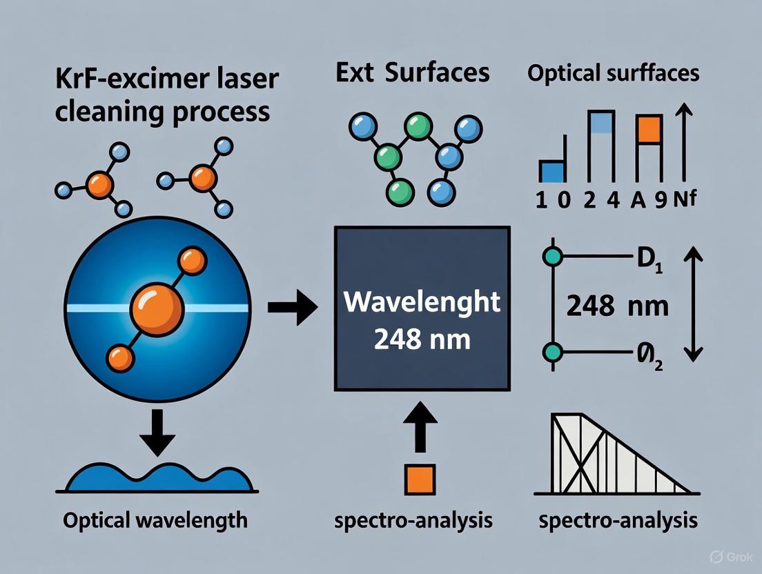
KrF Excimer Laser Cleaning at 248 nm: Mechanisms, Applications, and Optimization for Optical Surfaces
This article provides a comprehensive examination of KrF excimer laser cleaning at a wavelength of 248 nm, a advanced technique for the precise and controlled removal of contaminants, coatings, and...
KrF Excimer Laser Cleaning at 248 nm: Mechanisms, Applications, and Optimization for Optical Surfaces
Abstract
This article provides a comprehensive examination of KrF excimer laser cleaning at a wavelength of 248 nm, a advanced technique for the precise and controlled removal of contaminants, coatings, and particulates from sensitive optical surfaces. Tailored for researchers, scientists, and development professionals, the content spans from foundational principles and cleaning mechanisms to specific methodological applications across diverse materials. It delivers a thorough guide for troubleshooting and optimizing laser parameters to ensure efficacy and prevent substrate damage, supported by validation through modern analytical techniques and comparisons with traditional cleaning methods. The goal is to equip practitioners with the knowledge to implement this non-contact, environmentally friendly technology effectively in research and high-tech industrial settings.
Unveiling the Principles: How 248 nm KrF Laser Light Interacts with Contaminants and Substrates
Laser cleaning is an advanced, non-contact surface-processing technology that utilizes a high-energy laser beam to irradiate a component's surface, leading to the instant evaporation or stripping of contaminants, rust, and coatings. Compared with conventional cleaning methods, laser cleaning offers significant advantages, including high precision, efficiency, environmental friendliness, and superior controllability [1]. The interaction between a laser beam and a material involves complex physical and chemical processes such as decomposition, ionization, vibration, expansion, and vaporization. The effectiveness of the cleaning process is governed by three fundamental mechanisms: the laser thermal ablation mechanism, the laser thermal stress mechanism, and the plasma shock wave mechanism. The dominance of each mechanism depends on the laser parameters and the material properties of the contaminants and substrate [1]. This document details these mechanisms within the specific context of using a 248 nm KrF excimer laser for cleaning and preparing optical surfaces, providing application notes and experimental protocols for researchers.
Fundamental Mechanisms and Their Principles
Laser Thermal Ablation Mechanism
The laser thermal ablation mechanism primarily relies on the photothermal effect. When a pulsed laser beam irradiates the attachments on a substrate surface, the energy is absorbed, causing a rapid temperature increase. If the temperature exceeds the gasification threshold of the contaminant, it will be removed through processes like vaporization, combustion, or decomposition [1]. For a 248 nm KrF excimer laser, the high photon energy can also induce photochemical effects, where the photon energy directly breaks molecular bonds in the contaminant layer, transforming the material into a loose state that promotes its removal [1].
The temperature increase (ΔT) on the material surface under laser irradiation can be expressed as:
[
\Delta T = \frac{P}{\pi \omega_0 K}
]
where P is the incident laser power, ω₀ is the laser spot radius, and K is the thermal conductivity of the material [1].
The laser energy (W) required within a single pulse duration can be described as:
[
W = \rho h [Cs (Tm - T0) + Cp (Tb - Tm) + Lm + Lr]
]
where ρ is the density, h is the thickness of the attachment, C_s is the specific heat capacity, C_p is the specific heat, T_m is the melting point, T_b is the boiling point, T_0 is the initial temperature, L_m is the latent heat of melting, and L_r is the latent heat of evaporation [1].
A critical aspect of this mechanism is the ablation threshold. When the ablation threshold of the contaminant is lower than that of the substrate, the laser energy density can be controlled to remove the attachment without damaging the substrate. Successful application requires careful calibration of laser parameters to stay within this window [1].
Laser Thermal Stress Mechanism
Unlike the thermal ablation mechanism, the laser thermal stress mechanism utilizes stress effects induced by the laser rather than relying solely on thermal effects to vaporize material. When a short-pulse laser irradiates a surface, the attachment and a thin layer of the substrate absorb energy, causing rapid heating. The short pulse width leads to a very fast thermal expansion cycle, generating high compressive stresses. Upon cooling, this can create a high-pressure solid lifting force. When this lifting force surpasses the van der Waals force binding the contamination to the substrate, the contaminants are ejected from the surface [1].
The one-dimensional heat conduction equation governing this process is [1]:
[
\rho c \frac{\partial T(z,t)}{\partial t} = \lambda \frac{\partial^2 T(z,t)}{\partial z^2} + \alpha I_0 A e^{-Az}, (0 \leq z \leq l, 0 \leq t \leq \tau)
]
where ρ is density, c is specific heat capacity, λ is thermal conductivity, α is absorptivity, I₀ is laser intensity, A is the absorption coefficient, z is depth, and t is time.
The resulting thermal stress (σ) can be expressed as:
[
\sigma = Y \varepsilon = Y \frac{\Delta l}{l} = Y \gamma
]
where Y is Young's modulus, ε is strain, Δl is the change in length, l is the original length, and γ is the thermal expansion coefficient of the material [1]. This mechanism is particularly effective for removing particles or layers with a different thermal expansion coefficient from the substrate.
Plasma Shock Wave Mechanism
The plasma shock wave mechanism becomes significant when the laser fluence is high enough to ionize air or vaporized material above the surface, creating a laser-induced plasma. The rapid expansion of this plasma generates a propagating shock wave that travels towards the substrate surface. The impulsive pressure from this shock wave is capable of dislodging and removing tiny particles and thin films from the surface. This mechanism is highly effective for cleaning sub-micron particles without significant thermal loading of the substrate [1].
Quantitative Parameters for KrF Excimer Laser Cleaning
The following tables summarize critical laser parameters and their effects on the cleaning process and outcomes for different material systems, as reported in recent research.
Table 1: KrF Laser Parameters and Observed Cleaning Effects on Various Materials
| Material | Laser Fluence (J/cm²) | Pulse Number | Pulse Duration/ Frequency | Key Observations | Source |
|---|---|---|---|---|---|
| X-cut LiNbO₃ | Varied (Ablation study) | Varied (1 to 100) | 20 ns, 25 Hz | Surface damage (exfoliation, discoloration) observed at high fluence/pulses; Suppressed with SiO₂ overlayer. | [2] |
| WC-Co Composite | 5.5 J/cm² | 1, 5, 10, 20, 50, 100 | 20 ns, 1-100 Hz | Lower pulses (1, 5) caused Co removal & nano-structuring; Higher pulses (50, 100) induced micro-cracks. | [3] |
| Polyimide (PI) Film | 7-18 mJ/cm² | 6000-18000 | 20 ns, 10 Hz | Formation of Laser-Induced Periodic Surface Structures (LIPSS); Optimal at ~14 mJ/cm², 12000 pulses. | [4] [5] |
| General Polymer Ablation | Material-dependent | Material-dependent | Nanosecond pulses | Ablation occurs when fluence (F) exceeds material threshold; depends on pulses (P), frequency, and absorption coefficient. | [6] |
Table 2: Process Outcomes and Optimization Guidelines for KrF Laser Cleaning
| Process Outcome | Key Influencing Parameters | Optimal Conditions / Guidelines |
|---|---|---|
| Minimizing Substrate Damage | Fluence, Pulse Number, Presence of overlayer. | Use fluence just above contaminant's ablation threshold but below substrate's damage threshold; Use minimal pulses needed; A SiO₂ overlayer can protect the substrate [2] [1]. |
| Selective Binder Removal (WC-Co) | Fluence, Pulse Number. | Lower number of pulses (1-5) with high fluence (5.5 J/cm²) selectively removes Co binder without major cracking [3]. |
| Nanostructure Formation (LIPSS) | Energy Density, Pulse Number, Polarization. | For PI, 14.01 mJ/cm² with 12,000 pulses of linearly polarized 248 nm laser produces uniform ~200 nm period ripples [4] [5]. |
| Controlled Roughening | Pulse Number, Fluence. | Surface roughness increases with pulse number; can be controlled predictably (e.g., Ra from 0.55 nm to ~14.3 nm on PI) [4] [5]. |
Experimental Protocols for KrF Laser Cleaning
General Workflow for Laser Cleaning and Surface Preparation
The following diagram outlines a standard experimental workflow for laser cleaning and surface modification using a KrF excimer laser.
Protocol 1: Surface Decontamination and Coating Removal
This protocol is designed for removing contaminants or thin films from optical surfaces with minimal substrate damage.
- Objective: To selectively remove a surface contaminant or coating from a substrate (e.g., LiNbO₃) without inducing surface damage.
- Materials:
- KrF Excimer Laser (248 nm).
- Target sample (e.g., X-cut LiNbO₃).
- Optional: SiO₂ overlayer film (~1.0 µm thick).
- Solvents (ethanol, deionized water) for ultrasonic cleaning.
- Methodology:
- Sample Preparation: Clean the substrate ultrasonically in absolute ethanol and then deionized water for 10 minutes each. Dry in a vacuum oven. If using a protective overlayer, deposit a ~1.0 µm SiO₂ film on the LiNbO₃ surface [2].
- Laser Setup: Configure the KrF laser with a pulse repetition rate of 25 Hz and a pulse width (FWHM) of 20 ns. Use a mask-projection system to define the cleaning area [2].
- Parameter Calibration:
- Conduct a test with a low fluence (e.g., below 1 J/cm²) and a low number of pulses (e.g., 10).
- Gradually increase the fluence and pulse count in subsequent test areas while monitoring for the onset of surface damage (exfoliation, discoloration).
- Identify the optimal fluence that achieves cleaning while staying below the damage threshold. The use of a SiO₂ overlayer can allow for higher fluences without damage [2].
- Cleaning Execution: Irradiate the entire target area with the optimized parameters.
- Validation: Inspect the surface using Scanning Electron Microscopy (SEM) to confirm contaminant removal and check for laser-induced damage [2].
Protocol 2: Selective Binder Ablation for Surface Preparation
This protocol is used for surface engineering of composite materials, such as preparing WC-Co substrates for diamond film coating.
- Objective: To selectively remove the cobalt (Co) binder from a WC-Co composite surface to create a roughened, residual-stress-free surface ideal for diamond film adhesion.
- Materials:
- KrF Excimer Laser (248 nm, Compex Pro 201 or equivalent).
- Polished WC-Co composite sample.
- Diamond polishing compound (1-6 µm grit).
- Methodology:
- Sample Preparation: Polish the WC-Co sample using diamond polishing compounds of decreasing grit size (e.g., 6 µm, 3 µm, 1 µm) to achieve a smooth initial surface [3].
- Laser Setup: The laser is operated at a wavelength of 248 nm. The beam is delivered to the sample surface, which is placed in open atmosphere at room temperature [3].
- Parameter Calibration:
- For selective Co removal and nano-structuring without micro-cracks, use a lower number of pulses (1 to 10) with a higher fluence (e.g., 5.5 J/cm²) [3].
- Avoid higher pulse counts (e.g., 50, 100) at this fluence, as they induce micro-cracks.
- Processing: Irradiate the sample surface. A lower number of pulses will yield a darker surface region indicating Co removal and possible carbon phase formation [3].
- Validation:
- Use SEM to examine surface morphology and detect micro-cracks.
- Use Energy-Dispersive X-ray Spectroscopy (EDS) to confirm the reduction in surface cobalt content [3].
- Use X-ray Diffraction (XRD) to analyze for the presence of residual stress.
- Use Atomic Force Microscopy (AFM) to quantify the increase in surface roughness [3].
Protocol 3: Fabrication of Laser-Induced Periodic Surface Structures (LIPSS)
This protocol is for creating functional, periodic nanostructures on polymer surfaces to alter properties like wettability.
- Objective: To fabricate uniform nanoscale periodic ripples (LIPSS) on a polyimide (PI) film surface to enhance hydrophilicity.
- Materials:
- Linearly polarized KrF excimer laser (248 nm).
- Polyimide film (e.g., thickness 0.05 mm).
- Absolute ethanol and deionized water.
- Optical homogenizer to ensure a flat-top beam profile.
- Methodology:
- Sample Preparation: Cut PI film into 15x15 mm squares. Clean ultrasonically in absolute ethanol and deionized water for 10 minutes each. Dry in a vacuum drying oven [4] [5].
- Laser Setup:
- Use an optical path with a homogenizer to achieve a uniform energy distribution.
- Ensure the laser's inherent linear polarization is used; no additional polarizer is needed. The ripples will form perpendicular to the polarization direction [4] [5].
- Set the pulse width to 20 ns and the repetition rate to 10 Hz. Use normal beam incidence.
- Parameter Calibration:
- Processing: Irradiate the sample. The large number of pulses requires a stable sample stage and consistent laser output.
- Validation:
The Scientist's Toolkit: Essential Research Reagents and Materials
Table 3: Key Equipment and Materials for KrF Excimer Laser Cleaning Research
| Item | Specification / Example | Function in Research |
|---|---|---|
| KrF Excimer Laser | 248 nm wavelength, e.g., Lambda Physik LPX210i or Compex Pro 201. | The primary energy source for ablation and surface modification. The UV wavelength is well-absorbed by many materials. |
| UV-Grade Fused Silica Substrates/Mirrors | UV Fused Silica, Surface Quality: 10-5 scratch-dig. | Used as substrates or optics due to low thermal expansion, high UV transmission, and high damage threshold [7]. |
| Beam Delivery & Homogenizing Optics | Attenuators, beam splitters, plano-convex lenses, homogenizer. | Controls laser fluence, shapes the beam, and creates a uniform (flat-top) energy profile on the target surface [4] [5]. |
| Characterization: SEM | Scanning Electron Microscope. | Provides high-resolution imaging of surface morphology, ablation features, and damage assessment [2] [3]. |
| Characterization: EDS | Energy-Dispersive X-ray Spectroscopy. | Analyzes elemental composition changes on the surface (e.g., selective Co removal from WC-Co) [3]. |
| Characterization: AFM | Atomic Force Microscope, e.g., Cypher VRS. | Quantifies nanoscale topography, measures LIPSS period/depth, and evaluates surface roughness (Ra) [4] [5]. |
| Characterization: XPS | X-ray Photoelectron Spectroscopy. | Investigates chemical state changes and reactions with atmospheric gasses (e.g., nitrogen, oxygen) after laser treatment [3]. |
| Protective Overlayer | SiO₂ thin film (~1.0 µm thick). | Deposited on the sample to suppress surface damage (exfoliation) during deep ablation processes [2]. |
The Krypton Fluoride (KrF) excimer laser, operating at a wavelength of 248 nanometers (nm), is a cornerstone technology in advanced cleaning and processing applications. Its significance stems from the unique properties of ultraviolet-C (UV-C) light at this specific wavelength, which offers a powerful combination of high photon energy and strong material absorption. The high photon energy of approximately 4.99 eV enables the laser to directly break chemical bonds in organic materials and contaminants, facilitating a photochemical ablation process. Concurrently, this wavelength is strongly absorbed by a wide range of organic polymers, biological specimens, and degradation products found on cultural heritage objects, making it exceptionally effective for precise, non-contact cleaning. Within the broader thesis on KrF-excimer laser cleaning of optical surfaces, this document establishes the fundamental principles and provides detailed protocols for harnessing these properties in research and development.
Key Characteristics and Applications of 248 nm Radiation
The 248 nm wavelength occupies a critical position in the electromagnetic spectrum, offering distinct advantages for material processing.
Fundamental Properties
- High Photon Energy: At 248 nm, each photon carries an energy of about 4.99 eV. This energy is sufficient to directly dissociate covalent bonds (e.g., C-C, C-H, C-O) common in organic molecules and polymers, initiating photochemical decomposition [8] [9].
- Strong Absorption: Many materials, including organic coatings, microbial organisms, and pollution crusts, exhibit strong absorption bands in the UV-C region. This high absorption confines the laser energy to a thin surface layer, enabling precise removal without generating significant thermal stress to the underlying substrate [10] [11].
- Pulsed Operation: KrF excimer lasers typically operate with nanosecond (ns) to femtosecond (fs) pulse durations. This pulsed delivery allows for high peak powers that drive efficient ablation, while the short pulse width limits heat diffusion, minimizing thermal damage to sensitive surfaces in applications ranging from painting restoration to semiconductor cleaning [8] [12] [1].
Quantitative Performance Data
Table 1: Efficacy of 248 nm Pulsed Laser in Various Applications
| Application Field | Target Material | Key Laser Parameters | Efficacy / Outcome | Source |
|---|---|---|---|---|
| Pest Control | Spider mites (T. urticae) | 248 nm, 5 mJ pulse, 60 Hz, 80 kJ/m² dose | 98% mortality in adult mites | [8] |
| Pest Control | Spider mite eggs | 248 nm, 5 kJ/m² dose | ~100% egg mortality (prevented hatching) | [8] |
| Painting Cleaning | Aged varnish/overpaint | 248 nm, 0.1 - 1.1 J/cm² fluence | Successful removal of non-original layers | [10] |
| Water Treatment | Ibuprofen (pharmaceutical) | 266 nm (for reference), ns pulses | >95% degradation in pure water in <6 minutes | [9] |
| Stained Glass Cleaning | Encrustations & corrosion | 248 nm, varied fluence & rep. rate | Defined removal of crusts without damaging gel layer | [11] |
Experimental Protocols for KrF Excimer Laser Cleaning
This section provides detailed methodologies for key experiments, demonstrating the application of 248 nm laser cleaning across different fields.
Protocol 1: Cleaning of Aged Varnish from Paintings
This protocol is adapted from studies on laser cleaning of historical easel paintings [10].
1. Principle: Aged varnishes and overpaints strongly absorb 248 nm radiation. The laser energy ablates the material in a controlled, layer-by-layer manner, with selectivity achieved by tuning parameters below the damage threshold of the underlying original paint.
2. Materials and Equipment:
- KrF Excimer Laser System (e.g., Lambda Physik LPX series) emitting at 248 nm with nanosecond pulses.
- Beam delivery system with galvanometric mirrors for scanning.
- Non-invasive analytical tools: Optical Coherence Tomography (OCT), Reflection FT-IR spectrometer, Laser-Induced Fluorescence (LIF) spectrometer.
- Fume extraction system.
3. Procedure: 1. Pre-Cleaning Assessment: Characterize the painting surface using OCT to determine the stratigraphy and thickness of the varnish layers. Perform reflection FT-IR to identify the molecular composition of the surface coatings. 2. Laser Parameter Calibration: - Set the laser to a low fluence (e.g., 0.1 J/cm²). - Perform test cleaning on a small, inconspicuous area. - Gradually increase the fluence (up to ~1.1 J/cm²) and number of pulses (N) until optimal removal is observed, monitored in real-time if possible. 3. Cleaning Operation: Use the galvanometric scanner to direct the laser beam (shaped to a rectangular spot of ~0.08 x 1.00 cm²) over the target area. 4. In-situ Monitoring: After each cleaning pass, re-analyze the area with OCT and FT-IR to assess the amount of material removed and confirm the absence of the varnish layer without affecting the underlying paint. 5. Final Assessment: Conduct a final LIF measurement to verify the surface state and ensure no latent damage has occurred.
Protocol 2: Acaricidal Treatment Using 248 nm Irradiation
This protocol is based on research into the acaricidal efficacy of UV-C irradiation on spider mites [8].
1. Principle: High-intensity 248 nm radiation is lethal to arthropods and their eggs, causing molecular damage to DNA and proteins. The pulsed nature of the laser enhances efficacy through high peak power.
2. Materials and Equipment:
- Pulsed KrF Excimer Laser (e.g., CEX-100) with output at 248 nm.
- Laser power meter (e.g., thermopile sensor).
- Sample holders for mites and host leaves.
- Environmental chamber for rearing mites (26°C, 50-60% RH).
3. Procedure: 1. Sample Preparation: Rear two-spotted spider mites (Tetranychus urticae) on host plant leaves (e.g., common bean). Collect adult females and eggs of synchronized age for experiments. 2. System Setup: Place the laser source at a fixed distance from the target (e.g., 150 mm). Measure the power (mW) and calculate the energy density (kJ/m²) at the target surface. The spot size is typically small (e.g., 2.16 cm²). 3. Irradiation: Expose adult mites and eggs to the laser for varying durations (e.g., 1 to 4 minutes) and energy densities (e.g., 5 to 80 kJ/m²). The pulse repetition rate can be adjusted (e.g., up to 100 Hz). 4. Post-Treatment Evaluation: - Mite Mortality: Assess mortality immediately after irradiation and again at 24 hours post-irradiation. - Egg Hatchability: Observe irradiated eggs daily for up to 12 days to determine the percentage that fail to hatch.
The Scientist's Toolkit: Essential Research Reagents and Equipment
Table 2: Key Equipment and Reagents for 248 nm Laser Cleaning Research
| Item Name | Function / Role | Specific Example / Notes |
|---|---|---|
| KrF Excimer Laser | Generates high-energy 248 nm pulsed light. | CEX-100; Lambda Physik LPX 200/305i. Requires KrF gas mixture. |
| Beam Shaping Optics | Modifies beam profile and spot size on target. | Fused silica lenses, apertures. Enables even energy distribution. |
| Galvanometric Scanner | Precisely directs the laser beam over the surface. | Essential for automated cleaning of large or complex areas. |
| Calibrated Power/Energy Meter | Measures laser output at the target. | NIST-calibrated thermopile sensor (e.g., GreenTEG B05). Critical for dose control. |
| Non-Invasive Analysers (OCT, FT-IR) | Assesses surface pre- and post-cleaning. | OCT for stratigraphy; FT-IR for chemical composition. |
| Fume Extraction System | Removes ablated particulates and vapors. | Critical for operator safety and laboratory air quality. |
Workflow and Interaction Mechanisms
The following diagrams illustrate the experimental workflow for laser cleaning and the fundamental mechanisms of laser-material interaction at 248 nm.
KrF Laser Cleaning Experimental Workflow
Laser-Material Interaction Mechanisms at 248 nm
The 248 nm wavelength generated by KrF excimer lasers is a powerful tool for advanced cleaning, leveraging its high photon energy and strong material absorption. The provided application notes and detailed experimental protocols for painting conservation and acaricidal treatment showcase its versatility and efficacy. The fundamental mechanisms—photochemical, photothermal, and mechanical—often work in concert to enable the precise, controlled, and residue-free removal of unwanted surface layers. This makes 248 nm laser cleaning an invaluable technique for R&D professionals working with sensitive optical surfaces, cultural heritage artifacts, and in specialized industrial and biomedical fields.
Photochemical versus Photothermal Effects at Ultraviolet Wavelengths
The interaction of ultraviolet laser radiation with materials engages two primary mechanisms: photochemical and photothermal effects. Understanding the interplay between these phenomena is crucial for optimizing laser applications, particularly in precision fields such as laser cleaning of optical surfaces. At 248 nm, the wavelength of KrF-excimer lasers, photon energy reaches approximately 5 eV, sufficient to directly break molecular bonds in many materials [3]. This application note examines the conditions governing the dominance of each mechanism and provides experimental protocols for researchers requiring controlled laser processing of optical surfaces.
The photochemical effect occurs when high-energy photons directly break chemical bonds, causing material ablation through non-thermal pathways with minimal heat transfer to the substrate. In contrast, the photothermal effect relies on photon energy being converted to heat, enabling thermal processes like melting, vaporization, and stress-induced removal [1]. For KrF-excimer laser cleaning of optical surfaces, determining which mechanism dominates depends on specific laser parameters and material properties, requiring careful experimental control to achieve desired outcomes while preventing substrate damage.
Theoretical Background
Fundamental Mechanisms
Photochemical effects dominate when photon energy exceeds molecular bond energies, enabling direct bond dissociation without significant heat generation. At 248 nm (5 eV), this energy surpasses the binding energies of many organic compounds (C-C: 3.6 eV, C-H: 4.3 eV) and some inorganic bonds [13]. The process involves electronic excitation followed by bond cleavage, resulting in precise, cold ablation with minimal thermal damage to surrounding areas. This mechanism typically requires short pulse durations and high peak fluences to achieve multiphoton absorption when photon energy alone is insufficient for direct bond breaking [14].
Photothermal effects occur when materials absorb laser energy and convert it to heat, causing rapid temperature rise that leads to melting, vaporization, or thermal decomposition. This mechanism depends on the thermal properties of the material, including absorption coefficient, thermal conductivity, and specific heat capacity [1]. The resulting thermal stress can produce mechanical forces that remove material when thermal expansion differences between surface contaminants and substrate exceed adhesion forces [15].
Comparative Analysis
Table 1: Characteristics of Photochemical and Photothermal Effects at 248 nm
| Parameter | Photochemical Effect | Photothermal Effect |
|---|---|---|
| Energy Transfer | Direct bond breaking via electronic excitation | Phonon-mediated thermal activation |
| Primary Mechanism | Photon-induced molecular dissociation | Thermal vibration leading to phase changes |
| Spatial Resolution | High (sub-micron) | Moderate to high |
| Thermal Damage Risk | Low | High |
| Typical Pulse Duration | Nanosecond to femtosecond | Microsecond to continuous wave |
| Fluence Requirement | Material-dependent (exceed ablation threshold) | Sufficient for rapid heating |
| Material Selectivity | High (wavelength-dependent absorption) | Moderate (thermal property dependent) |
| Suitable Applications | Precision cleaning, polymer processing | Paint removal, large-area cleaning |
KrF Excimer Laser Specifics
KrF excimer lasers operating at 248 nm provide unique advantages for surface processing due to their high photon energy and typical nanosecond pulse durations. This wavelength is strongly absorbed by most organic materials, biological tissues, and many metal oxides, enabling precise ablation with minimal thermal penetration [3] [10]. The high absorption coefficient at UV wavelengths typically confines energy deposition to thin surface layers, facilitating both photochemical bond breaking and rapid thermal heating depending on pulse parameters.
Experimental Evidence and Parameter Optimization
Dominant Mechanism Identification
Recent research reveals that the dominant mechanism at 248 nm depends on specific laser parameters and material properties. For graphene oxide reduction, photochemical effects prevail under visible light irradiation despite temperatures remaining below the standard thermal reduction threshold of 200°C [14]. Conversely, laser cleaning of metallic contaminants typically employs photothermal mechanisms where thermal stress overcomes adhesion forces [15].
Table 2: Laser Parameters and Dominant Mechanisms for Different Materials
| Material | Laser Parameters | Dominant Mechanism | Observed Effects |
|---|---|---|---|
| Graphene Oxide | 532 nm, 300 μW, CW | Photochemical | Reduction without significant heating |
| WC-Co Composite | 248 nm, 5.5 J/cm², 1-50 pulses | Mixed (pulse-dependent) | Selective Co removal at low pulses |
| Historical Paintings | 248 nm, 0.1-1.1 J/cm², 1-50 pulses | Photochemical | Varnish removal without pigment damage |
| Polyimide Film | 248 nm, 14.01 mJ/cm², 12,000 pulses | Photothermal | LIPSS formation via thermal effects |
| Glass Insulators | 1064 nm, 8 W, 8 m/s scan | Photothermal | Contaminant removal via thermal stress |
KrF Laser Cleaning of Optical Surfaces
For KrF excimer laser cleaning of optical surfaces at 248 nm, the following parameter ranges have been established for different applications:
- Historical painting cleaning: Fluence: 0.1-1.1 J/cm², Pulse number: 1-50 pulses, Selective varnish removal via photochemical ablation [10]
- WC-Co composite treatment: Fluence: 1.5-5.5 J/cm², Pulse number: 1-100 pulses, Selective cobalt removal at lower pulse counts [3]
- Polyimide nanostructuring: Fluence: 7-18 mJ/cm², Pulse number: 6000-18000 pulses, LIPSS formation via photothermal mechanism [5]
The optimal working window for precision cleaning of optical surfaces typically employs lower fluences (0.5-2 J/cm²) with minimal pulse counts (1-20 pulses) to maximize photochemical effects while minimizing thermal contributions [3] [10].
Experimental Protocols
Protocol 1: KrF Excimer Laser Cleaning of Optical Surfaces
Objective: Remove contaminants from optical surfaces without substrate damage using dominant photochemical effects.
Materials and Equipment:
- KrF excimer laser (248 nm, nanosecond pulse duration)
- Optical components to be cleaned
- Beam delivery system with galvanometric mirrors
- Energy meter for fluence calibration
- Non-invasive monitoring equipment (OCT, reflection FT-IR)
Procedure:
- Surface Characterization:
- Perform baseline analysis using optical coherence tomography (OCT) to determine contaminant thickness
- Conduct reflection FT-IR spectroscopy to identify chemical composition of contaminants
Laser Parameter Setup:
- Set laser fluence between 0.5-1.5 J/cm² (below substrate damage threshold)
- Configure beam spot size based on contaminant distribution (typically 0.08 × 1.00 cm² rectangular area)
- Set initial pulse number to 5 pulses for test cleaning
Test Cleaning:
- Irradiate test areas with systematic variation of pulse numbers (1, 3, 5, 10, 20 pulses)
- Maintain constant fluence within ±5% variation across the beam profile
Post-Treatment Assessment:
- Re-examine cleaned areas using OCT to measure remaining contaminant thickness
- Perform FT-IR analysis to verify complete contaminant removal
- Inspect for surface damage using optical microscopy
Parameter Optimization:
- Select parameters that achieve complete contaminant removal without substrate damage
- For heat-sensitive substrates, prioritize lower pulse counts with moderate fluence
Troubleshooting:
- If contamination persists: Gradually increase pulse number (up to 50 pulses maximum)
- If substrate damage occurs: Reduce fluence by 0.1 J/cm² increments
- For non-uniform cleaning: Verify beam homogeneity and surface planarity
Protocol 2: Differentiation of Photochemical vs. Photothermal Effects
Objective: Determine the dominant mechanism in laser-material interaction at 248 nm.
Materials and Equipment:
- KrF excimer laser system with adjustable parameters
- Target materials (polymers, metals, or contaminants)
- Thermal imaging camera or Raman thermometry system
- XPS analysis capability
- AFM for surface topography
Procedure:
- Sample Preparation:
- Prepare identical samples of target material
- Ensure uniform surface roughness and contamination levels
Thermal Measurement Setup:
- Configure Raman thermometry with silicon nanowires for in situ temperature measurement
- Calibrate temperature measurement system
- Set safety limits to prevent material degradation
Laser Irradiation:
- Irplicate sample series with varying fluences (0.1-5 J/cm²) and pulse numbers (1-100)
- Monitor temperature evolution during irradiation
- Record maximum reached temperatures
Post-Irradiation Analysis:
- Perform XPS analysis to identify chemical changes
- Conduct AFM to examine surface topography modifications
- Compare results with control samples heated conventionally to same temperatures
Mechanism Identification:
- Photochemical dominance: Significant chemical modification with temperatures below thermal activation threshold
- Photothermal dominance: Material changes correlate with temperature profile and match conventional heating results
- Mixed mechanism: Both chemical and thermal changes observed
Data Interpretation:
- Temperatures <150°C with material modification indicate photochemical dominance
- Material changes only above specific temperature thresholds suggest photothermal processes
- Immediate ablation at low pulse numbers suggests photochemical pathways
- Gradual material modification over multiple pulses suggests thermal accumulation
The Scientist's Toolkit
Table 3: Essential Research Reagent Solutions for KrF Laser Studies
| Item | Function | Application Notes |
|---|---|---|
| KrF Excimer Laser | 248 nm photon source | 5 eV photons, nanosecond pulses |
| Beam Homogenizer | Creates uniform fluence distribution | Essential for reproducible processing |
| Galvanometric Mirror System | Precise beam positioning | Enables complex cleaning patterns |
| Energy Attenuator | Adjusts laser fluence | Fine-tunes energy density |
| Optical Coherence Tomography | Non-invasive stratigraphic analysis | Measures layer thickness pre/post cleaning |
| Reflection FT-IR Spectrometer | Chemical composition analysis | Identifies molecular changes |
| Atomic Force Microscope | Surface topography characterization | Nanoscale resolution of laser effects |
| X-ray Photoelectron Spectrometer | Surface chemistry analysis | Quantifies elemental and chemical changes |
| Raman Thermometry | In situ temperature measurement | Uses SiNWs for accurate thermal monitoring |
Visualizations
KrF Laser Cleaning Workflow
Diagram 1: KrF Laser Cleaning Workflow - This flowchart illustrates the systematic protocol for KrF excimer laser cleaning of optical surfaces, emphasizing parameter optimization and damage prevention.
Mechanism Differentiation Methodology
Diagram 2: Mechanism Differentiation Methodology - This workflow outlines the experimental approach for determining whether photochemical or photothermal effects dominate during UV laser processing.
The interaction of 248 nm laser radiation with materials involves complex competition between photochemical and photothermal effects. For KrF-excimer laser cleaning of optical surfaces, photochemical mechanisms typically dominate at lower fluences (0.5-2 J/cm²) and pulse numbers (1-20), enabling precise contaminant removal without substrate damage. Photothermal effects become increasingly significant at higher energy densities and pulse counts, particularly for materials with strong UV absorption.
Successful application requires careful parameter optimization based on the specific contaminant-substrate system, supported by non-invasive monitoring techniques. The protocols outlined herein provide researchers with methodologies to achieve controlled laser cleaning while identifying dominant mechanisms for process optimization. As research advances, continued investigation of the interplay between these mechanisms will further enhance precision and efficiency in UV laser processing of optical surfaces.
Within the scope of broader thesis research on KrF-excimer laser cleaning of optical surfaces at a wavelength of 248 nm, understanding the precise ablation thresholds of common contaminants and underlying optical substrates is paramount. This laser cleaning process depends on the principle of selective ablation, where the laser energy density is carefully controlled to exceed the removal threshold of the contaminant while remaining below the damage threshold of the optical substrate [1]. This application note consolidates critical quantitative data on these thresholds and provides detailed, reproducible experimental protocols for their determination, serving as an essential resource for researchers and scientists in the field.
Material Ablation and Damage Thresholds
The effectiveness and safety of laser cleaning are governed by the differential in ablation thresholds between the contaminant and the substrate. The data presented below are critical for defining the operational window of the process.
Table 1: Ablation and Damage Thresholds for Common Materials at 248 nm
| Material Type | Material Name | Ablation/Damage Threshold (mJ/cm²) | Key Findings / Context |
|---|---|---|---|
| Contaminant | Sulfide on Steel | 410 [1] | Removal threshold; successful cleaning between 410-8250 mJ/cm². |
| Contaminant | Aged Triterpenoid Varnishes | 200,000 - 1,800,000 [16] | "Optimum" photochemical ablation fluence range. |
| Contaminant | Parylene-C (on Iridium) | >1,000,000 [17] | Successful deinsulation; higher fluence improves uniformity. |
| Optical Substrate | Polyimide (PI) Film | 7,000 - 18,000 [5] | LIPSS formation range; optimal at ~14,010 mJ/cm². |
| Optical Substrate | CaF₂ (Highly Polished) | ~6,100,000 [18] | Laser-Induced Damage Threshold (LIDT) for front surface. |
| Optical Substrate | CaF₂ (Roughly Polished) | ~5,600,000 [18] | LIDT for front surface; lower due to surface defects. |
The data reveals a significant spread in threshold values, influenced by material properties and surface condition. For instance, the surface polishing level of CaF₂ substrates has a demonstrable impact on their laser-induced damage threshold (LIDT). Highly polished CaF₂ windows exhibit a higher LIDT (6.1 J/cm²) compared to their roughly polished counterparts (5.6 J/cm²), as surface defects like scratches and digs on the latter act as precursors to damage by enhancing local light absorption [18]. Furthermore, the LIDT of the rear surface is consistently lower than that of the front (incident) surface for both polishing levels, a phenomenon attributed to internal light field modulation [18].
Fundamental Laser Cleaning Mechanisms
The interaction of a 248 nm laser with materials can be described by three primary mechanisms, the dominance of which depends on the laser parameters and the material properties [1].
- Laser Thermal Ablation Mechanism: A high-energy laser beam irradiates the surface, causing the attachments to undergo rapid heating, leading to vaporization, combustion, or decomposition. For a 248 nm KrF excimer laser, the high photon energy (5 eV) can directly break molecular bonds (C-C, C-H, C-O) in organic contaminants, a process known as photochemical ablation or laser ablation [19].
- Laser Thermal Stress Mechanism: The short pulse width of the laser induces rapid, localized heating and cooling, leading to quick thermal expansion and contraction. This generates a high-pressure solid lifting force or shear force at the contaminant-substrate interface, mechanically peeling the contaminant away once this force surpasses the van der Waals force [1].
- Plasma Shock Wave Mechanism: When an ultra-short pulse, high-peak-power laser irradiates a surface, it can generate vapor and subsequently a laser-induced plasma. This plasma, continuing to absorb laser energy, produces an instantaneous shock wave (1-100 kbar) that fragments and removes surface contaminants [19].
The following workflow diagram illustrates the decision-making process for selecting the appropriate cleaning mechanism based on the contaminant and substrate properties.
Experimental Protocols
Protocol for Determining Single-Pulse Ablation Thresholds
This protocol outlines a standard method for determining the ablation threshold of a material using a KrF excimer laser, based on industry-standard practices [16] [18].
Objective: To determine the minimum laser fluence required to initiate ablation of a material with a single laser pulse.
Materials and Equipment:
- KrF excimer laser (λ = 248 nm)
- calibrated laser energy meter
- beam homogenizer and attenuator
- sample material
- optical microscope (for post-irradiation analysis)
Procedure:
- Sample Preparation: Clean and dry the sample surface to remove any ambient contaminants.
- Laser Setup: Configure the laser optical path, ensuring the use of a beam homogenizer to achieve a flat-top beam profile and an attenuator for precise fluence control.
- Irradiation Test: Fire a single laser pulse onto the sample surface at a predetermined starting fluence.
- Inspection: Examine the irradiated spot under an optical microscope for signs of material modification or removal.
- Iteration: Repeat steps 3 and 4, systematically increasing or decreasing the fluence in small increments.
- Threshold Identification: The ablation threshold is identified as the fluence at which a permanent, microscopically visible change to the surface is first observed.
Protocol for Systematic Investigation of Multi-Pulse LIPSS Formation on Polymers
This protocol provides a detailed methodology for studying the formation of Laser-Induced Periodic Surface Structures (LIPSS) on polymer surfaces like polyimide, as described in recent literature [5].
Objective: To systematically investigate the effects of laser energy density and pulse number on the morphology and surface roughness of LIPSS on polyimide films.
Materials and Equipment:
- KrF excimer laser (λ = 248 nm, pulse width ~20 ns, 10 Hz repetition rate)
- beam delivery system with homogenizer and attenuator
- motorized X-Y sample stage
- polyimide film samples (e.g., 15 mm x 15 mm, ultrasonically cleaned)
- Atomic Force Microscope (AFM)
Procedure:
- Parameter Definition: Define a matrix of experimental parameters. For example:
- Laser energy density: A range from 7 to 18 mJ/cm².
- Pulse number: A range from 6000 to 18,000 pulses.
- Sample Irradiation: Mount the PI sample on the stage. Using computer control, irradiate different areas of the sample with specific combinations of energy density and pulse number from the parameter matrix.
- AFM Characterization: After laser processing, characterize the surface morphology of each irradiated area using AFM in tapping mode to avoid sample damage.
- Data Analysis: For each parameter set, measure the spatial period of the ripples, the ripple depth, and calculate the surface roughness (Ra). The most uniform and well-defined LIPSS are typically achieved at an optimal combination of parameters (e.g., ~14 mJ/cm² and 12,000 pulses for polyimide) [5].
The Scientist's Toolkit: Research Reagent Solutions
Table 2: Essential Materials and Equipment for KrF Laser Cleaning Research
| Item Name | Function / Relevance in Research |
|---|---|
| KrF Excimer Laser | The core light source, emitting at 248 nm with high photon energy (5 eV) suitable for both photochemical and photothermal processes. |
| Beam Homogenizer | Creates a uniform flat-top beam profile, which is critical for achieving consistent ablation and accurate threshold measurements across the processed area. |
| Precision Energy Attenuator | Allows for fine, continuous adjustment of the laser fluence incident on the sample, enabling precise determination of ablation thresholds. |
| Atomic Force Microscope | Used for high-resolution characterization of surface topography, including the measurement of LIPSS periodicity, depth, and surface roughness (Ra). |
| Optical Microscope | For initial, rapid inspection of irradiated spots to identify the onset of ablation or surface damage. |
| Polyimide Films | A model polymer substrate with high thermal stability, often used for fundamental studies on laser-polymer interactions and LIPSS formation. |
| Calcium Fluoride (CaF₂) Windows | A common optical substrate material with high transmission in the UV range; used for studying laser-induced damage thresholds (LIDT). |
This application note has synthesized key data and methodologies central to the KrF-excimer laser cleaning of optical surfaces. The compiled ablation and damage thresholds provide a critical foundation for defining safe and effective processing windows. Furthermore, the detailed experimental protocols empower researchers to generate reproducible, high-quality data specific to their contaminant-substrate systems. A deep understanding of the interplay between laser parameters and material properties, as outlined in these guidelines, is essential for advancing the application of 248 nm laser cleaning in precision industrial and research settings.
Laser cleaning, particularly using KrF excimer lasers at a wavelength of 248 nm, has emerged as an advanced, controllable surface-processing technology with significant applications in the conservation of artworks and the processing of optical components [20] [1]. The interaction between the high-energy ultraviolet laser beam and material involves complex physical and chemical processes, including decomposition, ionization, vibration, expansion, and ablation [1]. Understanding the nature and extent of the chemical, physical, and morphological changes induced by laser irradiation is critical for optimizing cleaning procedures, minimizing substrate damage, and developing future applications. This Application Note details the fundamental mechanisms, characterizes the effects on diverse materials, and provides standardized protocols for analyzing these laser-induced modifications, serving as a practical resource within the broader context of KrF-excimer laser cleaning of optical surfaces.
Fundamental Mechanisms of Laser-Material Interaction
The interaction of a 248 nm laser with materials is governed by several competing mechanisms. The dominance of each mechanism depends on the laser parameters (e.g., fluence, pulse width), the properties of the contaminant or coating, and the substrate material [1].
- Laser Thermal Ablation Mechanism: When a pulsed laser beam irradiates a surface, the absorbed energy causes a rapid temperature increase. If the temperature exceeds the material's vaporization threshold, the attachments undergo combustion, decomposition, and ablation [1]. This mechanism is predominant when the laser energy density is set above the ablation threshold of the contaminant but below that of the substrate, enabling selective removal.
- Laser Thermal Stress Mechanism: This mechanism utilizes the stress effect induced by the laser rather than its thermal effect. The short pulse width causes rapid heating and cooling, resulting in quick thermal expansion and generating a high-pressure solid lifting force. The attachment is removed when this solid lifting force surpasses the van der Waals force binding it to the substrate [1].
- Plasma Shock Wave Mechanism: When a high-energy laser ionizes the surrounding air or surface material, it creates plasma. The rapid expansion of this plasma generates a shock wave that propagates across the surface, effectively dislodging and removing tiny particles and contaminants without direct ablation [1].
The following workflow illustrates the decision-making process for selecting the appropriate analysis techniques based on the observed laser-induced changes:
Material-Specific Laser-Induced Alterations
The effects of 248 nm KrF excimer laser irradiation are highly material-dependent. The following sections and tables summarize key changes observed in different material classes.
Optical Crystals: Calcium Fluoride (CaF₂)
Calcium fluoride is an important optical window material for ultraviolet (UV) and deep-ultraviolet (DUV) applications. Its laser-induced damage threshold (LIDT) directly limits the operational power of laser systems [18].
Table 1: Laser-induced damage characteristics of CaF₂ crystal planes at 248 nm [18] [21]
| Crystal Plane | LIDT (Zero Probability) - Highly Polished | LIDT (Zero Probability) - Roughly Polished | Primary Damage Characteristics |
|---|---|---|---|
| (100) | ~6.1 J/cm² | ~5.6 J/cm² | Damage morphology linked to {111} cleavage planes and <110> sliding systems. |
| (110) | ~6.1 J/cm² | ~5.6 J/cm² | Damage morphology linked to {111} cleavage planes and <110> sliding systems. |
| (111) | ~6.1 J/cm² | ~5.6 J/cm² | Damage morphology linked to {111} cleavage planes and <110> sliding systems. |
Key findings include:
- The rear surface of an optical window has a lower LIDT than the front surface due to higher localized electric field intensity [18].
- Superior surface polishing significantly increases the LIDT, as scratches and digs act as precursors that enhance light absorption and reduce mechanical strength [18].
- The specific damage morphology (e.g., cracking patterns) is intrinsically linked to the crystal's structural characteristics, particularly its cleavage planes {111} and slip systems {100} <110> [21].
Polymers and Paints
The response of organic materials to 248 nm laser irradiation varies significantly based on their composition.
Table 2: Chemical and physical changes in polymers and paints induced by 248 nm laser irradiation
| Material | Laser Parameters | Observed Chemical Changes | Observed Physical/Morphological Changes |
|---|---|---|---|
| Polyimide Film [5] | 248 nm, 20 ns, 7-18 mJ/cm², 6000-18000 pulses | - | Formation of Laser-Induced Periodic Surface Structures (LIPSS). Period ~200 nm, depth ~60 nm. Surface roughness (Ra) increased ~26x. Water contact angle decreased from 73.7° to 19.7°. |
| Egg Tempera Paints [20] [22] | 248 nm KrF laser | Degradation of binding medium (especially with inorganic pigments). Alterations in pigment molecular composition in some cases. | Various degrees of discoloration (ΔE), strongly dependent on pigment. Effects occur primarily in the surface layer. |
| Designed Polymer (Triazeno) [23] | 248 nm (absorption minimum) vs 308 nm (absorption max) | Surface oxidation for both wavelengths below ablation threshold. Different decomposition products above threshold. | Below threshold: smooth surface (chemical modification). Above threshold: pronounced differences in surface morphology between wavelengths. |
Metals: Aluminum (Al)
Laser ablation of aluminum in different ambient environments allows for controlled surface engineering, enhancing its mechanical properties [24].
Table 3: Surface and mechanical property changes of Al after 248 nm laser ablation in different ambients (100 torr, 100 pulses) [24]
| Ambient Environment | Laser Fluence (J/cm²) | Surface Structuring | Chemical Composition Changes | Change in Nanohardness |
|---|---|---|---|---|
| Non-reactive (Ar) | 0.86 - 1.27 | Formation of nanoparticles, LIPSS, and other micro/nano structures. | Minimal change in composition. | Moderate increase due to laser-induced work hardening. |
| Reactive (O₂) | 0.86 - 1.27 | Development of complex structures enhanced by oxidation. | Formation of aluminum oxides (AlO, Al₂O₃) on the surface. | Significant increase due to the synthesis of hard alumina phases. |
The Scientist's Toolkit: Essential Reagents and Materials
Table 4: Key research reagents and materials for studying 248 nm laser-induced changes
| Item | Function/Relevance |
|---|---|
| Calcium Fluoride (CaF₂) Crystals | Model substrate for UV optical material studies. Used to determine LIDT and damage mechanisms related to crystal orientation [18] [21]. |
| Polyimide (PI) Films | A robust polymer substrate for studying laser-induced nanostructuring (LIPSS) and wettability changes due to its high thermal and chemical stability [5]. |
| Egg Tempera Paint Dosimeters | Well-defined, artificially aged model systems for simulating historical paints. Critical for evaluating laser-induced discoloration and binder degradation in conservation science [20] [22]. |
| High-Purity Aluminum Targets | A ductile metal substrate for investigating the interplay between laser ablation, ambient gas, and the synthesis of surface structures and hard phases like alumina [24]. |
| Controlled Atmosphere Chamber | Essential for experiments requiring reactive (O₂, N₂) or non-reactive (Ar, He) environments to study the role of ambient gas in laser-material interactions [24]. |
Experimental Protocols
Protocol: Determining Laser-Induced Damage Threshold (LIDT) of Optical Windows
This protocol outlines the procedure for determining the LIDT of materials like CaF₂ using a 1-on-1 damage test [18].
5.1.1 Materials and Equipment
- KrF excimer laser (248 nm wavelength, ~20 ns pulse duration)
- Test samples (e.g., CaF₂ with different crystal planes and polishing levels)
- Laser energy measurement device (e.g., joule meter)
- Dark-field optical microscope
- Computer with data analysis software
5.1.2 Procedure
- Sample Preparation: Prepare and clean samples. Divide into groups (e.g., highly polished vs. roughly polished) and characterize initial surface defects using dark-field microscopy [18].
- Laser Irradiation:
- Mount the sample in the laser path.
- For each test site, irradiate with a single laser pulse (1-on-1 mode).
- Systemically increase the laser fluence for subsequent test sites.
- For each fluence level, test 10 sites to determine damage probability [18].
- Damage Inspection:
- After irradiation, inspect each site under a dark-field optical microscope.
- Define an irreversible, significant change in the surface as "damage" [18].
- Record the number of damaged sites (N_damage) for each fluence level.
- Data Analysis:
- Calculate damage probability for each fluence: P = Ndamage / Ntotal.
- Plot damage probability (P) versus laser fluence (F).
- Perform a linear fit of the data points. The fluence at which the fit line intersects zero probability is the zero-probability LIDT [18].
Protocol: Analyzing Laser-Cleaning Effects on Painted Artwork
This protocol describes a multi-technique approach to assess chemical and physical changes in paints after KrF laser irradiation, simulating conservation treatment [20] [22].
5.2.1 Materials and Equipment
- KrF excimer laser (248 nm)
- Model paint systems (e.g., artificially aged egg tempera on inert substrate)
- Profilometer
- Colorimeter
- FTIR Spectrometer
- Raman Spectrometer
- LIBS System
- Mass Spectrometer (e.g., DTMS, LDI-TOF-MS)
5.2.2 Procedure
- Sample Preparation and Baseline Characterization:
- Use well-defined, artificially aged paint dosimeters.
- Before irradiation, characterize a non-irradiated region (nv1) of the sample using all techniques listed to establish a baseline [20].
- Laser Irradiation:
- Irradiate adjacent sample regions using a range of laser fluences, from low (non-ablative, nv3) to high (ablative, nv2) [20].
- Ensure precise control over the beam spot and fluence.
- Post-Irradiation Analysis:
- Profilometry: Measure the irradiated areas to determine morphological changes and surface roughness (Ra) [20].
- Colorimetry: Measure the color (CIE Lab*) of irradiated and non-irradiated areas. Calculate the total color difference (ΔE) to quantify discoloration [20] [22].
- Chemical Analysis:
- Use FTIR and Raman spectroscopy to detect molecular-level changes in the binding medium and pigments [20] [22].
- Employ LIBS for elemental analysis and to stratigraphically probe the paint layer [20].
- Apply DTMS or LDI-TOF-MS to determine the nature of chemical changes in the organic components [20] [22].
- Data Integration and Interpretation:
- Correlate data from all techniques.
- Determine the fluence threshold for the onset of discoloration and binder degradation.
- Establish a safe operating window for cleaning where the varnish/contaminant is removed without altering the underlying paint [22].
The following diagram summarizes the workflow for analyzing laser-induced changes in artworks:
The analysis of laser-induced changes from 248 nm KrF excimer laser irradiation requires a systematic, multi-technique approach. The effects are profoundly material-dependent, necessitating careful a priori investigation on model systems. For optical crystals like CaF₂, the damage threshold and morphology are governed by surface polish and intrinsic crystallography. For polymers and paints, outcomes range from beneficial nanostructuring to detrimental discoloration and chemical degradation. In metals, the ambient environment critically influences the resulting surface chemistry and mechanical properties. The protocols provided herein offer a foundation for researchers to safely and effectively characterize these interactions, enabling the advancement of laser cleaning and processing technologies for optical, cultural heritage, and industrial applications.
From Theory to Practice: Implementing KrF Laser Cleaning on Diverse Optical Materials
KrF excimer laser operating at a wavelength of 248 nm is a cornerstone technology for the precise cleaning and processing of optical surfaces. Its effectiveness stems from its short wavelength, high photon energy, and ability to initiate primarily photochemical ablation processes, which minimize thermal damage to the substrate. The precision of this technique is not inherent but is meticulously controlled by four interdependent process parameters: fluence, pulse number, repetition rate, and spot size. This application note provides a detailed framework for researchers and scientists to define, optimize, and validate these critical parameters within the specific context of cleaning optical surfaces, ensuring reproducible, efficient, and safe outcomes.
Defining the Key Parameters and Their Physical Roles
Laser Fluence
- Definition: Laser fluence (Φ) is the ratio of a single pulse's energy (E) to the area (A) over which it is distributed (Φ = E/A). It is typically measured in Joules per square centimeter (J/cm²).
- Physical Role: Fluence is the primary driver of the ablation mechanism. It determines whether the interaction with the contaminant layer is dominated by photochemical, photothermal, or photomechanical processes [25] [16]. Operating below the ablation threshold of the target material leads to inefficient cleaning and heat accumulation, while excessive fluence can cause plasma shielding, reducing ablation efficiency and potentially damaging the underlying substrate [16].
Pulse Number
- Definition: The total count of laser pulses delivered to a single spot on the surface.
- Physical Role: The pulse number controls the total energy dose and the depth of material removal. In KrF excimer laser ablation of aged organic varnishes, multi-pulse irradiation can lead to incubation effects, where the ablation threshold fluence decreases with successive pulses due to the accumulation of defects [26] [16]. This effect must be accounted for to achieve uniform depth control.
Repetition Rate
- Definition: The frequency at which laser pulses are emitted, measured in Hertz (Hz) or kilohertz (kHz).
- Physical Role: The repetition rate governs the thermal load on the substrate. A high repetition rate can lead to significant heat accumulation between pulses, potentially increasing the removal rate of thin contaminants but also risking the formation of a heat-affected zone (HAZ). Conversely, a low repetition rate allows for longer cooling intervals, making it suitable for heat-sensitive materials [25] [27].
Spot Size
- Definition: The cross-sectional area of the laser beam at its focus on the workpiece surface. For a Gaussian beam, it is often defined by the 1/e² diameter.
- Physical Role: Spot size directly determines the laser fluence for a given pulse energy and defines the lateral resolution of the processing operation [26]. A smaller spot size enables finer feature definition but requires higher precision in beam delivery and sample positioning. The accurate measurement of spot size is critical for the correct calculation of fluence [26].
Table 1: Core Parameter Definitions and Their Roles in the Ablation Process
| Parameter | Definition | Unit | Primary Role in Ablation |
|---|---|---|---|
| Fluence | Pulse Energy / Beam Area | J/cm² | Determines the dominant ablation mechanism (photochemical vs. thermal) and removal efficiency. |
| Pulse Number | Total pulses on a single spot | Unitless | Controls ablation depth and is linked to incubation effects that lower the damage threshold. |
| Repetition Rate | Pulses emitted per second | Hz, kHz | Governs thermal accumulation on the substrate, affecting processing speed and HAZ. |
| Spot Size | 1/e² diameter of beam at focus | µm, mm | Defines lateral resolution and fluence for a given pulse energy. |
Quantitative Parameter Ranges and Interactions
The optimal value for each parameter is not absolute but is determined by the specific properties of the contaminant and the optical substrate. The following data, synthesized from research into laser cleaning and ablation, provides a foundational guideline for parameter selection.
Table 2: Experimentally-Derived Parameter Ranges for Different Cleaning Scenarios using a KrF (248 nm) Laser
| Application Scenario | Fluence Range (J/cm²) | Typical Repetition Rate | Key Considerations | Primary Ablation Mechanism |
|---|---|---|---|---|
| Aged Triterpenoid Varnishes (e.g., Dammar, Mastic) [16] | 0.6 - 1.2 (Optimum) | Single to few Hz | "Optimum" fluence maximizes ablation yield per photon; low fluences cause photothermal melting, high fluences induce screening [16]. | Photochemical |
| Thin Oxide Layers & Paints [25] | Medium | 20 - 50 kHz | Medium-high repetition rate provides uniform energy distribution for even removal of thin layers. | Photothermal/Photochemical |
| Thick Rust, Coatings & Stubborn Contaminants [25] | High | < 10 kHz | Low repetition rate ensures high single-pulse energy for effective cracking and peeling. | Photomechanical |
| Microprocessing of Polymers (PU, PI, PC) [28] | Specific to polymer | Low (Single pulse) | Ablation depth is linear with pulse accumulation; minimal chemical change to remaining surface [28]. | Photochemical |
| Fine Micromachining (PMMA) [29] | - | - | Low thermal effect and high precision are paramount; requires precise overlap and scanning control. | Photochemical |
The parameters do not function in isolation but interact in complex ways. For instance, the effective fluence is a function of both pulse energy and spot size. Furthermore, a high repetition rate can effectively lower the ablation threshold through thermal accumulation, while a high number of pulses can do the same via incubation. A critical interaction is the pulse number-dependent decrease in ablation threshold fluence, which has been successfully modeled for silicon and is a vital consideration for controlling depth penetration and avoiding substrate damage [26].
Detailed Experimental Protocols
Protocol: Determining the Ablation Threshold Fluence
This protocol is adapted from methodologies used to study the ablation of aged varnishes and silicon [26] [16].
1. Objective: To empirically determine the minimum fluence required to initiate ablation of a specific contaminant layer without damaging the underlying optical substrate.
2. Materials & Equipment:
- KrF excimer laser (248 nm)
- Beam profiling apparatus
- Attenuator
- Sample with contaminant/substrate
- White-light interferometer
3. Procedure: 1. Characterize Beam: Measure the beam's spatial profile and spot size at the sample plane using a beam profiler. For a non-Gaussian excimer beam, employ a method like the multi-pulse crater measurement to determine the effective spot diameter and profile [26]. 2. Prepare Sample: Mount the sample securely and ensure the surface is perpendicular to the beam. 3. Set Parameters: Fix the pulse number and repetition rate to low values. 4. Create Test Matrix: Fire a series of single pulses or a fixed, low number of pulses (e.g., 10 pulses) onto the sample surface, with each site at a systematically increased fluence. 5. Measure & Analyze: Use a white-light interferometer to measure the ablated depth at each site. Plot the ablated depth per pulse (d) versus the natural logarithm of the fluence (Φ). 6. Calculate Threshold: Fit the data to the relationship d = (1/α_eff) * ln(Φ/Φ_th), where α_eff is the effective absorption coefficient. The ablation threshold fluence, Φ_th, is the intercept on the fluence axis where the ablation depth becomes zero.
Protocol: Optimizing Fluence and Pulse Number for Uniform Layer Removal
This protocol is critical for applications like the removal of aged varnishes from sensitive surfaces [16].
1. Objective: To find the combination of fluence and pulse number that ensures complete contaminant removal while preserving a protective layer of varnish above the underlying paint or substrate.
2. Procedure: 1. Start with Threshold: Begin with the ablation threshold fluence (Φ_th) determined in Protocol 4.1. 2. Ablation Rate Curve: Perform ablation rate studies by firing a range of fluences (e.g., from 0.2 to 1.8 J/cm²) and measuring the depth ablated per pulse. Identify the "optimum fluence" which provides the highest ablation rate per pulse without inducing screening effects [16]. 3. Depth Profiling: At the optimum fluence, create a series of spots with an increasing number of pulses. Measure the ablated depth after each pulse number to establish a depth vs. pulse number calibration curve. 4. Safety Margin: Calculate the total pulse number required to ablate the contaminant layer down to a predetermined safe depth, ensuring the remaining layer thickness is greater than the optical penetration depth of the laser light in the varnish [16].
The Scientist's Toolkit: Essential Research Reagents and Materials
Table 3: Key Equipment and Materials for KrF Excimer Laser Cleaning Research
| Item | Function/Description | Example Use Case |
|---|---|---|
| KrF Excimer Laser | Pulsed UV laser source (248 nm). High photon energy enables photochemical ablation. | The core tool for all cleaning and ablation experiments [16] [28] [29]. |
| Beam Attenuator | Precisely controls the energy of the laser pulse reaching the sample. | Essential for conducting fluence-scaling experiments to find the ablation threshold [16]. |
| Beam Profiler | Measures the spatial intensity distribution and spot size of the laser beam at the focus. | Critical for accurate fluence calculation and process reproducibility [26]. |
| Aged Triterpenoid Varnish Films | Standardized test substrate (e.g., dammar, mastic). | Model system for studying the laser cleaning of historical artworks; exhibits depth-dependent aging gradients [16]. |
| Polymer Films (PU, PI, PC, PMMA) | Well-characterized substrates with known UV absorption. | Used for fundamental ablation studies and micromachining applications [28] [29]. |
| White-Light Interferometer | Non-contact 3D surface profiler for measuring ablation depth and surface topography. | Used to quantify ablation rates and inspect surface quality after processing [16]. |
| Laser-Induced Breakdown Spectroscopy (LIBS) | Analytical technique that uses the laser plasma to perform elemental analysis of ablated material in real-time. | Can be used for depth-profiling and as an endpoint detection method to prevent substrate damage [16]. |
KrF-excimer laser cleaning, operating at a wavelength of 248 nm, has emerged as a powerful, dry, and selective method for decontaminating sensitive surfaces in the semiconductor industry [30]. This advanced cleaning technology offers a solution to the limitations of conventional wet cleaning processes, which often involve hazardous solvents and can be inefficient at removing sub-micrometer particles [1] [30]. Laser cleaning is particularly valuable for cleaning wafer surfaces and semiconductor components where even infinitesimally small contaminants can lead to device defects and failures [31]. The process leverages precise laser ablation to remove organic contaminants, particles, and other undesirable layers without damaging the underlying substrate, providing a high degree of control and enabling in-situ monitoring [30].
Fundamental Mechanisms of KrF Excimer Laser Cleaning
The interaction between a 248 nm laser beam and material involves complex physico-chemical processes. The primary mechanisms responsible for cleaning are laser thermal ablation, laser thermal stress, and plasma shock waves [1]. For KrF excimer laser cleaning of organic contaminants from wafers, the laser thermal ablation mechanism is often dominant.
Laser Thermal Ablation
When a pulsed 248 nm laser beam irradiates a contaminated wafer surface, the contaminant layer (e.g., photoresist) absorbs the laser energy, causing its temperature to rise rapidly. If the energy exceeds the contaminant's vaporization threshold, it leads to instant evaporation, combustion, or decomposition [1]. The high-photon energy of the 248 nm wavelength (5 eV) can also directly break molecular bonds in organic contaminants through photochemical effects, transforming them into a loose state that is more easily removed [1]. The ablation process is highly selective when the ablation threshold of the contaminant is lower than that of the substrate, allowing for effective removal without substrate damage [1].
Role of Wavelength and Pulse Duration
The short wavelength (248 nm) and nanosecond pulse duration of the KrF excimer laser are key to its effectiveness [10] [30]. Organic materials and degraded varnishes strongly absorb radiation in the UV region, enabling highly selective removal. The short pulse duration confines thermal energy, minimizing heat diffusion into the substrate and allowing for precise layer-by-layer material removal [10].
Experimental Protocols for Wafer Surface Cleaning
Protocol 1: Removal of Organic Photoresist from Silicon Wafers
This protocol details a method for removing spin-coated photoresist, a common organic contaminant, from silicon wafers using a KrF excimer laser [30].
Research Reagent Solutions & Essential Materials
| Item | Function/Description |
|---|---|
| Silicon Wafer Substrate | Base material to be cleaned. |
| Photoresist (PFR 7790G) | Representative organic contaminant for protocol validation. |
| KrF Excimer Laser | Light source (λ=248 nm, pulse duration=20 ns). |
| Laser Fluence | Energy density per pulse (e.g., 0.1-0.3 J/cm²). |
| Beam Homogenizer | Creates a uniform energy profile across the beam spot. |
| Profilometer | Measures ablation depth and verifies cleaning efficacy. |
Step-by-Step Methodology
- Sample Preparation: Clean silicon wafers are spin-coated with photoresist (e.g., PFR 7790G) to a uniform thickness, typically around 1.2 μm [30].
- Laser Setup: Configure the KrF excimer laser (e.g., Lambda Physik LPX 100). Pass the beam through a homogenizer to ensure a top-hat energy profile. Use an external lens to focus the beam onto the wafer surface [30].
- Parameter Calibration: Set the laser fluence below the ablation threshold of silicon but above that of the photoresist. Fluence values between 0.1 J/cm² and 0.3 J/cm² are typical [30].
- Cleaning Procedure: Irradiate the coated wafer surface with the laser. The number of pulses (N) required is a function of the coating thickness (d) and the ablation rate (δ), given by N = d/δ. The wafer can be translated under the beam using a computer-controlled X-Y stage for large-area processing [30].
- Efficacy Assessment: Use a profilometer to measure the ablation depth and confirm complete removal of the photoresist layer. Inspect the surface for any residual contamination or damage.
Quantitative Data and Process Optimization
Ablation rates for photoresist at different fluences are quantified in the table below [30].
Table 1: Ablation rate of photoresist at 248 nm for different fluence values
| Laser Fluence (J/cm²) | Ablation Rate (μm/pulse) |
|---|---|
| 0.10 | 0.05 |
| 0.20 | 0.07 |
| 0.30 | 0.09 |
The relationship between the number of laser pulses (N) and the thickness of the removed layer (d) can be modeled linearly for a constant fluence: d = δ · N, where δ is the ablation rate per pulse [30]. This model is crucial for predicting the appropriate number of pulses needed to remove a contaminant layer of known thickness without damaging the substrate.
Protocol 2: In-situ Monitoring and Optimization of Laser Cleaning
This protocol leverages non-invasive analytical techniques to monitor the cleaning process in real-time, which is critical for optimizing parameters and ensuring safety, especially for valuable components [10].
Research Reagent Solutions & Essential Materials
| Item | Function/Description |
|---|---|
| KrF Excimer Laser Workstation | Includes galvanometric mirrors for beam steering. |
| Optical Coherence Tomography (OCT) | Non-invasive cross-sectional imaging of layer thickness. |
| Reflection FT-IR Spectroscopy | Identifies molecular composition of surface chemicals. |
| Laser-Induced Fluorescence (LIF) | Monifies fluorescence properties of surfaces. |
Step-by-Step Methodology
- Pre-cleaning Assessment: Perform initial OCT and Reflection FT-IR measurements on the wafer or component to determine the initial thickness and chemical identity of the contaminant or varnish layer [10].
- Laser Parameter Definition: Set the initial laser fluence (e.g., between 0.1 and 1.1 J/cm²) and a low number of pulses (N) [10].
- Iterative Cleaning and Monitoring:
- Apply a limited number of laser pulses to a test area.
- Use OCT to measure the remaining layer thickness and confirm material removal without substrate damage.
- Use Reflection FT-IR to detect chemical changes and verify the removal of target compounds (e.g., aged varnishes, oxalates) [10].
- Parameter Optimization: Gradually adjust the fluence and pulse count based on the feedback from OCT and FT-IR until the optimal parameters for complete contaminant removal with no substrate alteration are identified.
- LIF Monitoring: Use Laser-Induced Fluorescence (LIF) with the same laser beam at significantly attenuated energy densities to potentially provide on-line feedback during the cleaning process [10].
Results and Discussion
Efficacy and Process Window
KrF excimer laser cleaning at 248 nm has proven highly effective at removing organic contaminants from silicon wafers. The process window is defined by the ablation thresholds of the contaminant and the substrate [1]. Successful cleaning without substrate damage is achieved when the laser fluence is maintained above the contaminant's threshold but below the substrate's. For example, in cleaning martensitic stainless steel, a fluence between 0.41 J/cm² and 8.25 J/cm² successfully removed sulfides without damaging the steel substrate [1].
Advantages Over Conventional Methods
This dry laser cleaning process offers significant advantages, making it a promising alternative to conventional methods [1] [30].
- Environmental Friendliness: It eliminates or drastically reduces the need for hazardous chemical solvents, aligning with green manufacturing principles [30].
- Precision and Selectivity: The high absorption of 248 nm light by organic materials and the ability to control fluence and pulse count enable selective, layer-by-layer removal [10].
- Non-contact Process: The laser cleaning process avoids mechanical contact, eliminating the risk of surface scratching or mechanical stress induced by physical contact methods.
- Potential for In-situ Monitoring and Automation: The process is compatible with real-time monitoring techniques like OCT, FT-IR, and LIF, paving the way for automated, closed-loop control systems for high-reliability manufacturing [10].
This application note demonstrates that KrF excimer laser cleaning at 248 nm is a highly effective, precise, and environmentally friendly technology for cleaning wafer surfaces and semiconductor components. The detailed protocols for removing organic contaminants and for in-situ monitoring provide a framework for researchers and engineers to implement this advanced cleaning method. The quantitative data on ablation rates and the clear process windows enable the optimization of cleaning parameters for specific applications. As the semiconductor industry continues to demand higher purity and smaller critical dimensions, the adoption of controlled laser cleaning techniques is expected to grow, driving advancements in yield and device performance.
Laser cleaning has emerged as a transformative technology for the conservation of cultural heritage, offering a level of precision unattainable with traditional mechanical or chemical methods. This non-contact, environmentally friendly process utilizes controlled laser energy to remove unwanted surface contaminants—such as varnish, coatings, corrosion, and dirt—through ablation, vaporizing them without damaging the underlying substrate [32] [33]. Its selectivity and controllability make it particularly suited for delicate and historically significant objects.
This case study frames the application of laser cleaning within the specific context of research on KrF-excimer laser systems operating at a wavelength of 248 nm. This deep ultraviolet (UV) laser is notable for its high absorption by many organic materials and thin surface layers, allowing for precise removal of contaminants with minimal thermal impact on sensitive surfaces [34]. We will explore the fundamental principles, provide detailed application notes and protocols, and present quantitative data supporting its use for varnish and coating removal on sensitive cultural heritage materials.
Fundamental Principles of KrF-Excimer Laser at 248 nm
The interaction between laser light and material is the cornerstone of effective and safe cleaning. The 248 nm wavelength of the KrF-excimer laser is critically important for several reasons. This energy is strongly absorbed by a wide range of organic compounds and thin oxide layers, meaning the laser's energy is deposited in an extremely shallow surface layer [35]. This leads to direct bond breaking and ablation of the contaminant with minimal heat transfer to the underlying substrate, a process often referred to as "cold ablation" [35].
The primary mechanism for removal at this wavelength is photoablation, a photochemical process that effectively vaporizes targeted materials. This stands in contrast to longer wavelengths (e.g., IR), which rely more on thermal effects and pose a higher risk of damage to heat-sensitive substrates [36]. Research has demonstrated the application of 248 nm lasers in both nanosecond and femtosecond (500 fs) pulse regimes for the restoration of 19th-century daguerreotypes, highlighting its suitability for highly sensitive and complex degradation layers [34]. The precision offered by this wavelength allows conservators to stop the cleaning process at the interface between the unwanted coating and the original surface, preserving patina and other historically valuable features.
Experimental Protocols & Methodologies
Pre-Cleaning Assessment and Preparation
A systematic approach is vital for successful and safe laser cleaning. The following workflow outlines the key stages from initial assessment to final treatment.
1. Initial Characterization:
- Visual and Microscopic Inspection: Document the object's state using high-resolution photography and optical microscopy (OM) to understand surface topography and contamination [34].
- Material Identification: Employ analytical techniques to determine the composition of both the substrate and the unwanted coating. Key methods include:
- μ-Raman Spectroscopy: Identifies specific molecular compounds and pigments [34].
- Fourier Transform Infrared Spectroscopy (FTIR): Effective for characterizing organic varnishes, binders, and lacquers [36].
- Scanning Electron Microscopy with Energy Dispersive X-ray Spectroscopy (SEM-EDS): Provides data on surface morphology and elemental composition [34].
2. Parameter Selection and Testing:
- Wavelength Selection: A KrF-excimer laser at 248 nm is selected for its high absorption by organic coatings and minimal thermal penetration [34].
- Fluence Calibration: Determine the ablation threshold (in J/cm²) for both the contaminant and the substrate. Operating parameters must be set above the contaminant's threshold but well below the substrate's damage threshold [35]. Testing must be performed on an inconspicuous area or a representative sample.
- Pulse Duration: Consider both nanosecond and femtosecond pulses. Ultrashort (fs) pulses can further reduce thermal effects [34].
Laser Cleaning Execution and Monitoring
1. Real-Time Process Control:
- Laser-Induced Breakdown Spectroscopy (LIBS): This is a critical tool for controlled cleaning. LIBS analyzes the plasma emission generated by each laser pulse, providing real-time, elemental composition data of the material being ablated [36]. The process can be stopped as soon as the signal from the contaminant (e.g., carbon from varnish) diminishes and the signal from the underlying substrate stabilizes, ensuring the process stops at the correct interface.
2. Execution:
- Use a systematic raster pattern with sufficient overlap to ensure uniform cleaning.
- Implement a robust fume extraction system to remove ablated particulates and gases.
Post-Cleaning Evaluation
- Verification Analysis: Repeat characterization techniques (OM, FTIR, SEM-EDS) used in the initial assessment to verify the complete removal of contaminants and confirm the absence of damage to the substrate [34] [36].
- Long-Term Stability Assessment: For some materials, evaluate quantum efficiency (QE) or other functional properties post-cleaning to quantify performance restoration [37].
Key Research Reagent Solutions and Materials
The following table details essential materials and their functions in laser cleaning research and application.
| Item Name | Function & Application in Research | Key Characteristics |
|---|---|---|
| KrF-Excimer Laser | Generates laser light at 248 nm for precise, shallow-surface ablation of organic coatings and corrosion products [34]. | Pulsed operation (ns-fs), high peak power in the UV spectrum. |
| LIBS Spectrometer | Enables real-time, elemental monitoring of the ablation process to determine the cleaning endpoint and prevent substrate damage [36]. | High spectral resolution, fast data acquisition linked to laser pulse. |
| Optical Microscope | Used for pre- and post-cleaning surface inspection at high magnifications to assess cleaning efficacy and detect micro-damage [34] [36]. | High-resolution capabilities, digital imaging. |
| μ-Raman Spectrometer | Identifies and characterizes molecular compounds in surface layers (e.g., pigments, varnish types, corrosion products) [34]. | Confocal capability, non-destructive analysis. |
| FTIR Spectrometer | Analyzes organic functional groups in coatings like varnishes, lacquers, and binders, confirming their removal post-treatment [36]. | Attenuated Total Reflection (ATR) accessory for surface analysis. |
Data Presentation and Quantitative Analysis
Laser Parameters and Cleaning Outcomes on Diverse Materials
Empirical data from various studies demonstrates the effectiveness and safety windows for laser cleaning. The table below summarizes key parameters and results.
| Substrate Material | Target Contaminant/Condition | Laser Parameters (Wavelength, Pulse Duration, Fluence) | Analysis Method | Result & Efficacy |
|---|---|---|---|---|
| Daguerreotypes [34] | Tarnish; Complex layers (cyanides, CaCO₃, organics) | 248 nm KrF, ns & fs regimes, Fluence: N/S | OM, μ-Raman, SEM-EDS | Successful removal of complex degradation layers; first report of 248 nm for this application. |
| Metallic Photocathodes (Cu) [37] | Surface oxides & hydrides | Laser Cleaning, parameters N/S | Quantum Efficiency (QE) | QE enhanced from 5.0×10⁻⁵ to 1.2×10⁻⁴. |
| Brass [36] | Protective varnish, decorative inks, lacquers | Nd:YAG @ 532 nm, 5 ns, ~0.8 J/cm² | OM, FTIR, LIBS | Effective removal of coatings; LIBS provided accurate control to prevent substrate damage. |
| Wooden Cultural Monuments [33] | Paint, varnish, dirt, grime | Pulsed Laser (e.g., SHARK P CL 300M), parameters N/S | Visual Inspection | Gentle yet effective paint stripping and dry cleaning without water/chemical damage. |
| Metallic Photocathodes (Nb) [37] | Surface oxides & hydrides | Laser Cleaning, parameters N/S | Quantum Efficiency (QE) | QE enhanced from 2.1×10⁻⁷ to 2.5×10⁻⁵. |
Note: N/S = Not Specified in the source material. Specific parameters are highly dependent on the exact material system and must be determined empirically.
Quantum Efficiency (QE) Enhancement via Laser Cleaning
The impact of laser cleaning on functional properties is clearly demonstrated in the rejuvenation of metallic photocathodes. The removal of surface oxides and hydrides directly restores performance.
| Photocathode Material | Initial QE | QE after Laser Cleaning | Relative Enhancement |
|---|---|---|---|
| Copper (Cu) | 5.0 × 10⁻⁵ |
1.2 × 10⁻⁴ |
2.4x [37] |
| Magnesium (Mg) | 5.0 × 10⁻⁴ |
1.8 × 10⁻³ |
3.6x [37] |
| Yttrium (Y) | 1.0 × 10⁻⁵ |
3.3 × 10⁻⁴ |
33x [37] |
| Lead (Pb) | 3.0 × 10⁻⁵ |
8.0 × 10⁻⁵ |
~2.7x [37] |
| Niobium (Nb) | 2.1 × 10⁻⁷ |
2.5 × 10⁻⁵ |
~119x [37] |
Critical Safety and Operational Considerations
The use of high-power lasers necessitates strict safety protocols to protect operators and artifacts.
- Laser Safety Enclosures and Hazard Zones: Use interlocked safety enclosures to prevent exposure to the laser beam. For portable systems, establish a clearly marked Optical Hazard Zone [38].
- Personal Protective Equipment (PPE): All personnel within the hazard zone must wear laser safety glasses specifically rated for the 248 nm wavelength. Protective clothing may also be required [33] [38].
- Fume Extraction and Hazardous Materials: The ablation of certain coatings (e.g., lead-based paints) can produce hazardous vapors or nanoparticles. A high-efficiency fume extraction and filtration system is mandatory to capture these by-products [38].
- Electrical Safety: Laser systems often require high-voltage (e.g., 480V) power supplies. Adhere to strict lockout/tagout procedures during maintenance and setup [38].
The application of KrF-excimer laser cleaning at 248 nm represents a significant advancement in the conservation of cultural heritage and treatment of sensitive surfaces. The methodology outlined in this document—emphasizing rigorous pre-characterization, real-time monitoring with LIBS, and systematic parameter optimization—provides a reliable framework for researchers and conservators. The quantitative data presented confirms the technology's ability to effectively remove unwanted varnishes and coatings while restoring the functional properties of delicate substrates. As laser technology continues to evolve, its role as a precise, non-contact, and environmentally sustainable tool in heritage science is firmly established.
KrF excimer laser cleaning, operating at a wavelength of 248 nm, is an advanced, non-contact surface processing technology. Its high photon energy (5.0 eV) and short pulse duration (typically nanoseconds) enable precise material removal through primarily photothermal and photochemical ablation mechanisms [11] [1]. This precision makes it particularly suitable for cleaning complex composite materials like cobalt-cemented tungsten carbide (WC-Co), where selective removal of the cobalt binder without damaging the hard WC grains is crucial for enhancing subsequent processes such as diamond coating adhesion [3]. This case study details the application notes and experimental protocols for the precision laser cleaning of WC-Co composites within the broader research context of 248 nm KrF excimer laser processing of optical surfaces.
Laser Cleaning Mechanisms at 248 nm
The interaction of a 248 nm KrF excimer laser with materials involves several fundamental mechanisms, with their dominance depending on the material's properties and laser parameters.
- Photothermal Ablation: The laser beam irradiates the surface, causing rapid heating. When the temperature exceeds the vaporization threshold of the contaminant or binder material (e.g., cobalt), it instantaneously vaporizes, leading to material removal [1]. The process can be described by the laser energy
Wrequired:W = ρh[Cs(Tm - T0) + Cp(Tb - Tm) + Lm + Lr], whereρis density,his thickness,CsandCpare specific heats,Tmis melting point,Tbis boiling point,T0is initial temperature,Lmis latent heat of melting, andLris latent heat of evaporation [1]. - Photochemical Ablation: The high photon energy of the 248 nm laser (5.0 eV) can directly break molecular bonds in the surface material (e.g., polymer contaminants or the Co matrix), causing it to transform into a loose state and be ejected. This mechanism is dominant when the photon energy exceeds the material's molecular binding energy [11] [1].
- Thermal Stress Mechanism: The short pulsed laser causes rapid, localized thermal expansion, generating high stress waves. When this stress overcomes the adhesive forces (e.g., van der Waals forces) between the surface attachment and the substrate, the contaminant is spalled off [1].
For WC-Co composites, the process typically involves a combination of these mechanisms, aiming to selectively remove the cobalt binder from the surface layer while preserving the structural integrity of the tungsten carbide grains [3].
Application to WC-Co Composite Cleaning
Material Challenges and Objectives
WC-Co composites are widely used as cutting tools due to their high hardness and wear resistance. A key challenge in applying diamond coatings to these tools is the presence of the cobalt (Co) binder. During chemical vapor deposition (CVD) processes, Co can diffuse to the surface, catalyzing the formation of non-diamond carbon (graphite) instead of diamond, which severely compromises coating adhesion and performance [3]. Laser cleaning with a KrF excimer laser offers a solution for the selective removal of surface cobalt, creating a Co-depleted layer ideal for diamond nucleation. The primary objectives are:
- Selective removal of the cobalt binder from the surface.
- Generation of a surface free from micro-cracks.
- Creating a roughened surface topography to enhance mechanical interlocking for diamond coatings.
- Ensuring the process does not induce detrimental residual stresses in the substrate [3].
Quantitative Laser Processing Parameters
Systematic investigation has identified optimal and threshold parameters for effective WC-Co cleaning. The following table summarizes key findings from recent studies.
Table 1: Laser Parameters and Their Effects on WC-Co Cleaning Outcomes
| Laser Parameter | Typical Range Studied | Optimal/Threshold Value | Observed Effect on WC-Co | Citation |
|---|---|---|---|---|
| Wavelength (λ) | 248 nm | 248 nm | Fundamental parameter for KrF excimer laser; enables photochemical interactions. | [11] [3] |
| Fluence (F) | 1.5 - 5.5 J/cm² | 5.5 J/cm² | Higher fluence (5.5 J/cm²) with low pulse count effectively removes Co with minimal cracking. Lower fluence (1.5 J/cm²) leaves residual Co. | [3] |
| Number of Pulses (P) | 1 - 50 pulses | 1-5 pulses (low), 50 pulses (high) | Lower pulses (1, 5) with high fluence prevent cracking. Higher pulses (50) induce micro-cracks and increase surface roughness. | [3] |
| Repetition Rate (f) | Up to 50 Hz | Not specified | Affects the average power and heat accumulation. Must be controlled to avoid thermal damage to the substrate. | [11] |
| Pulse Width (τ) | ~20-40 ns (FWHM) | ~20-40 ns | Standard pulse width for the KrF laser system used; determines the peak power and interaction time. | [11] |
Table 2: Material Response and Surface Characterization Post-Cleaning
| Aspect Analyzed | Methodology | Key Findings | Citation |
|---|---|---|---|
| Cobalt Removal | Energy-Dispersive X-ray Spectroscopy (EDS) | Significant reduction in surface Co weight% after laser treatment with optimal parameters (e.g., from ~12% to near-complete removal). | [3] |
| Surface Morphology | Scanning Electron Microscopy (SEM) | Revealed selective removal of Co binder, exposing WC grains. Micro-cracks were observed with non-optimal parameters (high pulse counts). | [3] |
| Chemical Composition | X-ray Photoelectron Spectroscopy (XPS) | Detection of oxides (WO₃, CoO, Co₃O₄) and nitrides (WN) due to reactions with atmospheric O₂ and N₂ during open-air laser treatment. | [3] |
| Surface Roughness | Atomic Force Microscopy (AFM) | Surface roughness (Sa) increased with higher pulse counts (e.g., ~0.45 µm after 50 pulses vs. ~0.15 µm for 1 pulse at 5.5 J/cm²). | [3] |
| Residual Stress | X-ray Diffraction (XRD) | No significant residual stress was detected on the laser-ablated WC-Co surface, which is beneficial for subsequent coating adhesion. | [3] |
Experimental Protocols for KrF Laser Cleaning of WC-Co
Sample Preparation Protocol
- Material Specification: Use commercially available WC-Co composite workpieces. Document the initial composition (e.g., ~12% Co by weight) and physical dimensions.
- Polishing: Sequentially polish the sample surface using diamond polishing compounds with grit sizes of 6 µm, 3 µm, and 1 µm to achieve a uniform, smooth initial surface.
- Cleaning: Ultrasonically clean the polished samples in acetone for 10 minutes, followed by ethanol for another 10 minutes, to remove organic contaminants and polishing residues.
- Drying: Dry the samples in an oven at 60°C for 30 minutes or under a stream of dry, clean air (e.g., nitrogen).
Laser Ablation Procedure
- Laser System Setup:
- Laser Source: Configure a KrF excimer laser (e.g., Compex Pro 201, Lambda Physik) to operate at λ = 248 nm.
- Beam Delivery: Use an optical system comprising a beam homogenizer to create a flat-top profile, a variable aperture, and a focusing lens. The beam should be directed perpendicularly onto the sample surface.
- Atmosphere: Experiments can be conducted in ambient atmosphere at room temperature. Note that this will lead to surface oxidation and nitridation.
- Parameter Calibration:
- Set the laser repetition rate (e.g., 10 Hz).
- Systematically vary the fluence (e.g., 1.5 to 5.5 J/cm²) and the number of pulses (e.g., 1, 5, 10, 50) to create a parameter matrix on a single sample or across multiple identical samples.
- Measure the fluence precisely using a calibrated energy meter placed in the sample plane.
- Ablation Execution:
- Secure the sample on an X/Y/Z translation stage.
- Align the sample so the laser beam irradiates the desired area.
- Execute the ablation sequence according to the parameter matrix. Use a mechanical shutter to control the number of pulses accurately.
Post-Cleaning Analysis and Validation
- Morphological Inspection (SEM):
- Image the ablated regions using Scanning Electron Microscopy (SEM).
- Acceptance Criterion: The surface should show exposed, intact WC grains with no visible micro-cracks under optimal parameters.
- Chemical Analysis (EDS/XPS):
- Perform EDS to quantify the reduction in surface cobalt content.
- Perform XPS to identify the formation of new chemical compounds (e.g., oxides, nitrides).
- Acceptance Criterion: EDS shows a significant decrease in Co weight% on the surface. XPS confirms the chemical state of the surface elements.
- Topographical Analysis (AFM):
- Use Atomic Force Microscopy (AFM) to measure the 3D surface roughness (Sa) of selected ablated spots.
- Acceptance Criterion: Achieve a controlled increase in surface roughness to promote adhesion for subsequent coating.
- Structural Analysis (XRD):
- Use X-ray Diffraction (XRD) to check for phase changes and measure residual stress.
- Acceptance Criterion: XRD patterns should not indicate the development of significant residual stresses in the WC phase.
Diagram 1: WC-Co Laser Cleaning Experimental Workflow
The Scientist's Toolkit: Research Reagent Solutions
Table 3: Essential Materials and Equipment for KrF Laser Cleaning of WC-Co
| Item Name | Specification / Type | Function / Purpose | Citation / Rationale |
|---|---|---|---|
| KrF Excimer Laser | e.g., Compex Pro 201, λ=248 nm, Pulse Width 20-40 ns | Provides the high-energy, short-pulse ultraviolet light source for precise ablation. | [11] [3] |
| Beam Homogenizer | Optical component (e.g., fly's eye integrator) | Creates a uniform "flat-top" beam profile across the sample, ensuring consistent fluence and cleaning results. | [11] [3] |
| Polishing Compounds | Diamond-based, 6 µm, 3 µm, 1 µm grit | To prepare a smooth, standardized initial surface on the WC-Co composite before laser treatment. | [3] |
| Ultrasonic Cleaner | Standard Laboratory Unit | For removing contaminants from the sample surface pre- and post-laser processing using solvents like acetone and ethanol. | Standard Practice |
| Energy Meter | Calibrated Photodetector | Crucial for accurately measuring laser fluence (J/cm²) at the sample plane. | Implied in [3] |
| X-Y-Z Translation Stage | Motorized, computer-controlled | Allows for precise positioning of the sample under the laser beam and creation of parameter matrices. | [11] |
| High-Purity Solvents | Acetone, Ethanol (Analytical Grade) | For effective degreasing and cleaning of samples without introducing new contaminants. | Standard Practice |
Diagram 2: Parameter-Mechanism-Outcome Relationships in WC-Co Laser Cleaning
This application note demonstrates that KrF excimer laser cleaning at 248 nm is a highly effective and precise method for preparing WC-Co composite surfaces. The key to success lies in the careful selection of parameters, particularly a higher fluence (~5.5 J/cm²) coupled with a lower number of pulses (1-5), to selectively remove the cobalt binder while avoiding the initiation of micro-cracks. The process generates a Co-depleted, roughened surface with no significant residual stress, creating an ideal substrate for subsequent diamond coating deposition. This protocol provides a validated, structured framework for researchers and engineers to implement this advanced cleaning technique, contributing to the enhancement of cutting tool performance and longevity.
Laser cleaning has emerged as a advanced, non-contact surface decontamination technology that offers significant advantages over traditional methods such as mechanical scrubbing, chemical cleaning, or ultrasonic processing. For optical surfaces and critical electrical components like glass insulators, the 248 nm KrF excimer laser presents a particularly suitable solution due to its short wavelength, high photon energy, and precision ablation capabilities [1]. This application note details specific protocols and case studies for implementing KrF excimer laser cleaning of glass and insulator surfaces, framed within broader research on ultraviolet laser surface processing.
The fundamental mechanisms governing laser cleaning include laser thermal ablation, thermal stress mechanism, and plasma shock wave effects [1]. For glass insulators, contamination removal occurs primarily through thermal stress mechanisms where the rapid thermal expansion of contaminants creates a solid lifting force that exceeds the van der Waals adhesion forces binding them to the substrate [15] [1]. The precision of the 248 nm wavelength enables selective removal of surface contaminants while preserving the underlying substrate integrity.
Theoretical Framework and Cleaning Mechanisms
Fundamental Interaction Mechanisms
The interaction between 248 nm laser radiation and surface contaminants involves three primary mechanisms that may operate independently or synergistically:
Laser Thermal Ablation: Contaminants absorb laser energy, leading to rapid temperature increase that surpasses vaporization thresholds, resulting in direct removal through evaporation and combustion [1]. The temperature increase ΔT can be expressed as ΔT = P/(πω₀K), where P represents laser power, ω₀ is the beam radius, and K is thermal conductivity [1].
Laser Thermal Stress Mechanism: Short pulse widths (typically nanoseconds) create rapid thermal expansion and contraction cycles, generating stresses that dislodge particles when the induced stress exceeds adhesion forces [1]. The thermal stress (σ) can be quantified as σ = Yγ, where Y is Young's modulus and γ is the thermal expansion coefficient [1].
Plasma Shock Wave Mechanism: At sufficient energy densities, laser irradiation induces plasma formation with expanding shock waves that mechanically sweep away surface contaminants [1].
For glass insulator applications, the thermal stress mechanism typically dominates due to the differential thermal expansion between contaminant particles and the glass substrate [15].
Wavelength-Specific Considerations
The 248 nm wavelength of KrF excimer lasers offers distinct advantages for glass and insulator cleaning:
- High photon energy (5 eV) enables breaking of molecular bonds in organic contaminants through photochemical effects [3]
- Strong absorption by most contamination layers while maintaining sufficient transmission through glass substrates
- Limited thermal penetration depth minimizes heat-affected zones in sensitive components
- Capability for large-area processing through beam homogenization techniques [5]
Case Study: Laser Cleaning of Glass Insulators
Experimental Setup and Parameters
An experimental investigation was conducted to evaluate KrF excimer laser cleaning of artificially contaminated glass insulators according to IEC 60507 standards [15]. The setup incorporated a KrF excimer laser system (248 nm wavelength) with beam delivery optics, homogenizer for flat-top profile, and precision motion control for scanning. The experimental workflow encompassed the following stages:
Table 1: Laser Parameters for Glass Insulator Cleaning
| Parameter | Range Tested | Optimal Value | Effect on Cleaning |
|---|---|---|---|
| Laser Power | 10-50 W | 30 W | Positive correlation with cleaning efficiency up to damage threshold |
| Scanning Velocity | 5-15 m/s | 8 m/s | Lower velocity increases interaction time and cleaning effectiveness |
| Pulse Repetition Rate | 10-100 Hz | 50 Hz | Higher rates increase throughput but require cooling management |
| Energy Density | 5-25 mJ/cm² | 14.01 mJ/cm² | Must exceed contaminant ablation threshold but remain below substrate damage threshold |
| Spot Size Diameter | 0.1-1.0 mm | 0.5 mm | Smaller spots enable higher energy density for stubborn contaminants |
| Number of Passes | 1-10 | 3-5 | Dependent on initial contamination level |
Performance Metrics and Results
Cleaning efficiency was quantitatively evaluated through equivalent salt deposit density (ESDD) and non-soluble deposit density (NSDD) measurements before and after laser treatment [15]. The results demonstrated a clear correlation between laser parameters and decontamination effectiveness:
Table 2: Cleaning Efficiency Based on Contamination Levels
| Pollution Class | Laser Power | Scanning Velocity | ESDD Reduction | NSDD Reduction | Max Temperature |
|---|---|---|---|---|---|
| Light | 20 W | 10 m/s | 85.2% | 88.7% | 142°C |
| Medium | 30 W | 8 m/s | 92.5% | 94.1% | 185°C |
| Heavy | 40 W | 6 m/s | 96.8% | 97.3% | 231°C |
The maximum surface temperature exhibited a positive correlation with both laser power and pollution class, with the highest recorded temperature of 285°C at 50 W laser power on heavily contaminated samples [15]. Despite these elevated temperatures, no thermal damage to the glass substrate occurred when parameters remained within the recommended ranges.
Safety and Damage Threshold Analysis
Direct irradiation experiments on clean glass insulators confirmed the safety of the process when operating within established parameters. Electron microscopy analysis revealed no observable surface morphology changes, micro-cracks, or other damage at optimal cleaning parameters [15]. The damage threshold for glass insulators was established at approximately 8.25 J/cm² for nanosecond pulses at 248 nm, significantly above the recommended cleaning fluence of 14.01 mJ/cm² (0.01401 J/cm²) [15].
Experimental Protocols
KrF Excimer Laser Cleaning Protocol for Glass Insulators
Objective: Remove surface contaminants from glass insulators without substrate damage.
Materials and Equipment:
- KrF excimer laser system (248 nm)
- Beam homogenizer and delivery optics
- Precision motion control system (X-Y-Z stages)
- Thermal imaging camera
- ESDD/NSDD measurement apparatus
- Scanning Electron Microscope (SEM) or Atomic Force Microscope (AFM)
Step-by-Step Procedure:
Sample Preparation
- Clean samples ultrasonically in ethanol and deionized water for 10 minutes each
- Dry in vacuum oven at 60°C for 1 hour
- Artificially contaminate according to IEC 60507 standards if testing cleaning efficacy
Laser System Setup
- Configure laser for 248 nm wavelength operation
- Install beam homogenizer to create flat-top profile
- Set initial parameters: 14.01 mJ/cm² fluence, 50 Hz repetition rate, 0.5 mm spot size
- Align beam path to ensure normal incidence on sample surface
Cleaning Process
- Mount sample on motion stage
- Program scanning pattern with 30% overlap between adjacent tracks
- Set scanning velocity to 8 m/s for medium contamination
- Initiate laser operation with real-time temperature monitoring
- Perform multiple passes (3-5) for heavily contaminated surfaces
Post-Cleaning Analysis
- Measure ESDD and NSDD according to standard protocols
- Examine surface morphology using SEM/AFM
- Compare pre- and post-cleaning surface composition through EDS analysis
Protocol for Surface Analysis and Quality Control
Objective: Verify cleaning efficacy and assess potential substrate damage.
Procedure:
- ESDD/NSDD Measurement
- Prepare salt deposit density measurements per IEEE Standard 957
- Calculate removal efficiency using formula: [(Initial ESDD - Final ESDD)/Initial ESDD] × 100%
Surface Morphology Examination
- Image surface at 1000×, 5000×, and 10000× magnifications using SEM
- Perform AFM analysis on 10 μm × 10 μm areas to quantify surface roughness changes
- Compare with untreated reference samples
Chemical Composition Analysis
- Conduct EDS elemental analysis to detect residual contaminants
- Verify no chemical modification of glass substrate surface
The Scientist's Toolkit: Research Reagent Solutions
Table 3: Essential Materials for KrF Excimer Laser Surface Cleaning Research
| Item | Function | Application Notes |
|---|---|---|
| KrF Excimer Laser (248 nm) | Primary energy source for cleaning | 5 eV photon energy enables photochemical bond breaking |
| Beam Homogenizer | Creates uniform fluence distribution | Essential for consistent cleaning across entire surface |
| Precision Motion Stages | Enables controlled beam scanning | Minimum resolution ≤ 10 μm for precise patterning |
| Thermal Imaging Camera | Real-time temperature monitoring | Critical for process control and damage prevention |
| SEM/EDS System | Surface morphology and composition analysis | Verifies cleaning efficacy and detects substrate damage |
| AFM Instrument | Nanoscale topography measurement | Quantifies surface roughness changes post-cleaning |
| ESDD/NSDD Measurement Kit | Quantitative cleaning assessment | Standardized metric for insulator cleaning performance |
| Optical Emission Spectrometer | Plasma monitoring during processing | Detects cleaning mechanisms through plasma characteristics |
Technical Considerations and Optimization Guidelines
Parameter Optimization Strategy
Successful implementation of KrF excimer laser cleaning requires systematic parameter optimization:
Determine Ablation Thresholds
- Establish contaminant ablation threshold through single-pulse experiments
- Identify substrate damage threshold using clean reference samples
- Operate at 20-30% above contaminant threshold but ≥30% below damage threshold
Optimize Scanning Parameters
- Adjust velocity to balance throughput and cleaning efficiency
- Optimize overlap (typically 20-40%) to ensure complete coverage
- Program bidirectional scanning for more uniform treatment
Manage Thermal Effects
- Implement pulse trains with cooling periods for thermally sensitive substrates
- Use gas assist (air, N₂) for enhanced particle removal and cooling
- Monitor surface temperature in real-time to prevent thermal stress cracking
Troubleshooting Common Issues
- Incomplete Cleaning: Increase fluence (within safe limits) or reduce scanning velocity
- Substrate Damage: Reduce fluence, increase spot size, or implement beam defocusing
- Non-uniform Cleaning: Verify beam homogenizer alignment and profile uniformity
- Thermal Cracking: Introduce forced air cooling or reduce repetition rate
KrF excimer laser technology operating at 248 nm provides an effective, controllable method for decontaminating glass and insulator surfaces. The process enables precise removal of contaminants through primarily thermal stress mechanisms while preserving substrate integrity. Implementation of the protocols outlined in this application note will enable researchers and engineers to leverage this advanced cleaning technology for optical surfaces and electrical insulation components. The tabulated parameter sets and standardized procedures provide a foundation for reproducible, effective surface decontamination across research and industrial applications.
Step-by-Step Guide for a Standard Laser Cleaning Procedure
Laser cleaning is an advanced, non-contact surface cleaning technology that utilizes a high-energy laser beam to remove contaminants, coatings, or oxides from a substrate. For optical surfaces, where precision and surface integrity are paramount, the KrF excimer laser operating at a wavelength of 248 nm is particularly advantageous. This ultraviolet (UV) wavelength is strongly absorbed by many organic coatings and contamination layers, enabling high selectivity and minimal thermal impact on the underlying optical substrate [10]. This guide provides a standardized, step-by-step protocol for the laser cleaning of optical surfaces using a KrF excimer laser, framed within the context of academic research and industrial application.
Fundamental Principles and Mechanisms
Laser cleaning operates primarily through three physical mechanisms, the dominance of which depends on the laser parameters and the material properties of the contaminant and substrate.
Laser Thermal Ablation Mechanism: When the pulsed laser beam irradiates the surface, the contaminant layer absorbs the energy, causing rapid heating. If the temperature exceeds the material's vaporization threshold, the contaminant is removed through evaporation, combustion, or decomposition [1]. This mechanism is dominant when the attachment has a high absorption coefficient at the laser wavelength.
Laser Thermal Stress Mechanism: This mechanism relies on stress rather than pure thermal effects. The short laser pulse causes rapid, localized heating and thermal expansion of the surface material. This generates a thermoelastic stress wave. If the resulting solid lifting force exceeds the van der Waals forces binding the contaminant to the substrate, the particles are ejected from the surface [1].
Plasma Shock Wave Mechanism: When the laser energy is sufficient to induce ionization of the air or surface material, a plasma plume is formed. The rapid expansion of this plasma creates a shock wave that travels across the surface and physically dislodges microscopic particles. This is especially effective for removing sub-micron particulates [1].
For KrF excimer laser cleaning at 248 nm, the photo-thermal and photo-chemical effects are often most relevant. The high photon energy in the UV region can directly break molecular bonds in organic materials, facilitating their removal with high precision [10].
Research Reagent Solutions and Essential Materials
The following table details the key equipment and materials required to establish a KrF excimer laser cleaning laboratory.
Table 1: Essential Materials and Equipment for KrF Excimer Laser Cleaning
| Item | Specification / Function |
|---|---|
| KrF Excimer Laser | Wavelength of 248 nm, nanosecond pulse duration (e.g., ~24 ns). The primary energy source for ablation [10]. |
| Beam Delivery System | A system of galvanometric mirrors and an F-theta lens to precisely direct and focus the laser beam onto the workpiece surface [10]. |
| Computer Control Software | Tailor-made software to control the galvanometric system, laser parameters (fluence, repetition rate), and scanning patterns [10]. |
| Optical Coherence Tomography (OCT) | A non-invasive diagnostic tool for real-time, in-situ assessment of surface stratigraphy and layer thickness before, during, and after cleaning [10]. |
| Reflection FT-IR Spectroscopy | A non-invasive analytical method for identifying the molecular composition of surface coatings and contaminants (e.g., aged varnishes, oxalates) [10]. |
| Laser Induced Fluorescence (LIF) | A spectroscopic technique that can be used with the same laser (at attenuated energy) to monitor the fluorescence properties of the surface during cleaning [10]. |
| Sample Holder | A rigid, stable platform to hold the optical component during cleaning, ensuring consistent focal distance. |
Standard Operating Procedure (SOP)
Pre-Cleaning Assessment and Setup
- Initial Visual and Microscopic Inspection: Document the initial state of the optical surface using high-resolution photography and optical microscopy. Note the type and distribution of contaminants (e.g., dust, grease, oxidized layers, old coatings).
- Non-Invasive Surface Characterization:
- Laser System Calibration:
- Ensure the laser system is properly calibrated and the beam profile is characterized.
- Secure the optical component on the sample holder, ensuring it is perpendicular to the laser beam path.
- Precisely adjust the F-theta lens to focus the laser beam to the desired spot size on the workpiece surface. A typical focused beam size can be a rectangular area of
0.08 × 1.00 cm²[10].
Determination of Cleaning Parameters
The following workflow outlines the critical process for determining the optimal laser cleaning parameters. Adherence to this procedure is essential for achieving effective cleaning while preserving the substrate.
The key parameters to optimize, as identified in the workflow, are summarized in the table below.
Table 2: Key Laser Parameters and Their Optimization Ranges for KrF Laser Cleaning at 248 nm
| Parameter | Definition | Role in Cleaning Process | Typical Optimization Range for KrF (248 nm) |
|---|---|---|---|
| Fluence (F) | Energy delivered per unit area (J/cm²). | Must exceed the ablation threshold of the contaminant but remain below the damage threshold of the optical substrate. | 0.1 to 1.1 J/cm² [10] |
| Number of Pulses (N) | Total pulses delivered to a single spot. | Controls the depth of material removal. Multiple pulses may be needed for thicker layers. | 1 to 50 pulses [10] |
| Repetition Rate | Frequency of laser pulses (Hz or kHz). | Affects cleaning speed and thermal accumulation. High rates may cause heat buildup. | To be optimized based on material response. |
| Spot Size & Overlap | Size of the laser spot and the overlap between adjacent pulses. | Ensures uniform cleaning. Typical overlap ranges from 50% to 90% [39]. | To be optimized for homogeneity. |
Cleaning Execution and In-Process Monitoring
- Initiate Cleaning: Using the optimized parameters, initiate the cleaning process on the designated area. The galvanometric scanner will move the laser beam according to the pre-programmed pattern.
- In-Process Monitoring:
- LIF Spectroscopy: Use Laser Induced Fluorescence with the same laser beam (at a significantly attenuated energy density) to monitor changes in the surface fluorescence, which can indicate the removal of the contaminant layer [10].
- Periodic OCT/FT-IR Check: Pause the cleaning process at intervals to perform additional OCT and FT-IR measurements on the cleaned area. This provides direct feedback on the remaining layer thickness and chemical composition.
Post-Cleaning Validation
- Final In-Situ Assessment: Conduct a comprehensive post-cleaning assessment using OCT and FT-IR on the entire treated area to verify the complete removal of the target layer and confirm the absence of subsurface damage [10].
- Surface Quality Inspection: Use optical microscopy and scanning electron microscopy (SEM) to examine the surface morphology at high magnification for any signs of melting, cracking, or other laser-induced damage.
- Chemical Analysis: Employ energy-dispersive X-ray spectroscopy (EDS) or μ-Raman spectroscopy to confirm the elimination of contaminant elements or compounds and ensure no new undesirable chemical phases have formed [34].
Laser-Material Interaction and Parameter Selection
The following diagram illustrates the decision-making process for selecting the appropriate cleaning mechanism and corresponding laser parameters based on the properties of the contaminant and the optical substrate.
Achieving Precision and Safety: A Guide to Troubleshooting and Process Optimization
Laser cleaning using KrF excimer laser radiation at 248 nm is an advanced surface processing technology that enables highly selective removal of contaminants, coatings, and unwanted layers from optical surfaces. The process leverages the unique properties of ultraviolet laser light, which is strongly absorbed by many organic materials and surface contaminants, permitting precise ablation with minimal thermal penetration into the underlying substrate. This application note, framed within broader thesis research on KrF excimer laser cleaning of optical surfaces, details the primary damage mechanisms encountered during the process—micro-cracking, discoloration, and substrate alteration—and provides validated experimental protocols for their identification and mitigation. The content is structured to assist researchers, scientists, and drug development professionals in implementing safe and effective laser cleaning procedures for sensitive optical components.
The fundamental interaction between 248 nm laser radiation and material surfaces is complex, involving photochemical and photothermal processes. Successful application requires a thorough understanding of the laser-induced damage threshold (LIDT) of the substrate material, the absorption characteristics of the contaminant layers, and the precise control of laser parameters to exploit the difference in their ablation thresholds. The following sections will delineate the underlying damage mechanisms, provide quantitative data on damage thresholds for common optical materials, outline detailed experimental protocols for damage assessment, and present effective strategies for damage prevention.
Fundamental Damage Mechanisms in Laser Cleaning
The interaction of a high-energy 248 nm laser pulse with an optical surface can lead to several detrimental effects if the process parameters are not meticulously optimized. The three most prevalent damage types are explored below, with reference to their physical origins and manifestations.
Micro-cracking
Micro-cracking is a thermo-mechanical damage mechanism that arises from localized stress induced by rapid thermal expansion and contraction during and after laser irradiation. When a short laser pulse is absorbed by the substrate or a surface contaminant, the affected region experiences an instantaneous temperature rise, leading to thermal expansion. The subsequent rapid cooling generates significant tensile stress. If this stress exceeds the material's fracture strength, micro-cracks initiate and propagate. This mechanism is particularly prevalent in brittle optical materials like calcium fluoride (CaF₂) crystals.
Research on CaF₂ crystals has shown that micro-cracking is closely related to the material's intrinsic cleavage planes and sliding systems. The damage morphology is strongly influenced by the crystal's structural characteristics, leading to different mechanical properties on different crystal planes (e.g., (100), (110), and (111)) [21]. Furthermore, surface defects introduced during polishing, such as scratches and digs, act as stress concentrators, significantly reducing the LIDT and initiating micro-cracks at lower fluences [40]. Finite Element Method (FEM) simulations have semiquantitatively described this thermo-mechanical coupling, confirming that impurities like Ce₂O₃ from polishing processes can exacerbate crack formation [40] [41].
Discoloration
Discoloration, often manifesting as darkening or blackening of the treated surface, is primarily a photochemical damage mechanism. It occurs when the high-energy UV photons permanently alter the molecular or crystalline structure of the substrate, creating new color centers that absorb visible light.
This phenomenon is frequently observed when cleaning painted surfaces or polymers. The 248 nm photon energy is sufficient to break chemical bonds in many pigments and binder systems. For instance, during the cleaning of historical paintings, certain pigments are highly sensitive to laser radiation, which can cause irreversible color changes [10]. Discoloration can also result from the formation of oxide layers or other chemical compounds on metal surfaces following laser irradiation in an ambient atmosphere. Laser-Induced Fluorescence (LIF) spectroscopy has been identified as a valuable tool for in-situ monitoring of such photochemical alterations, helping to define safe operational windows for laser fluence and pulse number [10].
Substrate Alteration
Substrate alteration encompasses permanent changes to the surface morphology, composition, or microstructure of the optical material that fall outside the categories of discrete cracking or simple discoloration. This includes effects such as pitting, melting, and phase transformations.
A common form of substrate alteration is the formation of a tapered pore structure during laser ablation of polymer membranes. The ablative action of the laser beam inherently creates tapered pores, whose dimensions are controlled by laser fluence and the number of pulses [6]. In metallic photocathodes, laser cleaning can inadvertently alter the surface morphology after repetitive irradiation, even at energy densities below the ablation threshold, which can affect functional properties like quantum efficiency [42]. Additionally, the rear surface of optical windows like CaF₂ has been shown to be more susceptible to damage than the front surface due to higher electric field intensity, as revealed by 3D Finite-Difference Time-Domain (FDTD) simulations [40]. This can lead of a form of subsurface modification that compromises optical performance.
Quantitative Data on Laser-Induced Damage
Table 1: Experimentally Determined Laser-Induced Damage Thresholds (LIDT) for Selected Materials at 248 nm
| Material | Damage Type | LIDT (J/cm²) | Key Influencing Factors | Experimental Context |
|---|---|---|---|---|
| Calcium Fluoride (CaF₂), Highly Polished | Micro-cracking, Surface Pitting | ~6.1 (Front surface) [40] | Surface polish quality, Crystal plane orientation ({111} cleavage plane) [21] | 248 nm excimer laser, nanosecond pulses [40] |
| Calcium Fluoride (CaF₂), Roughly Polished | Micro-cracking, Surface Pitting | ~5.6 (Front surface) [40] | Density of surface defects (scratches, digs) [40] | 248 nm excimer laser, nanosecond pulses [40] |
| Aged Natural Varnish on Paintings | Ablation/Removal | 0.1 - 1.1 [10] | Chemical composition, Ageing degree, Number of laser pulses | KrF excimer laser for cleaning; monitored by OCT [10] |
| SU-8 Photoresist | Ablation/Removal | Varies with gas medium | Gaseous environment (highest rate in H₂) [43] | 248 nm laser, studied with RSM and ANN models [43] |
| Martensitic Stainless Steel (Sulfide contaminant) | Ablation/Substrate Damage | >0.41 to <8.25 (Safe window) [1] | Contaminant vs. substrate ablation threshold | 1064 nm fiber laser; thermal ablation mechanism [1] |
Table 2: Effects of Laser Parameters on Damage and Process Outcomes
| Laser Parameter | Influence on Damage & Process | Exemplar Data from Studies |
|---|---|---|
| Fluence (Energy Density) | Primary factor determining ablation rate and damage risk. Must be between contaminant and substrate LIDT. | Painting cleaning: 0.1 - 1.1 J/cm² [10]; Polymer ablation: Pore size increases with fluence [6]. |
| Number of Pulses (N) | Cumulative effect on material removal, heat accumulation, and damage probability. | Painting cleaning: 1 to 50 pulses [10]; Membrane pore size increases with pulse number [6]. |
| Polishing Quality | Determines surface defect density, which significantly lowers LIDT and initiates micro-cracks. | CaF₂ LIDT: 6.1 J/cm² (highly polished) vs. 5.6 J/cm² (roughly polished) [40]. |
| Crystal Orientation | Affects mechanical properties and damage morphology in anisotropic crystals. | CaF₂ shows different damage patterns on (100), (110), and (111) planes [21]. |
| Gaseous Environment | Can enhance removal rate or modify surface chemistry, reducing residual damage. | SU-8 removal rate: Highest in H₂ environment due to chemical assistance [43]. |
Experimental Protocols for Damage Identification and Mitigation
Protocol 1: LIDT Determination and Micro-cracking Assessment for Optical Crystals
This protocol provides a methodology for determining the Laser-Induced Damage Threshold and assessing micro-cracking in ultraviolet optical materials like CaF₂, based on established experimental procedures [40].
Research Reagent Solutions & Essential Materials
| Item | Function/Brief Explanation |
|---|---|
| KrF Excimer Laser (248 nm) | Primary energy source for irradiation. Nanosecond pulse duration is typical. |
| Polished CaF₂ Samples | Substrate under test. Must include samples with varying polish quality (highly polished and roughly polished). |
| Optical Microscope (Dark-field) | For characterizing surface defect distribution (scratches, digs) before testing and damage morphology after testing. |
| Pulse Energy Monitor | To accurately measure the energy of each laser pulse incident on the sample. |
| X-Y Translation Stage | To facilitate irradiation of multiple fresh sites on the sample for statistical significance. |
Methodology:
- Sample Preparation and Defect Characterization: Prepare at least two sets of CaF₂ samples with different surface polishing levels (highly polished and roughly polished). Characterize the initial surface condition of all samples using dark-field optical microscopy to map the distribution and density of pre-existing defects like scratches and digs [40].
- Laser Irradiation Test: Mount the sample on a computer-controlled X-Y translation stage. Irradiate multiple sites on the sample surface using a 1-on-1 testing mode (one site per laser shot). Systematically increase the laser fluence for each subsequent set of sites. Ensure a sufficient number of sites (e.g., 10) are tested at each fluence level to enable statistical analysis of damage probability [40].
- Damage Inspection and Threshold Calculation: Following irradiation, inspect each site using optical microscopy to determine if damage has occurred. "Damage" is defined as any observable, irreversible change under the microscope. Calculate the damage probability (P) at each fluence (F) as P = Ndamage/Ntotal. Plot P against F and use linear fitting to extrapolate the zero-probability damage threshold, which is defined as the LIDT [40].
- Morphological Analysis: Perform high-resolution microscopy (optical or SEM) on the damaged sites to classify the type of damage (micro-cracking, pitting, melting) and correlate its severity with laser parameters and initial surface quality.
Protocol 2: In-situ Monitoring for Discoloration Prevention in Artwork Cleaning
This protocol leverages non-invasive analytical techniques to optimize laser parameters for cleaning culturally sensitive surfaces, such as paintings, while preventing discoloration and other side effects [10].
Research Reagent Solutions & Essential Materials
| Item | Function/Brief Explanation |
|---|---|
| KrF Excimer Laser Workstation | For precise delivery of 248 nm laser pulses, often with galvanometric mirrors for beam steering. |
| Optical Coherence Tomography (OCT) System | For non-invasive, in-situ cross-sectional imaging of layer thickness and ablation progress. |
| Reflection FT-IR Spectrometer | For non-invasive, in-situ chemical analysis of the surface to identify molecular changes. |
| Laser-Induced Fluorescence (LIF) System | For monitoring fluorescence properties changes that can indicate photochemical alteration (discoloration). |
Methodology:
- Pre-cleaning Baseline Assessment: Select a representative but discreet area of the artwork. Perform baseline measurements using OCT to determine the stratigraphy and thickness of the contaminant/varnish layers. Acquire reflection FT-IR spectra to establish the chemical fingerprint of the surface. Record LIF spectra to understand the initial fluorescence signature [10].
- Stepwise Laser Cleaning with In-situ Monitoring: Apply laser pulses at a conservatively low fluence (e.g., starting at 0.1 J/cm²). After a small number of pulses (e.g., 1-5), repeat the OCT, FT-IR, and LIF measurements.
- Parameter Optimization: Analyze the in-situ data:
- OCT: Confirm the removal of the target layer and ensure no damage to underlying original layers.
- FT-IR: Monitor for the disappearance of contaminant bands and the absence of new chemical species indicating degradation.
- LIF: Watch for significant shifts or intensification in fluorescence, which can be an early warning of photochemical damage (discoloration) [10].
- Iterative Cleaning: Gradually increase the fluence or pulse number incrementally, repeating the measurement cycle after each step, until the desired cleaning endpoint is reached without inducing any detectable chemical or physical alteration of the substrate.
Protocol 3: Laser Cleaning of Metallic Photocathodes for Quantum Efficiency Recovery
This protocol outlines a "stepwise laser cleaning" procedure to remove contaminant layers from metallic photocathodes (e.g., Cu, Mg, Y) to restore Quantum Efficiency (QE) without causing surface alteration [42].
Research Reagent Solutions & Essential Materials
| Item | Function/Brief Explanation |
|---|---|
| Metallic Photocathode (e.g., Cu, Mg, Y) | The functional component whose QE is to be recovered. |
| Vacuum Chamber | High-vacuum environment is essential to prevent re-contamination during and after cleaning. |
| Pulsed Laser System | For cleaning (may be 248 nm or other suitable wavelength). |
| QE Measurement Apparatus | To quantitatively measure electron emission efficiency before and after cleaning. |
| Residual Gas Analyzer (RGA) | To monitor partial pressures of reactive species (H₂, O₂, H₂O) in the vacuum chamber. |
Methodology:
- Initial QE Measurement and Vacuum Baseline: Measure the initial degraded QE of the photocathode. Use a Residual Gas Analyzer (RGA) to record the mass spectrum of the residual gas in the vacuum chamber, noting partial pressures of key reactive species like H₂O and O₂ [42].
- Stepwise Laser Cleaning: Begin the cleaning process with a low laser energy density. Irradiate the cathode surface with a small number of pulses. The laser parameters (intensity, pulse repetition frequency, pulses per site) must be carefully balanced for each metal type [42].
- In-situ QE Monitoring and Parameter Ramping: After each cleaning step, re-measure the QE. Gradually and incrementally increase the laser intensity or the number of pulses in subsequent steps while monitoring the QE. The goal is to find the optimum exposure that maximizes QE recovery without causing surface morphological damage that could lead to performance degradation [42].
- Stabilization and Final Assessment: Once the QE has been optimized, monitor it over time to ensure stability. The RGA should continue to be used to ensure the vacuum environment does not lead to rapid re-degradation.
The application of KrF excimer lasers at 248 nm for optical surface cleaning is a powerful but nuanced technology. Success hinges on a rigorous, empirically-driven approach to parameter optimization. As detailed in these application notes, the primary risks—micro-cracking, discoloration, and substrate alteration—can be effectively identified and mitigated through the methodologies outlined. Key to this is the pre-determination of material-specific LIDTs, the implementation of in-situ monitoring techniques like OCT, FT-IR, and LIF for sensitive applications, and the adoption of stepwise cleaning protocols for functional surfaces like photocathodes. By adhering to these structured protocols, researchers can harness the precision of 248 nm excimer laser cleaning while safeguarding the integrity and performance of critical optical components.
Laser cleaning and surface modification using KrF excimer lasers at 248 nm represent advanced processing techniques across multiple disciplines, from optical component manufacturing to photocathode rejuvenation for particle accelerators. The precise control of laser parameters, particularly pulse number and fluence, directly determines the efficacy, efficiency, and safety of these processes. This application note delineates strategic approaches for employing lower and higher pulse regimes, providing researchers with structured protocols to optimize laser processing for diverse material systems. The interaction of ultraviolet laser energy with optical surfaces involves complex photothermal and photochemical mechanisms that must be carefully balanced to achieve desired surface modifications while avoiding damage [3] [37].
The fundamental challenge in laser processing lies in selecting appropriate parameter combinations that achieve target outcomes without inducing detrimental effects such as micro-cracking, unwanted chemical transformations, or irreversible damage to sensitive optical components. This document synthesizes experimental findings from recent research to establish clear guidelines for parameter selection, with particular emphasis on the interplay between cumulative pulse energy and instantaneous fluence in different operational regimes [3] [5].
Theoretical Framework: Laser-Matter Interactions at 248 nm
KrF excimer laser radiation at 248 nm interacts with materials primarily through photothermal and photochemical processes. The high photon energy (5.0 eV) enables direct bond breaking in many polymeric materials and some semiconductors, while in metals and other inorganic materials, thermal effects typically dominate [6] [44]. The spatial and temporal distribution of this energy deposition determines the resultant material response.
Pulse fluence, defined as energy per unit area per pulse (J/cm²), must exceed the material-specific ablation threshold to effect material removal. Below this threshold, non-ablative processes such as heating, annealing, or surface modification may occur. Pulse number determines the total energy deposited at a specific location, with cumulative effects often following an incubation behavior where the ablation threshold effectively decreases with successive pulses [5] [45].
The distinction between low and high pulse regimes is not merely numerical but functional. Lower pulse numbers (typically 1-100 pulses) are characterized by discrete, controlled interactions with minimal thermal accumulation, while higher pulse numbers (hundreds to thousands) involve significant thermal accumulation and often progressive modification of surface properties [3] [5].
Strategic Approaches to Pulse Number and Fluence
Lower Pulse Regime Strategy
The lower pulse regime employs limited pulses (1-100) often at higher fluences to achieve precise material removal with minimal thermal affected zones. This approach is particularly valuable for delicate optical surfaces where preserving substrate integrity is paramount.
In WC-Co composite processing, 1-5 pulses at 5.5 J/cm² effectively removed the cobalt binder phase from the surface with minimal damage to the tungsten carbide grains. This selective removal created a surface topography ideal subsequent diamond film deposition [3]. The key advantage observed was the controlled modification of surface chemistry and topography without generating micro-cracks, which became prevalent at higher pulse counts.
For metallic photocathode cleaning, a stepwise laser cleaning approach using progressively increased pulse counts effectively removed oxides and hydrides that degrade quantum efficiency. For copper photocathodes, this method enhanced quantum efficiency from 5×10⁻⁵ to 1.2×10⁻⁴, while for magnesium, improvements from 5.0×10⁻⁴ to 1.8×10⁻³ were documented [37].
Table 1: Lower Pulse Regime Applications and Parameters
| Material | Optimal Pulse Range | Fluence Range | Primary Application | Key Outcome |
|---|---|---|---|---|
| WC-Co Composite | 1-5 pulses | 5.5 J/cm² | Surface preparation for diamond coating | Selective Co removal, no micro-cracks [3] |
| Metallic Photocathodes | 10-100 pulses | Material-dependent | Quantum efficiency enhancement | Oxide/hydride removal, QE improvement [37] |
| Polymer Membranes | 1-100 pulses | Variable by material | Micropore creation | Tapered pores with controlled dimensions [6] |
| Aluminum Nitride | 1-100 pulses | 1-60 J/cm² | Conductive surface layer formation | Metallic layer creation at >30 J/cm² [46] |
Higher Pulse Regime Strategy
The higher pulse regime (hundreds to thousands of pulses), typically at moderate fluences, enables gradual surface modification, nanostructuring, and controlled roughening. This approach leverages cumulative incubation effects and is particularly effective for creating functional surface structures.
On polyimide films, 6,000-18,000 pulses at 7-18 mJ/cm² generated highly uniform laser-induced periodic surface structures with spatial periods of approximately 200 nm. The most regular structures formed at 14.01 mJ/cm² with 12,000 pulses, producing a surface roughness approximately 26 times greater than pristine polyimide and significantly enhancing surface wettability [5].
For ZnO single crystals, extensive irradiation (~5600 pulses) at 257 mJ/cm² modified the photoluminescence and electrical properties through defect engineering, despite causing a slight decline in crystallinity [45]. The higher pulse regime facilitated cumulative thermal effects that progressively altered the surface properties without catastrophic damage.
Table 2: Higher Pulse Regime Applications and Parameters
| Material | Pulse Range | Fluence Range | Primary Application | Key Outcome |
|---|---|---|---|---|
| Polyimide Film | 6,000-18,000 pulses | 7-18 mJ/cm² | LIPSS formation | Periodic nanostructures, enhanced wettability [5] |
| ZnO Single Crystal | ~5,600 pulses | 257 mJ/cm² | Property modification | Defect engineering, changed photoluminescence [45] |
| WC-Co Composite | >100 pulses | 5.5 J/cm² | Surface texturing | Micro-crack formation, increased roughness [3] |
| Block Copolymer Masks | ~100 pulses | 130 mJ/cm² | Nanodot fabrication | High-density nanodots (3-5 nm height) [6] |
Decision Framework for Parameter Selection
The selection between low and high pulse regimes depends on material properties and processing objectives. The following workflow provides a systematic approach to parameter selection:
Experimental Protocols
Protocol 1: Low-Pulse Selective Ablation for Surface Preparation
This protocol details the procedure for selective cobalt removal from WC-Co composites as representative of low-pulse high-fluence applications [3].
Materials and Equipment
- KrF excimer laser system (λ = 248 nm, pulse duration = 20-25 ns)
- Beam delivery system with homogenizer for uniform fluence distribution
- WC-Co composite workpiece (polished with diamond compound to 1 µm finish)
- Beam profiling equipment for spatial characterization
- Sample positioning system with precision translation stages
Procedure
Laser Parameter Setup
- Set laser to 1-5 pulses per site
- Adjust fluence to 5.5 J/cm² using calibrated energy meter
- Configure beam homogenizer for top-hat profile
- Set pulse repetition rate to 1-10 Hz to minimize thermal accumulation
Surface Processing
- Mount sample on precision stage
- Align beam normal to sample surface
- Process surface with predetermined pulse count
- Implement 50% spot overlap for large-area processing
Post-Processing Analysis
- Examine surface morphology by SEM
- Analyze chemical composition by EDS for Co/WC ratio
- Measure surface roughness by AFM
- Assess for micro-crack formation
Expected Outcomes
- Selective removal of cobalt binder phase
- Exposure of WC grains with minimal damage
- Increased surface roughness (Ra = 0.5-1.2 µm)
- No micro-crack formation at optimal parameters
Protocol 2: High-Pulse Nanostructuring for Surface Functionalization
This protocol describes the creation of laser-induced periodic surface structures on polyimide films, representative of high-pulse low-fluence applications [5].
Materials and Equipment
- KrF excimer laser (λ = 248 nm, tp = 20 ns, 10 Hz repetition rate)
- Beam homogenizing and masking system
- Linear polarization optics
- Polyimide films (50 µm thickness)
- Atomic force microscope for structural characterization
Procedure
Laser Parameter Configuration
- Set laser fluence to 14.01 mJ/cm²
- Configure for 12,000 pulses per site
- Ensure linear polarization perpendicular to desired ripple orientation
- Set pulse repetition rate to 10 Hz
Surface Processing
- Clean polyimide surface ultrasonically in ethanol and deionized water
- Mount sample perpendicular to beam path
- Process with predetermined pulse count
- For large areas, implement scanning with appropriate pulse overlap
Post-Processing Analysis
- Characterize surface morphology by AFM
- Measure ripple periodicity and depth
- Quantify surface roughness (Ra)
- Evaluate wettability by contact angle measurements
Expected Outcomes
- Regular LIPSS with periodicity ~200 nm
- Ripple depth approximately 60 nm
- Significant increase in surface roughness (Ra ~26× untreated)
- Enhanced wettability (contact angle reduction from 73.7° to 19.7°)
Research Reagent Solutions
Table 3: Essential Materials for KrF Excimer Laser Processing
| Material/Equipment | Specifications | Function | Application Examples |
|---|---|---|---|
| KrF Excimer Laser | λ = 248 nm, tp = 20 ns, 1-300 Hz | UV photon source for ablation/modification | All laser processing applications [3] [5] |
| Beam Homogenizer | Microlens array or diffractive optical element | Creates uniform fluence distribution | Critical for uniform processing [3] [5] |
| High-Energy Excimer Laser Mirrors | UV fused silica, >99% reflectivity | Beam steering without losses | High-power laser applications [7] |
| Precision Positioning Stages | <1 µm resolution, multi-axis | Sample translation for patterning | Creating structured surfaces [6] [5] |
| UV-Grade Fused Silica Substrates | Low thermal expansion, high damage threshold | Sample substrate for optical components | High-energy laser applications [7] |
The strategic implementation of pulse number and fluence parameters in KrF excimer laser processing enables precise control over surface properties for diverse applications. The low-pulse, high-fluence regime provides optimal solutions for selective material removal and precision processing with minimal thermal impact, while the high-pulse, moderate-fluence regime facilitates sophisticated surface nanostructuring and functional property modification. The protocols and guidelines presented herein offer researchers a structured framework for parameter selection and process optimization, contributing to enhanced reproducibility and efficacy in laser-based surface engineering. As laser technology continues to advance, further refinement of these strategies will undoubtedly expand the capabilities of excimer laser processing across increasingly diverse material systems and applications.
KrF excimer lasers, operating at a wavelength of 248 nm in the ultraviolet (UV) range, are a cornerstone of precision material processing. The ablation mechanism at this wavelength is primarily a photothermal process for many materials, where the high-energy photons efficiently break atomic bonds, leading to direct solid-vapor transition with minimal thermal damage to the surrounding area [47]. This characteristic makes the KrF excimer laser an exceptional tool for the controlled, layer-by-layer removal of material from optical surfaces, a process critical for applications requiring nanoscale precision, such as the restoration of cultural heritage artifacts or the fabrication of advanced optical components [6]. The exceptional controllability stems from the ability to finely tune laser parameters, which directly influence the ablation rate and final surface quality, enabling predictable and reproducible outcomes for research and industrial applications.
Quantitative Ablation Parameters and Data
The depth of ablation is not a function of a single parameter but is determined by the complex interplay of several key laser settings. Laser fluence, defined as the energy delivered per unit area, must exceed a material-specific threshold for ablation to initiate. Subsequently, the number of laser pulses applied to the same spot directly correlates with the total depth of material removed. The following tables summarize the quantitative relationships between these parameters and ablation outcomes for different material classes, providing a practical guide for researchers.
Table 1: Laser Parameters and Ablation Outcomes for Ceramics and Composites
| Material | Laser Fluence (J/cm²) | Number of Pulses | Key Ablation Outcome | Source |
|---|---|---|---|---|
| YSZ Ceramic Coating | >1.0 | Not Specified | Formation of a crack-free, dense surface layer with a re-deposited nanoparticle layer. | [47] |
| WC-Co Composite | 5.5 | 1 | No or very little surface cracking observed. | [3] |
| WC-Co Composite | 5.5 | 50 | Pronounced surface cracking and increased surface roughness. | [3] |
Table 2: Laser Parameters for Controlled Pore Creation in Polymers
| Material | Laser Fluence | Number of Pulses | Pore Size Outcome | Source |
|---|---|---|---|---|
| Polymer Films (General) | Optimized per material | Optimized per material | Creation of well-defined pores ranging from 600 nm to 25 μm. | [6] |
| Polymer Films (General) | Increased | Constant at a given energy | Larger pore sizes can be created. | [6] |
| Polymer Films (General) | Constant | Increased | Pore size increases with the number of pulses. | [6] |
Experimental Protocols for Depth-Controlled Ablation
Protocol: KrF Excimer Laser Glazing of Ceramic Optical Coatings
This protocol is designed for the surface modification of 8YSZ (Yttria-Stabilized Zirconia) thermal barrier coatings to create a dense, crack-free layer, based on the work of Yuan et al. [47].
- Objective: To achieve crack-free laser glazing of an APS 8YSZ coating, forming a dense surface layer and a nanoparticle deposition layer for enhanced high-temperature performance.
- Materials & Equipment:
- Methodology:
- Sample Preparation: Clean the surface of the 8YSZ coating with a mild solvent (e.g., isopropanol) in an ultrasonic bath to remove contaminants. Dry thoroughly in a stream of inert gas.
- Laser Setup: Configure the laser to operate with a fluence above 1.0 J/cm² [47]. The specific value within this range may require optimization. Set the pulse repetition rate and stage scanning speed to ensure sufficient overlap between adjacent laser spots for uniform coverage.
- Laser Processing: Conduct the glazing process in an open atmospheric environment. Ensure the laser beam is scanned across the sample surface in a pre-defined pattern.
- Post-Processing Analysis:
- Use Scanning Electron Microscopy (SEM) to examine surface morphology and cross-sectional microstructure to verify the formation of a dual-layer structure and the absence of cracks [47].
- Perform X-ray Diffraction (XRD) to confirm that the laser glazing has not induced a phase transformation from the metastable tetragonal (t') phase to the monoclinic (m) phase [47].
Protocol: Layer-by-Layer Ablation of WC-Co Composite for Surface Preparation
This protocol details the use of a KrF excimer laser to prepare a WC-Co composite surface for subsequent diamond film coating, focusing on controlling cracking and cobalt removal [3].
- Objective: To treat a WC-Co composite surface with a KrF laser to selectively remove cobalt, generate a residual-stress-free surface, and controllably roughen the topography.
- Materials & Equipment:
- KrF excimer laser (248 nm).
- Polished WC-Co composite workpiece.
- Beam homogenizer or mask projection system.
- Methodology:
- Sample Preparation: Polish the WC-Co sample using a series of diamond polishing compounds (e.g., 6 µm, 3 µm, and 1 µm grit sizes) to achieve a uniform initial surface [3].
- Parameter Selection for Low Cracking:
- For minimal surface cracking, use a lower number of pulses (1-5) with a higher fluence value (~5.5 J/cm²) [3].
- Note: A higher number of pulses (e.g., 50) at the same fluence will lead to pronounced micro-cracks.
- Laser Processing: Ablate the surface using the selected parameters. The use of a beam homogenizer can improve the uniformity of the ablated region.
- Post-Processing Analysis:
- Use SEM to inspect the ablated surface for microcracks and changes in microstructure [3].
- Employ Energy-Dispersive X-ray Spectroscopy (EDS) in conjunction with SEM to detect the change in the weight percentage of cobalt on the surface, confirming selective removal [3].
- Utilize Atomic Force Microscopy (AFM) to quantitatively analyze the change in surface roughness (Sa) [3].
Diagram 1: Workflow for controlled layer-by-layer ablation.
The Scientist's Toolkit: Essential Research Reagents and Materials
Successful and reproducible laser ablation research requires access to specific, high-purity materials and analytical tools. The following table details the key reagents and materials essential for experiments in KrF excimer laser cleaning of optical surfaces.
Table 3: Essential Research Reagents and Materials
| Item Name | Function / Application | Specific Example / Note |
|---|---|---|
| KrF Excimer Laser | Light source for ablation; provides 248 nm photons for precise photothermal/material processing. | E.g., Lambda Physik Compex Pro 201 [3]. |
| 8YSZ Coating | A model ceramic optical/thermal barrier coating material for ablation and glazing studies. | Applied via Atmospheric Plasma Spraying (APS) on a superalloy substrate [47]. |
| WC-Co Composite | A model hard metal composite for studying selective ablation of binder materials (Co). | Commercial grade, polished with diamond compound [3]. |
| Polymer Films (PET, PMMA) | Substrates for creating micro-pores, channels, or surface structures via laser ablation. | Used for manufacturing stimuli-responsive membranes [6]. |
| Scanning Electron Microscope (SEM) | For high-resolution analysis of surface morphology and cross-sectional microstructure post-ablation. | Often coupled with EDS for chemical analysis [47] [3]. |
| Atomic Force Microscope (AFM) | For quantitative 3D topographic analysis and measurement of surface roughness (Sa). | Used to measure nanoscale changes in surface topography [3]. |
| X-ray Diffractometer (XRD) | For phase identification and monitoring of laser-induced phase transformations. | Confirms no phase change in 8YSZ after correct laser glazing [47]. |
Within the field of laser cleaning and surface processing of optical components, the control of the ambient atmosphere is a critical, yet sometimes overlooked, variable. For processes employing a 248 nm KrF excimer laser, the surrounding gas environment directly influences fundamental mechanisms such as ablation efficiency, surface chemistry, and the final quality of the treated optic. The presence of reactive oxygen or inert nitrogen can steer these interactions toward vastly different outcomes, from beneficial surface oxidation to detrimental cracking or contamination. These application notes provide a structured overview of the role ambient gases play, consolidating quantitative data and standardizing experimental protocols to ensure reproducible and high-quality results in the KrF excimer laser cleaning of optical surfaces.
Fundamental Mechanisms and Atmospheric Interactions
The process of laser cleaning is governed by several core physical mechanisms, the dominance of which can be selected through laser parameters and the ambient environment. The laser thermal ablation mechanism involves the direct vaporization of contaminants or the substrate itself when the laser energy causes the temperature to exceed its vaporization threshold [1]. In a reactive atmosphere like oxygen, this mechanism is accompanied by chemical reactions. Conversely, the laser thermal stress mechanism relies on the rapid thermal expansion induced by short laser pulses, generating stresses that break the bonds between the contaminant and the substrate, a process less dependent on the ambient gas [1].
The surrounding atmosphere profoundly influences these mechanisms. The ambient gas can act as a reactive participant, an inert medium, or a source of contamination. For instance, laser ablation of a metal surface in a reactive oxygen environment can lead to the formation of an oxide layer, which may be desirable for passivation or detrimental if it increases optical absorption [24]. In contrast, an inert nitrogen environment can suppress such reactions, helping to maintain a chemically pure surface. Furthermore, the pressure of the ambient gas imposes a spatial confinement effect on the laser-generated plasma plume, affecting its expansion dynamics and the energy re-delivered to the surface, which in turn can modify ablation rates and surface morphology [24].
Table 1: Comparative Analysis of Laser-Induced Damage Threshold (LIDT) for Optical Materials under Different Conditions.
| Optical Material | Surface Condition | LIDT (J/cm²) - Front Surface | LIDT (J/cm²) - Rear Surface | Key Atmospheric Influence | Source |
|---|---|---|---|---|---|
| CaF₂ | Highly Polished | 6.1 | 5.6 | Polishing quality major factor; ambient gas role secondary in testing. | [18] |
| CaF₂ | Roughly Polished | 5.6 | 1.1 | Defects (scratches, digs) trap contaminants; lower LIDT, especially on rear surface. | [18] |
Quantitative Effects of Ambient Gas on Processing Outcomes
The choice between oxygen and nitrogen atmospheres leads to quantifiable differences in processing outcomes. The following table summarizes key findings from various studies on KrF excimer laser processing.
Table 2: Influence of Ambient Gas on KrF Excimer Laser (248 nm) Processing Outcomes.
| Target Material | Ambient Gas | Observed Effect | Quantitative Change / Key Result | Source |
|---|---|---|---|---|
| Aluminum (Al) | Oxygen (O₂) | Formation of hard aluminum oxide (Al₂O₃) layers. | Significant increase in nanohardness; creation of oxide structures. | [24] |
| Aluminum (Al) | Argon (Ar) | Formation of nanostructures without chemical reaction. | No oxide formation; lower surface hardness compared to O₂. | [24] |
| Photoresist | Hydrogen (H₂) | Remarkably higher ablation rate acting as a chemical agent. | Substantially higher removal rate vs. air, N₂, Ar, He. | [43] |
| Photoresist | Air / N₂ / Ar / He | Similar, lesser ablation rate through primarily thermal means. | Lower ablation rate compared to H₂ environment. | [43] |
| WC-Co Composite | Air (O₂ + N₂) | Surface oxidation and change in chemical composition. | Selective removal of Co binder; formation of oxides and micro-cracks. | [3] |
Experimental Protocols for Atmospheric Laser Processing
Protocol: Laser Ablation in a Controlled Gaseous Environment
This protocol outlines the procedure for studying the effect of different ambient gases on the laser ablation of solid targets, such as metals or optical materials.
1. Objective: To investigate the influence of reactive (O₂) and inert (N₂, Ar) gases on the surface morphology, chemical composition, and mechanical properties of a target material after KrF excimer laser ablation.
2. Research Reagent Solutions: Table 3: Essential Materials for Laser Ablation in Controlled Atmospheres.
| Item | Function / Specification | Justification |
|---|---|---|
| KrF Excimer Laser | Wavelength: 248 nm; Pulse Duration: 20-25 ns; Adjustable fluence and repetition rate. | Standard UV source for ablation and surface modification. |
| Vacuum Chamber | Base pressure ~10⁻³ mbar or lower, with gas inlet ports. | Ensures a controllable and pure ambient environment. |
| High-Purity Gases | O₂, N₂, Ar (e.g., 99.99% purity). | Minimizes interference from unexpected gas impurities. |
| Gas Pressure Regulator | To maintain constant pressure (e.g., 50-500 Torr). | Controls the degree of plasma confinement. |
| Polished Target Samples | e.g., Aluminum, CaF₂, WC-Co. | Provides a consistent and well-defined initial surface. |
3. Methodology:
- Step 1: Sample Preparation. Clean the target samples ultrasonically in acetone and ethanol for 10-15 minutes each to remove organic contaminants. Dry in a clean air or nitrogen stream.
- Step 2: Chamber Evacuation. Mount the sample in the vacuum chamber and evacuate to the base pressure to remove atmospheric contaminants, especially water vapor.
- Step 3: Gas Introduction. Introduce the desired high-purity gas (O₂, N₂, or Ar) into the chamber and maintain at a pre-determined pressure using the regulator.
- Step 4: Laser Irradiation. Irradiate the sample surface with the KrF excimer laser. Key parameters to control and document include:
- Laser Fluence (J/cm²): Must be above the ablation threshold of the target.
- Number of Pulses: This can be varied from a few pulses to hundreds.
- Repetition Rate (Hz): Typically 1-20 Hz to avoid cumulative heating.
- Spot Size: Must be measured to accurately calculate fluence.
- Step 5: Post-Processing Analysis. Remove the sample and characterize it using techniques such as:
- Scanning Electron Microscopy (SEM): For surface morphology and microstructure.
- Energy-Dispersive X-ray Spectroscopy (EDS): For chemical composition analysis.
- X-ray Diffraction (XRD): For phase identification and residual stress analysis.
- Nanohardness Tester: For measuring mechanical property changes.
Workflow Diagram: Atmospheric Laser Processing
The following diagram illustrates the logical workflow and decision points for a laser processing experiment in a controlled atmosphere.
The data and protocols presented confirm that the ambient atmosphere is a powerful parameter in KrF excimer laser processing. The choice between oxygen and nitrogen is not merely a procedural detail but a fundamental determinant of the chemical and physical outcome. Oxygen promotes reactive transformation, forming new compounds like oxides that can alter mechanical and optical properties. Nitrogen, being largely inert, favors physical and morphological modification while protecting the native surface chemistry.
For optical surface cleaning and preparation, this implies that an inert nitrogen atmosphere may be preferable for precision cleaning of delicate optics to prevent the formation of absorbing oxide layers or to avoid driving oxidative contaminants further into the surface. Conversely, a controlled oxygen environment could be utilized for intentional surface passivation or for aggressively removing carbon-based contaminants through combustion-like reactions.
Future work should focus on mapping the precise relationships between gas pressure, laser parameters, and final surface quality for specific optical materials like fused silica and calcium fluoride. A deeper understanding of the plasma-gas interactions at 248 nm will further enhance our ability to use the ambient environment not as a variable to be controlled, but as a tool to be wielded for advanced laser cleaning and surface engineering.
Substrate integrity is paramount for the performance and longevity of optical components in high-power laser systems and scientific instruments. Within the context of KrF-excimer laser cleaning at 248 nm, selective contaminant removal refers to the process of eliminating undesirable surface materials—such as particulate matter, hydrocarbons, and oxide layers—without damaging the underlying optical substrate or its delicate coatings [48] [37]. This application note details advanced strategies and protocols to achieve this critical objective, enabling the restoration of optical performance and the extension of component service life.
The interaction of a 248 nm laser pulse with a contaminated surface is a competition of absorption. Successful cleaning requires that the contaminant layer has a significantly higher absorption coefficient at this wavelength than the underlying substrate. This differential absorption allows the laser energy to be preferentially deposited in the contaminant, leading to its removal through ablation or desorption, while the reflective or transparent substrate remains unaffected [5] [37]. KrF excimer laser systems are particularly effective for this purpose due to their short pulse duration and high photon energy, which facilitate clean and controlled ablation processes.
Key Contaminants and Cleaning Mechanisms
Optical substrates in research and industrial environments are susceptible to a variety of contaminants, each requiring a specific removal strategy. The table below categorizes common contaminants and their respective removal mechanisms using 248 nm laser radiation.
Table 1: Common Contaminants and Their Removal Mechanisms with 248 nm Laser
| Contaminant Type | Example Sources | Primary Removal Mechanism | Laser Interaction |
|---|---|---|---|
| Particulate Matter | Dust, lint, abrasives [49] | Inertial ejection via rapid substrate vibration [50] | Rapid thermal expansion of substrate or particle. |
| Hydrocarbons & Oils | Skin oils, fingerprints, vacuum pump oils [49] [37] | Ablative photodecomposition | Direct breaking of molecular bonds by high-energy photons. |
| Oxide & Hydride Layers | Surface oxidation, hydration in vacuum systems [37] | Photothermal ablation & desorption | Contaminant layer absorbs energy, heats rapidly, and is desorbed. |
| Biofilms & Organic Residues | Microbial growth, organic spills [51] | UV photolysis and ablation | Direct bond breaking and thermal degradation. |
The efficacy of the cleaning process is highly dependent on the laser parameters. For instance, laser-induced periodic surface structures (LIPSS) can form on polyimide films at a fluence of around 14.01 mJ/cm² with 12,000 pulses from a 248 nm KrF laser [5]. This highlights the need for precise parameter control to achieve cleaning without inducing unintended surface modification.
Quantitative Laser Parameters for Selective Removal
Optimizing the laser parameters is critical for selective removal. The following table summarizes key parameters and their influence on the cleaning process for different substrate categories.
Table 2: Laser Parameter Guidelines for Selective Contaminant Removal at 248 nm
| Parameter | Typical Range for Cleaning | Influence on Cleaning Process | Substrate-Specific Considerations |
|---|---|---|---|
| Energy Density (Fluence) | 5 - 20 mJ/cm² [5] [37] | Determines energy delivered; must be above contaminant ablation threshold but below substrate damage threshold. | Delicate coatings (e.g., unprotected Au) require lower fluence (~5-10 mJ/cm²); robust metals (e.g., Cu) tolerate higher fluence. |
| Pulse Number | 1 - 10,000+ pulses [5] [37] | Cumulative effect; multiple pulses often needed for thick or tenacious layers. | "Stepwise cleaning" with incremental pulse counts is recommended for thin-film photocathodes to monitor and optimize QE [37]. |
| Repetition Rate | 10 - 100 Hz [5] [37] | Affects thermal load; lower rates allow for heat dissipation between pulses. | Thermally sensitive materials (e.g., polymers, some crystals) require lower rep rates (≤10 Hz). |
| Spot Size | 0.1 - 10 mm² (variable) | Affects energy distribution and processing time. | Larger spots enable faster processing of large areas; smaller spots allow precise targeting of specific contaminants. |
The desired outcome also dictates parameter selection. For example, laser cleaning of metallic photocathodes like Cu, Mg, and Y has been shown to enhance quantum efficiency (QE)—a key performance metric—by 2 to 30 times their pre-cleaned values, demonstrating the effectiveness of this method for functional restoration [37].
Experimental Protocol for KrF-Excimer Laser Cleaning
Pre-Cleaning Inspection and Handling
- Environment Setup: Perform all handling and cleaning in a clean, temperature-controlled environment to minimize the introduction of new contaminants [49].
- Personal Protective Equipment (PPE): Wear appropriate gloves (e.g., powder-free nitrile) to prevent transferring skin oils to optical surfaces [49].
- Visual Inspection:
- Illuminate the optic with a bright light and view it at a shallow angle for reflective surfaces, or look through it for transmissive optics [49].
- Use a magnification device to identify and categorize contaminants (e.g., particulates, films, stains) [49].
- Compare any surface defects against a scratch-dig paddle to ensure they fall within acceptable specifications for the application [49].
Laser Cleaning Procedure
- System Calibration:
- Verify the laser wavelength (248 nm), pulse duration (e.g., 20 ns), and beam profile.
- Calibrate the energy delivery system using a calibrated energy meter to ensure accurate fluence settings [5].
- Parameter Optimization:
- Begin with conservative parameters: a low fluence (e.g., 5-7 mJ/cm²) and a small number of pulses (e.g., 10-100) [37].
- Irradiate a non-critical area of the substrate or a representative sample.
- Stepwise Cleaning and In-situ Monitoring:
- Employ a stepwise cleaning approach, incrementally increasing the fluence or pulse count while monitoring the result after each step [37].
- If possible, use an in-situ monitoring technique such as quantum efficiency (QE) measurement for photocathodes or optical microscopy to assess cleaning progress and detect any potential substrate damage in real-time [37].
- Full-Area Processing:
- Once optimal parameters are established, translate the beam or substrate to process the entire contaminated area, ensuring appropriate overlap between adjacent laser spots for uniform cleaning.
Post-Cleaning Validation
- Re-inspection: Conduct a final visual and microscopic inspection to verify contaminant removal and confirm the absence of laser-induced damage [49].
- Performance Testing: For functional components like photocathodes, measure the post-cleaning QE to quantify the restoration of performance [37].
- Proper Storage: Store the cleaned optic in a clean, dry environment, preferably in an optical storage box wrapped in lens tissue, to prevent recontamination [49].
Diagram 1: Laser cleaning experimental workflow.
The Scientist's Toolkit: Research Reagent Solutions
The following table outlines essential materials and equipment required for implementing the KrF-excimer laser cleaning strategy.
Table 3: Essential Materials and Equipment for Laser Cleaning Research
| Item Name | Function/Application | Technical Notes |
|---|---|---|
| KrF Excimer Laser | Provides 248 nm radiation for the cleaning process. | Pulse duration ~20 ns; must allow precise control of fluence and repetition rate. |
| Beam Delivery & Shaping Optics | Guides and shapes the laser beam to the sample. | Includes mirrors, homogenizers, and masks to create a uniform fluence profile [5]. |
| Calibrated Energy Meter | Measures laser fluence at the sample plane. | Critical for reproducible parameter settings and process control [5]. |
| Optical Microscope | For pre- and post-cleaning inspection of surface contaminants and defects. | Should have high magnification and bright-field/dark-field capabilities. |
| Vacuum Chamber | Houses the sample during cleaning; provides a controlled environment. | Essential for cleaning reactive surfaces and for in-situ QE measurements [37]. |
| Scratch-Dig Paddle | Reference tool for quantifying the size of surface scratches and digs. | Used to verify if surface defects meet specification requirements [49]. |
| Vacuum Tweezers & Gloves | For safe handling of optics without causing contamination. | Prevents transfer of skin oils and particulate matter [49]. |
The application of a 248 nm KrF-excimer laser provides a highly effective and controllable method for the selective removal of contaminants from critical optical substrates. Success hinges on a deep understanding of the contaminant-substrate system and the careful optimization of laser parameters, primarily fluence and pulse count. The documented protocols of stepwise cleaning and in-situ monitoring ensure that the process enhances functionality, such as quantum efficiency, without inflicting damage. As optical systems continue to advance, these strategies will remain indispensable for maintaining substrate integrity and achieving peak performance in research and drug development applications.
Measuring Success: Validation Techniques and Comparative Analysis with Traditional Methods
The efficacy and safety of KrF-excimer laser cleaning at 248 nm for optical surfaces must be quantitatively assessed through a suite of complementary analytical techniques. This protocol details the application of profilometry, colorimetry, Scanning Electron Microscopy (SEM), and Atomic Force Microscopy (AFM) to characterize the physical, topological, and visual outcomes of the laser cleaning process. The integration of these techniques provides a multidimensional analysis, critical for validating cleaning parameters without inducing surface damage, discoloration, or undesirable chemical changes. Research on laser cleaning of cultural heritage materials, such as tempera paints, underscores the importance of such multifaceted analysis, as laser irradiation can lead to pigment-dependent discoloration and degradation of binding media [52]. The following sections outline standardized procedures for each analytical method, ensuring consistent and reliable data collection for research and development in laser-based surface processing.
Technique-Specific Application Notes and Protocols
Profilometry
Application Notes
Profilometry is a surface characterization technique used to measure two-dimensional (2D) and three-dimensional (3D) topography and surface roughness. It is particularly valuable for assessing the macroscopic and microscopic topological changes induced by laser cleaning, such as the removal of coatings, the presence of laser-induced grooves, or changes in surface texture. A comparative study on titanium specimens demonstrated that profilometry offers rapid scanning speeds (12 ± 5 seconds per image) and is well-suited for measuring surface roughness on the micron scale. The study found that for surface roughness values less than 0.2 μm, profilometry and AFM provide similar measurements. However, for rougher surfaces exceeding 0.3 μm, profilometry tends to report slightly higher values than AFM [53]. This makes profilometry an efficient tool for initial, large-area assessments of laser-cleaned optical surfaces.
Experimental Protocol
1. Instrument Calibration:
- Verify and calibrate the profilometer using a reference standard with known step height and roughness prior to measurement.
- Ensure the instrument's stylus is clean and undamaged.
2. Sample Preparation and Mounting:
- The optical surface sample must be clean, dry, and free of loose debris.
- Secure the sample firmly on the instrument stage to prevent any movement during scanning.
3. Parameter Setting:
- Scan Length: Set to a minimum of 5 mm to obtain a representative surface profile.
- Stylus Force: Adjust to the lowest possible force that maintains contact with the surface to prevent scratching (typically 1-3 mgf for delicate surfaces).
- Scan Speed: Set to a moderate speed (e.g., 0.5 mm/s) to balance data resolution and acquisition time.
4. Data Acquisition:
- Perform a minimum of five 2D linear scans at different, randomly selected locations on the sample.
- Perform one or more 3D area scans over a representative region (e.g., 1 mm x 1 mm) encompassing both treated and untreated areas, if possible.
5. Data Analysis:
- Extract key amplitude parameters (see Table 1) from the 2D scans: Arithmetic mean roughness (Ra), Root mean square roughness (Rq), and Maximum height of the profile (Rz).
- Use 3D topography data to visualize and qualify the surface morphology.
Table 1: Key Surface Roughness Parameters from Profilometry
| Parameter | Symbol | Description | Relevance to Laser Cleaning Assessment |
|---|---|---|---|
| Arithmetic Mean Roughness | Ra | The average of absolute values of the profile height deviations from the mean line. | Quantifies the overall change in surface texture post-cleaning. A significant increase may indicate laser-induced damage. |
| Root Mean Square Roughness | Rq | The root mean square average of the profile height deviations from the mean line. | More sensitive to extreme peaks and valleys than Ra; useful for detecting isolated features. |
| Maximum Height of Profile | Rz | The vertical distance between the highest peak and the deepest valley within the assessment length. | Identifies the worst-case surface irregularities, which are critical for optical performance. |
Colorimetry
Application Notes
Colorimetry is a quantitative technique used to measure color changes on a surface. In the context of KrF-excimer laser cleaning, it is essential for detecting subtle or pronounced discoloration (e.g., yellowing, whitening, or darkening) that may result from laser-induced chemical alterations. Such changes are highly dependent on the material composition, as evidenced by variable discoloration in different pigments during laser cleaning of tempera paints [52]. Colorimetry provides objective, numerical data to supplement visual inspection, ensuring that the cleaning process preserves the visual integrity of the optical surface.
Experimental Protocol
1. Instrument Calibration:
- Calibrate the colorimeter using the provided white and black reference tiles according to the manufacturer's instructions.
- Ensure consistent and uniform lighting conditions in the measurement environment.
2. Sample Preparation:
- The sample surface must be clean and dry.
- For a controlled experiment, measure an untreated (control) area of the same sample before laser cleaning, if feasible.
3. Parameter Setting:
- Use the CIE L*a*b* (CIELAB) color space, which is perceptually uniform.
- Set a standard illuminant (e.g., D65, which represents average daylight) and a standard observer angle (e.g., 10°).
4. Data Acquisition:
- Take a minimum of five measurements on the laser-cleaned area.
- Take a corresponding five measurements on an untreated, control area of the sample.
5. Data Analysis:
- Calculate the mean values for L*, a*, and b* for both the treated and control sets.
- Compute the total color difference, ΔE*ab, using the formula:
- ΔE*ab = √[(ΔL*)² + (Δa*)² + (Δb*)²]
- where ΔL*, Δa*, and Δb* are the differences in the respective coordinates between the cleaned and control areas.
- Interpret the values as outlined in Table 2.
Table 2: Interpretation of CIE L*a*b* Colorimetric Parameters
| Parameter | Description | Interpretation of Change |
|---|---|---|
| L* | Lightness | An increase indicates whitening; a decrease indicates darkening. |
| a* | Red-Green Axis | A positive increase indicates a shift toward red; a negative increase indicates a shift toward green. |
| b* | Yellow-Blue Axis | A positive increase indicates a shift toward yellow; a negative increase indicates a shift toward blue. |
| ΔE*ab | Total Color Difference | A value < 1 is typically imperceptible to the human eye; 1-2 is a slight difference; 2-10 is a perceptible difference; >10 is a very large difference. |
Scanning Electron Microscopy (SEM)
Application Notes
SEM provides high-resolution, topographical images of surfaces with great depth of field. It is indispensable for visualizing the micro-scale and nano-scale effects of laser cleaning, such as the removal of particulate contaminants, melting and re-solidification of the surface, the creation of micro-cracks, or changes in grain structure. SEM can reveal morphological features that are not detectable with optical techniques or profilometry.
Experimental Protocol
1. Sample Preparation:
- For non-conductive optical materials (e.g., glasses, some polymers), the sample must be coated with a thin, conductive layer (e.g., gold, gold-palladium, or carbon) of approximately 10-20 nm thickness using a sputter coater.
- Mount the sample securely on an SEM stub using conductive adhesive tape or paste to prevent charging.
2. Instrument Setup:
- Insert the sample into the SEM chamber and allow it to pump down to high vacuum.
- Select an accelerating voltage appropriate for the sample (typically 5-15 kV for coated non-conductors). A lower voltage can reduce charging and sample damage.
3. Data Acquisition:
- Start with a low magnification (e.g., 100x) to locate the region of interest (the laser-cleaned zone).
- Acquire micrographs at progressively higher magnifications (e.g., 500x, 1000x, 5000x, 10000x) to document the surface morphology at different scales.
- Use both secondary electron (SE) imaging for topographical contrast and backscattered electron (BSE) imaging for compositional contrast, if available.
4. Data Analysis:
- Qualitatively assess the micrographs for features such as:
- Completeness of contaminant removal.
- Evidence of surface melting or ablation.
- Micro-cracking or other laser-induced damage.
- Changes in porosity or microstructure.
Atomic Force Microscopy (AFM)
Application Notes
AFM provides ultra-high-resolution, three-dimensional topography of a surface at the nano-scale by physically scanning it with a sharp tip. It does not require conductive coating, making it ideal for direct analysis of optical surfaces. While its scanning speed is slower than profilometry (250 ± 50 seconds per image), its resolution is significantly higher [53]. AFM is the preferred technique for quantifying nano-roughness, observing nano-pitting, and assessing sub-micron morphological changes resulting from laser irradiation.
Experimental Protocol
1. Probe Selection:
- Select a sharp silicon or silicon nitride tip appropriate for the sample hardness and the required resolution (e.g., a tip radius < 10 nm).
2. Sample Preparation:
- The sample must be firmly mounted on a magnetic or adhesive disk. No conductive coating is required.
- Ensure the sample is clean and free from vibrations.
3. Instrument Setup and Engagement:
- Mount the probe and align the laser on the cantilever.
- Approach the tip to the surface carefully until engagement is achieved.
4. Data Acquisition:
- Select a scan size representative of the surface features. Common sizes for laser cleaning analysis are 1 µm x 1 µm, 5 µm x 5 µm, and 10 µm x 10 µm.
- Operate in a contact or tapping mode to minimize surface damage. Tapping mode is often preferred for soft or delicate surfaces.
- Acquire a minimum of three scans from different locations within the laser-cleaned area.
5. Data Analysis:
- Apply a first-order flattening algorithm to the raw data to remove sample tilt.
- Calculate nano-scale roughness parameters (Sa, Sq, Sz) analogous to the Ra, Rq, and Rz parameters from profilometry.
- Analyze the 3D images to identify nano-features such as laser-induced periodic surface structures (LIPSS), nanoparticles, or atomic-level rearrangements.
Integrated Workflow for Post-Laser Cleaning Assessment
The analytical techniques described above should be employed in a logical sequence to progress from macro- to micro- and nano-scale assessment. The following workflow diagram and accompanying description outline this integrated approach.
Workflow Description:
- Visual Inspection: The process begins with a qualitative visual examination of the laser-cleaned optical surface under standard lighting conditions to identify obvious defects, contamination remnants, or discoloration.
- Profilometry: Following visual inspection, profilometry is used to conduct a quantitative, macro-scale assessment of surface topography and roughness, providing data over relatively large areas quickly.
- Colorimetry: Conducted in parallel or subsequent to profilometry, colorimetry provides an objective measure of any laser-induced color changes on the surface.
- Scanning Electron Microscopy (SEM): Informed by the results of the previous techniques, SEM is used to perform a high-resolution micro-scale investigation of specific features (e.g., a seemingly clean area vs. a discolored one) to reveal morphological details.
- Atomic Force Microscopy (AFM): Finally, for the most detailed analysis, AFM is employed on regions identified by SEM to quantify nano-roughness and visualize atomic-level changes without the need for conductive coatings.
- Data Synthesis: Data from all techniques are correlated to form a comprehensive conclusion regarding the cleaning efficacy and the potential for surface damage.
Research Reagent and Material Solutions
Table 3: Essential Materials for Laser Cleaning Assessment Experiments
| Item | Function / Relevance | Specific Example / Note |
|---|---|---|
| Optical Surface Samples | The substrate under investigation. Material composition dictates laser interaction. | Fused silica, borosilicate glass, coated optics, or other relevant optical materials. |
| KrF Excimer Laser (248 nm) | The source for the cleaning process. Wavelength and pulse parameters are critical. | Standard KrF laser system with beam delivery and energy monitoring. |
| Profilometry Stylus | The physical probe for contacting profilometry. | Diamond-tipped stylus with low force (e.g., 1 mg) to prevent surface damage. |
| Colorimetry Calibration Tiles | Essential for ensuring the accuracy and repeatability of color measurements. | Certified white and black reflectance standard tiles. |
| SEM Sputter Coater | Required to apply a conductive layer to non-conductive samples to prevent charging. | Sputter coater capable of depositing a thin, uniform layer of Au/Pd or carbon. |
| Conductive Adhesive Tape | Used to mount samples onto SEM stubs, ensuring electrical grounding. | Carbon tape or silver paste. |
| AFM Cantilevers | The nano-scale probe for AFM imaging. | Silicon nitride tips for contact mode; doped silicon tips for tapping mode. |
| Surface Roughness Reference Standard | Used to verify the calibration and accuracy of both profilometry and AFM. | A standard with known, traceable Ra and Rz values. |
Within the broader research on KrF-excimer laser cleaning of optical surfaces at 248 nm, the precise validation of cleaning efficacy and the prevention of substrate damage are paramount. This protocol details the application of complementary spectroscopic techniques—Laser-Induced Breakdown Spectroscopy (LIBS), Laser-Induced Fluorescence (LIF), Fourier-Transform Infrared (FT-IR) Spectroscopy, and Raman Spectroscopy—for in-situ, real-time monitoring and ex-situ analysis of the laser cleaning process. The integration of these methods provides a comprehensive toolkit for verifying the removal of contaminants and ensuring the structural integrity of the underlying optical surface.
The Scientist's Toolkit: Research Reagent Solutions & Essential Materials
The following table catalogues the key reagents, materials, and their functions essential for experiments in KrF-excimer laser cleaning and spectroscopic validation.
Table 1: Essential Research Reagents and Materials
| Item Name | Function/Application in Research |
|---|---|
| KrF Excimer Laser (248 nm) | Primary cleaning and ablation source; its short wavelength and nanosecond pulses enable precise contaminant removal [54] [30] [55]. |
| Porous Gold Membrane (PAuM) | Plasmonic structure placed on a sample to enhance surface Raman signals by 3-4 orders of magnitude while suppressing bulk interference [56]. |
| Carbon Fiber Reinforced Polymer (CFRP) | A common substrate for laser cleaning studies; its epoxy coating is a target for removal, monitored via LIBS elemental signals [57]. |
| Isopropanol (IPA) / Ethanol | Liquid media used in Pulsed Laser Ablation in Liquid (PLAL) to synthesize nanoparticles or modify surfaces, influencing ablation chemistry and structure growth [55] [58]. |
| Sandstone & Historical Stained Glass | Model substrates in cultural heritage conservation studies for developing and validating laser cleaning protocols [54]. |
| Cadmium (Cd) Target | A metal used to study laser ablation in liquids (e.g., water, ethanol) for fabricating oxides and carbides with enhanced mechanical properties [55]. |
Spectroscopic Techniques: Principles and Quantitative Data
The successful monitoring of a laser cleaning process relies on selecting the appropriate spectroscopic technique based on the specific contaminant and substrate.
Table 2: Summary of Spectroscopic Techniques for Laser Cleaning Validation
| Technique | Core Principle | Key Measurable Parameters | Application in KrF Laser Cleaning |
|---|---|---|---|
| LIBS | Analysis of atomic emission from laser-induced plasma [57] [59] | - Intensity of elemental spectral lines (e.g., Na(I) at 588.9 nm) [57]- Signal-to-Bulk Ratio | Real-time, closed-loop control of cleaning depth; identifies the interface between contaminant and substrate [54] [57]. |
| Raman Spectroscopy | Inelastic scattering of light revealing molecular vibrations and crystal structure [56] | - Phonon frequency shifts (e.g., for strain measurement) [56]- Presence/absence of characteristic peaks | Ex-situ analysis of chemical composition and structural changes (e.g., strain) in the surface layer [56]. |
| FT-IR Spectroscopy | Absorption of infrared light by molecular bonds [55] | - Identification of functional groups (e.g., Cd–O, Cd–OH, C–O) [55]- Molecular composition of surface | Identifies the formation of new chemical compounds (e.g., oxides, carbides) on surfaces after laser ablation in liquid environments [55]. |
| LIF | Fluorescence emission from molecules excited by a laser | (Note: While LIF is mentioned as a complementary technique [59], the search results do not provide specific measurable parameters for it in this context.) | Potential for detecting fluorescent contaminants or molecular changes on the surface. |
Table 3: Exemplary Quantitative Data from Laser Cleaning and Analysis
| Parameter | Value / Observation | Context / Significance |
|---|---|---|
| LIBS Monitoring Threshold | Characteristic Na(I) line intensity: 128.22 a.u. [57] | Threshold for determining complete cleaning of epoxy from CFRP; correlated with 8.2 W average laser power [57]. |
| Raman Surface Enhancement | Surface-to-bulk signal ratio increased by 3 orders of magnitude [56] | Achieved using a Porous Gold Membrane (PAuM), enabling truly surface-sensitive Raman measurements [56]. |
| Laser Ablation Rate (Photoresist) | 0.07 µm/pulse at 0.2 J/cm²; 0.09 µm/pulse at 0.3 J/cm² [30] | Demonstrates the controllability of the KrF excimer laser cleaning depth for organic contaminants. |
| Ablation Threshold (Cd Metal) | Calculated value: 0.7146 J/cm² [55] | The minimum laser fluence required to initiate ablation of a cadmium target, as determined by material properties. |
Detailed Experimental Protocols
Protocol 1: Online LIBS Monitoring for Closed-Loop Cleaning Control
This protocol describes the setup for using LIBS as an online monitoring tool to control the laser cleaning of a carbon fiber reinforced polymer (CFRP) surface, ensuring the complete removal of an epoxy coating without damaging the underlying substrate [57].
Workflow Diagram: LIBS for Online Cleaning Control
Materials and Equipment
- Laser Cleaning System: Pulsed fiber laser (e.g., JPT) or KrF excimer laser (248 nm, 20 ns pulse duration) coupled with a scanning galvanometer for dynamic cleaning [57].
- LIBS Spectrometer: A spectrometer with a CCD detector capable of resolving elemental emission lines in the 200-650 nm range [54] [57].
- Optical Setup: A dichroic mirror to combine the cleaning laser path and the LIBS collection path. A focusing lens (e.g., f = 300 mm quartz cylindrical lens) to focus the laser onto the sample surface [54] [57].
- Sample: CFRP panel with a surface epoxy coating (approx. 1.2 µm thick, similar to photoresist) [30] [57].
Step-by-Step Procedure
System Calibration:
- Calibrate the LIBS spectrometer using a standard light source.
- Identify the characteristic spectral line for the contaminant. For epoxy on CFRP, this is the Sodium (Na) atomic line at 588.9 nm [57]. For other contaminants (e.g., black crust on stone), identify key elements like Silicon (Si) or Calcium (Ca) [54].
Initialization:
- Position the sample and focus the laser beam on its surface.
- Set the KrF excimer laser to a fluence above the ablation threshold of the contaminant but below the damage threshold of the substrate (e.g., 0.2 - 0.45 J/cm² for organics) [30].
Real-Time Monitoring and Control:
- Initiate the laser cleaning process.
- With each laser pulse, the generated plasma is collected by the spectrometer.
- In real-time, track the intensity of the chosen characteristic emission line (e.g., Na(I) 588.9 nm).
- The high intensity of this line indicates the presence of the contaminant layer.
- As the cleaning progresses and approaches the substrate, the intensity of the contaminant's spectral line will drop sharply.
- The cleaning process is automatically stopped when the intensity of the Na(I) signal falls below a predetermined threshold (e.g., 128.22 arbitrary units for the described CFRP setup) [57].
Protocol 2: Ex-situ Surface Analysis via Raman Spectroscopy with PAuM
This protocol is for the highly sensitive chemical and structural analysis of a laser-cleaned surface using Raman spectroscopy enhanced by a transferable Porous Gold Membrane (PAuM). This is particularly useful for detecting subtle surface changes, such as strain or the formation of new phases [56].
Workflow Diagram: Surface-Sensitive Raman Analysis
Materials and Equipment
- Raman Spectrometer: A standard Raman microscope system with a continuous-wave laser excitation source.
- Porous Gold Membrane (PAuM): A 20 nm thick, non-continuous gold film with irregular slot-shaped nanopores, fabricated by evaporation on a 30 nm SiO2/Si wafer and subsequently transferred onto the sample of interest [56].
- Sample: Laser-cleaned optical surface or thin film (e.g., strained Silicon, LaNiO3) [56].
Step-by-Step Procedure
PAuM Preparation and Transfer:
- Fabricate the PAuM as described in the literature [56].
- Carefully transfer a section of the PAuM onto the region of the laser-cleaned sample that is to be analyzed. The membrane should make conformal contact with the surface.
Raman Measurement:
- Focus the Raman laser beam onto the sample surface through the PAuM.
- Acquire the Raman spectrum. The plasmonic nanopores in the PAuM will enhance the Raman signal from the top 2.5 nm of the sample while simultaneously suppressing the Raman signal from the bulk material [56].
Data Analysis:
- Compare the PAuM-enhanced spectrum with a reference spectrum taken from an untreated area or a sample without PAuM.
- Identify surface-specific Raman peaks or peak shifts. For example:
Protocol 3: Analysis of Laser-Induced Surface Chemistry via FT-IR
This protocol uses FT-IR spectroscopy to characterize the chemical functional groups formed on a metal surface (e.g., Cadmium) after KrF excimer laser ablation in a liquid environment, a process relevant for designing functional surfaces [55].
Materials and Equipment
- FT-IR Spectrometer: Equipped with a reflectance accessory for surface analysis.
- Sample: Metal target (e.g., Cadmium) irradiated by a KrF excimer laser (248 nm, 20 ns) in different liquid environments (deionized water, ethanol) at a fluence of ~3.6 J/cm² for hundreds to thousands of pulses [55].
Step-by-Step Procedure
Surface Preparation:
- Irradiate the cadmium target using the KrF excimer laser in the desired liquid (deionized water or ethanol) as per the experimental design [55].
Spectra Acquisition:
- Remove the irradiated sample from the liquid and dry it.
- Acquire the FT-IR spectrum in reflectance mode, scanning typically from 4000 cm⁻¹ to 400 cm⁻¹.
Data Interpretation:
- Identify the functional groups based on characteristic absorption bands:
- Cd–O stretching vibration typically appears between 500-850 cm⁻¹, indicating the formation of Cadmium Oxide (CdO) [55].
- O–H stretching and bending from Cd(OH)2 appears as a broad band around 3400 cm⁻¹ and a sharp peak near 1384 cm⁻¹ [55].
- C–O stretching peaks around 1050 cm⁻¹ and 1418 cm⁻¹ indicate the formation of Cadmium Carbonate (CdCO₃), particularly when ablation is performed in ethanol [55].
- Identify the functional groups based on characteristic absorption bands:
Integrated Validation Workflow
For a comprehensive analysis, these techniques can be combined into a single validation strategy. The following diagram illustrates the logical relationship and decision-making process for using LIBS, Raman, and FT-IR in concert.
Workflow Diagram: Integrated Spectroscopic Validation
Within the broader research on KrF-excimer laser cleaning of optical surfaces at 248 nm, the precise quantification of cleaning efficacy is paramount. This process requires robust, standardized metrics to evaluate the removal of surface contaminants and assess potential substrate alterations. Equivalent Salt Deposit Density (ESDD) and Non-Soluble Deposit Density (NSDD) are the established, internationally recognized parameters for quantifying contaminant layers [15] [60] [61]. Post-cleaning, the analysis of surface roughness provides critical insight into the laser's interaction with the substrate, indicating potential laser-induced damage or morphological changes that can affect optical performance, such as light scattering and laser-induced damage threshold [62] [63]. This application note details the integrated experimental protocols for measuring ESDD, NSDD, and surface roughness, providing a comprehensive framework for evaluating the effectiveness and safety of 248 nm KrF-excimer laser cleaning processes for optical surfaces.
Contamination Assessment: ESDD and NSDD
Equivalent Salt Deposit Density (ESDD) and Non-Soluble Deposit Density (NSDD) are the standard metrics for evaluating the contamination level on insulators and other surfaces, and for quantifying the effectiveness of cleaning processes [15] [60]. ESDD represents the equivalent amount of NaCl salt per unit area that would yield the same electrical conductivity as the soluble contaminants present on the surface. NSDD measures the mass of non-soluble materials, such as dust and sediments, deposited per unit area [60] [61]. The following workflow and protocol outline the direct measurement of these parameters.
Experimental Protocol for ESDD/NSDD Measurement
Principle: Contaminants are washed from a defined surface area using deionized water. The electrical conductivity of the wash solution is measured and converted to an equivalent mass of NaCl (for ESDD). The insoluble matter is filtered, dried, and weighed (for NSDD) [15] [61].
Materials:
- Deionized water (Electrical conductivity < 5 μS/cm)
- conductivity meter
- Analytical balance (accuracy ±0.1 mg)
- Drying oven
- Filter paper (Whatman Grade 41 or equivalent)
- Beakers and volumetric flask
Procedure:
- Surface Delineation: Pre-clean the surface area to be sampled with a template to ensure no residues remain. Use a pre-cleaned template to define a known surface area (e.g., 100 cm²) on the test sample.
- Washing: Wash the defined area thoroughly using a wash bottle containing deionized water. Collect the entire wash solution in a clean beaker.
- ESDD Measurement:
- Measure the electrical conductivity (σ) and temperature (T) of the wash solution.
- Convert the conductivity to that at 20°C using standard temperature correction coefficients.
- Determine the equivalent NaCl concentration (mg/L) from the corrected conductivity using a pre-established calibration curve.
- Calculate the ESDD using the formula: ESDD (mg/cm²) = (Equivalent NaCl Mass in mg) / (Surface Area in cm²)
- NSDD Measurement:
- Filter the entire wash solution through a pre-weighed (W₁) filter paper.
- Dry the filter paper with the residue in an oven at 105°C for 2 hours.
- Cool the filter paper in a desiccator and weigh it again (W₂).
- Calculate the NSDD using the formula: NSDD (mg/cm²) = (W₂ - W₁) / (Surface Area in cm²)
Table 1: ESDD and NSDD Pollution Severity Grades Based on IEC 60507 [60] [61]
| Pollution Severity | ESDD (mg/cm²) | Typical NSDD (mg/cm²) |
|---|---|---|
| Very Light | < 0.03 | < 0.1 |
| Light | 0.03 - 0.06 | 0.1 - 0.2 |
| Medium | 0.06 - 0.10 | 0.2 - 0.4 |
| Heavy | > 0.10 | > 0.4 |
Surface Morphology Analysis: Surface Roughness
Laser cleaning can alter surface morphology. Quantifying surface roughness is essential to ensure the cleaning process does not damage the optical substrate. Key parameters include Ra (Arithmetic Average Roughness) and Rq (Root Mean Square Roughness) [62] [63]. The following workflow and protocol outline the measurement and analysis process.
Experimental Protocol for Surface Roughness Measurement
Principle: A high-resolution profilometer traces the surface topography to quantify fine-scale irregularities. The spatial frequency of these errors determines the appropriate measurement technology [62] [63].
Materials:
- Atomic Force Microscope (AFM) or White Light Interferometer (WLI)
- Vibration-isolated table
- Sample mounting equipment
Procedure:
- Sample Preparation: Clean the sample surface gently with filtered air or an appropriate solvent to remove loose particles. Mount the sample securely on the instrument stage.
- Measurement:
- For AFM: Operate in tapping mode to minimize sample damage. Scan multiple areas (e.g., 5 μm × 5 μm) at different locations on the sample. AFM is ideal for measuring high-spatial frequency errors (roughness) on ultrasmooth optics [62] [5].
- For Optical Profilometer: Use a 50x objective for high lateral resolution. Perform area scans (e.g., 250 μm × 250 μm) at multiple locations. This technique is well-suited for measuring mid-spatial frequency errors (waviness) [62] [63].
- Data Analysis:
- Extract standard roughness parameters, including:
- Ra (Arithmetic Average): The mean of absolute deviations from the mean line.
- Rq (Root Mean Square): The square root of the mean of squared deviations, more sensitive to peaks and valleys.
- Calculate the average and standard deviation from all measured areas.
- For laser optics, perform Power Spectral Density (PSD) analysis to understand how roughness is distributed across different spatial frequencies, which is critical for predicting scattering behavior [62] [63].
- Extract standard roughness parameters, including:
Table 2: Surface Roughness Measurement Techniques and Key Parameters [62] [63]
| Measurement Technique | Spatial Frequency Coverage | Key Parameters | Typical Application in Laser Cleaning |
|---|---|---|---|
| Atomic Force Microscopy (AFM) | High (Roughness) | Ra, Rq, Rz | Ultra-high resolution analysis of laser-induced nanostructures (LIPSS) [5]. |
| Optical Profilometry | Mid (Waviness) to High (Roughness) | Ra, Rq, PSD | Non-contact 3D mapping of cleaned areas for general optics. |
| Contact Profilometry | Mid (Waviness) to High (Roughness) | Ra, Rq, Rz | Robust measurement of less delicate surfaces. |
Integrated Laser Cleaning Efficacy Workflow
The following integrated protocol combines ESDD/NSDD measurement and surface roughness analysis to provide a complete picture of laser cleaning efficacy and safety for optical surfaces, with specific considerations for KrF excimer lasers at 248 nm.
Combined Pre- and Post-Cleaning Evaluation Protocol
Materials:
- KrF excimer laser system (248 nm)
- Equipment and reagents listed in Sections 2.1 and 3.1
- Artificially contaminated samples (e.g., coated with a mix of NaCl and kaolin powder to simulate specific ESDD/NSDD levels [15])
Procedure:
- Baseline Characterization (Pre-Cleaning):
- Contamination: Artificially contaminate samples to a target ESDD/NSDD level (e.g., 0.1 mg/cm² ESDD) as per Table 1.
- Surface Roughness: Measure and record the baseline surface roughness (Ra, Rq) of a clean, representative substrate using AFM or optical profilometry.
Laser Cleaning Intervention:
- Utilize a KrF excimer laser (248 nm). Note that this wavelength is particularly suitable for processing polymers and cleaning surfaces without excessive thermal damage to underlying substrates [5].
- Systematically vary laser parameters such as energy density (e.g., 7–18 mJ/cm² [5]) and pulse number to establish the optimal cleaning window.
Post-Cleaning Efficacy Analysis:
- Contamination Removal: Measure the ESDD and NSDD of the laser-cleaned area using the protocol in Section 2.1. Effective cleaning is indicated by a reduction of >90% in ESDD [15].
- Surface Morphology: Re-measure the surface roughness (Ra, Rq) in the cleaned area using the protocol in Section 3.1.
- Safety Verification: Compare post-cleaning Ra and Rq values to the baseline. A significant increase may indicate surface damage, such as the formation of Laser-Induced Periodic Surface Structures (LIPSS) or ablation [5] [63]. The surface should also be inspected under an electron microscope for micro-cracks or melting [15].
Table 3: Research Reagent Solutions and Essential Materials
| Item | Function/Brief Explanation | Example Specification/Note |
|---|---|---|
| Deionized Water | Solubilizing and rinsing soluble salts for ESDD measurement. | Electrical conductivity < 5 μS/cm [61]. |
| Kaolin Powder | Simulating non-soluble deposit (e.g., dust) for artificial contamination. | Used in mixture with NaCl to create controlled NSDD [15]. |
| Sodium Chloride (NaCl) | Simulating soluble salt contamination for artificial contamination. | Used to create controlled ESDD levels on test samples [15]. |
| KrF Excimer Laser | Laser cleaning source at 248 nm wavelength. | 248 nm is effective for organic polymer removal and precise ablation [5]. |
| Atomic Force Microscope | High-resolution surface roughness measurement. | Critical for detecting nanoscale LIPSS or other laser-induced morphology changes [5] [63]. |
| Optical Profilometer | Non-contact 3D surface topography mapping. | Ideal for measuring waviness and roughness over larger areas post-cleaning [62]. |
| Filter Paper | Capturing insoluble residues for NSDD measurement. | Whatman Grade 41 or equivalent; must be pre-weighed [61]. |
This application note provides a standardized framework for rigorously quantifying the efficacy of KrF-excimer laser cleaning at 248 nm. By integrating the direct measurement of contaminant removal (ESDD/NSDD) with the sensitive detection of substrate modification (surface roughness), researchers can fully characterize the cleaning process. This dual approach ensures that optical surfaces are not only freed from contaminants but also preserved in their structural and functional integrity, which is critical for high-performance applications in fields like photolithography, laser optics, and biomedical device manufacturing.
Within the domain of high-precision optical systems, the integrity of optical surfaces is paramount. Contaminants such as particulate matter, organic films, and chemical residues can significantly degrade optical performance by inducing scatter, absorption, and laser-induced damage. Maintenance of these surfaces necessitates cleaning methodologies that are not only effective but also preserve the stringent surface specifications. This application note provides a direct comparative analysis of 248 nm KrF excimer laser cleaning against traditional chemical and mechanical solvent methods. The context is specifically framed within a broader thesis on the application of KrF-excimer lasers for cleaning optical surfaces, detailing protocols, quantitative performance data, and experimental guidelines for researchers and drug development professionals who rely on high-fidelity optical components in instrumentation.
The fundamental mechanisms of laser cleaning technology include laser thermal ablation, laser thermal stress, and plasma shock wave effects [1]. KrF excimer laser cleaning operates primarily in the ultraviolet spectrum at 248 nm, where high photon energy enables photochemical decomposition of contaminant bonds in addition to thermal ablation processes [64] [1]. In contrast, chemical methods rely on dissolution and chemical reactions to break down contaminants, while mechanical methods employ physical force to dislodge and remove surface particulates and films [65].
Comparative Mechanism of Action
KrF Excimer Laser Cleaning (248 nm)
The 248 nm wavelength of the KrF excimer laser is strongly absorbed by most organic contaminants and surface layers. The cleaning action is a combination of:
- Photochemical Ablation: The high-energy UV photons directly break chemical bonds (e.g., C-C, C-H) in organic molecules, leading to their decomposition into volatile fragments that desorb from the surface [64] [1].
- Thermal Ablation: The laser pulse rapidly heats the contaminant layer, causing it to vaporize or undergo explosive sublimation. The short pulse duration (nanoseconds) confines thermal energy to the surface contaminant, minimizing heat transfer and damage to the underlying optical substrate [1].
- Thermal Stress: The rapid thermal expansion induced by the laser pulse generates stress waves that can mechanically spall or peel off the contaminant layer if its adhesion to the substrate is weaker than the induced stress [1].
This mechanism is non-contact, eliminating the risk of surface scratching or embedding particulate matter.
Chemical Solvent Cleaning
Chemical methods function via:
- Dissolution: Organic solvents (e.g., acetone, ethanol, hydrocarbon blends) physically dissolve non-polar contaminants like oils and greases [66] [65].
- Chemical Reaction: Specific solvents or aqueous solutions with surfactants can undergo saponification or other reactions to break down complex contaminant matrices [65]. This process carries risks of swelling, leaching, or generating chemical residues on the optical surface, which can be detrimental to performance [64]. Furthermore, chemical vapors pose environmental and operator health concerns, and their use is increasingly restricted by regulations such as REACH [66] [67].
Mechanical Solvent Cleaning
Mechanical methods involve physical interaction with the surface:
- Wiping/Brushing: A cloth, swab, or brush is used, often with a solvent, to physically dislodge and wipe away contaminants. The effectiveness depends on the force applied overcoming the particle adhesion forces [65].
- Abrasive Media Blasting: Plastic media or other soft abrasives are propelled at the surface to scour away contaminants like paint [68]. These methods are inherently contact-based, creating a high risk of microscratching, surface damage, and electrostatic charging that can attract new contaminants [68] [65]. Inconsistencies in manual pressure during wiping can also lead to non-uniform cleaning.
Table 1: Fundamental Comparison of Cleaning Mechanisms
| Feature | KrF Laser Cleaning | Chemical Solvent Cleaning | Mechanical Solvent Cleaning |
|---|---|---|---|
| Primary Mechanism | Photochemical decomposition & thermal ablation [64] [1] | Dissolution & chemical reaction [65] | Physical force & abrasion [65] |
| Contact with Surface | Non-contact | Contact (via liquid and swab) | Direct contact |
| Selectivity | High (based on absorption contrast) | Moderate (based on solubility) | Low |
| Chemical Consumption | None | High | Low to Moderate |
| Secondary Waste | Vapors (can be extracted) | Contaminated solvents and swabs [67] | Used media and swabs |
Quantitative Performance Data
Cleaning Efficiency and Surface Preservation
Experimental data from laser damage threshold studies on calcium fluoride (CaF2) optical windows provides a critical metric for comparing cleaning efficacy and safety. The Laser-Induced Damage Threshold (LIDT) is a standard measure of an optical surface's resilience, and a cleaning process should not lower this value.
Table 2: Cleaning Performance and Surface Integrity Metrics
| Parameter | KrF Laser Cleaning | Chemical Solvent Cleaning | Mechanical Solvent Cleaning |
|---|---|---|---|
| LIDT (CaF₂, Highly Polished) | 6.1 J/cm² [18] | Not explicitly quantified, but risk of residue-induced absorption | Not explicitly quantified, high risk of scratch-induced damage |
| LIDT (CaF₂, Roughly Polished) | 5.6 J/cm² [18] | Not explicitly quantified | Not explicitly quantified |
| Cleaning Precision | Micron-scale, can be confined to contaminant layer [1] | Millimetre-scale, depends on swab application | Millimetre-scale, highly operator-dependent |
| Risk of Substrate Damage | Low (with optimized fluence) [1] [18] | Moderate (chemical corrosion, leaching) [64] | High (microscratches, surface abrasion) [68] [65] |
| Process Control | High (computer-controlled parameters) | Low (often manual, variable pressure) | Low (highly operator-dependent) |
The data shows that KrF laser cleaning, when performed with fluence below the substrate's damage threshold, effectively removes contaminants while preserving the optical surface's intrinsic LIDT. Chemical and mechanical methods lack such precise control, inherently posing a higher risk to the surface.
Operational and Economic Factors
Beyond technical performance, the practical implementation of these methods differs significantly.
Table 3: Operational and Economic Comparison
| Factor | KrF Laser Cleaning | Chemical Solvent Cleaning | Mechanical Solvent Cleaning |
|---|---|---|---|
| Initial Capital Cost | Very High | Low | Very Low |
| Operational Cost | Low (electricity, maintenance) | Recurring (solvent purchase, disposal) [67] | Recurring (swab/media purchase) |
| Throughput Speed | Fast (automated scanning) | Slow (manual application) | Slow (manual application) |
| Environmental Impact | Low (no chemical waste) [1] | High (VOCs, hazardous waste) [68] [66] | Moderate (solid waste) |
| Operator Safety | Laser safety protocols | Exposure to chemical vapors and contact [68] | Risk of repetitive strain, aerosol generation |
| Cleaning Validation | Amenable to in-situ monitoring (e.g., LIBS, LIF) [64] | Requires post-cleaning residue analysis [67] | Visual inspection, often insufficient |
Experimental Protocols
Protocol for KrF Laser Cleaning of Optical Surfaces
This protocol is adapted from methodologies used in laser cleaning studies on delicate surfaces [64] [18].
Objective: To effectively remove surface contaminants from an optical component using a 248 nm KrF excimer laser without altering the substrate's surface morphology or reducing its LIDT.
The Scientist's Toolkit: Research Reagent Solutions
- KrF Excimer Laser System: Pulsed laser source at 248 nm. The core energy source for the cleaning process.
- Beam Delivery & Focusing Optics: Mirrors, lenses, and galvanometer scanners to direct and shape the laser beam on the target surface.
- Motorized XYZ Stage: For precise positioning and rastering of the optical component under the laser beam.
- Beam Profiler: To characterize laser fluence and spatial profile, ensuring uniform energy distribution.
- Fume Extraction Unit: To remove ablation byproducts from the processing area.
- In-situ Process Monitoring (Optional): Laser-Induced Fluorescence (LIF) or Plasma Spectroscopy setup to monitor the cleaning process in real-time [64].
Step-by-Step Procedure:
- Surface Characterization: Prior to cleaning, characterize the surface using microscopy (dark-field preferred [18]) and, if possible, determine the contaminant's composition. Measure the initial LIDT of a representative area if baseline data is required.
- Laser Parameter Calibration:
- Determine the ablation threshold of the contaminant and the LIDT of the optical substrate. This can be done by performing a damage test on a non-critical area or by referring to literature (e.g., ~6 J/cm² for CaF₂ [18]).
- Set the laser fluence to a value safely above the contaminant's ablation threshold but well below the substrate's LIDT. A typical starting point is 50-80% of the substrate LIDT.
- Set the pulse repetition rate (e.g., 10-100 Hz) and the scanning speed of the beam or stage to achieve the desired pulse overlap (typically 50-90%). Overlap ensures homogeneous cleaning.
- Test Cleaning:
- Perform cleaning trials on a small, non-critical area of the optic.
- Inspect the test area using dark-field microscopy and/or surface profilometry to confirm contaminant removal and check for any surface modification.
- Full-Area Cleaning:
- Once parameters are optimized, execute the cleaning protocol over the entire surface using a pre-programmed raster pattern.
- Ensure the fume extraction system is active throughout the process.
- Post-Cleaning Validation:
Protocol for Traditional Solvent Cleaning (Benchmark Method)
This protocol outlines a typical manual cleaning process for comparison.
Objective: To remove surface contaminants using solvents and swabs, minimizing residue and physical damage.
The Scientist's Toolkit: Research Reagent Solutions
- High-Purity Solvents: Selection based on solubility tests (e.g., acetone, ethanol, isopropanol, ligroin). Used to dissolve specific contaminants [64].
- Lint-Free Wipes/Swabs: Optically clean cotton swabs or microfiber cloths. The physical tool for applying solvent and wiping.
- Compressed Gas Duster: Filtered, oil-free gas (e.g., CDA, N₂). For removing loose particles before wet cleaning.
Step-by-Step Procedure:
- Dry Gas Blow-off: Use a stream of filtered, oil-free compressed gas or nitrogen to remove loose, particulate contamination from the surface.
- Solvent Selection: Perform a solubility test (e.g., Cremonesi test [64]) in an inconspicuous area to identify the most effective solvent that does not damage the optical coating or substrate.
- Wet Cleaning:
- Moisten a fresh, lint-free swab or wipe with a small amount of the selected solvent.
- Wipe the surface gently using a straight, overlapping stroke pattern. Do not apply excessive pressure.
- Continually turn the swab to present a clean surface and discard it after a single pass.
- Repeat with a dry swab to remove any remaining solvent residue.
- Final Inspection: Inspect the surface under bright and/or UV light to check for streaks, residue, or remaining contaminants [64].
Critical Analysis and Application Scope
The choice between KrF laser cleaning and traditional methods is dictated by the application's requirements for precision, surface integrity, and throughput.
KrF Laser Cleaning is recommended for:
- Cleaning high-value optics with a defined damage threshold.
- Applications requiring validated, residue-free results, such as in pharmaceutical manufacturing where cleaning validation is under regulatory scrutiny [67].
- Removing specific contaminant layers from sensitive substrates where chemical compatibility is an issue or mechanical contact is prohibited [68] [64].
Chemical/Mechanical Methods are suitable for:
- General-purpose cleaning of robust optical components.
- Situations with limited budget and low-throughput needs.
- Initial gross cleaning before a final precision laser cleaning step.
A significant challenge for KrF laser cleaning is the absorption dependency. The contaminant must absorb strongly at 248 nm for efficient ablation. Transparent contaminants on highly reflective substrates may not be removable and could even lead to substrate damage if the laser energy couples directly with the optic. Furthermore, surface defects from polishing can act as precursors for damage, lowering the effective LIDT and requiring even more precise fluence control [18].
For the demanding requirements of modern optical systems in research and drug development, KrF excimer laser cleaning presents a superior alternative to traditional chemical and mechanical methods. Its non-contact nature, high precision, and ability to preserve the surface's laser damage threshold make it an invaluable technique for maintaining ultimate performance. While the initial investment is substantial, the benefits of reproducibility, lack of chemical waste, and process control justify its adoption for critical applications. This direct comparison provides a foundation for researchers to make informed decisions and implement advanced cleaning protocols that ensure the longevity and reliability of sensitive optical components.
KrF excimer lasers, operating at a wavelength of 248 nm, represent a powerful tool for the precision cleaning and surface processing of optical materials. The underlying principle of this cleaning technology is laser ablation, a process where focused, high-energy laser pulses selectively remove contaminant layers from a substrate without damaging the underlying material [6]. The effectiveness of this process is governed by the high photon energy of the 248 nm wavelength, which is readily absorbed by many organic contaminants and polymer coatings, leading to their direct breakdown and ejection from the surface [6].
This document provides detailed application notes and experimental protocols for researchers evaluating this technology within the broader context of optical surface preparation. The focus is on quantifiable performance metrics across four critical advantage domains: precision, process control, environmental impact, and cost-effectiveness, with specific consideration for applications in semiconductor manufacturing, high-power laser optics, and sensitive R&D environments.
Core Advantages of KrF Excimer Laser Cleaning
The unique characteristics of the KrF excimer laser offer distinct advantages for high-value optical surface processing. Its performance is defined by its precision, operational control, and alignment with sustainable manufacturing goals.
Table 1: Core Advantages of KrF Excimer Laser Cleaning at 248 nm
| Advantage Category | Key Characteristics | Primary Applications |
|---|---|---|
| Precision & Selectivity | Micron and sub-micron scale material removal; selective ablation based on material absorption [6]. | Cleaning of optical coatings [48]; Micro-via drilling in glass interposers [69]. |
| Process Control | Precise manipulation of laser fluence, pulse width, and number of pulses [6] [69]. | Fabrication of stimuli-responsive membranes [6]; Cultural heritage restoration [70]. |
| Environmental Impact | Non-contact process; eliminates or reduces chemical solvents and abrasive media [71]. | Replacement of chemical cleaning in regulated industries [70]; Nuclear decontamination [72]. |
| Cost-Effectiveness | Reduced operational waste; minimal consumables; integration with automation [71]. | High-throughput industrial cleaning [72]; Automated surface treatment in manufacturing [70]. |
Precision and Selectivity
The short wavelength and high-energy photons of the KrF laser enable processing at the micrometer scale. Research demonstrates its capability to create well-defined pores in polymer films ranging from 600 nm to 25 μm by adjusting laser parameters and using metal mesh templates [6]. This precision is critical for applications like creating Through-Glass Vias (TGVs) in glass interposers for advanced semiconductor packaging, where high aspect ratios and fine features are required [69]. The process is inherently selective, as the laser energy can be tuned to ablate specific contaminant materials (e.g., organic polymers, oxide layers) while the underlying optical substrate (e.g., fused silica, coated glass) remains intact due to differences in absorption coefficients and ablation thresholds [48].
Process Control
A key strength of KrF laser cleaning is the high degree of control over the energy delivered to the surface. Critical parameters include:
- Laser Fluence (F): Energy per unit area, which must be maintained above the ablation threshold of the contaminant but below the damage threshold of the optical substrate [6].
- Number of Pulses (P): Determines the total energy dose and removal depth [6].
- Pulse Width: Studies show that extending the pulse width from 32 ns to 130 ns can increase the ablation rate in glass by a factor of 2.2, allowing for optimization between throughput and thermal management [69].
This precise parameter control enables tasks ranging from delicate surface cleaning to the energetic grafting of hydrogel polymers for creating smart membranes [6].
Environmental Impact
KrF laser cleaning aligns with green manufacturing goals by significantly reducing the environmental footprint of industrial processes. It is a dry process that eliminates the need for hazardous chemical solvents, such as perchloroethylene, which are facing increasing regulatory restrictions [71] [70]. Furthermore, it generates minimal secondary waste compared to abrasive methods like sandblasting, which produce significant spent media [71]. This characteristic is particularly valuable in strictly regulated environments like the nuclear industry for safe decontamination [72] and in cultural heritage restoration, where preserving the original substrate is paramount [70].
Cost-Effectiveness
While the initial capital expenditure for high-power laser systems can be significant, the total cost of ownership is often favorable. This is driven by the elimination of ongoing costs for chemical consumables and abrasive media, reduced waste disposal fees, and lower labor requirements, especially when integrated with robotic automation [71]. The market for laser cleaning is growing, with the global market projected to grow to US$1.02 billion by 2030, which is expected to further drive technological advancements and cost optimization [70].
Experimental Protocols and Data Analysis
Protocol: Laser Cleaning of Optical Glass Substrates
This protocol outlines a methodology for evaluating the cleaning efficacy of a KrF excimer laser on optically coated glass surfaces contaminated with sub-micron hydrocarbon particles.
1. Materials and Reagents Table 2: Research Reagent Solutions and Essential Materials
| Item | Function/Description |
|---|---|
| KrF Excimer Laser System | Pulsed laser source, 248 nm wavelength, with beam delivery and focusing optics. |
| Optical Substrates | Fused silica or borosilicate glass slides with AR/HR coatings [48]. |
| Contaminant Simulant | Standardized polystyrene latex (PSL) particles of defined size (e.g., 200 nm, 500 nm). |
| Characterization Tool: White Light Interferometer (WLI) | For non-contact 3D surface topography and post-cleaning damage inspection [48]. |
| Characterization Tool: Scanning Electron Microscope (SEM) | For high-resolution imaging of surface morphology and verification of contaminant removal. |
| Characterization Tool: FTIR Spectrometer | To verify the removal of organic contaminants by detecting C-H bond signatures. |
2. Experimental Workflow The following diagram illustrates the sequential workflow for the cleaning and evaluation process.
3. Step-by-Step Procedure
- Step 1: Substrate Preparation and Contamination
- Clean optical substrates using a standard solvent (e.g., isopropanol) in a cleanroom environment (ISO Class 5 or better).
- Apply a uniformly distributed layer of PSL particles onto the substrate surface using an aerosol deposition system. Characterize the initial contamination density using SEM.
Step 2: Laser Parameter Setup
- Mount the contaminated sample on a motorized X-Y translation stage within the laser processing chamber.
- Define the laser parameters based on a Design of Experiments (DoE) approach. A sample parameter matrix is provided in Table 3.
Step 3: Laser Cleaning Execution
- Evacuate the processing chamber or purged with an inert gas (e.g., N₂) to minimize plasma shielding and prevent ozone formation.
- Using a mask or focusing optic, define the laser spot size on the sample surface.
- Execute the cleaning process according to the predefined parameter matrix, moving the stage to treat the entire sample area.
Step 4: Post-Cleaning Analysis
- Inspect the cleaned surface with White Light Interferometry (WLI) to assess any changes in surface roughness or laser-induced damage.
- Use SEM to image the same areas characterized before cleaning to quantify the removal efficiency of PSL particles.
- Perform FTIR spectroscopy in reflectance mode to detect the presence or absence of organic residue.
Data Presentation and Analysis
The following tables summarize typical quantitative data obtained from systematic experiments based on the above protocol.
Table 3: Sample Laser Parameter Matrix and Cleaning Results
| Laser Fluence (J/cm²) | Number of Pulses | Particle Removal Efficiency (%) | Substrate Damage Observation | Estimated Cleaning Rate (cm²/h) |
|---|---|---|---|---|
| 0.5 | 10 | > 99.9 | No damage | 120 |
| 0.5 | 100 | > 99.9 | No damage | 12 |
| 1.0 | 10 | > 99.9 | No damage | 120 |
| 1.0 | 100 | > 99.9 | Minor surface modification | 12 |
| 2.0 | 10 | > 99.9 | No damage | 120 |
| 2.0 | 100 | > 99.9 | Coating damage | 12 |
Table 4: Comparative Analysis: KrF Laser Cleaning vs. Alternative Methods
| Performance Metric | KrF Excimer Laser Cleaning | Chemical Solvent Cleaning | Ultrasonic Cleaning (DI Water) |
|---|---|---|---|
| Cleaning Precision | Sub-micron [6] | Micron-scale | 10+ micron scale |
| Waste Generated | Minimal gaseous (managed by extraction) [71] | Hazardous liquid waste | Particle-contaminated liquid waste |
| Process Automation | High (Robotic integration) [70] | Moderate | Moderate |
| Operational Cost (per cycle) | Low (after CAPEX) | High (consumables, disposal) | Medium (consumables, disposal) |
| Substrate Damage Risk | Low (with parameter control) [15] | Low (chemical compatibility dependent) | High (for fragile structures) |
The relationships between key laser parameters and the resulting cleaning outcomes can be visualized through the following dependency diagram.
KrF excimer laser cleaning at 248 nm establishes a compelling profile as a high-precision, controllable, and environmentally friendly technology for maintaining and preparing optical surfaces. Its ability to be finely tuned allows for its application across a wide spectrum of tasks, from the delicate removal of nanoscale contaminants on high-value optics to the high-throughput fabrication of microstructures in glass and polymers. For researchers and engineers, the explicit experimental protocols and quantitative data provided here serve as a foundational guide for implementing and optimizing this advanced cleaning methodology within their own development and production cycles.
Conclusion
KrF excimer laser cleaning at 248 nm stands as a powerful, versatile, and highly controllable technology for maintaining and restoring critical optical surfaces. Its effectiveness hinges on a deep understanding of the fundamental laser-material interactions, careful optimization of operational parameters to avoid substrate damage, and rigorous validation using a suite of analytical techniques. When compared to traditional cleaning methods, it offers superior precision, minimal environmental impact, and the unique ability to clean complex and sensitive substrates. Future directions for this technology point towards increased automation, the development of real-time monitoring systems using techniques like LIBS, and its expanded adoption in biomedical manufacturing, where ultraclean and pristine optical components are paramount for diagnostic equipment and research instrumentation. The ongoing refinement of laser parameters and a deeper investigation into the interaction with novel composite materials will further solidify its role as an indispensable tool in advanced research and industry.
References
- 1. The Fundamental Mechanisms of Laser Cleaning ... [https://www.mdpi.com/2227-9717/11/5/1445]
- 2. Investigation of KrF excimer laser ablation and induced ... [https://www.sciencedirect.com/science/article/abs/pii/S0169433202009042]
- 3. Investigation on the effect of KrF excimer laser treatment ... [https://www.sciencedirect.com/science/article/abs/pii/S0030399225005353]
- 4. Laser-Induced Periodic Nanostructure on Polyimide Film ... [https://www.mdpi.com/2079-4991/15/10/742]
- 5. Laser-Induced Periodic Nanostructure on Polyimide Film ... [https://pmc.ncbi.nlm.nih.gov/articles/PMC12114464/]
- 6. Using Excimer Laser for Manufacturing Stimuli Responsive ... [https://pmc.ncbi.nlm.nih.gov/articles/PMC10146765/]
- 7. High-Energy Excimer Laser Mirrors [https://www.newport.com/f/high-energy-excimer-laser-mirrors]
- 8. Acaricidal efficacy of ultraviolet-C irradiation of Tetranychus ... [https://pmc.ncbi.nlm.nih.gov/articles/PMC8596343/]
- 9. Can UV-C laser pulsed irradiation be used for the removal ... [https://www.sciencedirect.com/science/article/abs/pii/S0048969720340298]
- 10. Laser cleaning of paintings: in situ optimization of operative ... [https://www.nature.com/articles/s40494-019-0284-8]
- 11. basic research of the potential for cleaning stained glass [https://www.sciencedirect.com/science/article/abs/pii/S1296207400001503]
- 12. Scientific Results – CNR – Istituto Nazionale di Ottica [https://www.ino.cnr.it/?page_id=3403&p=a14221]
- 13. Current Status of Pulsed UV Light Technology for Food ... [https://link.springer.com/article/10.1007/s12393-025-09428-3]
- 14. Photochemistry dominates over photothermal effects in the ... [https://pmc.ncbi.nlm.nih.gov/articles/PMC11550471/]
- 15. Simulation and experimental research on laser cleaning of ... [https://www.sciencedirect.com/science/article/abs/pii/S0030399223010915]
- 16. Depth-profile investigations of triterpenoid varnishes by KrF ... [https://www.sciencedirect.com/science/article/abs/pii/S016943320900796X]
- 17. Excimer laser deinsulation of Parylene-C on iridium for use in ... [https://pmc.ncbi.nlm.nih.gov/articles/PMC3783212/]
- 18. Nanosecond laser-induced surface damage and its ... [https://www.nature.com/articles/s41598-020-62469-y]
- 19. Precision Laser Cleaners: Disruptors in Industrial Cleaning [https://www.stylecnc.com/blog/industrial-precision-laser-cleaning.html]
- 20. Evaluation of the chemical and physical changes induced ... [https://www.sciencedirect.com/science/article/abs/pii/S1296207402011433]
- 21. Scientists Discover Excimer Laser-induced Damage ... [https://english.cas.cn/newsroom/research_news/tech/202203/t20220302_301793.shtml]
- 22. Analytical study of the chemical and physical changes ... [https://pubmed.ncbi.nlm.nih.gov/12349968/]
- 23. Laser induced chemical and physical modifications of ... [https://www.sciencedirect.com/science/article/abs/pii/S0169433296006630]
- 24. Study of Micro/Nano Structuring and Mechanical Properties of ... [https://pmc.ncbi.nlm.nih.gov/articles/PMC8269909/]
- 25. How Do Pulse Frequency and Repetition Rate Affect ... [https://www.accteklaser.com/how-do-pulse-frequency-and-repetition-rate-affect-laser-cleaning-efficiency/?srsltid=AfmBOoqr7u9Iy8dDXhwfLIWMsZwAUs4bZTts8DxOsIvxGyIlRFwBUBhp]
- 26. On determining the spot size for laser fluence measurements [https://www.sciencedirect.com/science/article/pii/S0169433205014352#!]
- 27. The Influence of the Processing Parameters on the Laser ... [https://pmc.ncbi.nlm.nih.gov/articles/PMC8779350/]
- 28. Microporous polymer surfaces prepared by an excimer ... [https://pubmed.ncbi.nlm.nih.gov/8555583/]
- 29. Preparation of Microlens Array Using Excimer Laser Motion ... [https://www.mdpi.com/2076-3417/15/19/10664]
- 30. Laser surface cleaning of organic contaminants [https://www.sciencedirect.com/science/article/abs/pii/S0169433299002378]
- 31. How silicon wafers are cleaned [https://waferpro.com/how-silicon-wafers-are-cleaned/?srsltid=AfmBOorrhxiEE6IkYNF8OaW48mXChVN-AuOF-aaqJPcbgXf9-ALrBrnp]
- 32. Laser Cleaning for Historical Artifact Restoration [https://dplaser.com/laser-cleaning-for-historical-artifact-restoration/]
- 33. Restoration of wooden cultural monuments [https://www.pulsar-laser.com/blog/?restoration-of-wooden-cultural-monuments]
- 34. Setting up a methodology for restoring degraded ... [https://www.sciencedirect.com/science/article/pii/S1296207425001311]
- 35. Can Laser Be Used for Cleaning Materials? - Maxcool CNC Sensitive [https://www.maxcoolcnc.com/can-laser-cleaning-be-used-for-sensitive-materials/]
- 36. Laser cleaning of varnishes and contaminants on brass [https://www.sciencedirect.com/science/article/abs/pii/S0169433208018710]
- 37. The Influence of Laser Cleaning Treatment on ... [https://www.mdpi.com/1996-1944/18/3/690]
- 38. The Basics of Laser Safety Surface Cleaning [https://adapt-laser.com/laser-cleaning-safety-basics/]
- 39. What Materials And Surfaces Can Be Cleaned With Laser ... [https://www.acctekgroup.com/what-materials-and-surfaces-can-be-cleaned-with-laser-cleaning-machines/]
- 40. Nanosecond laser-induced surface damage and its ... [https://pmc.ncbi.nlm.nih.gov/articles/PMC7099012/]
- 41. Nanosecond laser-induced surface damage and its ... [https://pubmed.ncbi.nlm.nih.gov/32218532/]
- 42. The Influence of Laser Cleaning Treatment on the [https://www.proquest.com/docview/3165851913/3AE9667FE5F40AFPQ/2]
- 43. Investigation of the performance of 248 nm excimer laser ... [https://www.sciencedirect.com/science/article/abs/pii/S1526612519300027]
- 44. Numerical simulation of KrF excimer laser treatment ... [https://www.sciencedirect.com/science/article/abs/pii/S0030399223008617]
- 45. Effect of KrF excimer laser irradiation on the surface ... [https://www.sciencedirect.com/science/article/abs/pii/S0925838816303899]
- 46. Fluence Dependence of Excimer Laser Ablation of Ain [https://portfolio.erau.edu/en/publications/fluence-dependence-of-excimer-laser-ablation-of-ain/]
- 47. Tailoring crack-free dual-layer surface structure via excimer ... [https://www.sciencedirect.com/science/article/abs/pii/S0257897225009284]
- 48. Research progress and prospects of laser coating technology [https://www.light-am.com/article/doi/10.37188/lam.2025.055]
- 49. Optics Handling and Care Tutorial [https://www.thorlabs.com/newgrouppage9.cfm?objectgroup_id=9025]
- 50. Contaminant Removal from Nature's Self-Cleaning Surfaces [https://pmc.ncbi.nlm.nih.gov/articles/PMC10214492/]
- 51. Surface Sampling and the Detection of Contamination - PMC [https://pmc.ncbi.nlm.nih.gov/articles/PMC7152397/]
- 52. Analytical Study of the Chemical and Physical Changes ... [https://www.academia.edu/110492651/Analytical_Study_of_the_Chemical_and_Physical_Changes_Induced_by_KrF_Laser_Cleaning_of_Tempera_Paints]
- 53. Profilometry and atomic force microscopy for surface ... [https://www.sciopen.com/article/10.26599/NTM.2023.9130017]
- 54. LIBS-spectroscopy for monitoring and control of the laser ... [https://www.sciencedirect.com/science/article/abs/pii/S1296207400001734]
- 55. Characterization of KrF Excimer Laser Ablation ... [https://www.mdpi.com/2079-6412/12/8/1193]
- 56. Bulk-suppressed and surface-sensitive Raman scattering ... [https://www.nature.com/articles/s41467-024-49130-2]
- 57. Laser induced breakdown spectroscopy online monitoring ... [https://www.sciencedirect.com/science/article/abs/pii/S0030399221005697]
- 58. KrF excimer laser for Pt–NPs synthesis by PLAL in ... [https://www.sciencedirect.com/science/article/pii/S2238785423009638]
- 59. Laser-Induced Breakdown Spectroscopy: A Closer Look at ... [https://www.spectroscopyonline.com/view/laser-induced-breakdown-spectroscopy-closer-look-capabilities-libs]
- 60. Quantitative analysis of glass insulator contamination ... [https://www.sciencedirect.com/science/article/abs/pii/S0263224125025230]
- 61. Spatial and Temporal Characteristics of Insulator ... [https://www.mdpi.com/1424-8220/15/2/3023]
- 62. Understanding Surface Roughness [https://www.edmundoptics.com/knowledge-center/application-notes/optics/understanding-surface-roughness/?srsltid=AfmBOorRGY04G5_Lx_KshVRP6m_NKeeiNJectI0QJU4DgtUsq6CK7liI]
- 63. Surface Roughness Control in High-Precision Optics [https://avantierinc.com/resources/knowledge-center/surface-roughness-high-precision-optics/]
- 64. Comparison of the Use of Traditional Solvents and ... [https://www.mdpi.com/2571-9408/6/2/53]
- 65. Review of Techniques to Achieve Optical Surface ... [https://pmc.ncbi.nlm.nih.gov/articles/PMC5603965/]
- 66. Electronics Cleaning Solvents Market Growth & Forecast ... [https://www.futuremarketinsights.com/reports/electronic-cleaning-solvents-market]
- 67. Cleaning Validation: Increasingly Under the Regulatory ... [https://www.bioprocessintl.com/validation/cleaning-validation-increasingly-under-the-regulatory-spotlight]
- 68. Research progress and prospects of laser cleaning for CFRP [https://www.sciencedirect.com/science/article/pii/S1359835X24003464]
- 69. Laser-based Micro- and Nanoprocessing XIX | (2025) [https://spie.org/Publications/Proceedings/Volume/13351]
- 70. Global Laser Cleaning Market Size will Grow to US$1.02 ... [https://en.gzlasertech.com/news/market-report-global-laser-cleaning-market-size-will-grow-to-us102-billion-by-2030.html]
- 71. How Laser Cleaning Supports Green Manufacturing Goals [https://laserphotonics.com/blog/how-laser-cleaning-supports-green-manufacturing-goals-in-2025/?srsltid=AfmBOorRvbc-uhsjY7okDLjyywWeUZY9qsPBI0TBy5fHudxn7vx2xKT_]
- 72. Laser Cleaning Market Size, Growth & Share Analysis by ... [https://www.metastatinsight.com/report/laser-cleaning-market]