Light and Matter: Decoding Molecular Secrets with Spectroscopic Principles for Biomedical Research
This article provides a comprehensive exploration of the fundamental principles governing the interaction of light and matter, which form the basis of spectroscopic techniques.
Light and Matter: Decoding Molecular Secrets with Spectroscopic Principles for Biomedical Research
Abstract
This article provides a comprehensive exploration of the fundamental principles governing the interaction of light and matter, which form the basis of spectroscopic techniques. Tailored for researchers, scientists, and drug development professionals, it details the quantum mechanical foundations of absorption, emission, and scattering phenomena. The scope extends from core theory to the practical application of UV-Vis, Infrared, Raman, and NIR spectroscopy in pharmaceutical and biomedical contexts. It further offers guidance on troubleshooting common analytical challenges, optimizing measurements, and validates findings through method comparison and data correlation, serving as a essential resource for analytical method development and material characterization in research and industry.
The Quantum Dance: How Light and Matter Interact at the Molecular Level
Light, or electromagnetic radiation, is a fundamental phenomenon that exhibits a dual nature, behaving as both a wave and a stream of particles. This wave-particle duality is central to our understanding of how light interacts with matter, forming the underlying principle of spectroscopic techniques used across scientific disciplines. In the context of spectroscopy research, light serves as a primary probe for investigating the composition, structure, and dynamics of physical systems at molecular and atomic levels [1].
Electromagnetic radiation is a self-propagating wave of the electromagnetic field that carries momentum and radiant energy through space, encompassing a broad spectrum classified by frequency and wavelength [2]. The interaction between the oscillating electric and magnetic fields that constitute light enables it to travel through a vacuum at a constant speed of approximately 3 × 10^8 m/s, without requiring a medium for propagation [2] [3].
The Wave Nature of Light and the Electromagnetic Spectrum
Fundamental Wave Properties
Light behaves as a transverse wave, with oscillations of the electric and magnetic fields occurring perpendicular to the direction of energy transfer [2]. These waves are characterized by several fundamental properties:
- Wavelength (λ): The distance between successive peaks of the wave [4] [3]
- Frequency (f): The number of wave cycles that pass a given point per second, measured in Hertz (Hz) [2] [3]
- Amplitude: The height or intensity of the wave, corresponding to the wave's energy
- Speed (c): In a vacuum, all electromagnetic waves travel at the speed of light (c ≈ 3 × 10^8 m/s) [2]
These properties are mathematically related through the fundamental equation: ( c = fλ ), where the speed of light equals the product of frequency and wavelength [2]. As waves cross boundaries between different media, their speeds change but their frequencies remain constant [2].
Like other waves, electromagnetic radiation can be polarized, reflected, refracted, diffracted, and can interfere with other waves [2]. The phenomenon of refraction occurs when a wave crosses from one medium to another of different density, altering its speed and direction according to Snell's law [2]. Dispersion, the wavelength-dependent refraction that creates spectra when composite light passes through a prism, is particularly crucial for spectroscopy [2] [1].
The Electromagnetic Spectrum
The electromagnetic spectrum encompasses all types of electromagnetic radiation, classified by frequency and wavelength into several regions [2] [4]. The table below summarizes the key regions, their wavelength and frequency ranges, and common applications:
Table 1: Regions of the Electromagnetic Spectrum
| Region | Wavelength Range | Frequency Range | Energy Range | Common Applications in Research |
|---|---|---|---|---|
| Gamma Rays | < 0.01 nm | > 30 EHz | Highest | Cancer treatment, nuclear research [5] |
| X-Rays | 0.01 nm - 10 nm | 30 EHz - 30 PHz | Very High | Medical imaging, material inspection [5] |
| Ultraviolet | 10 nm - 400 nm | 30 PHz - 750 THz | High | Sterilization, fluorescence studies [5] |
| Visible Light | 400 nm - 700 nm | 750 THz - 430 THz | Medium | Vision, microscopy, spectroscopy [4] [5] |
| Infrared | 700 nm - 1 mm | 430 THz - 300 GHz | Medium-Low | Thermal imaging, molecular vibrations [5] |
| Microwaves | 1 mm - 1 m | 300 GHz - 300 MHz | Low | Microwave ovens, satellite communications [5] |
| Radio Waves | > 1 mm to thousands of km | < 300 GHz to 3 Hz | Lowest | NMR, MRI, broadcasting [5] |
The visible spectrum that human eyes can detect represents only a small portion (400-700 nm) of the entire electromagnetic spectrum [4] [1]. Differences in wavelength within this range are perceived as different colors, with shorter wavelengths appearing bluer and longer wavelengths appearing redder [4].
Diagram 1: Electromagnetic spectrum showing wavelength and energy relationships
The Particle Nature of Light: Photons
Quantum Theory of Light
Complementing its wave behavior, light also exhibits particle-like properties, particularly when interacting with matter. The quantum theory of light describes electromagnetic radiation as consisting of discrete packets of energy called photons [2] [4]. Each photon carries a specific amount of energy proportional to its frequency, described by the equation:
[ E = hf ]
where ( E ) is the photon energy, ( h ) is Planck's constant (6.626 × 10^-34 J·s), and ( f ) is the frequency [2]. This relationship explains why higher-frequency radiation (e.g., ultraviolet, X-rays) carries more energy per photon than lower-frequency radiation (e.g., infrared, radio waves) [4].
Photons are uncharged elementary particles with zero rest mass that serve as the quanta of the electromagnetic field, responsible for all electromagnetic interactions [2] [3]. The particle nature of light becomes particularly evident when measuring small timescales and distances, or when electromagnetic radiation is absorbed by matter [2].
Wave-Particle Duality
The dual nature of light is not a contradiction but rather a fundamental aspect of quantum mechanics. Whether light manifests more obvious wave-like or particle-like characteristics depends on the experimental context and measurement technique [2] [3]:
- Wave characteristics are more apparent when electromagnetic radiation is measured over relatively large timescales and over large distances [2]
- Particle characteristics are more evident when measuring small timescales and distances, particularly when the average number of photons in the relevant wavelength cube is much smaller than 1 [2]
This duality is exemplified in experiments such as the self-interference of a single photon, where a low-intensity light source sent through an interferometer is detected along one arm (consistent with particle properties), yet the accumulated effect of many detections produces interference patterns (consistent with wave properties) [2].
Theoretical Framework: Maxwell's Equations and Quantum Electrodynamics
Classical Electromagnetic Theory
The mathematical foundation for understanding electromagnetic waves was established by James Clerk Maxwell in the 1860s and 1870s [2] [3] [5]. Maxwell's equations describe how electric and magnetic fields propagate and interact, mathematically predicting the existence of electromagnetic waves [3] [5]. Maxwell noticed that electrical fields and magnetic fields can couple together to form electromagnetic waves, and he summarized this relationship into four fundamental equations:
- Gauss's law for electricity: Relates electric flux to electric charge
- Gauss's law for magnetism: States that magnetic monopoles do not exist
- Faraday's law of induction: Describes how a changing magnetic field produces an electric field
- Ampère-Maxwell law: Shows how electric currents and changing electric fields produce magnetic fields
Maxwell derived a wave form from these equations, uncovering the wave-like nature of electric and magnetic fields and their symmetry [2]. The speed of EM waves predicted by his wave equation coincided with the measured speed of light, leading Maxwell to conclude that light itself is an electromagnetic wave [2] [3]. Heinrich Hertz later confirmed Maxwell's theories experimentally through his work with radio waves [2] [3].
Quantum Electrodynamics
For interactions at the atomic and molecular level, quantum electrodynamics (QED) provides the theoretical framework for understanding how electromagnetic radiation interacts with matter [2]. QED describes how charged particles interact by emitting and absorbing photons, and how photons interact with these charged particles [2].
In quantum mechanics, the electromagnetic field is quantized, and the interactions between light and matter are mediated by the exchange of photons. This quantum approach explains phenomena such as the photoelectric effect, where photons liberate electrons from materials [3], and atomic transitions, where electrons move between energy levels by absorbing or emitting photons with specific energies [2] [4].
Light-Matter Interactions: Fundamental Mechanisms for Spectroscopy
The interaction between light and matter occurs through several distinct mechanisms that form the basis for spectroscopic techniques:
Absorption
Absorption occurs when matter captures photons, converting their energy into other forms such as thermal energy or chemical energy [4]. The specific wavelengths absorbed depend on the electronic, vibrational, and rotational energy levels of atoms and molecules [4] [1]. For example:
- Plants absorb mostly red and blue wavelengths of sunlight for photosynthesis [4]
- Asphalt appears black because it absorbs all colors of visible light very efficiently [4]
- Atoms absorb specific wavelengths that correspond to electronic transitions between discrete energy levels [4]
Emission
Matter can emit light when excited electrons return to lower energy states, releasing photons with energies corresponding to the difference between these states [2] [6]. Every object emits thermal radiation proportional to its temperature, with cooler objects emitting primarily in the infrared and hotter objects emitting visible light [4].
Reflection and Scattering
Reflection occurs when light bounces off a surface without being absorbed, while scattering involves the redirection of light in various directions by irregularities or particles in a material [4]. Snow appears white because it reflects all colors of visible light efficiently, while grass appears green because it reflects predominantly green wavelengths [4].
Transmission
Transmission occurs when light passes through a material without significant absorption or reflection [4]. Window glass transmits all colors of visible light, while colored filters selectively transmit specific wavelength ranges [4].
Diagram 2: Primary light-matter interaction mechanisms
Experimental Protocols in Spectroscopy
Basic Spectrometer Setup and Operation
The fundamental components of a spectrometer, dating back to Isaac Newton's experiments with prisms, include [1]:
- Entrance Slit: Shapes and limits incoming light to create a defined beam
- Dispersive Element: Separates light into its constituent wavelengths (prism, diffraction grating)
- Detector: Captures and measures the intensity of separated wavelengths
Table 2: Essential Research Reagents and Materials for Spectroscopic Analysis
| Item | Function | Application Example |
|---|---|---|
| Prism/Diffraction Grating | Disperses light into component wavelengths | Wavelength separation in UV-Vis spectrometers |
| Photomultiplier Tube/CCD | Detects and quantifies light intensity | High-sensitivity detection in fluorescence spectroscopy |
| Monochromator | Selects specific wavelengths from a broadband source | Isolation of excitation wavelengths |
| Reference Standards | Provides calibration for quantitative measurements | Concentration determination in absorption spectroscopy |
| Optical Cells/Cuvettes | Contains liquid samples with defined path lengths | Sample presentation in liquid phase spectroscopy |
| Polarizers | Controls the polarization state of light | Studying anisotropic materials or molecular orientation |
Modern spectroscopic instruments have evolved from these basic principles to include sophisticated components such as monochromators for precise wavelength selection, sensitive detectors like photomultiplier tubes and CCD arrays, and computer interfaces for data acquisition and analysis [1] [7].
Protocol: Near-Infrared Spectroscopy for Material Classification
This protocol outlines the methodology for using Near-Infrared Spectroscopy (NIRS) to classify materials based on their chemical composition, adapted from studies on coffee bean analysis [7]:
Materials and Reagents:
- Near-infrared spectrometer (350-2500 nm range)
- Standard reference materials for calibration
- Sample containers or holders
- Data analysis software with chemometric capabilities
Procedure:
Sample Preparation:
- Ensure samples are uniformly prepared and presented to the spectrometer
- For solid materials, use consistent particle size or physical form
- Record environmental conditions (temperature, humidity)
Instrument Calibration:
- Collect background spectrum without sample
- Measure standard reference materials to establish baseline
- Verify wavelength accuracy using known absorption features
Data Acquisition:
- Expose each sample to NIR radiation across the 350-2500 nm range
- Collect reflectance or transmittance spectra with appropriate resolution
- Perform multiple scans per sample to assess reproducibility
- Maintain consistent measurement geometry and conditions
Chemometric Analysis:
- Pre-process spectra (normalization, baseline correction, derivatives)
- Perform Principal Component Analysis (PCA) to identify patterns
- Develop classification models using Linear Discriminant Analysis (LDA)
- Validate models with independent test sets
Interpretation:
- Correlate spectral features with material properties
- Establish classification criteria based on statistical analysis
- Report classification accuracy and confidence limits
This methodology has demonstrated classification accuracies up to 100% for distinct material categories and 91-95% for more similar groups in validation studies [7].
Applications in Research and Drug Development
Spectroscopy leverages the fundamental principles of light-matter interactions to provide critical analytical capabilities across scientific disciplines:
Chemical and Pharmaceutical Applications
- Compound Identification: Specific absorption patterns serve as molecular fingerprints for identifying chemical structures [6]
- Reaction Monitoring: Real-time tracking of chemical reactions by observing changes in spectral features [6]
- Quality Control: Verification of raw materials and finished products in pharmaceutical manufacturing [7]
- Kinetic Studies: Determination of reaction rates and mechanisms through time-resolved measurements [7]
Biomedical and Diagnostic Applications
- Medical Imaging: Techniques like MRI (based on NMR principles) and optical imaging for non-invasive diagnostics [6] [5]
- Disease Biomarker Detection: Identification of molecular signatures associated with pathological conditions [7]
- Therapeutic Drug Monitoring: Quantitative measurement of drug concentrations in biological fluids [6]
- Cellular and Tissue Analysis: Investigation of biological samples using fluorescence, infrared, and Raman spectroscopy [7]
The extensive applications of spectroscopic methods stem from their ability to provide both qualitative identification and quantitative measurement of substances across various states of matter (solids, liquids, and gases) through generally non-destructive analytical techniques [6].
Spectroscopy, fundamentally, is the study of physical systems by the electromagnetic radiation with which they interact or that they produce [1]. This interaction provides a window into the atomic and molecular structure of matter. When light—electromagnetic radiation—encounters matter, it can be absorbed, reflected, or transmitted. The specific wavelengths absorbed or emitted serve as a unique fingerprint, revealing the energy level structure of the atoms or molecules under investigation [4]. This principle is universal, applying from the analysis of complex drug molecules in the lab to the determination of elemental abundances in distant stars [8] [1].
The energy of light is directly linked to its wavelength; shorter wavelengths correspond to higher energy photons [4]. This energy relationship is key to spectroscopy because it must precisely match the energy difference between two quantized states within an atom or molecule to be absorbed. The subsequent sections will detail these atomic and molecular energy states, the experimental methods used to probe them, and how this knowledge is applied in cutting-edge research and industry, such as pharmaceutical development.
Atomic Structure: The Quantum Mechanical Model
The modern understanding of atomic structure is governed by quantum mechanics, which superseded earlier models like the Bohr model due to its ability to accurately describe multi-electron atoms and incorporate wave-particle duality [9].
Core Principles
The quantum mechanical model is founded on several key principles:
- Wave-Particle Duality: Electrons exhibit both wave-like and particle-like properties [9].
- The Schrödinger Equation: This is the central equation of quantum mechanics, describing the behavior of electrons as wave functions (ψ). The time-independent form is
Hψ = Eψ, whereHis the Hamiltonian operator (representing the total energy of the system) andEis the energy eigenvalue [9]. - Atomic Orbitals: Electrons do not orbit the nucleus in fixed paths. Instead, they occupy three-dimensional probability clouds called orbitals, which define the regions where an electron is most likely to be found [9].
- Heisenberg Uncertainty Principle: It is impossible to simultaneously know both the exact position and exact momentum of an electron. This principle sets a fundamental limit on measurement precision [9].
Quantum Numbers and Electron Configuration
Every electron in an atom is uniquely described by a set of four quantum numbers, which are solutions to the Schrödinger equation and define the electron's energy and spatial distribution [9].
Table 1: The Four Quantum Numbers Defining Atomic Orbitals
| Quantum Number | Symbol | Allowed Values | Description |
|---|---|---|---|
| Principal | n | 1, 2, 3, ... | Defines the main energy level or shell (n=1 is the lowest energy). |
| Azimuthal | l | 0, 1, 2, ... , n-1 | Defines the subshell or orbital shape (s, p, d, f for l=0,1,2,3). |
| Magnetic | mₗ | -l, ..., 0, ..., +l | Specifies the orientation of the orbital in space. |
| Spin | mₛ | +½ or -½ | Specifies the intrinsic spin direction of the electron. |
The arrangement of electrons in an atom, known as the electron configuration, is determined by the sequential filling of orbitals according to the Aufbau principle, the Pauli exclusion principle (no two electrons can have the same set of four quantum numbers), and Hund's rule (electrons fill degenerate orbitals singly before pairing up) [9]. This configuration dictates an element's chemical properties and its spectroscopic behavior.
Molecular Energy Levels and Electronic Transitions
While atoms have discrete electronic energy levels, molecules possess a more complex energy structure due to the combination of atomic orbitals and the addition of vibrational and rotational degrees of freedom.
Molecular Orbitals and Chromophores
When atoms bond to form molecules, their atomic orbitals combine to form molecular orbitals. Electrons in molecules can occupy bonding, non-bonding, or antibonding orbitals [10]. The highest-energy molecular orbital that contains electrons is the Highest Occupied Molecular Orbital (HOMO), and the lowest-energy unoccupied orbital is the Lowest Unoccupied Molecular Orbital (LUMO). The energy difference between the HOMO and LUMO is a critical parameter in electronic transitions [10] [11].
Sections of molecules that undergo detectable electron transitions are called chromophores. In conjugated systems, where single and double bonds alternate, the π-electrons are delocalized across the molecule. This delocalization lowers the energy required for a π→π* transition, shifting the absorption of light from the ultraviolet to the visible region [10] [1]. For instance, the chromophore lycopene, which gives tomatoes their red color, has a conjugated structure that absorbs blue and green light, allowing red light to be transmitted [1].
Types of Molecular Electronic Transitions
The primary electronic transitions in molecules, particularly organic molecules, are categorized based on the orbitals involved [11].
Table 2: Common Types of Molecular Electronic Transitions
| Transition Type | Orbitals Involved | Typical Energy (Wavelength) | Example |
|---|---|---|---|
| σ → σ* | Bonding sigma to antibonding sigma | High (short λ, e.g., <150 nm) | Ethane (135 nm) [11] |
| n → σ* | Non-bonding to antibonding sigma | High (short λ, ~150-250 nm) | Water (167 nm) [11] |
| π → π* | Bonding pi to antibonding pi | Variable; lower in conjugated systems | Ethene (165 nm); 1,3-butadiene (conjugated) [10] |
| n → π* | Non-bonding to antibonding pi | Low (long λ, ~270-300 nm) | Compounds with C=O and lone pairs |
| Aromatic π → π* | Aromatic pi system to antibonding pi | Characteristic bands | Benzene B-band (255 nm) [11] |
These transitions are not observed as infinitely sharp lines but as broad bands in solution. This broadening occurs because electronic transitions are superimposed on a backdrop of more closely spaced vibrational and rotational energy levels. When a molecule is excited electronically, it is also excited to higher vibrational states, leading to a band of absorption rather than a single line [11]. The solvent can also significantly influence the observed transition, causing bathochromic (red) or hypsochromic (blue) shifts [11].
Experimental Protocols and Methodologies
Quantitative Proton Nuclear Magnetic Resonance (qHNMR) Protocol
Quantitative ¹H NMR (qHNMR) is a powerful method for structure analysis, purity determination, and mixture analysis, especially relevant for bioactive molecules and natural products in drug development [12]. The following provides a detailed protocol for a routine 13C-decoupled qHNMR experiment.
1. Principle: qHNMR leverages the direct proportionality between the integrated signal intensity in a ¹H NMR spectrum and the number of nuclei giving rise to that signal. This allows for the simultaneous acquisition of qualitative (structural) and quantitative (purity/composition) data [12].
2. Experimental Setup and "Cookbook" Parameters:
- Pulse Sequence: Use a 13C GARP (Globally-optimized Alternating-phase Rectangular Pulses) broadband decoupled proton acquisition sequence. This removes 13C satellite signals from the 1H spectrum, reducing complexity and increasing accuracy for minor impurities [12].
- Sample Spinning: Acquire data in non-spinning mode. This eliminates spinning sidebands, which are artifacts that can be mistaken for or obscure low-level impurities [12].
- Shimming: Proper shimming of the sample is critical to achieve a homogeneous magnetic field and a good lineshape. Gradient shimming is recommended for best results in the shortest time [12].
- Relaxation Delay (D1): Set a sufficiently long relaxation delay (typically >5 times the longitudinal relaxation time T1 of the slowest-relaxing proton of interest) to ensure complete relaxation of all nuclei between pulses. This is essential for accurate integral quantification [12].
- Acquisition Time: A standard acquisition time is sufficient.
- Number of Scans: The number of scans should be increased to achieve a high signal-to-noise ratio (S/N), typically targeting a dynamic range of 300:1 (0.3%) or better [12].
3. Data Processing:
- Apply a mild line-broadening function (e.g., 0.3 Hz) to the Free Induction Decay (FID) to improve the S/N ratio before Fourier transformation.
- Manually integrate the peaks of interest. The purity or composition is calculated based on the relative ratios of the integrals, often using the 100% method when impurity molecular weights are unknown [12].
High-Resolution Fourier Transform Spectroscopy (FTS) for Atomic Data
This methodology is used for obtaining highly accurate atomic wavelengths and energy levels, which are critical for astrophysics and testing fundamental physics [8] [13].
1. Principle: High-resolution FTS measures the interference pattern of light from a source, and a Fourier transform converts this pattern into a spectrum of intensity versus wavelength with very high accuracy [8].
2. Experimental Workflow:
- Light Source: An emission source (e.g., a hollow cathode lamp) containing the element of interest is used to produce atomic spectral lines.
- Interferometer: Light from the source is passed through a Michelson interferometer. A moving mirror creates a path difference, generating an interferogram.
- Detection: A detector records the interferogram, which encodes the entire spectrum.
- Fourier Transformation: The interferogram is digitized and computationally transformed using a Fast Fourier Transform (FFT) algorithm to produce the final spectrum.
3. Data Analysis:
- Line Identification: Spectral lines in the measured spectrum are identified and their precise wavelengths are determined.
- Energy Level Optimization: The observed wavelengths, which correspond to transitions between energy levels, are used to compute a self-consistent set of atomic energy levels. Recent advances involve using artificial intelligence, specifically graph reinforcement learning, to accelerate this traditionally laborious "term analysis" from months to overnight, though human checking is still required for reference-quality data [8].
- Uncertainty Quantification: The high resolution of FTS allows for wavelength uncertainties at least ten times lower than previous methods, enabling a major refinement of atomic structures [8].
The Scientist's Toolkit: Key Research Reagents and Materials
Table 3: Essential Materials and Tools for Spectroscopic Analysis
| Item / Reagent | Function / Application |
|---|---|
| Fourier Transform Spectrometer | An instrument for measuring high-resolution atomic emission or absorption spectra over a wide wavelength range (UV to IR) [8]. |
| NMR Spectrometer | A core instrument for determining molecular structure and quantitative composition via qHNMR, particularly for complex natural products and pharmaceuticals [12]. |
| Deuterated Solvents (e.g., CDCl₃) | Used for NMR spectroscopy to provide a lock signal for the magnetic field and to dissolve samples without adding interfering ¹H signals [12]. |
| Internal Quantitative Standards (e.g., TMS) | A reference compound with a known concentration and well-defined NMR signal used for precise quantitation in qHNMR [12]. |
| High-Purity Elemental Lamps (e.g., Mn, Co, Nd) | Emission sources used in FTS to produce the sharp atomic spectral lines needed for precise wavelength and energy level measurements [8]. |
| Prisms & Diffraction Gratings | Dispersive elements used in spectrometers to separate light into its constituent wavelengths for measurement [1]. |
| AI-Assisted Term Analysis Software | Software utilizing graph reinforcement learning (e.g., adapted from DeepMind's Rainbow DQN) to rapidly identify atomic energy levels from thousands of spectral lines [8]. |
| Relativistic Coupled Cluster Code | High-accuracy computational software (e.g., Fock-space coupled cluster) used to predict atomic spectra and properties, especially for heavy elements where relativistic effects are significant [13]. |
Current Research and Applications
The precise measurement of atomic structure and molecular energy levels is a dynamically advancing field with profound implications across science and technology.
Supporting Astrophysics and Stellar Nucleosynthesis: High-resolution laboratory spectroscopy provides the fundamental atomic data needed to interpret astronomical observations. For example, the recent large-scale analysis of neutral manganese (Mn I) with unprecedented accuracy allows researchers to use manganese as a tracer for supernova yields and galactic chemical evolution with far greater accuracy [8]. Similarly, new data on doubly-ionized neodymium (Nd III) helps interpret light from colliding neutron stars detected via gravitational waves [8].
AI and Automation in Spectral Analysis: A major recent innovation is the application of artificial intelligence to the complex task of "term analysis"—the reconstruction of an atomic energy level system from observed spectral lines. A new system using graph reinforcement learning can achieve hundreds of energy level identifications overnight, a task that traditionally took PhD students years, thereby boosting efficiency tremendously [8].
Testing Fundamental Physics and the Standard Model: Precision spectroscopy of heavy atoms and molecules provides a pathway to search for physics beyond the Standard Model, such as charge-parity violation and an electron electric dipole moment. The sensitivity to these effects scales rapidly with proton number (Z² to Z⁵), making heavy elements like radium, thorium, and nobelium ideal candidates. Theory plays a crucial role in identifying promising systems and interpreting the results of these ultra-sensitive experiments [13].
Drug Development and Natural Products Analysis: qHNMR has become an indispensable tool in the natural product and pharmaceutical research workflow. It allows for the confirmation of chemical structure, provides insight into structural equilibria (e.g., tautomerism), determines the purity of bioactive isolates, and explores the composition of complex metabolomic mixtures. This is critical for establishing reliable structure-activity relationships, as the biological activity of a compound is closely related to its purity and impurity profile [12].
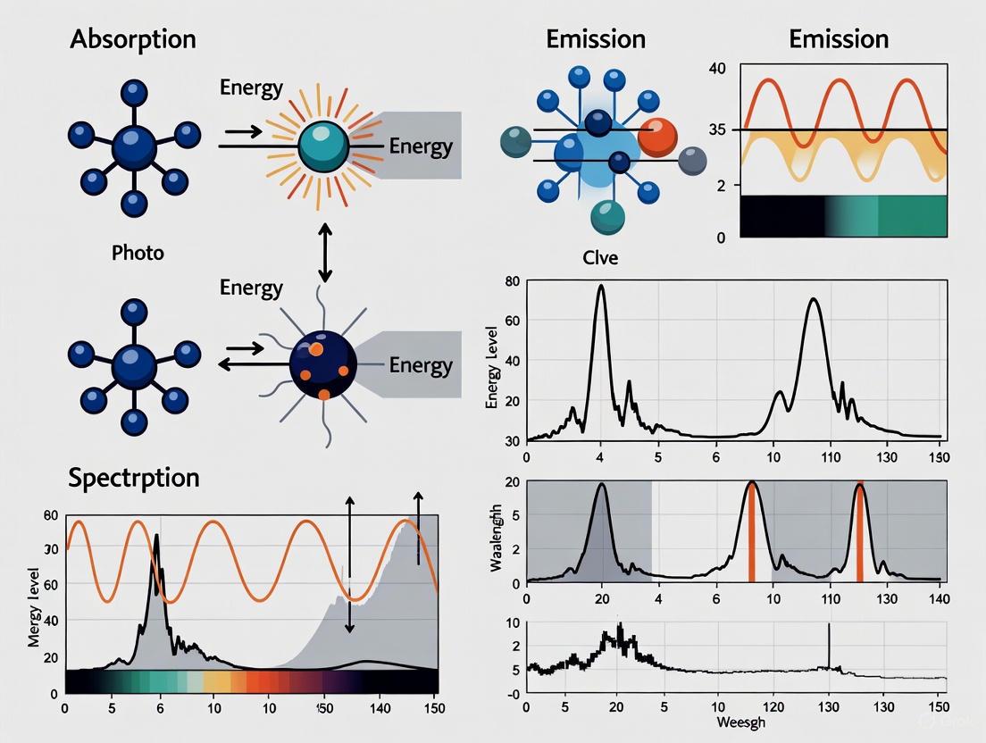
The interaction of light with matter constitutes the fundamental basis of spectroscopic analysis, providing critical insights into molecular structure, dynamics, and composition. Within the broader context of light-matter interaction in spectroscopy research, three primary mechanisms—absorption, emission, and scattering—govern how energy is exchanged between photons and materials. These processes enable researchers to decode the intricate energy-level structures of atoms and molecules, facilitating advances across scientific disciplines from drug development to materials science [14]. Understanding these core mechanisms is indispensable for interpreting spectroscopic data and developing innovative analytical methodologies for research applications.
This technical guide examines the fundamental principles, theoretical frameworks, and experimental manifestations of absorption, emission, and scattering processes. By establishing a coherent foundation of these interaction mechanisms, scientists can better leverage spectroscopic techniques to address complex analytical challenges in chemical research and pharmaceutical development.
Theoretical Foundations
Fundamental Principles
The interaction between light and matter occurs through quantized energy exchanges, wherein molecules transition between discrete energy states. When a molecule interacts with electromagnetic radiation, it may undergo changes in its electronic, vibrational, or rotational states through the absorption or emission of photons [15]. The specific energy transitions are dictated by the quantum mechanical properties of the system, with each transition corresponding to a precise energy difference between initial and final states [16].
The energy of electromagnetic radiation is inversely proportional to its wavelength, making different spectral regions sensitive to distinct molecular processes. Ultraviolet and visible radiation typically induce electronic transitions, infrared radiation corresponds to vibrational changes, and microwave radiation activates rotational modifications [16]. These energy-dependent interactions form the basis for various spectroscopic techniques that probe different molecular properties and characteristics.
Energy Transfer and Spectral Characteristics
The distinct nature of absorption, emission, and scattering processes produces characteristically different spectral signatures that convey specific molecular information. Table 1 summarizes the key spectral characteristics of each interaction mechanism.
Table 1: Spectral Characteristics of Light-Matter Interaction Mechanisms
| Interaction Mechanism | Spectral Pattern | Energy Transfer | Intensity Dependence |
|---|---|---|---|
| Absorption | Discrete peaks corresponding to specific molecular transitions [15] | Involves energy transfer from radiation to molecule [15] | Proportional to population of lower energy state [15] |
| Emission | Discrete peaks corresponding to specific molecular transitions [15] | Involves energy transfer from molecule to radiation [15] | Proportional to population of higher energy state [15] |
| Scattering | Generally continuous and less structured [15] | No net energy transfer (elastic) or modified energy transfer (inelastic) [15] | Depends on molecular polarizability and concentration [15] |
Absorption and emission spectra typically display discrete, well-defined peaks that correspond to specific quantum mechanical transitions between molecular energy states. In contrast, scattering spectra generally exhibit continuous, less structured profiles that reflect the distribution of energy modifications during photon-molecule interactions [15]. The intensity of absorbed or emitted radiation follows Boltzmann distribution statistics, depending fundamentally on the population of molecules in the initial energy state preceding the transition.
Absorption Processes
Quantum Mechanical Basis
Absorption occurs when a molecule incorporates energy from incident electromagnetic radiation, promoting itself from a lower energy state to a higher energy state. This transition activates when the energy of the incoming photons precisely matches the energy difference between two quantum states of the molecule [15]. The probability of absorption is governed by the transition dipole moment, a quantum mechanical property that depends on the change in the electronic, vibrational, or rotational configuration of the molecule during the transition [15].
The absorption process follows the Beer-Lambert law, which quantitatively relates the absorption of light to the properties of the material through which the light is passing. This fundamental relationship enables the determination of substance concentration in analytical applications, making absorption spectroscopy an indispensable quantitative tool in chemical analysis [16].
Absorption Spectroscopy Techniques
Absorption spectroscopy encompasses diverse techniques across the electromagnetic spectrum, each targeting specific molecular transitions and providing unique analytical capabilities. Table 2 outlines the primary absorption spectroscopy methods and their respective applications.
Table 2: Absorption Spectroscopy Techniques and Applications
| Technique | Spectral Region | Transition Type | Primary Applications |
|---|---|---|---|
| UV-Vis Spectroscopy | Ultraviolet-Visible (200-800 nm) | Electronic transitions [16] | Determination of conjugated systems, aromatic compounds, and chromophores [14] |
| IR Spectroscopy | Infrared (0.8-1000 μm) | Vibrational transitions [16] | Functional group identification, molecular structure determination [17] |
| X-ray Absorption Spectroscopy | X-ray (0.01-10 nm) | Inner shell electron excitation [16] | Elemental analysis, oxidation state determination |
| Microwave Spectroscopy | Microwave (1 mm-1 m) | Rotational transitions [16] | Molecular geometry determination, bond length precision |
The absorption spectrum of a material reveals its electronic and molecular composition, as absorption lines occur at frequencies that match energy differences between quantum states [16]. The positions, intensities, and widths of these absorption lines provide detailed information about the molecular structure, including functional groups, chemical environment, and intermolecular interactions.
Emission Processes
Emission Mechanisms
Emission processes involve the release of electromagnetic radiation from molecules transitioning from higher energy states to lower energy states. Two distinct emission mechanisms occur in molecular systems: spontaneous emission and stimulated emission.
Spontaneous emission occurs when a molecule in an excited state spontaneously decays to a lower energy state, releasing a photon with energy corresponding to the difference between the two states [15]. This process happens naturally without external influence, with the emitted photon possessing random phase and direction.
Stimulated emission takes place when an incident photon interacts with a molecule already in an excited state, inducing the emission of a second photon identical in energy, phase, and direction to the incident photon [15]. This process forms the fundamental basis of laser operation, enabling the amplification of coherent light.
The following diagram illustrates the fundamental emission mechanisms and their relationship to molecular energy states:
Emission Spectroscopy Applications
Emission spectroscopy leverages the characteristic radiation emitted by excited molecules to determine chemical composition and quantify substances. When molecules are excited by thermal, electrical, or optical energy, they emit radiation at specific wavelengths that form unique spectral fingerprints, enabling precise identification of elements and compounds.
In analytical chemistry, emission techniques such as fluorescence spectroscopy and laser-induced breakdown spectroscopy (LIBS) provide exceptional sensitivity for trace analysis and elemental characterization [18]. These methods are particularly valuable in pharmaceutical research for studying drug-receptor interactions, monitoring metabolic processes, and detecting minute quantities of biomarkers in complex biological matrices.
Scattering Processes
Elastic Scattering
Elastic scattering occurs when incident light interacts with a molecule and is re-emitted at the same frequency, with no net energy exchange between the photon and the molecule. The most prevalent form of elastic scattering is Rayleigh scattering, where incident electromagnetic radiation causes molecular oscillation and re-emission at the identical frequency [15].
The intensity of Rayleigh scattering exhibits a strong dependence on wavelength, proportional to the inverse fourth power of the wavelength (I ∝ 1/λ⁴) [15]. This wavelength dependency explains why shorter wavelengths (blue/violet light) are scattered more efficiently in the atmosphere, creating the blue appearance of the sky. Rayleigh scattering represents the dominant scattering mechanism for particles significantly smaller than the wavelength of incident light.
Inelastic Scattering
Inelastic scattering processes involve energy exchange between incident photons and molecules, resulting in scattered radiation with modified frequency. The primary inelastic scattering phenomenon is Raman scattering, which occurs when incident light interacts with a molecule, inducing a transition to a different vibrational or rotational state and re-emitting radiation at a shifted frequency [15] [19].
Raman scattering encompasses two distinct processes:
- Stokes Raman scattering: The scattered radiation has lower frequency (longer wavelength) than the incident radiation, corresponding to the molecule transitioning to a higher vibrational or rotational state [15] [19].
- Anti-Stokes Raman scattering: The scattered radiation has higher frequency (shorter wavelength) than the incident radiation, corresponding to the molecule transitioning from a higher to a lower vibrational or rotational state [15] [19].
Stokes Raman scattering is significantly more intense than anti-Stokes scattering at standard temperatures because most molecules initially reside in the ground vibrational state, as described by Boltzmann distribution statistics [19].
The following workflow diagram illustrates the experimental process for Raman spectroscopy, highlighting key scattering mechanisms:
Advanced Scattering Techniques
Brillouin scattering represents another inelastic scattering process involving the interaction of electromagnetic radiation with acoustic phonons (collective vibrational modes) in materials [15]. This interaction produces small frequency shifts determined by the velocity of acoustic phonons and the incident radiation wavelength, providing valuable information about elastic properties and sound velocities in materials.
Surface-Enhanced Raman Spectroscopy (SERS) utilizes metallic nanostructures to amplify local electromagnetic fields, dramatically increasing Raman scattering signals by several orders of magnitude [19]. This enhancement enables single-molecule detection and expands Raman applications to trace analysis, surface science, and biological sensing where conventional Raman signals would be undetectable.
Experimental Methodologies
Absorption Spectroscopy Protocol
Objective: Determine the absorption spectrum of a sample to identify chemical composition and quantify concentration.
Materials and Methods:
- Radiation Source: Select appropriate broadband source (e.g., deuterium lamp for UV, tungsten lamp for visible, globar for IR) [16]
- Monochromator/Spectrometer: Disperse light into component wavelengths
- Sample Chamber: Hold sample in appropriate container (cuvette, ATR crystal, gas cell)
- Detector: Measure transmitted light intensity (photodiode, PMT, bolometer)
- Reference Standard: Use for background correction
Procedure:
- Collect reference spectrum (I₀) without sample or with blank solvent
- Introduce sample into the light path
- Measure transmitted light intensity (I) across wavelength range
- Calculate absorbance: A = -log₁₀(I/I₀)
- Plot absorbance versus wavelength to generate absorption spectrum
- Apply Beer-Lambert law for quantitative analysis: A = εlc, where ε is molar absorptivity, l is path length, and c is concentration
Data Analysis: Identify characteristic absorption peaks, correlate with known transitions, determine sample composition, and calculate concentrations using established calibration curves.
Raman Spectroscopy Protocol
Objective: Obtain Raman spectrum to determine molecular structure and identify chemical compounds based on vibrational fingerprints.
Materials and Methods:
- Laser Source: Monochromatic light source (e.g., 532 nm, 785 nm lasers) [19]
- Sample Platform: Stable mounting for solid, liquid, or gaseous samples
- Filter System: Bandpass filter for laser line cleaning, longpass filter for Rayleigh rejection [19]
- Spectrometer: High-resolution instrument with diffraction grating
- Detector: CCD for visible region, InGaAs for NIR region [19]
Procedure:
- Align laser excitation source to optimally illuminate sample
- Collect scattered light at 90° or 180° geometry relative to excitation
- Filter collected light to remove elastically scattered Rayleigh component
- Disperse inelastically scattered light using spectrometer
- Detect Raman signal with appropriate detector
- Record spectrum of intensity versus Raman shift (cm⁻¹)
Data Analysis: Identify characteristic Raman shifts, assign vibrational modes, compare with reference spectra for compound identification, and determine molecular symmetry and structure.
Research Reagent Solutions
Successful spectroscopic analysis requires specific materials and reagents tailored to each technique. Table 3 details essential research reagents and their functions in spectroscopic experiments.
Table 3: Essential Research Reagents for Spectroscopy Experiments
| Reagent/Material | Function | Application Examples |
|---|---|---|
| Monochromator | Wavelength selection and dispersion [16] | Isolation of specific wavelengths in absorption spectroscopy |
| ATR Crystals (ZnSe, Diamond) | Internal reflection element for sample contact | FTIR sampling of solids, liquids, and gels without preparation |
| Spectroscopic Solvents (CDCl₃, DMSO-d₆) | NMR-compatible solvents with deuterated isotopes | Solubilization of samples for NMR analysis without interfering signals |
| Bandpass Filters | Laser line cleaning [19] | Removal of unwanted wavelengths from laser source in Raman spectroscopy |
| Longpass Filters | Rayleigh line rejection [19] | Blocking of elastically scattered light in Raman detection |
| Reference Standards | Instrument calibration and quantification | Creation of calibration curves for quantitative analysis |
| Metallic Nanoparticles (Au, Ag) | Signal enhancement substrates | Surface-enhanced Raman spectroscopy (SERS) for trace detection |
Comparative Analysis of Interaction Mechanisms
Selection Guidelines for Analytical Applications
Choosing the appropriate spectroscopic technique depends on the analytical requirements, sample characteristics, and information objectives. Table 4 provides a comparative framework for selecting interaction mechanisms based on analytical needs.
Table 4: Technique Selection Guide for Analytical Applications
| Analytical Requirement | Recommended Technique | Key Advantages | Limitations |
|---|---|---|---|
| Quantitative Concentration Measurement | UV-Vis Absorption Spectroscopy | Simple operation, high precision, wide linear dynamic range | Limited structural information, potential spectral overlap |
| Functional Group Identification | Infrared Absorption Spectroscopy | Extensive spectral libraries, non-destructive, minimal sample prep | Limited sensitivity for trace analysis, water interference |
| Molecular Structure Elucidation | NMR Spectroscopy | Detailed atomic-level structural information, quantitative capability | Expensive instrumentation, relatively low sensitivity |
| Chemical Fingerprinting | Raman Scattering Spectroscopy | Minimal sample preparation, aqueous compatibility, spatial mapping | Fluorescence interference, inherently weak signal |
| Trace Analysis | Surface-Enhanced Raman Spectroscopy | Exceptional sensitivity, single-molecule detection, multiplex capability | Complex substrate preparation, potential reproducibility issues |
Complementary Information from Multiple Techniques
Integrating multiple spectroscopic approaches often provides comprehensive molecular understanding that surpasses the capabilities of individual techniques. For example, combining IR and Raman spectroscopy offers complementary vibrational information, as IR absorption requires a change in dipole moment while Raman scattering depends on polarizability changes during molecular vibrations [15] [17]. This complementary nature allows complete assignment of vibrational modes and enhanced molecular structure determination.
Similarly, correlation of UV-Vis electronic transition data with NMR structural information enables researchers to establish structure-property relationships for novel compounds. Such multi-technique approaches are particularly valuable in pharmaceutical research for characterizing active pharmaceutical ingredients (APIs), studying drug-polymer interactions in formulations, and monitoring chemical reactions in real time.
Emerging Applications in Drug Development
Spectroscopic techniques leveraging absorption, emission, and scattering mechanisms play increasingly vital roles in modern pharmaceutical research and development. Confocal Raman microscopy provides label-free chemical imaging of pharmaceutical formulations, enabling visualization of active ingredient distribution within solid dosage forms without destructive sample preparation [19]. This capability is invaluable for optimizing manufacturing processes and ensuring product quality.
UV-Vis absorption spectroscopy remains fundamental for solubility studies, dissolution testing, and pharmacokinetic analysis, while fluorescence spectroscopy offers exceptional sensitivity for monitoring protein-ligand interactions and conformational changes in biopharmaceutical characterization. Additionally, the non-destructive nature of Raman spectroscopy enables counterfeit drug identification through packaging, supporting regulatory compliance and patient safety initiatives [19].
These applications demonstrate how fundamental light-matter interaction mechanisms translate into practical analytical solutions that accelerate drug development, enhance quality control, and advance therapeutic innovation. As spectroscopic technologies continue evolving with improved sensitivity, miniaturization, and computational integration, their impact on pharmaceutical research will undoubtedly expand, enabling new approaches to complex analytical challenges.
Quantum transitions between discrete energy states are the fundamental mechanism by which matter interacts with light, creating unique spectral fingerprints that form the basis of spectroscopy. This whitepaper delineates the quantum mechanical principles governing these transitions, presents a contemporary experimental breakthrough in detecting weak transitions, and provides detailed methodologies for their study. Framed within the broader context of light-matter interaction, this guide serves as a technical resource for researchers and drug development professionals in deploying spectroscopic techniques for material and molecular analysis.
Spectroscopy, the scientific study of the interaction between light and matter, is predicated on the quantum mechanical principle that atoms and molecules can only exist in specific, discrete energy states [6]. A quantum transition occurs when a particle absorbs or emits a photon, causing it to move between these energy levels. The energy of the photon must exactly match the energy difference between the two states, as described by the Bohr frequency condition: ΔE = hν, where ΔE is the energy difference, h is Planck's constant, and ν is the frequency of the photon [6].
Every element and molecule possesses a unique set of allowed energy levels, dictated by its chemical structure, electronic configuration, and nuclear properties. When photons are absorbed or emitted at the characteristic frequencies corresponding to these energy differences, they produce a pattern of lines or bands—a spectral fingerprint—that serves as a unique identifier for the substance [6]. The strength of a transition is governed by its transition matrix element, a quantum mechanical parameter that determines the probability of the transition occurring. Transitions are categorized as "allowed" (strong) or "forbidden" (weak) based on selection rules derived from quantum mechanics.
Breaking the Scaling Law: Enhancing Weak Transitions
A significant challenge in spectroscopy is the detection of weak transitions, which have small cross-sections due to their small transition matrix elements. These transitions are often buried in noise or obscured by stronger signals. Recent research has demonstrated a method to break the traditional scaling law, which states that the absorption cross-section (σ) is proportional to the absolute square of the transition matrix element (∣T∣²) [20].
Theoretical Concept of Enhancement
The conventional approach is described by the optical theorem: σ ∝ Im(A), where for a resonance in the linear regime, the forward scattering amplitude A is proportional to ∣T∣² [20]. The new concept introduces an additional, stronger laser-coupled pathway to the same excited state. In the presence of this intense light, the response function modifies to à ∝ T ˣ (T + T ′), where T ′ represents the contribution from the additional pathway [20]. For a weakly coupled state, if T ′ can be made much larger than T, the spectral visibility of the weak transition can be significantly enhanced, effectively boosting its transition probability [20].
Experimental Demonstration in Helium
This enhancement concept was experimentally validated using attosecond transient absorption spectroscopy in helium atoms [20]. The experiment targeted the quasi-forbidden weak transitions from the ground state (1s²) to the doubly excited 2p3d and sp²,4⁻ states. The transition probability for these states is orders of magnitude lower than for the strongly coupled 2s2p state [20].
The methodology involved:
- Light Sources: A weak, broadband extreme-ultraviolet (XUV) pulse generated via high-harmonic generation in neon, and a stronger, few-cycle visible (VIS) pulse centered at 700 nm [20].
- Beam Path: The VIS and XUV pulses propagated collinearly and were focused into a helium-filled gas cell. A variable time delay (τ) was introduced between the two pulses [20].
- Detection: The transmitted XUV radiation was dispersed by a grating and detected by a CCD camera. The absorption spectrum was characterized by the optical density, OD(ω, τ) = -log₁₀[ I(ω, τ) / I₀(ω) ], where I and I₀ are the transmitted and incident XUV spectra, respectively [20].
In the absence of the VIS pulse, the weak 2p3d and sp²,4⁻ transitions were barely visible. When the VIS pulse was applied, it strongly coupled the 2s2p state (populated by the XUV) to the target 2p3d and sp²,4⁻ states via a two-VIS-photon pathway through the 2p² intermediate state. This coupling transferred quantum-state amplitude, boosting the spectral signal of the weak transitions by an order of magnitude and making their relative spectral amplitude comparable to neighboring strong lines [20].
Quantitative Data on Helium Doubly Excited States
The table below summarizes key quantitative data for the doubly excited states in helium discussed in the experimental demonstration, illustrating the relative strengths of different transitions [20].
Table 1: Transition Strengths of Selected Helium Doubly Excited States
| State Series | Example State | Energy (eV) | Relative Transition Strength from Ground State |
|---|---|---|---|
| sp²,n+ | sp²,3+ | ~63.7 | Strong |
| sp²,n- | sp²,4- | ~64.1 | Weak (Quasi-forbidden) |
| 2pnd | 2p3d | ~64.1 | Weak (Quasi-forbidden) |
Note: The 2p3d and sp²,4⁻ states are nearly degenerate, with an energy spacing of less than 20 meV, making them indistinguishable in the reported measurement [20].
Detailed Experimental Protocol: Attosecond Transient Absorption
This protocol details the methodology for enhancing and measuring weak quantum transitions, as demonstrated in [20].
Experimental Workflow
Step-by-Step Methodology
Beam Preparation and Delay
- Generate a broadband XUV pulse via high-harmonic generation (HHG) in a noble gas target (e.g., Neon) driven by an intense infrared laser [20].
- Split the fundamental laser beam to generate a few-cycle visible (VIS) or near-infrared pulse.
- Use a high-precision, piezo-driven split mirror or delay stage to introduce a variable time delay (τ) between the XUV and VIS pulses. The convention is that a positive τ means the XUV pulse arrives first [20].
Sample Interaction
- Recombine the XUV and VIS pulses collinearly and focus them into a gas cell or jet containing the sample atoms or molecules (e.g., Helium at ~200 mbar backing pressure) [20].
- The weak XUV pulse preferentially excites the system from the ground state to various excited states, including both strongly and weakly coupled states.
- The delayed, intense VIS pulse couples these excited states, transferring population from the strongly coupled state (e.g., 2s2p in He) to the weakly coupled state (e.g., 2p3d) via a resonant multi-photon pathway [20].
Spectral Detection
- The transmitted XUV spectrum, I(ω, τ), is dispersed using a high-resolution grating spectrometer [20].
- The dispersed light is recorded by a CCD camera as a function of photon energy (ω) and time delay (τ) [20].
- A reference spectrum, I₀(ω), is recorded without the sample or without the VIS pulse to calculate the optical density: OD(ω, τ) = -log₁₀[ I(ω, τ) / I₀(ω) ] [20].
Data Analysis
- Analyze the OD(ω, τ) map to identify spectral features that emerge or are enhanced when the XUV and VIS pulses overlap in time (τ ≈ 0).
- The enhancement of a weak transition manifests as a pronounced absorption peak or a window-type resonance (a spectral dip) that appears only during temporal overlap and persists for a duration comparable to the lifetime of the involved states [20].
The Scientist's Toolkit: Essential Research Reagents and Materials
Table 2: Key Reagents and Materials for Advanced Spectroscopic Experiments
| Item | Function / Role in Experiment |
|---|---|
| Ultrafast Laser System | Primary source for generating both the pump and probe pulses. Typically a Ti:Sapphire laser producing femtosecond pulses at near-infrared wavelengths (e.g., ~800 nm). |
| High-Harmonic Generation (HHG) Source | A gas target (e.g., Neon, Argon) where the intense laser is converted to coherent XUV pulses through non-linear interaction. |
| Gas Cell / Jet | Contains the atomic or molecular sample under study (e.g., Helium gas). Ensures a uniform density of target particles in the interaction region. |
| Monochromator / Spectrometer | Disperses the broad-band, transmitted light after interaction with the sample, allowing wavelength-resolved detection. |
| CCD Camera | Detects the dispersed light, recording the intensity as a function of photon energy to construct the absorption spectrum. |
| High-Precision Delay Stage | A piezo-actuated optical stage that controls the path length of one beam, enabling sub-femtosecond precision in the time delay between pump and probe pulses. |
Implications for Research and Industry
The ability to enhance weak transitions has profound implications. In fundamental physics, it enables precision tests of quantum mechanics and the study of exotic, correlated electron states [20]. For drug development and the life sciences, this methodology can be applied to boost the spectral visibility of weak but functionally critical transitions in complex biomolecules, such as proteins and nucleic acids, improving diagnostic capabilities and the understanding of molecular interactions [20]. Furthermore, the general principle of controlling quantum pathways opens avenues for manipulating chemical reactions and energy transfer processes at the quantum level.
The Beer-Lambert Law is a fundamental principle in optical spectroscopy that provides a quantitative relationship between the attenuation of light through a substance and the properties of that substance [21]. This law, unquestionably the most important law in optical spectroscopy, is indispensable for the qualitative and quantitative interpretation of spectroscopic data [22]. Its development spans centuries, beginning with the work of Pierre Bouguer in 1729, who discovered the exponential decay of light intensity during his astronomical observations of the atmosphere [23] [22]. Johann Heinrich Lambert later formalized this mathematical relationship in his 1760 work Photometria, establishing that the loss of light intensity when propagating through a medium is directly proportional to both the intensity and the path length [23]. The law was completed in 1852 when August Beer extended the concept to include the concentration of colored solutions, noting that transmittance remained constant provided the product of concentration and path length stayed constant [23] [22].
The modern formulation of the law, which merges these contributions into the absorbance equation we use today, was first presented by Robert Luther in 1913 [22]. This law serves as a cornerstone in the broader thesis of light-matter interaction, providing researchers across chemistry, biology, pharmaceutical development, and environmental science with a critical tool for quantifying molecular species in solution [24]. Despite its widespread utility, modern research continues to explore the boundaries of this fundamental law, particularly through the lens of electromagnetic theory, which reveals significant limitations and opportunities for refinement in advanced spectroscopic applications [25] [22] [24].
Theoretical Foundation
Fundamental Concepts of Light Attenuation
When monochromatic light passes through a solution-containing cuvette, several physical processes occur that reduce the intensity of the transmitted light. The incident light with intensity (I_0) undergoes attenuation primarily through absorption by the solute molecules, though scattering and reflection at interfaces also contribute to reduced transmission [21] [26]. The fundamental quantities describing this attenuation are:
- Transmittance (T): The fraction of incident light that passes through the sample, defined as (T = I/I0), where (I) is the transmitted intensity [21] [27]. It is often expressed as a percentage: (\%T = (I/I0) \times 100\%) [21].
- Absorbance (A): A dimensionless quantity defined as the negative logarithm of transmittance: (A = -\log{10}T = \log{10}(I_0/I)) [21] [27]. This logarithmic relationship means that each unit increase in absorbance corresponds to a tenfold decrease in transmittance [21].
Table 1: Relationship Between Absorbance and Transmittance
| Absorbance (A) | Transmittance (T) | % Transmittance |
|---|---|---|
| 0 | 1 | 100% |
| 0.3 | 0.5 | 50% |
| 1 | 0.1 | 10% |
| 2 | 0.01 | 1% |
| 3 | 0.001 | 0.1% |
Mathematical Formulation
The Beer-Lambert Law establishes a linear relationship between absorbance and both the concentration of the absorbing species and the path length through the solution [21] [27] [23]. The standard mathematical form is:
[A = \varepsilon \cdot c \cdot l]
Where:
- (A) is the measured absorbance (dimensionless)
- (\varepsilon) is the molar absorptivity or molar extinction coefficient (typically in L·mol⁻¹·cm⁻¹)
- (c) is the concentration of the absorbing species (in mol·L⁻¹ or M)
- (l) is the optical path length through the sample (in cm) [21] [27] [28]
The molar absorptivity ((\varepsilon)) is a substance-specific property that measures how strongly a chemical species absorbs light at a particular wavelength [28] [29]. A higher (\varepsilon) value indicates a greater probability of electronic transitions and thus stronger absorption [26].
For systems with multiple absorbing species, the law becomes:
[A = l \sumi \varepsiloni c_i]
where the total absorbance equals the sum of contributions from all absorbing components [23].
Diagram 1: Fundamental relationships in the Beer-Lambert Law showing how sample properties affect light attenuation.
Derivation from First Principles
The Beer-Lambert Law can be derived by considering the differential attenuation of light passing through an infinitesimally thin layer of absorbing medium [30] [23] [28]. For a monochromatic light beam traversing a thickness (dx) of a solution containing concentration (c) of absorbing species, the decrease in intensity (dI) is proportional to the incident intensity (I), the path length (dx), and the concentration (c):
[ -\frac{dI}{dx} = \alpha \cdot I \cdot c ]
Where (\alpha) is the proportionality constant representing the absorption characteristics [30] [26]. Rearranging and integrating both sides:
[ \int{I0}^{I} \frac{dI}{I} = -\alpha c \int_{0}^{l} dx ]
[ \ln\left(\frac{I}{I_0}\right) = -\alpha c l ]
Converting from natural logarithm to base-10 logarithm:
[ \log{10}\left(\frac{I0}{I}\right) = \frac{\alpha}{2.303} c l ]
Substituting (A = \log{10}(I0/I)) and (\varepsilon = \alpha/2.303) yields the familiar form:
[ A = \varepsilon c l ]
This derivation assumes that: (1) the light is monochromatic, (2) the absorbing species act independently, (3) the solution is homogeneous, (4) the incident radiation is parallel and perpendicular to the surface, and (5) the concentration is sufficiently low to avoid molecular interactions [30] [26].
Practical Implementation in Research
Essential Equipment and Reagents
Table 2: Essential Research Reagents and Equipment for Beer-Lambert Law Applications
| Item | Function/Description | Typical Specifications |
|---|---|---|
| Spectrophotometer | Instrument for measuring light intensity before and after sample interaction [21] [28] | UV-Vis range (190-1100 nm); monochromator or diode array detector |
| Cuvettes | Containers for holding liquid samples during measurement [21] | Path length: 1 cm (standard); material: quartz (UV), glass (Vis) |
| Molar Extinction Coefficient Reference Materials | Substances with known ε values for calibration and verification | Holmium oxide filters (wavelength accuracy) [24]; potassium dichromate (absorbance standards) |
| Chemical Standards | High-purity compounds for preparing calibration solutions | Potassium permanganate, methyl orange, copper sulfate [24] |
| Solvent Systems | Chemically inert media for dissolving analytes | Distilled water, spectral-grade organic solvents [24] |
Experimental Protocol for Concentration Determination
The primary application of the Beer-Lambert Law in research involves determining unknown concentrations of solutions through spectrophotometric measurement [21]. The following protocol provides a detailed methodology:
Solution Preparation: Prepare a series of standard solutions with known concentrations of the analyte, typically using serial dilution techniques. Ensure the concentration range produces absorbance values between 0.1 and 1.0 AU for optimal accuracy [21] [27].
Spectrophotometer Calibration:
- Turn on the instrument and allow it to warm up for 15-30 minutes.
- Perform wavelength accuracy verification using a holmium oxide filter with known absorption peaks at 361 nm, 445 nm, and 460 nm [24].
- Set the desired analytical wavelength based on the analyte's absorption maximum.
Blank Measurement:
- Fill a cuvette with the pure solvent (without analyte).
- Place in the sample compartment and set the instrument to 100% transmittance (zero absorbance).
Standard Curve Generation:
- Measure the absorbance of each standard solution at the predetermined wavelength.
- Record both concentration and absorbance values in tabular form.
- Plot absorbance versus concentration and perform linear regression to obtain the calibration curve [21].
Unknown Sample Measurement:
- Measure the absorbance of the unknown solution under identical conditions.
- Calculate the concentration using the equation from the linear regression: (c = A/\varepsilon l).
Quality Control:
- Verify measurement precision through replicate analyses (typically n=3).
- Analyze a certified reference material if available to assess accuracy.
Diagram 2: Experimental workflow for quantitative analysis using the Beer-Lambert Law.
Advanced Application: Multi-Component Analysis
For systems containing multiple absorbing species with overlapping absorption bands, the Beer-Lambert Law can be extended through matrix algebra [23]. The total absorbance at wavelength (i) is:
[Ai = l \sum{j=1}^n \varepsilon{ij} cj]
Where (\varepsilon_{ij}) is the molar absorptivity of component (j) at wavelength (i). Measurements at multiple wavelengths ((i = 1, 2, ..., m)) yield a system of equations:
[\begin{bmatrix}A1 \ A2 \ \vdots \ Am\end{bmatrix} = l \begin{bmatrix}\varepsilon{11} & \varepsilon{12} & \cdots & \varepsilon{1n} \ \varepsilon{21} & \varepsilon{22} & \cdots & \varepsilon{2n} \ \vdots & \vdots & \ddots & \vdots \ \varepsilon{m1} & \varepsilon{m2} & \cdots & \varepsilon{mn}\end{bmatrix} \begin{bmatrix}c1 \ c2 \ \vdots \ c_n\end{bmatrix}]
This approach requires prior knowledge of the molar absorptivity matrix, typically determined by measuring pure standards of each component at all analytical wavelengths.
Limitations and Modern Theoretical Refinements
Fundamental Limitations of the Classical Law
Despite its widespread utility, the Beer-Lambert Law has several significant limitations that researchers must recognize:
High Concentration Effects: At elevated concentrations (typically >0.01 M), the average distance between absorbing molecules decreases, leading to electrostatic interactions that alter absorption characteristics [30] [25] [24]. The refractive index of the solution may also change significantly with concentration, violating one of the law's fundamental assumptions [25] [24].
Chemical Deviations: Molecular associations such as dimerization or complex formation at higher concentrations change the absorption spectrum [25]. Shifts in chemical equilibrium due to changes in pH, temperature, or solvent composition can also cause deviations [24].
Instrumental Deviations: The use of polychromatic light sources causes deviations because molar absorptivity varies with wavelength [25] [29]. Stray light reaching the detector without passing through the sample similarly violates the law's assumptions [25].
Electromagnetic Effects: The classical derivation neglects the wave nature of light, including interference effects that become significant in thin films or at interfaces between media with different refractive indices [25] [22]. Multiple reflections in cuvettes with parallel windows can create etalon effects that distort measurements [25].
Table 3: Common Limitations and Practical Solutions
| Limitation Type | Underlying Cause | Practical Mitigation Strategies |
|---|---|---|
| Fundamental Deviations | High concentration effects; refractive index changes | Dilute samples to <0.01 M; use shorter path length cuvettes |
| Chemical Deviations | Molecular interactions; equilibrium shifts | Control pH, temperature; use chemical buffers |
| Instrumental Deviations | Polychromatic light; stray light; detector nonlinearity | Use narrow bandwidth; double-beam instruments; regular calibration |
| Electromagnetic Effects | Interference; scattering; reflection losses | Use non-parallel cuvettes; index-matching techniques |
Electromagnetic Theory Refinements
Recent research has addressed the limitations of the classical Beer-Lambert Law through electromagnetic theory, which provides a more rigorous foundation for light-matter interactions [22] [24]. The classical law assumes light propagates as rays rather than waves, neglecting polarization effects and the complex refractive index.
The electromagnetic approach begins with the complex refractive index:
[\hat{n} = n + ik]
Where (n) is the real part (governing refraction) and (k) is the imaginary part (governing absorption). The absorption coefficient (\alpha) relates to (k) through:
[k = \frac{\alpha}{4\pi\nu}]
Where (\nu) is the wavenumber. For dilute solutions, the refractive index can be approximated as:
[n \approx 1 + c\frac{NA\alpha'}{2\epsilon0}]
Where (\alpha') is the polarizability and (N_A) is Avogadro's number. This leads to:
[k \approx \beta c]
Which recovers the classical Beer-Lambert Law at low concentrations [24]. However, at higher concentrations, higher-order terms become significant:
[k = \beta c + \gamma c^2 + \delta c^3]
This results in a modified absorbance expression:
[A = \frac{4\pi\nu}{\ln 10}(\beta c + \gamma c^2 + \delta c^3)l]
This electromagnetic extension successfully models the non-linear behavior observed at high concentrations, where molecular interactions and local field effects become significant [24]. Experimental validation with potassium permanganate, potassium dichromate, methyl orange, copper sulfate, and iron chloride solutions demonstrates superior performance compared to the classical law, with root mean square errors below 0.06 for all tested materials [24].
Applications in Pharmaceutical Research and Development
The Beer-Lambert Law finds extensive application in drug development, where precise quantification of chemical compounds is essential throughout the research, development, and manufacturing processes:
API Quantification: Determination of active pharmaceutical ingredient (API) concentration in bulk solutions and formulated products using validated spectrophotometric methods [28].
Dissolution Testing: Monitoring the release profile of APIs from solid dosage forms by measuring concentration in dissolution media at specific time points [28].
Impurity Profiling: Detection and quantification of trace impurities and degradation products through differential spectrophotometry, often employing multi-wavelength analysis [28].
Biomolecule Analysis: Quantification of proteins, nucleic acids, and other biomolecules using their characteristic absorption bands (e.g., proteins at 280 nm, DNA at 260 nm).
Pharmacokinetic Studies: Measuring drug concentrations in biological fluids during ADME (absorption, distribution, metabolism, excretion) studies, often after appropriate sample preparation to eliminate matrix effects.
The robustness of Beer-Lambert-based methods makes them indispensable for quality control in pharmaceutical manufacturing, where they are incorporated into numerous pharmacopeial monographs and regulatory submission documents.
The Beer-Lambert Law remains a cornerstone of analytical spectroscopy, providing an essential link between absorbance measurements and chemical concentration. Its mathematical elegance and practical utility have ensured its continued relevance across diverse scientific disciplines for nearly three centuries. However, modern research has clearly delineated its limitations, particularly at high concentrations where electromagnetic effects and molecular interactions become significant.
The ongoing refinement of this fundamental law through electromagnetic theory represents an important advancement in spectroscopic science, offering more accurate models for quantitative analysis under non-ideal conditions. For researchers in drug development and other applied fields, understanding both the classical formulation and its modern extensions is crucial for designing robust analytical methods and correctly interpreting spectroscopic data.
As spectroscopic techniques continue to evolve, the Beer-Lambert Law will undoubtedly remain central to quantitative analysis while simultaneously serving as a foundation for developing more sophisticated models of light-matter interaction that push the boundaries of analytical science.
A Practical Guide to Spectroscopic Techniques in Drug Discovery and Development
Ultraviolet-Visible (UV-Vis) spectroscopy operates on the fundamental principle of light-matter interaction, specifically the absorption of electromagnetic radiation in the ultraviolet (190-400 nm) and visible (400-800 nm) regions by molecules [31] [32]. This absorption occurs when photons carrying specific amounts of energy interact with molecular electrons, promoting them from their ground state to higher energy excited states [32]. The energy of a photon is inversely proportional to its wavelength, meaning shorter ultraviolet wavelengths carry more energy than longer visible wavelengths, which directly influences which electronic transitions can be excited [33].
The technique measures this interaction quantitatively, providing data that can be used for both identification and concentration determination of analytes [31]. The specific wavelengths absorbed, and the extent of absorption, create a characteristic absorption spectrum that serves as a molecular fingerprint, influenced by the molecular structure, particularly the presence of chromophores—functional groups with electrons that can be excited at these energy levels [32]. When a compound absorbs light in the visible region, it appears to the human eye as the complementary color to the wavelength absorbed; for instance, absorption around 500-520 nm (green) makes a substance appear red [32].
Electronic Transitions and Spectral Interpretation
Types of Electronic Transitions
The absorption of UV or visible light energy promotes electrons in a molecule from the highest occupied molecular orbital (HOMO) to the lowest unoccupied molecular orbital (LUMO) [32]. The primary transitions involved for organic molecules are summarized in the table below.
Table 1: Common Electronic Transitions in UV-Vis Spectroscopy
| Transition Type | Electron Origin | Typical Wavelength Range | Example Chromophores | Molar Absorptivity (ε) |
|---|---|---|---|---|
| σ → σ* | Sigma bonding orbital | < 200 nm (Far UV) | C-C, C-H | High |
| n → σ* | Non-bonding orbital | 150 - 250 nm | H2O, CH3OH, CH3Cl | Medium (100-3000) |
| π → π* | Pi bonding orbital | 200 - 700 nm (Conjugated) | Alkenes, Carbonyls, Aromatics | High (10,000-250,000) |
| n → π* | Non-bonding orbital | 250 - 400 nm | C=O, NO2 | Low (10-100) |
For molecules with conjugated π-electron systems, the energy gap between the HOMO and LUMO decreases as the extent of conjugation increases [32]. This results in a bathochromic shift (shift to longer wavelength) and often a hyperchromic effect (increase in absorbance) [32]. For instance, while ethene absorbs at 171 nm, increasing conjugation in butadiene (λmax = 217 nm) and lycopene (λmax = 470 nm) shifts the absorption into the visible region, producing color [32].
Advanced Spectral Analysis Using the Pekarian Function
For detailed interpretation of complex spectra, advanced fitting functions beyond simple Gaussian or Lorentzian models are employed. The modified Pekarian Function (PF) is highly effective for fitting both absorption and fluorescence spectra with high accuracy, especially for conjugated organic compounds [34]. The PF for an absorption spectrum (PFa) is defined as:
$$PFa(ν) = \sum{k=0}^{n} [S^k \times G(1, νk, σ_0) / k!]$$
Where:
- S is the Huang-Rhys factor, representing the average number of vibration quanta dissipated.
- ν₀ is the wavenumber for the 0-0 transition.
- Ω is the wavenumber of the principal vibrational mode.
- σ₀ is the Gaussian broadening parameter.
- δ is a global correction for contributions from other vibrational modes.
- ν_k = ν₀ + kΩ for absorption.
- k = 0–8 is typically sufficient for accurate fitting [34].
This approach allows for the deconvolution of overlapping bands and provides optimized parameters that offer deeper insight into the electronic and vibrational structure of the molecule [34]. The weighted average transition energy can be calculated as 〈νge*〉 = ν₀ + Ω × S for comparison with quantum mechanical calculations like TD-DFT [34].
Instrumentation and Methodology
Core Components of a UV-Vis Spectrophotometer
A UV-Vis spectrophotometer consists of several key components that work in concert to measure light absorption accurately [33].
Table 2: Key Components of a UV-Vis Spectrophotometer
| Component | Function | Common Types & Notes |
|---|---|---|
| Light Source | Provides broad-spectrum UV and/or visible light. | Deuterium lamp (UV), Tungsten/Halogen lamp (Visible). Xenon lamps can cover both but are less stable [33]. |
| Wavelength Selector | Isolates specific, narrow wavelengths from the broad source. | Monochromators (most common, using diffraction gratings), Absorption filters, Interference filters [33]. |
| Sample Container | Holds the sample and reference for measurement. | Cuvettes (quartz for UV, glass/plastic for visible only). Cuvette-free systems exist for micro-samples [33]. |
| Detector | Measures the intensity of light transmitted through the sample. | Photomultiplier Tube (PMT, high sensitivity), Photodiodes, Charge-Coupled Devices (CCD) [33]. |
The most common instrument designs are single-beam and double-beam. A double-beam instrument splits the light from the source, directing one beam through the sample and the other through a reference blank, allowing for instantaneous comparison and more stable baseline correction [31].
Quantitative Analysis: The Beer-Lambert Law
The foundation of quantification in UV-Vis spectroscopy is the Beer-Lambert Law, which states that the absorbance (A) of a sample is directly proportional to its concentration (c) and the path length (l) of the light through the sample [31] [33].
The law is mathematically expressed as: A = ε * c * l
Where:
- A is the measured absorbance (no units).
- ε is the molar absorptivity or extinction coefficient (L mol⁻¹ cm⁻¹), a compound-specific constant at a given wavelength.
- c is the concentration of the analyte (mol L⁻¹).
- l is the path length of the cuvette (cm) [33].
The relationship between the intensities of incident light (I₀) and transmitted light (I) is defined as A = log₁₀(I₀/I) [33]. For accurate quantification, absorbance values should ideally be kept below 1 to remain within the instrument's linear dynamic range, which can be achieved by diluting the sample or using a shorter path length cuvette [33].
Experimental Protocols for Quantification and Purity Assessment
Protocol 1: Quantification of True-to-Life Nanoplastics
This protocol, adapted from recent research, details the use of microvolume UV-Vis for quantifying nanoplastics in suspension, highlighting its advantages for scarce samples [35].
1. Materials & Reagent Solutions:
- True-to-Life Nanoplastics: Generated from fragmented polystyrene items via mechanical fragmentation under cryogenic conditions and separated via sequential centrifugations [35].
- Solvent: MilliQ water for suspension [35].
- Reference: A blank of the suspension solvent (e.g., MilliQ water) [35].
- Instrument: Microvolume UV-Vis spectrophotometer.
2. Methodology:
- Sample Preparation: Prepare stock suspensions of nanoplastics. The protocol is particularly suited for samples with limited volume [35].
- Baseline Correction: Measure the reference (MilliQ water) to establish a baseline [35] [33].
- Sample Measurement: Load the nanoplastic suspension onto the microvolume instrument. The non-destructive nature of UV-Vis allows for sample recovery for subsequent analysis [35].
- Data Analysis: Record the absorption spectrum. Quantification is achieved by applying the Beer-Lambert law, using the absorbance value at a characteristic wavelength. While this method may slightly underestimate concentration compared to mass-based techniques like Py-GC/MS, it provides consistent results of the correct order of magnitude [35].
Protocol 2: Purity Assessment of Nucleic Acids (DNA/RNA)
UV-Vis spectroscopy is a standard method for the rapid verification of DNA and RNA sample purity and concentration [31].
1. Materials & Reagent Solutions:
- Analyte: Purified DNA or RNA sample in aqueous buffer.
- Solvent: The same buffer used to dilute or elute the nucleic acids (e.g., TE buffer).
- Reference: A cuvette containing the blank buffer solution.
- Instrument: Standard UV-Vis spectrophotometer, often with a cuvette-free option for micro-samples.
2. Methodology:
- Blank Measurement: Place the blank buffer in the instrument and perform a baseline correction.
- Sample Measurement: Introduce the nucleic acid sample and obtain an absorption spectrum from 220 nm to 350 nm.
- Data Analysis & Purity Calculation:
- Nucleic Acid Concentration: Use the absorbance at 260 nm (A₂₆₀). An A₂₆₀ of 1.0 corresponds to approximately 50 µg/mL for double-stranded DNA and 40 µg/mL for single-stranded RNA.
- Purity Ratios: Calculate the following ratios:
- A₂₆₀/A₂₈₀: Assesses protein contamination. A pure DNA sample has a ratio of ~1.8; pure RNA ~2.0.
- A₂₆₀/A₂₃₀: Assesses contamination from chaotropic salts or other compounds. A pure nucleic acid sample typically has a ratio of 2.0-2.2.
The Scientist's Toolkit: Essential Research Reagent Solutions
Table 3: Essential Reagents and Materials for UV-Vis Experiments
| Item | Function / Rationale | Technical Considerations |
|---|---|---|
| Quartz Cuvettes | Sample holder for UV-Vis measurements. | Quartz is transparent down to ~200 nm. Plastic and glass cuvettes are unsuitable for UV measurements as they absorb UV light [33]. |
| High-Purity Solvents | To dissolve and dilute the analyte. | Must be UV-transparent in the spectral region of interest. Common choices include water, hexane, methanol, and acetonitrile. The solvent can affect the spectrum (solvatochromism) [32]. |
| Standard White Reference Plate | For reflectance spectroscopy and color analysis. | Used with an integrating sphere for highly reproducible color evaluation of solid samples [36]. |
| Buffers (e.g., Phosphate Buffer) | To maintain a stable pH for analytes like biomolecules. | Prevents shifts in absorption maxima or shape that can occur with pH-sensitive chromophores. |
| Certified Reference Materials | For instrument calibration and method validation. | Ensures accuracy and compliance with regulatory standards, crucial in pharmaceutical analysis and quality control [31]. |
Applications in Research and Industry
The applications of UV-Vis spectroscopy are vast, spanning multiple scientific and industrial disciplines.
- Pharmaceutical Analysis: Used in drug discovery and development to identify chemical components, quantify impurities, and ensure product quality with minimal effect on the drug sample [31].
- Environmental Monitoring: Employed to detect and quantify contaminants in air, soil, and water. Its use in analyzing emerging pollutants like true-to-life nanoplastics supports environmental health risk assessments [35] [31].
- Food and Beverage Quality Control: Applied to measure the concentration of ingredients (e.g., caffeine) and additives to ensure compliance with labeling laws and quality standards [31].
- Color Analysis: Used with an integrating sphere to perform highly reproducible color measurements and calculate color coordinates (e.g., L*a*b*) for solid pellets and other materials, which is vital for product development and quality assurance [36].
- Biochemical Research: Essential for checking the purity and concentration of biomolecules like DNA, RNA, and proteins, which is a critical step before applications like sequencing or PCR [31].
UV-Vis spectroscopy remains a cornerstone analytical technique in modern laboratories due to its robust principle of light-matter interaction, versatility, and quantitative power. Its ability to probe electronic transitions provides critical insights for identification, quantification, and purity assessment across diverse fields from nanotechnology to pharmaceuticals. The technique's ongoing relevance is underscored by its adaptation to new challenges, such as the analysis of nanoplastics, and by advanced methods for spectral interpretation, ensuring its continued place in the scientist's analytical arsenal.
Infrared (IR) spectroscopy is a fundamental spectroscopic technique that probes the interaction of infrared light with matter, specifically through the absorption of photons that induce vibrational excitations in covalently bonded atoms and groups [37] [38]. The energy associated with the infrared region of the electromagnetic spectrum (typically studied from 2,500 to 16,000 nm) is not sufficient to excite electrons but is perfectly matched to the energy required to cause bonds to stretch, bend, and twist [39]. The resulting infrared spectrum provides a unique fingerprint of a molecular structure, making it an indispensable tool for the identification of chemical substances and the analysis of functional groups in solid, liquid, and gaseous forms [37] [38].
The interaction is governed by a key selection rule: for a vibrational mode to be "IR active," it must be associated with a change in the dipole moment of the molecule [38]. This principle is central to understanding why certain vibrations appear in an IR spectrum while others do not. For example, symmetrical diatomic molecules like N₂ do not absorb IR radiation, whereas asymmetrical molecules like CO do [38]. The analysis of these vibrational modes allows researchers to deduce critical structural information, playing a vital role in fields ranging from organic chemistry and pharmaceuticals to food analysis and catalysis research [40] [38].
Theoretical Foundations of Molecular Vibrations
The Origin of Molecular Vibrations
A molecule composed of n atoms possesses 3n degrees of freedom. For non-linear molecules, three of these are translations and three are rotations, leaving 3n-6 fundamental vibrational modes. Linear molecules have 3n-5 vibrational modes [39] [38]. These vibrations can be conceptually likened to the stretching and compression of springs, though real molecular bonds are anharmonic [37] [38].
The frequency, or energy, of a fundamental vibration is determined by the strengths of the bonds involved and the mass of the component atoms [39]. This relationship leads to general trends that are invaluable for spectral interpretation [39]:
- Stretching frequencies occur at higher energies than corresponding bending frequencies.
- Bonds to hydrogen have higher stretching frequencies than those to heavier atoms.
- Triple bonds have higher stretching frequencies than double bonds, which are higher than single bonds.
The IR Spectrum and the Fingerprint Region
An IR spectrum is typically presented as a graph of absorbance (or transmittance) on the vertical axis versus frequency, expressed in reciprocal centimeters (cm⁻¹), on the horizontal axis [38]. The spectrum can be divided into two key regions [39]:
- The Group Frequency Region (4000–1450 cm⁻¹): This region contains absorptions arising from stretching vibrations of specific diatomic functional groups (e.g., O-H, C=O, N-H). These bands are often highly diagnostic for identifying the presence of major functional groups.
- The Fingerprint Region (1450–600 cm⁻¹): This area features a complex series of absorptions mainly due to bending vibrations and single-bond stretching. The pattern in this region is unique to each molecule, making it invaluable for confirming the identity of a compound [41] [39].
Experimental Protocols in IR Spectroscopy
Sample Preparation Methodologies
The method of sample preparation is critical and depends on the physical state of the sample. The overarching goal is to present the sample in a form that is transparent to IR radiation. Key methodologies include [39]:
- Liquid Samples: A thin film of the liquid is sandwiched between two polished salt plates (e.g., NaCl or KBr), which are transparent to IR light. Glass cannot be used as it absorbs strongly in the IR region [39].
- Solid Samples – KBr Disk: The solid is ground thoroughly with a small amount of powdered potassium bromide (KBr). This mixture is then placed in a die and subjected to high pressure to form a transparent pellet. The entire pellet is placed in the spectrometer for analysis [39].
- Solid Samples – Nujol Mull: The solid is ground to a fine paste with a non-volatile, IR-transparent liquid (e.g., Nujol, a mineral oil). This paste is then smeared between two salt plates for analysis [39].
- Gaseous Samples: Gases are analyzed in a sealed cell with a long pathlength (e.g., 5-10 cm) to compensate for the low sample density. The cell is equipped with IR-transparent windows [38].
FTIR Spectrometry and Quantitative Analysis
Fourier Transform Infrared (FTIR) spectrometers are the modern standard. They acquire the entire wavelength range simultaneously, providing higher resolution and lower noise compared to older dispersive instruments [37] [42]. For quantitative analysis, the following protocol, as applied in the analysis of coal mine gases, can be followed [42]:
- Instrument Parameters: Configure the FTIR spectrometer. A typical setup might include:
- Spectral range: 400–4000 cm⁻¹
- Resolution: 1 cm⁻¹
- Number of scans: 8 (to average out random noise)
- Apodization function: Norton–Beer medium (for favorable linearity)
- Baseline Correction: Correct for baseline drift, a common issue caused by environmental interference, using algorithms like adaptive smoothness parameter penalized least squares (asPLS) [42].
- Model Development:
- For analytes with distinct absorption peaks: Select the absorption peak and its adjacent troughs. Use curve-fitting methods (e.g., spline fitting, polynomial fitting) to establish a functional relationship between characteristic spectral parameters and concentration.
- For analytes with overlapping absorption peaks: Employ variable selection methods (e.g., based on impact values and population analysis). Use the selected spectral variables as input features for a quantitative model, such as a backpropagation (BP) neural network.
- Validation: Validate the quantitative model using standard samples with known concentrations to determine detection limits, quantification limits, and prediction errors [42].
Table 1: Summary of Sample Preparation Techniques for IR Spectroscopy
| Sample State | Preparation Technique | Key Materials | Critical Considerations |
|---|---|---|---|
| Liquid | Thin Film | Salt plates (NaCl, KBr) | Plates are water-soluble; avoid aqueous samples. |
| Solid | KBr Pellet | Potassium bromide (KBr), hydraulic press | KBr must be dry; high pressure required for transparency. |
| Solid | Nujol Mull | Nujol (mineral oil), salt plates | Nujol has its own IR spectrum (C-H bands). |
| Gas | Gas Cell | Sealed cell with IR-transparent windows | Pathlength must be long enough for low-concentration gases. |
Data Interpretation and Functional Group Analysis
The interpretation of an IR spectrum relies on correlating the observed absorption bands with specific vibrational modes of functional groups. The following table compiles characteristic frequencies for common organic functional groups, synthesized from multiple sources [39] [38].
Table 2: Characteristic Infrared Absorption Frequencies of Common Functional Groups
| Functional Class | Bond/Vibration Type | Frequency Range (cm⁻¹) | Intensity & Notes |
|---|---|---|---|
| Alkanes | C-H stretch | 2850-3000 | Strong, 2-3 bands |
| CH₂ & CH₃ bend | 1350-1470 | Medium | |
| Alkenes | =C-H stretch | 3020-3100 | Medium |
| C=C stretch | 1630-1680 | Variable; symmetry can reduce intensity | |
| Alkynes | ≡C-H stretch | ~3300 | Strong, sharp |
| C≡C stretch | 2100-2250 | Variable; symmetry can reduce intensity | |
| Arenes | C-H stretch | ~3030 | Variable |
| C=C (in-ring) | 1600 & 1500 | Medium-weak, 2-3 bands if conjugated | |
| Alcohols | O-H stretch (free) | 3580-3650 | Sharp |
| O-H stretch (H-bonded) | 3200-3550 | Broad | |
| C-O stretch | 970-1250 | Strong | |
| Aldehydes | C-H stretch (aldehyde) | 2690-2840 | Medium, two bands |
| C=O stretch | 1720-1740 | Strong | |
| Ketones | C=O stretch | 1705-1720 | Strong |
| Carboxylic Acids | O-H stretch | 2500-3300 | Very broad |
| C=O stretch | 1705-1720 | Strong | |
| Amines | N-H stretch (1°) | 3400-3500 | Weak, two bands |
| N-H stretch (2°) | 3300-3400 | Weak | |
| Amides | C=O stretch (Amide I) | 1640-1690 | Strong |
| N-H bend (Amide II) | 1500-1560 | Medium | |
| Nitriles | C≡N stretch | 2240-2260 | Medium |
The Scientist's Toolkit: Essential Research Reagents and Materials
Successful IR spectroscopy requires specific materials to handle and prepare samples correctly. The following table details key reagents and their functions in the laboratory.
Table 3: Essential Research Reagents and Materials for IR Spectroscopy
| Item | Function/Application | Key Considerations |
|---|---|---|
| Potassium Bromide (KBr) | Matrix for preparing solid sample pellets; transparent to IR radiation. | Must be kept dry (hygroscopic); high purity required to avoid spectral interference. |
| Sodium Chloride (NaCl) Plates | Windows for liquid sample cells and mulls. | Transparent above 650 cm⁻¹; attacked by water, small alcohols, and amines. |
| Calcium Fluoride (CaF₂) Plates | Alternative window material. | Transparent down to 1300 cm⁻¹; insoluble in most solvents. |
| Nujol (Mineral Oil) | Mulling agent for suspending solid particles. | Provides a non-volatile, IR-transparent medium; its own C-H absorptions can obscure sample's C-H region. |
| Perchlorated Solvents (CCl₄, CHCl₃) | Solvents for dissolving samples for analysis. | Transparent in key regions of the IR spectrum; care must be taken to avoid obscuring spectral regions of interest. |
| FTIR Spectrometer | Instrument for acquiring infrared spectra. | Modern FTIR instruments offer high speed, sensitivity, and resolution. Often hyphenated to other techniques like chromatography. |
| Standard Calibration Gases | Certified gas mixtures for quantitative analysis. | Essential for building and validating quantitative models in gas analysis; traceable to national standards [42]. |
Workflow and Data Analysis Visualization
The following diagram illustrates the logical workflow for conducting a quantitative analysis of a complex mixture using FTIR spectroscopy, integrating steps from sample preparation to model validation.
Raman spectroscopy is a powerful analytical technique grounded in the fundamental principles of light-matter interaction, specifically the phenomenon of inelastic light scattering. When light interacts with matter, most photons are elastically scattered (Rayleigh scattering), meaning they retain their original energy. However, approximately one in 10^7 photons undergoes inelastic scattering, where energy exchange occurs with the molecular vibrational modes, resulting in scattered light of different frequencies. This inelastic process, known as the Raman effect, provides a direct probe into the vibrational fingerprint of molecular structures, enabling detailed chemical analysis without destruction of the sample [43] [44] [45].
The technique has gained prominence across chemistry, materials science, and biomedicine due to its non-destructive nature, minimal sample preparation requirements, and ability to provide detailed molecular fingerprints. Driven by technological advancements and the growing need for precise chemical imaging, Raman spectroscopy continues to evolve, finding new applications in pharmaceutical development, clinical diagnostics, and biological imaging [46] [47].
Theoretical Foundations of the Raman Effect
Fundamental Physics of Inelastic Scattering
The Raman effect occurs when incident light, typically from a monochromatic laser source, interacts with a molecule. Most scattered light is elastically scattered (Rayleigh scattering) with the same frequency as the incident photon. A tiny fraction undergoes inelastic scattering (Raman scattering), where the photon loses (Stokes) or gains (anti-Stokes) energy corresponding exactly to the energy of a molecular vibration [43] [45].
The energy transfer is quantized, and the energy difference between incident and scattered light, known as the Raman shift, is independent of the laser wavelength and characteristic of specific molecular vibrations. This shift, measured in reciprocal centimeters (cm⁻¹), provides the chemical specificity that makes Raman spectroscopy so valuable [43] [45].
The diagram above illustrates the quantum energy transitions during Raman scattering. The virtual state is not a true eigenstate of the molecule but rather a short-lived intermediate during the photon interaction. Stokes Raman scattering (lower energy than incident light) occurs when the molecule starts in the ground vibrational state and ends in an excited vibrational state. Anti-Stokes Raman scattering (higher energy) occurs when the molecule begins in an excited vibrational state and returns to the ground state. At room temperature, Stokes scattering is significantly more intense because most molecules populate the ground vibrational state [48] [45].
Molecular Vibrations and Raman Activity
For a molecular vibration to be Raman-active, the interaction with light must induce a change in the molecular polarizability - a measure of how easily the electron cloud around a molecule can be distorted by an electric field. This contrasts with infrared (IR) spectroscopy, which requires a change in the permanent dipole moment [45].
Molecular vibrations are typically classified into two main categories:
- Symmetric stretching vibrations often cause a change in polarizability and are typically strong in Raman spectra.
- Asymmetric stretching vibrations may not significantly affect polarizability and can be weak or absent in Raman spectra but strong in IR spectra.
This complementary relationship explains why Raman and IR spectroscopy often provide different, yet complementary, information about molecular structure [45].
Advanced Raman Techniques and Their Mechanisms
Enhanced Raman Methodologies
While spontaneous Raman scattering is inherently weak, several enhanced techniques have been developed to amplify the signal, each with distinct mechanisms and applications.
Surface-Enhanced Raman Spectroscopy (SERS) utilizes metallic nanostructures (typically gold or silver) to amplify Raman signals by factors up to 10¹⁴-10¹⁵. The enhancement arises from two primary mechanisms: (1) electromagnetic enhancement due to localized surface plasmon resonances that dramatically increase the electric field strength, and (2) chemical enhancement through charge transfer between the analyte and metal surface. SERS enables single-molecule detection and is widely applied in biosensing, forensic analysis, and environmental monitoring [48] [44] [46].
Coherent Anti-Stokes Raman Spectroscopy (CARS) employs multiple laser beams to coherently drive molecular vibrations, resulting in a nonlinear optical process where the signal is generated as a coherent beam. CARS provides significantly stronger signals than spontaneous Raman scattering and allows for high-speed imaging. It is particularly effective for imaging molecules with high vibrational mode densities, such as lipids (C-H bonds) in biological tissues [44] [46].
Tip-Enhanced Raman Spectroscopy (TERS) combines scanning probe microscopy with Raman spectroscopy by using a metallicized nanoscale tip to confine the optical field to a few nanometers. TERS provides exceptional spatial resolution below the diffraction limit, enabling chemical imaging at the molecular scale. Applications include characterization of 2D materials, single nanostructures, and biological macromolecules [46].
Comparative Analysis of Raman Techniques
Table 1: Comparison of Advanced Raman Spectroscopy Techniques
| Technique | Enhancement Mechanism | Spatial Resolution | Key Applications | Advantages | Limitations |
|---|---|---|---|---|---|
| Spontaneous Raman | Inelastic scattering | ~0.5-1 μm (diffraction-limited) | Chemical identification, material characterization | Label-free, non-destructive, works with aqueous samples | Weak signal, fluorescence interference |
| SERS | Plasmonic enhancement on metal surfaces | ~0.5-1 μm (diffraction-limited) | Trace detection, biosensing, single-molecule studies | Extreme sensitivity (up to 10¹⁵ enhancement), single-molecule detection | Substrate-dependent, reproducibility challenges |
| CARS | Coherent nonlinear excitation | ~0.5-1 μm (diffraction-limited) | Biological tissue imaging, lipid metabolism studies | High speed, directional signal, reduced fluorescence | Non-resonant background, complex instrumentation |
| TERS | Plasmonic enhancement at scanning tip apex | <10 nm (nanometer scale) | Nanomaterial characterization, single-molecule studies | Nanoscale spatial resolution, single-molecule sensitivity | Complex tip fabrication, slow acquisition |
Applications in Pharmaceutical and Biomedical Research
Drug Development and Quality Control
Raman spectroscopy has become indispensable in pharmaceutical research and development, addressing critical needs in drug purity, authenticity, and efficacy verification [49] [50].
Polymorph Characterization and Control: Different crystalline forms (polymorphs) of an Active Pharmaceutical Ingredient (API) can exhibit varying solubility, stability, and bioavailability, directly impacting drug performance and safety. Raman spectroscopy differentiates polymorphs by their unique spectral fingerprints, quantifies proportions in mixtures through intensity ratios of specific peaks, and monitors polymorphic stability under different environmental conditions (e.g., temperature, humidity) to predict shelf-life [49].
Formulation Analysis and Process Monitoring: Ensuring uniform distribution of APIs and excipients throughout a formulation is critical for dose consistency and therapeutic effect. Raman microscopy maps spatial distribution of different components within tablets or dosage forms. Real-time in-line monitoring during manufacturing allows immediate process adjustments to ensure uniformity and quality [49].
Regulatory Compliance and Quality Assurance: The pharmaceutical industry operates under stringent regulations (e.g., CFR 21 Part 11, pharmacopeial standards). Raman spectroscopy supports compliance through non-destructive analysis, detailed chemical composition data, and secure, traceable electronic records. It enables rapid identification of raw materials, APIs, and finished products while detecting impurities or compositional deviations [49].
Biomedical Imaging and Disease Diagnosis
Raman spectroscopy has emerged as a powerful tool in biomedical research and clinical diagnostics, leveraging its label-free chemical specificity for disease detection and tissue characterization [44] [47].
Cancer Detection and Diagnosis: Raman spectroscopy identifies molecular changes in tissues associated with carcinogenesis. It has demonstrated diagnostic potential for cancers of various organs including esophagus, breast, lung, bladder, and skin. The technique differentiates benign and malignant lesions based on their unique biochemical compositions, providing physicians with real-time diagnostic information [44].
Tissue Characterization and Pathological Analysis: Raman spectroscopy provides detailed information on the chemical composition of cells and tissues without exogenous labels. The Raman spectrum of a cell represents a biochemical "fingerprint" containing molecular-level information about all biopolymers within the cell. This enables characterization of distribution of multiple cellular components and study of sub-cellular dynamics with excellent spatial resolution [44].
AI-Enhanced Clinical Diagnostics: Integration of artificial intelligence, particularly deep learning algorithms (CNNs, LSTMs, GANs), has revolutionized Raman spectral analysis in clinical settings. AI enhances pattern recognition in complex spectral data, enables early disease detection through biomarker identification, and supports personalized treatment planning. High-resolution component mapping using Raman imaging, enhanced by deep learning, identifies disease biomarkers at earlier stages than conventional diagnostics [51] [47].
Experimental Protocols and Methodologies
Standard Raman Spectroscopy Protocol
Instrument Setup and Calibration:
- Laser Selection: Choose appropriate laser wavelength based on sample properties. Visible lasers (532 nm, 785 nm) are common, but near-infrared (1064 nm) reduces fluorescence interference [48].
- Optical Configuration: Configure microscope with appropriate objective lens (typically 10× to 100× magnification). Ensure laser path alignment through mirrors and beam splitters.
- Spectrometer Calibration: Perform wavelength calibration using known standards (e.g., silicon peak at 520 cm⁻¹). Verify intensity response with reference materials.
- Filter Integration: Install bandpass filter for laser line cleaning and longpass/notch filter for Rayleigh rejection to ensure only Raman-shifted light reaches the detector [48].
Sample Preparation and Measurement:
- Mounting: Place solid samples on aluminum slides or glass substrates. Liquid samples may require capillary tubes or specialized liquid cells.
- Laser Focusing: Focus laser beam to a spot size of approximately 1 μm using microscope objectives. Adjust laser power to avoid sample damage (typically 0.1-10 mW at sample).
- Spectral Acquisition: Set integration time (typically 1-10 seconds) and accumulate multiple scans if necessary to improve signal-to-noise ratio.
- Data Collection: Acquire spectra across predetermined wavenumber range (typically 100-4000 cm⁻¹) with resolution of 2-4 cm⁻¹.
Data Analysis and Interpretation:
- Preprocessing: Apply cosmic ray removal, background subtraction, and intensity normalization.
- Peak Identification: Identify characteristic Raman shifts and compare with spectral libraries.
- Multivariate Analysis: Employ principal component analysis (PCA) or other chemometric methods for complex sample classification [47].
Essential Research Reagents and Materials
Table 2: Essential Research Reagents and Materials for Raman Spectroscopy
| Item | Function | Application Examples | Technical Considerations |
|---|---|---|---|
| Metallic Nanoparticles (Au/Ag) | SERS substrate for signal enhancement | Biosensing, trace detection, single-molecule studies | Size (10-100 nm), shape (spheres, rods), surface functionalization |
| Raman Reporter Dyes | Provide strong Raman signals for tagging | SERS nanotags, immunoassays, cellular imaging | High cross-section, photostability, specific binding chemistry |
| Silicon Wafer | Reference material for calibration | Instrument calibration, background subtraction | Standard peak at 520 cm⁻¹, clean surface requirement |
| Specialized Substrates | Low-background sample support | Low-concentration analysis, thin samples | Low fluorescence, chemical inertness, appropriate reflectance |
| Bandpass & Longpass Filters | Laser cleaning and Rayleigh rejection | Standard component in Raman systems | Optical density >6 at laser line, sharp cut-on transition |
| CCD/InGaAs Detectors | Signal detection and quantification | Spectral acquisition across UV-Vis-NIR | High quantum efficiency, low dark noise, linear response |
Future Perspectives and Emerging Trends
The field of Raman spectroscopy continues to evolve rapidly, with several emerging trends shaping its future applications. The integration of artificial intelligence and machine learning is revolutionizing spectral analysis, enabling automatic identification of complex patterns in noisy data and reducing manual feature extraction. Deep learning algorithms such as convolutional neural networks (CNNs) and long short-term memory (LSTM) networks are particularly impactful, though model interpretability remains a challenge being addressed through attention mechanisms and explainable AI approaches [47].
Portable and handheld Raman systems are expanding applications in field analysis, pharmaceutical verification, and point-of-care diagnostics. These systems leverage miniaturized optics and efficient algorithm implementation to provide laboratory-grade analysis in portable formats. The BRAVO handheld Raman spectrometer, for example, is used for incoming goods verification in pharmaceutical manufacturing [43].
Advanced multimodal imaging approaches that combine Raman spectroscopy with complementary techniques (e.g., FT-IR, mass spectrometry) provide comprehensive molecular characterization. Similarly, the development of Raman endoscopy enables in vivo chemical imaging for medical diagnostics, particularly in cancer detection and characterization [44] [46].
The experimental workflow diagram illustrates the standard process for Raman spectroscopic analysis, highlighting both conventional pathways (solid lines) and specialized modern approaches (dashed lines). The integration of SERS enhancement, AI/ML analysis, and hyperspectral imaging represents the cutting edge of Raman technology, enabling researchers to extract more detailed chemical information with higher sensitivity and specificity than ever before [51] [46] [47].
As these technological advancements continue, Raman spectroscopy is poised to expand its impact across fundamental research, industrial applications, and clinical diagnostics, solidifying its position as an essential tool in the analytical sciences.
Near-Infrared (NIR) spectroscopy is a powerful analytical technique grounded in the fundamental principles of light-matter interaction. Operating in the electromagnetic spectrum region of 780 to 2500 nanometers, NIR spectroscopy leverages the unique vibrational characteristics of molecular bonds when exposed to near-infrared light [52] [53]. This rapid, non-destructive method has become indispensable across pharmaceutical, food, and chemical industries for real-time process monitoring and quality control [54] [55]. The technique's effectiveness stems from how specific molecular bonds absorb NIR energy at characteristic wavelengths, creating distinctive spectral fingerprints that can be quantitatively analyzed to determine chemical composition [56] [57]. Unlike destructive analytical methods that require extensive sample preparation, NIR spectroscopy enables direct measurement of solids, liquids, and gases in their native states, making it particularly valuable for continuous manufacturing processes where timely analytical data is critical for maintaining product quality [53] [55].
Fundamental Principles: How NIR Spectroscopy Works
The Physics of Light-Matter Interaction in the NIR Region
The operational principle of NIR spectroscopy centers on the interaction between NIR light and molecular vibrations, specifically the overtones and combinations of fundamental molecular vibrations occurring in the mid-infrared region [52] [57]. When NIR radiation interacts with a sample, energy is absorbed at specific wavelengths corresponding to the vibrational frequencies of chemical bonds within the molecules [56]. This absorption process follows the Beer-Lambert law, where the concentration of chemical compounds determines how much radiation is absorbed [56]. The primary molecular bonds detected in NIR spectroscopy are those involving hydrogen atoms, including C-H, O-H, N-H, and S-H bonds, which undergo vibrational transitions—including stretching, bending, and combination motions—when exposed to NIR radiation [58] [56]. These vibrational transitions create a unique absorption pattern that serves as a molecular fingerprint for qualitative identification and quantitative analysis [57].
Sampling Techniques and Measurement Configurations
The interaction between NIR light and matter can be measured through different optical configurations depending on the sample physical state and analytical requirements. The four primary measurement methods include:
- Diffuse Reflection: Used for solid samples such as powders, granules, and pastes where NIR light penetrates the sample and unabsorbed energy is reflected back to the detector [53].
- Diffuse Transmission: Employed for solid dosage forms like tablets and capsules where light scatters throughout the sample before being transmitted to the detector [53].
- Transmission: Ideal for clear liquid solutions or suspensions where light passes directly through the sample to the detector [53] [57].
- Transflectance: Combines transmission and reflection principles using a reflector behind the sample, suitable for liquids and gels [53] [57].
The selection of appropriate sampling technique is critical for method success and depends on factors including sample physical state, transparency, homogeneity, and the specific information required [57].
NIR Instrumentation and the Scientist's Toolkit
Essential Research Reagent Solutions
Successful implementation of NIR spectroscopy for process monitoring requires specific materials and instrumentation. The following table details key components of the NIR research toolkit:
| Item | Function & Application |
|---|---|
| NIR Spectrometer | Instrument with wavelength range of 780-2500 nm; features halogen lamp source and InGaAs detector [56] [57]. |
| Chemometrics Software | Mathematical software for developing calibration models using PCA, PLS, and other multivariate algorithms [58] [57]. |
| Reference Samples | Samples with known properties analyzed by primary methods for calibration development [53] [59]. |
| Liquid Sample Cells | Cuvettes/vials with NIR-transparent windows for transmission or transflection measurements [53]. |
| Solid Sample Holders | Containers for powders, tablets with appropriate optical characteristics for diffuse reflection/transmission [53]. |
| Fiber Optic Probes | Enable remote sampling for online process monitoring in industrial settings [55]. |
The Role of Chemometrics in NIR Analysis
NIR spectroscopy relies heavily on chemometrics—the application of mathematical and statistical methods to chemical data—to extract meaningful information from complex spectral data [58] [57]. Since NIR absorption bands are typically broad and overlapping, advanced multivariate analysis is essential for accurate qualitative and quantitative analysis [58]. Principal Component Analysis (PCA) is commonly used for pattern recognition and identifying inherent spectral patterns, while Partial Least Squares (PLS) regression establishes relationships between spectral data and reference values for quantitative predictions [58] [57]. The development of robust calibration models requires careful sample selection covering the expected variability in composition, physical properties, and process conditions that may be encountered during routine analysis [57] [59].
NIR Spectroscopy in Pharmaceutical Process Monitoring
Applications Throughout the Pharmaceutical Manufacturing Workflow
The pharmaceutical industry has widely adopted NIR spectroscopy as a Process Analytical Technology (PAT) tool for real-time monitoring and control of manufacturing processes [54] [59]. The following diagram illustrates key application points in a typical pharmaceutical manufacturing workflow:
Quantitative Performance in Pharmaceutical Applications
NIR spectroscopy delivers robust quantitative performance across multiple pharmaceutical applications, as demonstrated by the following experimental data compiled from recent studies:
| Application | Methodology | Performance Metrics | Reference Technique |
|---|---|---|---|
| Residual Moisture in API | PLS regression on 5 drug substance batches | SEP: 0.42% (w/w) | Karl Fisher titration [59] |
| Powder Blend Homogeneity | Spectral residual standard deviation | Endpoint: ≤1% RSD | Off-line HPLC testing [59] |
| Tablet Content Uniformity | PLS modeling in reflectance/transmittance modes | Meets pharmacopeial standards | USP content uniformity [59] |
| Film Coating Thickness | Reflectance mode NIRS | Accuracy: 99.6%, Precision: <0.6% RSD | Weight gain, SEM [59] |
Detailed Experimental Protocol: Powder Blend Homogeneity Monitoring
Objective: To monitor and control powder blend homogeneity in real-time using NIR spectroscopy.
Materials and Equipment:
- NIR spectrometer with diffuse reflection probe
- Powder blending equipment
- Representative samples spanning expected compositional variability
- Reference analytical method (e.g., HPLC)
Methodology:
- Installation: Mount NIR probe at appropriate location in blender to ensure representative sampling.
- Spectral Collection: Acquire NIR spectra at regular intervals (e.g., every 30 seconds) throughout blending process.
- Endpoint Determination: Calculate mean spectral residual standard deviation (RSD) as function of time.
- Calibration: Develop quantitative model using limited off-line samples analyzed by reference method.
- Monitoring: Track API concentration and blend homogeneity in real-time.
- Endpoint Criteria: Establish predetermined endpoint criteria (e.g., RSD ≤1% for consecutive measurements).
Validation Parameters:
- Specificity: Ability to detect blend homogeneity
- Linearity: Across expected concentration range
- Accuracy: Comparison to reference methods
- Precision: Repeatability under normal operating conditions
This method enables real-time release of blending operations, significantly reducing processing time and improving overall efficiency while maintaining product quality [59].
Advantages and Implementation Considerations
Key Benefits of NIR Spectroscopy for Process Monitoring
The widespread adoption of NIR spectroscopy across industries stems from its significant advantages over traditional analytical methods:
- Rapid Analysis: Provides results in under one minute, enabling real-time process decisions [53]
- Non-destructive: Preserves sample integrity, allowing further testing or use of valuable materials [52] [55]
- No Sample Preparation: Eliminates extensive preparation steps, reducing analysis time and potential errors [53] [55]
- Environmentally Friendly: Generates no chemical waste, unlike many chromatographic methods [53]
- Versatile: Applicable to solids, liquids, and gases across multiple industries [52] [56]
- Cost-effective: Low cost per analysis after initial investment in instrumentation and calibration [53]
Method Validation and Regulatory Compliance
For pharmaceutical applications, NIR methods require rigorous validation to ensure reliability and regulatory compliance. According to regulatory guidelines from FDA, EMA, and USP, validation should address specificity, linearity, accuracy, precision, and robustness [59]. The sample population for any NIR method must encompass all possible variations encountered during normal production, including sample age, temperature fluctuations, process variations, and changes in material suppliers [59]. Lifecycle management of NIR methods is essential, with ongoing monitoring and maintenance of calibration models to ensure continued accuracy and reliability throughout method deployment [59].
Near-Infrared spectroscopy represents a powerful analytical technique grounded in the fundamental principles of light-matter interaction. Its ability to provide rapid, non-destructive analysis makes it ideally suited for real-time process monitoring across pharmaceutical, food, and chemical manufacturing. As regulatory agencies continue to encourage the adoption of Process Analytical Technologies, NIR spectroscopy stands poised to play an increasingly critical role in quality by design initiatives and continuous manufacturing paradigms. Ongoing advancements in instrumentation, chemometrics, and methodology development continue to expand the applications and capabilities of this versatile analytical technique, solidifying its position as an indispensable tool for modern process analytics.
The field of biopharmaceutical development relies fundamentally on the principles of light-matter interaction to understand and characterize potential therapeutic proteins. When light interacts with a protein molecule, the resulting phenomena—including absorption, scattering, and emission—provide critical information about its structure, purity, and stability [60]. This technical guide explores how these basic spectroscopic principles are applied to protein characterization, active pharmaceutical ingredient (API) identification, and formulation analysis, forming the cornerstone of biologics development. Proteins, comprised of 21 amino acids arranged in nearly infinite ways and folded into complex three-dimensional structures, present a formidable analytical challenge [61] [62]. The process is further complicated by the fact that proteins are never present in isolation, with a single cell potentially containing up to 10,000 different proteins [62]. Through advanced analytical techniques rooted in light-matter interactions, researchers can decipher this complexity to ensure the safety, efficacy, and quality of biological therapies including antibodies, recombinant proteins, and vaccines [61] [63].
Protein Characterization: Structural Elucidation Through Analytical Interrogation
Protein characterization elucidates the primary sequence, higher-order structure, post-translational modifications, interactions, and biological activities of proteins [61]. This multifaceted process requires a diverse toolkit of analytical technologies because no single "one-size-fits-all" technology can determine the chemical makeup, structure, and function of large molecules with different aggregation states, charges, sizes, and three-dimensional configurations [61] [62].
Structural Hierarchy Analysis
The character of a protein is defined through its hierarchical structural organization:
- Primary Structure: The unique linear sequence of amino acids that determines the protein's folding pattern and intramolecular bonds [63].
- Secondary Structure: Local folding patterns along the polypeptide chain, such as alpha helices or beta sheets [63].
- Tertiary Structure: The overall three-dimensional shape formed by the combination of folds and formations along the amino acid chain [63].
- Quaternary Structure: The arrangement of multiple polypeptide chains in proteins that contain more than one chain [63].
- Post-Translational Modifications (PTMs): Changes occurring after protein biosynthesis, such as glycosylation and PEGylation, which critically influence protein function and cell biology [63] [62].
Key Characterization Techniques
The following techniques leverage various light-matter interactions to probe different aspects of protein structure and function:
Table 1: Fundamental Protein Characterization Techniques
| Technique | Core Principle | Key Applications | Information Obtained |
|---|---|---|---|
| Mass Spectrometry (MS) [61] [63] | Measures mass-to-charge ratio of ionized proteins/peptides | Protein identification, PTM analysis, sequence determination | Molecular weight, amino acid sequence, modification sites |
| Chromatography (SEC, IEC, HPLC) [61] [63] [62] | Separates proteins based on size, charge, or affinity | Purity analysis, separation of protein isoforms | Purity, aggregation state, charge variants |
| Spectroscopy (UV-Vis, Fluorescence, CD) [61] | Analyzes light absorption, emission, or optical activity | Structural analysis, folding, conformational changes | Secondary structure, stability, ligand binding |
| Dynamic Light Scattering (DLS) [61] [63] | Measures Brownian motion to determine hydrodynamic size | Size distribution, aggregation analysis | Hydrodynamic diameter, polydispersity, aggregation state |
| Electrophoresis (SDS-PAGE, IEF, CE) [61] | Separates proteins in electric field based on size/charge | Purity analysis, subunit composition, charge variants | Molecular weight, purity, isoelectric point |
Experimental Protocols: Methodologies for Comprehensive Protein Analysis
Protocol 1: Protein Identification via Mass Spectrometry
Principle: Proteins are ionized and their mass-to-charge ratios measured, providing precise molecular weight and structural information through interactions with electromagnetic fields [61] [63].
Procedure:
- Protein Digestion: Cleave the protein into smaller peptides using tryptic digestion or other proteolytic enzymes [62].
- Ionization: Ionize the peptides using matrix-assisted laser desorption/ionization (MALDI) or electrospray ionization (ESI) [61].
- Mass Analysis: Separate ions based on mass-to-charge ratio using time-of-flight (TOF) or quadrupole analyzers [61] [63].
- Tandem MS (MS/MS): Select specific peptide ions for collision-induced dissociation to generate fragment spectra for sequence determination [61].
- Database Searching: Compare experimental peptide masses and sequences to theoretical databases for protein identification [61].
Applications: Identification of unknown proteins, characterization of post-translational modifications, and quantification of protein expression levels [61] [63].
Protocol 2: Protein Purity and Aggregation Analysis
Principle: Utilizes size-based separation and light scattering techniques to assess protein purity and quantify aggregates [61].
Procedure:
- Size Exclusion Chromatography (SEC):
- Dynamic Light Scattering (DLS):
Applications: Determination of protein purity, quantification of aggregates and fragments, and assessment of product stability [61] [62].
Workflow Visualization: Protein Characterization Pathway
API Identification and Formulation Analysis: Ensuring Therapeutic Quality
Active Pharmaceutical Ingredient (API) Identification
API identification in biologics focuses on comprehensively characterizing the therapeutic protein's critical quality attributes:
- Molecular Weight Characterization: Using SDS-PAGE, MALDI-TOF MS, and size exclusion chromatography to confirm expected molecular weight and identify fragments [61].
- Charge Variant Analysis: Employing ion-exchange chromatography or capillary electrophoresis to detect enzymatic and chemical modifications that create charge heterogeneity [61].
- Host Cell Protein (HCP) Identification: Detecting and quantifying residual protein impurities from the host organism using highly sensitive immunoassays and mass spectrometry [61].
- Peptide Mapping: Using HPLC or UHPLC with MS detection to confirm the amino acid sequence and identify sequence variants [63] [62].
Formulation Development and Analysis
Formulation development aims to ensure the stability, efficacy, safety, and manufacturability of biopharmaceutical products by selecting appropriate excipients, buffers, pH, and dosage forms [61]. Key analytical challenges include:
- Aggregate Formation: Protein aggregates can significantly reduce stability and therapeutic efficacy [61]. Advanced systems like the Aura PTx use Backgrounded Membrane Imaging (BMI) with Fluorescence Membrane Microscopy (FMM) to rapidly size, count, and characterize particles, identifying them as proteins, non-proteins, or excipients [61].
- Thermal Stability Assessment: Using Differential Scanning Calorimetry (DSC) to measure enthalpy (ΔH) and temperature (Tm) of thermally-induced structural transitions [61].
- Excipient Effects: Analyzing how formulation components like polysorbate affect protein stability and aggregation propensity [61].
Table 2: Key Analytical Techniques for Formulation Development
| Technique | Application in Formulation | Critical Parameters |
|---|---|---|
| Differential Scanning Calorimetry (DSC) [61] | Thermal stability assessment | Tm (melting temperature), ΔH (enthalpy change) |
| Dynamic Light Scattering (DLS) [61] [63] | Size distribution and aggregation | Hydrodynamic diameter, polydispersity index |
| Size Exclusion Chromatography (SEC) [61] | Quantification of aggregates and fragments | Percentage of monomers, aggregates, fragments |
| Fluorescence Membrane Microscopy (FMM) [61] | Particle characterization and identification | Particle count, size, morphology, composition |
| UV-Vis Spectroscopy [61] | Protein concentration and purity | Absorbance at 280 nm, spectral profile |
The Scientist's Toolkit: Essential Research Reagent Solutions
Successful protein characterization requires a comprehensive set of specialized reagents and materials. The following table details key research reagent solutions essential for experimental workflows in this field.
Table 3: Essential Research Reagents for Protein Characterization
| Reagent/Material | Function | Application Examples |
|---|---|---|
| Endoproteases (Trypsin) [61] | Cleaves proteins into smaller peptides at specific residues | Sample preparation for mass spectrometry, peptide mapping |
| Specific Antibodies [61] | Binds to target proteins with high specificity | Western blotting, ELISA, immunoprecipitation, immunoassays |
| Chromatography Resins [61] | Separates proteins based on specific properties | Affinity, size-exclusion, and ion-exchange chromatography |
| Fluorescent Dyes [61] | Labels proteins for detection and characterization | Fluorescence microscopy, detection in immunoassays, aggregation studies |
| Matrix Compounds [61] | Facilitates protein ionization for MS analysis | MALDI-TOF mass spectrometry |
| Cell Culture Media [63] | Supports growth of host cells for protein expression | Production of recombinant proteins in host systems |
| Buffers and Stabilizers [61] | Maintains pH and protein stability during analysis | All protein characterization techniques, formulation development |
Advanced Light-Matter Techniques: Pushing the Boundaries of Protein Analysis
Emerging technologies continue to enhance our ability to probe protein structure and function through sophisticated light-matter interactions:
- Photonic Hypercrystals: Advanced photonic structures that manipulate topologically-protected Tamm Plasmon Polaritons to enhance light-matter interactions in linear and nonlinear photonic crystals [60]. These developments may lead to significantly more sensitive detection methods for protein characterization.
- Trapped Ion Mobility Spectrometry (TIMS): A gas-phase technique that captures a wide molecular weight range of signals, separating ions according to their mobility for detailed protein analysis [63].
- Quadrupole Time-of-Flight (QTOF) Mass Spectrometry: A high-resolution, advanced hybrid technique that can be used to characterize proteins and profile complex mixtures with exceptional speed and quantitative capacity [61] [63].
- Backgrounded Membrane Imaging (BMI) with FMM: Combines backgrounded membrane imaging with fluorescence membrane microscopy for rapid particle characterization, enabling researchers to understand protein stability and purity with greater efficiency and accuracy than traditional methods [61].
Technology Integration Workflow
The sophisticated application of light-matter interaction principles continues to drive advances in protein characterization, API identification, and formulation analysis. From fundamental spectroscopic techniques to emerging technologies like photonic hypercrystals, these analytical methods provide the critical insights necessary to ensure the development of safe and effective biopharmaceuticals [61] [60]. As the field evolves, the integration of multiple complementary techniques—mass spectrometry, chromatography, spectroscopy, and light scattering—will remain essential for comprehensively understanding the complex relationship between protein structure and function [61] [63] [62]. This multidisciplinary approach, rooted in the fundamental principles of how light interacts with matter, continues to push the boundaries of what's possible in biologics development and characterization.
Solving Spectral Puzzles: Troubleshooting and Enhancing Spectroscopic Data
Spectroscopic analysis is fundamentally the study of light-matter interactions. When light interacts with a sample, the resulting signal contains not only the desired chemical information but also various unwanted contributions from the measurement process itself. These unwanted contributions are classified as artifacts—features not naturally present but introduced by the preparative or investigative procedure—and anomalies, which are unexpected deviations from standard patterns [64]. Understanding and correcting for these artifacts is crucial for ensuring the reliability and accuracy of spectroscopic data, particularly in sensitive fields like pharmaceutical development and biomedical research.
This guide focuses on three pervasive categories of artifacts: solvent effects, light scattering, and baseline drift. Each arises from distinct physical principles of light-matter interaction and requires specific correction strategies. Solvent effects involve the modification of the signal by the sample's medium; scattering results from the physical interaction of light with particles or interfaces; and baseline drift often stems from instrumental or environmental factors that shift the signal's foundation [64] [65]. The following sections provide a detailed examination of each artifact's origins, its impact on data quality, and robust methodologies for its identification and correction.
Solvent Effects
Origins and Impact on Spectral Data
Solvent effects encompass any artifact introduced by the medium in which the analyte is dissolved or suspended. A prominent example in high-field Nuclear Magnetic Resonance (NMR) spectroscopy is radiation damping. This phenomenon occurs when the strong signal from a protonated solvent (e.g., water) induces a time-dependent magnetic field in the detection coil. This field, in turn, affects the precession frequencies of all nuclei in the sample, leading to nonlinear system behavior. The consequences can include distorted signal amplitude, phase, and frequency, which are particularly problematic when difference methods are used to obtain the final spectrum [66]. In vibrational spectroscopy like Raman, solvents can contribute their own spectral bands, which may overlap with the analyte's peaks, or they can alter the analyte's spectrum through solvent-analyte interactions.
Experimental Mitigation Strategies
Several experimental techniques can mitigate solvent-induced artifacts:
- Solvent Signal Suppression: Specialized pulse sequences in NMR are designed to selectively suppress the strong signal from the solvent resonance without significantly affecting the analyte signals [66].
- Active Electronic Feedback: In NMR, this technique is used to suppress radiation damping by counteracting the induced magnetic field from the solvent [66].
- Probeheads with Switchable Quality Factor (Q-factor): Switching to a lower Q-factor in an NMR probehead can reduce the coil's efficiency, thereby diminishing the radiation damping effect [66].
- Solvent Choice: When possible, selecting a deuterated solvent for NMR or a solvent with minimal spectral interference in Raman can preemptively reduce artifacts.
Scattering Effects
Theoretical Foundations of Light Scattering
Light scattering is a physical process where a beam of light is deflected from its original path due to interactions with particles or inhomogeneities in a sample. The core principle is that when light interacts with dispersed particles, it scatters in various directions [67]. The specific nature of this scattering provides information about the sample. Dynamic Light Scattering (DLS), for instance, relies on the Brownian motion of dispersed particles. Smaller particles move at higher speeds due to constant collisions with solvent molecules, causing faster fluctuations in the intensity of the scattered light. The relationship between particle speed (the translational diffusion coefficient, D) and its size (the hydrodynamic radius, R_H) is given by the Stokes-Einstein equation [68]:
D = kBT / (6πηRH)
where k_B is the Boltzmann constant, T is the temperature, and η is the viscosity of the dispersant [68]. Multiplicative scatter, common in diffuse reflectance spectroscopy, arises from particle size variation and sample packing, introducing both additive and multiplicative distortions to the spectrum [65].
Correction Techniques and Protocols
Table 1: Common Techniques for Correcting Scattering Effects in Spectroscopy.
| Technique | Primary Use | Underlying Principle | Key Advantages |
|---|---|---|---|
| Multiplicative Scatter Correction (MSC) | Diffuse Reflectance Spectra | Models each spectrum as a linear transformation of a reference spectrum [65]. | Corrects both additive and multiplicative effects; computationally efficient. |
| Standard Normal Variate (SNV) | Heterogeneous Samples | Centers and scales each spectrum individually [65]. | Does not require a reference spectrum; useful for heterogeneous samples. |
| Extended MSC (EMSC) | Complex Spectra with Interferents | Generalizes MSC by including polynomial baseline trends and known interferents in the model [65]. | Handles scatter, baseline, and interference simultaneously. |
| Multi-Angle DLS (MADLS) | Polydisperse Nano-Suspensions | Measures scattered light at multiple angles to remove angular bias [67]. | Provides a more accurate size distribution for complex, polydisperse samples. |
Protocol: Standard Normal Variate (SNV) Correction
Purpose: To remove multiplicative and additive scatter effects from a single spectrum without a reference. Procedure:
- Center the Spectrum: For a spectrum vector x, calculate the mean absorbance μ across all n wavelengths. Subtract this mean from every value in x to create a mean-centered vector x'_c = x - μ.
- Scale the Spectrum: Calculate the standard deviation σ of the original spectrum x. Divide every value in the mean-centered vector x'_c by σ.
- Result: The transformed SNV spectrum z is given by z = (x - μ) / σ [65].
This process is applied to each spectrum independently, making it suitable for samples with varying particle sizes or path lengths.
Baseline Drift
Causes and Characterization
Baseline drift is a widespread problem in spectroscopic techniques, including Raman, infrared, and terahertz spectroscopy. It manifests as a low-frequency, additive background that obscures the true spectral features. In Raman spectroscopy, a significant source of baseline drift is sample-induced fluorescence, which generates a broad background that can swamp the inherently weaker Raman signal [64] [69]. Instrumental factors are also major contributors; in Fourier Transform Infrared (FTIR) spectroscopy, changes in the temperature of the light source, mechanical vibrations, tilt of the moving mirror, or fluctuations in the laser's central wavelength can all lead to spectral baseline drift [70]. This drift introduces large errors in both quantitative and qualitative analysis, making its correction an essential preprocessing step.
Advanced Correction Algorithms and Workflows
Numerous computational methods have been developed for baseline correction. A widely used family of algorithms is based on Penalized Least Squares (PLS). These methods find a fitted baseline by balancing the fidelity of the fit to the original data with the roughness (smoothness) of the baseline itself [70].
Table 2: Comparison of Penalized Least Squares Baseline Correction Algorithms.
| Algorithm | Key Mechanism | Typical Performance | Best For |
|---|---|---|---|
| Asymmetric Least Squares (AsLS) [65] | Uses asymmetric weights to fit the baseline below the peaks. | Can be biased low in high noise [70]. | Simple, smooth baselines. |
| Adaptive Iterative Reweighted PLS (AirPLS) [70] | Iteratively updates weights based on the difference between the signal and fitted baseline. | Faster than AsLS, but can fit low in low signal-to-noise ratio (SNR) environments [70]. | General-purpose use with moderate noise. |
| Asymmetric Reweighted PLS (ArPLS) [70] | Uses a broad logistic function to adaptively update the weight vector. | More robust than AirPLS and AsLS in low SNR [70]. | Noisy spectra with complex baselines. |
| Non-sensitive Area PLS (NasPLS) [70] | Leverages known "non-sensitive" spectral regions to guide the fit. | High precision in baseline estimation by using prior knowledge [70]. | Spectra with known silent regions (e.g., gas spectra). |
| Improved Adaptive Gradient PLS (IagPLS) [69] | Integrates curvature-driven regularization and feature protection. | High accuracy (96.1% in glioma ID), preserves key features, fast processing [69]. | Complex biological spectra where feature preservation is critical. |
Protocol: Asymmetric Least Squares (AsLS) Baseline Correction
Purpose: To estimate and subtract a smooth baseline from a spectrum. Procedure:
- Parameter Selection: Choose two parameters: the penalty factor λ (controls smoothness, e.g., 10^2 to 10^9) and the asymmetry parameter p (typically 0.001 - 0.1) which determines how much the peaks are penalized.
- Optimization: The baseline z is found by solving the minimization problem: argminz { ∑i (wi (yi - zi))^2 + λ ∑i (Δ²z_i)^2 } where y is the original spectrum, w is a weight vector, and Δ² is a second-order difference operator that penalizes roughness [65].
- Iterative Reweighting: The weight vector w is updated iteratively. If a data point yi is above the current baseline estimate zi, it is likely a peak and given a low weight (e.g., p); if it is below, it is given a high weight (e.g., 1-p). This forces the baseline to fit the "valleys" and not the peaks.
- Subtraction: After convergence, subtract the fitted baseline z from the original spectrum y to obtain the corrected spectrum.
The Scientist's Toolkit: Essential Research Reagents and Materials
Table 3: Key reagents, materials, and instruments for managing spectroscopic artifacts.
| Item | Function/Application | Key Consideration |
|---|---|---|
| Deuterated Solvents (e.g., D₂O) | Minimizes solvent interference in NMR spectroscopy. | Reduces the strong proton signal that causes radiation damping [66]. |
| Stable, Single-Frequency Laser | Excitation source for Raman and DLS. | Laser instability can introduce noise and baseline fluctuations [64]. |
| High-Sensitivity Detectors (CCD, APD) | Captures low-intensity scattered light signals. | Crucial for analyzing small particles or low-concentration samples [67]. |
| Optical Filters (Notch, Bandpass) | Removes non-lasing lines and elastic scattering. | Provides a clean excitation and collection path, reducing spurious signals [64]. |
| Certified Reference Materials | Calibration and validation of instrument performance. | Ensures consistency in Raman shift values and particle size measurements [64] [67]. |
| Standard Operating Procedure (SOP) | Defines measurement parameters for consistent data collection. | Reduces operator-induced variability, a significant source of uncertainty [67]. |
Integrated Experimental Workflow for Artifact Management
The following diagram illustrates a logical, sequential workflow for managing multiple artifacts in a spectroscopic analysis, from experimental design to final interpretation.
Artifact Correction Workflow: This workflow outlines a systematic approach to managing spectroscopic artifacts. It begins with preventative measures in Experimental Design, such as selecting an appropriate solvent and laser wavelength [64] [66]. After Raw Data Acquisition, a critical Data Quality Check is performed, assessing the signal-to-noise ratio, baseline shape, and, for DLS, the correlation function [68]. If quality is poor, the user is directed to re-acquire data. High-quality data then enters a sequential correction pipeline, typically addressing Scatter Correction first (e.g., with SNV or MSC) [65], followed by Baseline Correction (e.g., with advanced PLS methods) [70] [69], and finally any Solvent-specific corrections [66]. This yields Final Corrected Data suitable for robust chemical analysis.
Managing solvent effects, scattering, and baseline drift is not merely a data preprocessing step but a fundamental requirement for deriving meaningful chemical information from spectroscopic data. By understanding the physical origins of these artifacts—rooted in the principles of light-matter interaction—researchers can select appropriate experimental and computational correction strategies. The field is advancing rapidly, with modern methods like IagPLS and NasPLS for baseline correction and MADLS for scattering analysis offering significant improvements in accuracy and robustness [67] [70] [69]. The future of artifact management lies in the deeper integration of these physical models with intelligent, data-driven algorithms, paving the way for fully automated, reliable, and reproducible spectroscopic analysis in research and drug development.
Combating Fluorescence in Raman Spectroscopy
Raman spectroscopy, which probes molecular vibrations through inelastic light scattering, provides invaluable chemical fingerprint information across scientific disciplines. However, its effectiveness is frequently compromised by fluorescence interference, a competing light-matter interaction that can overwhelm the inherently weak Raman signal by several orders of magnitude [71] [45]. This interference arises when the excitation laser promotes molecules to electronic excited states, from which they relax by emitting broadband fluorescence light [72] [45]. For researchers in pharmaceuticals and materials science, this phenomenon presents a substantial barrier to obtaining usable spectral data, particularly when analyzing complex organic compounds or biological samples that contain fluorescent impurities or inherently fluorescent molecules [73] [71]. Understanding and mitigating fluorescence is therefore essential for advancing spectroscopic research and applications. This guide examines the fundamental principles underlying fluorescence interference and presents a comprehensive framework of established and emerging strategies to combat it, enabling reliable Raman analysis across challenging sample types.
Fundamental Principles: Distinguishing Raman Scattering from Fluorescence
The core challenge in combating fluorescence lies in the fundamental differences between Raman scattering and fluorescence emission processes, both of which occur when light interacts with matter but follow distinct pathways and timescales [45].
Raman scattering is an instantaneous, non-resonant inelastic scattering process where photons exchange energy with molecular vibrations. The scattered photons exhibit shifted wavelengths (Raman shifts) corresponding precisely to vibrational energy levels of the molecule, providing its chemical fingerprint. Crucially, Raman shifts remain constant regardless of excitation wavelength [72] [45].
Fluorescence emission involves absorption of photons, promotion to an electronic excited state, followed by emission of longer-wavelength photons during relaxation to the ground state. Unlike Raman scattering, fluorescence is a resonant process with a finite lifetime (typically 10⁻⁵ to 10⁻¹⁰ seconds) and produces a broad, structureless background that can mask Raman signals [71] [45].
The critical distinction for experimental identification is that Raman peaks maintain constant energy shifts (in cm⁻¹) when excitation wavelength changes, while fluorescence features remain at fixed wavelengths (in nm) [72]. This fundamental difference provides the basis for many fluorescence mitigation strategies.
Figure 1: Jablonski diagram comparing the fundamental processes of Raman scattering and fluorescence emission, highlighting key differences in pathways and timescales. Raman scattering occurs instantaneously via a virtual state, while fluorescence involves a finite lifetime in an electronic excited state.
Methodological Approaches to Fluorescence Mitigation
Instrumental Techniques
Wavelength Selection
The most straightforward approach to minimizing fluorescence involves shifting excitation to longer wavelengths where photon energy is insufficient to promote electrons to excited states. Moving from visible (514 nm, 633 nm) to near-infrared (785 nm, 1064 nm) excitation significantly reduces fluorescence background, as demonstrated in studies of hydroxypropyl methylcellulose where 785 nm excitation provided superior signal-to-noise ratio compared to 514 nm [71]. However, this approach involves trade-offs, as Raman scattering efficiency decreases with longer wavelengths (∝ν⁴), potentially necessitating increased integration times or laser power [74].
Table 1: Comparison of Common Laser Excitation Wavelengths for Fluorescence Suppression
| Wavelength (nm) | Relative Raman Efficiency | Fluorescence Risk | Typical Applications |
|---|---|---|---|
| 488-514 | High | Very High | Inorganic materials, resonance Raman |
| 633 | Moderate | High | Carbon materials, semiconductors |
| 785 | Moderate | Low | Pharmaceuticals, organic compounds |
| 1064 | Low | Very Low | Highly fluorescent materials, biological tissues |
Time-Gated Raman Spectroscopy
Time-gated techniques exploit the temporal disparity between instantaneous Raman scattering (10⁻¹⁴ seconds) and slower fluorescence emission (10⁻⁵ to 10⁻¹⁰ seconds) [73]. Using pulsed lasers and gated detectors like complementary metal-oxide semiconductor single-photon avalanche diodes (CMOS SPADs), researchers can selectively detect Raman photons while rejecting fluorescence based on arrival time differences [73]. This approach has proven effective for quantitative analysis of fluorescent pharmaceuticals, with kernel-based regularized least-squares regression further enhancing performance by optimizing data use in both spectral and time dimensions [73].
Advanced Raman Techniques
Surface-Enhanced Raman Spectroscopy (SERS) amplifies Raman signals by several orders of magnitude through plasmonic effects when molecules adhere to nanostructured metal surfaces, effectively raising Raman intensity above fluorescence background [74]. Spatially Offset Raman Spectroscopy (SORS) and related techniques like Raman Spectral Projection Tomography (RSPT) utilize spatial discrimination to probe subsurface layers or enable 3D molecular imaging in turbid media, providing complementary approaches to fluorescence management in complex samples [75] [76].
Computational and Post-Processing Methods
Background Subtraction Algorithms
When fluorescence cannot be prevented instrumentally, computational approaches offer post-acquisition solutions. Sophisticated algorithms such as automated polynomial fitting, kernel-based methods, and reference subtraction techniques can mathematically separate broad fluorescence background from sharp Raman features [75] [71]. The solvent subtraction method, where a pure solvent spectrum is measured under identical conditions and subtracted from the sample spectrum, is particularly effective for liquid samples where solvent Raman peaks may interfere with analyte signals [72].
Artificial Intelligence and Machine Learning
Recent advances incorporate artificial intelligence (AI) and machine learning (ML) for enhanced fluorescence rejection. These approaches can optimize data acquisition parameters, automate background subtraction, and extract meaningful spectral patterns from noisy data [75]. Compressive detection strategies have shown particular promise for automated high-speed chemical analysis in the presence of fluorescence background, potentially outperforming conventional subtraction methods [71]. The movement toward FAIR (Findable, Accessible, Interoperable, and Reusable) data principles supports the development of robust, standardized AI tools by providing large, curated datasets for model training and validation [75].
Sample Preparation and Treatment
Photobleaching
Controlled laser-induced photobleaching can permanently reduce fluorescence by destroying fluorescent impurities or altering their electronic structure through prolonged exposure to intense laser light before data acquisition [71]. Experimental studies with microcrystalline cellulose demonstrated that fluorescence background decreases exponentially with irradiation time (from seconds to hours, depending on the sample), while Raman peak areas remain unchanged, confirming the selective nature of this approach [71].
Table 2: Photobleaching Protocol for Microcrystalline Cellulose [71]
| Parameter | Specification | Effect on Fluorescence |
|---|---|---|
| Laser wavelength | 785 nm | Optimal balance of penetration and energy |
| Laser power at sample | 80 mW | Sufficient for bleaching without damage |
| Objective lens | 20x NIR (NA 0.40) | Adequate spatial resolution and collection |
| Exposure duration | 60 minutes total | Exponential decrease observed |
| Acquisition parameters | 60 s integration, 40 spectra total | Monitoring fluorescence decay kinetics |
| Result | Fluorescence decreased exponentially | Raman signals preserved unchanged |
Sample Purification and Modification
Chemical purification to remove fluorescent impurities represents a straightforward preventive approach. For solid materials, recrystallization or chromatography can eliminate fluorescent contaminants, while liquid samples may benefit from filtration or chemical treatment. In some cases, sample modification through pH adjustment, chemical quenching, or encapsulation in non-fluorescent matrices can suppress intrinsic fluorescence, though researchers must verify that such treatments do not alter the chemical properties under investigation.
Experimental Protocols and Methodologies
Protocol: Time-Gated Raman Spectroscopy for Quantitative Pharmaceutical Analysis
This protocol adapts methodologies from time-gated Raman studies of piroxicam solid-state forms [73]:
Instrument Setup: Employ a time-gated Raman spectrometer with picosecond pulsed laser (e.g., 532 nm Nd:YVO₄, 150 ps pulse width, 40 kHz repetition rate) and CMOS SPAD array detector.
Sample Preparation: Prepare ternary powder mixtures according to a special cubic mixture design. For piroxicam, forms included β (as received), α₂ (recrystallized from absolute ethanol), and monohydrate (recrystallized from saturated aqueous solution). Verify polymorph purity using XRPD, FTIR, and DSC.
Data Acquisition: Collect time-gated Raman spectra using appropriate gate parameters (e.g., Bin 3 providing strongest Raman signal in referenced setup). Maintain consistent laser power (14 mW average power after probe) and acquisition geometry.
Multivariate Analysis:
- Apply Partial Least-Squares (PLS) regression to relate spectral variation (X-matrix) to sample composition (Y-matrix).
- Implement kernel-based Regularized Least-Squares (RLS) regression with greedy feature selection to statistically optimize data use in both spectral and time dimensions.
- Validate model performance using center point replicates and independent test sets.
Protocol: Solvent Background Subtraction for Fluorescent Solutions
This protocol follows established procedures for addressing solvent Raman interference in fluorescence spectroscopy [72]:
Sample Preparation: Prepare analyte solution in appropriate solvent at working concentration. Ensure sample and solvent are optically matched.
Instrument Configuration: Use a fluorescence spectrometer with reference detector for excitation intensity monitoring. Set appropriate spectral bandwidth (e.g., Δλex = 3 nm, Δλem = 3 nm).
Spectral Acquisition:
- Measure emission spectrum of the analyte solution (λex = desired excitation wavelength).
- Under identical instrumental parameters, measure emission spectrum of pure solvent.
- Apply excitation reference correction to both spectra to account for lamp intensity fluctuations.
Background Subtraction:
- Subtract solvent spectrum from sample spectrum using appropriate software.
- Verify subtraction quality by ensuring removal of characteristic solvent Raman peaks (e.g., water Raman peak at ~3400-3600 cm⁻¹ shift).
- Examine residual spectrum for any artifactual features introduced by subtraction.
Figure 2: Comprehensive experimental workflow for fluorescence suppression in Raman spectroscopy, integrating instrumental, sample treatment, and computational approaches for optimal results.
The Scientist's Toolkit: Essential Research Reagents and Materials
Table 3: Key Research Materials for Fluorescence Management in Raman Spectroscopy
| Material/Reagent | Function/Application | Technical Considerations |
|---|---|---|
| NIR Lasers (785 nm, 1064 nm) | Primary excitation sources for fluorescence minimization | 785 nm offers balance between fluorescence rejection and signal strength; 1064 nm provides maximum fluorescence suppression [71] |
| CMOS SPAD Detectors | Time-gated detection for temporal fluorescence rejection | Enables sub-nanosecond temporal resolution; requires pulsed laser systems [73] |
| High-Purity Solvents | Sample preparation and background subtraction | Essential for solvent subtraction method; must be spectroscpure [72] |
| Reference Materials (e.g., Silicon) | Instrument calibration and spectral validation | Provides known Raman peak (520.7 cm⁻¹) for wavelength calibration and performance verification |
| Photobleaching Pre-treatment Setup | Fluorescence reduction through controlled irradiation | Requires stable laser system with precise power control; optimization of exposure time needed [71] |
| SERS Substrates | Signal enhancement for fluorescent samples | Gold/silver nanoparticles or nanostructured surfaces; molecule-dependent enhancement [74] |
Fluorescence interference represents a significant but surmountable challenge in Raman spectroscopy. A hierarchical approach combining strategic wavelength selection, advanced instrumental techniques, appropriate sample preparation, and sophisticated computational methods enables researchers to obtain high-quality Raman data even from highly fluorescent samples. The most effective fluorescence suppression typically involves integrating multiple complementary approaches tailored to specific sample properties and analytical requirements. As Raman technologies continue to evolve, particularly in time-resolved detection, AI-enhanced processing, and open science frameworks, researchers will possess an increasingly powerful arsenal for combating fluorescence interference, further expanding the application scope of this versatile analytical technique.
Sample Preparation Best Practices for Solids, Liquids, and Biologics
Sample preparation is a foundational step in analytical chemistry, directly determining the validity and accuracy of spectroscopic findings. Inadequate sample preparation is the cause of as much as 60% of all spectroscopic analytical errors [77]. This technical guide details best practices for preparing solid, liquid, and biological samples, framed within the core principle that proper preparation ensures optimal light-matter interaction—the fundamental phenomenon underlying all spectroscopic analysis [77].
Spectroscopy studies the interaction between electromagnetic radiation and matter, where atoms and molecules absorb, emit, or scatter light at specific wavelengths, creating spectral "fingerprints" that reveal material composition and structure [77]. The quality of these spectral signatures depends profoundly on sample characteristics including surface quality, particle size, homogeneity, and matrix composition [77]. This guide provides researchers, scientists, and drug development professionals with detailed methodologies to control these variables across diverse sample types.
Core Principles: How Sample Preparation Affects Light-Matter Interaction
The primary goal of sample preparation is to optimize the conditions under which radiation interacts with your sample. Several physical and chemical properties must be controlled to ensure accurate, reproducible spectroscopic results.
Key Physical and Chemical Properties Influencing Spectral Quality
Surface and Particle Characteristics: Rough surfaces scatter light randomly, while uniform particle size ensures consistent interaction with radiation. Particle size variations exceeding a small margin create sampling error that compromises quantitative analysis [77].
Matrix Effects: Sample matrix components can absorb radiation or contribute additional spectral signals, thereby obscuring or enhancing analyte response. Proper preparation techniques remove these interferences through dilution, extraction, or matrix matching [77].
Homogeneity Requirements: Heterogeneous samples yield non-reproducible results because the analyzed portion may not represent the whole. Grinding, milling, and mixing techniques create homogeneous samples that yield reliable data [77].
Contamination Control: Introduction of foreign materials generates spurious spectral signals that can render results worthless. Proper cleaning techniques and appropriate material selection throughout preparation are essential [77].
Spectroscopic Method Requirements
Table 1: Sample Preparation Requirements for Major Spectroscopic Techniques
| Technique | Primary Information | Key Preparation Requirements | Critical Parameters |
|---|---|---|---|
| XRF | Elemental composition | Flat, homogeneous surfaces; Consistent density | Particle size <75 μm; Pellet/bead formation [77] |
| ICP-MS | Elemental composition (trace level) | Complete dissolution; Particle removal | Accurate dilution; Filtration (0.2-0.45 μm); High-purity acidification [77] |
| FT-IR | Molecular structure | Appropriate solvent selection; Pathlength optimization | IR-transparent solvents; Absorbance values 0.1-1.0 [77] |
| LC-MS | Molecular composition & structure | Matrix interference removal; Analyte concentration | Selective extraction; Phospholipid removal; Compatibility with mobile phase [78] |
Solid Sample Preparation Techniques
Solid samples require careful processing to achieve the homogeneity, particle size, and surface quality necessary for valid spectroscopic analysis. The physical transformation of raw materials into analyzable specimens follows several distinct pathways.
Grinding and Milling
Grinding reduces particle size through mechanical friction, while milling provides more controlled particle size reduction with superior surface finish.
Equipment Selection Criteria: Choose equipment based on material hardness, required final particle size (typically <75μm for XRF), and contamination risks [77].
Swing Grinding Applications: Ideal for tough samples like ceramics and ferrous metals. The oscillating motion reduces heat formation that might alter sample chemistry [77].
Milling Advantages: Creates even, flat surfaces that minimize light scattering, improve signal-to-noise ratios, and provide consistent density across the sample surface [77].
Pelletizing and Fusion
For XRF analysis, powdered samples must be transformed into solid forms with uniform density and surface properties.
Pelletizing Process: Involves blending ground sample with a binder (wax or cellulose), then pressing at 10-30 tons pressure to create flat, smooth pellets with even thickness [77].
Fusion Techniques: The most stringent preparation method for refractory materials involves blending ground sample with flux (lithium tetraborate), melting at 950-1200°C in platinum crucibles, and casting into homogeneous glass disks [77].
Technique Selection: Fusion completely breaks down crystal structures in silicates, minerals, and ceramics, eliminating matrix effects that hinder quantitative analysis, though at higher cost than pressing methods [77].
Liquid Sample Preparation Techniques
Liquid samples present unique challenges requiring specialized preparation methods to optimize their interaction with spectroscopic instrumentation.
Dilution and Filtration for ICP-MS
Inductively Coupled Plasma Mass Spectrometry demands stringent liquid sample preparation due to its exceptional sensitivity.
Dilution Fundamentals: Places analyte concentrations within optimal detection range, reduces matrix effects, and prevents damage to instrument components from high salt content [77].
Filtration Protocols: Removal of suspended materials using 0.45 μm membrane filters (0.2 μm for ultratrace analysis) prevents nebulizer contamination and ionization interference [77].
Acidification and Standardization: High-purity acidification with nitric acid (typically to 2% v/v) maintains metal ions in solution, while internal standardization compensates for matrix effects and instrument drift [77].
Solvent Selection for Spectroscopic Techniques
Solvent choice critically influences spectral quality in both UV-Visible and FT-IR spectroscopy.
UV-Vis Solvent Requirements: Consider cutoff wavelength (below which solvent absorbs strongly), polarity, and purity grade. Common choices include water (~190 nm cutoff), methanol (~205 nm cutoff), and acetonitrile (~190 nm cutoff) [77].
FT-IR Solvent Considerations: Solvent absorption bands must not overlap with analyte features. Deuterated solvents like CDCl₃ provide excellent mid-IR transparency with minimal interfering absorption bands [77].
Concentration Optimization: Target absorbance values between 0.1 and 1.0 for UV-Vis to avoid detector saturation or poor signal-to-noise ratios [77].
Table 2: Liquid Sample Preparation Parameters for Spectroscopic Analysis
| Preparation Step | Key Parameters | Optimal Conditions | Technique Applications |
|---|---|---|---|
| Dilution | Dilution factor | 1:1000 for high dissolved solids | ICP-MS, UV-Vis |
| Filtration | Pore size, Membrane material | 0.2-0.45 μm PTFE membranes | ICP-MS, HPLC |
| Acidification | Acid type, Concentration | 2% v/v high-purity nitric acid | ICP-MS, Metal analysis |
| Solvent Selection | Cutoff wavelength, Polarity | Match polarity to analyte; Avoid spectral interference | UV-Vis, FT-IR |
Biological Sample Preparation Techniques
Biological samples present distinct challenges due to their complex composition, broad molecular weight distribution, and varying component concentrations [79]. Proper preparation is essential to extract, separate, purify, and enrich target analytes while maintaining their integrity.
Solid-Phase Extraction (SPE) Techniques
SPE works by passing a liquid sample through a solid adsorbent material that retains analytes of interest through selective interactions [78].
SPE Protocol Fundamentals: Most methods follow either a "load-wash-elute" sequence (retaining analytes and washing away interferences) or a "pass-through" approach (where interferences are captured and cleaned analytes pass through) [78].
Sorbent Selection Guide:
- Oasis HLB: Hydrophilic-lipophilic balanced sorbent with high capacity for acids, bases, and neutrals [78]
- Mixed-Mode Ion Exchange (MCX, MAX, WCX, WAX): Highest specificity for analytes demanding selective interactions [78]
- WAX Sorbents: Specifically designed for PFAS analysis due to ability to bind acidic PFAS compounds [80] [78]
Device Format Selection: Choose among syringe-based cartridges for individual samples, 96-well plates for high throughput, or μElution plates for peptide samples where non-specific binding concerns exist [78].
Advanced Materials in Biological Sample Preparation
Novel materials have significantly improved biological sample preparation efficiency through enhanced selectivity and capacity [79].
Porous Organic Frameworks: Feature unique framework structures, abundant surface functional groups, and tunable porosity for high extraction efficiency [79].
Molecularly Imprinted Polymers (MIPs): Provide artificial recognition sites with specificity comparable to natural antibodies [79].
Carbon Nanomaterials: Including graphene, carbon nanotubes, and fullerenes, offer large surface areas and versatile functionalization options [79].
Ionic Liquids: Feature low volatility, good solubility, and designable structures that can be tailored for specific extraction needs [79].
Biological Matrix-Specific Considerations
Table 3: Biological Sample Types and Preparation Considerations
| Sample Type | Key Preparation Challenges | Recommended Techniques | Stability Considerations |
|---|---|---|---|
| Blood/Serum/Plasma | High protein content, Complex matrix | Protein precipitation, SPE, Phospholipid removal [78] | Antioxidants, Low temperature storage [81] |
| Urine | Variable composition, Low analyte levels | Dilution, SPE, Derivatization [81] | pH adjustment, Antioxidants [81] |
| Brain Tissue | Complex lipid content, Low analyte levels | Homogenization, Lipid removal, Microextraction [81] | Flash-freezing, Protease inhibitors [81] |
| CSF & Microdialysates | Low volume, Ultra-low analyte levels | Minimal processing, Concentration techniques [81] | Immediate analysis, Low binding containers [81] |
Emerging Trends and Methodologies
The field of sample preparation continues to evolve with several prominent trends enhancing efficiency, sensitivity, and sustainability.
Miniaturized Extraction Techniques
Miniaturized SPE techniques have emerged as powerful alternatives to traditional methods, offering reduced sample and solvent consumption, simplified workflows, and compatibility with modern analytical platforms [82].
Solid-Phase Microextraction (SPME): Utilizes a fiber coated with extraction phase exposed to the sample for a specified time, then transferred to instrumentation for desorption and analysis [82].
Stir-Bar Sorptive Extraction (SBSE): Employs a magnetic stir bar coated with a thick layer of polydimethylsiloxane for high extraction capacity and sensitivity [82].
Microextraction by Packed Sorbent (MEPS): A miniaturized version of conventional SPE that can be connected directly to chromatographic systems without need for phase separation [82].
Automation in Sample Preparation
Automated systems address challenges of reproducibility, throughput, and operator dependency in complex preparation workflows.
The Samplify System: An automated sampling system for unattended, routine, periodic sampling of liquid sources with features including adjustable sample volumes (5-500 µL), automatic mixing, and improved reproducibility [80].
Alltesta Mini-Autosampler: A multifunctional instrument that operates as a fraction collector, reactor sampling probe, or automated sample storage and delivery system for microfluidic devices [80].
Workflow Integration: Automated systems can perform quenching reagents addition, thorough probe cleaning to prevent cross-contamination, and in-vial extraction with precise reagent quenching [80].
Method Validation Parameters
Evaluating sample preparation protocol effectiveness involves measuring three key parameters [78]:
Percentage Recovery: The percentage of analyte recovered from the sample, indicating extraction efficiency [78].
Matrix Effect: The impact of other substances in the sample on analyte detection, particularly crucial in LC-MS analysis [78].
Mass Balance: The total amount of analyte accounted for throughout the extraction process, ensuring comprehensive tracking [78].
The Scientist's Toolkit: Essential Research Reagent Solutions
Table 4: Key Reagents and Materials for Sample Preparation
| Reagent/Material | Function | Application Examples | Technical Notes |
|---|---|---|---|
| Oasis HLB Sorbent | Hydrophilic-lipophilic balanced extraction | Broad-spectrum analyte extraction from biological fluids [78] | High capacity for acids, bases, neutrals; Simplified protocols [78] |
| Mixed-Mode Ion Exchange Sorbents (MCX, MAX) | Selective ion-exchange extraction | Basic/acidic drugs, peptides, targeted biomarker extraction [78] | Enhanced specificity; Combines reverse-phase and ion-exchange mechanisms [78] |
| Enhanced Matrix Removal (EMR) Cartridges | Selective matrix interference removal | PFAS, mycotoxins, lipids from complex samples [80] | Pass-through cleanup; Automation-friendly format [80] |
| Lithium Tetraborate | Flux for fusion techniques | Dissolution of refractory materials for XRF [77] | Fusion at 950-1200°C; Platinum crucibles required [77] |
| Molecularly Imprinted Polymers | Artificial antibody-like recognition | Selective extraction of target biomarkers [79] | High stability; Customizable for specific analytes [79] |
| PTFE Membrane Filters | Particulate removal | Sample clarification for ICP-MS [77] | 0.2-0.45 μm pore size; Low analyte adsorption [77] |
| Deuterated Solvents (CDCl₃) | IR-transparent media | FT-IR sample preparation [77] | Minimal interfering absorption bands; Replacement for hazardous solvents [77] |
Proper sample preparation remains the most critical factor in obtaining accurate, reproducible spectroscopic data across all sample types. By understanding the fundamental principles of light-matter interaction and implementing the appropriate techniques outlined in this guide, researchers can overcome matrix interference, enhance sensitivity, and generate reliable analytical results. The continued advancement of materials-based media, miniaturized techniques, and automated systems promises even greater efficiency and capability in sample preparation, supporting the evolving needs of spectroscopic analysis in research and drug development.
Instrument Calibration and Validation for Regulatory Compliance
Spectroscopy, the scientific study of the interaction between light and matter, serves as a cornerstone technique in pharmaceutical analysis. Regulatory compliance in this context ensures that spectroscopic instruments consistently produce reliable data for critical decisions in drug development and quality control. The fundamental principle underpinning this field is that when light and matter interact, atoms and molecules undergo energy level transitions, absorbing or emitting photons at characteristic wavelengths that create unique spectral fingerprints [4] [6]. These fingerprints enable researchers to identify substances, determine composition, and quantify analytes with high specificity. This technical guide details the processes—calibration, qualification, and validation—that ensure these analytical measurements meet stringent regulatory standards throughout an instrument's lifecycle.
Fundamental Principles of Spectroscopy
Light-Matter Interaction Mechanisms
The theoretical foundation of spectroscopy is rooted in quantum mechanics, which describes how molecules and atoms exist at discrete energy levels. The core mechanisms of light-matter interaction include:
- Absorption: Occurs when a photon's energy precisely matches the energy difference between two molecular or atomic energy levels, causing the photon to be absorbed and the molecule to transition to a higher energy state [6]. This is the basis for techniques like Ultraviolet-Visible (UV-Vis) and Infrared (IR) spectroscopy.
- Emission: The reverse process, where an excited molecule returns to a lower energy state, emitting a photon of characteristic energy [6].
- Scattering: The redirection of light by the sample. Raman spectroscopy, a key technique in pharmaceutical analysis, relies on inelastic (Raman) scattering, where the scattered light undergoes energy shifts corresponding to molecular vibrations [47] [6].
These interactions provide a "spectral fingerprint" unique to each material, forming the basis for qualitative and quantitative analysis [6].
From Physical Principle to Regulatory Tool
The transition from observing these physical phenomena to employing them in a regulated environment requires a rigorous framework. The accuracy of an analytical result depends not only on the fundamental science but also on the instrument's performance and the validated analytical procedure. A properly calibrated and qualified spectrometer ensures that the detected spectral fingerprints are accurate and reproducible, forming the foundation for data integrity and regulatory compliance [83] [84].
The Compliance Framework: Calibration, Qualification, and Validation
In a Good Manufacturing Practice (GMP) environment, the terms calibration, qualification, and validation have distinct and specific meanings. Understanding their hierarchy and interaction is crucial for effective compliance [83].
Core Concepts and Their Distinctions
Table 1: Defining Calibration, Qualification, and Validation
| Concept | Definition | Key Focus | Example |
|---|---|---|---|
| Calibration [83] | A set of operations establishing the relationship between values indicated by a measuring instrument and corresponding known values from a reference standard. | Accuracy and precision of individual measurements. | Checking a thermometer reads 100.0°C in boiling water [83]. |
| Qualification [83] | The action of proving and documenting that premises, systems, and equipment are properly installed and work correctly. | Equipment readiness and functionality in its operating environment. | Verifying an oven is installed correctly (IQ) and holds a stable temperature (OQ) [83]. |
| Validation [83] | The action of proving and documenting that any process, procedure, or method consistently leads to the expected results. | Consistency and reproducibility of an entire process over time. | Baking three consecutive perfect cakes using the full recipe and qualified oven [83]. |
The relationship can be summarized as: Calibration ensures individual measurements are correct, Qualification ensures the equipment is fit for use, and Validation ensures the entire process (using calibrated and qualified equipment) is robust and reliable [83].
The Integrated Lifecycle Approach
Modern regulatory guidance, such as the updated United States Pharmacopoeia (USP) general chapter <1058> on Analytical Instrument and System Qualification (AISQ), promotes an integrated, risk-based lifecycle model [85]. This model aligns with FDA process validation guidance and consists of three phases:
This lifecycle approach ensures that instruments remain in a state of control and are metrologically capable throughout their operational use, with their contribution to measurement uncertainty being well-understood and controlled [85].
Implementing the Framework: Methodologies and Protocols
Analytical Instrument Qualification (AIQ) - The 4Q Model
The traditional 4Q model provides a structured methodology for qualifying equipment, often implemented within Phase 2 of the overall lifecycle.
- Design Qualification (DQ): This is the first step, where the instrument's design attributes are verified against a User Requirements Specification (URS) to ensure they are acceptable for the intended use before purchase [84]. The URS must include operating parameters and acceptance criteria defined in relevant pharmacopoeial chapters [85].
- Installation Qualification (IQ): This document-based process ensures the instrument is received as designed, is properly installed in its operating environment, and that the installation aligns with factory specifications [84]. It typically verifies components, utilities, and software installation.
- Operational Qualification (OQ): The OQ confirms that the instrument adheres to its intended use specifications in its operational environment [84]. This phase involves executing tests based on the manufacturer's specifications and relevant pharmacopoeia requirements (e.g., USP, European Pharmacopoeia) to verify key performance parameters like wavelength accuracy, photometric accuracy, and reproducibility [84].
- Performance Qualification (PQ): This is the final stage of initial qualification, which distinctly defines and verifies the instrument's intended performance for specific applications [84]. Unlike the OQ, which tests broad instrument function, PQ often involves running a specific method or assay with known standards to prove the system performs as needed for its routine analytical tasks.
Calibration: Ensuring Metrological Traceability
Calibration is a periodic activity that occurs throughout the instrument's life, primarily supporting the Ongoing Performance Verification (OPV) phase. It involves comparing the instrument's measurements against a certified reference standard with known uncertainty [83]. The goal is to establish metrological traceability to national or international standards [85]. For an FTIR spectrometer, a common calibration step involves verifying the instrument's wavelength accuracy using a polystyrene standard traceable to NIST (National Institute of Standards and Technology) [84]. The calibration process must be documented, and the frequency determined based on the instrument's criticality, performance history, and manufacturer recommendations.
System Validation for Spectroscopy Software
Modern spectrometers are controlled by sophisticated software, which must be validated to ensure data integrity and regulatory compliance. Key requirements include:
- Electronic Records and Signatures: Compliance with regulations like 21 CFR Part 11, which sets rules for electronic records and electronic signatures [84].
- Integrated Qualification: Software validation is often integrated with the instrument qualification process. For instance, the software's installation and operational qualification (IQ/OQ) are performed alongside the hardware's [84].
- Data Security: Controlling access and ensuring the integrity and confidentiality of spectral data is paramount [86].
Advanced Applications and Current Trends
AI-Enhanced Spectroscopy
The integration of Artificial Intelligence (AI), particularly deep learning, is revolutionizing Raman spectroscopy and other spectroscopic techniques. Convolutional Neural Networks (CNNs) and other algorithms can automatically identify complex patterns in noisy spectral data, improving the accuracy and efficiency of analysis [47]. This is being applied in pharmaceutical analysis for:
- Drug development and impurity detection [47] [87].
- Biopharmaceutical research, including monitoring drug-biomolecule interactions [47].
- Clinical diagnostics, enabling early disease detection and personalized treatment planning [47]. A key challenge is the "black box" nature of some AI models, which is being addressed through research into interpretable AI methods like attention mechanisms to enhance transparency for regulatory approval [47].
Market Growth and Regulatory Impact
The critical role of these analytical techniques is reflected in market data. The global molecular spectroscopy market is projected to grow from USD 6.97 billion in 2024 to USD 9.04 billion by 2034 [87]. Furthermore, the spectroscopy software market, driven by demand from the pharmaceutical industry, was valued at around USD 1.1 billion in 2024 and is estimated to grow at a compound annual growth rate (CAGR) of 9.1% [86]. This growth is fueled by stringent regulatory requirements for drug quality and safety, which necessitate advanced analytical tools [86].
Table 2: Molecular Spectroscopy Market Overview and Drivers
| Metric | Value | Source |
|---|---|---|
| Market Size (2024) | USD 6.97 Billion | [87] |
| Projected Market Size (2034) | USD 9.04 Billion | [87] |
| Forecast CAGR (2025-2034) | 2.64% | [87] |
| Leading Application Segment (2024) | Pharmaceutical Applications | [87] |
| Key Growth Driver | Expanding pharmaceutical and biotechnology R&D, and drug safety regulations. | [87] [86] |
The Scientist's Toolkit: Essential Research Reagents and Materials
Successful calibration, qualification, and validation require specific, high-quality materials. The following table details key items used in these processes.
Table 3: Essential Research Reagents and Materials for Spectroscopy Compliance
| Item | Function / Purpose | Example in Practice |
|---|---|---|
| Certified Reference Standards | Provide a metrologically traceable benchmark with a known value and uncertainty for instrument calibration. | NIST-traceable polystyrene standard for wavelength verification in FTIR [84]. |
| Pharmacopoeia Reference Standards | Used during OQ/PQ to verify system suitability and performance as mandated by regulatory monographs. | USP reference standards for testing against compendial methods [85] [84]. |
| Stable Control Samples | A homogeneous, stable sample with known properties used for ongoing performance verification (OPV) and periodic precision checks. | A stable polymer film for daily FTIR system checks or a standard solution for UV-Vis. |
| AI/Machine Learning Software | Advanced software for automated spectral analysis, pattern recognition, and predictive modeling, enhancing data interpretation. | Deep learning algorithms (e.g., CNNs) for identifying complex patterns in noisy Raman data [47]. |
| Calibration Kits | Manufacturer-provided sets of tools and standards specifically designed for the calibration and qualification of a particular instrument model. | Often includes alignment tools, reflectance standards, and wavelength standards. |
Instrument calibration, qualification, and validation form an interdependent system that transforms the fundamental science of light-matter interaction into a reliable, regulatory-compliant analytical capability. By adhering to the integrated lifecycle approach—from defining intended use in a URS to ongoing performance verification—pharmaceutical researchers and scientists can ensure their spectroscopic instruments generate data that is accurate, reliable, and defensible. As technologies evolve with AI integration and the market expands, the underlying principles of metrological traceability, documented evidence, and a proactive quality culture remain the bedrock of regulatory compliance in spectroscopy.
Optimizing Signal-to-Noise Ratio and Spectral Resolution
This technical guide examines the interdependent relationship between signal-to-noise ratio (SNR) and spectral resolution in spectroscopic analysis. Optimizing these parameters is fundamental to advancing research across fields from material science to pharmaceutical development. Within the framework of light-matter interactions, we present validated experimental protocols, quantitative models, and practical methodologies for enhancing spectroscopic performance, enabling researchers to make informed decisions in instrument selection and experimental design.
Fundamental Principles of Light-Matter Interaction
Spectroscopy fundamentally probes the interactions between light and matter, which are governed by the absorption, emission, or scattering of photons. Light, or electromagnetic radiation, exhibits both wave-like and particle-like properties. Its wave nature is characterized by wavelength—the distance between successive peaks—which the human eye perceives as color. As a stream of particles (photons), each carries a discrete amount of energy inversely proportional to its wavelength [4].
Matter consists of atoms and molecules, with electrons occupying discrete energy levels. When light interacts with matter, several key processes can occur, forming the basis for all spectroscopic techniques:
- Absorption: Photons with energy matching the difference between two electron energy levels are absorbed, promoting electrons to higher energy states.
- Emission: Excited electrons returning to lower energy states emit photons of characteristic energies.
- Reflection and Transmission: Light not absorbed is either reflected from or transmitted through the material [4].
These interactions produce spectra that serve as fingerprints, revealing the chemical composition, structure, and dynamics of the sample. The quality of this spectral information is directly determined by the achieved SNR and spectral resolution.
Defining Spectral Resolution and Signal-to-Noise Ratio
Spectral Resolution
Spectral resolution (R) is a measure of a spectrometer's ability to distinguish between two closely spaced spectral features. It is quantitatively defined as: R = λ / Δλ where λ is the wavelength of interest and Δλ is the smallest resolvable wavelength difference [88]. For example, a high-resolution spectrometer can separate two narrow emission lines that a low-resolution instrument would detect as a single broad peak.
The resolution is primarily determined by the spectrometer's optical components [89]:
- Entrance Slit Width: A narrower slit reduces the range of wavelengths entering the instrument, improving resolution but decreasing light throughput and thus signal intensity.
- Diffraction Grating: Gratings with a higher density of grooves per millimeter angularly disperse wavelengths more effectively, increasing resolution.
- Detector Pixel Array Density: Detectors with more pixels per unit wavelength can sample the dispersed spectrum more finely, supporting higher resolution measurements.
There is an inherent trade-off: enhancing spectral resolution typically reduces the instrument's sensitivity, as less light reaches the detector [89].
Signal-to-Noise Ratio (SNR)
The Signal-to-Noise Ratio quantifies the level of a desired analytical signal relative to the background noise. A higher SNR yields more reliable and detectable spectral features. Key sources of noise in spectroscopic systems include [90]:
- Photon Noise (Shot Noise): Fundamental noise arising from the statistical variation in photon arrival rates.
- Detector Noise: Includes readout noise and dark current noise, the latter being highly dependent on the detector material and operating temperature [90].
Table 1: Common Noise Sources in Spectroscopic Systems
| Noise Type | Origin | Dependence |
|---|---|---|
| Photon (Shot) Noise | Quantum nature of light | Square root of total signal intensity |
| Dark Current Noise | Thermal generation of charge carriers in the detector | Material properties and temperature (approximately T^1.5 * e^(-E/2kT)) [90] |
| Readout Noise | Electronic readout process of the detector | Largely independent of signal level |
Optimization Strategies and Methodologies
Optimizing Signal-to-Noise Ratio
SNR optimization often requires strategic instrumental and computational approaches.
a) Instrumental Filtering for Background Reduction In X-ray fluorescence (XRF) spectrometry, a significant challenge for analyzing solutions is the high X-ray scattering background, which degrades the SNR. Implementing optimized X-ray filters can dramatically improve sensitivity.
Table 2: SNR Optimization via X-ray Filter Design for Chromium Analysis [91]
| Parameter | Optimal Condition | Impact on SNR |
|---|---|---|
| Filter Material | Copper (Cu) | Absorbs primary photons that cause interfering background scattering |
| Filter Thickness | 100 μm to 140 μm | SNR increases with thickness up to a saturation point; beyond this, only measurement time increases |
| Achieved Result | Limit of Quantitation (LOQ) of 0.32 mg/L for Cr | Well below the 2.5 mg/L Swedish legal limit, enabling direct environmental monitoring |
Experimental Protocol: XRF Filter Optimization [91]
- Objective: Direct measurement of Chromium (Cr) in leachate from incineration fly ash with sufficient sensitivity for environmental compliance monitoring.
- Method: Combine Monte Carlo N-Particle (MCNP) code simulations with experimental energy-dispersive XRF spectrometry.
- Procedure:
- Simulate and test different filter materials and thicknesses to identify those that most effectively remove primary photons with energies interfering with the Cr fluorescence peak.
- Experimentally measure the SNR of the Cr peak as a function of filter thickness.
- Determine the optimal thickness as the point where the SNR reaches saturation.
- Outcome: An optimized filter design that lowers background scattering, enabling a low limit of quantitation suitable for an environmental alarm system.
b) Computational Resolution Enhancement In Raman spectroscopy, overlapping peaks from complex samples like biological tissues complicate analysis. Computational resolution enhancement methods can effectively "sharpen" spectra post-measurement [92].
Table 3: Computational Methods for Spectral Resolution Enhancement [92]
| Method Category | Example Techniques | Principle of Operation |
|---|---|---|
| Band Narrowing | Node Narrowing (NN) | Applies a filter derived from the first and second derivatives of the spectrum to narrow peaks [92]. |
| Deconvolution | Blind Deconvolution (BD), Weighted Over-deconvolution (WO) | Algorithmically reverses the blurring effect of the instrument's point spread function (IPSF). |
| Peak Fitting | Moving Window Multiple Peak Fitting, Fityk | Fits theoretical model profiles (e.g., Voigt) to overlapping peaks to deconvolve them. |
Optimizing Spectral Resolution
Optimization requires a systems-level approach considering the entire spectroscopic instrument chain.
Diagram 1: Spectral resolution optimization involves balancing component selection with the resulting signal-to-noise ratio.
Experimental Protocol: Measuring Spectrometer Resolution [89]
- Objective: Determine the practical spectral resolution of a spectrometer.
- Required Material: A monochromatic light source (e.g., a single-mode laser or a low-pressure mercury emission lamp).
- Procedure:
- Direct the light from the monochromatic source into the spectrometer's entrance slit.
- Acquire the spectrum. The light source, being monochromatic, should theoretically produce an infinitely narrow peak.
- Measure the Full Width at Half Maximum (FWHM) of the resulting peak. This measured Δλ is the direct experimental spectral resolution of the instrument at that wavelength (λ).
- Calculation: The Spectral Resolution (SR) is given by SR = λ / Δλ.
Advanced Coupling Models and Trade-Off Analysis
For advanced applications like remote sensing, a holistic model that combines instrument parameters with the environmental context is essential. A study on polarization multispectral imaging remote sensors developed a 6SV-SNR coupling model. This model integrates the internal SNR parameters of the sensor (including the polarization extinction ratio) with the atmospheric vector radiative transfer process. The findings confirmed that the central wavelength of the detection spectrum, the observation zenith angle, and the extinction ratio all significantly impact the final SNR [90].
The core challenge in optimization is balancing the trade-off between SNR and resolution. Increasing the slit width improves light throughput and SNR but worsens spectral resolution. Conversely, using a high-density diffraction grating or a narrow slit improves resolution at the cost of signal intensity and SNR [89]. The optimal balance is always application-dependent.
Applications in Pharmaceutical Research and Development
The optimization of SNR and resolution is critical in the pharmaceutical industry, where spectroscopy is indispensable from discovery to quality control.
- Drug Discovery and Development: Nuclear Magnetic Resonance (NMR) spectroscopy relies on high resolution to determine the structure of organic compounds and confirm molecular identity [93] [94]. Quantitative NMR (qNMR) leverages a high SNR for precise concentration determination without compound-specific calibration curves [93].
- Process Analytical Technology (PAT): Near-Infrared (NIR) spectroscopy is used for real-time monitoring of manufacturing processes. Robust SNR is vital for detecting deviations immediately to minimize batch failures [94] [95].
- Impurity and Degradation Analysis: Enhanced spectral resolution allows for the separation and identification of closely eluting peaks from impurities or degradation products, which is crucial for stability studies and ensuring product safety [93].
The Scientist's Toolkit: Essential Research Reagents and Materials
Table 4: Key Materials and Reagents for Spectroscopic Experimentation
| Item | Function / Application |
|---|---|
| Monochromic Light Source (e.g., Single-mode laser, Argon/Hg lamp) | Essential for the experimental measurement of a spectrometer's spectral resolution [89]. |
| Internal Standard for qNMR (e.g., Caffeine, 3-(trimethylsilyl)propionic acid salt) | Used as a reference of known concentration for the quantitative determination of analytes in quantitative NMR [93]. |
| Deuterated Solvents (e.g., D₂O, CDCl₃) | Standard solvents for NMR spectroscopy to avoid a large signal from protonated solvents interfering with the sample signal [93]. |
| Custom X-ray Filters (e.g., Cu filters of specific thickness) | Used to selectively attenuate parts of the X-ray spectrum to reduce background scattering and improve SNR for specific elements like chromium [91]. |
| Referencing Solutions (e.g., ERETIC, PULCON) | Artificial electronic reference signals generated by the NMR instrument to enable quantification without adding physical internal standards to the sample [93]. |
Diagram 2: A workflow for spectroscopic analysis shows how hardware and software components contribute to a final optimized spectrum.
Choosing Your Tool: A Comparative Analysis of Spectroscopic Methods
Vibrational spectroscopy, encompassing both Infrared (IR) and Raman techniques, is a cornerstone of modern analytical science, providing unparalleled insights into molecular structure, dynamics, and interaction. These methods are fundamentally rooted in the basic principles of light-matter interaction, where electromagnetic radiation probes the vibrational energy levels of molecules. The interaction mechanisms, however, differ profoundly between the two techniques, making them powerfully complementary.
In IR spectroscopy, molecules absorb infrared light, directly exciting them to a higher vibrational energy level. For this absorption to occur, the incident photon's energy must precisely match the energy difference between vibrational states, and the vibration must cause a change in the molecule's dipole moment [37] [38]. In contrast, Raman spectroscopy relies on the inelastic scattering of light. Here, a photon is temporarily absorbed, exciting the molecule to a short-lived "virtual" state. Upon relaxation, a photon is re-emitted with a different energy—either lower (Stokes shift) or higher (Anti-Stokes shift) than the incident photon. This energy shift corresponds to the vibrational energy gained or lost by the molecule, and the process requires a change in the molecule's polarizability during the vibration [96] [97]. This fundamental difference in light-matter interaction—absorption versus scattering—governs their respective selection rules and sensitivities, defining their unique applications in research and industry, particularly in drug development.
Theoretical Foundations: Selection Rules and Molecular Vibrations
The "selection rules" governing whether a molecular vibration is observable in a spectrum are the most critical differentiator between IR and Raman spectroscopy. These rules are dictated by the underlying light-matter interaction mechanism.
Infrared Spectroscopy Selection Rules
For a vibrational mode to be IR active, it must result in a change in the dipole moment of the molecule [38] [98]. The alternating electric field of the infrared radiation interacts with the molecular dipole. If the vibration alters this dipole, energy can be transferred from the radiation field to the molecule, resulting in absorption. Molecules do not need a permanent dipole; a transient change during the vibration is sufficient. This makes IR spectroscopy highly sensitive to polar functional groups such as hydroxyl (O-H), carbonyl (C=O), and amine (N-H) groups [37].
Raman Spectroscopy Selection Rules
For a vibrational mode to be Raman active, it must cause a change in the polarizability of the molecule [96] [97] [98]. Polarizability refers to the ease with which an external electric field (from the incident photon) can distort the electron cloud surrounding the molecule. During a vibration, if the electron cloud's distribution changes in a way that alters its polarizability, it can induce a temporary dipole moment, leading to inelastic scattering. Consequently, Raman spectroscopy is exceptionally sensitive to non-polar but highly polarizable bonds and molecular frameworks, such as carbon-carbon backbones, sulfur-sulfur bonds, and aromatic rings [96] [99].
The Rule of Mutual Exclusion
The complementary nature of IR and Raman is formally expressed for molecules with a center of symmetry. For such molecules, the rule of mutual exclusion applies: no vibrational mode can be both IR and Raman active. A vibration that is symmetric (gerade) is Raman active but IR inactive, while an antisymmetric (ungerade) vibration is IR active but Raman inactive [97]. A classic example is carbon dioxide (CO₂). Its symmetric stretch vibration does not change the dipole moment (which remains zero) but does change the polarizability, making it Raman active but IR inactive. Conversely, its asymmetric stretch vibration changes the dipole moment and is therefore IR active but Raman inactive [98].
Comparative Analysis: Sensitivity and Practical Considerations
The theoretical differences in selection rules translate directly into distinct practical sensitivities, advantages, and limitations for each technique.
Table 1: Comparative Analysis of IR and Raman Spectroscopy
| Parameter | Infrared (IR) Spectroscopy | Raman Spectroscopy |
|---|---|---|
| Fundamental Process | Absorption of IR radiation [37] | Inelastic scattering of light [96] |
| Selection Rule | Change in dipole moment [38] [98] | Change in polarizability [96] [98] |
| Sample Form | Solids, liquids, gases (requires specific cells) [38] | Solids, liquids, gases, powders |
| Water Compatibility | Poor (strong IR absorber) [98] | Excellent (weak Raman scatterer) [98] |
| Fluorescence Interference | Not prone to fluorescence | Can be severe, may obscure spectrum [98] |
| Sensitivity to Functional Groups | Excellent for polar groups (O-H, C=O, N-H) [37] | Excellent for non-polar frameworks (C-C, C=C, aromatic rings) [96] |
| Typical Excitation Source | Globar, NIR laser | Monochromatic laser (Vis, NIR) [97] |
| Key Application Strength | Identifying functional groups; reaction monitoring | Lattice vibrations; polymorphism; aqueous solutions [99] [98] |
Analysis of Sensitivity and Limitations
- Sensitivity to Aqueous Samples: Raman spectroscopy holds a significant advantage for analyzing biological samples or reactions in aqueous solutions. Water is a very weak Raman scatterer, allowing for direct analysis of samples in water. In contrast, water strongly absorbs IR radiation, making it difficult to use standard IR techniques for aqueous solutions without sophisticated background subtraction or very short pathlength cells [98].
- Fluorescence Interference: A major practical limitation of Raman spectroscopy is fluorescence, which can be orders of magnitude stronger than the Raman signal and completely swamp it. Fluorescence can be induced by the laser in the sample or impurities. IR spectroscopy, using lower energy radiation, does not cause electronic excitation and is therefore not affected by fluorescence [98].
- Sensitivity for Specific Analyses: While IR is generally more sensitive for direct absorption measurements, each technique has domains where it excels. Raman is uniquely sensitive to lattice vibrations and polymorphism in crystalline materials due to its ability to probe low-frequency phonon modes, which is critical in pharmaceutical development [99]. IR, on the other hand, is particularly sensitive to studying reaction intermediates, even in low concentrations [98].
Experimental Protocols and Methodologies
Successful application of IR and Raman spectroscopy requires careful experimental design. Below are detailed protocols for key analyses.
Protocol for Polymorph Screening in Pharmaceuticals using Raman Spectroscopy
Objective: To identify and differentiate crystalline polymorphs of an Active Pharmaceutical Ingredient (API) [99].
Sample Preparation:
- Gently grind a small amount of the API crystal to a fine powder to improve homogeneity and packing. Avoid excessive pressure that may induce a phase transition.
- Load the powder into a standard Raman sample holder or a glass capillary tube. Ensure a smooth, flat surface for consistent laser focus.
Instrument Calibration:
- Turn on the Raman spectrometer and the chosen laser (e.g., 785 nm NIR laser to minimize fluorescence). Allow 30 minutes for the light source to stabilize.
- Perform a wavelength calibration using a standard reference material such as silicon (peak at 520.7 cm⁻¹) or neon lamp emission lines.
Data Acquisition:
- Place the sample under the microscope objective (e.g., 20x magnification).
- Focus the laser beam onto the sample surface. Use a low laser power initially (e.g., 1-5 mW) to prevent sample degradation.
- Set the spectrometer to a high spectral resolution (e.g., <2 cm⁻¹) to resolve subtle peak shifts between polymorphs [99].
- Acquire the spectrum over a range of 50-2000 cm⁻¹, with a particular emphasis on the low-frequency region (50-300 cm⁻¹) where external lattice vibrational modes appear.
- Use an appropriate integration time (e.g., 10-30 seconds) and accumulate multiple scans to improve the signal-to-noise ratio.
Data Analysis:
- Pre-process the spectra by applying cosmic ray removal and a baseline correction to subtract any fluorescent background.
- Compare the acquired spectrum to reference Raman spectra of known polymorphs.
- Identify the polymorph based on characteristic peak positions, particularly in the low-energy region, which is highly sensitive to crystal packing and intermolecular interactions.
Protocol for In-Situ Reaction Monitoring using FTIR Spectroscopy
Objective: To monitor the consumption of a starting material and formation of a product in real-time during a chemical reaction.
Reaction Setup and Probe Alignment:
- Set up the reaction in a round-bottom flask equipped with an ATR (Attenuated Total Reflectance)-FTIR probe immersed directly in the reaction mixture.
- Alternatively, use a flow cell with IR-transparent windows (e.g., CaF₂) for continuous monitoring.
- Ensure the IR source and detector are activated and stable.
Background Measurement:
- Before starting the reaction, acquire a background single-beam spectrum of the solvent or reaction mixture at the starting temperature.
Kinetic Data Collection:
- Initiate the reaction (e.g., by adding a catalyst).
- Program the FTIR software to collect spectra at regular intervals (e.g., every 30 seconds or 1 minute).
- Use a resolution of 4 cm⁻¹ and accumulate 16-32 scans per spectrum to achieve a good signal-to-noise ratio rapidly.
Data Processing and Quantification:
- For each time-point spectrum, the instrument software will automatically ratio it against the background to generate an absorbance spectrum.
- Select a characteristic absorption band for a key reactant (e.g., C=O stretch at ~1700 cm⁻¹) and product.
- Plot the absorbance (or integrated peak area) of these bands as a function of time to generate kinetic profiles.
- Use calibration curves or multivariate analysis to convert absorbance changes into concentration data, providing real-time reaction kinetics.
The Scientist's Toolkit: Essential Research Reagents and Materials
The following table details key materials and their functions for experiments in vibrational spectroscopy.
Table 2: Research Reagent Solutions for Vibrational Spectroscopy
| Item | Function / Application | Technical Notes |
|---|---|---|
| ATR Crystals (e.g., Diamond, ZnSe) | Enables direct, minimal sample preparation for FTIR analysis of solids and liquids. Diamond is durable; ZnSe offers a good balance of performance and cost. | [38] |
| IR-Transparent Windows (e.g., CaF₂, KBr) | Used in liquid cells and gas cells for transmission IR measurements. CaF₂ is water-resistant; KBr is hygroscopic. | [38] |
| 785 nm NIR Laser | Excitation source for Raman spectroscopy. Minimizes fluorescence from organic samples compared to visible lasers. | [97] [98] |
| Notch/Edge Filters | Critical optical component in Raman spectrometers to filter out the intense Rayleigh scattered laser light, allowing the weak Raman signal to be detected. | [97] |
| Silium Wafer Standard | Used for wavelength calibration of Raman spectrometers. The sharp peak at 520.7 cm⁻¹ provides a precise reference. | [99] |
| Hollow Cathode Lamps (e.g., Ne, Ar) | Provides emission lines with known wavelengths for accurate calibration of Raman spectrometers. | [97] |
| Microscope Objectives (e.g., 50x, 100x) | Used in micro-Raman spectroscopy to focus the laser onto a small sample area and collect the scattered light, enabling spatial resolution down to ~1 µm. | [99] |
Advanced Applications and Future Perspectives
The complementary nature of IR and Raman spectroscopy continues to be exploited in cutting-edge research. A powerful example is in the study of polaritonics, where strong light-matter interactions in optical cavities create new hybrid states. Recent research demonstrates ultrafast all-optical switching by manipulating the strong coupling regime in microcavities embedded with 2D semiconductors like MoS₂. Femtosecond laser pulses are used to induce a transient collapse of the polariton gap (Rabi splitting) by saturating excitons, effectively providing a switch to control light-matter coupling strengths on sub-picosecond timescales [100]. This application leverages the fundamental principles of light-matter interaction probed by spectroscopic techniques, pushing the boundaries toward ultrafast optical logic devices and neural networks.
Furthermore, the integration of machine learning and artificial intelligence with spectroscopic data is a growing trend. For instance, IR spectroscopy combined with machine learning shows significant potential for the rapid, accurate, and non-invasive classification of bacteria and antimicrobial susceptibility testing, which could revolutionize clinical diagnostics [38]. As instrumentation becomes more sophisticated and data analysis more powerful, the synergistic use of IR and Raman spectroscopy will remain a vital paradigm for scientific discovery and industrial innovation.
Comparative Strengths and Weaknesses of UV-Vis, NIR, and IR
The interaction of light with matter provides the foundational basis for a wide array of spectroscopic techniques used in scientific research and industrial applications. When electromagnetic radiation encounters a molecular species, the energy transfer that occurs is quantized, meaning molecules will only absorb radiation of specific energies that correspond to the energy difference between their allowed quantum states. The specific manner in which molecules interact with different energy photons gives rise to the distinctive capabilities of ultraviolet-visible (UV-Vis), near-infrared (NIR), and mid-infrared (mid-IR) spectroscopy.
UV-Vis spectroscopy utilizes high-energy photons from the ultraviolet (typically 190-400 nm) and visible (400-800 nm) regions of the electromagnetic spectrum [33] [101]. This energy corresponds to the amount needed to promote electrons from their ground state to higher energy excited states, resulting in electronic transitions [101]. These transitions typically occur in molecules containing chromophores—functional groups with delocalized electrons, such as C=C, C=O, and aromatic rings.
In contrast, infrared spectroscopy (including both mid-IR and NIR) exploits lower-energy radiation that induces molecular vibrations rather than electronic transitions [102]. When IR radiation interacts with a molecule, the energy absorbed corresponds to specific vibrational frequencies of the chemical bonds within that molecule. Mid-IR spectroscopy (4000-400 cm⁻¹ or 2.5-25 μm) probes the fundamental vibrational modes of molecules, resulting in strong, well-defined absorption bands that provide a unique "molecular fingerprint" for compound identification [102].
NIR spectroscopy (780-2500 nm or 12500-4000 cm⁻¹) accesses overtone and combination bands of the fundamental vibrations seen in the mid-IR region, primarily involving hydrogen-containing functional groups such as C-H, O-H, and N-H [58] [102]. These higher-energy overtones and combinations are an order of magnitude weaker than fundamental absorptions, which enables direct analysis of thicker samples without preparation and makes NIR particularly suitable for quantitative analysis of complex matrices.
The following diagram illustrates these core light-matter interaction mechanisms across the electromagnetic spectrum:
Technical Comparison of Spectroscopic Techniques
Table 1: Fundamental characteristics of UV-Vis, NIR, and mid-IR spectroscopy
| Parameter | UV-Vis Spectroscopy | Near-IR (NIR) Spectroscopy | Mid-IR Spectroscopy |
|---|---|---|---|
| Spectral Range | 190-800 nm [101] | 780-2500 nm [58] | 2500-25000 nm (4000-400 cm⁻¹) [102] |
| Primary Interaction | Electronic transitions [101] | Overtone/combination vibrational bands [102] | Fundamental vibrational modes [102] |
| Sample Form | Primarily liquids (solutions) [103] | Solids, liquids, powders [58] | Solids, liquids, gases, powders [104] |
| Sample Preparation | Often requires dilution and precise pathlength [33] | Minimal to none [105] [58] | Varies (ATR requires good contact) [104] |
| Penetration Depth | Low (typically μm to mm) | High (mm to cm) [106] | Low (μm range for ATR) [102] |
| Detection Limits | High sensitivity (ppm to ppb for chromophores) [33] | Lower sensitivity (% to ppm range) [58] | High sensitivity for fundamental vibrations [102] |
| Quantitative Strength | Excellent for single chromophores [33] | Excellent for complex mixtures with chemometrics [58] | Good for specific functional groups [104] |
| Key Applications | Concentration measurement, DNA/RNA analysis, bacterial culture [101] | Raw material identification, agricultural products, pharmaceutical QA/QC [105] [107] | Compound identification, structural analysis, functional group detection [104] [102] |
Table 2: Performance comparison in analytical applications
| Application | UV-Vis Performance | NIR Performance | Mid-IR Performance |
|---|---|---|---|
| Compound Identification | Limited to chromophore detection [103] | Good with spectral libraries [107] | Excellent (molecular fingerprint) [102] |
| Quantitative Analysis | Excellent for single components (Beer-Lambert law) [33] [101] | Excellent for complex mixtures with multivariate calibration [58] | Good for specific functional groups [104] |
| Complex Mixtures | Limited without separation [103] | Excellent with chemometrics [58] [103] | Good with chemometrics [104] |
| Water-Based Samples | Good (with appropriate reference) [33] | Challenging (strong water absorption) [58] | Challenging (strong water absorption) [102] |
| Speed of Analysis | Fast (seconds to minutes) [33] | Very fast (seconds) [105] [58] | Fast (seconds to minutes) [104] |
| Field Analysis | Portable systems available [103] | Excellent (robust portable systems) [105] [107] | Limited (increasing with new portable systems) [104] |
Experimental Protocols and Methodologies
UV-Vis Spectroscopy: Protocol for Nucleic Acid Purity Assessment
Principle: This method exploits the specific absorption maxima of nucleic acids (260 nm) and proteins (280 nm) to assess purity and concentration [101]. The ratio of absorbances at these wavelengths provides a sensitive measure of protein contamination in nucleic acid samples.
Materials and Equipment:
- UV-Vis spectrophotometer with deuterium lamp for UV region [33] [101]
- Quartz cuvettes with 10 mm path length (required for UV transmission) [33]
- Matched reference cuvette with appropriate buffer solution
- Purified nucleic acid sample (DNA or RNA)
- Dilution buffer (typically TE buffer: 10 mM Tris-HCl, 1 mM EDTA, pH 8.0)
Procedure:
- Power on the spectrophotometer and allow the deuterium lamp to warm up for 15-30 minutes.
- Set instrument to scan mode or select fixed wavelength measurements at 230 nm, 260 nm, and 280 nm.
- Fill reference cuvette with an appropriate blank solution (typically the same buffer used for sample dilution).
- Dilute the nucleic acid sample to achieve an absorbance at 260 nm between 0.1 and 1.0 AU (within the linear range of the Beer-Lambert law) [33].
- Measure absorbance of the diluted sample at 230 nm, 260 nm, and 280 nm against the blank reference.
- Calculate nucleic acid concentration using the Beer-Lambert law: For double-stranded DNA, Concentration (μg/mL) = A₂₆₀ × 50 μg/mL × dilution factor.
- Calculate purity ratios: A₂₆₀/A₂₈₀ for protein contamination (pure DNA ~1.8, pure RNA ~2.0) and A₂₆₀/A₂₃₀ for organic compound contamination [101].
Data Interpretation:
- Pure DNA typically shows A₂₆₀/A₂₈₀ ratio of ~1.8 and A₂₆₀/A₂₃₀ ratio >2.0
- Significant deviation from these values indicates contamination requiring further purification
NIR Spectroscopy: Protocol for Pharmaceutical Raw Material Identification
Principle: This method utilizes the unique NIR spectral patterns of pharmaceutical materials combined with chemometric analysis for rapid, non-destructive identification [105] [107]. The protocol is particularly valuable for Good Manufacturing Practice (GMP) environments where rapid raw material verification is essential.
Materials and Equipment:
- FT-NIR or diode-array NIR spectrometer with diffuse reflectance capability [107]
- Standard reference materials for model development
- Chemometric software with pattern recognition capabilities
- Sample cups or holders appropriate for solid materials
Procedure:
- Develop a spectral library using known reference standards:
- Collect spectra of all approved raw materials using appropriate sampling accessories (typically diffuse reflectance for powders)
- For each material, collect multiple spectra from different lots and under varying environmental conditions to capture natural variability
- Preprocess spectral data using Standard Normal Variate (SNV) or Multiplicative Scatter Correction (MSC) to minimize physical light scattering effects [58].
- Apply Principal Component Analysis (PCA) to reduce data dimensionality and identify inherent patterns [108] [58].
- Develop a classification model using algorithms such as Soft Independent Modeling of Class Analogy (SIMCA) or Partial Least Squares-Discriminant Analysis (PLS-DA) [58].
- Validate the model using independent test sets not included in model development.
- For routine analysis:
- Collect spectrum of unknown material using the same instrumental parameters as model development
- Apply the same preprocessing methods used during model development
- Use the validated classification model to identify the material
- Apply quality metrics (e.g., spectral distance, residual variance) to assess match quality
Data Interpretation:
- Positive identification occurs when the unknown spectrum matches a reference class within established statistical limits
- Failed identification triggers investigation for material rejection or additional testing
Mid-IR Spectroscopy: Protocol for Compound Identification Using ATR-FTIR
Principle: Attenuated Total Reflectance (ATR)-FTIR spectroscopy measures the fundamental vibrational modes of molecules, providing unique spectral fingerprints for compound identification [104] [102]. The ATR technique minimizes sample preparation by enabling direct analysis of solids and liquids.
Materials and Equipment:
- FTIR spectrometer with ATR accessory (diamond or zinc selenide crystal) [104]
- Pressure device for ensuring good sample-crystal contact
- Cleaning solvents (e.g., methanol, isopropanol) for crystal cleaning
Procedure:
- Background Collection:
- Clean the ATR crystal thoroughly with appropriate solvents and allow to dry
- Collect background spectrum with no sample present to account for atmospheric contributions
- Sample Measurement:
- Place a representative portion of the sample directly onto the ATR crystal
- Apply consistent pressure using the integrated pressure device to ensure good crystal contact
- Collect sample spectrum (typically 16-32 scans at 4 cm⁻¹ resolution) [104]
- Clean crystal thoroughly between samples to prevent cross-contamination
- Spectral Processing:
- Apply atmospheric compensation (typically CO₂ and water vapor correction)
- Apply baseline correction if necessary to remove scattering effects
- Normalize spectra if quantitative comparisons are required
- Compound Identification:
- Compare unknown spectrum to reference spectral libraries using correlation algorithms
- Examine key functional group regions (e.g., carbonyl stretch ~1700 cm⁻¹, OH/NH stretch ~3300 cm⁻¹)
- Confirm identification by matching multiple characteristic absorption bands
Data Interpretation:
- High spectral correlation (>95%) with reference library suggests positive identification
- Characteristic functional group absorptions confirm presence of specific molecular motifs
Research Reagent Solutions and Essential Materials
Table 3: Essential research reagents and materials for spectroscopic analysis
| Category | Specific Items | Function and Application |
|---|---|---|
| Sample Containment | Quartz cuvettes (UV-Vis) [33] | Allows UV light transmission for accurate measurement in UV region |
| Glass cuvettes (UV-Vis) | Cost-effective containment for visible region measurements only | |
| ATR crystals (diamond, ZnSe, Ge) [104] [102] | Enables direct sampling of various materials for mid-IR analysis | |
| Diffuse reflectance cups and accessories [107] | Facilitates non-destructive analysis of powders and solids in NIR | |
| Reference Materials | Solvent-grade reference materials [33] | Provides accurate blanks for background correction |
| Certified reference materials [107] | Enables method validation and instrument qualification | |
| Spectral libraries [104] | Provides reference data for compound identification | |
| Instrument Components | Deuterium and tungsten lamps (UV-Vis) [33] [101] | Provides broad-spectrum UV and visible light source |
| Xenon lamps (UV-Vis) [33] | High-intensity source for both UV and visible regions | |
| NIR light sources (tungsten halogen) [107] | Optimized for NIR spectral region | |
| Photomultiplier tubes (UV-Vis) [33] | High-sensitivity detection for low-light applications | |
| Photodiode arrays (UV-Vis, NIR) [103] | Enables rapid full-spectrum acquisition | |
| Pyroelectric detectors (mid-IR) [102] | Thermal detection for FTIR instruments | |
| Data Analysis Tools | Chemometric software packages [58] [103] | Enables multivariate analysis of complex spectral data |
| Spectral preprocessing algorithms (SNV, MSC, derivatives) [58] | Corrects for physical light scattering effects | |
| Multivariate calibration methods (PLS, PCA, SVM) [108] [58] | Extracts meaningful information from complex spectral data |
Comparative Analysis of Strengths and Limitations
UV-Vis Spectroscopy: Advantages and Constraints
Strengths:
- High sensitivity for chromophores: UV-Vis offers exceptional detection limits (ppm to ppb range) for molecules with conjugated systems or chromophoric functional groups, making it ideal for quantitative analysis of compounds like nucleic acids, proteins, and pharmaceuticals [33] [101].
- Quantitative precision: The technique follows the Beer-Lambert law, providing excellent quantitative accuracy for single-component systems without requiring complex calibration models [33] [101].
- Instrument accessibility: UV-Vis spectrometers are widely available, cost-effective compared to IR systems, and relatively simple to operate [101] [103].
- Rapid analysis: Measurements can be completed in seconds to minutes, enabling high-throughput screening applications [33].
Limitations:
- Limited structural information: UV-Vis spectra typically contain broad, overlapping absorption bands that provide limited information about molecular structure compared to IR techniques [103].
- Matrix interference: Complex samples often require separation or extensive sample preparation to isolate the analyte of interest [103].
- Narrow applicability: Only molecules with chromophores in the UV-Vis region can be directly analyzed, limiting its utility for many organic compounds [33] [103].
NIR Spectroscopy: Capabilities and Challenges
Strengths:
- Minimal sample preparation: The technique is renowned for its ability to analyze samples in their native state—solids, liquids, and powders—without destruction or extensive preparation [105] [58].
- Rapid, non-destructive analysis: NIR enables real-time monitoring and high-throughput screening, making it ideal for process analytical technology (PAT) applications in pharmaceutical and food industries [105] [107].
- Deep penetration: NIR radiation can penetrate several millimeters into a sample, providing more representative sampling of heterogeneous materials [106].
- Field portability: The availability of robust, miniaturized NIR instruments has enabled the technology to move from the laboratory to field applications [105] [107].
Limitations:
- Indirect spectral interpretation: NIR spectra consist of overlapping overtone and combination bands that require sophisticated chemometric analysis for meaningful interpretation [58] [102].
- Lower sensitivity: Compared to mid-IR and UV-Vis, NIR has inherently lower sensitivity, typically limited to major and minor components (% to ppm range) rather than trace analysis [58].
- Model dependency: Quantitative and qualitative analysis requires development and validation of multivariate calibration models that must be carefully maintained and updated [58].
Mid-IR Spectroscopy: Power and Practical Considerations
Strengths:
- Molecular fingerprinting: Mid-IR provides detailed structural information through fundamental vibrational modes, enabling definitive compound identification [104] [102].
- Comprehensive functional group analysis: Specific absorption bands correspond directly to molecular functional groups, facilitating structural elucidation [104].
- High sensitivity: Fundamental vibrations produce strong absorption signals, enabling detection of minor components and subtle molecular changes [102].
- Versatile sampling techniques: Various accessories (ATR, transmission, reflectance) accommodate diverse sample types from gases to solids [104].
Limitations:
- Sample preparation challenges: Traditional transmission methods often require careful sample preparation (e.g., KBr pellets), though ATR has mitigated this limitation [104] [102].
- Strong water interference: Aqueous samples are challenging due to strong water absorption across key spectral regions [102].
- Limited penetration depth: Particularly with ATR techniques, the information depth is typically limited to a few micrometers, potentially unrepresentative for heterogeneous samples [102].
The following workflow illustrates the decision process for selecting the appropriate spectroscopic technique:
The complementary nature of UV-Vis, NIR, and IR spectroscopy provides scientists with a powerful analytical toolkit for investigating molecular properties across diverse applications. UV-Vis spectroscopy remains unparalleled for quantitative analysis of chromophore-containing compounds, while NIR spectroscopy offers exceptional utility for rapid, non-destructive analysis of complex matrices, particularly with advancements in portable instrumentation and chemometric data analysis [105] [107] [103]. Mid-IR spectroscopy continues to be the gold standard for molecular identification and structural elucidation through its detailed spectral fingerprints [104] [102].
Future developments in spectroscopic technologies are focusing on several key areas. The miniaturization of instruments and development of portable field-deployable systems is particularly evident in NIR and increasingly in mid-IR technologies, expanding applications in point-of-care diagnostics, environmental monitoring, and food safety [105] [104]. The integration of advanced chemometric techniques with artificial intelligence and machine learning is enhancing the analytical capabilities of all spectroscopic methods, particularly for complex sample analysis [58] [103]. Furthermore, the fusion of multiple spectroscopic techniques is emerging as a powerful approach for comprehensive sample characterization, overcoming the limitations of individual methods [106] [103].
As these technologies continue to evolve, their applications in pharmaceutical development, biomedical research, food quality control, and environmental analysis will expand, further solidifying their essential role in scientific advancement and industrial quality assurance. The fundamental understanding of light-matter interactions that underpins all these techniques ensures their continued relevance and adaptation to new analytical challenges.
The evolution of analytical science is increasingly defined by hybrid approaches that combine multiple spectroscopic and chromatographic techniques to overcome the limitations of individual methods. These integrated strategies provide a more comprehensive analytical picture by harnessing complementary information from different instrumental sources. The fundamental principle underpinning these methodologies lies in the distinct ways various forms of electromagnetic radiation interact with matter, providing unique yet interrelated insights into molecular composition, structure, and dynamics [4] [109].
The drive toward hybrid analytics stems from growing analytical challenges, particularly in complex sample matrices such as natural products, biological systems, and environmental samples. Single-technique analysis often yields incomplete data due to limitations in sensitivity, specificity, or resolution. However, by strategically combining techniques through data fusion methodologies and hyphenated instrumentation, researchers can achieve synergistic analytical capabilities exceeding what any single method can provide [110] [111]. This whitepaper explores the theoretical foundations, methodological frameworks, and practical applications of these hybrid approaches, with particular emphasis on their implementation in pharmaceutical and biochemical research.
Theoretical Foundations: Light-Matter Interactions in Spectroscopy
Basic Principles of Spectroscopy
All spectroscopic techniques are fundamentally rooted in the interactions between electromagnetic radiation and matter. When light encounters a material, several phenomena can occur: absorption, emission, transmission, or scattering [4]. The specific interaction that takes place provides characteristic information about the sample's molecular composition and structure.
The electromagnetic spectrum encompasses a broad range of wavelengths and energies, from high-energy gamma rays to low-energy radio waves. Different spectroscopic techniques utilize specific regions of this spectrum, each probing distinct molecular properties and transitions [109]:
- Ultraviolet-Visible (UV-Vis) spectroscopy (190-780 nm) involves electronic transitions where molecules absorb energy, promoting electrons to higher energy orbitals.
- Infrared spectroscopy probes molecular vibrations, with mid-infrared (MIR) examining fundamental vibrations and near-infrared (NIR) investigating overtones and combination bands.
- Raman spectroscopy also provides vibrational information but through inelastic scattering of light, complementing IR absorption data.
- Terahertz spectroscopy explores lower-energy molecular motions, including intermolecular vibrations and collective modes.
The energy of electromagnetic photons is directly correlated to their wavelength through the relationship E = hc/λ, where h is Planck's constant, c is the speed of light, and λ is the wavelength. This relationship determines which specific molecular transitions can be excited by different regions of the electromagnetic spectrum [1].
Molecular Transitions and Spectral Fingerprints
At the molecular level, spectroscopy examines how atoms and molecules interact with specific energies of light. When the energy of an incoming photon matches the energy difference between two quantum states, resonance occurs, leading to absorption or emission of radiation [1]. Molecules exist in specific quantized energy states, and transitions between these states yield characteristic spectral "fingerprints" that enable material identification and quantification [77].
The relationship between molecular structure and spectral response is particularly evident in vibrational spectroscopy. In infrared spectroscopy, molecules absorb light at frequencies that match their natural vibrational frequencies, provided the vibrations cause a change in the dipole moment. In contrast, Raman spectroscopy relies on induced dipole moments during light scattering and is sensitive to different vibrational modes [112] [109]. This complementary nature makes the two techniques powerfully synergistic when combined.
Methodological Frameworks for Data Integration
Data Fusion Strategies
The combination of multiple analytical techniques requires systematic approaches to data integration. Data fusion methodologies formally combine multiple data sources to increase the accuracy and precision of predictive models beyond what single-source analysis can achieve [111]. These approaches are particularly valuable in spectroscopy, where each technique provides unique but partial information about sample composition.
Data fusion strategies are generally categorized into three distinct levels, each with specific applications and requirements:
Low-Level Data Fusion: This approach involves the direct concatenation of raw or preprocessed data from multiple sources into a single composite dataset. While methodologically straightforward, low-level fusion requires careful data scaling to ensure all data blocks present with equal variance. The main limitation of this approach is the high dimensionality of the resulting dataset, which often contains correlated information and requires significant computational resources [111].
Mid-Level Data Fusion: Also known as feature-level fusion, this method employs dimensionality reduction techniques (such as Principal Component Analysis or Partial Least Squares) to extract meaningful features from each data source before combination. The reduced feature sets are then concatenated into a single feature vector for subsequent classification or regression analysis. Mid-level fusion effectively removes redundant information and noise while reducing computational demands [111].
High-Level Data Fusion: This sophisticated approach builds separate prediction models for each data source and subsequently combines these individual outputs to generate a consensus prediction. High-level fusion often outperforms low and mid-level approaches because it preserves the unique characteristics of each data source while eliminating irrelevant information through the modeling process [111].
A review of information fusion in the food industry found that in 81% of studies, fusion methods positively affected results, with only 2% having negative effects compared to non-fusion methods [111].
Multi-Block Analysis Methods
Multi-block methods provide specialized statistical approaches for analyzing data from multiple sources while maintaining the unique structure of each data block. These methods combine data from several instruments to provide complementary information that more completely describes analytical samples [111].
Common multi-block techniques include:
- Sequential and Orthogonal Partial Least Squares (SO-PLS): Suitable when data blocks have a natural order or sequence in how they should be processed.
- Parallel and Orthogonal Partial Least Squares (PO-PLS): Applied when equal importance is assigned to each data block with no inherent sequence.
- Multi-Block Component Analysis: Enhances discrimination between sample classes by identifying common and distinct components across multiple data sources.
These multi-block approaches add an "augmented layer" containing the combined data from all sources, enabling predictions based on either the integrated model or comparisons between individual data groups [111].
Experimental Protocols and Implementation
Design Considerations for Hybrid Studies
Implementing successful hybrid analytical approaches requires careful experimental design and methodological rigor. A comprehensive guideline for reporting experimental protocols recommends including 17 key data elements to ensure reproducibility and sufficient methodological detail [113]. These elements encompass detailed descriptions of samples, reagents, instruments, procedural steps, and data analysis methods.
Critical considerations for hybrid experimental design include:
- Sample Compatibility: Ensure sample preparation methods are appropriate for all techniques in the hybrid approach. For example, samples for spectroscopic analysis often require specific particle sizes, surface characteristics, or matrix conditions [77].
- Data Alignment: Establish protocols for temporal and spatial registration of data from different instruments, particularly when analyzing dynamic processes or heterogeneous samples.
- Quality Controls: Implement appropriate controls and standards that can be measured across all analytical platforms to verify data quality and instrumental performance.
Representative Protocol: Hybrid Spectroscopy for Plastic Waste Identification
A recent study demonstrated an effective hybrid approach combining near-infrared and terahertz spectroscopy with machine learning for accurate identification of plastic waste materials [114]. The experimental protocol provides an exemplary model for implementing hybrid spectroscopic analysis:
Sample Preparation:
- Collected 264 plastic samples from municipal waste in Sendai City, Japan
- Cleaned containers of food residue and removed non-plastic components
- Used label information and FT-IR spectroscopy to establish reference identifications
- Prepared samples for spectral analysis with appropriate surface characteristics for both NIR and THz measurements
Instrumental Analysis:
- Acquired NIR spectra using a dedicated spectrophotometer system
- Collected THz transmission spectra across multiple frequencies (0.075-0.140 THz)
- Ensured consistent sample positioning and orientation for both measurements
Data Integration and Modeling:
- Combined spectral datasets from both techniques
- Implemented XGBoost algorithm with Bayesian optimization for hyperparameter tuning
- Achieved classification precision exceeding 90% for challenging plastic materials
- Employed explainable AI techniques to identify the most discriminatory spectral features from each technique
This protocol successfully addressed the analytical challenge of identifying visually similar plastics (transparent PET and black PS) that pose difficulties for conventional sorting systems. The research demonstrated that different plastics require different spectral frequencies for optimal identification, with THz transmittance at 0.140 THz particularly effective for transparent PS, while 0.075 THz was critical for distinguishing transparent PET. In contrast, NIR spectroscopy proved most effective for identifying black PS, which typically challenges conventional optical methods [114].
The Scientist's Toolkit: Essential Research Reagents and Materials
Table 1: Key Research Reagent Solutions for Hybrid Spectroscopic Analysis
| Item | Function | Application Examples |
|---|---|---|
| Lithium Tetraborate | Flux material for fusion techniques, creates homogeneous glass disks from refractory materials | XRF analysis of minerals, ceramics, and other difficult-to-dissolve materials [77] |
| Potassium Bromide (KBr) | Infrared-transparent matrix for FT-IR sample preparation | Creating pellets for transmission FT-IR analysis of solid samples [77] |
| Deuterated Solvents | Spectroscopically inert solvents with minimal interfering absorption bands | FT-IR and NMR analysis where solvent signals must be minimized [77] |
| Internal Standards | Reference compounds for signal normalization and quantification | ICP-MS analysis to correct for matrix effects and instrument drift [77] |
| Binding Agents | Materials to create cohesive pellets from powdered samples | XRF pellet preparation using wax or cellulose binders [77] |
| Certified Reference Materials | Standards with known composition for method validation | Calibration and quality control across multiple analytical techniques [113] |
Analytical Workflows and Signaling Pathways
The integration of multiple analytical techniques requires well-defined workflows that ensure proper data acquisition, processing, and interpretation. The following diagram illustrates a generalized workflow for hybrid spectroscopic analysis, from sample preparation through final interpretation:
Data Processing Pathways
Following data acquisition, spectroscopic information typically undergoes multiple processing steps before fusion and modeling. The specific pathway depends on the data fusion strategy employed, as illustrated in the following diagram:
Applications in Pharmaceutical and Biomedical Research
Enhanced Material Characterization
The pharmaceutical industry extensively utilizes hybrid spectroscopic approaches for comprehensive material characterization, particularly for complex natural products and synthetic compounds. Combining separation techniques like chromatography with spectroscopic detection provides powerful capabilities for identifying and quantifying chemical constituents in complex mixtures [110].
Chromatography-Spectroscopy Platforms represent a well-established category of hybrid analytical systems:
- Gas Chromatography-Mass Spectrometry (GC-MS): Separates complex mixtures before spectroscopic identification, greatly simplifying spectra and enabling precise component identification [115].
- Liquid Chromatography-NMR/MS: Combines the separation power of liquid chromatography with the structural elucidation capabilities of NMR and mass spectrometry.
- Hyphenated Spectroscopic Systems: Integrate multiple spectroscopic techniques (e.g., NIR-THz, IR-Raman) to provide complementary molecular information from a single sample analysis.
These platforms have proven particularly valuable for analyzing natural products, where chemical complexity often challenges single-technique analysis. The integration of separation and detection technologies enables de novo identification, distribution analysis, quantification, and authentication of constituents found in biogenic raw materials and natural medicines [110].
Biomedical Diagnostics and Disease Monitoring
Hybrid spectroscopic approaches are advancing biomedical research and clinical diagnostics by enabling more precise correlation of spectral signatures with pathological states. The combination of Raman spectroscopy with multivariate statistical analysis has demonstrated remarkable accuracy in classifying tissue types and disease states [112].
A representative application involves the classification of breast cancer subtypes using Raman spectroscopy combined with principal component and linear discriminant analysis. Research has demonstrated classification accuracies of 70% for luminal A, 100% for luminal B, 90% for HER2, and 96.7% for triple-negative subtypes based on spectral features related to tissue lipid, collagen, and nucleic acid content [112].
The integration of artificial intelligence with hybrid spectroscopic data further enhances diagnostic capabilities. Machine learning algorithms, particularly deep learning models, can automatically identify and classify spectral patterns with high accuracy and speed, detecting subtle spectral changes that might escape human analysts [115]. This approach shows significant promise for real-time tissue characterization during surgical procedures and for development of non-invasive diagnostic platforms.
Comparative Analysis of Hybrid Techniques
Table 2: Performance Comparison of Hybrid Analytical Approaches
| Technique Combination | Applications | Advantages | Limitations |
|---|---|---|---|
| NIR + THz + ML [114] | Plastic waste identification, material classification | >90% accuracy for challenging materials; complementary molecular information | Requires specialized instrumentation; complex data processing |
| Raman + IR Spectroscopy [112] [109] | Biomedical tissue analysis, polymer characterization | Complementary vibrational information; enhanced molecular specificity | Different sample requirements; data alignment challenges |
| GC-MS + Spectroscopy [115] | Environmental analysis, food authenticity | Separation reduces mixture complexity; powerful identification | Destructive analysis; longer processing time |
| NIR + Raman + Data Fusion [111] | Dairy product authentication, quality control | Enhanced prediction robustness; complementary data structures | Complex chemometric modeling; requires significant samples for calibration |
| Hyperspectral Imaging + ML [115] | Environmental monitoring, agricultural quality | Spatially resolved chemical information; large area assessment | Large data volumes; computationally intensive |
Future Perspectives and Emerging Trends
The trajectory of hybrid analytical approaches points toward increasingly sophisticated integration of spectroscopic data with advanced computational methods. Several emerging trends are particularly noteworthy:
AI-Enhanced Spectral Interpretation is transforming how spectroscopic data is processed and understood. Machine learning algorithms, particularly deep learning networks, are increasingly capable of automatically identifying spectral patterns and correlating them with material properties or biological states [115]. Explainable AI approaches further enhance these capabilities by providing insights into which spectral features contribute most significantly to classification decisions, improving transparency and scientific understanding [114].
Miniaturized and Field-Portable Systems represent another significant trend, bringing laboratory-grade analytical capabilities to field settings. Advances in instrumentation, particularly in optical design and detector technology, are enabling the development of portable hybrid systems that combine multiple spectroscopic techniques in compact formats. These systems support real-time monitoring and decision-making in agricultural, environmental, and clinical settings.
Standardization and Protocol Development will be crucial for broader adoption of hybrid approaches. As these methodologies mature, establishing standardized protocols for data acquisition, processing, and fusion will ensure reproducibility and comparability across different laboratories and instrumentation platforms [113]. The development of reference materials specifically designed for hybrid method validation will further support quality assurance in these complex analytical workflows.
The continued evolution of hybrid spectroscopic approaches promises to address increasingly complex analytical challenges across pharmaceutical development, clinical diagnostics, environmental monitoring, and materials science. By strategically combining complementary analytical techniques and leveraging advanced data fusion methodologies, researchers can extract deeper insights from complex samples than previously possible with single-technique analysis.
Validation protocols are a foundational component of modern pharmaceutical analysis, ensuring that analytical techniques based on light-matter interaction produce reliable, reproducible, and meaningful data. Within the framework of spectroscopic research, validation provides the critical link between theoretical principles and their practical application in drug discovery and development. As therapeutic modalities have expanded from small molecules to complex biologics and messenger RNA vaccines, the demands on analytical characterization have intensified significantly [116]. The process of validation transforms spectroscopic methods from mere observational tools into rigorously quantified techniques capable of supporting regulatory submissions and critical quality decisions.
The International Council for Harmonisation (ICH) Q2(R2) guideline provides the foundational framework for the validation of analytical procedures, defining them as "methods to confirm that the analytical procedure employed for a specific test is suitable for its intended use" [117]. This guidance applies to procedures used for release and stability testing of commercial drug substances and products, encompassing both chemical and biological/biotechnological entities. The principles of accuracy, precision, and specificity form the core of this validation framework, ensuring that spectroscopic methods yield data that accurately represents the molecular phenomena under investigation.
Core Validation Parameters and Their Spectroscopic Significance
Defining Key Validation Parameters
In spectroscopic analysis, validation parameters translate fundamental principles of light-matter interaction into measurable, quantifiable performance characteristics. Accuracy represents the closeness of agreement between the value obtained by the spectroscopic method and the true value or accepted reference value. It reflects how well the analytical results correspond to the actual molecular property being measured, whether it be concentration, structural feature, or functional group presence [117]. Precision expresses the closeness of agreement between a series of measurements obtained from multiple sampling of the same homogeneous sample under prescribed conditions, indicating the reproducibility of the spectroscopic response [117]. Specificity is the ability to assess unequivocally the analyte in the presence of components that may be expected to be present, such as impurities, degradation products, or matrix components – a critical parameter for spectroscopic methods distinguishing between similar molecular structures [117].
Quantitative Requirements for Validation Parameters
Table 1: Validation Parameters and Their Acceptance Criteria for Spectroscopic Methods
| Parameter | Definition | Spectroscopic Application | Typical Acceptance Criteria |
|---|---|---|---|
| Accuracy | Closeness to true value | Recovery studies using spiked samples | Recovery of 98-102% for API; 90-107% for impurities |
| Precision | Repeatability of measurements | Multiple injections/preparations of homogeneous sample | RSD ≤ 1% for assay; RSD ≤ 5-10% for impurities |
| Specificity | Ability to measure analyte uniquely | Spectral resolution in presence of interferents | No interference from placebo, impurities, degradation products |
| Linearity | Proportionality of response to concentration | Calibration curve across specified range | Correlation coefficient r² ≥ 0.998 |
| Range | Interval between upper and lower concentrations | Demonstrated linearity and precision across range | Typically 80-120% of test concentration for assay |
| Detection Limit (LOD) | Lowest detectable amount | Signal-to-noise ratio of 3:1 | Verified by actual measurement at detection limit |
| Quantitation Limit (LOQ) | Lowest quantifiable amount | Signal-to-noise ratio of 10:1 | Precision ≤ 5% RSD; Accuracy 90-110% |
These validation parameters must be demonstrated through carefully designed experiments that reflect the intended use of the spectroscopic method. For techniques employed in quality control environments, the validation requirements are particularly stringent, as they must satisfy regulatory standards for drug approval and commercialization [117]. The precision of measurements should be stated with clearly defined statistical parameters, and figures should include error bars where appropriate to represent experimental uncertainty [118].
Experimental Protocols for Validation Studies
Protocol for Accuracy Determination
Objective: To demonstrate that the spectroscopic method produces results that accurately reflect the true value of the analyte.
Materials and Equipment:
- Reference standard of known purity (preferably ≥99.5%)
- Placebo/excipient mixture representing the sample matrix
- Appropriate solvent systems of spectroscopic grade
- Validated spectroscopic instrument (UV-Vis, FTIR, NMR, etc.)
- Volumetric glassware class A
Procedure:
- Prepare a minimum of nine determinations over a minimum of three concentration levels covering the specified range (e.g., 80%, 100%, 120% of target concentration).
- For each concentration level, prepare three separate samples by spiking the placebo/material with known quantities of reference standard.
- Process each sample according to the analytical procedure, including all preparation steps.
- Measure the response for each preparation using the spectroscopic method.
- Calculate the recovery for each concentration using the formula: Recovery (%) = (Found Concentration / Theoretical Concentration) × 100.
- Calculate mean recovery and relative standard deviation (RSD) across all determinations.
Data Analysis: The mean recovery at each concentration level should be within 98-102%, with an overall RSD not more than 2%. Results should be presented in tabular form showing theoretical concentration, measured concentration, individual recovery, mean recovery, and RSD for each level [118] [117].
Protocol for Precision Assessment
Objective: To establish the repeatability and intermediate precision of the spectroscopic method.
Materials and Equipment:
- Homogeneous sample of reference standard or actual product
- All reagents, solvents, and equipment as specified in the method
- Multiple analysts (for intermediate precision)
- Different days (for intermediate precision)
Procedure for Repeatability:
- Prepare a minimum of six independent sample preparations from a single homogeneous sample lot at 100% of test concentration.
- Each preparation should be performed independently, including all extraction and processing steps.
- Analyze each preparation following the complete analytical procedure.
- Record all spectroscopic responses and calculate the analyte concentration for each preparation.
- Calculate the mean, standard deviation, and relative standard deviation (RSD) of the results.
Procedure for Intermediate Precision:
- Repeat the repeatability study using a second analyst on a different day.
- Use different instrumentation of the same type if available.
- Maintain all other method parameters identical.
- Combine data from both experiments to calculate overall mean, standard deviation, and RSD.
Data Analysis: For assay of active pharmaceutical ingredients, the RSD for repeatability should be not more than 1%. For intermediate precision, the RSD should be not more than 2%, and no significant statistical difference should be observed between the two sets of results using appropriate statistical tests (e.g., F-test, t-test) [117].
Protocol for Specificity Demonstration
Objective: To prove that the spectroscopic method can unequivocally distinguish and quantify the analyte in the presence of potential interferents.
Materials and Equipment:
- Pure analyte reference standard
- Placebo formulation (all components except analyte)
- Forced degradation samples (acid, base, oxidative, thermal, photolytic stress)
- Known impurities and degradation products if available
- Diode array detector or equivalent for spectral purity assessment
Procedure:
- Analyze the following samples using the complete analytical procedure:
- Blank (solvent or mobile phase)
- Placebo formulation
- Analyte reference standard
- Placebo spiked with analyte at target concentration
- Individual impurity standards
- Stressed samples (separately stressed with acid, base, oxidant, heat, light)
- For spectroscopic methods with chromatographic separation, demonstrate baseline resolution between analyte and closest eluting potential interferent (Resolution > 2.0).
- For spectroscopic methods without separation (e.g., direct UV, FTIR), demonstrate that the spectral characteristics of the analyte are distinct from potential interferents and that mathematical processing can resolve the analyte signal.
- Use diode array detection or similar technology to demonstrate spectral homogeneity of the analyte peak.
Data Analysis: The method is specific if:
- The blank and placebo show no interference at the retention time or spectral position of the analyte.
- All potential impurities and degradation products are separated from the analyte or can be distinguished spectrally.
- The analyte peak in all samples shows spectral purity, indicating no co-elution [119] [117].
Workflow Visualization for Method Validation
Spectroscopic Techniques in Pharmaceutical Analysis
The Scientist's Toolkit: Essential Research Reagent Solutions
Table 2: Essential Research Reagents and Materials for Spectroscopic Method Validation
| Reagent/Material | Specification | Function in Validation | Critical Quality Attributes |
|---|---|---|---|
| Reference Standards | Certified, high purity (≥99.5%) | Serves as benchmark for accuracy and quantitative calibration | Certified purity, stability, proper storage conditions, documentation of traceability |
| Spectroscopic Grade Solvents | Low UV cutoff, minimal fluorescent impurities | Ensure minimal background interference in spectroscopic measurements | Spectral purity, water content, peroxide levels (for ethers), absorbance specifications |
| Mobile Phase Additives | HPLC grade or better | Modify separation characteristics in LC-UV/LC-MS methods | Purity, pH consistency, filterability, compatibility with detection system |
| Sample Preparation Materials | Appropriate for intended use | Enable reproducible sample extraction and preparation | Binding characteristics, recovery validation, leachable testing, lot-to-lot consistency |
| System Suitability Standards | Well-characterized mixtures | Verify instrument performance before and during validation experiments | Stability, reproducibility, ability to detect system performance changes |
| Forced Degradation Reagents | Analytical grade | Generate degradation products for specificity demonstration | Concentration accuracy, purity, storage stability |
| Filter Membranes | Low extractable, appropriate pore size | Clarify samples for spectroscopic analysis | Extractable profiles, recovery validation, chemical compatibility |
The selection and qualification of research reagents represents a critical component of the validation process. Materials must be properly identified with their source, purity, and lot number, particularly when compounds are not widely available or when the source is critical for the experimental result [118]. The evidence for purity should be clearly stated, and for known compounds synthesized via literature procedures, references to previously published characterization data should be provided [118].
Data Presentation and Reporting Standards
The reporting of validation data requires meticulous attention to detail to ensure transparency, reproducibility, and regulatory compliance. All data associated with the research should be Findable, Accessible, Interoperable, and Reusable (FAIR), enabling other researchers to replicate and build on the research [118]. Experimental procedures and compound characterization data should be provided in sufficient detail to enable skilled researchers to accurately reproduce the work [118].
For spectroscopic data, the following presentation standards are recommended:
NMR Data Presentation:
- Report δ values with nucleus indicated by subscript if necessary (e.g., δH, δC)
- Specify instrument frequency, solvent, and standard
- Example format: δH(100 MHz; CDCl3; Me4Si) 2.3 (3 H, s, Me), 2.5 (3 H, s, COMe), 3.16 (3 H, s, NMe) and 7.3–7.6 (5 H, m, Ph) [118]
Mass Spectrometry Data:
- Present in the form: m/z 183 (M+, 41%), 168 (38), 154 (9), 138 (31)
- Indicate the type of spectrum (field desorption, electron impact, etc.)
- Exact masses for identification should be accurate to within 5 ppm (EI and CI) or 10 ppm (FAB or LSIMS) [118]
IR Absorption Data:
- Present as νmax/cm⁻¹ 3460 and 3330 (NH), 2200 (conj. CN), 1650 (CO) and 1620 (CN)
- Signal type (s, w, vs, br) can be indicated by appended letters [118]
The accuracy of primary measurements should be clearly stated, with error bars included on figures where appropriate, and results should be accompanied by an analysis of experimental uncertainty [118]. Care should be taken to report the correct number of significant figures throughout the manuscript, and the method used to reduce the primary data should be explained to provide a clear chain linking the measurements and the results [120].
Validation protocols serve as the critical bridge between the theoretical principles of light-matter interaction and their reliable application in pharmaceutical research and quality control. By rigorously demonstrating accuracy, precision, and specificity through standardized experimental protocols, spectroscopic methods transition from research tools to validated analytical procedures capable of supporting regulatory submissions and critical quality decisions. The integration of these validation principles ensures that spectroscopic data maintains its integrity throughout the drug development lifecycle, from early discovery through commercial manufacturing. As spectroscopic technologies continue to evolve and therapeutic modalities become increasingly complex, the fundamental validation protocols outlined in this guide will remain essential for ensuring data quality, product safety, and ultimately, patient wellbeing.
The structural elucidation of unknown compounds represents a cornerstone of analytical chemistry, with critical applications across pharmaceutical development, agrochemicals, and materials science. This process fundamentally relies on the principles of light-matter interaction, where various forms of electromagnetic radiation probe different aspects of molecular structure. When light interacts with matter, it can be absorbed, emitted, or scattered, producing characteristic spectral signatures that reveal molecular fingerprints.
Advanced spectroscopic techniques leverage these interactions to extract complementary structural information. Nuclear Magnetic Resonance (NMR) spectroscopy exploits the interaction between atomic nuclei and radio waves to reveal carbon-hydrogen frameworks. Mass Spectrometry (MS) involves the interaction between high-energy electrons and molecules to produce characteristic fragmentation patterns. Infrared (IR) and Raman spectroscopy measure molecular vibrations excited by specific light frequencies, revealing functional groups. The synergy of these techniques enables researchers to solve complex structural puzzles, transforming spectral data into definitive molecular structures [121] [122].
This case study explores the practical application of these principles through the identification of an unknown compound found in a suspected counterfeit pharmaceutical product, demonstrating a systematic workflow from data acquisition to structural confirmation.
Analytical Techniques and Theoretical Foundations
The following table summarizes the primary spectroscopic techniques used in structural elucidation and their underlying light-matter interaction principles.
Table 1: Analytical Techniques and Their Theoretical Bases
| Technique | Type of Radiation Used | Fundamental Light-Matter Interaction | Structural Information Obtained |
|---|---|---|---|
| MS | High-energy electrons | Ionization and fragmentation | Molecular weight, elemental composition, fragmentation patterns |
| NMR | Radiofrequency waves | Nuclear spin transitions | Carbon-hydrogen framework, connectivity, functional groups |
| IR | Infrared light | Molecular vibrations | Functional groups, bond types |
| UV-Vis | Ultraviolet-visible light | Electronic transitions | Chromophores, conjugated systems |
| XRD | X-rays | Electron cloud diffraction | Crystal structure, bond lengths, atomic arrangement |
The Light-Matter Interaction Framework
At the quantum level, light-matter interactions involve discrete energy exchanges. When photons of specific energy strike a molecule, they can promote transitions between quantized energy states if the photon energy (E = hν) matches the energy difference between states. In IR spectroscopy, this results in vibrational excitation when IR light frequencies match natural molecular vibration frequencies. In NMR, resonant absorption occurs when radiofrequency photons match energy differences between nuclear spin states in a magnetic field. The photocatalysis and photothermoelectrics fields represent more complex applied examples of these fundamental interactions [121].
Recent research has revealed that broken symmetry in crystal structures can significantly enhance light-matter interactions, leading to the formation of hybrid light-matter particles called polaritons. These quasiparticles can trap light at the nanometer scale, creating new possibilities for controlling light at molecular dimensions [122]. Furthermore, studies using transition metal dichalcogenides have demonstrated ultrafast switching between strong and weak light-matter coupling regimes, highlighting the dynamic nature of these interactions [100].
Experimental Workflow for Structure Elucidation
A systematic, multi-technique approach is essential for successful structure elucidation. The following diagram illustrates the comprehensive workflow from sample preparation to final confirmation.
Sample Assessment and Analytical Strategy
The initial phase involves understanding the sample origin, possible structure types, elemental composition, and molecular weight. This preliminary assessment informs the selection of appropriate analytical techniques. For pharmaceutical impurities or counterfeit products, this includes considering commercially available, inexpensive compounds that might serve as substitutes [123] [124].
Multi-Technique Screening Protocol
Comprehensive structural analysis requires multiple complementary techniques:
Liquid Chromatography-Mass Spectrometry (LC-MS/MS): A generic 10-minute LC-gradient separation with a C18 reversed-phase column (1.8-μm particle size) effectively separates components. Eluents typically consist of 0.05 M ammonium acetate in water and acetonitrile. Positive-ion electrospray ionization enables analysis of most pharmaceutical compounds. Mass analyzers capable of accurate mass measurement (such as Q-TOF instruments) are essential for determining elemental composition [123] [125].
Nuclear Magnetic Resonance (NMR) Spectroscopy: Both 1D (¹H, ¹³C) and 2D (COSY, HSQC, HMBC) experiments provide crucial information about carbon-hydrogen connectivity and framework. Samples are typically dissolved in deuterated solvents, and spectra are referenced to tetramethylsilane (TMS) [124] [126].
Fourier-Transform Infrared (FTIR) Spectroscopy: Samples are prepared as KBr pellets or analyzed via ATR (Attenuated Total Reflectance). Spectral range of 4000-400 cm⁻¹ captures characteristic functional group vibrations [124].
Case Study: Unknown Compound in Counterfeit Antimalarial Medication
Initial Detection and Analysis
Analysis of a suspected counterfeit antimalarial tablet revealed a major chromatographic peak at 2.8 minutes retention time, with no evidence of the expected active pharmaceutical ingredient (nominal mass 499). The unknown compound showed an [M+H]⁺ ion at m/z 152, with an acetonitrile adduct at m/z 193 increasing confidence in protonated molecule identification [123].
Accurate Mass Measurement and Elemental Composition
Using a quadrupole time-of-flight (Q-TOF) mass spectrometer capable of accurate mass measurement, the protonated molecule was measured at m/z 152.0679. The isotope pattern indicated absence of chlorine or bromine. With a mass accuracy limit of 5 mDa and considering elements C, H, N, O, F (maximum 3), and S (maximum 1), six possible elemental compositions were generated [123].
Table 2: Possible Elemental Compositions for m/z 152.0679
| Elemental Composition | Theoretical Mass | Mass Error (mDa) | Identification |
|---|---|---|---|
| C₈H₉NO₂ | 152.0706 | 2.7 | Acetaminophen |
| C₉H₁₀N₃ | 152.0822 | 14.3 | Unknown |
| C₆H₁₀N₅ | 152.0938 | 25.9 | Unknown |
| C₅H₆NO₃F | 152.0352 | 32.7 | Unknown |
| C₅H₆N₃OF | 152.0465 | 21.4 | Unknown |
| C₄H₆N₃O₂F | 152.0414 | 26.5 | Unknown |
Structural Elucidation and Confirmation
The elemental composition C₈H₉NO₂ was selected as most probable based on mass accuracy and common pharmaceutical occurrence. Database searching identified this composition as acetaminophen (paracetamol), a readily available, inexpensive analgesic consistent with counterfeiting patterns. Additional spectroscopic confirmation included:
MS/MS Fragmentation: Characteristic fragments at m/z 110, 93, and 65 corresponding to neutral losses of ketene (CH₂=C=O, 42 Da) and water (H₂O, 18 Da) [123].
NMR Validation: ¹H NMR showed characteristic aromatic proton signals (δ 6.7-7.4 ppm) and methyl singlet (δ 2.3 ppm). ¹³C NMR confirmed carbonyl carbon (δ 170 ppm), aromatic carbons (δ 115-135 ppm), and methyl carbon (δ 24 ppm) [126].
IR Spectroscopy: Strong absorption at 3320 cm⁻¹ (N-H stretch), 1650 cm⁻¹ (amide C=O), and 1610 cm⁻¹ (aromatic C=C) [124].
This identification demonstrated a deliberate substitution of the authentic antimalarial with acetaminophen, a pharmacologically inactive substitute in this context, highlighting significant product quality and patient safety concerns.
Advanced Methodologies and Computational Approaches
Innovative Algorithm for Structure Elucidation
Recent advances in computational methods have introduced innovative algorithms that combine MS and NMR data for enhanced structure elucidation. One such algorithm operates through three distinct phases:
Database Creation: A set of compounds from PubChem or user data is fragmented in silico, generating millions of independent fragments for comparison [126].
Candidate Generation: m/z values from input MS/MS data are queried against the fragment database, with rational combinations generating potential structural candidates [126].
NMR Validation: Predicted NMR spectra for candidate structures are compared with experimental data, ranking possibilities by combined MS and NMR fit [126].
This approach successfully elucidated the structure of 10P-909 (2-chloro-5-[[4-[3-(trifluoromethyl) phenyl] piperazin-1-yl]methyl]-1,3-thiazole) from a pool of 500,000 potential structures, demonstrating powerful integration of complementary data types [126].
Computer-Assisted Structure Elucidation (CASE) Systems
Modern CASE systems significantly accelerate the elucidation process by:
Automated Data Correlation: Integrating multiple spectral data types (LC/MS, GC/MS, NMR, UV, IR, Raman) in a single application [127].
Structure Verification Tools: Providing numerical match factors between proposed structures and experimental data, suggesting alternative structures that fit spectra [127].
Spectral Prediction and Comparison: Predicting NMR and MS spectra for candidate structures and comparing with experimental results [127].
One pharmaceutical researcher noted: "We have just used CASE software to solve an amazing structure. It worked like a dream... producing just one possible structure which was the correct one!" [127]
Essential Research Reagent Solutions
The following table details key reagents and materials essential for structural elucidation workflows.
Table 3: Essential Research Reagents and Materials for Structural Elucidation
| Reagent/Material | Function/Application | Technical Specifications |
|---|---|---|
| Ammonium Acetate | LC-MS mobile phase additive | 0.05 M in water for reversed-phase chromatography |
| Deuterated Solvents | NMR sample preparation | DMSO-d₆, CDCl₃, D₂O with TMS reference |
| C18 Chromatography Columns | LC separation | 1.8-μm particle size, reversed-phase |
| Potassium Bromide (KBr) | IR spectroscopy | FTIR sample preparation as KBr pellets |
| Reference Standards | Compound confirmation | Purified authentic compounds for comparison |
| Solid Phase Extraction Cartridges | Sample clean-up | C18, mixed-mode, ion exchange for matrix removal |
The structural elucidation of unknown compounds remains a challenging yet essential endeavor across multiple scientific disciplines. The case study presented demonstrates how a systematic, multi-technique approach leveraging complementary light-matter interactions can successfully identify unexpected compounds in pharmaceutical products. Through the integrated application of LC-MS/MS, NMR, and IR spectroscopy—supported by accurate mass measurement, fragmentation pattern analysis, and computational algorithms—the unknown compound in a counterfeit antimalarial was definitively identified as acetaminophen.
This workflow highlights critical considerations for analysts: the importance of accurate mass measurement for elemental composition determination, the value of orthogonal techniques for structural confirmation, and the necessity of understanding sample context. Furthermore, emerging computational approaches that combine MS and NMR data show significant promise for accelerating the elucidation process while improving accuracy.
As light-matter interaction research continues to advance—with developments in polariton dynamics, broken symmetry effects, and ultrafast switching—these fundamental insights will undoubtedly translate into enhanced spectroscopic methods, pushing the boundaries of what is possible in molecular structure determination.
Conclusion
The interaction between light and matter provides a powerful and versatile foundation for analyzing and understanding molecular structure and composition. By mastering the fundamental principles explored in this article—from quantum transitions to practical application—researchers can effectively select, optimize, and validate spectroscopic methods to solve complex challenges in drug development. The future of spectroscopy in biomedical research points toward increased miniaturization, portability for point-of-care diagnostics, and the integration of advanced data analytics like machine learning to extract deeper insights from complex spectral data. These advancements will further solidify spectroscopy's role as an indispensable tool for innovation in clinical research and therapeutic development.
References
- 1. Session 1: The Basic Physics of Spectroscopy [https://andor.oxinst.com/learning/view/article/session-1-the-basic-physics-of-spectroscopy-light-matter-interaction-for-fun-and-profit]
- 2. Electromagnetic radiation [https://en.wikipedia.org/wiki/Electromagnetic_radiation]
- 3. Anatomy of an Electromagnetic Wave [https://science.nasa.gov/ems/02_anatomy/]
- 4. Spectroscopy 101 – Light and Matter [https://science.nasa.gov/mission/webb/science-overview/science-explainers/spectroscopy-101-light-and-matter/]
- 5. Electromagnetic Spectrum: Applications, Regions, and ... [https://openmedscience.com/electromagnetic-spectrum-applications-regions-and-impact-on-technology-and-health/]
- 6. Spectroscopy: The Science of Light and Matter Interaction [https://www.scitechnol.com/peer-review/spectroscopy-the-science-of-light-and-matter-interaction-yTpS.php?article_id=27752]
- 7. Spectrosc. J., Volume 3, Issue 2 (June 2025) – 10 articles [https://www.mdpi.com/2813-446X/3/2]
- 8. News | Research groups [https://www.imperial.ac.uk/a-z-research/atomic-and-molecular-spectroscopy/news/]
- 9. Quantum Mechanical Model of the Atom: 2025 Ultimate ... [https://www.spinquanta.com/news-detail/ultimate-guide-to-the-quantum-mechanical-model-of-atom]
- 10. 3.3: Electronic Transitions [https://chem.libretexts.org/Bookshelves/Organic_Chemistry/Introduction_to_Organic_Spectroscopy/03%3A_Conjugated_Compounds_and_Ultraviolet_Spectroscopy/3.03%3A__Electronic_Transitions]
- 11. Molecular electronic transition [https://en.wikipedia.org/wiki/Molecular_electronic_transition]
- 12. A Routine Experimental Protocol for qHNMR Illustrated ... [https://pmc.ncbi.nlm.nih.gov/articles/PMC3392896/]
- 13. Relativistic atomic structure calculations in support of ... [https://www.sciencedirect.com/science/article/abs/pii/S014664102500047X]
- 14. Principles of Organic Spectroscopy - Open Access Journals [https://www.openaccessjournals.com/articles/organic-spectroscopy-principles-of-organic-spectroscopy-16479.html]
- 15. 7.2 Absorption, emission, and scattering processes [https://fiveable.me/molecular-physics/unit-7/absorption-emission-scattering-processes/study-guide/wzTHOTcyEdht7BAH]
- 16. Absorption spectroscopy [https://en.wikipedia.org/wiki/Absorption_spectroscopy]
- 17. IR Spectrum | Table of IR Spectroscopy Values [https://chemistrytalk.org/ir-spectrum-values/]
- 18. 9th International Symposium on Light Matter Interaction (ILMI) [https://www.ncp.edu.pk/lmi-2025.php]
- 19. Basic Principles of Raman Scattering and Spectroscopy [https://www.edmundoptics.com/knowledge-center/application-notes/lasers/basic-principles-of-raman-scattering-and-spectroscopy/?srsltid=AfmBOor3-3dXeb29vG3ZWy0FYamovRlPkVUeKbK_htjTOEhzvOGrogga]
- 20. Bringing weak transitions to light [https://www.nature.com/articles/s41467-025-60701-9]
- 21. Beer-Lambert Law | Transmittance & Absorbance [https://www.edinst.com/resource/the-beer-lambert-law/]
- 22. The Bouguer‐Beer‐Lambert Law: Shining Light on the ... [https://pmc.ncbi.nlm.nih.gov/articles/PMC7540309/]
- 23. Beer–Lambert law [https://en.wikipedia.org/wiki/Beer%E2%80%93Lambert_law]
- 24. beyond the Bouguer–Beer–Lambert law | Discover Chemistry [https://link.springer.com/article/10.1007/s44371-025-00214-y]
- 25. Understanding the Limits of the Bouguer-Beer-Lambert Law [https://www.spectroscopyonline.com/view/understanding-the-limits-of-the-bouguer-beer-lambert-law]
- 26. Derivation of Beer-Lambert Law [https://www.geeksforgeeks.org/physics/derivation-of-beer-lambert-law/]
- 27. The Beer-Lambert Law [https://chem.libretexts.org/Bookshelves/Physical_and_Theoretical_Chemistry_Textbook_Maps/Supplemental_Modules_(Physical_and_Theoretical_Chemistry)/Spectroscopy/Electronic_Spectroscopy/Electronic_Spectroscopy_Basics/The_Beer-Lambert_Law]
- 28. Beer-Lambert Law: Statement, Equation, Advantage & ... [https://www.sciencefacts.net/beer-lambert-law.html]
- 29. Beer's Law [https://spie.org/publications/spie-publication-resources/optipedia-free-optics-information/fg08_p65_beers_law]
- 30. Derivation of Beer Lambert Law [https://byjus.com/physics/derivation-of-beer-lambert-law/]
- 31. What Is the Principle of UV-Vis Spectroscopy and Why It ... [https://connect.ssllc.com/learning-center/principle-of-uv-vis-spectroscopy/]
- 32. UV-Visible Spectroscopy [https://www2.chemistry.msu.edu/faculty/reusch/virttxtjml/spectrpy/uv-vis/spectrum.htm]
- 33. UV-Vis Spectroscopy: Principle, Strengths and Limitations ... [https://www.technologynetworks.com/analysis/articles/uv-vis-spectroscopy-principle-strengths-and-limitations-and-applications-349865]
- 34. Analyzing experimental UV–vis spectra of conjugated ... [https://pubs.rsc.org/en/content/articlehtml/2025/nj/d4nj05537c]
- 35. Quantitative evaluation of true-to-life nanoplastics using UV -visible... [https://pubs.rsc.org/en/content/articlehtml/2025/en/d5en00502g]
- 36. Color Analysis using a UV-Visible Spectrophotometer | JASCO [https://jascoinc.com/applications/color-analysis-uv-visible-spectrophotometer/]
- 37. Infrared Spectroscopy [https://chem.libretexts.org/Bookshelves/Physical_and_Theoretical_Chemistry_Textbook_Maps/Supplemental_Modules_(Physical_and_Theoretical_Chemistry)/Spectroscopy/Vibrational_Spectroscopy/Infrared_Spectroscopy/Infrared_Spectroscopy]
- 38. Infrared spectroscopy [https://en.wikipedia.org/wiki/Infrared_spectroscopy]
- 39. Infrared Spectroscopy [https://www2.chemistry.msu.edu/faculty/reusch/virttxtjml/spectrpy/infrared/infrared.htm]
- 40. Mid-Infrared Spectroscopy for the Qualitative and ... [https://chemrxiv.org/engage/chemrxiv/article-details/689a00be728bf9025e047fdb]
- 41. Infrared Spectroscopy [https://chem.libretexts.org/Bookshelves/Physical_and_Theoretical_Chemistry_Textbook_Maps/Supplemental_Modules_(Physical_and_Theoretical_Chemistry)/Spectroscopy/Vibrational_Spectroscopy/Infrared_Spectroscopy]
- 42. Research on Quantitative Analysis Method of Infrared ... [https://pmc.ncbi.nlm.nih.gov/articles/PMC12299426/]
- 43. Guide to Raman Spectroscopy [https://www.bruker.com/en/products-and-solutions/raman-spectroscopy/raman-basics/what-is-raman-spectroscopy.html]
- 44. Imaging with Raman Spectroscopy - PMC [https://pmc.ncbi.nlm.nih.gov/articles/PMC2917525/]
- 45. Basics of Raman spectroscopy [https://wiki.anton-paar.com/us-en/basics-of-raman-spectroscopy/]
- 46. Raman Techniques: Fundamentals and Frontiers - PMC [https://pmc.ncbi.nlm.nih.gov/articles/PMC6626094/]
- 47. AI-Powered Raman Spectroscopy Signals New Era for ... [https://www.spectroscopyonline.com/view/ai-powered-raman-spectroscopy-signals-new-era-for-drug-development-and-disease-diagnosis]
- 48. Basic Principles of Raman Scattering and Spectroscopy [https://www.edmundoptics.com/knowledge-center/application-notes/lasers/basic-principles-of-raman-scattering-and-spectroscopy/?srsltid=AfmBOootkhXDsodKTWaw2FjwErocbxYJfK3bO0AZug-vre-gdXVxP0JA]
- 49. Raman Spectroscopy for Pharmaceutical [https://www.horiba.com/usa/scientific/products/raman-imaging-and-spectrometers/raman-application-corner/raman-spectroscopy-for-pharmaceutical/]
- 50. Principles and applications of Raman spectroscopy in ... [https://pubmed.ncbi.nlm.nih.gov/25399993/]
- 51. Recent research progress in the integration of Raman ... [https://www.sciopen.com/article/10.26599/NR.2025.94907834]
- 52. How does Near-infrared spectroscopy work? [https://www.nirlab.com/nir-spectroscopy-nutshell/]
- 53. NIR Spectroscopy: What It Is, Principles, Advantages, and ... [https://www.azom.com/article.aspx?ArticleID=23386]
- 54. Recent applications of near infrared to pharmaceutical ... [https://pubmed.ncbi.nlm.nih.gov/40413709/]
- 55. Near-Infrared Spectroscopy Overview [https://www.thermofisher.com/us/en/home/industrial/spectroscopy-elemental-isotope-analysis/molecular-spectroscopy/near-infrared-spectroscopy.html]
- 56. Near-Infrared (NIR) Spectroscopy - Detailed Explanation [https://tec5usa.com/nir-spectroscopy/]
- 57. Inclusive Insights: Grasping the Principles of NIR ... [https://www.si-ware.com/blogs/from-light-to-insight-grasping-the-principles-of-nir-spectroscopy]
- 58. Research progress on the application of near-infrared ... [https://bjbas.springeropen.com/articles/10.1186/s43088-025-00660-8]
- 59. Monitoring and controlling drug products and ... [https://www.europeanpharmaceuticalreview.com/article/78184/monitoring-and-controlling-drug-products-and-manufacturing-processes-with-nirs/]
- 60. Amplifying light - matter by manipulating... interactions [https://opg.optica.org/abstract.cfm?uri=IPRSN-2025-JTu3A.23]
- 61. Complete Guide to Protein Identification and Characterization [https://www.halolabs.com/blog/protein-identification-and-characterization-guide/]
- 62. A Guide to Protein Characterization, Identification and ... [https://www.azom.com/article.aspx?ArticleID=19635]
- 63. Protein Characterization & Identification: What To Know [https://www.verisimlife.com/publications-blog/protein-characterization-identification-what-to-know]
- 64. Artifacts and Anomalies in Raman Spectroscopy: A Review on ... [https://pmc.ncbi.nlm.nih.gov/articles/PMC11478279/]
- 65. Baseline and Scatter: Correcting the Spectral Chameleons [https://www.spectroscopyonline.com/view/baseline-and-scatter-correcting-the-spectral-chameleons]
- 66. Solvent Magnetization Artifacts in High-Field NMR Studies ... [https://www.sciencedirect.com/science/article/pii/S1090780797912878]
- 67. Shaping the Future of Particle Characterization with Light ... [https://www.malvernpanalytical.com/en/learn/knowledge-center/insights/shaping-the-future-of-particle-characterization-with-light-scattering]
- 68. The principles of dynamic light scattering [https://wiki.anton-paar.com/us-en/the-principles-of-dynamic-light-scattering/]
- 69. A novel IagPLS baseline correction method for glioma ... [https://pubs.rsc.org/en/content/articlelanding/2025/ay/d5ay00792e]
- 70. An automatic baseline correction method based on ... [https://www.sciencedirect.com/science/article/abs/pii/S0924203125000402]
- 71. Photobleaching profile of Raman peaks and fluorescence ... [https://www.europeanpharmaceuticalreview.com/article/70503/raman-peaks-fluorescence-background/]
- 72. Raman Scattering in Fluorescence Emission Spectra [https://www.edinst.com/resource/raman-scattering-in-fluorescence-emission-spectra-common-errors-in-fluorescence-spectroscopy/]
- 73. Time-Gated Raman Spectroscopy for Quantitative ... [https://pmc.ncbi.nlm.nih.gov/articles/PMC6150637/]
- 74. Basic Principles of Raman Scattering and Spectroscopy [https://www.edmundoptics.com/knowledge-center/application-notes/lasers/basic-principles-of-raman-scattering-and-spectroscopy/?srsltid=AfmBOooU3nsmSeKFZmHtQMoblp7ermSTKw1F8WpJEiClHW-Q82UHKbCr]
- 75. Artificial Intelligence-Powered Raman Spectroscopy through ... [https://pmc.ncbi.nlm.nih.gov/articles/PMC12613844/]
- 76. Label-free 3D molecular imaging of living tissues using ... [https://pmc.ncbi.nlm.nih.gov/articles/PMC11384735/]
- 77. Spectroscopic Sample Preparation: Techniques for ... [https://www.metkon.com/spectroscopic-sample-preparation/]
- 78. Solid-Phase Extraction: From Sample Prep Fundamentals ... [https://www.waters.com/blog/solid-phase-extraction-from-sample-prep-fundamentals-to-best-practices/?srsltid=AfmBOoplyPiDHIOQljwFJbzWqOzj77y3PCm_lfZlBlg8krYo_BtrcWGC]
- 79. Recent progress on media for biological sample preparation [https://www.sciencedirect.com/science/article/abs/pii/S0021967324006678]
- 80. New Sample Preparation Products and Accessories for ... [https://www.chromatographyonline.com/view/new-sample-preparation-products-and-accessories-for-2024-2025]
- 81. Mass Spectrometry Analysis of Catecholamines and Their ... [https://www.sciencedirect.com/science/article/abs/pii/S0039914025013281]
- 82. Miniaturized solid-phase extraction techniques in sample ... [https://www.sciencedirect.com/science/article/pii/S0026265X25011488]
- 83. Calibration, Validation, & Qualification: Clarifying the Core ... [https://www.webofpharma.com/2025/11/calibration-validation-qualification.html]
- 84. Compliance Requirements for Spectroscopy Solutions [https://www.thermofisher.com/blog/materials/compliance-requirements-for-spectroscopy-solutions/]
- 85. An Enhanced Approach to Analytical Instrument and ... [https://www.spectroscopyonline.com/view/an-enhanced-approach-to-analytical-instrument-and-system-qualification]
- 86. Spectroscopy Software Market: 2025-2034 Growth ... [https://www.gminsights.com/industry-analysis/spectroscopy-software-market]
- 87. Molecular Spectroscopy Market to Reach USD 9.04 Billion by [https://www.globenewswire.com/news-release/2025/10/28/3175584/0/en/Molecular-Spectroscopy-Market-to-Reach-USD-9-04-Billion-by-2034-Driven-by-Expanding-Healthcare-Applications-and-Research-Growth.html]
- 88. Spectral resolution [https://en.wikipedia.org/wiki/Spectral_resolution]
- 89. Spectral Resolution of Spectrometer [https://www.ossila.com/pages/spectrometer-resolution]
- 90. Optimization Model of Signal-to-Noise Ratio for a Typical ... [https://pmc.ncbi.nlm.nih.gov/articles/PMC9460696/]
- 91. Signal-to-noise ratio optimization in X-ray fluorescence ... [https://www.sciencedirect.com/science/article/pii/S0039914021001570]
- 92. Critical Evaluation of Spectral Resolution Enhancement ... [https://pmc.ncbi.nlm.nih.gov/articles/PMC8750138/]
- 93. NMR spectroscopy in drug discovery and development [https://pmc.ncbi.nlm.nih.gov/articles/PMC8963582/]
- 94. The Role of Spectroscopy in Modern Pharmaceutical Industry [https://www.simsonpharma.com/us/blog-details/the-role-of-spectroscopy-in-modern-pharmaceutical-industry]
- 95. Process Spectroscopy Market Size, Share, Trends & ... [https://www.consegicbusinessintelligence.com/process-spectroscopy-market]
- 96. 4.3: Raman Spectroscopy - Chemistry LibreTexts [https://chem.libretexts.org/Bookshelves/Analytical_Chemistry/Physical_Methods_in_Chemistry_and_Nano_Science_(Barron)/04%3A_Chemical_Speciation/4.03%3A_Raman_Spectroscopy]
- 97. Raman spectroscopy [https://en.wikipedia.org/wiki/Raman_spectroscopy]
- 98. Infrared or Raman Spectroscopy? [https://www.edinst.com/resource/infrared-or-raman-spectroscopy/]
- 99. Raman Spectroscopy and Polymorphism [https://www.spectroscopyonline.com/view/raman-spectroscopy-and-polymorphism]
- 100. Femtosecond switching of strong light-matter interactions in ... [https://www.nature.com/articles/s41467-025-61607-2]
- 101. UV-Vis Spectroscopy: Principle, Parts, Uses, Limitations [https://microbenotes.com/uv-vis-spectroscopy/]
- 102. Why Choose Mid-IR Spectroscopy? [https://www.monospektra.com/why-choose-mid-ir-spectroscopy/]
- 103. How Chemometrics Revives the UV-Vis Spectroscopy ... [https://www.mdpi.com/2227-9040/11/1/8]
- 104. A Review of the Latest Research Applications Using FT-IR ... [https://www.spectroscopyonline.com/view/a-review-of-the-latest-research-applications-using-ft-ir-spectroscopy]
- 105. Unveiling the transformative power of near-infrared ... [https://www.sciencedirect.com/science/article/pii/S0928098725001745]
- 106. Comparative analysis of hyperspectral and near-infrared ... [https://pubs.rsc.org/en/content/articlehtml/2025/ay/d4ay02167c]
- 107. Near Infrared Spectrometer (NIR) Strategic Insights [https://www.archivemarketresearch.com/reports/near-infrared-spectrometer-nir-491524]
- 108. Wavelength-specific information in benchtop and handheld ... [https://www.sciencedirect.com/science/article/pii/S0963996925018095]
- 109. The Essentials of Analytical Spectroscopy: Theory and ... [https://www.spectroscopyonline.com/view/the-essentials-of-analytical-spectroscopy-theory-and-applications]
- 110. A comprehensive review-current development in ... [https://www.sciencedirect.com/science/article/pii/S2211715625003248]
- 111. Spectroscopic technologies and data fusion: Applications ... [https://pmc.ncbi.nlm.nih.gov/articles/PMC9875022/]
- 112. Artificial Intelligence in Analytical Spectroscopy, Part II [https://www.spectroscopyonline.com/view/artificial-intelligence-in-analytical-spectroscopy-part-ii-examples-in-spectroscopy]
- 113. A guideline for reporting experimental protocols in life sciences [https://pmc.ncbi.nlm.nih.gov/articles/PMC5978404/]
- 114. New Hybrid Spectroscopy Method Boosts Accuracy in ... [https://www.spectroscopyonline.com/view/new-hybrid-spectroscopy-method-boosts-accuracy-in-plastic-waste-sorting]
- 115. Spectroscopic Data Analysis → Term [https://pollution.sustainability-directory.com/term/spectroscopic-data-analysis/]
- 116. Tools shaping drug discovery and development - PMC [https://pmc.ncbi.nlm.nih.gov/articles/PMC10903431/]
- 117. ICH Q2(R2) Validation of analytical procedures [https://www.ema.europa.eu/en/ich-q2r2-validation-analytical-procedures-scientific-guideline]
- 118. Experimental reporting [https://www.rsc.org/publishing/publish-with-us/publish-a-journal-article/experimental-reporting]
- 119. Innovations in Drug Spectroscopy Methods for Pharmaceutical ... [https://ijhsm.umsida.ac.id/index.php/ijhsm/article/view/38]
- 120. Guidelines for the Reporting of Numerical Data and ... [https://pmc.ncbi.nlm.nih.gov/articles/PMC6706558/]
- 121. Advanced Methods in Exploring Light-Matter Interactions ... [https://www.frontiersin.org/research-topics/62025/advanced-methods-in-exploring-light-matter-interactions-and-their-applications/magazine]
- 122. Light-matter interaction: broken symmetry drives polaritons [https://www.fhi.mpg.de/1440498/2024-01-12-Light-matter-interaction]
- 123. Techniques for Structure Elucidation of Unknowns [https://www.chromatographyonline.com/view/techniques-structure-elucidation-unknowns-finding-substitute-active-pharmaceutical-ingredients-cou-0]
- 124. Structure Analysis of Unknown Compounds - T,C&A LAB [https://tcalab.alfa-chemistry.com/material-testing/structure-analysis-of-unknown-compounds.html]
- 125. Strategies for structure elucidation of small molecules ... [https://pmc.ncbi.nlm.nih.gov/articles/PMC9489805/]
- 126. An innovative algorithm to elucidate the structure of ... [https://mass-analytica.com/an-innovative-algorithm-to-elucidate-the-structure-of-unknown-compounds-using-tandem-mass-spectrometry-and-nmr-data/]
- 127. Structure Elucidation & Verification | Software & Automation [https://www.acdlabs.com/solutions/structure-elucidation-verification/]