Matrix Effects in Bioanalysis: From Theory to Practice in LC-MS/MS Method Development
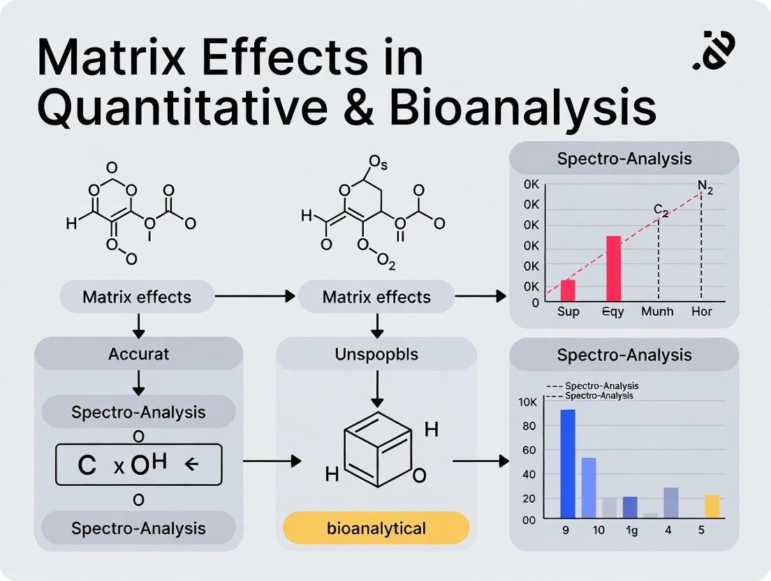
This article provides a comprehensive guide to matrix effects in quantitative bioanalysis using LC-MS/MS, focusing on their impact on accuracy, precision, and reliability of pharmacokinetic and biomarker data.
Matrix Effects in Bioanalysis: From Theory to Practice in LC-MS/MS Method Development
Abstract
This article provides a comprehensive guide to matrix effects in quantitative bioanalysis using LC-MS/MS, focusing on their impact on accuracy, precision, and reliability of pharmacokinetic and biomarker data. We explore the fundamental sources of matrix effects, practical methodologies for assessment and mitigation, advanced troubleshooting strategies, and systematic validation approaches. Targeted at researchers and drug development professionals, this resource offers actionable insights to enhance bioanalytical method robustness for regulatory compliance and reliable clinical decision-making.
What Are Matrix Effects? Defining the Invisible Interference in Bioanalytical Data
Ion suppression and ion enhancement are phenomena collectively known as matrix effects in mass spectrometry (MS). They refer to the alteration of analyte ionization efficiency in the ion source due to the presence of co-eluting compounds from the sample matrix. This is a critical variable in the broader thesis on how matrix effects impact quantitative bioanalysis research, as it directly compromises accuracy, precision, and reproducibility. These effects are most pronounced in complex biological matrices like plasma, urine, or tissue homogenates, which are central to drug development. Ion suppression leads to a decrease in signal, while ion enhancement causes an artificial increase. Both phenomena skew the correlation between the actual analyte concentration and the observed MS signal, leading to potentially erroneous pharmacokinetic or toxicokinetic conclusions.
Underlying Mechanisms: A Physicochemical Perspective
The primary mechanisms occur within the electrospray ionization (ESI) source, which is more susceptible than atmospheric pressure chemical ionization (APCI).
Key Mechanisms:
- Competition for Charge: Co-eluting matrix components compete with the analyte for available protons (in positive mode) or protons are abstracted (in negative mode) at the droplet surface during the final stages of solvent evaporation.
- Altered Droplet Properties: High concentrations of non-volatile compounds (e.g., salts, phospholipids) increase droplet viscosity and surface tension, impeding solvent evaporation and the efficient release of gas-phase ions.
- Gas-Phase Reactions: After desolvation, gas-phase ions from the matrix can react with analyte ions, leading to charge transfer or neutralization.
- Suppression of Analyte Evaporation: Matrix components can form a crust or envelope around the analyte molecule, physically preventing its evaporation into the gas phase.
Experimental Protocols for Assessment and Mitigation
The following are standard methodologies cited in current literature for evaluating and addressing matrix effects.
Protocol 3.1: Post-Column Infusion Experiment (Qualitative Assessment)
This method visually identifies chromatographic regions affected by matrix effects.
- Prepare a neat solution of the analyte at a constant concentration.
- Connect a syringe pump containing this solution to the LC stream via a T-connector post-column and pre-MS inlet.
- Infuse the analyte at a constant low flow rate (e.g., 5-10 µL/min) while the LC pump runs a gradient elution.
- Inject a blank matrix extract (e.g., processed plasma) onto the LC column.
- Monitor the selected reaction monitoring (SRM) trace for the infused analyte. A stable signal indicates no matrix effect. A depression in the signal indicates ion suppression; an increase indicates enhancement.
Protocol 3.2: Post-Extraction Spiking Method (Quantitative Assessment)
This method calculates the Matrix Factor (MF) to quantify the effect.
- Prepare three sets of samples in six replicates:
- Set A (Neat Solution): Analyte in mobile phase.
- Set B (Post-Extraction Spike): Blank matrix is extracted, then the analyte is spiked into the purified extract.
- Set C (Extracted Calibrators): Analyte is spiked into matrix prior to extraction and carried through the full sample preparation.
- Analyze all sets by LC-MS/MS.
- Calculate the Matrix Factor (MF) for each sample:
MF = Peak Area (Set B) / Peak Area (Set A). - Calculate the Internal Standard Normalized MF:
MF_IS = MF (Analyte) / MF (Internal Standard). - An MF of 1 indicates no effect; <1 indicates suppression; >1 indicates enhancement. A CV of MF_IS > 15% typically signifies unacceptable variability.
Protocol 3.3: Key Mitigation Strategies
- Improved Chromatographic Separation: Optimize LC methods to separate analytes from early-eluting, ionic matrix components (primary cause).
- Selective Sample Preparation: Utilize solid-phase extraction (SPE) or liquid-liquid extraction (LLE) over protein precipitation to remove phospholipids and salts.
- Stable Isotope-Labeled Internal Standards (SIL-IS): The most effective method. The SIL-IS co-elutes with the analyte, experiences identical matrix effects, and corrects for them upon peak area ratio calculation.
- Standard Addition or Matrix-Matched Calibration: Use calibration curves prepared in the same biological matrix as the samples.
- Source Parameter Optimization: Adjust source gas flows, temperatures, and capillary position to promote efficient desolvation.
- Alternative Ionization: Switch to APCI or APPI, which are generally less prone to certain matrix effects.
Data Presentation: Quantitative Impact
The following tables summarize typical data from matrix effect studies in bioanalysis.
Table 1: Calculated Matrix Factor (MF) for a Panel of Drug Analytes in Human Plasma
| Analyte | Retention Time (min) | Mean MF (n=6) | MF CV (%) | Mean IS-Norm. MF | IS-Norm MF CV (%) | Interpretation |
|---|---|---|---|---|---|---|
| Drug A | 2.1 | 0.45 | 25.3 | 1.05 | 6.2 | Severe suppression, corrected by SIL-IS |
| Drug B | 5.8 | 0.95 | 8.7 | 0.98 | 5.1 | Minimal effect |
| Drug C | 1.8 | 1.65 | 32.1 | 1.25 | 18.4 | Severe enhancement, poorly corrected |
Table 2: Impact of Sample Preparation on Matrix Effect (Phospholipid Removal)
| Preparation Method | Phospholipid Residual (%) | Mean MF for Early-Eluter | MF CV (%) | Key Advantage/Disadvantage |
|---|---|---|---|---|
| Protein Precipitation | 100 | 0.31 | 28.5 | Simple, high recovery, poor cleanliness |
| Liquid-Liquid Extraction | <5 | 0.89 | 10.1 | Excellent lipid removal, selective, variable recovery |
| SPE (C18) | 15 | 0.75 | 15.3 | Good balance, automatable, method development needed |
| Hybrid SPE (Phospholipid) | <2 | 0.97 | 7.8 | Superior phospholipid removal, additional cost |
Visualizing Mechanisms and Workflows
The Scientist's Toolkit: Key Research Reagent Solutions
| Item | Function in Addressing Ion Suppression/Enhancement |
|---|---|
| Stable Isotope-Labeled Internal Standard (SIL-IS) | Chemically identical to analyte but with e.g., ¹³C or ²H; co-elutes and undergoes identical matrix effects, enabling precise correction during quantification. |
| HybridSPE-Phospholipid Cartridges | Specialized solid-phase extraction sorbent designed to selectively precipitate proteins and bind phospholipids via zirconia-coated silica, drastically reducing a major source of suppression. |
| LC-MS Grade Ammonium Acetate/Formate | High-purity volatile buffers for LC mobile phases; improve chromatographic peak shape and provide consistent ionization without leaving non-volatile residues in the source. |
| SPE Cartridges (C18, HLB, Ion Exchange) | Selective stationary phases for sample clean-up to remove specific classes of interfering matrix components (lipids, acids, bases) prior to LC-MS injection. |
| Passivated (deactivated) LC Liner Vials/Inserts | Reduce nonspecific adsorption of analyte to glass surfaces, ensuring consistent recovery and preventing loss that can be confused with ion suppression. |
| Methanol & Acetonitrile (LC-MS Grade) | High-purity organic solvents for protein precipitation and mobile phases; minimize background ions that contribute to chemical noise and charge competition. |
Within the framework of understanding how matrix effects impact quantitative bioanalysis research, this technical guide examines four major classes of endogenous and exogenous interferences of biological origin: phospholipids, salts, metabolites, and co-administered drugs. These components represent the core of matrix effect challenges, directly influencing ionization efficiency, chromatographic performance, and assay reproducibility in LC-MS/MS.
Core Interferents: Mechanisms and Quantitative Impact
Phospholipids
Phospholipids are major constituents of cell membranes and are ubiquitous in biological samples like plasma. They cause severe matrix effects, primarily through ion suppression in the ESI source, and can accumulate on column and MS components.
Table 1: Common Phospholipids and Their Impact on Ionization
| Phospholipid Class | Primary m/z (Negative Mode) | Typical Plasma Concentration (µg/mL) | Relative Ion Suppression Potential (%)* |
|---|---|---|---|
| Phosphatidylcholine (PC) | 184.1 (head group) | 1000-2000 | 60-85 |
| Lysophosphatidylcholine (LPC) | 184.1, 496.3, 524.3 | 50-200 | 40-70 |
| Phosphatidylethanolamine (PE) | 196.0, 714.5 | 300-800 | 30-60 |
| Sphingomyelin (SM) | 184.1 | 200-500 | 50-75 |
*Data from pooled LC-MS/MS studies; suppression varies by analyte and platform.
Endogenous Salts and Metabolites
High concentrations of salts (e.g., Na⁺, K⁺, Ca²⁺) and small molecule metabolites (e.g., urea, bilirubin) can co-elute with analytes, forming adducts and competing for charge.
Table 2: Key Salts and Metabolites Causing Matrix Effects
| Component | Typical Conc. in Plasma | Primary Interference Mechanism | Common Adduct Formed |
|---|---|---|---|
| Sodium (Na⁺) | 135-145 mM | Ion suppression, [M+Na]⁺ adduction | [M+Na]⁺ |
| Potassium (K⁺) | 3.5-5.0 mM | Ion suppression, [M+K]⁺ adduction | [M+K]⁺ |
| Urea | 2.5-6.7 mM | Ion suppression in ESI- | - |
| Bilirubin | 0.1-1.2 mg/dL | Ion suppression, column adsorption | - |
| Fatty Acids (C16-C20) | Varies | Ion suppression, column retention shift | [M-H]⁻ |
Co-administered Drugs (Polypharmacy)
In clinical studies, concomitant medications are a major source of unpredictable matrix effects. Their interference is highly variable and patient-dependent.
Table 3: Classes of Co-administered Drugs Known to Cause Matrix Effects
| Drug Class | Example Compounds | Risk of Ion Suppression/Enhancement | Potential for Isobaric Interference |
|---|---|---|---|
| NSAIDs | Ibuprofen, Diclofenac | High | Low-Moderate |
| Proton Pump Inhibitors | Omeprazole, Pantoprazole | Moderate | Low |
| Statins | Atorvastatin, Simvastatin acid | High | High (active metabolites) |
| Antidepressants (SSRIs) | Sertraline, Fluoxetine | Moderate-High | Moderate |
| Antibiotics | Ciprofloxacin, Azithromycin | Moderate | Low |
Experimental Protocols for Assessing Matrix Effects
Protocol 2.1: Post-Column Infusion Experiment for Phospholipid Monitoring
Objective: To visualize the time-region of ion suppression/enhancement caused by phospholipids and other matrix components. Materials: LC-MS/MS system, analytical column, post-column T-connector, infusion pump, blank plasma extract. Procedure:
- Prepare a constant infusion of the analyte of interest (e.g., 100 ng/mL in 50:50 mobile phase) via a syringe pump connected post-column.
- Inject a neat solution (mobile phase) to establish a baseline response.
- Inject a processed blank matrix sample (e.g., 10 µL of precipitated plasma) using the chromatographic method.
- Monitor the analyte signal. A dip in the baseline indicates ion suppression; a peak indicates enhancement.
- Correlate suppression regions with MRM transitions for phospholipids (e.g., m/z 184→184 for PC, 496→184 for LPC 16:0).
Protocol 2.2: Quantitative Assessment via Post-Extraction Spike Method
Objective: To calculate the Matrix Factor (MF) and IS-normalized MF. Materials: Blank matrix from at least 6 different sources, calibration standards in matrix, QC samples. Procedure:
- Prepare two sets of samples at low and high QC concentrations. Set A: Spike analyte into blank matrix before extraction. Set B: Spike analyte into the supernatant of processed blank matrix after extraction ("post-extraction spike").
- Process Set A according to the validated method. Reconstitute Set B extracts with solution containing the analyte.
- Analyze both sets and calculate the peak area for each.
- Calculate MF = (Peak area of post-extraction spike / Peak area of neat solution at same concentration). Calculate IS-normalized MF = (MF of analyte / MF of internal standard). An MF of 1 indicates no effect; <1 indicates suppression; >1 indicates enhancement. CV of normalized MF > 15% indicates significant variability.
Protocol 2.3: Monitoring for Co-administered Drug Interference
Objective: To proactively assess potential interference from common concomitant medications. Procedure:
- Compile a list of the top 20 most likely co-administered drugs for the patient population.
- Prepare individual solutions of each drug at its maximum therapeutic plasma concentration (Cmax).
- Spike each drug individually into blank matrix containing the analyte and internal standard at the LLOQ level.
- Analyze and compare analyte/IS response to that in neat matrix. A deviation >±15% indicates interference.
- If interference is found, investigate chromatographic resolution or MS/MS selectivity (different MRM transition).
Mitigation Strategies: Experimental Workflows
Diagram Title: Workflow for Mitigating Biological Matrix Effects
Phospholipid Removal and Elution Pathway
Diagram Title: Phospholipid Removal SPE (PRP) Process Flow
The Scientist's Toolkit: Key Research Reagent Solutions
Table 4: Essential Materials for Managing Biological Matrix Effects
| Item/Category | Specific Example/Product | Primary Function in Mitigating Matrix Effects |
|---|---|---|
| Phospholipid-Removal SPE Plates | HybridSPE-PPT (Sigma), Ostro (Waters), Captiva ND Lipids (Agilent) | Selective binding and removal of phospholipids from plasma/serum extracts prior to LC-MS. |
| Stable-Labeled Internal Standards | d₃-, ¹³C-, ¹⁵N-labeled analogs of the analyte | Compensates for ion suppression/enhancement by co-eluting with the analyte, normalizing recovery. |
| Matrix Enhancement/Modifier Additives | Ammonium fluoride, formic acid, acetic acid, ammonium acetate | Improves ionization efficiency, promotes consistent adduct formation ([M+H]⁺ or [M-H]⁻), reduces sodium adducts. |
| Specialty LC Columns | Fused-core C18, HILIC, Charged Surface Hybrid (CSH) | Provides superior chromatographic resolution, separating analytes from early-eluting phospholipids and salts. |
| LC Guard Columns/In-Line Filters | UHPLC guard cartridges (e.g., 0.2 µm), active divert valve | Protects analytical column from particulate and non-eluting matrix buildup, prolonging column life. |
| Blank Matrix Pools | Charcoal-stripped plasma, dialyzed serum, matrix from >6 individual donors | Used for preparing calibration standards and assessing specificity, ensuring lack of endogenous interference. |
| Mass Spectrometer Cleaner/Descaler | LC-MS system wash solvents (e.g., 50:50 IPA:ACN with 0.1% formic acid) | Regular cleaning of ion source and sampling cone removes accumulated non-volatile salts and lipids. |
The biological origins of matrix effects—phospholipids, salts, endogenous metabolites, and co-administered drugs—present a multi-faceted challenge that must be systematically addressed throughout method development. A strategy combining selective sample preparation, chromatographic resolution, stable-isotope internal standardization, and rigorous matrix factor assessment is critical for developing robust, reproducible quantitative bioanalytical methods. This approach directly supports the overarching thesis that understanding and mitigating matrix effects is not merely a validation requirement but a fundamental pillar of reliable quantitative bioanalysis research.
Within the broader thesis of How do matrix effects impact quantitative bioanalysis research, matrix effects are recognized as a critical, non-ignorable source of analytical bias. They constitute the direct, unwanted alteration of an analyte's ionization efficiency in mass spectrometry (MS) detection due to the presence of co-eluting, non-target matrix components. This phenomenon, central to liquid chromatography-tandem mass spectrometry (LC-MS/MS) workflows, directly and insidiously compromises the fundamental analytical figures of merit: Accuracy, Precision, and Linearity. This guide deconstructs the mechanisms of this impact and provides a roadmap for its identification and mitigation.
Mechanisms of Matrix-Induced Skew
Matrix effects primarily manifest as ion suppression or, less commonly, ion enhancement in the MS source. This occurs when endogenous phospholipids, salts, metabolites, or drug metabolites co-elute with the analyte, competing for access to the droplet surface during electrospray ionization (ESI) or altering droplet viscosity and solvent evaporation rates. The consequence is a response for the analyte that is not proportional to its true concentration in the prepared sample.
Quantitative Impact on Analytical Figures of Merit
The following table summarizes the direct, quantitative impact of unmitigated matrix effects on core validation parameters, based on current regulatory guidance (EMA, FDA) and research literature.
Table 1: Direct Impact of Matrix Effects on Analytical Parameters
| Analytical Parameter | Definition | Impact of Matrix Effects | Typely Accepted Threshold (Deviation) |
|---|---|---|---|
| Accuracy | Closeness of mean test results to the true value. | Systematic bias. Results are consistently lower (suppression) or higher (enhancement) than the true concentration. | ±15% from nominal value (±20% at LLOQ). |
| Precision | Closeness of agreement among individual test results (Repeatability & Reproducibility). | Increased variability. Ionization efficiency fluctuates with inconsistent matrix component levels, increasing scatter. | RSD ≤15% (≤20% at LLOQ). |
| Linearity | Ability to obtain test results directly proportional to analyte concentration. | Non-linear response. The degree of suppression/enhancement may vary with concentration, causing curvature and a non-linear calibration curve. | Correlation coefficient (r) >0.99, visual inspection of residuals. |
Table 2: Experimental Evidence of Matrix Effect Magnitude
| Study Focus | Matrix | Analyte Class | Observed Ion Suppression/Enhancement | Impact on Accuracy at LLOQ |
|---|---|---|---|---|
| Phospholipid Removal Efficiency | Human Plasma | Basic Drugs | -25% to -90% suppression without cleanup | -18% to -45% bias |
| Post-Column Infusion Analysis | Rat Microdialysate | Neuropeptides | -40% average suppression in specific RT window | N/A (Qualitative) |
| Stable-Isotope Dilution Assessment | Human Serum | Endogenous Metabolites | -15% to +30% variability vs. stable-label IS | Precision RSD increased from 5% to 18% |
Core Experimental Protocols for Assessment
Protocol 1: Post-Column Infusion Analysis (Qualitative)
- Objective: To visualize regions of ion suppression/enhancement across the chromatographic run time.
- Methodology:
- A constant infusion of the analyte(s) of interest is introduced post-column into the mobile phase flow entering the MS.
- A blank matrix extract (e.g., precipitated plasma) is injected onto the LC column.
- The MRM channel for the infused analyte is monitored. A stable baseline indicates no matrix effect. A dip in signal indicates ion suppression; a peak indicates ion enhancement at that retention time.
Protocol 2: Calculation of Matrix Factor (MF) & IS-Normalized MF (Quantitative)
- Objective: To quantitate the absolute and relative matrix effect.
- Methodology:
- Prepare six different lots of blank matrix. For each lot, prepare Low (L) and High (H) concentration QC samples (n=3-5 per lot/concentration).
- Prepare equivalent L and H concentration samples in neat solution (mobile phase or reconstitution solvent).
- Inject all samples and record analyte and internal standard (IS) peak areas.
- Calculate:
- MF (Analyte) = (Peak Area in Matrix Extract) / (Peak Area in Neat Solution)
- MF (IS) = (Peak Area of IS in Matrix Extract) / (Peak Area of IS in Neat Solution)
- IS-Normalized MF = MF (Analyte) / MF (IS)
- Interpretation: An MF ≠ 1 indicates a matrix effect. An IS-normalized MF close to 1 (e.g., 0.85-1.15) indicates the IS successfully compensates for the effect. High variability (%CV) in MF across lots indicates a concerning relative matrix effect.
Visualization of Workflow & Impact Logic
Title: Matrix Effects Disrupt the LC-MS/MS Workflow
Title: Direct Impact on Accuracy, Precision, and Linearity
The Scientist's Toolkit: Key Research Reagent Solutions
Table 3: Essential Materials for Matrix Effect Investigation & Mitigation
| Item / Reagent Solution | Function in Addressing Matrix Effects |
|---|---|
| Phospholipid Removal SPE Plates (e.g., HybridSPE, Ostro) | Selective removal of primary endogenous cause of ion suppression in ESI+ from biological matrices. |
| Stable Isotope-Labeled Internal Standards (SIL-IS) | Gold standard for compensation. Co-elutes with analyte, experiences identical matrix effect, enabling normalization. |
| Diversified Blank Matrix Lots (≥6 individual donors) | Essential for assessing "relative matrix effect" and establishing method robustness across a population. |
| Post-Column Infusion Tee & Syringe Pump | Hardware setup required to perform the qualitative post-column infusion experiment. |
| LC Columns with Alternative Selectivity (e.g., HILIC, different C18 phases) | Changing retention characteristics can separate analytes from regions of suppression identified via infusion. |
| Enhanced Sample Clean-up Solvents (e.g., MTBE for LLE, low-ionic strength washes) | Improves selectivity of extraction, removing more interfering matrix components than simple protein precipitation. |
1. Introduction: A Universal Challenge in Bioanalysis
Matrix effects—the alteration of an analytical signal due to the presence of co-eluting or co-detected components from a sample matrix—are a central thesis in quantitative bioanalysis. While extensively discussed in Liquid Chromatography-Tandem Mass Spectrometry (LC-MS/MS), these effects are platform-agnostic and can critically impact accuracy, precision, and sensitivity across all analytical techniques. This guide details the manifestation, evaluation, and mitigation of matrix effects in key alternative platforms, framing them as a universal variable that must be controlled for robust bioanalytical research.
2. Matrix Effects Across Analytical Platforms: Mechanisms and Data
The core mechanisms differ from LC-MS/MS's ionization suppression/enhancement, but the impact on quantitative reliability is equally significant.
Table 1: Manifestation of Matrix Effects Across Analytical Platforms
| Analytical Platform | Primary Mechanism of Matrix Effect | Impact on Quantitative Signal | Common Source of Interference |
|---|---|---|---|
| Immunoassays (e.g., ELISA) | Non-specific binding, cross-reactivity, or heterophilic antibodies. | False elevation or suppression of signal. | Human anti-animal antibodies, rheumatoid factor, complement, endogenous analytes. |
| GC-MS | Enhancement or suppression during electron/chemical ionization; active sites in liner/column. | Altered detector response for target analyte. | Non-volatile matrix components, co-extracted compounds, derivatization by-products. |
| ICP-MS | Spectral overlaps (isobaric, polyatomic), non-spectral interferences (signal suppression). | Inaccurate concentration/isotope ratio. | Plasma-based matrices (Cl, Ar, Ca, Na), organic solvents, sample viscosity. |
| High-Performance Liquid Chromatography (HPLC) with UV/FL | Co-elution of interfering compounds with similar spectral properties. | Inaccurate peak area/height (false positive/negative). | Metabolites, endogenous compounds, drug components, sample prep reagents. |
| Capillary Electrophoresis (CE) | Changes in sample conductivity, viscosity, or adsorption to capillary wall. | Migration time shift, peak broadening, efficiency loss. | Salts, proteins, phospholipids. |
Table 2: Quantitative Impact Assessment of Matrix Effects in Selected Platforms (Representative Data)
| Platform | Experiment | Result: No Matrix Effect | Result: With Matrix Effect | Observed Deviation |
|---|---|---|---|---|
| Immunoassay | Spike recovery of 10 ng/mL analyte in 10 different plasma lots. | Mean recovery = 100% (Target) | Mean recovery = 135%; Range: 85-210% | +35% (Severe, variable) |
| GC-MS | Response factor for analyte in pure solvent vs. post-extraction spiked matrix. | RF = 15,000 ± 500 | RF = 12,000 ± 1,800 | -20% (Signal suppression) |
| ICP-MS | Measurement of 5 ppb As in 1% HNO3 vs. in urine matrix. | Signal (cps) = 50,000 | Signal (cps) = 42,000 | -16% (Non-spectral suppression) |
3. Experimental Protocols for Assessing Matrix Effects
Protocol 3.1: Post-Column Infusion for Immunoassay & HPLC-UV
- Objective: Visually identify regions of non-specific interference in an immunoassay or chromatographic run.
- Materials: Analytical column (for HPLC), assay plate/beads, mobile phase/buffer, neat analyte solution, infusion pump.
- Procedure:
- Perform the analytical run (chromatographic separation or immunoassay incubation step) with a blank matrix sample (no analyte).
- During the detection phase, continuously infuse a constant concentration of the analyte post-column (HPLC) or into the detection system (simulated for immunoassay).
- Record the detection signal (UV absorbance, fluorescence, or chemiluminescence).
- A stable baseline indicates no matrix effect. Signal depression or elevation corresponds to the elution/time zone where matrix components cause interference.
Protocol 3.2: Post-Extraction Addition (Spike Recovery) for GC-MS & CE
- Objective: Quantitatively determine the absolute matrix effect by comparing responses in neat solution to matrix.
- Materials: Blank matrix from at least 6 different sources, calibration standards in neat solvent, extraction reagents/equipment.
- Procedure:
- Prepare two sets of samples:
- Set A (Neat Standards): Prepare standards in reconstitution solvent or buffer.
- Set B (Matrix Spikes): Extract blank matrix from multiple sources. After extraction is complete, spike the analyte at Low, Mid, and High QC levels into the extracted matrix.
- Analyze all samples (Set A and B).
- Calculate % Absolute Matrix Effect (ME) for each source and level:
- ME (%) = (Mean Peak Area of Set B / Mean Peak Area of Set A) x 100%.
- A value of 100% indicates no effect. <100% = suppression; >100% = enhancement. CV of ME across sources > 15% indicates a significant variable matrix effect.
- Prepare two sets of samples:
Protocol 3.3: Isotopic Dilution & Standard Addition for ICP-MS
- Objective: Correct for both spectral and non-spectral interferences.
- Materials: Stable isotope-enriched internal standard (IS), sample matrix, multi-element calibration standards.
- Procedure (Isotopic Dilution):
- Spike the sample with a known amount of the analyte's stable isotope (e.g., ^111^Cd for Cd analysis) before digestion/dilution.
- Measure the ratio of the native analyte signal to the isotopically enriched IS signal.
- This ratio is inherently corrected for most matrix-induced signal fluctuations. Calibrate using certified reference materials.
4. Visualization of Workflows and Relationships
Matrix Effect Influence on Bioanalytical Workflow
Mitigation Strategy Framework for Matrix Effects
5. The Scientist's Toolkit: Key Research Reagent Solutions
Table 3: Essential Materials for Managing Matrix Effects
| Item / Reagent | Primary Function in Mitigating Matrix Effects |
|---|---|
| Stable Isotope-Labeled Internal Standards (SIL-IS) | Gold standard for MS techniques. Co-elutes with analyte, correcting for ionization suppression/enhancement and extraction losses. |
| Anti-Heterophilic Antibody Blocking Reagents | Added to immunoassay buffers to prevent false results from human anti-animal antibodies. |
| Certified Reference Materials (CRMs) & Matrix-Matched Standards | For ICP-MS, GC-MS. Calibrants with a matrix similar to the sample to account for non-spectral interferences. |
| Solid-Phase Extraction (SPE) Cartridges (e.g., Mixed-mode, HLB) | Selective cleanup to remove phospholipids, proteins, and salts that cause matrix effects in LC/GC-MS and CE. |
| Immunoaffinity Depletion Columns | Remove high-abundance proteins (e.g., albumin, IgG) from plasma/serum to reduce interference in HPLC and CE assays. |
| Derivatization Reagents (for GC) | Improves volatility and detectability of analytes, potentially separating them from underivatized matrix interferences. |
| Collision/Reaction Cell Gases (e.g., He, H2, O2 for ICP-MS) | Used in collision/reaction cell ICP-MS to break apart polyatomic interferences before detection. |
Within the broader thesis on How do matrix effects impact quantitative bioanalysis research, a thorough understanding of the regulatory expectations is paramount. Matrix effects—the alteration of ionization efficiency by co-eluting matrix components—are a critical source of bias and variability in quantitative bioanalytical methods, particularly when using Liquid Chromatography-Tandem Mass Spectrometry (LC-MS/MS). This guide details the current regulatory positions of the U.S. Food and Drug Administration (FDA) and the European Medicines Agency (EMA) on the assessment and control of matrix effects, providing a technical framework for compliance.
Regulatory Framework: FDA vs. EMA
The FDA’s 2018 Bioanalytical Method Validation Guidance for Industry and the EMA’s 2011 Guideline on bioanalytical method validation (with 2021 draft for revision) form the cornerstone documents. Both emphasize the need to evaluate matrix effects, but with nuanced differences in approach and emphasis.
Table 1: Comparison of FDA and EMA Guidelines on Matrix Effect Assessment
| Aspect | FDA (2018 Guidance) | EMA (2011 Guideline, 2021 Draft Concepts) |
|---|---|---|
| Core Requirement | Assessment of matrix effect is "recommended" for MS-based assays. | Assessment is "essential" for validation of MS-based methods. |
| Primary Metric | Matrix Factor (MF). | Matrix Factor and Internal Standard Normalized Matrix Factor. |
| MF Calculation | MF = Peak response in presence of matrix / Peak response in neat solution |
Same as FDA. |
| IS-Normalized MF | Implied but not explicitly mandated. | Explicitly required: IS-normalized MF = MF(analyte) / MF(IS) |
| Acceptance Criteria | No universal numerical criteria provided. CV of MF should be ≤15%. | IS-normalized MF should be close to 1.0. CV of IS-normalized MF should be ≤15%. |
| Number of Lots | A minimum of 10 matrices from individual subjects should be evaluated. | At least 10 batches from individual sources. EMA 2021 draft suggests 6 for rare matrices. |
| Focus on Selectivity | Linked to matrix effect assessment; should test for lots with potential interferences (e.g., hemolyzed, lipemic). | Strong emphasis on testing matrices from different species/disease states, and abnormal samples. |
Detailed Experimental Protocols for Matrix Effect Assessment
Protocol 3.1: Determination of Matrix Factor (MF)
Objective: To quantitatively measure the ion suppression/enhancement for the analyte and internal standard (IS) across multiple lots of matrix.
- Prepare Solutions:
- Set A (Post-extraction Spiked): Extract 10 or more individual matrix lots (e.g., plasma) using the validated sample preparation protocol. After extraction, spike the analyte and IS at known concentrations into the cleaned matrix extract.
- Set B (Neat Solution): Prepare analyte and IS in mobile phase or a protein-free solution at the same concentrations as Set A.
- Analysis: Inject and analyze Sets A and B using the finalized LC-MS/MS method.
- Calculation: For each matrix lot and each analyte/IS:
MF = Peak Area (Set A) / Peak Area (Set B)- MF = 1 indicates no effect; MF < 1 indicates suppression; MF > 1 indicates enhancement.
- Data Presentation: Report the mean, standard deviation, and coefficient of variation (CV%) of the MF for the analyte and the IS across all matrix lots.
Protocol 3.2: Determination of IS-Normalized Matrix Factor
Objective: To assess whether the internal standard adequately compensates for matrix effects observed on the analyte.
- Prerequisite: Complete Protocol 3.1 to obtain MF(analyte) and MF(IS) for each matrix lot.
- Calculation: For each individual matrix lot:
IS-Normalized MF = MF(analyte) / MF(IS)
- Acceptance Criteria (EMA): The CV% of the IS-normalized MF across all matrix lots should be ≤15%. The mean value should be close to 1.0 (typically 0.85-1.15).
Protocol 3.3: Assessment of Matrix Effect on Calibration and QC Samples
Objective: To evaluate the impact of variable matrix lots on quantitative accuracy and precision.
- Preparation: Prepare calibration standards and Quality Control (QC) samples in at least 6 different individual matrix lots. Include one lot used for the original calibration curve.
- Analysis: Analyze all samples against a single calibration curve prepared in a separate, distinct matrix lot.
- Evaluation: Calculate the accuracy (% bias) of the QC samples prepared in different lots. The accuracy should be within ±15% (±20% at LLOQ), demonstrating that the method is robust to inter-subject matrix variability.
Visualizing the Matrix Effect Assessment Workflow
Matrix Effect Assessment Decision Workflow
Mitigation Strategies for Significant Matrix Effects
When matrix effect criteria are not met, systematic investigation and mitigation are required.
- Improved Sample Cleanup: Optimize solid-phase extraction (SPE) or introduce liquid-liquid extraction (LLE) to remove more phospholipids and salts.
- Chromatographic Optimization: Increase retention time, improve separation of the analyte from the ionization front (eluting matrix components), or use alternative stationary phases.
- Alternative Internal Standard: Switch from a stable-label analog (SIL-IS) to a structural analog or deuterated IS that co-elutes more precisely with the analyte.
- Method Re-Development: In extreme cases, consider changing the ionization mode (e.g., APCI instead of ESI) or the sample type (e.g., use of dried blood spots).
The Scientist's Toolkit: Essential Research Reagents & Materials
Table 2: Key Reagents and Materials for Matrix Effect Studies
| Item | Function & Rationale |
|---|---|
| Individual Donor/Subject Matrix Lots (≥10) | To capture biological variability in phospholipids, salts, and endogenous compounds that cause ion suppression/enhancement. |
| Stable Isotope-Labeled Internal Standard (SIL-IS) | Ideally deuterated or 13C-labeled; corrects for variability in extraction and ionization, crucial for calculating IS-normalized MF. |
| Control Matrix (e.g., Stripped/Charcoal-Treated) | Used for preparing calibration standards; must be verified for absence of interference with the analyte. |
| Phospholipid Mix Standards | Used during method development to identify chromatographic regions of high ion suppression in ESI+ mode. |
| Mobile Phase Additives (Ammonium Formate/Acetate) | Volatile buffers that improve chromatographic separation and peak shape without causing persistent source contamination. |
| Solid-Phase Extraction (SPE) Cartridges | For method development of selective sample cleanup to remove phospholipids and proteins. |
| Hemolyzed and Lipemic Plasma Samples | Required for selectivity testing to ensure method robustness against pathological matrix variants. |
Detecting and Quantifying Matrix Effects: Proven Experimental Strategies
1. Introduction and Thesis Context Matrix effects—the alteration of analyte ionization efficiency by co-eluting, non-target compounds—represent a pivotal challenge in quantitative bioanalysis via Liquid Chromatography-Mass Spectrometry (LC-MS/MS). Their impact can severely compromise assay accuracy, precision, and sensitivity, leading to erroneous pharmacokinetic or toxicokinetic data. This whitepaper details the post-column infusion experiment, a core diagnostic technique framed within the broader thesis: Understanding and mitigating matrix effects is fundamental to achieving reliable quantitative bioanalysis in drug development. This guide provides the technical framework for visualizing and characterizing ion suppression (or enhancement) zones, a critical first step in method development and validation.
2. Experimental Protocols
2.1. Core Post-Column Infusion Protocol Objective: To visualize regions of ion suppression/enhanceance in chromatographic time for a given sample matrix and LC-MS method. Materials: LC system, tandem mass spectrometer, syringe pump, post-column T-connector, analytical column, mobile phases, neat analyte standard solution, extracted matrix samples (blank plasma, urine, tissue homogenate). Procedure:
- Prepare a continuous infusion of a neat solution of the analyte of interest (typical concentration: 50-500 ng/mL) using a syringe pump.
- Connect the syringe pump output post-column via a low-dead-volume T-connector between the column outlet and the MS ion source.
- Inject a blank matrix extract (e.g., 5-10 µL) onto the LC column and start the chromatographic gradient.
- The MS, operating in selected reaction monitoring (SRM) mode for the infused analyte, monitors the signal intensity continuously.
- A stable signal baseline is established when only the infusion is present. As matrix components elute from the column and enter the source, they cause deviations from this baseline.
- Data Interpretation: A dip in the signal indicates ion suppression; a peak indicates ion enhancement. The retention time of the deviation corresponds to the elution time of the interfering component.
2.2. Complementary Experiments for Quantitative Assessment Protocol A: Absolute Matrix Factor (MF) Determination.
- Prepare six replicates of Low and High Quality Control (QC) samples in matrix (post-extraction spiked).
- Prepare six replicates of neat reference solutions at identical concentrations in mobile phase.
- Analyze all samples and calculate peak areas.
- Calculate MF = (Mean Peak Area of Post-extraction Spiked Sample) / (Mean Peak Area of Neat Solution). An MF of 1 indicates no effect; <1 indicates suppression; >1 indicates enhancement.
Protocol B: Internal Standard Normalized MF Assessment.
- Repeat Protocol A, but spike analyte into matrix before extraction.
- Use a stable isotope-labeled internal standard (SIL-IS), added at a fixed point in the workflow (usually post-extraction for this test).
- Calculate IS-normalized MF = (Mean Peak Area Ratio of Analyte/IS in Matrix) / (Mean Peak Area Ratio of Analyte/IS in Neat Solution). This assesses whether the IS adequately corrects for matrix effects.
3. Data Presentation: Summary of Quantitative Metrics
Table 1: Matrix Effect Classification Based on Calculated Matrix Factor
| Matrix Factor (MF) Value | Interpretation | Impact on Assay |
|---|---|---|
| 0.80 - 1.20 | Acceptable | Minimal |
| 0.50 - 0.80 or 1.20 - 1.50 | Moderate Effect | Requires Investigation |
| < 0.50 or > 1.50 | Severe Effect | Method Modification Required |
Table 2: Comparison of Matrix Effect Assessment Methods
| Method | Measures | Advantage | Limitation |
|---|---|---|---|
| Post-Column Infusion | Location (retention time) of effect | Rapid visualization, qualitative map | Not quantitative, requires extra setup |
| Absolute MF | Magnitude of effect on analyte | Quantitative, simple calculation | Does not account for IS correction |
| IS-Normalized MF | Magnitude after IS correction | Most relevant to final assay performance | Requires a reliable SIL-IS |
4. Visualizing the Workflow and Impact
Diagram 1: Post-column infusion experimental setup workflow.
Diagram 2: Role of post-column infusion in addressing matrix effects.
5. The Scientist's Toolkit: Key Research Reagent Solutions
Table 3: Essential Materials for Post-Column Infusion & Matrix Effect Studies
| Item | Function / Role | Example/Typical Use |
|---|---|---|
| Stable Isotope-Labeled Internal Standard (SIL-IS) | Corrects for variability in ionization efficiency and sample preparation; gold standard for bioanalysis. | Deuteration (D₃, D₅) or ¹³C/¹⁵N labeled analogs of the analyte. |
| Matrix Samples (Blank) | Provides the source of interfering compounds for assessment. Must be from relevant biological fluid or tissue. | Blank human plasma from multiple donors, urine, tissue homogenates. |
| Post-Column T-Connector | Low-dead-volume union for merging the column eluent with the continuous infusion stream. | PEEK or stainless steel, with < 1 µL internal volume. |
| Syringe Pump | Delivers a constant, pulse-free flow of the analyte infusion solution. | Used at flow rates of 5-50 µL/min. |
| Solid-Phase Extraction (SPE) Kits | For selective sample clean-up to remove phospholipids and other common causes of suppression. | Mixed-mode cation/anion exchange or dedicated phospholipid removal plates. |
| Phospholipid Standards | Used to identify and confirm the elution profile of major suppressors in plasma samples. | SPC (16:0/18:1) and LPC (16:0) for LC-MS/MS monitoring. |
Within the context of a broader thesis on How do matrix effects impact quantitative bioanalysis research, the assessment of matrix effects (ME) is critical for ensuring the reliability, accuracy, and precision of bioanalytical methods, particularly in ligand binding assays (LBAs) and liquid chromatography-tandem mass spectrometry (LC-MS/MS). The Matrix Factor (MF) is a key quantitative measure used to evaluate these effects. This guide provides an in-depth technical review of MF calculation methodologies, acceptance criteria, and experimental protocols.
Defining the Matrix Factor
Matrix effects occur when co-eluting matrix components alter the ionization efficiency of the analyte in the mass spectrometer source (ion suppression/enhancement) or interfere with antibody binding in LBAs. The MF quantifies this effect by comparing the instrument response for an analyte spiked into a post-extraction biological matrix sample to the response for the same analyte in a neat solution.
Core Formulas
The fundamental formula for calculating the MF in LC-MS/MS is:
MF = (Response of analyte in post-extracted spiked matrix) / (Response of analyte in neat solution)
Where "Response" typically refers to the chromatographic peak area (or area ratio to internal standard).
To account for variability and the role of the internal standard (IS), two primary calculations are employed:
Table 1: Matrix Factor Formulas
| Formula Type | Equation | Description |
|---|---|---|
| Absolute MF | MF_abs = (A_analyte in matrix / A_analyte in neat) |
Measures absolute ion suppression/enhancement for the analyte. |
| Internal Standard Normalized MF | MF_IS Norm = (A_analyte in matrix / A_IS in matrix) / (A_analyte in neat / A_IS in neat) |
Normalizes the effect using a stable isotope-labeled IS, which should experience similar ME. |
A = Peak Area (or Area Ratio)
An MF = 1 indicates no matrix effect. MF < 1 indicates ion suppression, and MF > 1 indicates ion enhancement. The IS-normalized MF is generally preferred as it corrects for extraction efficiency and instrument variability, providing a purer measure of the ionization effect.
Acceptance Criteria
Regulatory guidance from the FDA and EMA, as well as industry white papers (e.g., from the European Bioanalysis Forum), recommend specific acceptance criteria. These are summarized below.
Table 2: Acceptance Criteria for Matrix Factor Assessment
| Parameter | Recommended Criteria | Rationale & Notes |
|---|---|---|
| Number of Matrix Lots | Minimum of 6 individual donor lots from relevant population (e.g., healthy, diseased). 10 lots are often used for increased confidence. | To assess biological variability. Hemolyzed or lipemic lots may be tested separately. |
| Calculation & Reporting | Report both absolute and IS-normalized MF for each lot at low and high QC concentrations. | Provides a complete picture. |
| Precision (CV%) of IS-normalized MF | CV ≤ 15% across all matrix lots tested. | Indicates consistent matrix effect and proper performance of the IS. |
| Mean IS-normalized MF | Ideally close to 1.00. A mean value of 0.80-1.20 is often considered acceptable, but consistency (low CV) is more critical. | Significant deviation from 1 suggests a systematic, but consistent, matrix effect compensated by the IS. |
| Per-Lot MF Value | No definitive universal cutoff. Values outside 0.80-1.20 often trigger investigation but may be acceptable if IS-normalized and CV is within limits. | The IS must effectively track the analyte. An outlier lot may indicate an interfering substance. |
Experimental Protocol for LC-MS/MS
A detailed methodology for determining MF via the post-extraction addition method is outlined below.
Protocol: Post-Extraction Spike Method for MF Determination
Objective: To quantify matrix effects by comparing analyte response in extracted matrix versus neat solution.
Materials: See "The Scientist's Toolkit" below.
Procedure:
- Prepare Matrix Lots: Obtain at least 6 individual lots of blank matrix (e.g., human plasma). Pool a small portion of each to create a "pooled" matrix lot.
- Prepare Solutions:
- Analyte Stock Solutions: In appropriate solvent.
- Internal Standard Stock Solution: In appropriate solvent.
- Sample Set Preparation (in duplicate or triplicate):
- Set A (Post-extraction Spike): a. Aliquot blank individual matrix lots (e.g., Lot 1-6) and pooled matrix. b. Process these blanks through the entire sample preparation procedure (e.g., protein precipitation, SPE, SLE). c. After extraction and reconstitution in mobile phase, spike a known concentration of analyte and IS into the extracted matrix. This represents the "post-extracted spiked matrix."
- Set B (Neat Solution): a. Prepare neat solutions of analyte and IS in mobile phase/reconstitution solvent at the same concentrations as used in Set A. These represent the "neat solution."
- LC-MS/MS Analysis: Inject and analyze all samples from Set A and Set B in the same batch.
- Data Analysis:
- Record peak areas for the analyte and IS.
- Calculate the absolute MF and IS-normalized MF for each individual matrix lot and the pooled matrix using the formulas in Table 1.
- Calculate the mean and CV% of the IS-normalized MF across all individual lots.
Diagram Title: Experimental Workflow for Matrix Factor Determination
The Scientist's Toolkit
Table 3: Essential Research Reagent Solutions for MF Experiments
| Item | Function in MF Assessment |
|---|---|
| Individual Donor Matrix Lots (>6 lots) | Represents biological variability. The core substrate for testing the specificity of the method against endogenous interferences. |
| Stable Isotope-Labeled Internal Standard (SIL-IS) | Gold standard for LC-MS/MS. Ideally experiences identical matrix effects as the analyte, enabling accurate normalization in the MF_IS Norm calculation. |
| Analyte Stock Solutions (at LQC/HQC levels) | Prepared in appropriate solvent. Used for spiking post-extracted blanks and preparing neat solutions for comparison. |
| Sample Preparation Materials (SPE cartridges, PPT plates, SLE cartridges) | Used to process blank matrix lots. The choice of clean-up method directly impacts the severity of residual matrix effects. |
| LC-MS/MS Mobile Phase & Reconstitution Solvents (HPLC-grade) | Consistent, high-purity solvents are critical to avoid introducing variability that could confound MF results. |
| Quality Control (QC) Samples (in pooled matrix) | While not for MF calculation itself, they are analyzed concurrently to ensure system suitability and assay performance during the MF experiment. |
Contextualizing MF in Bioanalysis
The Matrix Factor is a diagnostic tool, not a pass/fail test in isolation. Its primary value is in method development and troubleshooting. A high CV% in IS-normalized MF signals that the internal standard is not adequately compensating for matrix effects, jeopardizing assay accuracy. This finding would prompt method modification—such as altering the sample clean-up, chromatography, or source conditions—or investigation of a more suitable IS. Ultimately, a well-understood and consistent MF, even if deviating from 1, supports the validity of a quantitative bioanalytical method within the broader thesis on managing matrix effects.
Within the thesis that matrix effects critically compromise the accuracy, precision, and reproducibility of quantitative bioanalysis, the experimental design for their assessment is paramount. Matrix effects, defined as the alteration of analyte ionization efficiency by co-eluting endogenous or exogenous matrix components, can lead to significant quantitative bias. This guide details the foundational step of such an investigation: the rigorous selection of matrix lots and sources to ensure that method robustness is evaluated against real-world biological variability.
Quantitative Data on Matrix Variability
Recent literature and regulatory guidelines emphasize testing a minimum of 10 individual matrix lots from independent sources to capture inherent biological diversity. The following table summarizes key quantitative targets and acceptance criteria for a robust matrix effect experiment.
Table 1: Experimental Design Parameters for Matrix Lot Selection
| Parameter | Recommended Value/Range | Rationale & Acceptance Criteria |
|---|---|---|
| Number of Individual Lots | Minimum of 10 (≥6 for rare matrices) | Captures population variability; EMA & FDA recommendations. |
| Source Diversity | At least two distinct geographical/ demographic sources (e.g., different collection sites, suppliers). | Accounts for potential population-specific differences (diet, medication, genetics). |
| Matrix Types per Lot | For each donor: Plain, K₂EDTA, Heparin, Citrate (as applicable). | Evaluates impact of common anticoagulants on ionization. |
| Hemolyzed & Lipemic Lots | Include at least 2 lots each with marked hemolysis (≥500 mg/dL Hb) and lipemia (≥3+ visually or >1000 mg/dL triglycerides). | Stress-tests the method against common interferents. |
| Internal Standard Response Variation | IS-normalized matrix factor (MF) CV ≤ 15% across all lots. | Primary quantitative metric for consistency. |
| Accuracy in Individual Matrix | Measured concentration within ±15% (±20% at LLOQ) of nominal for ≥80% of lots. | Confirms assay performance is not lot-dependent. |
Experimental Protocol: Matrix Effect Assessment via Post-Column Infusion
This protocol evaluates ionization suppression/enhancement across the chromatographic run time for multiple matrix lots.
Materials: LC-MS/MS system, syringe pump, T-union, 10+ individual lots of blank matrix, analyte and IS stock solutions, mobile phases.
Procedure:
- Standard Solution: Prepare a solution containing the analyte(s) of interest at a concentration near the mid-range of the calibration curve.
- Infusion Setup: Connect the syringe pump loaded with the standard solution via a T-union between the HPLC column outlet and the MS ion source. Start infusion at a constant low flow rate (e.g., 5-10 µL/min).
- Blank Matrix Injection: While continuously infusing the analyte, inject an extract of a single blank matrix lot (processed through the entire sample preparation protocol).
- Chromatographic Run: Perform the standard LC gradient. The MS signal now reflects the constant analyte level modified by the co-eluting matrix components from the injected blank.
- Data Acquisition: Monitor the selected MRM transition. A stable signal indicates no matrix effect. Suppression appears as a negative peak (signal dip); enhancement as a positive peak (signal rise).
- Replication: Repeat Steps 3-5 for each of the 10+ individual blank matrix lots and abnormal (hemolyzed, lipemic) lots.
- Analysis: Overlay the extracted ion chromatograms from all lots. The region of eluting analyte should be examined for signal variability across lots.
Title: Post-Column Infusion Workflow for Matrix Effects
Experimental Protocol: Calculation of Matrix Factor (MF)
This quantitative method compares analyte response in matrix to response in neat solution.
Procedure:
- Prepare Three Sets (in replicates of n=3-5):
- Set A (Neat Solution): Analyte and IS spiked into mobile phase or solvent.
- Set B (Post-Extraction Spiked): Blank matrix carried through extraction, then analyte and IS spiked into the processed extract.
- Set C (Pre-Extraction Spiked): Analyte and IS spiked into blank matrix, then carried through the entire extraction process.
- LC-MS/MS Analysis: Analyze all sets.
- Calculation:
- Matrix Factor (MF) = Peak Area Response (Set B or C) / Peak Area Response (Set A).
- Internal Standard Normalized MF = MF (Analyte) / MF (IS).
- Assessment: Calculate the IS-normalized MF for each of the 10+ individual lots. The coefficient of variation (CV%) across all lots should be ≤15%. This directly tests the impact of lot-to-lot variability.
Title: Matrix Factor Calculation and Assessment Workflow
The Scientist's Toolkit: Research Reagent Solutions
Table 2: Essential Materials for Matrix Effect Experiments
| Item | Function & Rationale |
|---|---|
| Charcoal-Stripped / Dialyzed Matrix | Serves as a "cleaned" control to confirm the removal of specific interfering components; useful for troubleshooting. |
| Stable Isotope-Labeled Internal Standard (SIL-IS) | The most critical reagent. Compensates for variable ionization efficiency when co-eluting with the analyte, improving MF precision. |
| Commercially Sourced, Well-Characterized Blank Matrix Pools | Provide a consistent, readily available baseline for initial method development and system suitability testing. |
| Single-Donor, Individual-Donor Matrix Lots | The gold standard for robustness testing. Purchased from certified biological suppliers with donor demographic data. |
| Quality Control Materials for Hemolysis & Lipemia | Prepared by spiking lysed RBCs or lipid emulsions (e.g., Intralipid) into normal matrix to create standardized abnormal lots. |
| Automated Liquid Handlers | Essential for high-precision, reproducible spiking of analytes and IS across dozens of matrix samples, minimizing manual error. |
The core thesis of modern quantitative bioanalysis research investigates how matrix effects—unwanted alterations in analytical signal caused by co-eluting substances from the sample matrix—compromise assay accuracy, precision, and reproducibility. Hemolyzed (containing lysed red blood cells) and hyperlipidemic (excess lipids) plasma samples represent two of the most prevalent and challenging problematic matrices. These matrices introduce endogenous interferents such as hemoglobin, intracellular enzymes, lipids, and lipoproteins, which can cause significant ion suppression/enhancement in LC-MS/MS, spectral interference in immunoassays, and physical obstructions.
Characterizing the Impact of Problematic Matrices
The impact of hemolysis and hyperlipidemia manifests through several quantifiable mechanisms. Understanding these is critical for developing effective mitigation strategies.
Table 1: Quantitative Impact of Hemolysis and Hyperlipidemia on Bioanalytical Assays
| Matrix Interferent | Primary Components | Key Impact Mechanisms | Typical Affected Assays | Reported Signal Alteration Range |
|---|---|---|---|---|
| Hemolyzed Plasma | Hemoglobin, Lactate Dehydrogenase (LDH), Potassium, Iron, Hepcidin | Ion suppression (ES+), Chemical quenching (FL), Increased viscosity, Adsorption to surfaces | LC-MS/MS, Fluorimetry, Potentiometry (ISE), Immunoassays | -20% to -60% ion suppression for basic compounds in ESI+ |
| Hyperlipidemic Plasma | Chylomicrons, VLDL, LDL, Triglycerides, Cholesterol | Ion suppression (ES-), Elevated background, Clogging of columns/nebulizers, Non-specific binding | LC-MS/MS (especially for acidic compounds), Spectrophotometry, Turbidimetric assays | -15% to -50% ion suppression for acidic compounds in ESI- |
| Lipemic & Hemolyzed | Combination of above | Additive or synergistic matrix effects, Compound-specific unpredictability | All chromatographic and ligand-binding assays | Signal variation can exceed ±70% without correction |
Experimental Protocols for Assessment and Mitigation
Protocol for Quantifying Matrix Effect (ME% and IS-Normalized ME%)
Objective: To quantitatively assess ion suppression/enhancement using the post-column infusion method and the post-extraction spike method. Materials: Blank plasma from at least 6 individual sources (normal, hemolyzed, hyperlipidemic), analyte stock solution, internal standard (IS) stock solution, HPLC system with tandem mass spectrometer (MS/MS). Procedure:
- Post-Column Infusion (Qualitative):
- Prepare a continuous infusion of the analyte at a constant rate (e.g., 500 ng/mL) via a T-connector between the HPLC column outlet and the MS/MS inlet.
- Inject extracted blank plasma from each matrix condition.
- Monitor the MRM transition. A dip in the baseline indicates ion suppression; a peak indicates enhancement. Map the chromatographic region of interference.
- Post-Extraction Spike (Quantitative - ME%):
- Prepare two sets of samples for each matrix source (n=6 per matrix type).
- Set A (Post-extracted spike): Spike analyte and IS into the supernatant of extracted blank plasma.
- Set B (Neat solution): Prepare analyte and IS in reconstitution solvent at identical concentrations.
- Analyze all samples. Calculate ME% = (Peak Area of Set A / Peak Area of Set B) × 100%. An ME% of 100% indicates no effect; <100% indicates suppression; >100% indicates enhancement.
- IS-Normalized Matrix Factor (Quantitative - MF%):
- From the data above, calculate the matrix factor for the analyte (MFanalyte = AreaAanalyte / AreaBanalyte) and IS (MFIS).
- Calculate the IS-normalized MF = MFanalyte / MFIS. The variability (CV%) of the IS-normalized MF across different matrices should be ≤15% for a robust method.
Protocol for High-Speed Centrifugation and Ultracentrifugation
Objective: To remove lipoprotein particles from hyperlipidemic plasma. Materials: Hyperlipidemic plasma samples, high-speed centrifuge (capable of >100,000× g), ultracentrifuge, fixed-angle or vertical rotor. Procedure:
- High-Speed Centrifugation (Simple Clean-up):
- Transfer plasma to a sealed centrifuge tube.
- Centrifuge at 15,000× g for 30 minutes at 4°C.
- Carefully collect the middle layer of plasma using a pipette, avoiding the top lipid layer and bottom pellet.
- Ultracentrifugation (Gold Standard):
- Load plasma into ultracentrifuge tubes. Ensure proper balancing.
- Centrifuge at ~100,000× g for 1-2 hours at 4°C.
- A dense, white lipoprotein cake forms at the top. Aspirate the infranatant (clarified plasma) from the bottom two-thirds of the tube.
- Process the infranatant through solid-phase extraction (SPE) or protein precipitation (PPT) as needed.
Protocol for Solid-Phase Extraction (SPE) Optimization for Problematic Matrices
Objective: To selectively isolate analyte from hemoglobin and lipid interferents. Materials: Mixed-mode cation-exchange (MCX) or phospholipid removal (PLR) SPE cartridges (e.g., 30 mg/1 mL), hemolyzed/hyperlipidemic plasma, conditioning and elution solvents. Procedure:
- Condition the cartridge with 1 mL methanol, followed by 1 mL water or buffer.
- Load 100-200 µL of plasma (pre-diluted 1:1 with a loading buffer, e.g., 2% formic acid for MCX).
- Wash with 1 mL of a wash solution to remove interferents:
- For MCX (basic analytes): 2% formic acid in water (removes proteins, phospholipids), followed by methanol (removes neutral lipids).
- For PLR: Specific proprietary solvents to elute lipids while retaining a wide range of analytes.
- Elute the analyte with an appropriate strong solvent (e.g., 5% NH4OH in methanol for MCX).
- Evaporate the eluent under nitrogen and reconstitute in mobile phase compatible solvent.
Visualization of Workflows and Relationships
Flowchart: Assessment and Mitigation of Problematic Matrices
Workflow: Sample Preparation for Problematic Matrices
The Scientist's Toolkit: Key Research Reagent Solutions
Table 2: Essential Materials for Handling Problematic Matrices
| Item Name / Category | Specific Example/Format | Primary Function in Addressing Matrix Effects |
|---|---|---|
| Phospholipid Removal (PLR) SPE | HybridSPE-PPT, Ostro Pass-through | Selectively binds phospholipids and lipids from plasma extracts, significantly reducing ion suppression in LC-MS/MS. |
| Mixed-Mode SPE Sorbents | MCX (Cation Exchange), MAX (Anion Exchange) | Provide dual retention (reversed-phase + ion-exchange) for selective clean-up, removing ionic interferents like hemoglobin fragments. |
| Stable Isotope-Labeled Internal Standard (SIL-IS) | ¹³C, ¹⁵N, or ²H-labeled analog of analyte | Compensates for analyte-specific ion suppression/enhancement by co-eluting and experiencing identical matrix effects, normalizing the signal. |
| Matrix-Matched Calibrators & QCs | Prepared in pooled hemolyzed or lipemic plasma | Essential for method validation to demonstrate accuracy and precision in the target problematic matrix, correcting for recovery differences. |
| Hemoglobin & Lipid Standard Kits | Quantification kits (e.g., spectrophotometric) | To quantify and standardize the degree of hemolysis (g/dL Hb) or lipemia (Triglyceride mM) in validation samples. |
| Enhanced Mass Spec Solvents & Additives | LC-MS Grade solvents, Ammonium Fluoride, FA, AA | Improve ionization efficiency and chromatographic peak shape, helping to out-compete interferents for charge. |
| Ultracentrifugation Equipment | Table-top ultracentrifuge with fixed-angle rotor | Gold-standard physical separation of chylomicrons and VLDL from hyperlipidemic plasma prior to extraction. |
Within the central thesis of How do matrix effects impact quantitative bioanalysis research, this case study exemplifies the practical challenges and solutions required for a robust, regulatory-compliant bioanalytical method. Matrix effects—ion suppression or enhancement caused by co-eluting endogenous compounds—are a primary threat to method accuracy, precision, and sensitivity. For a challenging small molecule drug (e.g., one with poor ionization, high polarity, or structural instability), developing a method that minimizes and controls matrix effects is not just a step in the process; it is the core determinant of success. This document details the systematic development, optimization, and validation of an LC-MS/MS method for such a molecule.
The Challenging Molecule: Hypothetical Compound X
For this study, we define a hypothetical "Compound X" with the following challenging properties, synthesized from common obstacles in contemporary drug pipelines:
- Low Molecular Weight (<250 Da): Prone to endogenous interference.
- High Polarity (LogP < 1): Difficult to retain on reverse-phase columns.
- Poor Ionization Efficiency: Inefficient in both ESI+ and ESI- modes.
- pH-Sensitive Stability: Degrades outside a narrow pH range (5-7).
- Active Metabolite: Requires simultaneous quantification of a structurally similar, isobaric metabolite.
Systematic Method Development & Optimization
Initial Strategy and Critical Parameters
The primary objective was to achieve a lower limit of quantification (LLOQ) of 1 ng/mL in human plasma with <15% matrix effect. The strategy focused on three pillars: (1) Chromatographic separation to resolve interference, (2) Sample preparation to remove phospholipids (a major source of matrix effects), and (3) MS/MS optimization for sensitivity.
Experimental Protocols
Protocol 1: Phospholipid Removal Efficiency Assessment. Objective: Compare sample preparation techniques for their ability to remove phospholipids, measured by post-column infusion analysis. Procedure:
- Prepare six lots of human K2EDTA plasma (including one hemolyzed and one lipemic lot).
- Spike each lot with internal standard (ISTD) only.
- Apply the following sample preparation techniques in parallel:
- Protein Precipitation (PPT) with acetonitrile (1:3 ratio).
- Liquid-Liquid Extraction (LLE) with methyl tert-butyl ether (1:5 ratio, pH 6.0 buffer).
- Solid-Phase Extraction (SPE) using a mixed-mode cation-exchange cartridge (60 mg).
- Reconstitute all extracts in initial mobile phase.
- Continuously infuse a solution of Compound X (50 ng/mL) post-column via a T-union at 10 µL/min.
- Inject the processed plasma extracts and monitor the ion trace of Compound X. A dip (suppression) or peak (enhancement) indicates matrix effects.
- Quantify the area of the disturbance relative to the stable baseline. Calculate % Matrix Effect = (Area in Presence of Extract / Area of Neat Solution - 1) * 100.
Protocol 2: Optimization of Hydrophilic Interaction Liquid Chromatography (HILIC). Objective: Achieve adequate retention and peak shape for polar Compound X. Procedure:
- Test three HILIC columns: Bare silica, amino, and amide (all 2.1 x 100 mm, 1.7-1.8 µm).
- Prepare mobile phase A: 10 mM ammonium formate in water, pH 4.5. Mobile phase B: 10 mM ammonium formate in 90% acetonitrile.
- Use a gradient from 95% B to 60% B over 5 minutes. Flow rate: 0.4 mL/min. Column temp: 40°C.
- Inject neat solutions of Compound X and its metabolite.
- Evaluate based on retention factor (k' > 2), peak symmetry (As < 1.5), and resolution from metabolite (Rs > 2.0).
Protocol 3: Post-Extraction Addition Experiment for Absolute Matrix Effect. Objective: Quantify the absolute matrix effect (ME) and extraction recovery (RE) for the final method. Procedure:
- Prepare three sets of samples (n=6 per lot, 4 lots of plasma):
- Set A (Neat): Compound X/ISTD spiked into mobile phase.
- Set B (Post-Extraction): Blank plasma extracted, then Compound X/ISTD spiked into the extract.
- Set C (Pre-Extraction): Compound X/ISTD spiked into blank plasma, then extracted.
- Analyze all sets. Calculate:
- ME (%) = (Mean Peak Area of Set B / Mean Peak Area of Set A) x 100.
- RE (%) = (Mean Peak Area of Set C / Mean Peak Area of Set B) x 100.
- Process Efficiency (PE%) = (Mean Peak Area of Set C / Mean Peak Area of Set A) x 100 = (ME% x RE%)/100.
Data Presentation: Optimization Results
Table 1: Comparison of Sample Preparation Techniques
| Technique | Avg. Phospholipid Removal (%) | Avg. Matrix Effect for Compound X (%) | Recovery of Compound X (%) | Pros/Cons |
|---|---|---|---|---|
| Protein Precipitation | < 10% | -45.2 (± 12.7) | 98.5 | Fast, but severe suppression, high variability. |
| Liquid-Liquid Extraction | ~ 75% | -15.3 (± 8.5) | 82.4 | Good lipid removal, moderate ME, recovery pH-dependent. |
| Mixed-Mode SPE | > 95% | +3.1 (± 4.8) | 91.7 | Excellent cleanup, minimal & consistent ME. Selected. |
Table 2: HILIC Column Screening Results
| Column Chemistry | Retention Factor (k') | Peak Asymmetry (As) | Resolution from Metabolite (Rs) | Conclusion |
|---|---|---|---|---|
| Bare Silica | 1.5 | 1.8 | 1.0 | Poor peak shape, insufficient resolution. |
| Amino | 4.2 | 1.1 | 2.5 | Strong retention, good shape. Risk of column degradation. |
| Amide | 3.0 | 1.0 | 3.2 | Optimal retention, excellent symmetry and resolution. Selected. |
Table 3: Final Method Validation Summary (Key Parameters)
| Validation Parameter | Result (Compound X) | Acceptance Criteria |
|---|---|---|
| LLOQ | 1.0 ng/mL | Signal/Noise > 5, Accuracy 80-120%, Precision <20% CV |
| Linearity Range | 1.0 – 500 ng/mL | R² > 0.995 |
| Intra-day Accuracy/Precision | 98.5% / 4.2% CV | 85-115% / <15% CV |
| Inter-day Accuracy/Precision | 97.8% / 5.8% CV | 85-115% / <15% CV |
| Absolute Matrix Effect (4 lots) | 101.3% (± 4.5% CV) | Mean 85-115%, CV < 15% |
| Extraction Recovery | 91.7% (± 2.1% CV) | Consistent and high |
| Process Efficiency | 93.0% | N/A |
Visualizations
Title: Method Development Workflow for Challenging Molecules
Title: Mixed-Mode SPE & HILIC Integration Workflow
The Scientist's Toolkit: Key Research Reagent Solutions
Table 4: Essential Materials for Mitigating Matrix Effects in Bioanalysis
| Item / Reagent Solution | Function / Rationale |
|---|---|
| Mixed-Mode SPE Cartridges (e.g., MCX, MAX, WAX) | Selective retention based on ionic and hydrophobic interactions, enabling superior removal of phospholipids and endogenous acids/bases compared to reverse-phase SPE. |
| HILIC Columns (e.g., Amide, BEH Silica) | Retain highly polar analytes that elute at or near the void volume in RPLC, improving separation from matrix interferences and MS detection. |
| Stable Isotope-Labeled Internal Standard (SIL-ISTD) | Co-elutes with the analyte, correcting for variability in ionization efficiency, recovery, and matrix effects. Critical for accurate quantification. |
| Phospholipid Removal Plates (PRiMe) | Specialized SPE sorbents designed for selective capture and removal of phospholipids from biological samples. |
| Dual LC System with Trap-and-Elute Configuration | Allows for large-volume injection and online cleanup, concentrating the analyte while diverting early-eluting salts and phospholipids to waste. |
| Post-Column Infusion Assembly (T-union, syringe pump) | Essential hardware for visually identifying regions of chromatographic matrix effects during method development. |
| Multi-Lot, Variable Quality Blank Matrix (Normal, Hemolyzed, Lipemic, Hyperbilirubinemic) | Required for rigorous assessment of matrix effect variability and selectivity. |
Solving Matrix Effect Problems: A Step-by-Step Mitigation Guide
Quantitative bioanalysis is fundamental to drug development, from pharmacokinetic studies to biomarker validation. However, the accuracy and reliability of data generated by LC-MS/MS are critically dependent on the effective removal of matrix components that cause ion suppression or enhancement—collectively known as matrix effects. This whitepaper positions sample preparation not merely as a preliminary step, but as the primary strategic defense against matrix effects, detailing the optimization of Protein Precipitation (PPT), Liquid-Liquid Extraction (LLE), and Solid-Phase Extraction (SPE).
The Matrix Effect Challenge in Quantitative Bioanalysis
Matrix effects occur when co-eluting analytes from the biological sample alter the ionization efficiency of the target analyte in the MS source. This leads to inaccurate quantification, reduced sensitivity, and poor reproducibility. The core thesis is that proactive, optimized sample preparation is the most effective and controllable method to mitigate matrix effects at their source, thereby ensuring data integrity more reliably than post-hoc instrumental or data processing corrections.
Core Techniques: Mechanisms and Optimization for Matrix Cleanup
Protein Precipitation (PPT)
- Principle: Disruption of protein structure using an organic solvent, acid, or salt to precipitate proteins, which are then removed by centrifugation.
- Optimization for Matrix Defense: The primary goal is to maximize protein removal while minimizing the co-precipitation of phospholipids—a major source of ion suppression in ESI.
- Solvent Selection: Acetonitrile is superior to methanol or acetone for phospholipid removal, as it precipitates a wider range of proteins and leaves more phospholipids in solution.
- Ratio: A sample-to-solvent ratio of 1:3 (v/v) is typical, but increasing to 1:4 can improve cleanup for lipid-rich matrices.
- Limitation: PPT is a "dilute-and-shoot" method; it removes proteins but many endogenous small molecules and phospholipids remain, offering moderate defense against matrix effects.
Liquid-Liquid Extraction (LLE)
- Principle: Partitioning of analytes based on solubility between two immiscible liquids (aqueous sample and organic solvent).
- Optimization for Matrix Defense: Leverages differential polarity to selectively isolate the analyte from hydrophilic matrix interferences.
- Solvent Selection: Ethyl acetate or methyl tert-butyl ether (MTBE) are preferred for extracting a broad range of mid-to-low polarity analytes while leaving polar phospholipids and salts in the aqueous layer.
- pH Adjustment: Critical for ionizable analytes. Adjusting the aqueous phase pH to ensure the analyte is in its neutral form (e.g., pH 2 for basic compounds, pH > pKa for acids) maximizes recovery into the organic phase.
- Ionic Strength: Adding salts (e.g., ammonium acetate, sodium chloride) can improve partitioning efficiency ("salting out" effect).
- Strength: Excellent removal of proteins, salts, and polar phospholipids, offering a high degree of protection against matrix effects.
Solid-Phase Extraction (SPE)
- Principle: Selective retention and elution of analytes from a liquid sample using a solid sorbent, based on specific chemical interactions.
- Optimization for Matrix Defense: The most selective technique, capable of targeted removal of specific interference classes.
- Sorbent Chemistry: Choice is critical.
- Reversed-Phase (C18, C8): Retains non-polar analytes; can be washed with water/ low organic to remove polar salts and sugars.
- Mixed-Mode (Cation/Anion Exchange + RP): Provides orthogonal selectivity. A wash at neutral pH can remove neutral phospholipids and interferences, followed by a wash at an opposite pH to remove strong ion-exchange interferences before analyte elution. This is exceptionally powerful for eliminating phospholipids and drug metabolites.
- Wash Optimization: The key to mitigating matrix effects lies in rigorous, selective washing steps before elution.
- Elution Solvent: Must be strong enough to quantitatively recover the analyte but developed to minimize elution of retained matrix.
- Sorbent Chemistry: Choice is critical.
Table 1: Comparative Analysis of PPT, LLE, and SPE for Matrix Effect Mitigation
| Parameter | Protein Precipitation (PPT) | Liquid-Liquid Extraction (LLE) | Solid-Phase Extraction (SPE) |
|---|---|---|---|
| Simplicity/Speed | High | Medium | Low to Medium |
| Cost per Sample | Very Low | Low | Medium to High |
| Selectivity | Low (non-specific) | Medium (polarity-based) | High (multi-mechanism) |
| Matrix Effect Reduction | Moderate (~30-70% ME remaining) | Good (~10-40% ME remaining) | Excellent (~5-20% ME remaining) |
| Key Matrix Removed | Proteins | Proteins, polar phospholipids | Proteins, phospholipids, salts, specific interferences |
| Typical Recovery | High (but variable) | High (optimizable) | High and Consistent |
| Best For | High-throughput screening, stable analyte. | Mid-polarity analytes, robust methods. | Complex matrices, low-concentration analytes, regulatory bioanalysis. |
Experimental Protocols for Optimization Studies
Protocol 1: Evaluating Matrix Effect via Post-Column Infusion.
- Objective: Visually map regions of ion suppression/enhancement across the chromatographic run.
- Method:
- Infuse a solution of the analyte at a constant rate directly into the MS detector post-column.
- Inject a neat solution (in mobile phase) to establish a baseline response.
- Inject an extracted blank matrix sample (prepared via PPT, LLE, or SPE).
- Monitor the signal. A dip indicates suppression; a peak indicates enhancement.
Protocol 2: Quantitative Assessment via Post-Extraction Spiking.
- Objective: Calculate the Matrix Factor (MF) to quantitatively compare sample prep efficiency.
- Method:
- Prepare six replicates of blank matrix from different sources.
- Extract using the candidate method (optimized PPT, LLE, or SPE).
- Spike the analyte into the extracted blanks (Post-Extracted Spikes, PES).
- Prepare equivalent neat solutions in mobile phase.
- Analyze all samples by LC-MS/MS.
- Calculate: MF = Peak Area (PES) / Peak Area (Neat Solution). An MF of 1 indicates no matrix effect. IS-normalized MF (MFanalyte / MFIS) should be close to 1.
Protocol 3: SPE Wash Optimization for Phospholipid Removal.
- Objective: Develop a wash scheme that removes phospholipids without eluting the analyte.
- Method:
- Condition a mixed-mode cation-exchange (MCX) plate with methanol, then water/buffer.
- Load samples (adjusted to pH ~3 for basic analytes).
- Wash 1: Apply 2 mL of 2% formic acid in water.
- Wash 2: Apply 2 mL of methanol (removes neutral interferences).
- Critical Step: Perform a selective wash with 2 mL of a solvent like acetonitrile or a buffer at neutral pH. Monitor phospholipid content in the wash and analyte loss in the eluate.
- Elute with 5% ammonium hydroxide in acetonitrile.
- Analyze eluates for analyte recovery and phospholipid content via LC-MS/MS with a positive ion mode scan for m/z 184 → 184 (phospholipid marker).
The Scientist's Toolkit: Key Research Reagent Solutions
Table 2: Essential Materials for Advanced Sample Preparation
| Item | Function & Rationale |
|---|---|
| Mixed-Mode SPE Sorbents (MCX, MAX, WAX, WCX) | Provide orthogonal selectivity (ionic + hydrophobic) for superior cleanup of phospholipids and acidic/basic interferences. |
| Phospholipid Removal SPE Plates (e.g., HybridSPE-PPT, Ostro) | Specialized sorbents that selectively bind phospholipids during PPT, offering cleaner extracts than traditional PPT. |
| Supported Liquid Extraction (SLE) Plates | A modern, more reproducible format of LLE using a diatomaceous earth support, eliminating emulsion issues. |
| 96-Well Plate Format SPE & LLE | Enables high-throughput processing essential for modern drug development pipelines. |
| Ammonium Formate/Bicarbonate Buffers | Provide precise pH control during SPE load/wash steps without leaving non-volatile residues that harm MS instrumentation. |
| Methyl tert-Butyl Ether (MTBE) | Preferred organic solvent for LLE; offers good recovery for many drugs and efficient partitioning away from phospholipids. |
| Internal Standards (Stable-Labeled, e.g., d3, 13C) | Correct for variability in recovery and matrix effects; deuterated IS should elute identically to the analyte. |
Workflow and Decision Pathways
Decision Workflow for Selecting Sample Prep Technique
Matrix Effect Impact on LC-MS/MS Bioanalysis
Within the critical discipline of quantitative bioanalysis, the accurate measurement of analytes (e.g., drugs, metabolites) in complex biological matrices is paramount for pharmacokinetic, toxicokinetic, and biomarker studies. A pervasive challenge in Liquid Chromatography-Mass Spectrometry (LC-MS) based assays is the phenomenon of matrix effects—ion suppression or enhancement caused by co-eluting matrix components—which directly compromise accuracy, precision, and sensitivity. This whitepaper posits that chromatographic optimization is the primary, most controllable defense against matrix effects. By strategically altering analyte retention, mobile phase composition, and stationary phase chemistry, the bioanalyst can achieve superior separation of the analyte from matrix interferents, thereby ensuring the integrity of quantitative results.
Core Chromatographic Parameters and Their Impact on Matrix Effects
Matrix effects predominantly occur when ionizable matrix components co-elute with the analyte, interfering in the MS ion source. Chromatographic resolution is the key to their mitigation. The following parameters offer direct control.
Altering Retention Time (t_R)
Retention time is governed by the partition coefficient (K) of the analyte between the stationary and mobile phases. Shifting t_R moves the analyte away from regions of high matrix interference, typically observed in the solvent front or at the elution times of phospholipids and salts.
Experimental Protocol: Method Scouting for Optimal t_R
- Preparation: Prepare analyte stock solutions and a matrix blank (e.g., processed plasma extract).
- Initial Gradient: Employ a generic fast gradient (e.g., 5-95% organic in 2 min).
- Post-Column Infusion: Continuously infuse the analyte into the MS while injecting the matrix blank via the LC. This creates a "matrix effect chromatogram" showing regions of ion suppression/enhancement.
- Gradient Optimization: Adjust gradient slope, starting %B, and gradient time to position the analyte's
t_Rin a "quiet" zone with minimal matrix signal. - Isocratic Comparison: For methods with a narrow range of analytes, test isocratic elution at a calculated %B that places
t_Rwell beyond the solvent front (typicallyt_R> 2-3 xt_0).
Modifying Mobile Phase Composition
The choice of organic modifier, aqueous phase pH, and buffer strength directly impacts analyte selectivity and ionization efficiency.
Experimental Protocol: Systematic Mobile Phase Screening
- Organic Modifiers: Compare acetonitrile (ACN) vs. methanol (MeOH). ACN generally offers different selectivity and lower backpressure. Test in increments of 5% across a plausible range (e.g., 20-80%).
- pH Adjustment: For ionizable analytes, adjust aqueous phase pH using volatile buffers (e.g., ammonium formate, ammonium acetate). Set pH at least 1.5 units away from the analyte's pKa to ensure it exists predominantly in one ionic form.
- For acids: Use pH ~3.5 (suppresses dissociation, increases retention on RP columns).
- For bases: Use pH ~7-8 (suppresses protonation, reduces retention).
- Buffer Concentration: Test buffer concentrations from 2-20 mM. Higher concentrations can improve peak shape but may increase source contamination.
Table 1: Impact of Mobile Phase Parameters on Selectivity and Matrix Effects
| Parameter | Variation | Typical Effect on Retention (RP) | Impact on Matrix Effect Mitigation |
|---|---|---|---|
| Organic Modifier | Acetonitrile vs. Methanol | Alters selectivity; MeOH is stronger eluent for many polar compounds. | Can shift elution of phospholipids vs. analyte differently, improving resolution. |
| Aqueous pH | Low (pH ~3) vs. High (pH ~7-8) | Acids: retained longer; Bases: retained less. | Dramatically changes ionization state of both analyte and interferents, enhancing separation. |
| Buffer Strength | 2 mM vs. 10 mM | Minimal effect on t_R; improves peak shape for ionics. |
Ensures consistent pH, improving reproducibility of separation critical for avoiding matrix effects. |
| Additives | Formic Acid (0.1%) vs. Ammonium Fluoride (10-20 mM) | Can alter ionization efficiency in MS. | Alternative additives can change ionization pathways of interferents, reducing competition. |
Changing Column Chemistry
The stationary phase is the most powerful tool for altering selectivity. Beyond standard C18, numerous phases are available.
Experimental Protocol: Column Screening for Bioanalysis
- Select Phase Library: Choose 3-5 columns with distinct chemistries:
- C18 (Benchmark): Standard reversed-phase (RP).
- Phenyl-Hexyl or Biphenyl: Offers π-π interactions with aromatics.
- Pentafluorophenyl (PFP): Strong dipole and π-π interactions, excellent for positional isomers.
- HILIC (Hydrophilic Interaction): For very polar analytes; different matrix interferents are retained.
- Charged Surface Hybrid (CSH): Low-pH positive surface charge can improve peak shape for bases.
- Constant Conditions: Test all columns with the same mobile phase and gradient to isolate stationary phase effects.
- Evaluate: Measure resolution between analyte and nearest endogenous peak (via matrix blank), peak asymmetry, and matrix effect magnitude (via post-column infusion or post-extract spike experiments).
Table 2: Stationary Phase Chemistries and Their Application to Matrix Challenge
| Column Chemistry | Primary Interactions | Best For Mitigating Matrix Effects From... | Typical Application |
|---|---|---|---|
| Classical C18 | Hydrophobic | Non-polar interferents. | Standard small molecule drugs. |
| Phenyl-Hexyl | Hydrophobic, π-π | Co-eluting aromatics from matrix. | Compounds with aromatic rings. |
| PFP | Dipole-dipole, π-π, hydrophobic | Polar isomers and phospholipids. | Complex mixtures, halogenated compounds. |
| HILIC | Partitioning, hydrogen bonding, electrostatic | Non-polar interferents (they elute early). | Very polar, basic analytes; direct injection possible. |
| CSH/C18+ | Hydrophobic + electrostatic (at low pH) | Improving peak shape of basic drugs, separating from acidic interferents. | Basic compounds with tailing peaks in standard RP. |
Integrated Workflow for Chromatographic Optimization
The following diagram outlines a systematic, decision-based workflow for developing a rugged LC-MS method resistant to matrix effects.
Diagram Title: Workflow for Chromatographic Optimization to Minimize Matrix Effects
The Scientist's Toolkit: Key Research Reagent Solutions
Table 3: Essential Materials for Method Development Against Matrix Effects
| Item | Function & Rationale |
|---|---|
| Stable-Labeled Internal Standards (IS) | Isotope-dilution is the gold standard for correcting residual matrix effects; co-elutes with analyte, experiences identical ME. |
| SPE Plates (Mixed-Mode) | For robust sample clean-up; mixed-mode (e.g., C18/SCX) removes phospholipids and salts, major sources of ME. |
| Phospholipid Removal SPE | Specific sorbents (e.g., hybrid zirconia) to deplete phospholipids, the primary cause of ion suppression in plasma. |
| Volatile Buffers | Ammonium formate & acetate. Allow precise pH adjustment without MS source contamination. |
| LC-MS Grade Solvents | Minimize background noise and contaminant-related ME. |
| Column Selector Valve | Enables automated screening of up to 6 different column chemistries without manual intervention. |
| Post-Column Infusion Kit | Custom T-fitting and syringe pump for generating matrix effect chromatograms during development. |
| Matrix Lot Library | A collection of plasma/liver homogenate from ≥10 individual donors to test method ruggedness. |
Quantitative Assessment of Matrix Effects
Matrix effects must be quantified to validate any chromatographic solution. The standard protocol is as follows:
Experimental Protocol: Quantitative Matrix Effect Evaluation
- Prepare Samples:
- Set A: Analyte spiked into neat mobile phase (n=5).
- Set B: Analyte spiked into post-extraction matrix blank from ≥6 individual sources (n=5 per source).
- Analyze: Inject Sets A and B in the same batch.
- Calculate: Matrix Factor (MF) = (Peak Area of Set B) / (Peak Area of Set A).
- MF = 1 indicates no effect.
- MF < 1 indicates ion suppression.
- MF > 1 indicates ion enhancement.
- Acceptance: For a valid method, the coefficient of variation (CV%) of the MF across different matrix lots should be <15%. The use of a stable-labeled IS should normalize MF to near 1.0.
Table 4: Example Data from Mobile Phase pH Optimization for a Basic Analyte (pKa 9.2)
| Aqueous pH | Analyte t_R (min) | Phospholipid Elution Window (min) | Mean MF (No IS) | CV% of MF (6 lots) | Mean MF (With SIL-IS) | Normalized MF CV% |
|---|---|---|---|---|---|---|
| 2.7 | 4.2 | 1.8 - 2.5 | 0.45 (Suppression) | 25.1 | 0.98 | 3.2 |
| 7.0 | 1.8 | 1.8 - 2.5 | 0.15 (Co-elution) | 48.7 | 1.05 | 22.5 |
| 10.0 | 6.5 | 1.8 - 2.5 | 0.90 (Mild Suppression) | 8.5 | 1.01 | 2.1 |
Matrix effects represent a non-negotiable challenge in quantitative LC-MS bioanalysis, with direct consequences for the reliability of drug development data. This guide demonstrates that a strategic, systematic approach to chromatographic method development—focused on deliberate manipulation of retention time, mobile phase chemistry, and stationary phase selection—provides the most effective means of overcoming this challenge. By creating temporal separation between the analyte and matrix-derived ion interferents, the bioanalyst achieves not just compliance with validation guidelines, but true data integrity. The presented workflows, protocols, and toolkit form a comprehensive strategy for developing rugged, reliable methods that ensure the accuracy essential for critical pharmaceutical research decisions.
The Role of Stable Isotope-Labeled Internal Standards (SIL-IS)
Within the broader thesis investigating How do matrix effects impact quantitative bioanalysis research, Stable Isotope-Labeled Internal Standards (SIL-IS) emerge as a critical technological cornerstone for ensuring data integrity. Matrix effects—the alteration of analyte ionization efficiency by co-eluting endogenous compounds from the biological sample—represent a paramount challenge in liquid chromatography-mass spectrometry (LC-MS/MS) bioanalysis. They can cause significant signal suppression or enhancement, leading to inaccurate quantification, compromised precision, and ultimately, flawed pharmacokinetic or metabolomic conclusions. SIL-IS directly combat this source of error by providing a chemically identical reference that co-elutes with the native analyte, experiences identical matrix effects, and thereby enables precise correction.
Core Principle and Mechanism of Action
SIL-IS are analogues of the target analyte where one or more atoms are replaced with their stable isotopes (e.g., ^2H, ^13C, ^15N). This substitution increases the molecular mass without altering the chemical structure or chromatographic behavior. During LC-MS/MS analysis, the SIL-IS and the native analyte are distinguished by their distinct mass-to-charge (m/z) ratios in the mass spectrometer.
The fundamental correction mechanism is summarized in the following relationship:
Corrected Analyte Response = (Analyte Peak Area / SIL-IS Peak Area) × Known Concentration of SIL-IS
Because the SIL-IS is added to the sample matrix at a known concentration before any preparation steps, it tracks the analyte through extraction, chromatography, and ionization. Any matrix-induced suppression affecting the analyte will affect the SIL-IS proportionally, preserving their response ratio and yielding an accurate concentration measurement.
Title: Workflow of SIL-IS Correction for Matrix Effects
Quantitative Impact of Matrix Effects and SIL-IS Correction
The following tables consolidate data from recent studies illustrating the severity of matrix effects and the efficacy of SIL-IS.
Table 1: Impact of Matrix Effects on Analytical Performance Without SIL-IS
| Analyte Class | Sample Matrix | Observed Signal Suppression/Enhancement (%) | Resulting Accuracy Deviation (%) | Reference (Year) |
|---|---|---|---|---|
| Small Molecule Drug | Human Plasma | -25 to +15 | -30 to +22 | Yang et al. (2023) |
| Lipid Metabolite | Liver Homogenate | -60 | -55 | Sharma & Lee (2024) |
| Peptide Biomarker | Rat Serum | +40 | +35 | Petrović et al. (2023) |
| Pesticide Residue | Plant Extract | -85 | -80 | Nielsen & Bauer (2024) |
Table 2: Performance Improvement with SIL-IS Implementation
| Experiment Condition | Precision (CV%) Without SIL-IS | Precision (CV%) With SIL-IS | Accuracy (%) With SIL-IS | Key Finding |
|---|---|---|---|---|
| Post-Column Infusion (Plasma) | N/A (Signal Unstable) | N/A | N/A | SIL-IS response mirrored analyte suppression, enabling correction. |
| Standard Curve in 10 Donor Matrices | 15-25% | 2-5% | 95-105% | SIL-IS normalized inter-subject variability. |
| Low Concentration (LLOQ) | 20% | 6% | 98% | SIL-IS critical for reliable detection at sensitivity limits. |
Detailed Experimental Protocols
Protocol 1: Post-Column Infusion Experiment to Visualize Matrix Effects
Objective: To empirically map regions of ion suppression/enhancement across the chromatographic run. Materials: LC-MS/MS system, syringe pump, neat analyte solution, blank matrix extract. Procedure:
- Infuse a constant stream of the analyte solution directly into the MS ion source post-column using a syringe pump.
- Inject a blank, processed sample matrix (e.g., precipitated plasma) onto the LC column.
- Monitor the MS signal for the infused analyte throughout the chromatographic run.
- Observe signal drops (suppression) or rises (enhancement) corresponding to the elution of matrix components.
- Repeat, co-infusing the SIL-IS. The signals for analyte and SIL-IS will dip and rise in unison, demonstrating their co-experience of the effect.
Title: Post-Column Infusion Setup for Matrix Effect Detection
Protocol 2: Validation of SIL-IS Efficacy using Calibration Standards in Matrix
Objective: To quantitatively demonstrate how SIL-IS corrects for matrix effects and improves accuracy. Materials: Calibration standards (native analyte), SIL-IS, blank matrix, LC-MS/MS. Procedure:
- Prepare two identical sets of calibration standards in the relevant biological matrix (e.g., plasma) across the required range (e.g., 1-1000 ng/mL).
- Set A: Add SIL-IS at a fixed concentration to all standards and quality controls (QCs) before sample processing.
- Set B: Do not use any SIL-IS, or use a non-isotopic, structural analogue internal standard.
- Process both sets identically (e.g., protein precipitation, solid-phase extraction).
- Analyze via LC-MS/MS. For Set A, calculate concentrations using the analyte/SIL-IS peak area ratio. For Set B, use the absolute analyte peak area.
- Compare the accuracy (%% nominal) and precision (%%CV) of back-calculated concentrations for both sets. Set A with SIL-IS will show significantly improved accuracy, especially at the lower limit of quantification (LLOQ) and in different individual matrix lots.
The Scientist's Toolkit: Key Research Reagent Solutions
| Item | Function & Rationale |
|---|---|
| ^13C- or ^15N-Labeled SIL-IS | Preferred over ^2H due to nearly identical retention time (no isotopic fractionation), ensuring perfect co-elution with the native analyte. |
| Certified Blank Matrix | Matrix from the target species (e.g., human, rat) guaranteed to be free of the target analyte, essential for preparing calibration standards. |
| Stable Isotope-Labeled Analogues of Metabolites | For metabolomics, used to quantify endogenous compounds and trace metabolic flux in pathway studies. |
| Hybrid SPE-PPT Plates | Combination solid-phase extraction/protein precipitation plates for efficient, high-throughput sample clean-up to minimize matrix components. |
| Mobile Phase Additives (e.g., Ammonium Fluoride) | Can improve ionization efficiency and reduce adduct formation, complementing the role of SIL-IS. |
| Membrane-Based Microelution SPE | Allows for very low elution volumes, concentrating the analyte and SIL-IS equally, improving sensitivity. |
Quantitative bioanalysis of drugs, metabolites, and biomarkers in complex biological matrices (e.g., plasma, urine, tissue) is plagued by matrix effects. These effects, primarily ion suppression or enhancement in the mass spectrometer ion source, compromise analytical accuracy, precision, and sensitivity. This whitepaper, framed within the thesis "How do matrix effects impact quantitative bioanalysis research," explores the synergistic integration of Two-Dimensional Liquid Chromatography (2D-LC) and Differential Mobility Spectrometry (DMS) as a powerful orthogonal strategy to mitigate matrix effects, thereby improving data reliability and throughput in drug development.
Technical Foundations and Synergistic Principles
1. Two-Dimensional Liquid Chromatography (2D-LC): 2D-LC separates analytes based on two independent chemical or physicochemical properties (e.g., reversed-phase in 1D, hydrophilic interaction in 2D). Its primary contribution to combating matrix effects is the enhanced peak capacity and selective fractionation, isolating analytes from isobaric or co-eluting interferences present in the biological matrix before they reach the mass spectrometer.
2. Differential Mobility Spectrometry (DMS): DMS is an ion mobility technique that operates at atmospheric pressure. Ions are separated based on the difference in their mobility under high and low electric fields in an asymmetric RF waveform. Separation is achieved by applying a compensation voltage (CoV). Crucially, DMS can exploit chemical modifiers (e.g., isopropanol, acetone) introduced into the carrier gas, which selectively cluster with or decluster from analyte ions, altering their mobility and providing a second, gas-phase dimension of separation orthogonal to both LC and MS.
3. Synergy: 2D-LC reduces the overall burden of matrix components entering the MS source. DMS then acts as a selective, high-speed filter at the front of the MS, further separating the analyte from any residual isobaric interferences that co-elute chromatographically. This dual-layer filtration dramatically reduces chemical noise and matrix-induced ion suppression/enhancement.
Experimental Protocols
Protocol 1: Comprehensive 2D-LC-DMS-MS Method Development for Plasma Metabolomics
Objective: To quantitatively profile low-abundance metabolites in human plasma with high fidelity.
Materials:
- LC System: Dual ternary pumps with a 2-position/10-port dual-loop interface for comprehensive modulation.
- 1D Column: XBridge BEH Amide (150 mm x 2.1 mm, 3.5 µm) for HILIC separation.
- 2D Column: Acquity UPLC BEH C18 (50 mm x 2.1 mm, 1.7 µm) for RPLC separation.
- DMS-MS: SelexION DMS device coupled to a triple quadrupole or Q-TOF mass spectrometer.
- Modifier: HPLC-grade isopropanol for DMS chemical modification.
Method:
- Sample Prep: Precipitate 50 µL of plasma with 200 µL of cold acetonitrile containing internal standards. Centrifuge, evaporate, and reconstitute in starting mobile phase.
- 1D-LC (HILIC): Gradient elution at 0.1 mL/min. Fractions are collected in a sample loop every 30 seconds (modulation time).
- 2D-LC (RPLC): Each fraction is rapidly transferred and re-separated using a fast gradient (0.5 mL/min).
- DMS Parameters: The total effluent from 2D-LC is introduced into the DMS cell.
- SV (Separation Voltage): 3800 V.
- CoV Scan: Perform a preliminary CoV scan (±30 V) for target ions to determine optimal settings.
- Chemical Modifier: Introduce isopropanol vapor at 2% of total gas flow.
- MS Detection: Operate in scheduled MRM (for QqQ) or high-resolution MS/MS mode. Use DMS as a selective filter for each transition/mass.
Protocol 2: Targeted Bioanalysis of a Therapeutic Peptide using Selective 2D-LC (Heart-Cutting) with DMS
Objective: Achieve robust quantification of a peptide drug in rat plasma amidst a high background of endogenous peptides.
Materials:
- LC System: 2D-LC system with a 2-position/6-port switching valve for heart-cutting.
- 1D Column: Strong Cation Exchange (SCX) column (50 mm x 1.0 mm).
- 2D Column: Kinetex C18 (50 mm x 2.1 mm, 1.7 µm).
- DMS-MS: DMS-equipped QTRAP system.
Method:
- Sample Prep: Solid-phase extraction (SPE) to desalt and concentrate.
- 1D-LC (SCX): Isocratic elution at low pH. Monitor UV signal; at the retention window of the target peptide (±0.3 min), trigger the valve to "cut" the heart of the peak to the trapping column.
- 2D-LC (RPLC): After switching, back-flush the trapped analyte onto the 2D C18 column for fine separation using an acetonitrile/water gradient.
- DMS Optimization: For the specific peptide m/z, optimize CoV in the presence of 1.5% acetone modifier to resolve it from a known isobaric interference.
- Quantification: Use stable isotope-labeled internal standard (SIL-IS) and perform MRM detection with the optimized, fixed CoV value.
Data Presentation: Comparative Performance Metrics
Table 1: Impact of 2D-LC and DMS on Key Bioanalytical Figures of Merit
| Analytical Parameter | 1D-LC-MS/MS | 1D-LC-DMS-MS/MS | 2D-LC-MS/MS | 2D-LC-DMS-MS/MS |
|---|---|---|---|---|
| Signal-to-Noise Ratio (for LLOQ) | 1x (Baseline) | 5-10x Improvement | 3-8x Improvement | 15-50x Improvement |
| Matrix Effect (Ion Suppression, %RE) | -25% to +30% | -15% to +20% | -10% to +15% | -5% to +8% |
| Precision (%RSD at LLOQ) | 15-20% | 8-12% | 10-15% | <8% |
| Analyte Recovery (%) | 85% | 85% | 80%* | 80%* |
| Chromatographic Peak Capacity | ~100 | ~100 | ~1000 | ~1000 |
| Selectivity Gain | 1-D (LC) | 2-D (LC, Gas-Phase) | 2-D (LCxLC) | 3-D (LCxLCxGas-Phase) |
Note: Slight reduction due to additional transfer steps, offset by vastly improved selectivity.
Table 2: Optimized DMS Conditions for Selected Analyte Classes
| Analyte Class | Chemical Modifier | Typical SV Range (V) | Typical CoV Window (V) | Primary Interference Removed |
|---|---|---|---|---|
| Small Molecule Drugs | Isopropanol | 3500 - 4000 | -5 to +5 | Metabolites, phospholipids |
| Therapeutic Peptides | Acetone | 3800 - 4200 | +10 to +20 | In-source fragments, other peptides |
| Phospholipids | None (Pure N₂) | 3000 - 3500 | -15 to -5 | Isobaric lipid species |
| Oxidized Metabolites | Methanol | 3600 - 3800 | -2 to +8 | Reduced form of analyte |
Visualizations
Title: 2D-LC-DMS-MS Integrated Workflow
Title: How 2D-LC and DMS Mitigate Matrix Effects
The Scientist's Toolkit: Research Reagent Solutions
| Item / Reagent | Function in 2D-LC-DMS Workflow |
|---|---|
| Orthogonal LC Phases (e.g., HILIC & RPLC) | Provides two distinct separation mechanisms to maximize resolution of analytes from matrix components. |
| DMS Chemical Modifiers (e.g., IPA, Acetone) | Selectively alters ion mobility, enabling separation of otherwise indistinguishable isobaric species in the gas phase. |
| Stable Isotope-Labeled Internal Standards (SIL-IS) | Compensates for residual matrix effects and variability in sample prep/ionization; essential for accurate quantification. |
| Low-Binding Vials & Tubes | Minimizes adsorptive losses of low-abundance or sticky analytes (e.g., peptides, lipids) throughout the process. |
| High-Purity Solvents & Additives | Reduces chemical noise, prevents system contamination, and ensures reproducible DMS modifier vapor pressure. |
| Atmospheric Pressure Ionization (API) Source Cleaner | Specialized cleaning solutions for maintaining optimal MS sensitivity by removing matrix buildup from LC-DMS effluent. |
Within the broader thesis on "How do matrix effects impact quantitative bioanalysis research," phospholipid interference stands as a preeminent and persistent challenge. Matrix effects, defined as the alteration of ionization efficiency by co-eluting endogenous compounds, directly compromise the accuracy, precision, and sensitivity of Liquid Chromatography-Mass Spectrometry (LC-MS/MS) assays. Phospholipids are a primary causative agent due to their ubiquitous presence in biological matrices (e.g., plasma, serum), their varied elution profiles, and their potent ion suppression capabilities in electrospray ionization (ESI). Effective mitigation is therefore not optional but fundamental to generating reliable pharmacokinetic, toxicokinetic, and biomarker data. This guide details contemporary, specific protocols and column technologies designed to address this core interference.
Mechanisms of Phospholipid Interference
Phospholipids cause ion suppression via two primary mechanisms in the ESI source: 1) competition for charge (protons or adducts) during droplet formation, and 2) competition for droplet surface area, preventing efficient evaporation and ion release of the analyte. The most problematic phospholipids are lysophosphatidylcholines (LPCs) and phosphatidylcholines (PCs), which elute across a wide chromatographic range.
Column Technologies for Phospholipid Mitigation
The strategic selection of chromatographic stationary phases is the first line of defense. The following table summarizes key technologies.
Table 1: Comparison of Column Technologies for Phospholipid Management
| Technology | Principle/Phase | Key Feature | Primary Phospholipid Removal Mechanism | Best Suited For |
|---|---|---|---|---|
| Hydrophilic Interaction Liquid Chromatography (HILIC) | Polar stationary phase (e.g., silica, amide, cyano) with organic-rich mobile phase. | Retains polar analytes; phospholipids elute early in dead volume. | Elution order reversal. Phospholipids are less retained and elute before the analyte of interest. | Polar, hydrophilic analytes. |
| Charged Surface Hybrid (CSH) / Ionizable Phases | Low-level positive charge incorporated into C18 particles at low pH. | Enhanced retention of basic analytes via weak electrostatic interaction. | Alters selectivity; can separate analytes from phospholipids based on both hydrophobicity and charge. | Basic and neutral compounds. |
| Specialty "Phospholipid-Retention" Columns | Proprietary phases (e.g., F5 PFP, advanced polar embedded C18). | Engineered to retain phospholipids more strongly than traditional C18. | Trapping and delayed elution. Phospholipids are retained and eluted after the analyte window. | A broad range of small molecule analytes. |
| Ultra-High-Performance LC (UHPLC) with Small Particles | Sub-2µm C18 or equivalent particles. | Very high chromatographic efficiency and peak capacity. | Improved separation resolution. Sharper peaks allow better temporal separation from phospholipid bands. | General use, when paired with optimized methods. |
Experimental Protocols for Mitigation and Assessment
Protocol 4.1: Systematic Post-Column Infusion Experiment for Phospholipid Zone Mapping
Objective: To visually identify regions of ion suppression caused by endogenous phospholipids in a specific bioanalytical method. Materials: LC-MS/MS system, infusion pump, analytical column, processed blank matrix extract. Procedure:
- Connect a tee-union between the column outlet and the MS source.
- Infuse a constant flow (e.g., 10 µL/min) of a neat solution of the analyte(s) of interest post-column via the infusion pump.
- Inject a processed sample of blank matrix extract (e.g., 10 µL of extracted plasma) onto the LC system with the standard analytical gradient.
- Monitor the MRM transition of the infused analyte. A stable signal indicates no interference. A dip or suppression in the baseline corresponds directly to the elution time of ion-suppressing agents (primarily phospholipids).
- Use the resulting "suppression chromatogram" to identify critical time windows requiring method optimization.
Protocol 4.2: Comparison of Phospholipid Removal Efficiency across Extraction Techniques
Objective: To quantitatively compare the ability of different sample preparation methods to remove phospholipids. Materials: Blank plasma, Protein Precipitation (PPT), Liquid-Liquid Extraction (LLE), Solid-Phase Extraction (SPE; various sorbents), LC-MS/MS system with a Sciex 5500+ QTrap or equivalent capable of MRM scans. Procedure:
- Prepare 6 replicates of blank plasma samples using each technique:
- PPT: 1:3 ratio with acetonitrile containing 0.1% formic acid. Vortex, centrifuge, dilute supernatant.
- LLE: 1:5 ratio with methyl tert-butyl ether (MTBE). Shake, centrifuge, evaporate organic layer, reconstitute.
- SPE: Use three different 96-well plates: a) Reverse-Phase C18, b) Mixed-Mode Cation Exchange (MCX), c) HybridSPE-PPT (ZrO2-based).
- Reconstitute all extracts in a standard injection solvent.
- Inject each extract (e.g., 10 µL) using a generic C18 gradient (e.g., 5-95% organic over 3 min).
- Detection: Use a phospholipid-specific MRM scan monitoring the m/z transitions for the most common phospholipid fragments: LPC (184→184 and precursor scans for 496, 524), PC (184→184 and precursor scans for 758, 786). See table below.
- Integrate the total peak area for the summed phospholipid signals in the chromatographic region from 0.5 to 2.5 minutes. Normalize area counts across techniques.
Table 2: Quantitative Phospholipid Removal Efficiency Data (Representative)
| Extraction Method | Total Phospholipid MRM Area Count (Mean ± SD) | % Reduction vs. PPT | Key Phospholipids Remaining |
|---|---|---|---|
| Protein Precipitation (PPT) | 5.2e7 ± 4.1e6 | 0% (Baseline) | High levels of LPCs, PCs |
| Liquid-Liquid Extraction (LLE, MTBE) | 8.5e6 ± 9.2e5 | 83.7% | Moderate late-eluting PCs |
| SPE - Reverse Phase C18 | 1.1e7 ± 1.3e6 | 78.8% | Early and mid-eluting LPCs |
| SPE - Mixed-Mode Cation Exchange (MCX) | 3.4e6 ± 5.5e5 | 93.5% | Very low levels across range |
| SPE - Hybrid/Polymerized ZrO2 | 7.8e5 ± 1.1e5 | 98.5% | Near-total removal |
Protocol 4.3: Method Development using a Phospholipid-Retention Column
Objective: To develop an analytical method that temporally separates analytes from phospholipids. Materials: Specialty column (e.g., 50 x 2.1 mm, 1.7µm particles designed for phospholipid retention), analyte standards, spiked plasma samples. Procedure:
- Perform a standard method scouting gradient (e.g., 5-95% B in 5 min) with the specialty column.
- Inject a blank matrix extract and use a mass spectrometer in negative ESI mode with a precursor ion scan of m/z 255 (for oleic acid fragment) to map the elution profile of major phospholipid classes.
- Based on the phospholipid map, adjust the starting %B and gradient slope to ensure the analyte(s) of interest elute in a "phospholipid-free" window, typically before the main phospholipid band (which is strongly retained).
- Validate the absence of matrix effects via post-column infusion (Protocol 4.1) and by calculating the matrix factor (MF) per regulatory guidelines (e.g., EMA) using 6 different lots of matrix.
Visualizing the Mitigation Strategy Workflow
Diagram Title: Workflow for Mitigating Phospholipid Interference in LC-MS/MS
The Scientist's Toolkit: Key Research Reagent Solutions
Table 3: Essential Materials for Phospholipid Mitigation Experiments
| Item | Function/Benefit | Example Vendor/Product |
|---|---|---|
| HybridSPE-PPT 96-Well Plates | Zirconia-coated silica sorbent that selectively chelates phospholipids via Lewis acid-base interaction during protein precipitation. | Sigma-Aldrich (Supelco), HybridSPE-Phospholipid. |
| Mixed-Mode Cation Exchange (MCX) SPE Sorbents | Combine reversed-phase and strong cation exchange; effective for removing phospholipids and other interferences from basic/neutral analytes. | Waters, Oasis MCX; Agilent, Bond Elut Plexa PCX. |
| Phospholipid Removal (PLR) Cartridges | Specialized polymer-based sorbents designed for selective phospholipid capture in a pass-through format. | Phenomenex, Phree; Thermo, HyperSep Retain PEP. |
| Phospholipid-Retention UHPLC Columns | Stationary phases engineered to retain phospholipids longer than analytes, creating a clean detection window. | Waters, ACQUITY UPLC BEH C18 Shield; Supelco, Ascentis Express F5. |
| Synthetic Phospholipid Standards | For creating calibration curves to quantify phospholipid removal efficiency or for use as internal standards. | Avanti Polar Lipids (LPC 17:0, PC 17:0/17:0). |
| Stable Isotope-Labeled Internal Standards (SIL-IS) | Critical for compensating for any residual, non-uniform matrix effects that persist after mitigation. | Analyst's compound-specific requirement (e.g., ²H, ¹³C, ¹⁵N labeled). |
Validation and Comparison: Ensuring Method Robustness and Reliability
Incorporating Matrix Effect Assessments into Full Method Validation (ICH M10)
Introduction The reliable quantification of analytes in biological matrices is the cornerstone of pharmacokinetic and toxicokinetic studies. A core thesis in quantitative bioanalysis research posits that matrix effects—ion suppression or enhancement caused by co-eluting matrix components—are a primary source of method bias and variability, directly impacting data accuracy and regulatory acceptance. ICH M10 on Bioanalytical Method Validation and Study Sample Analysis mandates a systematic assessment of matrix effects as an integral component of full method validation. This guide details the technical implementation of these assessments.
The Matrix Effect Phenomenon and ICH M10 Requirements Matrix effects occur during the ionization process in LC-MS/MS, where non-volatile compounds (phospholipids, salts, metabolites) alter the ionization efficiency of the target analyte. ICH M10 requires the evaluation of matrix effect for each analyte and internal standard (IS) to establish method robustness. The guideline specifies the use of the post-extraction addition method for quantitative assessment.
Experimental Protocols for Matrix Effect Assessment
1. Post-Extraction Addition (Matrix Factor Determination)
- Objective: To quantitatively measure the matrix factor (MF) and IS-normalized matrix factor (IS-MF).
- Protocol: a. Prepare six lots of matrix from individual donors (e.g., human plasma), avoiding pooled matrix. Include at least one hemolyzed and one lipemic lot. b. For each lot, prepare two sets: * Set A (Post-extraction spike): Extract blank matrix. Spike the analyte and IS into the extracted blank eluent. * Set B (Neat solution): Prepare the analyte and IS in mobile phase or reconstitution solvent at the same concentration. c. Analyze all samples (6 lots x 2 sets = 12 samples) in one batch. d. Calculate for each lot and at each concentration level (Low and High QC): * Matrix Factor (MF) = Peak response (Set A) / Peak response (Set B) * IS-Normalized MF = MFAnalyte / MFIS
- Acceptance Criteria: The coefficient of variation (CV%) of the IS-normalized MF across the six lots should be ≤15%. A value of 1.0 indicates no matrix effect.
2. Quantitative Assessment via Spiked Samples
- Objective: To confirm that the precision and accuracy of quality control (QC) samples prepared in different matrix lots are acceptable.
- Protocol: a. Prepare QC samples at Low and High concentrations in at least six individual matrix lots. b. Analyze against a calibration curve prepared in a single batch of matrix. c. Calculate the measured concentration for each QC in each matrix lot.
- Acceptance Criteria: The accuracy (85-115%) and precision (CV% ≤15%) must meet standard bioanalytical criteria across all lots.
Summary of Quantitative Data Requirements
Table 1: Core Experimental Design for Matrix Effect Assessment per ICH M10
| Assessment Type | Matrix Lots Required | Concentration Levels | Key Calculated Metric | Acceptance Criterion |
|---|---|---|---|---|
| Matrix Factor (MF) | 6 minimum (individual) | Low & High QC | MF = Peak ResponsePost-extract / ResponseNeat | IS-normalized MF CV% ≤15% |
| IS-Normalized MF | Same as above | Low & High QC | IS-MF = MFAnalyte / MFIS | Mean IS-MF close to 1.0 |
| QC Precision/Accuracy | 6 minimum (individual) | Low & High QC | Calculated Concentration | Accuracy 85-115%, Precision CV% ≤15% |
Table 2: Troubleshooting Matrix Effect Outcomes
| Observation | Possible Cause | Mitigation Strategy |
|---|---|---|
| High CV% for IS-MF (>15%) | Variable ionization, lot-specific interferences | Improve sample cleanup (SPE, better phospholipid removal), optimize chromatography, test alternative IS (stable isotope-labeled). |
| IS-MF consistently << 1.0 | Significant ion suppression for analyte | Modify extraction, increase chromatographic separation (retention time shift), evaluate alternative ionization mode. |
| IS-MF consistently >> 1.0 | Significant ion enhancement for analyte | Ensure complete removal of matrix components, check for carryover, optimize source conditions. |
| Acceptable IS-MF but failing QC accuracy | Inaccurate IS compensation or recovery issues | Re-evaluate IS choice, ensure extraction recovery is consistent and high. |
Visualization of Workflows and Relationships
Matrix Effect Assessment Workflow per ICH M10
Matrix Effect Sources and Impact Pathway
The Scientist's Toolkit: Key Research Reagent Solutions
Table 3: Essential Materials for Matrix Effect Investigations
| Item / Reagent Solution | Function in Matrix Effect Assessment |
|---|---|
| Individual Donor Matrices (≥6 lots) | To assess inter-individual variability in matrix composition; includes normal, hemolyzed, and lipemic lots. |
| Stable Isotope-Labeled Internal Standard (SIL-IS) | The ideal IS to compensate for matrix effects via identical chromatography and co-ionization. |
| Phospholipid Removal SPE Plates/Cartridges | To selectively remove a major class of matrix effect-causing compounds during sample preparation. |
| Matrix Lot Comparison Kits | Commercially available sets of characterized individual donor matrices for standardized testing. |
| Mobile Phase Additives (e.g., ammonium fluoride, formic acid) | To modify ionization efficiency and improve analyte peak shape, potentially mitigating matrix effects. |
| QC Materials in Specific Matrices (e.g., diseased patient samples) | To validate method performance in the presence of potential unique interferents from the study population. |
Conclusion Incorporating a rigorous matrix effect assessment as mandated by ICH M10 is non-negotiable for validating robust quantitative bioanalytical methods. The experimental data generated not only fulfills regulatory requirements but also directly tests the core thesis that matrix effects are a critical source of analytical variance. By employing the detailed protocols, visual workflows, and specialized tools outlined, scientists can systematically identify, quantify, and mitigate matrix effects, thereby ensuring the generation of reliable data to support drug development.
Within the broader thesis investigating how matrix effects impact quantitative bioanalysis research, the rigorous validation of analytical methods is paramount. Matrix effects—ion suppression or enhancement caused by co-eluting sample constituents—can critically compromise assay accuracy, precision, and reproducibility. Cross-validation, the process of comparing two or more validated bioanalytical methods, is essential when transferring methods between laboratories or platforms, especially when analyzing samples from complex biological matrices (e.g., plasma, urine, tissue). This technical guide provides an in-depth comparison of cross-validation methodologies across analytical platforms with varying susceptibility to matrix effects.
Core Cross-Validation Methodologies: A Comparative Framework
Cross-validation strategies must be tailored to account for the anticipated matrix effect profile of the assay. The following table summarizes the primary approaches.
Table 1: Comparison of Cross-Validation Methods in Bioanalysis
| Method | Primary Use Case | Key Statistical Metrics | Sensitivity to Matrix Effects | Recommended Platform Context |
|---|---|---|---|---|
| Standard Curve-Based | Routine method transfer between similar LC-MS/MS systems. | Slope, intercept, correlation coefficient (R²) of paired standard curves. | Low to Moderate. Assumes consistent matrix interference across batches. | LC-MS/MS to LC-MS/MS (same manufacturer). |
| Sample Reanalysis (ISR) | Gold standard for confirming incurred sample stability and method robustness. | Percent difference between original and reanalyzed concentration for incurred samples. | High. Directly tests accuracy in real, variable matrix. | Any platform transfer (e.g., HPLC-UV to LC-MS/MS). |
| Bland-Altman Analysis | Assessing agreement between two methods measuring the same analyte. | Mean bias (average difference) and 95% limits of agreement. | High. Visualizes bias relative to concentration, where matrix effects often manifest. | Immunoassay to LC-MS/MS cross-validation. |
| Passing-Bablok Regression | Method comparison without assumption of error-free reference. | Regression line with confidence interval for slope and intercept. | Moderate. Robust to outliers from sporadic matrix interference. | Comparing a novel point-of-care device to a central lab assay. |
| Accuracy Profiles (Total Error) | Holistic assessment combining trueness (bias) and precision. | β-expectation tolerance intervals (e.g., ±15% total error). | High. Provides probability that future results will be within acceptance limits despite matrix variance. | Critical cross-validation for GLP studies across labs. |
Platform-Specific Susceptibility and Mitigation Protocols
Matrix effects are platform-dependent. The choice of detection fundamentally dictates the nature and extent of interference.
Table 2: Analytical Platform Susceptibility to Matrix Effects
| Platform | Dominant Matrix Effect Mechanism | Typical Impact on Quantification | Key Mitigation Strategy in Protocol |
|---|---|---|---|
| LC-MS/MS (ESI) | Ion suppression/enhancement in the source due to co-eluting, ionizable compounds. | Can be severe (>50% signal alteration). Most susceptible. | Stable Isotope-Labeled Internal Standard (SIL-IS) is mandatory. Extensive post-column infusion studies. |
| LC-MS/MS (APCI) | Gas-phase reactions; less prone to non-volatile salts. | Generally lower than ESI, but still significant. | Chemical analog IS can be sufficient; still recommend SIL-IS. Adjust vaporizer temperature. |
| HPLC-UV/FLD | Co-elution of interfering compounds with similar spectral properties. | Baseline noise, shifted retention times, peak overlap. | Sophisticated sample clean-up (SPE, LLE). Gradient elution optimization. |
| Immunoassay | Non-specific binding, cross-reactivity with heterophilic antibodies, or interfering substances. | False elevation or suppression. | Use of blocking agents, sample dilution, and platform-specific diluents. |
Detailed Experimental Protocol: Post-Column Infusion for LC-MS/MS Matrix Effect Assessment
This protocol is critical for diagnosing matrix effects during method development and must be repeated during cross-validation.
- Solution Preparation: Prepare a neat solution of the analyte at a concentration that yields a steady signal (~1e5 counts). Prepare a mobile phase stream (without split) for constant infusion via a tee-union post-column.
- Infusion Setup: Connect the infusion syringe pump to a tee-union placed between the HPLC column outlet and the MS source. The LC flow and infusion flow combine at the union.
- Chromatographic Run: Inject a blank matrix extract (e.g., processed plasma from 6 different lots) using the intended sample preparation and chromatographic method. The analyte is not present in the injected sample.
- Data Acquisition: Monitor the selected MRM transition for the infused analyte throughout the chromatographic run. A stable signal indicates no matrix effects. Signal depression (suppression) or elevation (enhancement) at specific retention times indicates co-elution of interfering compounds.
- Analysis: Overlay the ion chromatograms of the infused analyte from all matrix lots. The region around the analyte's retention time must be free of significant deviation (<±20% signal variation is often acceptable).
Detailed Experimental Protocol: Incurred Sample Reanalysis (ISR)
ISR is a mandatory component of cross-validation to demonstrate real-world reproducibility.
- Sample Selection: Select incurred samples (from dosed subjects) covering the entire study: C~max~, mid-range, and near the lower limit of quantification (LLOQ). Minimum 10% of total samples or 50 samples, whichever is greater.
- Reanalysis: Re-assay the selected samples in a separate batch, prepared by a different analyst if possible, using the cross-validated method on the new platform/lab.
- Calculation: For each pair, calculate the percent difference: % Difference = [(Repeat - Original) / Mean] * 100.
- Acceptance Criterion: Two-thirds (67%) of the repeated results must be within ±20% of the original value. This confirms the method's reliability for the actual matrix composition of study samples.
Diagram 1: Incurred Sample Reanalysis (ISR) Workflow
The Scientist's Toolkit: Key Research Reagent Solutions
Table 3: Essential Materials for Cross-Validation Studies
| Item | Function & Rationale |
|---|---|
| Stable Isotope-Labeled Internal Standard (SIL-IS) | Gold standard for correcting matrix effects in MS. Co-elutes with analyte, shares extraction/ionization properties, but differs in mass. Corrects for losses and ion suppression. |
| Blank Matrix from Multiple Sources | (e.g., ≥6 individual lots of human plasma, hemolyzed, lipemic). Used to assess variability of matrix effects and prepare calibration standards. |
| Incurred Study Samples | Actual samples from previously dosed subjects. Contain all metabolites and processed components necessary for ISR, the ultimate test of method robustness. |
| Post-Column Infusion Tee-Union | Allows continuous introduction of analyte into the mobile phase stream for direct visualization of matrix-induced signal changes throughout the chromatographic run. |
| Solid-Phase Extraction (SPE) Plates/Cartridges | For sample clean-up to remove phospholipids and proteins—major contributors to ESI matrix effects. Different chemistries (C18, HLB, ion-exchange) are evaluated. |
| Matrix Effect Evaluation Kits | Commercial kits containing standardized mixes of phospholipids or other interferents to spike into blanks for systematic matrix effect testing. |
Diagram 2: Matrix Effect Impact on Analysis & Cross-Validation Need
Integrated Cross-Validation Decision Pathway
A systematic approach is required to select the appropriate cross-validation strategy based on platform susceptibility and study phase.
Diagram 3: Cross-Validation Method Selection Pathway
Effective cross-validation in quantitative bioanalysis cannot be decoupled from a thorough understanding of matrix effects. The susceptibility of an analytical platform dictates the necessary rigor of the comparison. While LC-MS/MS offers superior sensitivity, it demands exhaustive matrix effect tests and SIL-IS use, with ISR as a cornerstone of cross-validation. For platforms like HPLC-UV, interference mitigation focuses on chromatography and sample clean-up. By selecting cross-validation methods—from Bland-Altman to total error profiles—that directly probe the accuracy and precision under the influence of variable matrix, researchers can ensure data integrity, supporting robust decision-making in drug development. This process is a critical operationalization of the broader thesis, proving that acknowledging and controlling for matrix effects is fundamental to reliable bioanalytical science.
Quantitative bioanalysis of drugs and metabolites in biological matrices (e.g., plasma, serum) is foundational to pharmacokinetic and toxicokinetic studies. A core thesis in this field posits that matrix effects—ion suppression or enhancement caused by co-eluting matrix components—are a critical, systematic source of bias that can compromise assay accuracy, precision, and reproducibility. Ignoring these effects can lead to erroneous PK conclusions and flawed regulatory submissions.
This whitepaper addresses a pivotal operational component of this thesis: the transition from characterizing matrix effects during method validation to actively managing them in routine batch analysis. The Matrix Factor (MF), a quantitative measure of matrix effects, is central to this process. We propose a framework for establishing batch acceptance criteria based on MF results, ensuring that matrix-related variability is controlled throughout a study's lifecycle.
Quantifying Matrix Effects: The Matrix Factor
The Matrix Factor is calculated to assess the extent of ionization interference.
- Internal Standard Normalized MF (IS-MF): Most commonly used, as it corrects for variability in sample processing and instrument response.
IS-MF = (Peak Response Ratio of analyte in post-extraction spiked matrix) / (Peak Response Ratio of analyte in neat solution)Where the Peak Response Ratio = (Analyte Peak Area / Internal Standard Peak Area). - Interpretation: An IS-MF of 1.0 indicates no matrix effect. Significant deviation from 1.0 signals suppression (<1.0) or enhancement (>1.0).
Table 1: Matrix Factor Data from a Representative Method Validation (n=6 lots)
| Matrix Lot | Analyte IS-MF | Metabolite A IS-MF | Metabolite B IS-MF | %CV across lots |
|---|---|---|---|---|
| Lot 1 | 0.95 | 0.98 | 1.12 | - |
| Lot 2 | 1.05 | 1.03 | 0.97 | - |
| Lot 3 | 0.89 | 1.10 | 1.05 | - |
| Lot 4 | 1.12 | 0.95 | 0.93 | - |
| Lot 5 | 0.97 | 1.01 | 1.08 | - |
| Lot 6 | 1.04 | 0.97 | 0.99 | - |
| Mean | 1.00 | 1.01 | 1.02 | - |
| Std Dev | 0.09 | 0.05 | 0.07 | - |
| Overall %CV | 8.7% | 5.2% | 6.9% | 6.9% |
Protocol: Establishing Batch-Specific MF QC
Objective: To verify that matrix effects in a given analytical batch are consistent with those characterized during validation.
Materials & Workflow:
Diagram Title: Matrix Factor QC Batch Integration Workflow
Detailed Protocol:
- QC Pool Preparation: During method validation, prepare a large-volume matrix pool from at least 10 individual donor lots. Aliquot and store at ≤ -70°C.
- MF QC Sample Preparation (Post-extraction spike): For each batch, prepare six replicates of MF QC samples.
- Extract aliquots of the pooled matrix without analyte or IS (blank matrix extraction).
- Post-extraction, spike a known concentration of analyte and IS (at QC mid-level) into the processed matrix extract.
- Also, prepare six replicates of neat solution samples (analyte and IS in reconstitution solution) at the same concentration.
- Batch Analysis: Integrate the six MF QC samples and six neat solution samples at appropriate positions within the routine analytical batch sequence.
- Calculation: Calculate the IS-MF for each of the six replicates. Determine the mean IS-MF for the batch.
- Acceptance Criterion Application: The mean batch IS-MF must fall within a pre-defined acceptance range (e.g., 0.85–1.15 or ±X% of the validation mean). The precision (%CV) of the six replicates should also meet pre-set limits (e.g., ≤15%).
- Batch Decision: Meeting the MF acceptance criteria is a mandatory requirement, in addition to traditional QC sample acceptance, for the batch to be deemed acceptable.
Defining Scientifically Justified Acceptance Ranges
The acceptance range should be derived from validation data, not arbitrary.
Protocol for Range Setting:
- Calculate the overall mean and standard deviation (SD) of the IS-MF from the validation experiment (e.g., 6 lots, 3 concentrations, replicate analysis). See Table 1.
- Define the acceptance range as: Mean IS-MF ± k * SD, where k is a coverage factor.
- The factor k can be determined statistically (e.g., using tolerance intervals) or based on risk. A common pragmatic approach is to use ±3*SD or the observed min-max from validation if the sample size is large (>20).
Table 2: Derivation of Acceptance Criteria from Validation Data
| Analyte | Validation Mean IS-MF | Validation SD | Proposed Acceptance Range (Mean ± 3*SD) | Proposed Range (Numeric) |
|---|---|---|---|---|
| Analyte | 1.00 | 0.09 | 1.00 ± 0.27 | 0.73 – 1.27 |
| Metabolite A | 1.01 | 0.05 | 1.01 ± 0.15 | 0.86 – 1.16 |
| Metabolite B | 1.02 | 0.07 | 1.02 ± 0.21 | 0.81 – 1.23 |
The Scientist's Toolkit: Research Reagent Solutions
Table 3: Essential Materials for Matrix Factor Assessment
| Item | Function & Rationale |
|---|---|
| Charcoal-Stripped Matrix | Used during method development to prepare calibration standards, providing a cleaner baseline. It is not a substitute for evaluating matrix effects in normal matrix. |
| Individual Donor Matrix Lots (≥10) | Essential for assessing inter-lot variability in matrix effects. Sourced from diverse demographics for robustness. |
| Stable Isotope-Labeled Internal Standard (SIL-IS) | The gold standard for correcting matrix effects. Co-elutes with the analyte, sharing its extraction and ionization pathway, thereby normalizing suppression/enhancement. |
| Matrix Pool (QC Pool) | A homogenized mixture of many individual lots. Serves as a consistent, representative material for preparing batch-specific MF QC samples and routine validation QCs. |
| Post-extraction Spike Solution | A working solution of analyte and IS in reconstitution solvent/mobile phase. Used specifically for preparing MF QC samples to isolate ionization effects from extraction recovery. |
Integrating matrix factor results into routine batch acceptance criteria operationalizes the thesis that matrix effects are a continuous risk in quantitative bioanalysis. This proactive, QC-based approach moves beyond one-time validation checks, providing ongoing assurance that matrix-related variability remains controlled. It enhances data reliability, supports regulatory compliance, and ultimately strengthens the scientific conclusions drawn from bioanalytical studies.
Within the broader thesis on How do matrix effects impact quantitative bioanalysis research, the assessment of mitigation strategies is paramount. Matrix effects—ion suppression or enhancement caused by co-eluting endogenous compounds—critically compromise the accuracy, precision, and sensitivity of Liquid Chromatography-Tandem Mass Spectrometry (LC-MS/MS) assays. This whitepaper provides a comparative analysis of common mitigation techniques, evaluating their technical efficacy against practical implementation costs, to guide method development for researchers and drug development professionals.
Key Mitigation Strategies & Methodologies
Sample Preparation: Solid-Phase Extraction (SPE)
- Protocol: Condition SPE sorbent (e.g., mixed-mode cation exchange) with methanol followed by water or buffer. Load pre-treated sample (e.g., protein-precipitated plasma). Wash with water and a mild organic/aqueous solution to remove interfering polar components. Elute analytes with a solvent containing a volatile acid/base in organic medium. Evaporate and reconstitute in mobile phase.
- Cost Factors: High reagent consumption, expensive cartridges/plates, labor-intensive, longer development time.
- Benefit: Excellent removal of phospholipids (primary source of matrix effects) and salts, leading to significant sample cleanup and analyte enrichment.
Chromatographic Optimization: Increased Separation
- Protocol: Employ longer analytical columns (e.g., 100-150mm) with sub-2µm or superficially porous particles. Optimize a gradient elution method to increase the analyte's retention factor (k). Specifically, shift the analyte's retention time away from the column void volume (where most matrix interferences elute). Use a rigorous column washing program post-run.
- Cost Factors: Higher backpressure, potential for reduced throughput, increased mobile phase usage, shorter column lifetime with biological samples.
- Benefit: Directly reduces co-elution, physically separating the analyte from interfering substances. Fundamental and highly effective.
Analytical Design: Stable Isotope-Labeled Internal Standard (SIL-IS)
- Protocol: Synthesize or procure an internal standard that is chemically identical to the analyte but substituted with non-radioactive isotopes (e.g., ¹³C, ¹⁵N, ²H). Add the SIL-IS at the earliest possible point in the sample preparation workflow (preferably before protein precipitation). The SIL-IS will co-elute with the analyte but be distinguished by a different mass-to-charge (m/z) ratio.
- Cost Factors: Very high synthesis and procurement cost, especially for novel compounds. Limited availability.
- Benefit: Gold standard for compensation. The SIL-IS experiences nearly identical matrix effects as the analyte, perfectly correcting for ion suppression/enhancement during quantification.
Source Modification: Alternative Ionization
- Protocol: Switch from standard Electrospray Ionization (ESI) to Atmospheric Pressure Chemical Ionization (APCI) or Atmospheric Pressure Photoionization (APPI). Re-optimize source parameters (nebulizer gas, heater temperature, vaporizer current). These techniques are less susceptible to non-volatile salts and phospholipids.
- Cost Factors: Requires hardware change or a dedicated source, may not be suitable for thermally labile or polar compounds, method re-development needed.
- Benefit: Can dramatically reduce matrix effects for certain analyte classes by changing the ionization mechanism from solution-based (ESI) to gas-phase.
Post-Data Acquisition: Mathematical Correction
- Protocol: Perform post-column infusion experiments to map matrix effect regions. Quantify a known endogenous interference (e.g., monitor a specific phospholipid transition) and establish a correlation between its signal intensity and the observed matrix effect on the analyte. Develop a mathematical model to correct calibration curves.
- Cost Factors: Complex development, requires extensive validation, risk of over-correction, not universally accepted by regulatory bodies.
- Benefit: Low incremental cost post-development, useful when other strategies are insufficient.
Comparative Cost-Benefit Analysis
Table 1: Quantitative Assessment of Mitigation Strategies
| Strategy | Relative Efficacy (% Reduction in Matrix Effect)* | Development Cost | Per-Sample Cost | Throughput Impact | Regulatory Acceptance |
|---|---|---|---|---|---|
| SIL-IS | 95-100% (Full compensation) | Very High | Low | Minimal | High |
| Advanced SPE | 70-90% | High | High | Low-Medium | High |
| Chromatographic Optimization | 60-85% | Medium | Low-Medium | Low-Medium | High |
| Alternative Ionization (APCI/APPI) | 50-80% (Analyte dependent) | Medium-High | Low | Minimal | High |
| Mathematical Correction | 30-60% (Model dependent) | Very High | Very Low | Minimal | Low |
*Efficacy is presented as a typical range observed in literature for mitigating ion suppression in plasma/serum LC-MS/MS assays.
Visualizing the Decision Workflow
Matrix Effect Mitigation Decision Workflow
The Scientist's Toolkit: Key Research Reagent Solutions
Table 2: Essential Materials for Matrix Effect Investigation & Mitigation
| Item | Function & Rationale |
|---|---|
| Stable Isotope-Labeled Internal Standards (SIL-IS) | Ideal for compensating for matrix effects; identical chemical behavior ensures accurate quantification. |
| Mixed-Mode SPE Sorbents (e.g., Oasis MCX, WCX) | Provide selective cleanup by combining reverse-phase and ion-exchange mechanisms to remove phospholipids and acidic/basic interferences. |
| Phospholipid Removal PLR Cartridges (e.g., HybridSPE, Ostro) | Specifically designed to precipitate proteins and sequester phospholipids via zirconia-coated phases, dramatically reducing a major source of suppression. |
| High-Purity LC-MS Grade Solvents & Ammonium Salts | Minimize background noise and system-induced ion suppression; essential for reproducible gradients and stable baseline. |
| Post-Column Infusion T-Union & Syringe Pump | Critical hardware for conducting post-column infusion experiments to visually map matrix effect regions in chromatographic time. |
| Synthetic Phospholipid Standards | Used as marker ions to monitor and quantify phospholipid carryover in methods, enabling targeted optimization. |
The optimal mitigation strategy balances uncompromised data integrity with project constraints. While SIL-IS offers unparalleled compensation, its cost is prohibitive for early research. A combined approach of robust chromatographic separation (a foundational, moderate-cost benefit) with selective sample cleanup (higher cost, high benefit) is often most practical. The choice must be validated via rigorous matrix factor experiments and the analysis of incurred samples to ensure the method's reliability within the critical context of quantitative bioanalysis research.
Quantitative bioanalysis is the cornerstone of pharmacokinetics, toxicokinetics, and biomarker assessment in drug development. A core thesis in this field posits that matrix effects—the alteration of analyte ionization efficiency by co-eluting, non-target constituents of a sample—are the single most significant contributor to method failure, inaccuracy, and irreproducibility in Liquid Chromatography-Mass Spectrometry (LC-MS/MS) assays. Failed method validations often trace their root cause to inadequate assessment or mitigation of these effects. This guide deconstructs common validation failures, directly linking them to matrix effect phenomena, and provides robust experimental protocols to preempt them.
Quantitative Impact: Data from Failed Validations
The following tables summarize common validation criteria failures and their quantitative linkage to matrix effects, based on current regulatory guidance (FDA, EMA) and literature.
Table 1: Common Bioanalytical Method Validation Failures Linked to Matrix Effects
| Validation Parameter | Acceptance Criteria | Typical Failure Manifestation | Direct Link to Matrix Effects |
|---|---|---|---|
| Accuracy & Precision | Within ±15% (20% at LLOQ) of nominal value. | Inconsistent bias (high or low) across batches or concentration levels. | Ion suppression/enhancement varies between individual matrix lots, causing non-reproducible bias. |
| Selectivity & Specificity | Response in blank matrix <20% of LLOQ and <5% of internal standard (IS). | High background, interfering peaks, or significant signal in blanks. | Co-eluting isobaric or isomeric interferences from matrix cause false positive signals or elevate baseline. |
| Calibration Curve Linearity | R² ≥ 0.99, residuals within ±15%. | Non-linear behavior, especially at low concentrations; poor fit. | Saturation of ionization source or competition for charge at high concentrations; ion suppression disproportionately affects low levels. |
| IS Response Consistency | Consistent IS response across all samples (no formal criteria, but monitored). | Highly variable IS response between different matrix lots. | Definitive diagnostic for matrix effects. IS co-elutes with analyte and is similarly affected, but variability indicates non-uniform effects. |
Table 2: Results of a Post-Mortem Matrix Effect Investigation in a Failed Validation
| Matrix Lot Tested | Calculated Matrix Factor (MF)* for Analyte | IS-Normalized MF | Impact on Accuracy at QC Mid |
|---|---|---|---|
| Lot 1 (Used for Calibrators) | 0.85 (15% suppression) | 1.00 | Nominal (-2%) |
| Lot 2 | 0.45 (55% suppression) | 0.95 | -8% |
| Lot 3 | 1.30 (30% enhancement) | 1.05 | +12% |
| Lot 4 (Hemolyzed) | 0.25 (75% suppression) | 0.65 | -22% (FAIL) |
| Lot 5 (Lipemic) | 0.50 (50% suppression) | 1.10 | +5% |
*MF = Peak response in post-extraction spiked matrix / Peak response in neat solution. An IS-normalized MF = (MF Analyte / MF IS).
Experimental Protocols for Proactive Matrix Effect Evaluation
To avoid failures, these protocols must be integrated early in method development.
Protocol 1: Comprehensive Matrix Factor Assessment
- Prepare Solutions: Prepare analyte and IS at medium QC concentration in neat mobile phase (Solution A). Prepare identical concentrations spiked into the processed extract of at least 10 individual donor matrix lots (plasma, serum, etc.), including lots with potential pathologies (hemolyzed, lipemic, hyperbilirubinemic) (Solution B).
- Extraction: Process blank matrix from the same 10+ lots. After extraction is complete, spike the analyte and IS into the resulting matrix extract (post-extraction addition, Solution B). This measures the post-extraction matrix effect.
- LC-MS/MS Analysis: Inject Solution A and all Solution B replicates (n=3-5 each) in a single batch.
- Calculation: For each lot, calculate MF = Mean Peak Area (Solution B) / Mean Peak Area (Solution A). Calculate IS-normalized MF = MF (Analyte) / MF (IS). Acceptance: IS-normalized MF should be consistent (e.g., CV% < 15%) across all lots.
Protocol 2: Post-Column Infusion Experiment for Chromatographic Mapping
- Setup: Infuse a constant concentration of analyte and IS directly into the MS source via a T-connector, producing a stable baseline signal.
- Run Chromatography: Inject a blank matrix extract onto the LC column. Start the gradient elution.
- Monitoring: The MS signal will drop (suppression) or rise (enhancement) at retention times where matrix components elute and affect ionization. This visually identifies "danger zones" in the chromatogram.
- Mitigation: The method must be optimized so that the analyte and IS elute in a region of minimal matrix interference, often by adjusting the chromatographic gradient or improving sample cleanup.
Visualizing Workflows and Relationships
Figure 1: Diagnostic Workflow for Matrix Effect-Linked Failures
Figure 2: Ionization Competition Causing Matrix Effects
The Scientist's Toolkit: Research Reagent Solutions
Table 3: Essential Materials for Matrix Effect Investigation and Mitigation
| Item | Function & Relevance to Matrix Effects |
|---|---|
| Charcoal-Stripped / Dialyzed Matrix | Matrix depleted of endogenous interferences (lipids, proteins). Used as a "clean" control to benchmark matrix effects from normal lots. |
| Individual Donor Matrix Lots (≥10) | To assess biological variability in matrix effect. Must include lots from diverse donors and disease states (hemolyzed, lipemic). |
| Stable Isotope-Labeled Internal Standard (SIL-IS) | Gold standard for correction. Co-elutes with analyte, shares chemical properties, and experiences nearly identical matrix effects, allowing for normalization. |
| Solid-Phase Extraction (SPE) Plates/Cartridges | For selective sample cleanup. Removing phospholipids (major cause of ion suppression) prior to LC-MS is a primary mitigation strategy. |
| Post-Column Infusion T-connector | Allows continuous infusion of analyte during LC run of blank matrix to visually map regions of ion suppression/enhancement across the chromatogram. |
| Phospholipid Removal SPE Sorbents (e.g., HybridSPE) | Specialized sorbents designed to selectively bind phosphatidylcholines and lysophosphatidylcholines, significantly reducing a major source of matrix effects. |
| Hydrophilic Interaction Liquid Chromatography (HILIC) Columns | Alternative chromatographic mode that can separate polar matrix components from analytes, shifting analyte RT away from common suppression zones in reversed-phase. |
Conclusion
Matrix effects represent a critical, yet manageable, challenge in quantitative bioanalysis. A thorough understanding of their origins (Intent 1) is the foundation for developing reliable methods. Systematic detection and quantification (Intent 2) are non-negotiable for robust method development. Proactive troubleshooting and mitigation strategies (Intent 3) transform a problematic method into a validated, robust assay. Finally, rigorous validation and comparative practices (Intent 4) ensure data integrity for regulatory submission and clinical decision-making. The future lies in leveraging advanced instrumentation, AI-driven method development, and standardized cross-laboratory protocols to further minimize this ubiquitous analytical variable, thereby enhancing the quality and reproducibility of biomedical research.