Optimizing Dilution and Filtration Protocols for ICP-MS in Biomedical Research: A Guide to Accuracy and Efficiency
This article provides a comprehensive guide to dilution and filtration protocols for Inductively Coupled Plasma Mass Spectrometry (ICP-MS), specifically tailored for researchers, scientists, and drug development professionals.
Optimizing Dilution and Filtration Protocols for ICP-MS in Biomedical Research: A Guide to Accuracy and Efficiency
Abstract
This article provides a comprehensive guide to dilution and filtration protocols for Inductively Coupled Plasma Mass Spectrometry (ICP-MS), specifically tailored for researchers, scientists, and drug development professionals. It covers the foundational principles of sample preparation, detailed methodologies for handling complex biological matrices, advanced troubleshooting strategies to mitigate analyte loss and matrix effects, and rigorous validation techniques to ensure regulatory compliance. By synthesizing current best practices and recent research, this resource aims to enhance the accuracy, sensitivity, and reliability of trace element and nanoparticle analysis in pharmaceuticals, biomonitoring, and clinical diagnostics.
The Critical Role of Sample Preparation in ICP-MS: Why Dilution and Filtration Matter
Inductively Coupled Plasma Mass Spectrometry (ICP-MS) is a powerful analytical technique for trace element and isotopic analysis, whose performance is profoundly influenced by sample introduction protocols. This application note details the critical role of sample preparation—specifically dilution and filtration—within the broader ICP-MS workflow. We provide optimized, detailed methodologies for preparing natural water and biological fluid samples, emphasizing how proper introduction techniques mitigate matrix effects, reduce interferences, and enhance data quality for researchers and drug development professionals.
The core principle of ICP-MS involves the ionization of a sample in a high-temperature argon plasma (up to 10,000 K) and the subsequent separation and detection of these ions based on their mass-to-charge ratio (m/z) [1]. The technique is renowned for its extremely low detection limits (ranging from parts per trillion to parts per billion), wide dynamic range, and capability for multi-element analysis [2] [1]. However, the accuracy and precision of these measurements are critically dependent on the state of the sample when it is introduced into the plasma.
The sample introduction system is the gateway to the instrument and comprises the nebulizer and spray chamber. This system is responsible for creating a fine, consistent aerosol from a liquid sample for efficient transport into the plasma [1]. Inadequate sample preparation, such as the presence of particulates or a high matrix load, can lead to nebulizer clogging, signal drift, plasma instability, and spectroscopic interferences, ultimately compromising the analytical results [3] [4]. Therefore, robust dilution and filtration protocols are not merely preliminary steps but foundational to a successful and reliable ICP-MS analysis, especially within a research context demanding high data integrity.
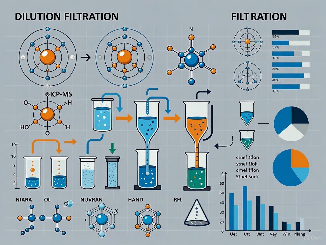
The ICP-MS Workflow: A Visual Guide
The entire ICP-MS process, from sample collection to data analysis, is a tightly integrated sequence. The following workflow diagram summarizes the key stages, highlighting the central role of sample preparation.
Figure 1: A simplified workflow for ICP-MS analysis. The sample preparation stage (green) is critical for ensuring the quality of the sample introduced into the instrumental components (red).
Critical Sample Preparation Parameters and Protocols
The sample introduction process is the most common source of error in ICP-MS analysis. Controlling the following parameters is essential for method robustness.
Filtration and Particulate Removal
Samples must be free of particulates, gels, or undigested material to prevent clogging the sample introduction system. Filtration through a 0.45 µm membrane is a standard practice for water samples and digested solutions [5] [3]. The cost of neglecting this step is high, as blockages can lead to downtime and expensive repairs, with micro-nebulizer replacements costing approximately $600 [3].
Dilution and Total Dissolved Solids (TDS)
To minimize matrix effects that alter ionization efficiency and to prevent rapid coating of the interface cones, samples should be diluted to a Total Dissolved Solids (TDS) content of ≤ 0.2% (2000 ppm) [3] [2] [6]. For biological fluids like blood, a dilution factor between 10 and 50 is typically sufficient to achieve this [2]. Conductivity meters can provide a rough estimate of TDS, with saline samples ideally having a conductivity below 4000 µS/cm and freshwater samples below 2860-3330 µS/cm after dilution [3].
Acidification and Sample Stability
Samples and calibration standards are typically prepared in a matrix of 1-2% high-purity nitric acid (HNO₃) to stabilize elements in solution and prevent adsorption to container walls [3] [6]. The use of the highest purity "trace metal grade" acids is critical to avoid introducing contamination that elevates background levels [6].
Experimental Protocols
Protocol 1: Preparation of Natural Water Samples for Strontium Isotope Analysis
This protocol, optimized for high-throughput analysis, allows filtered and acidified samples to be directly introduced into an automated separation system [5].
- Step 1: Field Collection and Filtration. Collect water sample using standard field practices. Immediately filter the sample through a 0.45 µm membrane filter.
- Step 2: Acidification for Stability. Acidify the filtered sample with ultra-high purity nitric acid (HNO₃) to a concentration of approximately 0.09 mol/L (pH of 1–2) to stabilize the analytes.
- Step 3: Automated Sr Separation. Directly load the prepared sample into a High-Pressure Ion Chromatography (HPIC) system. The system automatically separates Sr from matrix elements like Ca.
- Step 4: Collection and Analysis. Collect the purified Sr isolate in ultrapure water. Directly acidify this isolate to 0.5 mol/L HNO₃ for immediate analysis by MC-ICP-MS without the need for dry-down. This automated method can process 40–50 samples in a 24-hour period [5].
Protocol 2: Dilution of Biological Fluids for Trace Element Analysis
This protocol outlines two primary methods for preparing biological fluids, balancing simplicity with completeness of matrix removal [4].
- Method A: Direct Dilution
- Select Diluent. For elements like Cd, Hg, and Pb, use a 2% (v/v) high-purity nitric acid solution. For a broader panel (As, Cd, Co, Cr, Cu, Mn, Pb), a mixture of ammonia and nitric acid with a 50-fold dilution is more effective [4].
- Perform Dilution. Combine one part sample with the appropriate number of parts diluent (e.g., 1:19 for a 20x dilution) in a pre-cleaned polypropylene tube.
- Vortex Mix. Ensure complete homogenization of the solution.
- Method B: Acid Mineralization (Microwave Digestion)
- Aliquot Sample. Transfer a known volume (e.g., 0.5 mL) of blood or plasma into a dedicated microwave digestion vessel.
- Add Acid. Add 2-5 mL of 65% high-purity nitric acid. Optionally, add 1-2 mL of hydrogen peroxide (H₂O₂) for more complete organic matrix destruction [4] [6].
- Digest. Run the microwave digestion program according to the manufacturer's instructions (e.g., a temperature-controlled ramp to 180-200°C).
- Dilute Digestate. After cooling, quantitatively transfer the digestate to a volumetric flask and dilute to mark with Type 1 (18.2 MΩ·cm) water to achieve a final acid concentration of ~2% HNO₃.
Key Data and Quality Control
Typical Calibration Ranges and Limits of Detection
The table below provides an example of calibration ranges and estimated Limits of Detection (LOD) for various elements, illustrating the wide concentration range of the technique.
Table 1: Example ICP-MS Calibration Ranges and Limits of Detection (LOD) [3].
| Element Group | High Calibration Standard (ppb) | Estimated LOD (ppb) |
|---|---|---|
| Na, Ca | 25,000 | < 10 |
| Mg | 12,500 | < 2 |
| Si | 10,000 | < 3 |
| K | 6,250 | < 3 |
| P, B, Al, Fe, Li, Sr, Zn | 1,000 | < 1 |
| All other trace elements | 200 | < 1 |
The Scientist's Toolkit: Essential Research Reagents and Materials
Table 2: Essential materials and reagents for ICP-MS sample preparation protocols.
| Item | Function and Critical Specifications |
|---|---|
| High-Purity Nitric Acid (HNO₃) | Primary diluent and digestion acid; stabilizes metal ions in solution. Must be "trace metal grade" to minimize background contamination [4] [6]. |
| 0.45 µm Membrane Filters | Removes particulates to prevent nebulizer and tubing clogs. A standard for natural waters and final filtrations [5] [3]. |
| Polypropylene Tubes (15 mL) | Sample storage and preparation; less likely to contribute trace contamination than borosilicate glass with cardboard seals [3]. |
| Type 1 Ultrapure Water (18.2 MΩ·cm) | Preparation of all blanks, standards, and diluents; high resistivity ensures low ionic background [6]. |
| Internal Standard Solution | Corrects for non-spectroscopic matrix effects and instrument drift. Often a mix of Sc, Ge, Rh, In, Tb, Lu/Bi, added online or to all samples and standards [4]. |
| Certified Reference Materials (CRMs) | Validates method accuracy and precision. Should match the sample matrix (e.g., Seronorm for blood) [3]. |
The path to generating reliable, high-quality ICP-MS data is paved long before the sample reaches the plasma. As detailed in this note, a meticulously optimized sample introduction protocol—encompassing strategic dilution, rigorous filtration, and appropriate acidification—is not a mere preliminary step but the foundation of the entire analytical workflow. By adopting these standardized protocols for dilution and filtration, researchers can effectively bridge the "throughput gap" between sample preparation and modern, fast ICP-MS analysis, thereby ensuring data integrity, enhancing productivity, and achieving the exceptional sensitivity that makes ICP-MS an indispensable tool in scientific research and drug development.
In inductively coupled plasma mass spectrometry (ICP-MS), the sample introduction and ionization processes are highly sensitive to the physical and chemical properties of the sample. The core objectives of sample preparation—minimizing matrix effects and preventing contamination—are therefore critical for generating accurate, reproducible data. Sample preparation transforms a raw sample into an analyzable form while ensuring it is compatible with the instrument's operational parameters [7]. Inadequate sample preparation is in fact the cause of as much as 60% of all spectroscopic analytical errors [7]. This application note, framed within a broader thesis on dilution and filtration protocols, details the strategies and methodologies to achieve these fundamental objectives for ICP-MS research.
The Critical Role of Sample Preparation in ICP-MS
Sample preparation serves as the foundational step that dictates the success of any subsequent ICP-MS analysis. Its primary purposes are:
- Achieving Instrument Compatibility: ICP-MS instruments require liquid samples free of particulates that could clog the nebulizer or torch assembly. Micro-nebulizers, which are particularly sensitive, can incur replacement costs of approximately $600 [3].
- Ensuring Representative Data: Proper preparation creates homogeneous samples that yield reproducible results, as heterogeneous samples can lead to non-representative sampling [7].
- Protecting Instrument Integrity: Samples with high total dissolved solids (TDS) can rapidly coat sampler and skimmer cones, altering orifice geometries and becoming a source of contamination [3].
Core Objective I: Minimizing Matrix Effects
Matrix effects occur when components of the sample matrix interfere with the ionization efficiency of target analytes in the plasma, leading to suppressed or enhanced signals and inaccurate quantification.
Quantitative Assessment of Matrix Interferences
The following table summarizes key matrix-related challenges and their documented impacts on analytical data.
Table 1: Common Matrix Effects and Their Impact on ICP-MS Analysis
| Matrix Effect Type | Cause | Impact on Analysis | Supporting Data |
|---|---|---|---|
| Spectral Interference | Polyatomic ions (e.g., ArO⁺ on ⁵⁶Fe⁺) | False positives, elevated backgrounds, and poorer detection limits [8]. | Requires collision/reaction cell for removal [8]. |
| Non-Spectral Interference | High Total Dissolved Solids (TDS) | Changed ionization efficiency, signal suppression/enhancement, and coating of interface cones [3]. | Samples must be diluted to ≤ 200 ppm TDS for ideal analysis [3]. |
| Physical Interference | Presence of undissolved solids/particulates | Clogging of nebulizer and sample introduction system, leading to instrumental downtime [9] [3]. | Replacement cost for a clogged micro-nebulizer is ~$600 [3]. |
| Particle Loss | Sample pre-treatment (filtration/centrifugation) | Significant loss of analyte, invalidating quantitative data for nanoparticles [10]. | Up to 99% loss of natural Fe-containing particles post-filtration [10]. |
Protocols for Mitigating Matrix Effects
A. Dilution Protocols
Dilution is the primary strategy for reducing matrix effects.
- Recommended Diluent: Dilutions should be made in 2% high-purity nitric acid (HNO₃) to match the matrix of most conventional calibration standards [3] [11]. Some elements, such as gold, are more stable in a chloride matrix (e.g., 2% HCl) [3].
- TDS Guidelines: Samples should be diluted to a TDS content of ≤ 200 parts per million (ppm) for ideal analysis [3]. Conductivity meters can provide a rough estimate of TDS.
- Analyte-Specific Considerations: If major and trace elements are both of interest, running the sample at two separate dilutions may be necessary to keep all analyte concentrations within the optimal calibration range [3].
B. Filtration Protocols
Filtration removes suspended particulates to prevent nebulizer clogging.
- Pore Size: Use a 0.45 µm membrane filter for most applications. For ultratrace analysis, a 0.2 µm filter may be required [7].
- Filter Material: Select filter materials that do not introduce contamination or adsorb the analyte. PTFE (Polytetrafluoroethylene) membranes are generally recommended for their chemical resistance and low background [7].
- Critical Consideration for Nanoparticle Analysis: For single-particle ICP-MS (spICP-MS) studies, standard filtration can cause catastrophic particle loss. One study recorded losses of up to 99% for naturally occurring Fe-containing particles, demonstrating that filtration directly impedes quantitative particle analysis in environmental samples [10].
The sample introduction system itself can be optimized to handle complex matrices.
- Nebulizer Selection: Conventional concentric nebulizers with thin capillaries are prone to clogging. Using an innovative, robust non-concentric nebulizer with a larger sample channel internal diameter can improve resistance to clogging from high salt levels or small particulates, eliminating the need for labor-intensive filtration or centrifugation steps in some applications [12].
- Automation: Automated systems like the FiltrationStation can standardize the process, performing filtration, dilution, and acidification in a controlled manner, thereby improving reproducibility [13].
Core Objective II: Preventing Contamination
Contamination during sample preparation introduces exogenous elements that produce spurious spectral signals, compromising data integrity at ultra-trace concentration levels.
Table 2: Common Contamination Sources and Prevention Strategies
| Contamination Source | Impact | Prevention Strategy |
|---|---|---|
| Reagents & Acids | High and variable blank levels, adversely affecting accuracy and precision [3]. | Use high-purity, "trace metal grade" acids and reagents. Acidify samples to 2% HNO₃ with high-purity acid [3]. |
| Labware (Tubes, Vials) | Leaching of elements from container walls or introduction of contaminants from caps/seals [3]. | Use 15 mL conical bottom polypropylene tubes instead of borosilicate glass. Perform acid-washing of all labware [3]. |
| Sample Processing Equipment | Cross-contamination between samples from grinders, mills, or presses [7]. | Clean equipment intensively between samples. Use grinding surfaces that will not introduce interfering elements [7]. |
| Airborne Particulates | Introduction of ambient dust, skin cells, or fibers into samples [3]. | Work in a clean laboratory environment, use laminar flow hoods, and minimize sample exposure to the open air [3]. |
Contamination Control Protocols
- Labware Cleaning: All labware (polypropylene tubes, etc.) should be cleaned with high-purity acid, rinsed thoroughly with Type 1 (18.2 MΩ·cm) water, and dried in a controlled, clean environment before use [3].
- Method Blanks: Include method blank(s) with every batch of samples. These blanks undergo the entire preparation process using the same reagents to quantify and correct for contamination introduced during preparation [3].
- Workflow Management: To reduce sample-to-sample carryover, analyze samples in order of increasing concentration (blanks and controls first) [3].
Experimental Workflow for Reliable ICP-MS Analysis
The following diagram synthesizes the core objectives into a logical workflow for preparing liquid samples for ICP-MS analysis.
The Scientist's Toolkit: Essential Research Reagents & Materials
The following table details key consumables and equipment critical for executing the protocols described in this note.
Table 3: Essential Materials for ICP-MS Sample Preparation
| Item | Specification / Recommended Type | Primary Function |
|---|---|---|
| Nitric Acid (HNO₃) | High Purity, Trace Metal Grade | Primary diluent and acidifying agent; oxidizes organic matter during digestion [3] [11]. |
| Hydrochloric Acid (HCl) | High Purity, Trace Metal Grade | Alternative matrix for specific elements (e.g., Au); dissolves oxides and carbonates [3] [11]. |
| Laboratory Pure Water | Type 1 (18.2 MΩ·cm) | Used for all dilutions and final rinsing of labware to minimize background contamination [3]. |
| Syringe Filters | 0.45 µm or 0.2 µm Pore Size, PTFE Membrane | Removes suspended particulates to prevent nebulizer clogging [7]. |
| Sample Tubes | 15 mL Conical Polypropylene | Preferred container for analysis; less likely to contribute trace contamination than glass [3]. |
| Internal Standards | e.g., Sc, Ge, In, Bi, Li, Y | Added to all samples and standards to correct for instrument drift and matrix-induced suppression/enhancement [7]. |
| Certified Reference Materials (CRMs) | Matrix-matched to samples | Verifies analytical accuracy and method validation [3]. |
| Automated Filtration System | e.g., FiltrationStation | Automates filtration, dilution, and acidification to improve reproducibility and throughput [13]. |
The pursuit of reliable data in ICP-MS research hinges on a disciplined approach to sample preparation. The dual objectives of minimizing matrix effects and preventing contamination are not merely preliminary tasks but are integral to the analytical method itself. By adhering to the detailed protocols for dilution, filtration, and contamination control outlined in this note, researchers can ensure their samples are introduced to the instrument in an optimal state. This rigorous approach to sample preparation lays the necessary foundation for generating data that is both accurate and reproducible, thereby upholding the integrity of scientific conclusions in drug development and environmental research.
The accuracy of Inductively Coupled Plasma Mass Spectrometry (ICP-MS) analysis is fundamentally dependent on proper sample preparation. For biological matrices, which present challenges such as complex compositions and high total dissolved solids (TDS), implementing scientifically sound dilution protocols is essential to generate reliable data. This document outlines the core principles, calculations, and practical protocols for diluting biological samples for ICP-MS analysis, providing researchers with a framework to optimize their analytical methods.
The primary goals of sample dilution are threefold: first, to reduce the matrix effect and minimize signal suppression or enhancement; second, to ensure that the analyte concentration falls within the instrument's linear dynamic range; and third, to lower the total dissolved solids content to prevent instrumental drift and physical blockages [14]. Modern ICP-MS applications increasingly favor simple "dilute-and-shoot" approaches for their efficiency and reduced contamination risk. For instance, a recent 2025 method for simultaneously determining 40 elements in urine and blood samples used only a 1% nitric acid direct dilution, completing sample pretreatment within minutes [15].
Calculating Dilution Factors
The dilution factor (DF) is a critical numerical value that quantifies the extent of dilution and must be applied to instrument-read concentrations to determine the original concentration in the sample. The fundamental formula for calculating a dilution factor is:
Dilution Factor (DF) = Final Volume / Initial Volume
For serial dilutions, the overall dilution factor is the product of the individual dilution factors at each step. The original concentration in the sample is then calculated as:
Original Concentration = Instrument Concentration × Dilution Factor
Practical Application and Considerations
In practice, the required dilution factor depends on the analyte concentrations and the sample matrix. For unknown samples, it is advisable to analyze the sample at multiple dilution levels (e.g., 10x, 100x, 1000x) to ensure that all analytes of interest are within the calibration range [3]. The optimal dilution should place the analyte signal comfortably within the calibration curve while maintaining a matrix simple enough to avoid significant interferences. For biological samples like urine and serum, a simple direct dilution often suffices, as demonstrated by a method that achieved recovery rates of 81.92–108.66% in urine and 83.47–110.32% in serum with just a 1% nitric acid dilution [15].
Table 1: Typical Dilution Scenarios for Biological Samples in ICP-MS
| Sample Type | Typical Dilution Factor | Diluent | Key Considerations |
|---|---|---|---|
| Urine | 1:10 to 1:50 | 1% HNO₃ | Minimizes matrix effects; common for multi-element analysis [15]. |
| Serum/Blood | 1:10 to 1:50 | 1% HNO₃, sometimes with Triton-X-100 or methanol | Helps solubilize proteins and maintain analyte stability [15]. |
| Digested Tissue | Variable (to achieve ≤ 0.2% TDS) | 1-2% HNO₃ | Final TDS content is more critical than a fixed factor [14]. |
The following workflow outlines the logical process for determining the correct dilution factor for an analysis.
Selecting the Appropriate Diluent
The choice of diluent is as critical as the dilution factor. An ideal diluent matches the calibration standard matrix, stabilizes the analytes in solution, and minimizes the sample's introduction system and plasma.
Common Diluents and Their Applications
- Nitric Acid (1-2% v/v): This is the most universal and recommended diluent for elemental analysis. It provides a low and consistent matrix, helps keep metals in solution, and is compatible with the ICP-MS sample introduction system. Most calibration standards are prepared in 1-2% nitric acid, making it the default choice [3] [11]. A 2025 study successfully used 1% nitric acid for the direct dilution of both urine and serum for the analysis of 40 elements [15].
- Hydrochloric Acid (HCl): Used for stabilizing certain elements that form chloro-complexes, such as mercury and platinum group metals. A concentration of 2% or higher is often necessary to prevent precipitation and ensure accurate quantification of these elements [6].
- Surfactants and Additives: For complex biological matrices like serum, additives such as Triton X-100 (e.g., 0.02%) or small percentages of methanol (e.g., 2%) can be added to the acidic diluent. These help to solubilize proteins, ensure a homogeneous solution, and improve aerosol generation and transport efficiency during nebulization [15].
- Ammonium Hydroxide and Organic Solvents: For specific applications involving organic compounds or specialized matrices, basic diluents like 4% ammonium hydroxide or mixtures containing n-butanol and EDTA may be used [16].
Table 2: Guide to Diluent Selection for Biological Matrices
| Diluent | Common Concentration | Primary Use Case | Advantages | Cautions |
|---|---|---|---|---|
| Nitric Acid (HNO₃) | 1-2% (v/v) | Universal; urine, digests, general multi-element analysis [15] [3]. | Matches calibration standards; keeps most metals in solution. | May not stabilize elements like Hg without Cl⁻. |
| HCl | ≥ 2% (v/v) | Stabilizing Hg, Pt, Au, and other elements forming chloro-complexes [6] [3]. | Prevents precipitation of volatile species. | Can create polyatomic interferences (e.g., ArCl⁺ on As⁺). |
| Triton X-100 | 0.02-0.1% (v/v) | Added to diluent for serum/plasma or viscous samples [15]. | Reduces surface tension, improves nebulization, solubilizes proteins. | Requires thorough rinsing to avoid memory effects; contributes organics to plasma. |
| Methanol | 1-2% (v/v) | Added to diluent to enhance sensitivity for some elements. | Can improve ionization for certain elements. | Alters plasma conditions, requires stable plasma tuning. |
Experimental Protocol: Direct Dilution of Paired Serum and Urine Samples
This protocol is adapted from a 2025 study that developed a high-throughput method for the simultaneous determination of 40 metal and non-metallic elements in paired biological samples [15].
Reagents and Materials
- Ultrapure water: Resistivity of 18.2 MΩ·cm
- Nitric acid (HNO₃): Trace metal grade
- Triton X-100 (for serum dilution)
- Calibration standards: Multi-element standard solution, custom mixed or commercial
- Internal standard solution: Contains elements not present in the sample (e.g., Sc, Ge, Rh, In, Tb, Bi)
- Pipettes: Adjustable, with disposable tips
- Sample tubes: 15 mL conical-bottom polypropylene tubes, pre-cleaned with 1% nitric acid [16]
Step-by-Step Procedure
Diluent Preparation:
- Urine Diluent: Prepare a 1% (v/v) solution of trace metal grade nitric acid in ultrapure water.
- Serum Diluent: Prepare a solution of 1% (v/v) nitric acid and 0.02% (v/v) Triton X-100 in ultrapure water.
Sample Dilution:
- For Urine: Pipette 100 µL of well-mixed urine into a 15 mL tube. Add 9.9 mL of the 1% HNO₃ diluent. This yields a 1:100 (v/v) dilution. Mix vigorously on a vortex mixer for 10 seconds.
- For Serum: Pipette 100 µL of serum into a 15 mL tube. Add 9.9 mL of the 1% HNO₃ / 0.02% Triton X-100 diluent. This yields a 1:100 (v/v) dilution. Mix vigorously on a vortex mixer for 30 seconds to ensure complete solubilization.
Calibration and Quality Control:
- Prepare calibration standards in the same diluent as the samples (1% HNO₃ for urine simulant, 1% HNO₃ / 0.02% Triton for serum simulant) to minimize matrix effects.
- Incorporate a method blank (diluent only), a certified reference material (CRM), and a spike recovery sample to validate the analytical run.
ICP-MS Analysis:
- Use the Kinetic Energy Discrimination (KED) mode with a collision gas (e.g., He) to minimize polyatomic interferences.
- Add the internal standard online post-nebulization or to all samples and standards to correct for instrument drift and matrix suppression.
Method Validation Data
The original study reported the following performance characteristics for this protocol, demonstrating its robustness for clinical biomonitoring [15].
Table 3: Method Validation Data from the 2025 Protocol for 40 Elements
| Parameter | Urine | Serum |
|---|---|---|
| Linear Range | R² ≥ 0.999 | R² ≥ 0.999 |
| Limit of Detection (LOD) | As low as 2 ng/L | As low as 20 ng/L |
| Recovery Rate | 81.92% – 108.66% | 83.47% – 110.32% |
| Precision (RSD) | < 15% | < 15% |
| Analysis Time | < 6 minutes per sample | < 6 minutes per sample |
The Scientist's Toolkit: Essential Research Reagents and Materials
Successful sample preparation requires high-purity reagents and dedicated labware to prevent contamination, which is a significant concern at trace metal concentrations.
Table 4: Essential Reagents and Materials for ICP-MS Sample Preparation
| Item | Function / Purpose | Critical Specifications |
|---|---|---|
| Nitric Acid (HNO₃) | Primary diluent and digestion acid; oxidizes organic matter and stabilizes metals in solution. | Trace metal grade; sub-boiling distilled is ideal for ultratrace work. |
| Hydrochloric Acid (HCl) | Used for stabilizing specific elements (e.g., Hg, Au) and digesting some inorganic matrices. | Trace metal grade. |
| Ultrapure Water | Diluent and rinsing agent for all solutions and labware. | Resistivity of 18.2 MΩ·cm at 25°C. |
| Triton X-100 | Non-ionic surfactant added to diluents for viscous biological fluids (e.g., serum) to improve homogeneity and aerosol generation. | High purity. |
| Single-Element & Multi-Element Stock Standards | For preparation of calibration curves and quality control materials. | Certified Reference Materials (CRMs) from a national metrology institute (NMI) or accredited commercial supplier. |
| Internal Standard Stock Solution | Corrects for instrument drift and matrix-induced signal suppression/enhancement. | Contains elements (e.g., Sc, Ge, Rh, In, Bi) not expected in the samples. |
| Polypropylene Tubes & Vials | For storing and diluting samples, standards, and reagents. | Pre-cleaned; certified metal-free. |
| Pipette Tips | For accurate and precise liquid transfer. | With aerosol barrier; certified metal-free. |
Mastering the fundamental principles of dilution is non-negotiable for generating high-quality, reliable data in ICP-MS analysis of biological matrices. The process involves a careful balance of calculating the correct dilution factor to bring analytes into the optimal range while minimizing the matrix, and selecting a diluent that ensures analyte stability and compatibility with the instrument. The provided protocol, based on a recent and validated "dilute-and-shoot" method, serves as a powerful template that can be adapted for a wide range of research applications, from clinical biomonitoring to pharmaceutical development. By adhering to these principles and rigorously applying quality control measures, researchers can ensure the accuracy and integrity of their elemental analyses.
In the realm of inductively coupled plasma mass spectrometry (ICP-MS) research, sample preparation is not merely a preliminary step but a critical determinant of analytical success. Effective filtration protocols ensure that samples introduced into the high-temperature plasma are free of particulates that could compromise instrument integrity and analytical accuracy. The fundamental principles of filtration—encompassing separation mechanisms, precise pore size selection, and chemical compatibility—form the cornerstone of reliable ICP-MS methodologies in pharmaceutical development and environmental research [17] [3]. This application note delineates comprehensive protocols for integrating filtration practices within sample preparation workflows for ICP-MS analysis, addressing the specific needs of researchers and scientists engaged in trace element analysis and drug development.
The consequences of inadequate filtration are far-reaching, including nebulizer clogging, signal drift, and inaccurate quantification [3]. Particulates in samples can obstruct the delicate sample introduction system of ICP-MS instruments, requiring costly repairs exceeding $600 for micro-nebulizer replacements [3]. Furthermore, undigested particles or gels can introduce spectral interferences and matrix effects that compromise the exceptional sensitivity and detection limits that make ICP-MS indispensable for measuring trace elements in biological fluids [2]. Thus, a meticulous approach to filtration is not optional but essential for any rigorous ICP-MS research protocol.
Theoretical Foundations of Filtration
Filtration Mechanisms and Pore Size Significance
Membrane filtration operates as a selective barrier, separating components based on size exclusion through microscopic pores. The pore size, defined as the average diameter of these openings, directly determines the cut-off point for separation and is typically measured in micrometers (µm) or nanometers (nm) [18]. In the context of ICP-MS sample preparation, filtration serves to remove particulate matter that could potentially clog the nebulizer and sample introduction system, while simultaneously ensuring that the analyzed sample represents the truly dissolved fraction of metals [3].
The relationship between pore size and filtration efficiency follows a fundamental principle: smaller pore sizes retain finer particles but typically require higher pressure and result in reduced flow rates due to increased flow resistance [18]. This trade-off necessitates careful selection based on application requirements. For ICP-MS analysis, where the objective is to remove particulates without altering the dissolved elemental composition, pore sizes typically reside within the microfiltration range (0.1-10 µm) [18]. The precise selection depends on the specific sample matrix and analytical objectives, balancing the need for particle-free samples with the practical considerations of filter capacity and flow rates.
Classification of Membrane Filtration Technologies
Filtration technologies are systematically classified based on their pore sizes and separation capabilities, as detailed in Table 1. This classification provides a framework for selecting appropriate filtration methods for specific applications within ICP-MS workflows.
Table 1: Classification of Membrane Filtration Technologies
| Filtration Type | Typical Pore Size Range | Key Separations | Common Applications in ICP-MS Research |
|---|---|---|---|
| Microfiltration (MF) | 0.1 to 10 µm [18] | Suspended solids, bacteria, large colloids [18] | Sample clarification; removal of undigested particles from digested samples [3] |
| Ultrafiltration (UF) | 0.01 to 0.1 µm (1,000-500,000 Daltons MWCO*) [18] | Viruses, proteins, macromolecules [18] | Separating protein-bound elements from free ions in biological fluids; sample fractionation |
| Nanofiltration (NF) | 0.001 to 0.01 µm (150-1,000 Daltons MWCO) [18] | Divalent ions, small organic molecules [18] | Specialized separations for complex matrices |
| Reverse Osmosis (RO) | < 0.001 µm (non-porous) [18] | Virtually all dissolved salts and ions [18] | Production of ultrapure water for diluent preparation [6] |
*MWCO: Molecular Weight Cut-Off
For most ICP-MS sample preparation applications, microfiltration membranes with pore sizes of 0.45 µm or 0.2 µm are employed as the standard for ensuring particulate-free solutions without removing dissolved analytes of interest [3]. These pore sizes effectively retain particulates that could clog the nebulizer while allowing dissolved elements to pass through for analysis.
Filter Media Selection and Compatibility
Filter Material Properties and Applications
The chemical composition of filter membranes significantly influences their performance and suitability for specific sample types. Table 2 outlines the properties and applications of common filter materials in ICP-MS research, with chemical compatibility being a paramount consideration.
Table 2: Filter Material Properties and Applications for ICP-MS
| Filter Material | Available Pore Sizes (µm) | Material Features | ICP-MS Applications & Compatibility |
|---|---|---|---|
| Polyethersulfone (PES) | 0.1, 0.2, 0.45, 0.65, 0.8, 1.2 [17] | Hydrophilic, low protein binding, high flow rate [17] | Ideal for aqueous samples, biological fluids, culture media; compatible with dilute acids and alkalis used for sample dilution [17] [2] |
| Polytetrafluoroethylene (PTFE) | 0.1, 0.2, 0.45, 1.0, 3.0, 5.0, 10 [17] | Hydrophobic, chemically inert, high-temperature resistance [17] | Suitable for aggressive chemicals, organic solvents, ketones; excellent for digestates containing strong acids [17] [6] |
| Polypropylene (PP) | 0.1, 0.2, 0.45, 0.65, 0.8, 1.0, 3.0, 5.0, 10 [17] | High chemical resistance, economical [17] | Effective for pre-filtration of complex matrices; not recommended for final sterilizing filtration [17] |
Chemical compatibility between the filter membrane and the sample matrix is critical to prevent both membrane degradation and sample contamination. In ICP-MS analysis, where samples may contain various acids (e.g., nitric, hydrochloric) or alkaline diluents, verifying compatibility is essential [17] [6]. Incompatible filters can leach contaminants into samples or introduce interferences that compromise analytical results at trace levels.
Pore Size Selection for Specific Applications
The selection of appropriate pore size represents a balance between filtration efficiency and practical considerations. For ICP-MS sample preparation, the following guidelines apply:
- 0.45 µm pore size: Recommended for general clarification of solutions, removing larger particulates and microorganisms without achieving full sterility. This pore size is suitable for most aqueous samples and digestates where the primary concern is nebulizer protection [18].
- 0.2 µm pore size: Considered "sterilizing grade" for bacterial removal [17] [18]. Essential for samples where any microbial contamination could interfere with analysis or for biological samples requiring sterility. It's important to note that while 0.2 µm filtration removes most bacteria, some very small bacteria (like Mycoplasma) and viruses can pass through [18].
- 0.1 µm pore size: Provides enhanced retention of fine particles and colloids that might otherwise pass through larger pores and contribute to matrix effects [17].
The distinction between 0.2 µm and 0.22 µm, while seemingly minor, can be significant in certain applications. For instance, Brevundimonas diminuta, a benchmark organism used in validation studies, may pass through a 0.22 µm filter but is reliably retained by an absolute 0.2 µm filter [17]. For ICP-MS applications, 0.2 µm filtration provides sufficient particulate removal while maintaining reasonable flow rates.
Integrated Filtration and Dilution Protocols for ICP-MS
Comprehensive Sample Preparation Workflow
The integration of filtration within the broader context of ICP-MS sample preparation requires careful planning and execution. The following workflow diagram illustrates the complete pathway from sample collection to instrumental analysis:
Figure 1: Comprehensive ICP-MS Sample Preparation Workflow Integrating Filtration Steps
Step-by-Step Filtration Protocol for Liquid Samples
Protocol Title: Particulate Removal from Aqueous Samples for ICP-MS Analysis
Principle: This protocol describes the procedure for removing particulate matter from liquid samples using membrane filtration to prevent nebulizer clogging and reduce matrix effects in ICP-MS analysis [3].
Materials and Reagents:
- Sample filtration apparatus (syringe filter assembly or vacuum filtration system)
- Membrane filters (0.45 µm or 0.2 µm pore size, PES or PTFE based on compatibility)
- Syringes (if using syringe filters, 5-60 mL volume, plastic-free)
- Sample collection vials (15 mL conical bottom polypropylene tubes)
- Ultrapure water (18.2 MΩ·cm resistivity) [6]
- High-purity nitric acid (trace metal grade)
Procedure:
- Sample Pre-treatment: For complex matrices with high particulate load, consider a pre-filtration step using a 1.0 µm pore size filter to extend the life of the final filter [18].
- Filter Assembly: Assemble the filtration apparatus according to manufacturer instructions. Ensure all components are clean and contamination-free.
- Filter Conditioning: Pre-wet the filter with 5-10 mL of ultrapure water or dilute acid matching the sample matrix to remove potential contaminants from the filter itself.
- Sample Filtration: Pass the sample through the filter using gentle pressure. Discard the first 1-2 mL of filtrate to avoid dilution effects from the conditioning step.
- Filtered Sample Collection: Collect the filtered sample in appropriately cleaned polypropylene tubes. For ICP-MS, a minimum of 5 mL is recommended for conventional sample introduction systems [3].
- Post-filtration Treatment: Acidify the filtered sample to 2% (v/v) with high-purity nitric acid to maintain element stability, unless analyzing elements that require different stabilization (e.g., gold in hydrochloric acid) [3].
- Quality Control: Process method blanks through the entire filtration and preparation procedure to identify potential contamination sources.
Notes:
- For samples with high dissolved solids content (>0.2%), additional dilution may be necessary after filtration to minimize matrix effects [3].
- Always process samples in order of increasing concentration to minimize carry-over effects [3].
- Automated filtration systems (e.g., FiltrationStation) can enhance reproducibility and throughput for high-volume laboratories [13].
Essential Research Reagent Solutions
Table 3: Essential Reagents and Materials for ICP-MS Sample Preparation and Filtration
| Reagent/Material | Specification/Purity | Function in ICP-MS Sample Preparation |
|---|---|---|
| Nitric Acid (HNO₃) | Trace metal grade, high purity [6] | Primary diluent for elemental analysis; aids in sample stabilization and digestion |
| Hydrochloric Acid (HCl) | Trace metal grade, high purity [6] | Used in digestions (e.g., aqua regia); stabilizes certain elements (e.g., Hg, Au) |
| Hydrogen Peroxide (H₂O₂) | High purity [6] | Oxidizing agent for digesting organic matrices in combination with nitric acid |
| Ultrapure Water | 18.2 MΩ·cm resistivity [6] | Diluent preparation, equipment rinsing; minimal elemental background |
| Polypropylene Tubes | 15 mL conical bottom, acid-cleanable [3] | Sample collection and storage; minimal elemental leaching |
| PES Membrane Filters | 0.2 µm and 0.45 µm pore sizes [17] | Primary filtration for aqueous samples; low protein binding |
| PTFE Membrane Filters | 0.2 µm and 0.45 µm pore sizes [17] | Filtration of aggressive chemicals and organic solvents |
Quality Assurance and Troubleshooting
Quality Control Measures
Implementing rigorous quality control measures is essential for validating filtration protocols in ICP-MS research:
- Method Blanks: Process blanks through the entire filtration and preparation procedure to identify contamination from reagents, equipment, or the environment [3]. High and variable blank levels will adversely affect the quality and uncertainty of reported results [3].
- Matrix Spikes: Add known concentrations of analytes to samples after filtration to evaluate recovery efficiencies and identify potential matrix effects [3].
- Replicate Analyses: Perform replicate analyses of samples to evaluate analytical reproducibility and precision [3].
- Certified Reference Materials: Analyze appropriate certified reference materials with similar matrices to validate method accuracy [3].
Troubleshooting Common Filtration Issues
- Slow Filtration Rates: May indicate filter clogging due to high particulate load. Consider pre-filtration with a larger pore size filter (0.8-1.0 µm) or sample dilution prior to filtration [18].
- Poor Element Recovery: Could result from analyte adsorption to filter membranes. Validate recovery for target elements and consider alternative membrane materials (e.g., PES for low protein binding) [17].
- Contamination: evidenced by elevated blanks. Systematically evaluate all potential contamination sources including filters, reagents, and labware. Use only high-purity acids and conduct leach tests for plasticware [6].
- Filter Incompatibility: Manifested as membrane degradation or unusual results. Verify chemical compatibility between filter material and sample matrix, especially with organic solvents or strong acids [17].
The integration of appropriate filtration methodologies within ICP-MS sample preparation workflows is a critical component of robust analytical protocols. By understanding the fundamental principles of filtration mechanisms, pore size selection, and material compatibility, researchers can develop optimized approaches that ensure sample integrity and analytical reliability. The protocols outlined in this document provide a framework for implementing effective filtration strategies that protect instrumentation, minimize interferences, and generate high-quality data for pharmaceutical development and environmental research applications. As ICP-MS technology continues to evolve toward increasingly sensitive detection, the importance of meticulous sample preparation through proper filtration will only grow in significance.
Inductively Coupled Plasma Mass Spectrometry (ICP-MS) is a powerful technique for ultra-trace elemental analysis, known for its high sensitivity, good precision, and wide dynamic range [19]. The technique has become a dominant tool across diverse fields, including environmental monitoring, clinical research, pharmaceuticals, and geochemistry [12]. However, the reliability of the analytical results—embodied by the key figures of merit (detection limits, accuracy, and precision)—is highly dependent on the entire analytical workflow. Sample preparation, particularly dilution and filtration, is a critical pre-analytical step that can significantly influence these figures of merit. Within the context of a broader thesis on ICP-MS research, this application note details how specific dilution and filtration protocols directly impact the detection capability, trueness of results, and measurement reproducibility. Adherence to meticulously designed protocols is not merely a procedural formality but a fundamental requirement for generating data that is both reliable and traceable [19].
Current Application Landscape and the Centrality of Sample Preparation
The ICP-MS marketplace installs approximately 2,000 systems annually worldwide, with single quadrupole instruments comprising about 80% of these installations [12]. The technique's applicability has expanded far beyond specialized research laboratories into high-throughput contract laboratories, necessitating robust and rugged methodologies that can handle complex sample matrices with minimal operator intervention [12]. This expansion underscores the need for standardized, optimized sample preparation protocols.
A core challenge is that samples often contain high concentrations of dissolved solids or particulate matter, which can cause instrumental blockages, matrix effects, and spectral interferences [12]. Consequently, sample preparation strategies like dilution and filtration are frequently employed to produce a sample introduction solution that is compatible with the instrument. However, these strategies must be chosen and validated with care, as they can inadvertently alter the sample's composition and directly affect the final analytical results.
Impact of Dilution and Filtration on Analytical Figures of Merit
Impact on Detection Limits
Detection limits in ICP-MS are influenced by the signal-to-noise ratio, which can be optimized through measurement protocol [20]. However, sample preparation plays an equally crucial role. While dilution can mitigate matrix effects, it proportionally reduces the analyte concentration. For a target analyte present at an ultra-trace level (e.g., 10 ppt), a 1:10 dilution pushes its concentration closer to the instrument's background noise, potentially degrading the reported detection limit [12]. Furthermore, dilution can alter the physical properties of the solution, such as viscosity, which may affect nebulization efficiency and, consequently, the sensitivity [19].
Filtration, aimed at removing particulates that could clog the sample introduction system, can have a more complex and detrimental impact on detection limits for particulate analysis. In Single Particle ICP-MS (SP ICP-MS) studies, common sample preparation strategies like syringe filtration or centrifugation can lead to catastrophic losses of nano- and microparticles.
Table 1: Impact of Sample Preparation on Particle Recovery in SP ICP-MS
| Sample Preparation Strategy | Particle Type | Typical Particle Recovery | Key Findings |
|---|---|---|---|
| Filtration or Centrifugation | Gold Nanoparticles (spiked) | ≤10% | Significant loss of detectable particles, impeding quantitative analysis [10]. |
| Filtration or Centrifugation | Natural Fe-containing particles | ≤1% | Near-total loss of natural particles, indicating model nanoparticles are poor analogues for environmental samples [10]. |
| Addition of Surfactant (Triton X-100) | Gold Nanoparticles (spiked) | Up to ~30% | Provides moderate recovery improvement for some synthetic particles but remains ineffective for natural particles [10]. |
Impact on Accuracy
Accuracy—the closeness of a measured value to a true value—is paramount in quantitative analysis [19]. Dilution and filtration can introduce significant inaccuracies if not properly controlled.
- Dilution Errors: Inaccurate dilution, whether due to improper technique, miscalibration of volumetric equipment, or the use of impure reagents and acids, will systematically bias all subsequent results [20]. The purity of diluents is critical, as contaminants can lead to false positive signals or elevated baselines.
- Filtration Errors: As demonstrated in Table 1, filtration can selectively remove analyte-bearing particles. This leads to a substantial underestimation of particle number concentration and a skewing of particle size distribution in SP ICP-MS analysis [10]. The chemical and physical properties of the filter material can also interact with ionic or particulate analytes, leading to adsorptive losses and a reduction in the reported concentration for dissolved metals as well [10].
For accurate quantitative measurements, calibration using appropriate standards is essential. The use of matrix-matched standards or isotopic dilution can help compensate for some of these effects, but it cannot correct for the irreversible loss of analytes during preparation [19].
Impact on Precision
Precision refers to the closeness of agreement between independent measurement results obtained under stipulated conditions [19]. Inconsistent sample preparation is a major source of poor precision.
- Dilution Consistency: Manual performance of serial dilutions can introduce random variations. Automated dilution systems can improve reproducibility but must be regularly calibrated.
- Filtration Reproducibility: The process of filtration is prone to variability. Factors such as pressure applied, filter lot consistency, and potential clogging can cause the fraction of analyte passing through the filter to vary between samples, leading to poor precision [10]. This is particularly critical for particulate analysis, where the recovery is already low and highly variable.
Experimental Protocols
General Protocol for Dilution of Liquid Samples for Trace Element Analysis
1. Scope: This protocol describes the procedure for diluting liquid samples with an acidic matrix (e.g., digests) to a level suitable for ICP-MS analysis. 2. Principal: Samples are volumetrically diluted with high-purity dilute acid (e.g., 1-2% v/v HNO~3~) to bring analyte concentrations within the instrument's calibration range and to reduce matrix effects. 3. Reagents & Equipment: - High-purity water (e.g., 18.2 MΩ·cm) - High-purity concentrated nitric acid (HNO~3~) - Class A volumetric glassware or calibrated automatic pipettes - Trace metal-free vials and caps 4. Procedure: a. Calculate the required dilution factor based on prior knowledge or a qualitative scan. b. Pipette an appropriate aliquot of the well-homogenized sample into a clean vial. c. Add the calculated volume of diluent (e.g., 1% v/v HNO~3~). The acidification helps keep dissolved metals in solution. d. Cap the vial and mix thoroughly by inverting at least 10 times. e. The diluted sample is now ready for analysis. 5. Quality Control: - Process a method blank alongside samples to account for any contamination from reagents or equipment. - Analyze a certified reference material (CRM) that has undergone the same dilution procedure to verify accuracy. - Analyze duplicate samples to monitor precision.
Protocol for Evaluating Filtration Procedures for SP ICP-MS
1. Scope: This protocol evaluates the recovery efficiency of nanoparticles following filtration, using spiked surrogate nanoparticles. 2. Principal: A sample is spiked with a known concentration of well-characterized nanoparticles (e.g., 100 nm Au). The particle number concentration (PNC) is measured by SP ICP-MS before and after filtration to calculate recovery. 3. Reagents & Equipment: - Standard nanoparticle suspension (e.g., 100 nm ± 8 nm Au nanoparticles) - Various syringe filters (e.g., different pore sizes, membrane materials) - SP ICP-MS instrument 4. Procedure: a. Characterize the PNC and size distribution of the stock nanoparticle suspension (Pre-filtration measurement). b. Gently homogenize the sample and split it into two portions. c. Spike one portion with a known volume of the nanoparticle standard. d. Filter both the spiked and unspiked samples according to the standard operating procedure (e.g., discard first 1 mL of filtrate). e. Measure the PNC and size distribution of the filtrate of the spiked sample (Post-filtration measurement). f. Analyze the unspiked, filtered sample to determine the background. 5. Calculations & Evaluation: - Recovery (%) = (PNC~post~ / PNC~pre~) × 100 - A recovery of <90% indicates significant analyte loss, making the filtration protocol unsuitable for quantitative analysis [10]. - Compare the size distributions pre- and post-filtration to check for size-based bias.
Workflow and Decision Pathway
The following diagram visualizes the experimental workflow for developing and validating a sample preparation method for ICP-MS, highlighting critical decision points for dilution and filtration.
The Scientist's Toolkit: Key Research Reagent Solutions
Table 2: Essential Materials for Dilution and Filtration Protocols in ICP-MS
| Item | Function & Importance |
|---|---|
| High-Purity Acids (HNO~3~) | Primary diluent for digestates; purity is critical to prevent contamination and elevated background signals [20]. |
| High-Purity Water (18.2 MΩ·cm) | Base for all diluents and rinsing solutions; low elemental content is essential for ultra-trace analysis. |
| Certified Reference Materials (CRMs) | Used to validate the entire method (digestion, dilution, filtration, analysis); verifies accuracy and traceability [19]. |
| Single-Element & Multi-Element Standards | For instrument calibration and quality control checks; must be traceable to a national standard [19]. |
| Well-Characterized Nanoparticles (e.g., Au, 100 nm) | Vital surrogates for evaluating particle recovery in filtration studies for SP ICP-MS [10]. |
| Syringe Filters (Various Pore Sizes/Materials) | For sample clean-up; must be selected and evaluated to minimize analyte adsorption and particle loss [10]. |
| Surfactants (e.g., Triton X-100) | Can be added to improve nanoparticle stability in suspension and potentially mitigate losses during filtration [10]. |
Proven Dilution and Filtration Protocols for Complex Biomedical Samples
The dilute-and-shoot approach is a streamlined sample preparation strategy characterized by its simplicity, minimal analyte loss, and high sample throughput [21]. This technique is particularly advantageous for the analysis of liquid samples in high-volume clinical and pharmaceutical settings. When applied to inductively coupled plasma mass spectrometry (ICP-MS), it facilitates rapid, multi-element trace analysis of biological fluids such as urine and serum. The core principle involves a simple dilution of the sample in an appropriate aqueous matrix, which stabilizes the analytes and renders the sample compatible with the instrument's introduction system, thereby eliminating lengthy digestion procedures [22] [2]. This protocol outlines a validated dilute-and-shoot procedure for the determination of elemental impurities, framed within a broader research thesis on dilution and filtration sample preparation workflows for ICP-MS.
Principles and Advantages of the Dilute-and-Shoot Technique
The dilute-and-shoot technique significantly enhances laboratory efficiency. Its simplicity reduces sample preparation time, minimizes the consumption of reagents, and decreases the potential for contamination introduced by complex sample handling [22] [6]. For ICP-MS analysis, which is inherently a multi-element technique, this approach allows for the rapid screening of a large number of samples and analytes, making it ideal for routine analysis in drug development and clinical research [2].
A critical consideration for this strategy is managing the sample matrix. Direct introduction of a diluted biological sample can lead to matrix effects, affecting nebulization efficiency, plasma energy, and ultimately causing signal suppression or enhancement [22] [6]. Furthermore, the total dissolved solids (TDS) content must be controlled; for ICP-MS, a TDS content below 0.2% is generally recommended to prevent nebulizer clogging, cone blockage, and signal drift [2] [23]. These challenges can be effectively mitigated through optimized dilution factors and robust calibration methods, as detailed in the following sections.
Materials and Reagents
Research Reagent Solutions
| Item | Function/Justification |
|---|---|
| High-Purity Nitric Acid (HNO3) | Primary diluent component; stabilizes trace elements in solution and prevents adsorption to vial walls [2] [11]. |
| High-Purity Hydrochloric Acid (HCl) | Additive to stabilize specific elements (e.g., Hg, Au, Pt-group metals) by forming stable chloro-complexes [6] [23]. |
| Internal Standard Solution (e.g., Y, In, Sc, Bi) | Corrects for instrument drift, sample viscosity differences, and matrix-induced suppression/enhancement [22] [2]. |
| Triton-X-100 (Surfactant) | Helps solubilize and disperse lipids and membrane proteins in biological samples, improving homogeneity [2]. |
| Ammonium Hydroxide / Tetramethylammonium Hydroxide (TMAH) | Alkaline diluents; an alternative to acids for samples where protein precipitation at low pH is a concern [2]. |
Additional Materials: Deionized water (18.2 MΩ·cm resistivity), adjustable-volume pipettes and tips, sterile polypropylene tubes, 0.45 µm syringe filters (for filtration if required), and an ICP-MS instrument equipped with a pneumatic nebulizer.
Experimental Protocol
Sample Preparation Workflow
The following diagram illustrates the complete dilute-and-shoot procedure from sample collection to data analysis.
Step-by-Step Procedure
Sample Pretreatment:
Diluent Preparation:
- Prepare the dilution matrix as 2% (v/v) high-purity nitric acid in 18.2 MΩ·cm deionized water [11] [23].
- For elements such as Hg, Au, Pd, or Pt, add 0.5% (v/v) high-purity hydrochloric acid to the diluent to stabilize them [6] [23].
- Spike the diluent with a multi-element internal standard solution (e.g., Y, In, Sc, Bi) at a consistent concentration across all samples, blanks, and calibration standards [22] [2].
Dilution:
- Perform a 1:10 (v/v) dilution by adding 100 µL of the centrifuged sample to 900 µL of the prepared diluent in a polypropylene tube. For samples with high analyte concentrations or a complex matrix, a 1:20 dilution may be necessary [22].
- Vortex the mixture vigorously for at least 30 seconds to ensure complete homogenization.
Filtration (Optional):
Analysis:
- Transfer the final diluted and filtered solution to a labeled ICP-MS autosampler vial for analysis.
Calibration and Data Quality Control
Effective calibration is crucial for overcoming matrix-related interferences in the dilute-and-shoot approach.
- Internal Standardization (IS): This is the recommended calibration method. A constant concentration of an internal standard (e.g., Yttrium) is added to all samples, calibration standards, and blanks. The analyte-to-internal standard signal ratio is used for quantification, correcting for signal fluctuations and matrix effects [22].
- One-Point Standard Addition (OP SA): For complex matrices, this method can be highly effective. It involves analyzing the sample and the sample spiked with a known concentration of analyte, which corrects for matrix effects within the specific sample [22].
Table 1: Key Analytical Parameters for a 1:10 Dilute-and-Shoot ICP-MS Method
| Parameter | Specification | Justification |
|---|---|---|
| Typical Dilution Factor | 1:10 or 1:20 (v/v) | Achieves TDS <0.2% for serum/urine and reduces matrix effects [22] [2]. |
| Final Acid Concentration | 2% HNO3 | Standard matrix that matches calibration standards, stabilizes analytes [11] [23]. |
| Internal Standards | Y, In, Sc, Bi (at 10-50 µg/L) | Monitors and corrects for instrument drift and plasma-based interferences [22] [2]. |
| Expected LOD Change | Slight increase vs. digested samples | Dilution factor increases LODs, but is offset by high ICP-MS sensitivity; remains fit-for-purpose [22]. |
Troubleshooting and Method Validation
- Nebulizer Clogging: Ensure thorough centrifugation and/or filtration of samples. Verify that the TDS of the final solution is within the instrument's operating limits (typically <0.2% for ICP-MS) [2] [23].
- Carbon Deposition & Signal Drift: For samples with high organic content, use a smaller injector, platinum-tipped cones, and consider oxygen ashing by introducing a small amount of oxygen into the plasma [23].
- Spectral Interferences: Use a triple quadrupole (TQ) ICP-MS in mass-shift mode or collision/reaction cell technology to eliminate polyatomic interferences common in biological matrices [25] [2].
This protocol has been demonstrated as effective for the determination of up to 23 elemental impurities (Ag, As, Cd, Cr, Pb, etc.) in liquid pharmaceutical samples, with results comparable to those from microwave-assisted digestion [22]. Validation should include tests for accuracy, precision, linearity, limit of detection (LOD), and limit of quantitation (LOQ) specific to your analytical requirements.
The analysis of challenging matrices by Inductively Coupled Plasma Mass Spectrometry (ICP-MS) presents significant hurdles for researchers in drug development and related fields. Complex samples such as digested tissues, high-salt solutions, and organic-rich materials introduce substantial matrix effects that compromise data accuracy and instrument performance. These effects manifest as spectral interferences, signal suppression or enhancement, plasma instability, and instrumental drift [26] [27]. Within the broader context of dilution and filtration protocols for ICP-MS research, this application note provides detailed methodologies for mitigating these challenges through optimized sample preparation, specialized instrumentation, and robust quality control measures. The protocols outlined herein are designed to enable reliable trace element quantification while maintaining instrument integrity when analyzing difficult sample types commonly encountered in pharmaceutical and biomedical research.
Matrix-Specific Challenges and Mechanisms
High-Salt Matrices
High-salt samples, including physiological fluids and saline formulations, introduce multiple analytical complications in ICP-MS analysis. The primary challenges include matrix effects that alter plasma ionization efficiency, polyatomic spectral interferences (e.g., 40Ar35Cl+ interference on 75As), and physical deposition of salts on instrumental components [26] [27]. These effects collectively cause signal drift, reduced sensitivity, and inaccurate quantification. The high total dissolved solids (TDS) content, ideally kept below 0.2-0.5% for routine analysis, can lead to rapid cone orifice degradation and increased background signals [6] [3].
Organic-Rich Matrices
Digested tissues and other organic-rich samples present unique challenges primarily due to carbon-based interferences and plasma destabilization. The introduction of organic carbon into the plasma leads to formation of polyatomic ions (e.g., 12C40Ar+, 12C35Cl+) that overlap with analyte masses, particularly affecting elements like arsenic and vanadium [28]. Additionally, carbon deposition on sampler and skimmer cones progressively degrades instrument sensitivity. The high viscosity of many organic matrices also causes nebulization inefficiency and transport instability, while memory effects from adsorbed organic compounds necessitate extended washout times between samples [28].
Digested Tissue Matrices
Digested tissue samples combine challenges from both high organic content and potential residual acid matrices. Incomplete digestion can leave carbonaceous residues that contribute to spectral interferences, while high acid concentrations (>5%) can corrode instrumental components and suppress analyte signals [6] [11]. The complex composition of tissue digests also increases susceptibility to non-spectral matrix effects that differentially impact analyte ionization efficiency based on matrix composition.
Table 1: Primary Challenges Posed by Challenging Matrices in ICP-MS Analysis
| Matrix Type | Primary Challenges | Impact on Analysis | Common Examples |
|---|---|---|---|
| High-Salt | Matrix-induced signal suppression/enhancement; Polyatomic interferences (ArCl+); Salt deposition on cones; Plasma instability | Inaccurate quantification; Signal drift; Reduced instrument lifetime; Poor detection limits | Seawater; Physiological fluids; Saline formulations; Brines [26] [27] |
| Organic-Rich | Carbon-based polyatomic interferences; Plasma cooling/destabilization; Carbon deposition on interface cones; High viscosity effects | Spectral overlaps; Reduced ionization efficiency; Sensitivity degradation; Nebulization instability | Digested tissues; Biological fluids; Food extracts; Organic solvents [28] |
| Digested Tissues | Residual carbon interferences; High acid matrix effects; Incomplete digestion particulates; Variable matrix composition | Clogging of sample introduction system; Cone corrosion; Incomplete analyte recovery; Signal suppression | Tissue homogenates; Biological samples; Pharmaceutical ingredients [6] [11] |
Experimental Protocols
Sample Preparation Workflow for Challenging Matrices
The following standardized protocol establishes a systematic approach for preparing challenging matrices prior to ICP-MS analysis, incorporating critical quality control checkpoints to ensure data reliability.
Protocol for High-Salt Matrices
Materials and Equipment
- ICP-MS instrument equipped with collision/reaction cell technology
- High-salt resistant nebulizer (PFA or concentric type with large bore)
- Desolvating sample introduction system (e.g., Apex, Aridus)
- Platinum or nickel sampler/skimmer cones
- Ion-exchange resins (Dowex 50 W×8, boron-specific resins)
- Ultrapure water (18.2 MΩ·cm resistivity)
- Trace metal grade acids (HNO₃, HCl)
- Internal standard mix (Sc, Y, In, Bi at 1 mg/L in 2% HNO₃)
Step-by-Step Procedure
Initial Characterization: Determine total dissolved solids (TDS) content via conductivity measurement or gravimetric analysis. For saline samples, use conversion factor of approximately 0.5 ppm TDS per μS/cm [3].
Dilution Optimization: Prepare serial dilutions in 2% HNO₃ to achieve TDS content <0.2% for conventional ICP-MS or <0.5% for instruments with high matrix tolerance [6] [3]. Maintain final analyte concentrations within calibration range.
Matrix Removal (Alternative Approach):
- For brine samples targeting specific analytes like boron, use boron-specific resins to adsorb analyte while removing sodium chloride matrix [26].
- Pack resin in chromatography column (bed volume: 2-5 mL).
- Condition with 10 mL of 2% HNO₃.
- Load sample at flow rate of 1-2 mL/min.
- Elute bound analytes with 5-10 mL of 2M HCl at elevated temperature (60-80°C).
Internal Standardization: Add internal standards to all samples, blanks, and calibration standards at consistent concentration (~50 μg/L). Select elements with ionization potentials matching target analytes [27].
Instrument Configuration:
- Install high-salt resistant sample introduction system.
- Set plasma power to ~1550 W to maintain stability against matrix loading.
- Reduce sample flow rate to ~0.2 mL/min to minimize salt introduction.
- Configure collision/reaction cell with helium (for kinetic energy discrimination) or hydrogen (for reaction protocols) to mitigate polyatomic interferences [26] [27].
Quality Control:
- Include matrix-matched calibration standards with similar salt content to samples.
- Perform spike recovery tests (should be 85-115%).
- Analyze certified reference materials with comparable matrix.
- Monitor internal standard signals for signal drift (>30% deviation requires re-analysis) [29].
Protocol for Organic-Rich and Digested Tissue Matrices
Materials and Equipment
- Microwave digestion system with temperature and pressure control
- High-purity acids (HNO₃, HCl, H₂O₂)
- Water bath or heating block
- Centrifuge (capable of 10,000 × g)
- Syringe filters (0.45 μm pore size, PVDF or PES membrane)
- Oxygen gas supply for optional oxygen plasma addition
Step-by-Step Procedure
Sample Digestion (for solid tissues):
- Accurately weigh ~0.5 g of homogenized tissue into microwave digestion vessel.
- Add 8 mL HNO₃ and 2 mL H₂O₂ to effect complete oxidation of organic matter.
- Run stepped temperature program: ramp to 180°C over 20 minutes, hold for 30 minutes.
- Cool vessels, transfer digestates to volumetric flasks, and dilute to 50 mL with ultrapure water [6] [11].
Post-Digestion Treatment:
- Centrifuge aliquots at 10,000 × g for 10 minutes to remove particulate matter.
- Filter supernatant through 0.45 μm syringe filter to ensure removal of residual particles.
- For samples with high residual carbon content, consider secondary oxidation with H₂O₂ at 80°C for 30 minutes.
Dilution and Matrix Matching:
- Dilute digested samples with 2% HNO₃ to achieve <0.1% residual organic carbon content.
- For samples requiring ultra-trace analysis, implement pre-concentration techniques (e.g., evaporation, solid-phase extraction) to enhance detectability while maintaining manageable carbon levels.
Instrument Configuration:
- Addition of ~2% oxygen to argon plasma gas to minimize carbon deposition on cones.
- Increase RF power to ~1600 W to maintain robust plasma conditions.
- Use desolvating nebulizer to reduce solvent load entering plasma.
- Extend washout times between samples (60-90 seconds) with 2% HNO₃ + 0.5% HCl wash solution to minimize memory effects [28].
Interference Correction:
- Utilize collision/reaction cell technology with oxygen or ammonia to address carbon-based polyatomic interferences.
- Implement mathematical correction equations for persistent interferences.
- Monitor carbon-based polyatomics (e.g., 40Ar12C+) to assess interference severity.
Quality Control:
- Include method blanks processed through entire digestion procedure.
- Analyze certified reference materials with similar matrix (e.g., NIST SRM 1577c Bovine Liver).
- Perform standard addition calibrations for samples with severe matrix effects.
- Monitor carbon content in final solutions via C-related polyatomic signals [29].
Table 2: Troubleshooting Guide for Challenging Matrix Analysis
| Problem | Potential Causes | Solutions | Preventive Measures |
|---|---|---|---|
| Signal Drift | Salt/carbon buildup on cones; Changing plasma conditions | Clean cones; Use internal standardization; Matrix-match calibration standards | Regular cone maintenance; Limit TDS to <0.2%; Use oxygen addition for organic matrices [26] [28] |
| High Background | Spectral interferences; Contaminated reagents | Implement CRC technology; Use high-purity acids; Mathematical interference correction | Analyze method blanks; Use MS/MS mode for interference removal; Purify reagents by sub-boiling distillation [6] [29] |
| Nebulizer Clogging | Particulate matter; High viscosity samples | Filter through 0.45μm membrane; Dilute sample; Use large-bore nebulizers | Centrifuge samples prior to filtration; Use alternative nebulizer designs (e.g., parallel path) [12] [11] |
| Poor Spike Recovery | Severe matrix effects; Incomplete digestion | Standard addition calibration; Extended digestion time; Matrix matching | Optimize digestion protocol; Use microwave-assisted digestion; Validate with CRMs [6] [29] |
| Cone Occlusion | High TDS samples; Carbon deposition | Use high-plasma power; Regular cone cleaning; Platinum-tipped cones | Dilute samples to recommended TDS; Add oxygen to plasma; Schedule preventive cone maintenance [3] [27] |
The Scientist's Toolkit: Essential Research Reagents and Materials
Table 3: Essential Research Reagents and Materials for Challenging Matrix Analysis
| Item | Function/Application | Selection Criteria |
|---|---|---|
| Boron-Specific Resins | Selective removal of boron from high-salt matrices for accurate quantification | Use for brine samples; Effective in adsorbing H₃BO₃; Elution with HCl at elevated temperatures [26] |
| Ion-Exchange Resins (Dowex 50 W×8) | Removal of interfering ionic species from complex matrices | Combination of strong acid cation and weak-base anion resins provides broad matrix cleanup [26] |
| High-Purity Acids (HNO₃, HCl) | Sample digestion and dilution medium; Must exhibit low background contamination | Trace metal grade or better; Sub-boiling distillation improves purity; Essential for ultratrace analysis [6] [11] |
| Internal Standard Mix | Compensation for matrix effects and instrument drift | Elements (Sc, Y, In, Bi, Tb) with varied ionization potentials; Should be absent in samples [27] [29] |
| Certified Reference Materials | Method validation and accuracy verification | Matrix-matched to samples (e.g., NIST SRM 1640a for natural water); Include in each analytical batch [29] |
| Specialized Nebulizers | Sample introduction for challenging matrices | PFA for high organics; Parallel path for particulates; Flow rates matched to TDS content [12] [27] |
| Syringe Filters (0.45 μm) | Removal of particulate matter from digested samples | PVDF or PES membranes; Pre-wash with diluent to remove contaminants; Ensure chemical compatibility [3] [11] |
| Collision/Reaction Gases | Polyatomic interference reduction | Helium for KED; Hydrogen for reaction protocols; Oxygen for carbon removal [26] [27] |
The reliable ICP-MS analysis of challenging matrices requires integrated strategies addressing both sample preparation and instrumental analysis phases. For high-salt samples, matrix removal techniques and specialized interference mitigation are critical, while organic-rich and digested tissue matrices demand complete oxidation of organic matter and management of carbon-based interferences. Adherence to the protocols detailed in this application note, including systematic sample preparation, appropriate instrumentation configuration, and comprehensive quality control, enables researchers to obtain accurate and reproducible elemental data from these difficult matrices. These methodologies form an essential component of robust dilution and filtration protocols for ICP-MS research in pharmaceutical development and related fields, ensuring data quality while maintaining instrument performance when analyzing complex sample types.
Single-particle inductively coupled plasma mass spectrometry (spICP-MS) has emerged as a premier technique for the characterization of metallic nanoparticles (NPs), providing critical data on particle size, size distribution, and number concentration in complex samples [25]. However, the analysis of environmental, biological, or industrial samples often requires specific preparation strategies to handle complex matrices that could otherwise clog the sample introduction system or interfere with analysis [10]. This protocol details optimized procedures for filtration and chemical stabilization to prepare challenging samples for spICP-MS analysis, with a particular focus on preserving particle integrity and maximizing recovery rates.
Critical Considerations for Nanoparticle Recovery
Sample preparation strategies must be carefully evaluated as they can significantly impact particle recovery. Recent research demonstrates that common preparation techniques can cause substantial nanoparticle losses.
Table 1: Impact of Sample Preparation Strategies on Nanoparticle Recovery
| Preparation Strategy | Particle Type | Recovery Rate | Key Findings |
|---|---|---|---|
| Syringe Filtration | Gold NPs (spiked) | <10% | Significant losses observed even with appropriate pore size filters [10] |
| Ultra-Centrifugation | Gold NPs (spiked) | <10% | Substantial losses across both synthetic and natural particles [10] |
| Filtration/Centrifugation | Natural Fe-containing particles | ≤1% | Extreme losses observed for naturally occurring particles [10] |
| Surfactant Addition (Triton X-100) | Gold NPs (spiked) | Up to 30% | Improved recovery for spiked particles but limited effect on natural particles [10] |
Experimental Protocols
Evaluation of Filter Materials for Nanoparticle Extraction
The selection of appropriate filter media is critical for applications involving nanoparticle collection and subsequent analysis.
Table 2: Filter Material Evaluation for Nanoparticle Analysis
| Filter Material | Dissolution Properties | Fiber Release | NP Recovery Efficiency | Recommended Applications |
|---|---|---|---|---|
| Mixed Cellulose Ester (MCE) | Completely soluble in basic media during MAE | None after dissolution | Quantitative recovery for Au, Pt, ZrO~2~, TiO~2~ NPs [30] | Indoor air sampling; complete dissolution required |
| Micro-quartz | Partially degrades during MAE | Significant fiber release | Quantitative for citrate-stabilized NPs [30] | Limited use due to nebulizer clogging risk |
| PTFE | Chemically resistant | No fibers released | Requires evaluation per NP type | Chemically harsh environments |
| Nylon | Chemically resistant | No fibers released | Requires evaluation per NP type | General filtration applications |
| Polycarbonate | Chemically resistant | No fibers released | Requires evaluation per NP type | Size-selective filtration |
Procedure for Filter-Based Nanoparticle Extraction:
- Sample Collection: Draw air samples through MCE filters using appropriate sampling equipment at a calibrated flow rate. For liquid samples, deposit onto filters via vacuum filtration.
- Microwave-Assisted Extraction (MAE):
- Place the filter in a microwave digestion vessel.
- Add a suitable volume of sodium hydroxide solution (concentration optimized for complete filter dissolution).
- Process using a optimized microwave program (e.g., ramping to target temperature with appropriate hold time).
- Allow samples to cool to room temperature after digestion [30].
- Sample Reconstitution: Dilute the digestate to a standard volume (e.g., 40 mL) with ultrapure water. Centrifugation is typically unnecessary with MCE filters due to complete dissolution.
- spICP-MS Analysis: Analyze the supernatant directly by spICP-MS without further filtration to prevent additional nanoparticle losses.
Chemical Stabilization Protocol for Nanoparticle Suspensions
Chemical stabilizers can mitigate nanoparticle adhesion to container surfaces and maintain colloidal stability during sample preparation and analysis.
Reagents:
- Triton X-100 surfactant
- Sodium dodecyl sulfate (SDS)
- Tetrasodium pyrophosphate (TSPP)
- Ultra-pure water (18.2 MΩ·cm)
Stabilization Procedure:
- Sample Dilution: Dilute the nanoparticle suspension to the appropriate concentration for spICP-MS analysis (typically 1-100 μg/L) using ultrapure water.
- Stabilizer Addition: Add a chemical stabilizer to the diluted suspension. Common concentrations are:
- Triton X-100: 0.1-0.5% v/v
- SDS: 0.01-0.1% w/v
- TSPP: 0.1-1.0% w/v [10]
- Equilibration: Mix the stabilized suspension thoroughly and allow it to equilibrate for 5-10 minutes before spICP-MS analysis.
- Efficiency Monitoring: Include stabilized and non-stabilized aliquots of the same sample to quantify recovery improvement. Spike with known nanoparticle standards (e.g., 100 nm Au NPs) as an internal recovery control [10].
spICP-MS Instrumental Analysis Parameters
Proper instrumental setup is essential for accurate nanoparticle characterization.
Instrument Configuration:
- ICP-MS System: Triple quadrupole ICP-MS (e.g., Agilent 8900)
- Nebulizer: Micro-flow or concentric nebulizer
- Spray Chamber: Cyclonic type
- Data Acquisition Mode: Time-resolved analysis
Typical Operating Parameters:
- RF Power: 1550 W
- Nebulizer Gas Flow: 1.0 L/min
- Sample Uptake Rate: 0.1-0.4 mL/min
- Dwell Time: 100 μs
- Acquisition Time: 60-120 s
- Isotopes Monitored: Dependent on target nanoparticles (e.g., ( ^{197}Au ), ( ^{48}Ti ), ( ^{195}Pt ), ( ^{90}Zr ))
Data Processing:
- Process raw data using specialized spICP-MS software
- Calculate particle size based on signal intensity using external calibration with ionic standards
- Determine particle number concentration using nebulization efficiency
- Differentiate particulate signals from dissolved analyte based on signal pulse intensity and frequency [30]
The Scientist's Toolkit
Table 3: Essential Reagents and Materials for spICP-MS Sample Preparation
| Category | Item | Specification/Function |
|---|---|---|
| Filter Materials | Mixed Cellulose Ester (MCE) Filters | 37 mm diameter, 0.8 μm pore size; completely soluble for quantitative NP recovery [30] |
| Polycarbonate Membranes | 47 mm diameter, 0.22 μm pore size; size-selective filtration | |
| Chemical Stabilizers | Triton X-100 | Non-ionic surfactant (0.1-0.5% v/v) to reduce surface adhesion [10] |
| Sodium Dodecyl Sulfate (SDS) | Ionic surfactant (0.01-0.1% w/v) for electrostatic stabilization [10] | |
| Tetrasodium Pyrophosphate (TSPP) | Dispersing agent (0.1-1.0% w/v) for aggregated nanoparticles [10] | |
| Extraction Reagents | Sodium Hydroxide | High purity (≥99.996%) for basic extraction media preparation [30] |
| Reference Materials | Gold Nanoparticle Standards | 100 nm ± 8 nm, citrate stabilized; recovery and size calibration reference [10] |
| Mono-elemental Standards | 1000 mg L¯¹ for instrumental calibration (Au, Pt, Ti, Zr) [30] | |
| Equipment | Microwave Digestion System | Controlled temperature/pressure for assisted extraction (e.g., Milestone Ultraclave) [30] |
Workflow Visualization
Below is a decision pathway for selecting appropriate sample preparation strategies based on sample matrix and analytical objectives.
Diagram 1: Decision pathway for selecting filtration and stabilization methods in SP-ICP-MS sample preparation. MCE: Mixed Cellulose Ester; SDS: Sodium Dodecyl Sulfate; TSPP: Tetrasodium Pyrophosphate.
This protocol outlines comprehensive strategies for preparing nanoparticle samples for spICP-MS analysis, with emphasis on filtration and stabilization techniques. The critical finding from recent research is that standard preparation methods like filtration and centrifugation can cause extreme nanoparticle losses (>90%), highlighting the necessity for careful method selection and recovery assessment [10]. The use of Mixed Cellulose Ester filters, which dissolve completely during extraction, provides a robust solution for air sample analysis, while chemical stabilizers like Triton X-100 can moderately improve recovery in liquid matrices [30]. Researchers should validate their specific application and report recovery rates to ensure data quality in nanoparticle characterization studies.
The integration of automated sample preparation systems represents a transformative advancement in Inductively Coupled Plasma Mass Spectrometry (ICP-MS) workflows. In the context of a broader thesis on dilution and filtration protocols, automation addresses critical limitations of manual methods, significantly enhancing analytical reproducibility and operational throughput [31]. Traditional manual sample preparation is characterized by relatively low throughput, typically processing 1-2 minutes per sample, which becomes a significant bottleneck in environments demanding high efficiency and large sample volumes, such as pharmaceutical development and environmental monitoring [31]. This protocol details the implementation of automated systems to overcome these constraints while maintaining the exceptional sensitivity and accuracy that define ICP-MS analysis.
The primary objective of implementing automated preparation is to develop systems capable of processing hundreds to thousands of samples daily without compromising analytical performance [31]. Secondary objectives include reducing per-sample analysis costs, minimizing reagent consumption, and decreasing the environmental footprint of analytical processes—goals that align with broader industry trends toward more sustainable and economically viable laboratory practices [31].
Key Implementation Strategies for Automated Sample Preparation
Automation in ICP-MS sample preparation encompasses several technological approaches, each targeting specific workflow bottlenecks. The most effective implementations often combine multiple strategies to create seamless, integrated systems.
Table 1: Core Strategies for Automated Sample Preparation in ICP-MS
| Strategy | Key Components | Primary Benefits | Application Context |
|---|---|---|---|
| Automated Sample Introduction | Autosamplers, automated dilution systems, robotic sample handling [31]. | Reduces manual intervention, minimizes operator error, increases daily sample capacity. | High-volume routine testing laboratories. |
| High-Throughput Digestion Methods | Automated microwave-assisted digestion, parallel digestion systems [31]. | Addresses the sample preparation bottleneck, often the rate-limiting step in ICP-MS analysis. | Analysis of complex matrices requiring digestion (e.g., oils, biological tissues). |
| Sample Preparation Automation | Automated filtration, integrated sample tracking, batch processing [31] [32]. | Ensures consistent sample quality, reduces contamination risks, and improves traceability. | Protocols requiring filtration or other physical preparation steps. |
| Data Processing Automation | Integration with LIMS, machine learning algorithms for peak identification [31]. | Streamlines post-acquisition workflow, enables real-time data processing and reporting. | Environments where rapid decision-making is critical. |
Strategic Considerations for Dilution and Filtration Protocols
Within the specific context of dilution and filtration, automation introduces critical advantages for reproducibility. Manual dilution protocols are susceptible to human error, leading to variations in matrix effects and ultimately affecting the accuracy of elemental quantification [33]. Automated dilution systems ensure consistent dilution factors and complete mixing, thereby enhancing the reliability of subsequent ICP-MS measurements [31].
Similarly, automated filtration mitigates inconsistencies observed in manual methods. Studies comparing filtration sample pretreatments for wine analysis, for instance, found that the sequence of acidification and filtration significantly impacted results for multiple isotopes, highlighting the sensitivity of these protocols to manual execution [33]. Automation standardizes these steps, eliminating such sequence-dependent variabilities and improving method robustness.
Quantitative Performance Comparison
The transition from manual to automated sample preparation yields measurable improvements in key performance metrics. The following table summarizes comparative data highlighting the impact of automation on analytical operations.
Table 2: Impact of Automation on ICP-MS Workflow Performance
| Performance Metric | Manual Preparation | Automated Preparation | Improvement Factor |
|---|---|---|---|
| Sample Throughput | 1-2 minutes per sample [31]. | Hundreds to thousands of samples per day [31]. | >10x increase |
| Reproducibility (RSD) | Variable; highly operator-dependent. | Consistent; %RSD improvements documented in automated methods [31]. | Significant enhancement |
| Operational Cost | Higher labor costs, consumable use. | Reduced per-sample analysis costs [31]. | Notable reduction |
| Contamination Risk | Higher potential from manual handling [34]. | Minimized through reduced intervention [31]. | Significant reduction |
| Process Integration | Discontinuous workflow. | Seamless integration from preparation to analysis [31]. | Major workflow enhancement |
Experimental Protocol: Automated Sample Preparation for ICP-MS Analysis
Scope and Application
This protocol describes the procedure for automated preparation of liquid samples prior to ICP-MS analysis, with a focus on dilution and filtration steps. It is suitable for high-throughput environmental, pharmaceutical, and clinical applications where reproducibility and efficiency are paramount [31].
Principle
An automated liquid handling system performs pre-programmed dilution and filtration steps on samples in microplate format. The system transfers a precise aliquot of each sample to a clean well, adds the appropriate volume of diluent, mixes the solution, and then moves it through a filtration module. This process ensures consistent preparation for all samples, minimizing human error and variability [31].
Materials and Equipment
- Automated Liquid Handling System: Equipped with pipetting capabilities and a filtration module (e.g., systems from vendors like Agilent Technologies, Thermo Fisher Scientific, or PerkinElmer) [31].
- Sample Tubes/Vials: High-purity polypropylene (PP) or perfluoroalkoxy (PFA) vials. Note: Caps can be a significant source of contamination (e.g., Al, Zn, Ni, Cu) and should be acid-rinsed prior to use [34].
- Filtration Membranes: Compatible with the automated system, with pore size appropriate for the application (e.g., 0.45 µm for removing particulates).
- Diluent: Typically, a 1-5% high-purity nitric acid (HNO₃) solution in ultrapure water (18.2 MΩ·cm).
- Internal Standard Solution: A mixed-element standard (e.g., containing Sc, Ge, Rh, In, Tb, Lu, Bi) in a compatible acid matrix.
Step-by-Step Procedure
Step 1: System Initialization and Calibration
- Power on the automated liquid handling system and associated computer.
- Execute the system's initialization and self-check routine.
- Calibrate the pipetting units using the diluent to ensure volumetric accuracy.
Step 2: Labware and Reagent Setup
- Load a clean microplate or rack of vials for the final prepared samples.
- Place the source samples (in tubes or a microplate) into the designated positions on the deck.
- Position reservoirs containing the diluent and internal standard solution in their assigned locations.
- Install the appropriate filtration manifold or plate.
Step 3: Workflow Programming and Execution
- Load the pre-defined method for dilution and filtration. Key parameters to set include:
- Sample Volume: Typically 0.1-1 mL.
- Diluent Volume: To achieve the desired final dilution factor (e.g., 10-50x).
- Internal Standard Addition: Usually 0.5-1% of the final volume.
- Filtration Parameters: Pressure/vacuum and duration.
- Initiate the automated run. The system will:
- Transfer a precise aliquot of each sample to a new well.
- Add the specified volume of internal standard solution.
- Add the required volume of diluent.
- Mix the solution thoroughly by repeated aspiration and dispensing.
- Transfer the mixture through the filtration module into the final output container.
Step 4: System Shutdown and Cleanup
- Once the run is complete, remove the prepared samples for immediate analysis or storage.
- Discard the used filtration unit and any liquid waste.
- Execute the system's wash procedure to prevent cross-contamination between runs.
Quality Control and Notes
- Internal Standard Monitoring: The internal standard is added to correct for instrumental drift and suppression/enhancement effects. Consistently high or low recoveries across all samples may indicate an error in the automated dilution process [34].
- Blanks: Include at least one preparation blank (all reagents, no sample) in each run to monitor for contamination from the system or reagents.
- System Maintenance: Regular maintenance of the liquid handler's pipetting units and fluidic paths is critical for long-term reproducibility.
Workflow Visualization
The Scientist's Toolkit: Research Reagent Solutions
Table 3: Essential Materials for Automated ICP-MS Sample Preparation
| Item | Function/Description | Critical Considerations |
|---|---|---|
| High-Purity Acids | Electronic-grade (EL) nitric acid (HNO₃) for sample dilution and digestion [35]. | Minimizes background contamination; essential for achieving low detection limits. |
| Internal Standard Mix | A solution of elements (e.g., Sc, Ge, Rh, In, Tb, Lu, Bi) not present in samples, added to all solutions [34]. | Corrects for instrumental drift and matrix effects; critical for quantitative accuracy. |
| Ultrapure Water | Water with resistivity of 18.2 MΩ·cm from a system like Milli-Q [35]. | Prevents introduction of trace elements that compromise sensitive analyses. |
| High-Purity Vials | Sample tubes made of PFA, PP, or PE [34]. | Vial caps can be a significant source of contamination (Al, Zn, Ni, Cu); acid rinsing is recommended. |
| Certified Reference Materials (CRMs) | Matrix-matched standards with certified element concentrations. | Validates the entire analytical workflow, from preparation to instrumental analysis. |
| Automation-Compatible Filtration Plates | Disposable filter plates with defined pore sizes (e.g., 0.45 µm) for microplate-based systems. | Removes particulates that could clog ICP-MS nebulizers or cones; ensures stable analyte introduction. |
Inductively Coupled Plasma Mass Spectrometry (ICP-MS) has become a cornerstone technique in pharmaceutical analysis due to its exceptional sensitivity, specificity, and capability for multi-elemental analysis at ultra-trace levels. The technique provides unmatched analytical performance for quantifying elemental impurities in compliance with strict regulatory guidelines, investigating the pharmacokinetics of metal-based drugs, and measuring endogenous biomarkers in complex biological matrices. The drive toward lower detection limits in pharmaceutical quality control, with requirements now reaching 1-2 ppt for certain elements in semiconductor process chemicals, exemplifies the critical need for robust ICP-MS methodologies [12]. This application note details standardized protocols for sample preparation, focusing specifically on dilution and filtration strategies that ensure data integrity across these diverse pharmaceutical applications.
Analytical Approaches and Experimental Protocols
Regulatory Framework for Elemental Impurities
Pharmaceutical elemental impurity analysis is governed by a harmonized regulatory landscape. ICH Q3D and USP <232> guidelines classify 24 elements of concern based on their toxicity and likelihood of occurrence, setting Permissible Daily Exposure (PDE) limits according to the route of drug administration (oral, parenteral, inhalation) [36] [37]. USP <233> outlines validated analytical procedures, designating ICP-MS and ICP-OES as the primary techniques, replacing outdated wet chemistry methods that lacked specificity and sensitivity [36] [37].
The following table summarizes the key regulatory elements and their typical concentration limits for analysis:
Table 1: Regulatory Guidelines and Elemental Impurity Limits for ICP-MS Analysis
| Category | Description | Key Elements | Typical J-Value Concentration in Sample (µg/L) |
|---|---|---|---|
| Class 1 | Elements of significant safety concern | As, Cd, Hg, Pb | Varies by PDE and daily dose [36] |
| Class 2A | Elements with lower PDE, high likelihood of occurrence | Co, Ni, V | Varies by PDE and daily dose [36] |
| Class 2B | Elements with lower PDE, low likelihood of occurrence | Ag, Au, Ir, Os, Pd, Pt, Rh, Ru, Se, Tl | Varies by PDE and daily dose [36] |
| Class 3 | Elements with low toxic potential, high PDE | Ba, Cr, Cu, Li, Mo, Sb, Sn | Varies by PDE and daily dose [36] |
| Additional Elements | Assessed for patient safety impact | Al, B, Ca, Fe, K, Mg, Mn, Na, W, Zn | Varies by PDE and daily dose [36] |
Sample Preparation: Dilution and Filtration for ICP-MS
Sample preparation is a critical step, with inadequate preparation accounting for up to 60% of all spectroscopic analytical errors [7]. Proper dilution and filtration are paramount for generating reliable data and protecting the instrument.
Dilution Protocols: Samples require dilution to achieve two primary goals. First, analyte concentrations must fall within the optimal calibration range of the instrument. Second, the total dissolved solids (TDS) content must be reduced to ≤ 200 ppm to minimize matrix effects that suppress or enhance ionization in the plasma and to prevent rapid cone clogging [3]. Dilution factors are calculated based on the expected analyte concentration and sample matrix complexity, sometimes requiring dilutions greater than 1:1000 for concentrated solutions [7]. Dilutions should be performed with high-purity 2% nitric acid (v/v), which helps stabilize metal ions in solution and matches the matrix of most calibration standards [3]. For specific elements like gold, 2% hydrochloric acid (HCl) may be a more suitable diluent [3].
Filtration Strategies: Filtration is mandatory to remove suspended particles or gels that could clog the nebulizer or sample introduction tubing. Membrane filters with a 0.45 μm pore size are generally sufficient, though 0.2 μm filters are recommended for ultra-trace analysis or samples with very fine particulates [7]. Filter material should be selected to minimize contamination or analyte adsorption; PTFE membranes typically offer the best combination of chemical resistance and low background [7]. However, recent research highlights a critical consideration for nanoparticle analysis: syringe filtration can cause significant particle losses, up to 90-99% for certain natural and engineered nanoparticles, potentially biasing results in single-particle ICP-MS (SP-ICP-MS) studies [10]. For such applications, minimal preparation or ultra-centrifugation may be preferable, though the latter can also induce losses.
Key Experimental Workflows
The experimental workflow for pharmaceutical ICP-MS analysis follows a structured path from sample receipt to data reporting, with dilution and filtration as critical, interconnected steps in ensuring sample integrity and analytical accuracy.
Protocol: Quantification of Elemental Impurities in a Drug Product
This protocol is designed for compliance with ICH Q3D and USP <232>/<233>.
- Sample Digestion (for solid samples): Accurately weigh ~0.5 g of the homogenized drug product into a microwave digestion vessel. Add 5-10 mL of high-purity concentrated nitric acid. Perform microwave digestion using a validated temperature program (e.g., ramping to 180°C over 20 minutes and holding for 15 minutes). After cooling, quantitatively transfer the digestate to a 50 mL volumetric flask and dilute to volume with Type I water. For liquid samples, a simple dilution in 2% HNO3 may be sufficient [36] [7].
- Dilution and Filtration: Based on the expected concentration of target elements and the TDS content, perform a secondary dilution of the sample digest or liquid sample with 2% HNO3. Pass the final diluted sample through a 0.45 μm PTFE syringe filter into a clean autosampler tube [3] [7].
- ICP-MS Analysis:
- Calibration: Prepare a multi-element calibration standard series in 2% HNO3, covering the expected concentration range of the analytes.
- Internal Standardization: Use a mixed internal standard solution (e.g., Sc, Ge, Rh, Ir) added online via a T-piece or directly to all samples and standards to correct for instrumental drift and matrix suppression [7].
- Quality Control: Analyze a method blank, a continuing calibration verification (CCV) standard, and a certified reference material (CRM) or spiked sample to verify accuracy and precision. Recovery for the spike or CRM should be within 80-120% [3].
- Data Analysis: Quantify elemental concentrations against the calibration curve, applying internal standard correction. Compare results against the J-values calculated from the PDE limits and the maximum daily dose of the product [36].
Protocol: Single-Cell ICP-MS for Metallodrug Uptake Studies
This protocol quantifies platinum uptake in individual cancer cells to study drug resistance mechanisms [38] [39] [40].
- Cell Culture and Dosing: Culture sensitive (e.g., A2780) and resistant (e.g., A2780cis) ovarian cancer cell lines. Treat cells with the metallodrug (e.g., 30 μM cisplatin) for varying incubation times (e.g., 2, 4, 8, 16 h). Include an untreated control.
- Cell Harvesting and Washing: Gently trypsinize cells and quench the trypsin with complete medium. Centrifuge the cell suspension (e.g., 300 x g for 5 minutes) and carefully aspirate the supernatant. Wash the cell pellet twice with a buffered saline solution (e.g., PBS) to remove extracellular metal. Resuspend the final pellet in a known volume of PBS or high-purity water. Cell counting should be performed to determine cell density [40].
- Sample Introduction and SC-ICP-MS Analysis:
- Nebulizer: Use a low-flow nebulizer designed for high transport efficiency.
- Instrument Setup: Operate the ICP-MS in time-resolved analysis (TRA) mode with a very short dwell time (e.g., 100 μs) to detect the transient signal from individual cells.
- Calibration: Calibrate the mass response using a dissolved ionic Pt standard. Determine the cell transport efficiency to the plasma using a reference material like selenized yeast (SELM-1) or standardized gold nanoparticles [40].
- Data Analysis: Process the transient signal to identify individual cell events. Calculate the mass of Pt per cell and the cellular concentration based on the calibrated response and the predetermined average cell volume [38].
The Scientist's Toolkit: Essential Research Reagents and Materials
The following table lists key consumables and reagents critical for success in pharmaceutical ICP-MS analysis.
Table 2: Essential Research Reagents and Materials for Pharmaceutical ICP-MS
| Item | Function | Application Notes |
|---|---|---|
| High-Purity Acids | Sample digestion and dilution matrix. | Trace metal grade nitric acid (HNO₃) is essential to minimize background contamination [3]. |
| Multi-Element Standards | Instrument calibration and quality control. | Certified ICP-MS standard solutions for preparing calibration curves and QC samples [3]. |
| Internal Standard Mix | Correction for signal drift and matrix effects. | A mix of non-interfering, non-sample elements (e.g., Sc, Ge, Rh, Ir) added to all samples and standards [7]. |
| PTFE Syringe Filters | Removal of particulate matter from samples. | 0.45 μm or 0.2 μm pore size, low elemental background, to prevent nebulizer clogging [7]. |
| Single-Cell Reference Material | Validation of SC-ICP-MS data. | Selenized yeast (SELM-1) can be used to normalize and validate quantitative single-cell experiments [40]. |
| Certified Reference Materials (CRMs) | Verification of method accuracy. | Pharmaceutical-grade CRMs with certified elemental impurity levels for quality assurance [3]. |
ICP-MS is an indispensable tool for ensuring drug safety and advancing pharmaceutical development. Its applications span from rigorous quality control of elemental impurities to cutting-edge research on metallodrug mechanisms. The accuracy and reliability of this technique are profoundly dependent on robust sample preparation protocols. As demonstrated, meticulous dilution and evidence-based filtration strategies are not merely preliminary steps but are foundational to generating data that meets stringent regulatory standards and deepens our understanding of metal-inclusive therapeutics. Adherence to these detailed protocols empowers researchers and quality control professionals to leverage the full potential of ICP-MS, thereby strengthening the pharmaceutical pipeline and ensuring patient safety.
Solving Common Challenges: A Troubleshooting Guide for Dilution and Filtration
In the context of dilution and filtration protocols for Inductively Coupled Plasma Mass Spectrometry (ICP-MS), preventing analyte loss is paramount for achieving accurate and reliable quantitative results. Adsorption of target elements to container walls and filter membranes constitutes a significant, yet often overlooked, source of error that can compromise data integrity in pharmaceutical research and drug development. This application note details the mechanisms of these adsorption processes and provides evidence-based, practical protocols to mitigate them, thereby enhancing the robustness of sample preparation workflows for trace metal analysis.
Quantitative Data on Analyte Loss
The following tables consolidate empirical findings on analyte loss, providing a clear overview of the risks associated with different sample handling procedures.
Table 1: Analyte Loss Due to Container Wall Adsorption
| Analyte | Container Type | Matrix | Contact Time | Reported Loss | Reference |
|---|---|---|---|---|---|
| Gold (Au) | Polyethylene (PE) | 1% HNO₃ | 24 hours | Significant (ppb levels) | [41] |
| Gold (Au) | Polyethylene (PE) | 1% HCl | 24 hours | Stable | [41] |
| Mercury (Hg) | Polyethylene (PE) | 1% HNO₃ | 24 hours | Significant (ppb levels) | [41] |
Table 2: Analyte Loss Due to Filtration Membrane Adsorption
| Analyte / Particle Type | Filtration Protocol | Matrix | Reported Recovery/Loss | Reference |
|---|---|---|---|---|
| Gold Nanoparticles (100 nm) | Syringe Filtration (0.45 µm) | Soil/Water Extract | >90% loss of particle number concentration | [10] |
| Natural Fe-containing Particles | Syringe Filtration or Centrifugation | Soil/Water Extract | Up to 99% loss | [10] |
| Silver Nanoparticles (AgNPs) | 0.8 µm Polyether Sulfone Membrane | Sewage Effluent | 70.2% recovery of particle concentration | [42] |
| Silver Nanoparticles (AgNPs) | Heated Filters | Real Aqueous Solutions | Improved recovery (vs. unheated) | [42] |
Experimental Protocols for Mitigating Analyte Loss
Protocol: Stabilizing Gold and Mercury in Solution
This protocol addresses the instability of gold and mercury in nitric acid, a common preservative for other trace elements [41].
3.1.1 Principle: Gold (Au) and Mercury (Hg) at parts-per-billion (ppb) concentrations are known to chemi-adsorb onto the walls of standard polyethylene (PE) containers when in a nitric acid (HNO₃) matrix. Replacing the matrix with hydrochloric acid (HCl) effectively stabilizes these analytes.
3.1.2 Materials:
- Samples: Standard solutions or digested samples containing Au and/or Hg.
- Reagents: High-purity hydrochloric acid (HCl, e.g., TraceMetal Grade) and ultra-pure water (18.2 MΩ·cm).
- Labware: Pre-cleaned polypropylene or fluorinated ethylene propylene (FEP) containers. Avoid polyethylene for these specific analytes.
3.1.3 Step-by-Step Procedure:
- Preparation: After sample digestion or dilution, ensure the solution is cool.
- Matrix Adjustment: Dilute the sample into a calibration solution or storage solution containing a final concentration of 1% to 10% (v/v) HCl.
- Storage: Store the stabilized solutions in appropriate containers. Solutions are typically stable for at least 24 hours.
3.1.4 Quality Control:
- Analyze method blanks prepared in the same 1% HCl matrix.
- Perform recovery tests by spiking a sample aliquot with a known concentration of Au/Hg after acidification and comparing the measured value to the expected one.
Protocol: Filtration of Nanoparticle-Containing Samples for SP-ICP-MS
Single Particle ICP-MS (SP-ICP-MS) requires samples free of large particulates that can clog the nebulizer, but standard filtration can cause massive nanoparticle loss [10]. This protocol outlines a more recoverable filtration approach.
3.2.1 Principle: Filtration of environmental samples for nanoparticle analysis can lead to severe and biased losses due to particle adhesion to the filter membrane. Using membranes with a larger pore size and compatible material, along with potential heating, can improve recovery.
3.2.2 Materials:
- Samples: Aqueous samples containing engineered or natural nanoparticles (e.g., soil extracts, wastewater).
- Filters: Low-binding membranes such as Polyether Sulfone (PES). A pore size of 0.8 µm is recommended over 0.45 µm or 0.22 µm for better recovery of nanoparticles [42].
- Equipment: Syringe filter assembly, optional heating block.
3.2.3 Step-by-Step Procedure:
- Sample Pre-treatment: Gently homogenize the sample by inverting the container. Do not vortex vigorously.
- Filter Selection: Select a 0.8 µm PES syringe filter. For challenging matrices, pre-heat the filter on a heating block (e.g., 40-50°C) to improve recovery [42].
- Filtration: Pass the first 1-2 mL of filtrate through the membrane and discard it to condition the membrane and saturate adsorption sites. Collect the subsequent filtrate for analysis.
- Analysis: Analyze the filtrate by SP-ICP-MS immediately, if possible, to prevent post-filtration changes.
3.2.4 Quality Control:
- The recovery of nanoparticles should be quantified by comparing the particle number concentration in the filtrate to the original sample. This can be done using well-characterized, stable nanoparticle spikes (e.g., 100 nm Au NPs) [10].
- Note that recoveries can be highly variable, and additives like surfactants (e.g., Triton X-100) may be tested to promote stability, though their efficacy is matrix- and particle-dependent [10].
Workflow Visualization
The following diagram illustrates the decision-making process for selecting the appropriate protocol to prevent analyte loss during sample preparation for ICP-MS.
(Diagram 1: Decision workflow for preventing analyte loss in ICP-MS sample preparation.)
The Scientist's Toolkit: Research Reagent Solutions
Table 3: Essential Materials for Preventing Analyte Loss
| Item | Function & Rationale | Application Notes |
|---|---|---|
| Hydrochloric Acid (HCl), High-Purity | Stabilizing agent for Au and Hg ions; prevents adsorption to container walls by forming stable chloro-complexes. | Use at 1-10% (v/v) final concentration. Always use the same acid matrix for standards and samples [41]. |
| Polyether Sulfone (PES) Filters | Low-protein-binding membrane material for filtration; demonstrates higher recovery for nanoparticles like AgNPs compared to other materials [42]. | Use a larger pore size (e.g., 0.8 µm) to minimize nanoparticle retention. Pre-heating the filter can further improve recovery. |
| Triton X-100 Surfactant | Non-ionic detergent used to disperse and stabilize nanoparticles in suspension; reduces agglomeration and adhesion to surfaces. | Can improve recovery of spiked Au nanoparticles (up to 30% relative recovery), but is less effective for natural particles like Fe-containing ones [10]. |
| Polypropylene or FEP Labware | Container materials with lower propensity for analyte adsorption compared to standard polyethylene. | Use for sample storage and preparation, especially for critical analytes like Au and Hg at low concentrations. |
| Certified Nanoparticle Standards | Used as internal probes to quantify recovery efficiency during sample preparation steps like filtration [10]. | E.g., 100 nm citrate-stabilized Au nanoparticles. A significant loss of the spike indicates a problematic preparation method. |
Single-particle inductively coupled plasma-mass spectrometry (spICP-MS) has emerged as a premier technique for the analysis of nano- and micro-scale entities in environmental, pharmaceutical, and consumer product samples. The method provides unparalleled capability for determining particle size, size distribution, and particle number concentration at environmentally relevant levels. However, a significant challenge impedes its application to complex matrices: the requirement for dilute aqueous solutions free from large particles that could cause instrumental blockages. For environmentally relevant samples like soil extracts, biological tissues, or wastewater, this typically necessitates some form of sample clean-up prior to analysis. Among the most common preparation strategies are syringe filtration and centrifugation, yet these methods introduce substantial risks of particle loss and biased size distribution.
Recent research demonstrates that common sample preparation strategies directly impede possibilities for quantitative particle analysis by spICP-MS. In the vast majority of cases, at least 90% of detectable particles are lost when filtration or centrifugation is applied to complex environmental samples. This critical review evaluates the mechanisms of particle loss associated with these techniques, presents quantitative data on recovery efficiencies, and provides optimized protocols to mitigate these losses while maintaining analytical integrity for drug development and environmental research applications.
Critical Assessment of Conventional Sample Preparation Methods
Quantitative Evidence of Particle Loss
Current research reveals alarming rates of particle loss associated with standard preparation techniques. A comprehensive 2025 study examining common preparative strategies for natural and synthetic nanoparticles found catastrophic recovery issues across multiple sample types and particle compositions [10].
Table 1: Particle Recovery Rates After Sample Preparation
| Sample Type | Particle Type | Preparation Method | Recovery Rate | Key Finding |
|---|---|---|---|---|
| Mineral standards | Au nanoparticles (spiked) | Filtration | <10% | Majority of particles lost |
| Sediment standards | Fe-containing natural particles | Filtration | ~1% | Near-total particle loss |
| Mineral standards | Au nanoparticles (spiked) | Centrifugation | <10% | Significant particle loss |
| Various environmental | Au, Pt, ZrO₂, TiO₂ | Nylon filtration + MAE | Quantitative | Method preserved size distribution |
The data demonstrates that filtration and centrifugation consistently result in recovery rates below 10% for both reference Au nanoparticles and naturally occurring Fe-containing particles. This suggests that synthesized nanospheres do not necessarily reflect the reality of nano- and microparticles found in environmental samples, as natural particles exhibited even greater losses [10]. The near-total loss (99%) of natural Fe-containing particles indicates that chemical composition and surface properties significantly influence particle retention during preparation.
Mechanisms of Particle Loss and Analytical Bias
The physical and chemical mechanisms underlying particle loss during sample preparation are multifaceted and depend on both particle characteristics and matrix properties. Filtration systems are designed with specific particle size cut-offs intended to preserve particles smaller than the designated pore size. However, the reality is more complex, as changes in environmental conditions such as salinity or pH can alter interactions between filter material and analytes, affecting measurement accuracy beyond simple size exclusion [10].
Centrifugation introduces biases through differential settling velocities based on particle size, density, and aggregation state. The resulting size distribution in the supernatant no longer represents the original sample, compromising subsequent analysis. Furthermore, both techniques are susceptible to particle adhesion to container surfaces, a phenomenon influenced by surface chemistry, ionic strength, and particle coating. The addition of surfactants like Triton X-100 has shown promise in promoting particle recoveries of up to 30% for spiked Au particles, but notably, extracted Fe-containing particles continued to experience losses of up to 99% despite these interventions [10].
Improved Protocols for Particle Analysis in Complex Matrices
Alternative Sample Preparation Workflow
Based on recent methodological advances, the following protocol provides significantly improved particle recovery for complex samples. This approach is particularly suitable for wastewater, environmental extracts, and pharmaceutical formulations containing nanoparticles at low concentrations.
Table 2: Research Reagent Solutions for Improved Particle Recovery
| Reagent/Material | Function | Application Notes |
|---|---|---|
| Nylon membranes | Particle retention and matrix separation | Effective for diverse compositions (Au, Pt, ZrO₂, TiO₂) and sizes (20-150 nm) |
| NaOH (0.01 M) | Extraction solution | Preserves original particle size distribution during recovery |
| Triton X-100 | Surfactant stabilizer | Improves recovery for some particle types (up to 30% for Au) |
| Tetrasodium pyrophosphate | Dispersing agent | Reduces particle aggregation and adhesion |
| Sodium dodecyl sulfate | Surfactant | Enhances particle stability in suspension |
Protocol: Nylon Filtration with Microwave-Assisted Extraction for spICP-MS Sample Preparation
Sample Filtration
- Pass the sample through a nylon membrane filter under gentle vacuum.
- The nylon membrane effectively retains nanoparticles of different compositions (Au, Pt, ZrO₂, TiO₂), sizes (20-150 nm), and surface coatings while allowing dissolved matrix components to pass through [43].
Microwave-Assisted Extraction
- Transfer the filter to a microwave vessel and add 10 mL of 0.01 M NaOH solution.
- Perform microwave-assisted extraction at optimized conditions (10 minutes duration).
- This step quantitatively recovers nanoparticles retained on the filter while preserving their original particle size distribution [43].
Sample Reconstitution and Analysis
- Allow the extract to cool and adjust volume precisely to obtain the desired enrichment factor (up to 10-fold depending on initial sample volume).
- Analyze by spICP-MS using appropriate dwell times (typically 5-10 ms) and transport efficiency determination.
This method has been validated using wastewater samples spiked with different nanoparticles, showing quantitative recoveries while preserving particle size distribution. Importantly, the limits of detection for size and particle number concentration by spICP-MS were independent of the sample matrix, confirming the robustness of this approach for diverse sample types [43].
Figure 1: Workflow comparison showing improved particle recovery with nylon filtration and microwave-assisted extraction versus conventional methods that cause significant particle loss.
Method Optimization Considerations
Transport Efficiency Determination Accurate transport efficiency measurement is crucial for quantitative spICP-MS analysis. The transport efficiency (ηn) can be determined using several approaches:
Particle Frequency Method: Using well-characterized, monodisperse nanoparticle standards (e.g., 60 nm Au nanoparticles), measure the particle event frequency and calculate transport efficiency based on known particle number concentration [44].
Dissolved Standard Method: Compare the signal intensity from a dissolved standard with the signal generated from a nanoparticle suspension of known concentration [44].
Dwell Time Optimization The dwell time significantly impacts spICP-MS data quality. Shorter dwell times (0.5-1 ms) may miss particle events, while excessively long dwell times (>10 ms) can lead to multiple particles being detected simultaneously. Studies have demonstrated that optimal dwell times of 5-10 ms provide the best balance between particle detection efficiency and size resolution for most applications [45].
Matrix Effects Mitigation For high-salinity samples, advanced introduction systems such as all-matrix sampling (AMS) devices can be employed. These systems achieve online gas dilution by introducing argon gas perpendicular to the sample flow, effectively reducing matrix suppression effects while maintaining sensitivity for target analytes [46].
The critical evaluation of filtration and centrifugation methods for spICP-MS sample preparation reveals substantial limitations for quantitative analysis. Conventional approaches result in dramatic particle losses exceeding 90% for both engineered and natural nanoparticles, fundamentally compromising data quality and interpretation. The implementation of alternative methodologies, specifically nylon membrane filtration coupled with microwave-assisted extraction, provides significantly improved recovery while preserving original particle size distributions.
For researchers in pharmaceutical development and environmental analysis, the following recommendations are provided:
Avoid routine filtration and centrifugation for spICP-MS sample preparation unless method-specific recovery rates have been quantitatively validated for the specific particle types of interest.
Implement surfactant-assisted stabilization using agents such as Triton X-100 or tetrasodium pyrophosphate to improve particle recovery, while recognizing that effectiveness varies significantly with particle composition.
Adopt the nylon filtration with MAE protocol for complex matrices where concentration and matrix separation are required, as this approach provides demonstrated quantitative recovery across diverse nanoparticle types.
Validate preparation methods using relevant reference materials that closely match the chemical and physical properties of the target particles, as model nanoparticles (e.g., Au) may not accurately represent the behavior of environmental or drug-derived particles.
As spICP-MS continues to evolve as a cornerstone technique for nanomaterial characterization, the development and implementation of robust sample preparation strategies that minimize particle loss will be essential for generating accurate, representative data in pharmaceutical development and environmental monitoring applications.
Optimizing Dilution Factors to Balance Matrix Removal and Signal-to-Noise Ratio
Inductively Coupled Plasma Mass Spectrometry (ICP-MS) is a dominant technique for ultra-trace elemental analysis due to its high sensitivity, multi-element capability, and wide dynamic range [12]. However, a significant challenge in analyzing complex samples is the presence of a sample matrix, which can cause non-spectral interferences such as signal suppression or enhancement, leading to inaccurate quantification [47]. These matrix effects arise from components like dissolved solids, organic matter, and high salt concentrations, which can alter plasma conditions, deposit on interface components, and suppress analyte signals through space charge effects during ion extraction [2] [14].
Sample dilution serves as a primary strategy to mitigate these matrix effects. The core challenge lies in optimizing the dilution factor to sufficiently reduce the matrix to a manageable level—traditionally below 0.2% total dissolved solids (TDS)—while preserving a strong enough analyte signal to maintain a favorable signal-to-noise ratio (S/N) and meet method detection limit requirements [14]. This application note provides a structured framework and detailed protocols to achieve this critical balance, ensuring data integrity in pharmaceutical and bio-analytical research.
Theoretical Background: The Dilution Compromise
The relationship between dilution and analytical performance is governed by competing principles. Increasing the dilution factor reduces the concentration of matrix components, thereby minimizing their interfering effects and improving plasma robustness [14]. However, this simultaneously reduces the concentration of the target analytes. For trace and ultra-trace elements, over-dilution can push analyte signals closer to or below the method detection limit, degrading the S/N ratio and compromising the ability to quantify low-abundance elements accurately [33].
Modern ICP-MS instruments with high inherent sensitivity provide greater flexibility for employing higher dilution factors [48]. Furthermore, techniques like aerosol dilution offer an alternative to liquid dilution. This method uses an additional argon gas flow to dilute the aerosol after the spray chamber, reducing both matrix and water vapor loading into the plasma. This leads to a higher effective plasma temperature, improved decomposition of matrix components, and a reduction in oxide-based interferences, without the contamination risks or errors associated with liquid diluents [14].
Experimental Protocols
Protocol 1: Initial Scoping and Dilution Factor Screening
This protocol aims to determine a suitable starting dilution factor for a new sample type.
Materials & Reagents:
- High-purity water (e.g., 18 MΩ·cm)
- High-purity nitric acid (HNO₃), trace metal grade
- Single-element or multi-element stock standard solutions
- Sample to be analyzed
- Volumetric flasks and pipettes (class A)
Procedure:
- Sample Preparation: Prepare a concentrated acid digest or a suitable initial solution of the sample if it is a solid [2].
- Dilution Series: Create a serial dilution of the sample. Recommended dilution factors to test are 1:5, 1:10, 1:20, 1:50, and 1:100 [33]. Use a diluent of 1-2% v/v HNO₃.
- Internal Standard (ISTD) Addition: Add a known concentration of appropriate internal standard elements (e.g., Sc, Ge, In, Lu, Bi) to all diluted samples and calibration standards to monitor and correct for signal drift and matrix suppression [14] [47].
- ICP-MS Analysis: Analyze all diluted samples using robust plasma conditions. Monitor the CeO+/Ce+ ratio; a value of <2% is often indicative of robust plasma conditions suitable for handling matrices [48] [14].
- Data Analysis:
- Plot the measured analyte intensities (corrected by ISTD response) against the dilution factor.
- Identify the dilution factor where the ISTD signal stabilizes, indicating minimal matrix-induced suppression.
- Confirm that the analyte signals at this dilution factor are sufficiently above the instrument detection limit.
Protocol 2: Aerosol Dilution as an Alternative to Liquid Dilution
This protocol leverages the instrument's aerosol dilution capability to manage matrix effects.
Materials & Reagents:
- Prepared sample (may require minimal pre-dilution)
- Argon gas supply
Procedure:
- Instrument Setup: Configure the ICP-MS system for aerosol dilution as per the manufacturer's instructions (e.g., using a specific accessory or gas flow parameter).
- Method Development:
- Set a moderate liquid dilution factor (e.g., 1:10) to ensure the sample introduction system is not overwhelmed [14].
- Engage the aerosol dilution feature at a medium setting.
- While keeping the RF power and sampling depth constant, progressively reduce the nebulizer gas flow rate and increase the aerosol dilution gas flow.
- Optimization and Validation:
- Analyze the sample and monitor the sensitivity of key analytes and the stability of the ISTD signals.
- Compare the signal intensities and S/N ratios with those obtained from a traditional liquid dilution method that provides equivalent matrix tolerance.
- Validate the method by analyzing a matrix-matched quality control (QC) sample to ensure accuracy.
The following workflow diagram illustrates the decision-making process for selecting and optimizing a dilution strategy.
Data Presentation and Analysis
Quantitative Comparison of Sample Preparation Strategies
The choice of sample preparation method significantly impacts quantitative results. The following table summarizes a comparative study of different pretreatment methods for wine analysis, highlighting the trade-offs inherent in each approach [33].
Table 1: Comparison of ICP-MS sample preparation methods for complex matrices (exemplified by wine analysis).
| Sample Preparation Method | Key Advantages | Key Disadvantages / Observations | Impact on Measured Concentration |
|---|---|---|---|
| Direct Dilution (DD) | Simple, rapid, high throughput, minimal contamination risk [21]. | May not fully remove organic matrix interferences. | Generally provides a good compromise; results for many elements are comparable to other methods [33]. |
| Microwave-Assisted Digestion (MW) | Complete destruction of organic matrix. | Higher risk of contamination and volatilization losses; more complex and time-consuming. | Significantly higher results for 17 of 43 isotopes, suggesting better recovery or release from the organic matrix [33]. |
| Acidification then Filtration (AF) | Removes particulate matter. | May not dissolve or recover elements bound to particles or complexes. | Lower results for 11 isotopes compared to other methods, potentially due to loss of analyte during filtration [33]. |
| Filtration then Acidification (FA) | Investigates stability of metal complexes. | pH-dependent complexation can lead to analyte loss. | Similar to AF, can yield lower results depending on the element and its binding form [33]. |
The Scientist's Toolkit: Essential Reagent Solutions
Successful implementation of dilution protocols relies on high-quality reagents and materials to prevent contamination and ensure accuracy.
Table 2: Key research reagent solutions for dilution protocols in ICP-MS.
| Reagent / Material | Function / Purpose | Specification & Usage Notes |
|---|---|---|
| High-Purity Nitric Acid (HNO₃) | Primary diluent for inorganic analysis; helps stabilize elements in solution and prevent precipitation [2]. | Trace metal grade. Typically used at 1-2% v/v in high-purity water. |
| Internal Standard (ISTD) Mix | Corrects for instrument drift and non-spectral matrix effects [14] [47]. | A mix of non-analyte elements (e.g., Sc, Y, In, Tb, Bi) covering a range of masses. Added online or to all samples and standards. |
| High-Purity Water | Universal solvent and diluent base. | 18 MΩ·cm resistivity or better, to minimize elemental background. |
| Single-Element Stock Standards | For calibration standard preparation and spike recovery experiments. | Certified reference materials (CRMs) at 1000 mg/L in dilute acid. |
| Ammonium Hydroxide (NH₄OH) | Alternative alkaline diluent for proteinaceous samples (e.g., blood, serum) to prevent protein precipitation [2]. | Trace metal grade. Often used with a chelating agent like EDTA. |
Implementation in Research
Practical Application and Troubleshooting
When integrating these protocols into a research workflow, analysts should:
- Prioritize Internal Standards: The stability of the ISTD signal is the most direct indicator of successful matrix mitigation. A persistent drift or suppression of ISTDs across all masses indicates insufficient dilution or the need for aerosol dilution [14].
- Validate with Spike Recovery: For any chosen dilution factor, perform a spike recovery experiment using the sample matrix. Acceptable recoveries (85-115%) confirm that the method is accurate despite the remaining matrix [33].
- Monitor Long-term Stability: During a batch analysis, the response of the ISTDs for every sample should be plotted. This not only validates the calibration but also identifies any samples with an unexpectedly high matrix load that may require re-analysis at a higher dilution [14].
- Leverage Aerosol Dilution for High-Throughput: For laboratories analyzing diverse sample types with variable matrix loads, aerosol dilution provides a rapid way to apply a standardized "dilution" without physically preparing multiple liquid dilutions, thereby improving efficiency and reducing potential errors [14].
Optimizing the dilution factor in ICP-MS is a critical step that directly impacts data quality. A systematic approach, beginning with a scoping study and leveraging modern techniques like aerosol dilution, allows researchers to effectively balance matrix removal with signal preservation. By adhering to the detailed protocols and utilizing the essential reagents outlined in this document, scientists can develop robust, reliable, and high-throughput methods for accurate trace element analysis in even the most complex sample matrices.
The sample introduction system is a critical component of inductively coupled plasma mass spectrometry (ICP-MS), serving as the primary interface between the sample and the plasma. However, it is also the most vulnerable to performance degradation and failure. Issues such as clogging, contamination, and blockages are frequent sources of analytical error, causing signal drift, poor precision, and increased downtime [12] [49]. Within the context of dilution and filtration protocols for ICP-MS research, managing these challenges is paramount for generating reliable, high-quality data. This application note provides detailed protocols and best practices for preventing and troubleshooting common problems in the sample introduction system, ensuring robust analytical performance in trace element analysis.
Quantitative Impact of Sample Preparation on Analysis
The choice of sample preparation strategy directly influences particle recovery and the potential for introduction system blockages. Research demonstrates that common clean-up methods can significantly alter the apparent composition of a sample.
Table 1: Impact of Sample Preparation Strategies on Particle Recovery for SP-ICP-MS
| Sample Preparation Strategy | Recovery of Spiked Au NPs (100 nm) | Recovery of Natural Fe-containing Particles | Key Observations |
|---|---|---|---|
| Filtration (Syringe Filter) | >90% Loss | Up to 99% Loss | Significant loss of both spike and natural particles; not quantitative [10]. |
| Centrifugation | >90% Loss | Up to 99% Loss | High loss of detectable particles for both types [10]. |
| Surfactant Addition (Triton X-100) | Up to ~30% Recovery | Low Recovery (High Loss Persists) | Can improve recovery for some engineered particles but ineffective for complex natural particles [10]. |
| "Dilute and Shoot" | Varies with matrix | Varies with matrix | Minimizes manipulation but ionic strength changes can affect particle stability [10]. |
Experimental Protocols for Clogging and Contamination Management
Protocol 1: Sample Preparation and Pre-Filtration
This protocol is designed for liquid samples to minimize the risk of clogging from undissolved solids [23] [3].
- Assess Total Dissolved Solids (TDS): Determine the TDS of your sample. For robust ICP-MS operation, the TDS should ideally be diluted to ≤ 0.2% (2000 ppm) [23] [3]. Heavier element matrices (e.g., tungsten, lead) require lower TDS levels (e.g., 0.1%) than lighter element matrices (e.g., sodium, calcium), which may tolerate up to 0.5% TDS [23].
- Perform Dilution: Dilute samples using a high-purity diluent such as 2% nitric acid (v/v) prepared from trace metal-grade acid and 18.2 MΩ·cm water [23] [3]. This matrix stabilizes many elements and matches common calibration standards.
- Pre-Filtration (if necessary): If the sample contains suspended solids or a gel-like substance, pre-filtration or centrifugation is required [3]. Note that these steps may remove particulate analytes of interest, as shown in Table 1.
- Filtration: Use a syringe filter with an appropriate pore size (e.g., 0.45 µm). Be aware that this will remove all particles larger than the pore size.
- Centrifugation: Centrifuge the sample to pellet large particulates, then carefully extract the supernatant for analysis.
- Verification: Ensure the final solution is clear and free of visible particulates before introduction to the ICP-MS.
Regular cleaning is essential to remove accumulated residues that cause signal drift and carry-over [50].
- Disassembly: Carefully disconnect the nebulizer, spray chamber, and torch from the introduction system.
- Cleaning Solution Preparation: Prepare a cleaning solution. For inorganic residues, 1:1 (v/v) nitric acid (HNO₃) to water is effective. For organic residues or grease, a more severe approach using sulfuric acid and hydrogen peroxide can be used [50].
- Cleaning Process:
- Spray Chamber and Torch: Submerge the components in the cleaning solution for 15-30 minutes. Rinse thoroughly with high-purity water and allow to air dry in a clean environment [50].
- Nebulizer: Soak the nebulizer in the cleaning solution. Do not use an ultrasonic bath for glass concentric nebulizers, as this will destroy them. Instead, leaching or applying a gentle backpressure with water is recommended to dislodge particles [50].
- Reassembly and Check: Reassemble the introduction system and perform a connection check (see Protocol 3) before igniting the plasma.
The Scientist's Toolkit: Essential Research Reagents and Materials
Table 2: Key Reagent Solutions for ICP-MS Sample Preparation
| Reagent/Material | Function | Critical Specification |
|---|---|---|
| Nitric Acid (HNO₃) | Primary diluent and digestion acid; oxidizes organic matter [23] [6]. | Trace metal grade or higher purity to minimize background contamination [6]. |
| Hydrochloric Acid (HCl) | Stabilizes elements like mercury and platinum group metals; part of aqua regia for digesting metals [23] [6]. | Trace metal grade. |
| Hydrofluoric Acid (HF) | Digests samples high in silica (e.g., glass, soils) [23]. | High purity. Requires inert sample introduction system (e.g., PFA nebulizer, PTFE spray chamber) [6]. |
| Hydrogen Peroxide (H₂O₂) | Oxidizing agent; efficiently breaks down organic matter during digestion [23] [6]. | High purity, sub-boiling distilled if necessary. |
| Triton X-100 | Non-ionic surfactant; can improve nanoparticle recovery in some SP-ICP-MS applications by stabilizing suspensions [10]. | High purity. |
| High-Purity Water | Preparation of all diluents, standards, and cleaning solutions [6]. | Resistivity of 18.2 MΩ·cm. |
Troubleshooting and Preventive Maintenance Workflows
A systematic approach to troubleshooting and maintenance is key to minimizing instrument downtime. The following workflow diagrams outline clear pathways for addressing common issues.
Preventive Maintenance and Connection Integrity Workflow
Diagram 1: Daily Preventive Maintenance Check. This workflow outlines the essential daily checks for the sample introduction system to prevent issues related to contamination and poor connections [49] [50].
Troubleshooting Clogging and Signal Drift
Diagram 2: Troubleshooting Clogging and Signal Drift. A logical pathway for diagnosing and resolving common issues related to sample matrix and physical blockages [23] [12] [49].
Proactive management of the ICP-MS sample introduction system is fundamental to analytical success. By implementing the detailed protocols for sample preparation, systematic cleaning, and daily maintenance outlined in this document, researchers can significantly reduce the frequency and impact of clogging, contamination, and blockages. Adhering to TDS guidelines, using high-purity reagents, selecting appropriate hardware, and following a structured troubleshooting workflow will enhance data quality, improve instrument uptime, and ensure the reliability of results in trace element and single-particle analysis.
The Role of Surfactants and Stabilizing Agents in Improving Particle Recovery
In the field of single-particle inductively coupled plasma-mass spectrometry (SP-ICP-MS), accurate characterization of nanoparticles—including their size, concentration, and elemental composition—is heavily dependent on the effectiveness of sample preparation. Sample preparation strategies must produce suitable samples that can be effectively analyzed while ensuring high particle recoveries, preserving particle integrity, and minimizing size or species bias [10]. Within this framework, surfactants and stabilizing agents have emerged as critical components for improving particle recovery, particularly when dealing with complex matrices that can lead to significant particle losses through adsorption, agglomeration, or filtration [10]. These chemical additives function by modifying particle-surface interactions, promoting dispersion stability, and preventing irreversible transformations of particulate analytes during preparation steps [10] [51]. This application note examines the role of these agents within the broader context of dilution and filtration protocols for ICP-MS research, providing structured experimental data and detailed methodologies to enhance analytical accuracy for researchers, scientists, and drug development professionals.
Table 1: Surfactant Performance Across Different Sample Matrices
| Surfactant/Stabilizing Agent | Sample Matrix | Particle Type | Recovery Efficiency | Key Findings |
|---|---|---|---|---|
| Triton X-100 [10] | Mineral/sediment water extracts | Au nanoparticles | Up to 30% improvement | Promoted particle stability; effectiveness matrix-dependent |
| Triton X-114 [52] | Seawater | Ag nanoparticles (40-60 nm) | Effective extraction | Enabled successful cloud point extraction for saline samples |
| Tetrasodium Pyrophosphate (TSPP) [51] | Sewage sludge | Mixed metallic nanoparticles (Ag, Au, Ce, Cu, Pb, Ti, Zn) | High recovery across multiple particle types | Alkaline conditions enhanced organic matter solubilization and desorption |
| Sodium Hydroxide (NaOH) [53] | Mouse liver tissue | Ag nanoparticles | Optimal for detection | Effective for biological tissues; applicable to multiple organs |
| Tetramethylammonium Hydroxide (TMAH) [51] [54] | Sewage sludge; Ground beef | Ag nanoparticles | High recovery (>90%) but potential aggregation | Strong alkaline extraction; less stable processed samples than enzymatic approaches |
| Enzymatic Mixtures (Pancreatin/Lipase) [54] | Ground beef | Ag nanoparticles (40 nm) | ≈60% recovery | Gentle extraction; minimized particle alteration |
Quantitative Comparison of Stabilizing Agents
The effectiveness of surfactants and stabilizing agents varies significantly depending on the sample matrix, particle type, and specific analytical requirements. Systematic evaluation of these parameters is essential for selecting the optimal agent for a given application.
Table 2: Comprehensive Performance Metrics of Stabilizing Agents
| Agent Category | Representative Agents | Optimal Concentration | Mechanism of Action | Advantages | Limitations |
|---|---|---|---|---|---|
| Non-ionic Surfactants | Triton X-100, Triton X-114 [10] [52] | 12% (v/v) Triton X-114 [52] | Reduces surface tension; stabilizes particles | Effective in saline matrices; prevents agglomeration | Variable performance with natural particles [10] |
| Alkaline Extractants | NaOH, TMAH [53] [51] | 0.1 mol/L NaOH; 5-25% TMAH [53] [51] | Solubilizes organic components; promotes dispersion | High recovery rates; rapid processing | Potential particle aggregation; may alter particle properties [54] |
| Chelating Dispersants | Tetrasodium Pyrophosphate (TSPP) [51] | 0.5-1.0% [51] | Chelates metal cations; breaks organo-mineral bridges | Superior for environmental matrices; maintains particle integrity | Requires optimization of concentration and settling time |
| Enzymatic Systems | Proteinase K, Pancreatin/Lipase [54] | 10 U/mL Proteinase K; Pancreatin/Lipase mixtures [53] [54] | Degrades proteinaceous/lipid matrices | Gentle extraction; minimal particle alteration | Lower recovery in fatty tissues; longer processing times |
Experimental Protocols
Cloud Point Extraction with Triton X-114 for Saline Samples
Purpose: To separate and pre-concentrate silver nanoparticles from saline matrices such as seawater prior to SP-ICP-MS analysis [52].
Reagents: Triton X-114 (12% v/v), saturated EDTA solution, acetate buffer (pH 7.0), nitric acid (69% w/w), ultrapure water.
Procedure:
- Transfer 40 mL of seawater sample into a conical tube.
- Add 750 μL of saturated EDTA, 500 μL of 12% (v/v) Triton X-114, and 500 μL of acetate buffer (pH 7.0).
- Vortex the mixture thoroughly to ensure complete mixing.
- Incubate in a water bath at 60°C for 30 minutes to achieve phase separation.
- Centrifuge at 4500 rpm at 4°C for 15 minutes to complete phase separation.
- Carefully discard the supernatant aqueous phase.
- For ETAAS analysis: Digest the surfactant-rich phase with 25 μL of 69% HNO3 in an ultrasonic bath at 60°C for 30 minutes.
- For SP-ICP-MS analysis: Dilute the surfactant-rich phase 200-fold with ultrapure water before analysis [52].
Validation: Analyze particle size distribution in the extracts using SP-ICP-MS to confirm no alterations occurred during extraction. Compare results with TEM imaging for verification [52].
Alkaline Extraction with TMAH for Biological Tissues
Purpose: To extract nanoparticles from high-fat biological matrices with high recovery rates [54].
Reagents: Tetramethylammonium hydroxide (TMAH, 25%), phosphate-buffered saline (PBS), ultrapure water.
Procedure:
- Homogenize tissue sample with PBS at a 1:10 (w/v) ratio.
- Spike the homogenate with target nanoparticles for method validation.
- Add TMAH solution at a 1:1 (v/v) ratio to the homogenate.
- Incubate at 37°C for 3 hours with occasional mixing.
- Centrifuge at low speed (50× g) to separate undigested material without pelleting nanoparticles.
- Collect the supernatant for immediate analysis.
- Dilute the supernatant 500-fold with ultrapure water before SP-ICP-MS analysis [53] [54].
Optimization Notes: For ground beef matrix, ultrasound-assisted TMAH extraction reduced processing time to <20 minutes while maintaining >90% recovery for 40 nm Ag NPs [54].
Enzymatic Extraction for Protein-Rich Matrices
Purpose: To gently extract nanoparticles from protein-rich biological samples while maintaining particle integrity [54].
Reagents: Proteinase K (10 U/mL) in Tris-HCl buffer with EDTA and SDS; or Pancreatin/Lipase mixture in HEPES buffer.
Procedure:
- Homogenize tissue sample with appropriate buffer (PBS or Tris-HCl).
- Add enzymatic solution at 1:1 (v/v) ratio.
- Incubate at 37-50°C for 3 hours with gentle shaking (100 RPM).
- Centrifuge at low speed (50× g) to remove large aggregates while retaining nanoparticles in supernatant.
- Collect supernatant and filter through appropriate membrane if necessary (note potential particle losses).
- Dilute supernatant appropriately for SP-ICP-MS analysis [53] [54].
Performance: Enzymatic extraction typically achieves ≈60% recovery for 40 nm Ag NPs in ground beef, with better particle stability compared to alkaline methods [54].
Chelation-Assisted Dispersion with TSPP for Environmental Matrices
Purpose: To extract metallic nanoparticles from complex environmental samples like sewage sludge with high efficiency [51].
Reagents: Tetrasodium pyrophosphate (TSPP, 0.5-1.0% w/v), ultrapure water.
Procedure:
- Air-dry and sieve environmental samples through a 2-mm mesh.
- Prepare TSPP solution (0.5-1.0% w/v) in ultrapure water.
- Mix sample with TSPP solution at 1:100 ratio (w/v).
- Shake the mixture for 30 minutes at room temperature.
- Allow samples to settle for 30 minutes to separate larger particles.
- Collect the supernatant for immediate SP-ICP-MS analysis.
- For long-term storage, maintain neutral pH and refrigerate to prevent particle dissolution [51].
Applications: Effective for simultaneous extraction of seven environmentally relevant metallic nanoparticles (Ag, Au, Ce, Cu, Pb, Ti, and Zn) from sewage sludge [51].
Workflow Integration
The Scientist's Toolkit: Essential Research Reagents
Table 3: Essential Reagents for Nanoparticle Recovery in SP-ICP-MS
| Reagent Category | Specific Agents | Primary Function | Application Context | Considerations |
|---|---|---|---|---|
| Non-ionic Surfactants | Triton X-100, Triton X-114 [10] [52] | Reduce surface tension; stabilize emulsion | Cloud point extraction; preventing particle adhesion | Concentration-dependent effectiveness; potential interference in MS |
| Alkaline Reagents | TMAH, NaOH [53] [51] [54] | Digest organic matrix; promote particle dispersion | Biological tissues; organic-rich environmental samples | May cause particle aggregation; optimal concentration critical |
| Chelating Agents | Tetrasodium Pyrophosphate (TSPP), EDTA [51] [52] | Chelate metal cations; disrupt particle-matrix bonds | Soil sediments; sewage sludge; hard water samples | Preserves particle integrity; effective for polymetallic particles |
| Enzymatic Preparations | Proteinase K, Pancreatin, Lipase [53] [54] | Selective degradation of proteins and lipids | Protein-rich tissues; food matrices; medical samples | Gentle extraction; may require extended incubation times |
| Dispersion Salts | Sodium chloride, Calcium chloride [51] | Modify ionic strength; control aggregation | Environmental samples; testing dispersion stability | Concentration must be optimized to prevent aggregation |
| Buffer Systems | Acetate buffer, HEPES, Tris-HCl [54] [52] | Maintain optimal pH for extraction | Enzymatic digressions; chelation-assisted extraction | pH critical for reagent effectiveness and particle stability |
Surfactants and stabilizing agents play an indispensable role in optimizing particle recovery for SP-ICP-MS analysis, particularly when implemented within carefully designed dilution and filtration protocols. The data presented demonstrates that agent selection must be matrix-specific, with non-ionic surfactants like Triton X-系列 proving valuable for liquid matrices, alkaline reagents such as TMAH and NaOH effective for biological tissues, and chelating agents like TSPP optimal for environmental samples. Critically, researchers must validate their complete sample preparation workflow to account for particle-specific behaviors, as recovery rates can vary significantly even when using optimized stabilization approaches. By integrating these chemical strategies with appropriate physical separation techniques, analysts can significantly improve the accuracy and reliability of nanoparticle characterization across diverse research applications.
Ensuring Data Integrity: Validation, Comparative Analysis, and Regulatory Compliance
Inductively Coupled Plasma Mass Spectrometry (ICP-MS) has become a dominant technique for ultra-trace elemental analysis due to its exceptional detection limits, wide linear dynamic range, and multi-element capability [12]. However, the accuracy and reliability of ICP-MS results are profoundly influenced by sample preparation, a critical step that can introduce contamination, analyte loss, or matrix effects if not properly controlled and validated [55] [56]. Within the context of a broader thesis on dilution and filtration protocols for ICP-MS research, this application note details the essential method validation parameters—Recovery, Linearity, Repeatability, and Limit of Quantitation (LOQ). These parameters form the cornerstone of demonstrating that an analytical method is fit for its intended purpose, ensuring data integrity for researchers, scientists, and drug development professionals. This document provides a standardized framework and detailed protocols for validating sample preparation methods, with a specific focus on procedures relevant to pharmaceutical impurities testing as guided by standards such as USP chapters <232> and <233> [57] [56].
Core Validation Parameters: Definitions and Acceptance Criteria
Method validation for ICP-MS sample preparation provides objective evidence that the procedure is suitable for its intended use. The table below summarizes the four key parameters discussed in this note, their definitions, and typical acceptance criteria for a quantitative procedure.
Table 1: Key Method Validation Parameters for ICP-MS Sample Preparation
| Parameter | Definition | Typical Acceptance Criteria |
|---|---|---|
| Recovery | Measure of the accuracy of the method, expressing the percentage of a known amount of analyte recovered from the sample matrix [55]. | 70-150% for spiked samples at the target level [55] [57]. |
| Linearity | Ability of the method to obtain test results directly proportional to the concentration of analyte in the sample within a given range [58] [56]. | Correlation coefficient (r) ≥ 0.99 [58] [56]. |
| Repeatability (Precision) | Degree of agreement among independent test results under stipulated, identical conditions (e.g., same analyst, same equipment, short time interval) [59]. | Relative Standard Deviation (RSD) ≤ 15% for six replicates [59] [56]. |
| Limit of Quantitation (LOQ) | Lowest concentration of an analyte that can be quantitatively determined with acceptable precision and accuracy [58]. | Signal-to-noise ratio ≥ 10:1; precision and accuracy at the LOQ must meet predefined goals (e.g., RSD ≤ 20%, recovery 80-120%) [58]. |
Detailed Experimental Protocols
The following sections provide step-by-step protocols for evaluating each validation parameter.
Protocol for Determining Accuracy via Spike Recovery
Objective: To assess the accuracy of the sample preparation and analytical method by determining the recovery of known amounts of analyte spiked into the sample matrix.
Materials and Reagents:
- Test sample (e.g., topical cream, powdered API, excipient)
- Certified multi-element standard solution
- High-purity acids (e.g., TraceMetal Grade HNO₃, HCl)
- High-purity water (e.g., 18 MΩ·cm)
- Microwave digestion system and vessels
- Volumetric flasks and pipettes
Procedure:
- Homogenize Sample: Ensure the test sample is thoroughly homogenized.
- Prepare Samples: Weigh six separate portions of the test sample (e.g., 0.5 g each) [55].
- Spike Samples: To three of the six portions, add a known volume of a multi-element standard solution to achieve a concentration at the target level (e.g., 100% of the specification limit, or "J" value) [60]. The remaining three portions are the unspiked controls.
- Sample Preparation: Subject all six samples to the intended sample preparation procedure (e.g., closed-vessel microwave digestion with a program of ramping to 120°C in 2 min and holding for 20 min in a mixture of nitric and hydrochloric acids) [55] [56].
- Analysis: Analyze all prepared solutions using the validated ICP-MS method.
- Calculation:
- Calculate the concentration of each analyte in the spiked (Cₛₚᵢₖₑd) and unspiked (Cᵤₙₛₚᵢₖₑd) samples.
- The recovery (%) is calculated as:
Recovery (%) = [(Cₛₚᵢₖₑd - Cᵤₙₛₚᵢₖₑd) / Cₛₚᵢₖₑd ₜₕₑₒᵣₑₜᵢcₐₗ] × 100where Cₛₚᵢₖₑd ₜₕₑₒᵣₑₜᵢcₐₗ is the theoretical concentration of the spike.
Data Interpretation: The mean recovery for each analyte should fall within the acceptance criteria of 70-150% [55]. Results outside this range indicate potential matrix interference, incomplete digestion, or analyte loss during preparation.
Protocol for Establishing Method Linearity
Objective: To demonstrate that the sample preparation and instrumental response is linear over the required concentration range.
Materials and Reagents:
- Certified multi-element standard stock solution
- Matrix-matched blank solution (e.g., 2% HNO₃, 0.5% HCl) [60] [56]
Procedure:
- Prepare Calibration Standards: From a stock standard solution, prepare at least five calibration standards by serial dilution to cover the analytical range (e.g., from the LOQ to 150% or 200% of the target specification limit) [55] [60].
- Ensure Matrix Matching: Prepare all standards in the same acid matrix as the processed samples to minimize physical interferences [55] [3].
- Analysis: Analyze the calibration standards in random order.
- Calculation: Plot the instrument response (e.g., intensity) against the known concentration of each analyte. Perform a linear regression analysis to determine the correlation coefficient (r) and the calibration curve equation.
Data Interpretation: A correlation coefficient (r) of ≥ 0.99 is generally considered acceptable for quantitative analysis [58] [56].
Protocol for Assessing Repeatability (Precision)
Objective: To evaluate the precision of the entire method by analyzing multiple independently prepared samples under identical conditions.
Procedure:
- Prepare Replicates: Weigh and prepare six independent portions of a homogeneous test sample spiked at the target level (100% J) according to the validated sample preparation procedure [56].
- Analysis: Analyze all six prepared solutions in a single sequence under the same operational conditions.
- Calculation: For each analyte, calculate the mean concentration, standard deviation (SD), and Relative Standard Deviation (RSD).
Data Interpretation: The RSD for the six replicates should be ≤ 15% for each analyte, demonstrating acceptable method precision [59] [56].
Protocol for Determining the Limit of Quantitation (LOQ)
Objective: To determine the lowest concentration of an analyte that can be quantified with acceptable accuracy and precision.
Procedure:
- Prepare Low-Level Standards: Prepare a series of standards at progressively lower concentrations, typically near the expected detection limit.
- Analysis: Analyze each low-level standard multiple times (e.g., n=10) to establish the signal-to-noise ratio.
- Experimental LOQ Verification: Prepare and analyze multiple samples (e.g., n=6) spiked at the proposed LOQ concentration (often set at 0.25J or based on a signal-to-noise ratio of 10:1) [58] [56].
- Calculation:
- The LOQ can be estimated as:
LOQ = 10 × (SD of the response for low-level standards) / (Slope of the calibration curve). - For the experimental verification, calculate the RSD and recovery for the six replicates at the proposed LOQ.
- The LOQ can be estimated as:
Data Interpretation: The LOQ is confirmed if the precision (RSD) at this level is ≤ 20% and the mean recovery is within 80-120% [58].
Case Study and Data Presentation
A recent study on the development of an ICP-MS method for a topical cream containing ximenynic acid provides a clear example of successful validation [56]. The sample was digested using microwave-assisted digestion with nitric acid and hydrogen peroxide, and the method was validated for seven elemental impurities.
Table 2: Validation Data for ICP-MS Analysis of a Topical Cream [56]
| Element | Linearity (R) | Recovery (%) | Repeatability (RSD%) | LOQ Level |
|---|---|---|---|---|
| Arsenic (As) | >0.99 | 83.33 - 115.97 | <5% | 0.25J |
| Cadmium (Cd) | >0.99 | 83.33 - 115.97 | <5% | 0.25J |
| Mercury (Hg) | >0.99 | 83.33 - 115.97 | <5% | 0.25J |
| Lead (Pb) | >0.99 | 83.33 - 115.97 | <5% | 0.25J |
| Vanadium (V) | >0.99 | 83.33 - 115.97 | <5% | 0.25J |
| Cobalt (Co) | >0.99 | 83.33 - 115.97 | <5% | 0.25J |
| Nickel (Ni) | >0.99 | 83.33 - 115.97 | <5% | 0.25J |
The data in Table 2 demonstrates that the validated method met all acceptance criteria, proving it to be selective, accurate, and precise for the simultaneous determination of multiple elemental impurities in a complex matrix.
The Scientist's Toolkit: Research Reagent Solutions
The following table lists essential materials and reagents critical for successful ICP-MS sample preparation and method validation.
Table 3: Essential Reagents and Materials for ICP-MS Sample Preparation and Validation
| Item | Function/Application | Specification Notes |
|---|---|---|
| High-Purity Acids | Sample digestion and dilution; minimizing background contamination [55] [3]. | Use Trace Metal Grade or similar high-purity HNO₃, HCl. HCl is often added (e.g., 0.5%) to stabilize elements like Hg and Pt [57] [56]. |
| Certified Reference Materials (CRMs) | Method validation and verification of accuracy [3]. | Use matrix-matched CRMs (e.g., Seronorm Trace Elements Urine) or pure elemental standards traceable to NIST [58] [56]. |
| Internal Standards | Correcting for signal drift and matrix suppression/enhancement [61] [60]. | Use elements not present in the sample (e.g., Sc, Ge, In, Bi, Y). Prepare in the same matrix as samples and standards [61]. |
| Stabilizers | Preventing loss of volatile elements (e.g., Hg) [55]. | Gold (III) chloride (AuCl₃) is commonly used to stabilize mercury in solution [55]. |
| Microwave Digestion Vessels | Closed-vessel digestion for efficient decomposition and preventing loss of volatiles [55] [56]. | Use vessels made of Teflon or other fluoropolymers. Required for safety and completeness of digestion, especially for organic matrices. |
Workflow and Relationship Visualization
The following diagram illustrates the logical sequence and interrelationships of the key steps involved in validating a sample preparation method for ICP-MS.
Using Certified Reference Materials (CRMs) and Isotope Dilution for Accuracy Verification
Accurate elemental and isotopic analysis is a cornerstone of advanced scientific research and industrial quality control. For techniques as sensitive as Inductively Coupled Plasma Mass Spectrometry (ICP-MS), the reliability of data is paramount. Two fundamental pillars for ensuring this reliability are the use of Certified Reference Materials (CRMs) and the application of the Isotope Dilution (ID) methodology. This application note details integrated protocols for employing CRMs and ID-ICP-MS to verify analytical accuracy, specifically within the context of dilution and filtration workflows essential for handling complex sample matrices.
The Scientist's Toolkit: Research Reagent Solutions
The following table outlines the essential materials and reagents required for the accurate application of CRMs and Isotope Dilution in ICP-MS.
Table 1: Key Research Reagent Solutions for CRM and ID-ICP-MS Analysis
| Item | Function/Description | Key Considerations |
|---|---|---|
| Single/Multi-element CRM Standards [62] | Calibration and quality control; provide a known quantity of an element to establish a calibration curve and verify instrument performance. | Available in various acid matrices (e.g., HNO₃, HCl); concentrations typically 1000 mg/L; should be traceable to international standards. |
| Matrix-matched CRMs [63] | Method validation; used to validate the entire analytical procedure for a specific sample type (e.g., sediment, biological tissue). | Should closely mimic the sample's composition and analyte concentrations. |
| Isotopically Enriched Spikes [63] | Isotope Dilution analysis; added to the sample to act as an internal standard, correcting for losses and instrument drift. | Requires careful selection of an isotope not abundant in the sample; must be of high isotopic purity. |
| High-Purity Acids (HNO₃, HF, HCl) [63] | Sample digestion and dilution; used to dissolve samples and prepare standards without introducing contaminants. | "Trace metal grade" or higher purity is essential for ultra-trace analysis to minimize blank levels. |
| Specialized Additives (H₃BO₃, HBF₄) [63] | Digestion aid; used to complex fluoride ions or dissolve refractory phases after HF digestion, preventing precipitation of elements. | Critical for complete digestion of silicate-based matrices like sediments and soils. |
| Chromatographic Resins (e.g., UTEVA, TEVA) [64] | Matrix separation; used to isolate analytes from a complex or interfering matrix (e.g., U-Pu) prior to ICP-MS analysis. | Simplifies the matrix, reduces interferences, and lowers detection limits. |
Experimental Protocols for CRM and ID-ICP-MS
Protocol 1: Microwave-Assisted Digestion of Sediment CRMs for ID-ICP-MS
This protocol is adapted from a study evaluating digestion methods for the characterization of a lake sediment CRM [63].
- Objective: To achieve complete digestion of a sediment CRM for the accurate quantification of 14 elements (Fe, Mg, Ti, Ca, Ba, Zn, Cr, Ni, Pb, Cu, Sn, Sb, Cd, Se) using ID-ICP-MS.
- Materials and Reagents:
- Sediment CRM (e.g., KRISS CRM 109-05-002, NIST SRM 1646a).
- Isotopically enriched spikes for target elements.
- High-purity acids: HNO₃ (69%), HF (48-50%), HCl (37%), H₃BO₃, HBF₄.
- Microwave digestion system with appropriate vessels.
- Ultrapure water (18.2 MΩ·cm).
- Procedure:
- Weighing and Spike Addition: Accurately weigh approximately 0.1 g of the homogenized sediment CRM into a microwave digestion vessel. Add a precisely known amount of the mixed isotopically enriched spike solution.
- Acid Addition: Tested digestion methods include different acid combinations:
- Method A: HNO₃ + HF.
- Method B: HNO₃ + HF, followed by conversion to HBF₄.
- Method C: HNO₃ + HF, followed by H₃BO₃ addition.
- Method D: HNO₃ + HBF₄.
- Microwave Digestion: Close vessels and place them in the microwave system. Run the digestion program (typically a stepped program involving ramping to 180–200°C with appropriate hold times).
- Post-Digestion Treatment: After cooling, carefully vent the vessels. For methods using HF, add H₃BO₃ or convert to HBF₄ to complex fluoride ions and prevent precipitation of elements like Ca, Mg, and Ba.
- Dilution and Filtration: Quantitatively transfer the digestate to a volumetric flask. Make up to volume with ultrapure water. For particulates, filter the solution (e.g., using a 0.45 µm syringe filter) prior to analysis.
- Analysis: Analyze the digested solution by ID-ICP-MS.
Protocol 2: Rapid Analysis of High-Salinity Brines Using an All-Matrix Sampler
This protocol demonstrates a dilution and filtration workflow for challenging matrices, utilizing online gas dilution to mitigate matrix effects [46].
- Objective: To determine trace levels of Rb and Cs in high-salinity brine samples with minimal offline dilution.
- Materials and Reagents:
- Brine samples.
- Single-element standard solutions of Rb and Cs (1000 mg/L).
- Internal standard solution (e.g., Y and Rh, 100 mg/L).
- High-purity NaCl, KCl, MgCl₂·6H₂O, CaCl₂·2H₂O for interference studies.
- ICP-MS equipped with an All-Matrix Sampling (AMS) system.
- Procedure:
- Sample Pre-treatment/Filtration: Filter raw brine samples through medium-speed filter paper (15–20 µm pore size) to remove large particulates.
- Dilution: Perform a single-step, 10-fold dilution with ultrapure water to achieve a salinity of approximately 35 g·L⁻¹, which is within the tolerance of the AMS system.
- Calibration: Prepare a calibration curve (e.g., 5–400 µg·L⁻¹ for both Rb and Cs) in a dilute acid matrix (e.g., 2% HNO₃).
- Internal Standard Addition: Add Y and Rh to all samples, blanks, and standards online via a T-connector or through the instrument's internal standard line.
- ICP-MS Analysis with AMS:
- Instrument Setup: Equip the ICP-MS with the AMS device. The AMS introduces argon gas perpendicularly into the sample flow, achieving online gas dilution.
- Parameter Optimization: Optimize the dilution gas flow rate, RF power, and nebulizer gas flow rate to balance matrix suppression reduction and signal stability.
- Data Acquisition: Acquire data for Rb (using isotope ⁸⁵Rb) and Cs (¹³³Cs) with dynamic internal standard correction.
Quantitative Data and Performance
The efficacy of the described protocols is demonstrated by the following quantitative data.
Table 2: Performance Data for ID-ICP-MS Analysis of Sediment CRM Using Different Digestion Methods [63]
| Element | Certified Value (mg/kg) | Method A (HNO₃+HF) Recovery (%) | Method B (HNO₃+HF+HBF₄) Recovery (%) | Method C (HNO₃+HF+H₃BO₃) Recovery (%) | Method D (HNO₃+HBF₄) Recovery (%) |
|---|---|---|---|---|---|
| Fe | 45,900 | 98 | 101 | 99 | 102 |
| Ca | 7,120 | 75* | 98 | 97 | 99 |
| Ba | 112 | 72* | 105 | 102 | 104 |
| Cd | 0.87 | 99 | 101 | 98 | 100 |
| Sn | 3.45 | 95 | 102 | 104 | 101 |
Note: Low recovery for Ca and Ba in Method A is likely due to fluoride precipitation.
Table 3: Analytical Figures of Merit for AMS-ICP-MS Analysis of Trace Rb and Cs in Brines [46]
| Parameter | Rubidium (Rb) | Cesium (Cs) |
|---|---|---|
| Linear Range (µg/L) | 5 - 400 | 5 - 400 |
| Coefficient of Determination (R²) | > 0.999 | > 0.999 |
| Limit of Detection (LOD, µg/L) | 0.039 | 0.005 |
| Precision (RSD, %) | < 5 | < 5 |
| Recovery in Brine Samples (%) | 85 - 108 | 85 - 108 |
| Signal Suppression with AMS (%) | < 1.5 | < 1.5 |
Workflow for Accuracy Verification
The following diagram illustrates the logical workflow for integrating CRMs and Isotope Dilution to verify analytical accuracy in ICP-MS, incorporating key dilution and filtration steps.
Workflow for Accuracy Verification in ICP-MS
This workflow provides a robust framework for ensuring data integrity. The critical steps of sample preparation and post-digestion processing are where context-specific dilution and filtration protocols are implemented to manage matrix complexity [12] [46]. The final accuracy check against the CRM's certified values is the ultimate test of the entire method's validity [63] [65].
The accuracy of trace element analysis in complex matrices using Inductively Coupled Plasma Mass Spectrometry (ICP-MS) is critically dependent on effective sample preparation. This application note, framed within a broader thesis on ICP-MS methodologies, provides a detailed comparative analysis of filtration media and dilution protocols. Sample preparation is a pivotal step that directly influences data quality, impacting method detection limits, analyte recovery, and instrumental performance. Proper selection of filtration media prevents the introduction of particulates that can clog nebulizers and torch injectors, while optimized dilution protocols mitigate matrix effects that cause signal suppression or enhancement, polyatomic interferences, and salt deposition on interface cones [66] [2]. We present standardized experimental protocols and quantitative data to guide researchers and drug development professionals in selecting optimal sample preparation strategies for robust and reliable ICP-MS analysis.
Experimental Protocols
Protocol 1: Evaluation of Filtration Media for Particulate Removal
Principle: This protocol assesses the efficiency and suitability of various membrane filters in removing particulate matter from complex samples without introducing elemental contamination or causing significant analyte adsorption.
Materials:
- Samples: Undiluted seawater, wastewater, or biological fluids (e.g., urine, serum).
- Filtration Media:
- Cellulose Acetate (CA): 0.45 µm and 0.22 µm pore sizes.
- Nylon: 0.45 µm and 0.22 µm pore sizes.
- Polyethersulfone (PES): 0.45 µm and 0.22 µm pore sizes.
- Polytetrafluoroethylene (PTFE): 0.45 µm pore size.
- Equipment: Sterile syringe filters (25 mm diameter), luer-lock syringes (10 mL), vacuum filtration apparatus.
Procedure:
- Pre-rinsing: Pre-rinse each filter with 10 mL of ultrapure deionized water (18 MΩ·cm) to remove any manufacturing contaminants. Discard the rinseate.
- Sample Preparation: Homogenize the sample matrix thoroughly.
- Filtration: Pass 20 mL of the sample through the pre-rinsed filter. For syringe filters, use a luer-lock syringe; for larger volumes, use a vacuum filtration setup.
- Analysis: Analyze the filtrate immediately via ICP-MS for a panel of target elements (e.g., As, Cd, Pb, Hg, Zn, Cu). Simultaneously, analyze an unfiltered aliquot of the same sample.
- Blank Determination: Process 20 mL of ultrapure water through an identical filter and analyze to determine the filter blank.
Data Analysis:
- Analyte Recovery: Calculate the percentage recovery for each element and filter type:
(Concentration in Filtrate / Concentration in Unfiltered Sample) * 100. - Contamination Assessment: Compare analyte levels in the filter blank against the method detection limits.
Protocol 2: Assessment of Dilution Protocols for High-Matrix Samples
Principle: This protocol evaluates the effectiveness of different dilution strategies in reducing matrix effects while maintaining acceptable sensitivity for trace element analysis in high-total dissolved solids (TDS) samples.
Materials:
- Samples: Saline water, urine, or digested tissue samples.
- Diluents:
- 1% (v/v) Ultrapure Nitric Acid (HNO₃)
- 1% (v/v) Ultrapure HNO₃ with 0.5% (v/v) Hydrochloric Acid (HCl)
- 2% (v/v) Tetramethylammonium Hydroxide (TMAH)
- Acidified water (pH ~2) with 0.01% Triton-X100
- Internal Standard Solution: A multi-element internal standard (e.g., Sc, Ge, Rh, In, Tb, Lu) prepared in the same diluent.
- Equipment: Automated dilutor or calibrated pipettes, polypropylene tubes.
Procedure:
- Simple Dilution:
- Prepare a 1:10 dilution of the sample with each diluent.
- Add internal standard online or directly to the diluted sample for a final concentration of 10-50 µg/L.
- Analyze via ICP-MS.
- Aerosol Dilution (Instrument-based):
- Analyze the sample directly (undiluted) using an instrument equipped with an ultra-high matrix introduction (UHMI) system or analogous aerosol dilution technology [66].
- The UHMI system uses an additional argon gas flow to dilute the aerosol before it reaches the plasma, effectively reducing the matrix load [66].
- Internal Standardization: For all methods, use an internal standard mix to correct for signal drift and matrix-induced suppression/enhancement.
Data Analysis:
- Ionization Suppression: Monitor the internal standard signals. A significant drop in signal compared to a pure aqueous standard indicates ionization suppression.
- Signal Stability: Measure signal drift over a 1-hour analysis period for high-matrix samples.
- Accuracy: Analyze a certified reference material (CRM) processed with each dilution protocol and calculate recovery.
Results & Data Presentation
Performance of Filtration Media
Table 1: Analyte Recovery (%) for Different Filtration Media (0.45 µm) in Wastewater Matrix
| Analyte | Unfiltered | Cellulose Acetate | Nylon | PES | PTFE |
|---|---|---|---|---|---|
| Arsenic (As) | 100 | 99 | 98 | 95 | 101 |
| Cadmium (Cd) | 100 | 99 | 95 | 92 | 98 |
| Lead (Pb) | 100 | 102 | 101 | 98 | 99 |
| Copper (Cu) | 100 | 98 | 95 | 90 | 99 |
| Zinc (Zn) | 100 | 101 | 99 | 97 | 100 |
| Blank (ng/L) | < 0.01 | < 0.05 | < 0.01 | < 0.02 | < 0.01 |
Conclusion: Cellulose Acetate and PTFE membranes showed the most consistent analyte recovery with minimal adsorption and low blank levels, making them suitable for multi-element analysis. Nylon also performed well, particularly for blanks, while PES showed a tendency for greater adsorption of certain elements like Cu and Cd.
Efficacy of Dilution Protocols for Seawater Analysis
Table 2: Comparison of Dilution Protocols for the Analysis of Undiluted Seawater (Approx. 3.5% TDS)
| Parameter | Direct Analysis (No Dilution) | Simple Dilution (1:10 with 1% HNO₃) | Aerosol Dilution (UHMI) |
|---|---|---|---|
| Matrix Tolerance | Poor (rapid cone clogging) | Good | Excellent [66] |
| Internal Standard Recovery | 30-50% | 85-95% | 95-105% [66] |
| CeO+/Ce+ Ratio | > 3.0% | ~ 1.5% | < 0.5% [66] |
| Analysis Time Before Drift | < 10 minutes | ~ 30 minutes | > 60 minutes [66] |
| Effective LOD | Best | Degraded by 10x | Slightly higher than direct, but stable [66] |
| Major Advantage | Best theoretical sensitivity | Simple, universal | Robust, high-throughput, minimal handling [66] |
| Major Disadvantage | Unstable, not practical | Dilution-induced contamination, worse LOD | Requires specialized instrumentation [66] |
Conclusion: For high-matrix samples like seawater, aerosol dilution (e.g., UHMI) provides the most robust and reliable performance by improving plasma stability and reducing matrix effects, albeit requiring advanced instrument capabilities. Simple dilution remains a viable, though less ideal, option for many laboratories.
Workflow and Decision Pathway
The following diagram illustrates the logical decision process for selecting an appropriate sample preparation strategy based on sample matrix and analytical requirements.
The Scientist's Toolkit: Essential Research Reagent Solutions
Table 3: Key Reagents and Materials for ICP-MS Sample Preparation
| Item | Function & Importance | Exemplary Products / Notes |
|---|---|---|
| Ultrapure Nitric Acid (HNO₃) | Primary diluent and acid for digestion; oxidizes organic matter. Must be ultra-pure to prevent contamination. | TraceMetal Grade, ASTM D-1193 Type 1 |
| Internal Standard Mix | Corrects for signal drift and matrix-induced suppression/enhancement during analysis. | Multi-element mix (e.g., Sc, Ge, Rh, In, Tb, Lu) at 10-100 µg/L |
| Cellulose Acetate Syringe Filters | Removes particulate matter with minimal analyte adsorption for most elements. | 0.45 µm and 0.22 µm pore sizes, 25-33 mm diameter |
| Certified Reference Material (CRM) | Validates the entire analytical method, from preparation to analysis, ensuring accuracy. | NIST 1640a (Natural Water), Seronorm Trace Elements |
| Polypropylene Tubes | Sample storage and preparation; low in trace metal contaminants. | Conical-bottom tubes, pre-cleaned |
| Argon Humidifier | Reduces salt crystal buildup on nebulizer and injector when analyzing high-salinity matrices. | Optional accessory for robust analysis of seawater/brines [66] |
| Triton-X100 Surfactant | Added to diluents to aid in solubilizing and dispersing lipids and membrane proteins in biological samples [2]. | Use high-purity grade, typically at 0.01-0.1% concentration |
Inductively Coupled Plasma Mass Spectrometry (ICP-MS) has become the dominant technique for ultra-trace elemental analysis, offering part-per-trillion (ppt) detection limits that are essential for complying with strict elemental impurity regulations in the pharmaceutical industry [12] [61]. The technique's exceptional sensitivity and multi-element capabilities make it ideally suited for adhering to ICH Q3D, a comprehensive guideline that presents a risk-based process for assessing and controlling elemental impurities in drug products [67]. Regulatory frameworks worldwide, including those from the EMA and USP, mandate strict limits on elemental impurities to ensure drug safety, requiring analytical methodologies capable of verifying that drug products and ingredients meet these safety thresholds [67].
The ICH Q3D guideline classifies elemental impurities based on their toxicity and likelihood of occurrence, establishing Permitted Daily Exposures (PDEs) for different routes of administration [67]. A key aspect of the Q3D approach is the application of risk management principles, which provides a platform for developing a risk-based control strategy to limit elemental impurities [67]. Implementing these guidelines requires robust analytical techniques like ICP-MS, which can determine approximately 80 elements from the periodic table at the trace and ultra-trace levels needed for compliance [61].
ICP-MS Fundamentals and Regulatory Relevance
ICP-MS is an analytical technique used for elemental determinations where the instrument uses inert argon gas as a plasma source to generate the ionization state for elements [61]. A mass spectrometer, typically with a quadrupole mass filter, separates the produced ions for detection and investigation [61]. The sample is introduced by a peristaltic pump with a nebulizer system that converts the sample to an aerosol, which then passes into a spray chamber and is introduced into the plasma where temperatures reach 6000–10,000 K [61]. This high-temperature environment instantly decomposes the sample aerosol to produce analyte atoms, which subsequently ionize, forming single positive ions of the sample constituents [61].
Advantages for Regulatory Compliance
ICP-MS offers several distinct advantages over other elemental analysis techniques such as atomic absorption spectroscopy (AAS) or ICP-optical emission spectrometry (OES) [61]. Its exceptional detection limits for most elements equal or surpass those obtained by Graphite Furnace Atomic Absorption Spectroscopy, making it uniquely capable of measuring at the stringent levels required by modern regulatory standards [61]. The technique also provides high sample throughput and can perform isotopic analysis, which adds another dimension for method verification and traceability [12] [61]. As the technique has matured, it has become more accessible, with single quadrupole systems now comprising approximately 80% of the market and instrument costs decreasing significantly over the past 25 years [12].
Sample Preparation Protocols for Regulated Analysis
General Principle: Complete Dissolution and Matrix Considerations
For solution ICP-MS analysis, analytes must be completely dissolved to ensure accurate results, as undissolved materials will not be accurately measured [68]. The primary goal of digestion is to dissolve analytes and decompose solids while avoiding loss or contamination of the sample [68]. Different sample types require specific digestion approaches to ensure complete dissolution while maintaining the integrity of the analytes of interest. Throughout all preparation protocols, it is essential to use trace metal grade acids and reagents, and to employ plastic labware (polypropylene or PTFE) instead of glass, which can leach metals in acidic environments [68].
Sample Type-Specific Digestion Protocols
Table 1: Sample-Specific Digestion Protocols for ICP-MS Analysis
| Sample Type | Digestion Reagents | Heating Conditions | Special Considerations |
|---|---|---|---|
| Cell Pellets | 0.1 mL concentrated HNO₃, 0.05 mL H₂O₂ (30%) | 2 hours at 90°C, then additional hour after H₂O₂ | Wash cells to remove metal-containing media first; include method blank [68] |
| Proteins | 0.5 mL concentrated HNO₃ | Room temperature overnight, then 2 hours at 90°C | Suitable for samples unstable at low pH; digest to prevent denaturation [68] |
| Nanoparticle Biological Samples | 0.5 mL HNO₃ or aqua regia (for Pt group) | 2 hours at 90°C, additional hours with H₂O₂ if needed | Use aqua regia for gold and platinum; repeat digestion until no precipitate remains [68] |
| Plant Material | 1 mL HNO₃, 0.5 mL H₂O₂ (repeated if needed) | 2 hours at 90°C, additional hours after each H₂O₂ addition | Loosely cap tubes to prevent explosion; continue until no particulates visible [68] |
| Soils and Sediments | 5 mL HNO₃ (multiple additions), 3 mL H₂O₂ | 30 minutes at 90°C after each acid addition | Does not include HF digestion; continue until no brown fumes are emitted [68] |
Critical Filtration and Cleanliness Procedures
Samples must be free of particulates (gels, flocs, undigested material) that could clog sample line tubing and the nebulizer [3]. Proper filtration or centrifugation should be performed in advance to ensure samples contain only soluble metals [3]. For water samples intended for dissolved analyte analysis, filtration through a 0.45 µm membrane filter is recommended before acidification [68]. All containers and labware must be properly cleaned to avoid contamination; metal-free plastic sample tubes can be obtained commercially or acid-leached by filling with 2% nitric acid and heating for 24 hours at 60°C, followed by thorough rinsing with deionized water [68].
Dilution Strategies for Complex Matrices
Total Dissolved Solids (TDS) Management
A fundamental consideration in ICP-MS analysis is managing the total dissolved solids (TDS) content in samples. Ideally, samples should be diluted to contain ≤ 200 ppm TDS to minimize matrix effects that can affect ionization consistency in the plasma and ion focusing in the interface region [3]. High TDS samples can rapidly coat the sampler and skimmer cones with residues that change orifice geometries and flow dynamics, potentially constituting a source of contamination if partially ionized during analysis [3]. For routine analysis, the TDS level should generally be below approximately 0.5%; above this level, solids may precipitate in the nebulizer or overload the plasma [23]. Specialized nebulizers and spray chambers may be required for high-TDS samples, though sample dilution is more commonly employed [23].
Systematic Dilution Approaches
When the approximate concentration of samples is unknown, it is advisable to run a few representative samples over a range of dilutions (e.g., 10,000×, 1,000×, 100×, 10×) to determine the optimal approach [3]. In cases where both high and low concentration analytes are of interest, running samples at two separate dilutions may provide the best results [3]. For conductivity measurement estimation of TDS in natural waters, saline samples should have conductivities for diluted ICP-MS samples below 4,000 µS/cm, while freshwater samples should generally be below 2,860-3,330 µS/cm [3].
Table 2: Serial Dilution Strategy for Ultra-Trace Analysis (Adapted from SP-ICP-MS Protocols)
| Solution ID | Diluent Amount (mL) | Dilution Factor | Application Context |
|---|---|---|---|
| Stock (0.10 mL) | 9.90 mL | 1:100 | Initial dilution from concentrate [69] |
| 1:100 Solution (0.10 mL) | 9.90 mL | 1:10,000 | Secondary dilution for moderate sensitivity [69] |
| 1:10k Solution (0.10 mL) | 9.90 mL | 1:1,000,000 | High-sensitivity analysis for most nanoparticles [69] |
| 1:1M Solution (0.77 mL) | 9.23 mL | 1:13,000,000 | Ultra-trace analysis for smallest nanoparticles (30 nm) [69] |
Acid Matrix Considerations for Dilution
Dilutions should ideally be made in 2% HNO₃ if possible, as this matches conventional ICP laboratory standards [3]. The laboratory blank acid is typically 2% HNO₃ (vol/vol from trace metal grade reagent), similar to the matrix of most calibration standards [3] [70]. Increasing acidity and/or salt concentration beyond calibration standards will result in lower intensities [3]. Some elements require different matrices for stability; for example, gold is more stable in a chloride matrix and may be analyzed in 2% HCl (v/v) [3]. Nitric acid is generally preferred for ICP analysis, and hydrochloric acid should be used cautiously because of the severity of chloride ion interference on important analytes, particularly vanadium and arsenic [61] [70].
Standard Preparation and Quality Control
Preparation of Calibration Standards
Since standard solutions determine data accuracy in chemical analyses, their careful preparation is paramount [70]. ICP-MS analysis requires at least three standard solutions: blank, high standard, and quality control (QC) standard, though using more standards (e.g., five) improves data quality [70]. The blank should match the matrix of the unknown samples, while the high standard should contain all target elements at concentrations higher than the highest expected in unknowns (but not excessively higher) [70]. The QC standard should contain elements at approximately half the concentration of the high standard and should be prepared independently rather than by simple dilution of the high standard [70]. For weight-based preparation using an analytical balance, the formula V(ml) = weight(g)/density should be used if the stock solution density is not 1 g/mL [70].
Comprehensive Quality Control Framework
A robust quality control plan is essential for regulatory compliance and should include several key components [3]. Method blanks must be included to evaluate contamination related to representative sample processing, as ICP-MS measurement is only as good as the quality of reagents used and cleanliness of contacting labware [3] [68]. Matrix spikes should be analyzed to evaluate matrix bias, where unknowns are spiked with known concentrations of an analyte to measure recovery efficiencies [3]. Replicate analyses of unknowns are necessary to evaluate precision, while certified reference materials should be analyzed to verify accuracy [3]. These quality control measures collectively provide the necessary data integrity for regulatory submissions.
Analytical Workflow and Interference Management
Sample Preparation and Analysis Workflow
The following diagram illustrates the complete workflow for ICP-MS sample preparation and analysis under regulatory guidelines:
Interference Management Strategies
ICP-MS analysis faces several types of interferences that must be managed for accurate results [61]. Isobaric interferences occur when isotopes of different elements form ions with the same nominal mass-to-charge ratio (m/z) and can be addressed by the instrument's software correction equations [61]. Polyatomic interferences are caused by ions with more than one atom that have the same nominal m/z as the isotope of interest and can be reduced using collision/reaction cell technology, kinetic energy discrimination, or by careful adjustment of nebulizer gas flow and RF power [61]. Physical interferences related to viscosity, surface tension, and dissolved solids differences can be minimized by keeping dissolved solids below 0.1% and using internal standardization [61]. Memory interferences from previous samples require adequate rinse times between samples and proper cleaning of the sample introduction system [61].
The Scientist's Toolkit: Essential Research Reagents and Materials
Table 3: Essential Research Reagents and Materials for ICP-MS Sample Preparation
| Item | Specification | Function/Purpose |
|---|---|---|
| Nitric Acid | Trace metal grade, in plastic bottles | Primary digestion acid; oxidizes organic matter to form soluble nitrates [68] |
| Hydrochloric Acid | Trace metal grade, in plastic bottles | Supplemental digestion acid for metals insoluble in HNO₃ (e.g., tin, platinum group) [68] |
| Hydrogen Peroxide | Trace metal grade, 30% | Breaks down organic matter efficiently during digestion [23] [68] |
| Aqua Regia | Freshly prepared from HCl and HNO₃ | Digestion of noble metals (gold, platinum) and refractory materials [68] |
| Deionized Water | Type 1 (18.2 MΩ·cm) | All solution preparation and dilutions; minimizes contamination [68] |
| Polypropylene Tubes | 15 mL conical, acid-cleaned | Sample containers; less likely to contribute contamination than glass [3] [68] |
| Internal Standards | Mixed element solution (e.g., Sc, Ge, Rh, Bi) | Corrects for instrument drift, matrix effects, and signal suppression [23] [61] |
| Certified Reference Materials | Matrix-matched to samples | Verifies method accuracy and recovery efficiencies [3] |
| Membrane Filters | 0.45 µm pore size | Removes particulates from liquid samples before analysis [68] |
Method Validation and Regulatory Integration
Method validation for ICP-MS analysis under ICH Q3D and related guidelines must demonstrate several key performance parameters. The exceptional detection capability of ICP-MS, with instrument detection limits at or below the part-per-trillion level, must be formally established for all target elements [61]. The technique's specificity must be demonstrated, particularly regarding the management of isobaric and polyatomic interferences through appropriate correction methods or instrumental resolution [61]. Accuracy and precision should be established through spike recovery studies and replicate analyses, with particular attention to matrix effects that may influence results [3].
The linearity and range of the method should encompass the concentrations from the limit of quantitation to well above the expected levels in samples, with special consideration for elements that may exhibit non-linear behavior [70]. Robustness testing should evaluate the method's resilience to small changes in operating parameters, which is particularly important for maintaining data quality in high-throughput laboratory environments where instruments must handle samples containing high concentrations of mineral acids with minimal maintenance [12]. Finally, the method should demonstrate ruggedness through consistent performance when samples are analyzed over extended periods, by different analysts, or with different instrument configurations [12].
Successful integration of ICP-MS methodology into the pharmaceutical quality control system requires alignment with the risk-based principles outlined in ICH Q9, which forms the foundation of the ICH Q3D approach to elemental impurities control [67]. This includes appropriate documentation of the control strategy, validation of all sample preparation procedures, and maintenance of complete traceability from raw data through to regulatory submissions. By addressing these comprehensive requirements, pharmaceutical manufacturers can ensure their ICP-MS methodologies provide the necessary data quality and regulatory compliance for drug products across all routes of administration.
Inductively Coupled Plasma Mass Spectrometry (ICP-MS) is a powerful technique for trace element analysis, with its final analytical results being highly dependent on the sample preparation and measurement protocols employed [12]. This case study examines how specific procedures, particularly dilution and filtration, directly impact key analytical figures of merit, including recovery, detection limits, and precision. The objective is to provide researchers and drug development professionals with evidence-based guidance for optimizing their analytical workflows, ensuring data quality and reliability in complex matrices such as environmental, biological, and oil samples.
The fundamental strength of ICP-MS lies in its ability to perform ultra-trace multielement analysis; however, the technique is susceptible to various interferences and matrix effects that can compromise data integrity [71]. Modern single quadrupole systems, which comprise approximately 80% of the ICP-MS market, are especially reliant on robust sample preparation to achieve their performance potential [12]. As this study will demonstrate, protocol choices made prior to instrument analysis—often considered routine—can become primary determinants of analytical success or failure.
Impact of Sample Preparation on Analytical Recovery
Sample preparation is a critical step in ICP-MS analysis, with filtration and dilution strategies significantly influencing element recovery, especially in complex matrices. The following case studies provide quantitative evidence of these impacts.
Particle Recovery in Single-Particle ICP-MS Analysis
A 2025 study systematically evaluated the impact of common sample preparation strategies on the recovery of natural and synthetic nanoparticles during single-particle ICP-MS (SP ICP-MS) analysis [10]. The research examined water extracts from mineral and sediment standards spiked with 100 nm Au nanoparticles, testing physical preparation methods like syringe filtration and ultracentrifugation, as well as chemical stabilization with surfactants.
Table 1: Impact of Sample Preparation on Nanoparticle Recovery in SP-ICP-MS
| Sample Type | Preparation Method | Particle Type | Recovery (%) | Key Finding |
|---|---|---|---|---|
| Mineral/Sediment Extracts | Syringe Filtration | Au Nanoparticles | <10% | Severe particle loss |
| Mineral/Sediment Extracts | Syringe Filtration | Natural Fe-containing particles | <10% | Severe particle loss |
| Mineral/Sediment Extracts | Ultracentrifugation | Au Nanoparticles | <10% | Severe particle loss |
| Mineral/Sediment Extracts | Ultracentrifugation | Natural Fe-containing particles | <10% | Severe particle loss |
| Mineral/Sediment Extracts | Triton X-100 Surfactant | Au Nanoparticles | Up to 30% | Moderate improvement |
| Mineral/Sediment Extracts | Triton X-100 Surfactant | Natural Fe-containing particles | ~1% | Minimal improvement |
The data reveals that common preparation methods like filtration and centrifugation can cause near-total loss (>90%) of detectable particles for both spiked Au nanoparticles and naturally occurring Fe-containing particles [10]. The study highlights that while surfactants like Triton X-100 can improve recovery for engineered nanoparticles (up to 30% for Au), they remain ineffective for natural particles, which continued to experience losses up to 99% [10]. This demonstrates that engineered nanoparticles like Au do not accurately reflect the behavior of natural environmental particles, indicating a significant challenge for quantitative environmental SP ICP-MS analysis.
Comparative Study of Sample Preparation Methods for Olive Oil Analysis
A 2019 study compared three sample preparation methods for multielement analysis of olive oil by ICP-MS: microwave-assisted acid digestion, a combined microwave digestion-evaporation method, and ultrasound-assisted extraction [35]. The evaluation criteria included detection limits and method precision across multiple elements.
Table 2: Performance Comparison of Olive Oil Preparation Methods for ICP-MS
| Preparation Method | Detection Limit Range | Repeatability Range (RSD%) | Notable Advantages | Key Limitations |
|---|---|---|---|---|
| Microwave Digestion | 0.3–160 µg·kg⁻¹ | 5–21% | Complete matrix decomposition | High dilution factors reduce sensitivity |
| Combined Digestion-Evaporation | 0.012–190 µg·kg⁻¹ | 5.4–99% | Lower residual acidity | Variable precision for some elements |
| Ultrasound-Assisted Extraction | 0.00061–1.5 µg·kg⁻¹ | 5.1–40% | Best detection limits, simple procedure | Potential incomplete extraction for some matrices |
The ultrasound-assisted extraction method demonstrated superior detection limits for most elements, ranging from 0.00061 to 1.5 µg·kg⁻¹, significantly lower than both microwave-based methods [35]. This improvement is attributed to the use of dilute acid solutions instead of concentrated acids, minimizing subsequent dilution requirements and maintaining analytes at detectable concentrations. The microwave digestion method, while providing complete matrix decomposition, required substantial dilution (up to 250-fold) to reduce the corrosive nature of digests and minimize matrix effects, ultimately compromising detection capability for trace elements [35].
Experimental Protocols
Protocol for Evaluating Filtration Impact on Nanoparticle Recovery
Materials:
- Natural sample matrices (mineral standards, sediment standards)
- Reference nanoparticles (100 nm Au nanoparticles, citrate stabilized)
- Syringe filters (various pore sizes)
- Centrifuge equipment
- Surfactants (Triton X-100, SDS, TSPP)
- SP ICP-MS instrument (quadrupole, triple quadrupole, or time-of-flight)
Methodology:
- Sample Collection and Preparation:
- Prepare water extracts from standard reference materials (e.g., IAEA-B-8 clay, MESS-4 marine sediment)
- Spike samples with 100 nm Au nanoparticles as internal reference
Filtration Protocol:
- Pass samples through syringe filters with specific pore size cut-offs
- Apply consistent pressure to ensure uniform filtration rate
- Collect filtrate for immediate analysis
Centrifugation Protocol:
- Subject samples to ultracentrifugation at optimized g-force and duration
- Carefully collect supernatant without disturbing pellet
Surfactant Treatment:
- Add surfactants (e.g., 0.1% Triton X-100) to aliquots before filtration/centrifugation
- Allow equilibration for 30 minutes with gentle agitation
SP ICP-MS Analysis:
- Analyze prepared samples using SP ICP-MS with dwell times ≤ 100 µs
- Calibrate using nanoparticle size standards
- Calculate particle number concentration, size distribution, and recovery relative to untreated controls
Quality Control:
- Analyze unprocessed samples as recovery reference
- Include procedural blanks to account for background contamination
- Perform replicate analyses (n≥3) to assess method precision [10]
Protocol for Comparing Sample Preparation Methods for Organic Matrices
Materials:
- Olive oil samples
- High-purity nitric acid (HNO₃, 61%)
- Hydrogen peroxide (H₂O₂, 30%)
- Hydrochloric acid (HCl, 35%)
- Internal standard solution (e.g., Indium)
- Microwave digestion system
- Ultrasonic bath
- Hotplate with evaporation capability
Methodology:
- Microwave Digestion:
- Accurately weigh 0.5 g oil sample into digestion vessel
- Add 7 mL HNO₃ and 1 mL H₂O₂
- Execute digestion program: ramp to 180°C over 20 min, hold for 15 min
- Cool, transfer digest to volumetric tube, dilute to 25 mL with ultrapure water
Combined Microwave Digestion-Evaporation:
- Perform microwave digestion as above
- Transfer digest to heating vessel, evaporate to near dryness
- Reconstitute residue in 2% HNO₃ with minimal dilution
Ultrasound-Assisted Extraction:
- Weigh 0.5 g oil sample into extraction tube
- Add 5 mL of 2% HNO₃
- Sonicate at 55°C for 30 minutes
- Centrifuge at 3000 rpm for 10 minutes
- Collect aqueous layer for analysis
ICP-MS Analysis:
- Analyze all prepared samples using ICP-MS
- Employ internal standardization (e.g., 1 μg·L⁻¹ In)
- Use multiple measurement modes (standard, KED, DRC) to address interferences
- Construct calibration curves in matched matrices [35]
Signaling Pathways and Workflows
Sample Preparation Decision Pathway
The following workflow outlines a systematic approach for selecting appropriate sample preparation methods based on sample matrix and analytical objectives:
ICP-MS Signal Optimization Pathway
The measurement protocol in ICP-MS significantly influences signal quality and detection capability. The following pathway illustrates the relationship between measurement parameters and analytical performance:
The Scientist's Toolkit: Essential Research Reagent Solutions
Table 3: Key Reagents and Materials for ICP-MS Sample Preparation
| Reagent/Material | Function | Application Examples | Considerations |
|---|---|---|---|
| High-Purity Nitric Acid (HNO₃) | Primary digestant for organic matrices; stabilizes trace metals in solution | Microwave digestion of oils, biological tissues; sample dilution | Optimal for most elements; avoid for Hg, Au, Pt group |
| Hydrofluoric Acid (HF) | Dissolution of silica-based matrices | Digestion of soils, sediments, geological materials | Requires specialized HF-resistant labware and instrumentation |
| Hydrogen Peroxide (H₂O₂) | Oxidizing agent for organic matter | Combined with HNO₃ for complete digestion of biological samples | Enhances digestion efficiency; reduces carbon content |
| Triton X-100 | Surfactant for nanoparticle stabilization and dispersion | Improving nanoparticle recovery in SP-ICP-MS | Effectiveness varies by particle type; may not work for natural particles |
| Enzyme Cocktails (Proteinase K, Lipase) | Mild extraction of nanoparticles from biological tissues | SP-ICP-MS analysis of animal tissues | Preserves nanoparticle integrity; avoids harsh chemical conditions |
| Chelating Agents (EDTA, TPP) | Stabilizes elements at alkaline pH | Alkaline diluents for biological fluids | Prevents precipitation of trace elements |
| Certified Reference Materials | Quality control and method validation | Verification of preparation method accuracy | Matrix-matched materials essential for reliable results |
| Syringe Filters | Particulate removal; sample clarification | Preparation of environmental waters, extracts | Potential significant nanoparticle loss; use with caution |
| Extraction Chromatography Resins | Selective element separation | Radionuclide purification (e.g., Pd-107) | Overcomes isobaric interferences; simplifies complex matrices |
This case study demonstrates that sample preparation and measurement protocols significantly impact ICP-MS analytical results, with dramatic effects observed in quantitative recovery, detection capability, and method precision. Filtration and centrifugation protocols caused near-total loss (>90%) of nanoparticles in environmental samples, while dilution strategies directly influenced detection limits in organic matrices. The evidence underscores that protocol selection must be tailored to specific sample matrices and analytical objectives, with particular attention to potential biases introduced by seemingly routine procedures like filtration and dilution. Researchers should prioritize method validation using matrix-matched certified reference materials and implement quality control measures that account for preparation-induced artifacts, especially when analyzing nanoparticles or complex organic matrices.
Conclusion
Effective dilution and filtration are not mere preliminary steps but are integral to the success of any ICP-MS analysis in the biomedical field. This synthesis of intents demonstrates that a foundational understanding, combined with robust methodological protocols, proactive troubleshooting, and rigorous validation, is essential for generating reliable data. The move towards automation and the development of novel stabilization strategies represent key future directions. As research into metallodrugs, nanomedicine, and trace element biomarkers advances, the continued refinement of these sample preparation protocols will be paramount for ensuring drug safety, understanding disease mechanisms, and achieving regulatory compliance in clinical and pharmaceutical development.
References
- 1. ICP-MS: How Inductively Coupled Plasma Mass ... [https://www.spectro.com/inductively-coupled-plasma-mass-spectrometry-icp-ms-principle]
- 2. Inductively Coupled Plasma Mass Spectrometry: Introduction ... [https://pmc.ncbi.nlm.nih.gov/articles/PMC6719745/]
- 3. Solution Mode ICP-MS - Jackson School of Geosciences [https://www.jsg.utexas.edu/icp-ms/solution/]
- 4. Improving methods for sample preparation of biological ... [https://www.sciencedirect.com/science/article/abs/pii/S1387380624001660]
- 5. A workflow-optimized protocol for accelerated sample ... [https://pubs.rsc.org/en/content/articlelanding/2025/ja/d5ja00086f]
- 6. Key Steps to Create a Sample Preparation Strategy for ... [https://www.spectroscopyonline.com/view/key-steps-to-create-a-sample-preparation-strategy-for-inductively-coupled-plasma-icp-or-icp-mass-spectrometry-icp-ms-analysis]
- 7. Spectroscopic Sample Preparation: Techniques for ... [https://www.metkon.com/spectroscopic-sample-preparation/]
- 8. ICP-MS: Instrumentation and Analysis [https://www.technologynetworks.com/analysis/articles/icp-ms-icp-ms-instrumentation-icp-ms-analysis-strengths-and-limitations-341882]
- 9. Best Practices for ICP-MS Sample Introduction [https://www.sepscience.com/best-practices-for-icp-ms-sample-introduction-6527]
- 10. Assessing the impact of common sample preparation ... [https://pubs.rsc.org/en/content/articlehtml/2025/ja/d5ja00170f]
- 11. ICP Analysis [https://ral.cfans.umn.edu/tests-analysis/icp-analysis]
- 12. Best Practices for Optimizing ICP-MS Analytical Methodology [https://www.spectroscopyonline.com/view/best-practices-for-optimizing-icp-ms-analytical-methodology-impact-of-the-evolving-application-landscape]
- 13. FiltrationStation – Automatic Sample Filtration & Dilution [https://www.icpms.com/products/sample-preparation/filtrationstation/]
- 14. ICP-MS: Essential Steps to Optimize Matrix Tolerance [https://www.spectroscopyonline.com/view/icp-ms-essential-steps-optimize-matrix-tolerance]
- 15. Development of a direct dilution method for the ... [https://www.sciencedirect.com/science/article/abs/pii/S0039914025002553]
- 16. Preparing Sample Submissions [https://www.ohsu.edu/elemental-analysis-core/preparing-sample-submissions]
- 17. Understanding Filter Pore Sizes: Finding the Right Filter [https://internationalfilterproducts.com/blogs/ifp-blog/understanding-filter-pore-sizes-finding-the-right-filter?srsltid=AfmBOopy-pkVjdbkr_gusFX8i2DMTyLHXAORAgRTYxTrv86sT43UDu0I]
- 18. Membrane Filter Pore Size Guide: Understanding Filtration ... [https://www.nihaowater.com/news/understanding-membrane-filter-pore-size-a-comprehensive-guide.html]
- 19. Quantitative aspects of inductively coupled plasma mass ... [https://pmc.ncbi.nlm.nih.gov/articles/PMC5031635/]
- 20. Impact of Measurement Protocol on ICP-MS Data Quality ... [https://www.spectroscopyonline.com/view/impact-of-measurement-protocol-on-icp-ms-data-quality-objectives-part-i]
- 21. Redefining dilute and shoot: The evolution of the technique ... [https://www.sciencedirect.com/science/article/pii/S0165993621001072]
- 22. Evaluation of dilute-and-shoot procedure for determination ... [https://www.sciencedirect.com/science/article/abs/pii/S0026265X19302929]
- 23. ICP-MS Sample Preparation [https://www.thermofisher.com/us/en/home/industrial/spectroscopy-elemental-isotope-analysis/spectroscopy-elemental-isotope-analysis-learning-center/trace-elemental-analysis-tea-information/inductively-coupled-plasma-mass-spectrometry-icp-ms-information/icp-ms-sample-preparation.html]
- 24. Flow Chart for ICP analyses - geo - Cornell University [http://www.geo.cornell.edu/eas/PeoplePlaces/Faculty/derry-new/facilities/flow-chart.html]
- 25. Advances in ICP‐MS‐Based Nanoparticle Characterization [https://pmc.ncbi.nlm.nih.gov/articles/PMC12423364/]
- 26. Overcoming the Challenges of High-Salt Sample Analysis ... [https://aelabgroup.com/overcoming-high-salt-sample-analysis-challenges-in-icp-ms/]
- 27. How to Address the Challenges of High-Salt Sample ... [https://www.drawellanalytical.com/how-to-address-the-challenges-of-high-salt-sample-analysis-in-icp-ms/]
- 28. Analyzing Organic Samples Using ICP-MS: Methods and ... [https://eureka.patsnap.com/report-analyzing-organic-samples-using-icp-ms-methods-and-challenges]
- 29. How to Develop and Implement an Effective ICP-MS ... [https://eureka.patsnap.com/report-how-to-develop-and-implement-an-effective-icp-ms-quality-control-protocol]
- 30. Determination of metal oxide and metallic nanoparticles in ... [https://link.springer.com/article/10.1007/s00604-025-07139-4]
- 31. Implementing High Throughput Screening in ICP-MS [https://eureka.patsnap.com/report-implementing-high-throughput-screening-in-icp-ms]
- 32. Automated Sample Preparation for ICP/MS Analysis [https://www.kerstinthurow.de/2025/04/10/automated-sample-preparation-for-icp-ms-analysis/]
- 33. Comparison of Dilution, Filtration, and Microwave ... [https://pmc.ncbi.nlm.nih.gov/articles/PMC6151594/]
- 34. Understanding the Fundamental Components of Sample ... [https://www.spectroscopyonline.com/view/understanding-the-fundamental-components-of-sample-introduction-for-icp-oes-and-icp-ms]
- 35. Comparison of Sample Preparation Methods for ... [https://www.mdpi.com/2409-9279/2/3/72]
- 36. Impact and practicability of recently introduced ... [https://www.sciencedirect.com/science/article/abs/pii/S0165993617302716]
- 37. Oncoming Elemental Impurity Analysis [https://www.pharmtech.com/view/oncoming-elemental-impurity-analysis]
- 38. Beyond Single-Cell Analysis of Metallodrugs by ICP-MS [https://www.mdpi.com/1422-0067/22/17/9468]
- 39. Single-cell and bulk ICP-MS investigation of accumulation ... [https://pubmed.ncbi.nlm.nih.gov/36271844/]
- 40. Toward Normalization of Quantitative Single Cell ICP-MS ... [https://www.spectroscopyonline.com/view/toward-normalization-of-quantitative-single-cell-icp-ms-experiments]
- 41. Overcoming Gold Stability Issues: Nitric Acid Matrices [https://www.inorganicventures.com/advice/overcoming-gold-stability-issues:-nitric-acid-matrices]
- 42. Single particle ICP-MS combined with filtration membrane ... [https://pubmed.ncbi.nlm.nih.gov/37093556/]
- 43. Determination of nanoparticles in wastewaters by means ... [https://pubmed.ncbi.nlm.nih.gov/40946478/]
- 44. Determining transport efficiency for the purpose of counting ... [https://pmc.ncbi.nlm.nih.gov/articles/PMC3410750/]
- 45. Optimisation and application of an analytical approach for ... [https://www.sciencedirect.com/science/article/abs/pii/S0039914020311644]
- 46. A rapid method for detecting trace Rb and Cs in high ... [https://www.nature.com/articles/s41598-025-14552-5]
- 47. How to Mitigate Matrix Effects in ICP-MS for Complex ... [https://www.drawellanalytical.com/how-to-mitigate-matrix-effects-in-icp-ms-for-complex-sample-analysis/]
- 48. - ICP Instruments - PlasmaQuant MS - Analytik Jena MS [https://www.analytik-jena.us/products/elemental-analysis/icp-ms/plasmaquant-ms/]
- 49. Sample Introduction for ICP-MS and ICP-OES [https://www.spectroscopyonline.com/view/sample-introduction-icp-ms-and-icp-oes]
- 50. Sample Introduction Systems [https://www.inorganicventures.com/icp-guide/sample-introduction-systems]
- 51. Optimized extraction, quantification and characterization of ... [https://www.sciencedirect.com/science/article/abs/pii/S0304389425026536]
- 52. Development of a cloud point extraction method combined ... [https://link.springer.com/article/10.1007/s00604-025-07330-7]
- 53. Optimization and Evaluation of Pretreatment Method for sp- ... [https://pmc.ncbi.nlm.nih.gov/articles/PMC6538717/]
- 54. Analysis of Silver Nanoparticles in Ground Beef by Single ... [https://www.mdpi.com/1420-3049/28/11/4442]
- 55. Sample Preparation for Quantitative Metal Impurity Testing [https://www.pharmtech.com/view/sample-preparation-quantitative-metal-impurity-testing]
- 56. Development and validation of ICPMS methods for ... [https://fjps.springeropen.com/articles/10.1186/s43094-023-00498-w]
- 57. Validating ICP-MS for the Analysis of Elemental Impurities ... [https://www.spectroscopyonline.com/view/validating-icp-ms-analysis-elemental-impurities-according-draft-usp-general-chapters-and]
- 58. Development and Validation of a High-Performance Liquid ... [https://www.mdpi.com/2227-9040/13/3/78]
- 59. Method validation of an inductively coupled plasma mass ... [https://www.sciencedirect.com/science/article/pii/S2667145X24000336]
- 60. ICP-MS with Autodilution and Autocalibration for ... [https://www.spectroscopyonline.com/view/icp-ms-autodilution-and-autocalibration-implementing-new-usp-chapters-elemental-impurities]
- 61. Guideline of inductively coupled plasma mass ... [https://link.springer.com/article/10.1007/s42452-019-0825-5]
- 62. Standards for AAS, ICP, ICP-MS and IC [https://www.chromservis.eu/en/standards-for-aas-icp-icp-ms-and-ic]
- 63. Evaluation of microwave-assisted digestion methods for ID- ... [https://pubs.rsc.org/en/content/articlelanding/2025/ja/d5ja00211g]
- 64. Investigation of chromatographic procedures for the ... [https://www.sciencedirect.com/science/article/pii/S0584854725000217]
- 65. Certified Reference Materials | Analytical Standards | CRM [https://www.inorganicventures.com/]
- 66. ICP-MS Configuration and Optimization for Successful ... [https://www.spectroscopyonline.com/view/icp-ms-configuration-and-optimization-for-successful-routine-analysis-of-undiluted-seawater]
- 67. ICH Q3D Elemental impurities - Scientific guideline [https://www.ema.europa.eu/en/ich-q3d-elemental-impurities-scientific-guideline]
- 68. Sample Preparation Guidelines [https://cais.uga.edu/facilities/plasma-chemistry-laboratory/sample-preparation-guidelines/]
- 69. Table 4, Serial dilutions to achieve necessary ... [https://www.ncbi.nlm.nih.gov/books/NBK604918/table/ncipr74.tab4/?report=objectonly]
- 70. Procedures to make ICP-MS Standard Solution [https://sdssemf.stanford.edu/equipment/inductive-coupled-plasma-spectrometer-icp-ms/procedures-make-icp-ms-standard-solution]
- 71. Trace Analysis Guide [https://www.inorganicventures.com/trace-analysis-guide/icp-ms-measurement]