Qualitative vs. Quantitative Spectroscopy: A Strategic Guide for Pharmaceutical and Biomedical Researchers
This article provides a comprehensive framework for researchers and drug development professionals to strategically select and apply qualitative and quantitative spectroscopic methods.
Qualitative vs. Quantitative Spectroscopy: A Strategic Guide for Pharmaceutical and Biomedical Researchers
Abstract
This article provides a comprehensive framework for researchers and drug development professionals to strategically select and apply qualitative and quantitative spectroscopic methods. Covering foundational principles, technique-specific applications, and troubleshooting, it details how methods from UV-Vis and IR to NMR and ICP-MS serve distinct roles in identification, quantification, and structural analysis. The guide also explores advanced trends, including the integration of machine learning and innovative calibration methods, to optimize analytical workflows, ensure regulatory compliance, and enhance efficiency in pharmaceutical QA/QC and biomedical research.
Core Concepts: Defining the Roles of Qualitative and Quantitative Analysis
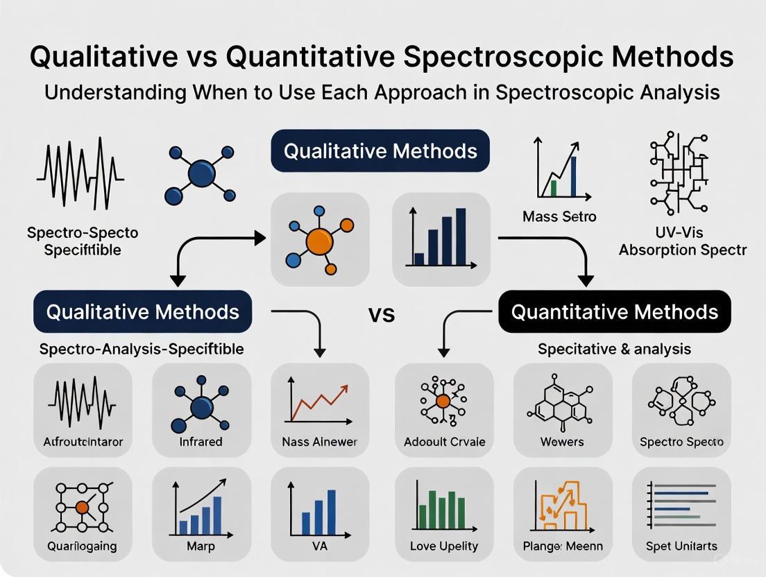
In chemical and pharmaceutical analysis, the separation between identification and measurement forms the cornerstone of spectroscopic methodology. This division directly maps onto the two primary approaches in analytical science: qualitative analysis, which identifies the presence or absence of specific chemical components in a sample, and quantitative analysis, which provides precise, numerical data regarding the concentration or amount of those components [1]. Understanding this distinction is not merely academic; it dictates experimental design, technique selection, and data interpretation strategies across research and development workflows. For drug development professionals, strategically leveraging this divide enables efficient resource allocation, from initial compound discovery through to final product quality control.
Qualitative analysis answers the fundamental question "What is present?" by characterizing complex mixtures and identifying functional groups, molecular structures, or specific compounds. Conversely, quantitative analysis addresses "How much is present?" through meticulous calibration to deliver concentration data indispensable for standardization, dosage formulation, and regulatory compliance [1]. While often presented as a binary choice, modern analytical workflows frequently integrate both approaches, using qualitative screening to inform subsequent quantitative validation. This guide explores the technical specifications, methodological workflows, and decision-making criteria governing the application of qualitative versus quantitative spectroscopic methods within pharmaceutical research and development.
Technical Foundations: Core Principles and Data Outputs
The fundamental principles of spectroscopy exploit the interaction between matter and electromagnetic radiation. When molecules are exposed to specific energy ranges, they undergo quantized transitions that result in the absorption, emission, or scattering of photons. The patterns of these interactions provide characteristic molecular fingerprints [2].
Qualitative Spectroscopic Analysis focuses on the position of spectral features. The presence of specific bonds, functional groups, or molecular structures is determined by identifying the characteristic wavelengths or frequencies at which these interactions occur. For instance, in Infrared (IR) spectroscopy, a sharp peak around 3300 cm⁻¹ strongly suggests an N-H bond, while a strong band between 1800-1600 cm⁻¹ is indicative of a carbonyl (C=O) group [3]. The output is typically a spectrum plotted as intensity versus wavelength, frequency, or wavenumber, which is then compared against reference libraries or databases for identification [1] [2].
Quantitative Spectroscopic Analysis focuses on the intensity of these spectral features. The underlying principle for many quantitative methods is the Beer-Lambert law, which establishes a linear relationship between the concentration of an analyte in a solution and the absorbance of light at a specific wavelength [2]. The key output is a numerical value—such as concentration, mass, or percentage purity—derived from calibrating signal intensity against known standards.
Table 1: Core Outputs of Qualitative vs. Quantitative Spectroscopic Analysis
| Aspect | Qualitative Analysis | Quantitative Analysis |
|---|---|---|
| Primary Question | What is present? | How much is present? |
| Analytical Focus | Spectral pattern and peak position | Peak intensity or signal amplitude |
| Key Data Output | Spectrum (e.g., IR, NMR, Raman) | Concentration, mass, percentage |
| Governing Principle | Functional group characteristics | Beer-Lambert law, calibration curves |
| Common Techniques | FTIR, NMR (for structure elucidation), Qualitative Raman | UV-Vis Spectrophotometry, Quantitative NMR, MS-based quantification |
Methodological Comparison: Techniques and Applications
The selection of an appropriate spectroscopic technique is guided by the analytical question, the nature of the sample, and the required information content. Both qualitative and quantitative methods span the electromagnetic spectrum, with each spectral region providing unique insights.
Ultraviolet-Visible (UV-Vis) spectroscopy, for example, is highly effective for quantifying the concentration of chromophores in solution. In contrast, Infrared (IR) spectroscopy is a powerhouse for qualitative identification, as the specific vibrational energies of bonds provide a detailed fingerprint of molecular structure [1] [2]. Mid-IR spectra are dominated by fundamental molecular vibrations, offering high specificity for identifying functional groups, while signals in the Near-IR (NIR) region are broader and result from overtones and combination bands, which can be leveraged for quantitative analysis using chemometrics [2].
Table 2: Common Spectroscopic Techniques and Their Primary Analytical Role
| Technique | Primary Qualitative Application | Primary Quantitative Application |
|---|---|---|
| IR / FTIR | Identification of functional groups and molecular structures [3] | Limited; used with multivariate calibration for complex samples |
| NMR | Elucidation of molecular structure, connectivity, and conformation | Determination of concentration and purity (qNMR) |
| UV-Vis | Identification of chromophores (e.g., conjugated systems) | High-precision concentration measurement of light-absorbing species [1] |
| Mass Spectrometry (MS) | Determination of molecular weight and fragmentation pattern for identity | High-sensitivity quantification (e.g., LC-MS/MS for bioanalysis) |
| Raman | Identification of functional groups and crystal forms; complementary to IR | Quantification of polymorphic ratios in solid dosage forms |
The choice between absorption-based and scattering-based techniques like Raman spectroscopy is often dictated by the sample matrix. Absorption methods are highly sensitive but can be complicated by strongly absorbing matrices, such as aqueous solutions. In such environments, Raman scattering, which has a low sensitivity to water, may provide a superior alternative [2].
Experimental Protocols and Workflows
Protocol for Qualitative Identification of an Organic Compound via FTIR
Objective: To identify the functional groups present in an unknown organic compound using Fourier-Transform Infrared (FTIR) spectroscopy.
Principle: Different chemical bonds and functional groups in a molecule absorb infrared radiation at characteristic frequencies. The resulting spectrum serves as a unique fingerprint for compound identification [3].
Materials and Reagents:
- FTIR spectrometer with attenuated total reflection (ATR) accessory
- Unknown organic compound (solid or liquid)
- Pure solvent (e.g., methanol, for cleaning the ATR crystal)
- Wiping materials (lint-free tissues)
Procedure:
- Instrument Preparation: Power on the FTIR spectrometer and computer. Allow the system to initialize and the IR source to warm up as per the manufacturer's instructions.
- Background Collection: Clean the ATR crystal with a lint-free tissue moistened with a small amount of pure solvent. Ensure the crystal is completely dry and free of residue. Collect a background spectrum with no sample present.
- Sample Preparation:
- For solids: Place a small amount (1-2 mg) of the finely powdered compound directly onto the ATR crystal.
- For liquids: Deposit 1-2 droplets directly onto the crystal.
- Sample Measurement: Clamp the sample applicator to ensure good contact between the sample and the crystal. Acquire the IR spectrum over a standard wavenumber range (e.g., 4000 to 600 cm⁻¹) with a specified resolution (e.g., 4 cm⁻¹).
- Data Analysis: Compare the acquired spectrum to reference libraries. Identify key absorption bands and their corresponding functional groups using a standard IR data table [3].
Protocol for Quantitative Analysis of a Drug Compound via UV-Vis Spectrophotometry
Objective: To determine the concentration of a known active pharmaceutical ingredient (API) in a solution using UV-Vis spectrophotometry.
Principle: The concentration of an analyte is directly proportional to the absorbance of light at a specific wavelength, as described by the Beer-Lambert law (A = εlc) [2].
Materials and Reagents:
- UV-Vis spectrophotometer
- Quartz cuvettes (1 cm pathlength)
- API standard (high purity)
- Appropriate solvent (e.g., buffer, methanol)
- Volumetric flasks and pipettes
Procedure:
- Preparation of Standard Solutions: Accurately weigh the API standard and dissolve it in the chosen solvent to prepare a stock solution of known concentration (e.g., 1 mg/mL). Serially dilute this stock solution to prepare a set of standard solutions covering a range of concentrations (e.g., 2, 4, 6, 8, 10 μg/mL).
- Blank Measurement: Fill a cuvette with the pure solvent and place it in the spectrophotometer. Use this as the blank to zero the instrument at the predetermined analytical wavelength (λ_max).
- Calibration Curve Generation: Measure the absorbance of each standard solution at λ_max. Plot the absorbance values against the corresponding concentrations to generate a calibration curve. Perform linear regression to obtain the equation of the line (y = mx + c) and the correlation coefficient (R²).
- Analysis of Unknown Sample: Prepare the unknown sample (e.g., a dissolved tablet) in the same solvent. Measure its absorbance at the same λ_max.
- Quantification: Use the equation from the calibration curve to calculate the concentration of the API in the unknown sample.
The Scientist's Toolkit: Essential Research Reagent Solutions
Successful spectroscopic analysis relies on a suite of essential materials and reagents. The following table details key items and their functions in the featured experiments and the broader field.
Table 3: Essential Research Reagents and Materials for Spectroscopic Analysis
| Item | Function / Application |
|---|---|
| FTIR Spectrometer with ATR | Enables rapid, non-destructive fingerprinting of solids and liquids without extensive sample preparation [2]. |
| UV-Vis Spectrophotometer | Measures the absorption of light by a solution, fundamental for quantitative analysis of chromophores [1] [2]. |
| ATR Crystals (Diamond, ZnSe) | The internal reflection element in ATR accessories that interfaces with the sample. Diamond is durable, while ZnSe offers a broader spectral range but is softer. |
| Quz Cuvettes | Contain liquid samples for UV-Vis analysis. Quartz is transparent down to UV wavelengths, unlike glass. |
| Deuterated Solvents (e.g., CDCl₃, D₂O) | Used in NMR spectroscopy to provide a lock signal for field stability and to avoid intense solvent proton signals that would overwhelm the sample spectrum. |
| Certified Reference Materials (CRMs) | High-purity standards with well-characterized composition; essential for accurate calibration and validation in quantitative analysis [1]. |
| KBr Pellets | A traditional method for preparing solid samples for transmission IR spectroscopy, where the sample is dispersed in potassium bromide and pressed into a pellet. |
Strategic Decision Framework: Selecting the Right Approach
Choosing between qualitative and quantitative methods depends on the project's stage and specific informational needs. The following diagram visualizes the key decision-making workflow for researchers.
Diagram 1: Method Selection Workflow
This decision framework highlights that the analytical strategy must be aligned with the research objective. Qualitative methods are the tool of choice during early discovery for de novo identification of unknown compounds, characterization of reaction products, and troubleshooting synthesis pathways [1] [3]. As a project matures, the emphasis shifts toward quantitative analysis for determining assay and purity, quantifying impurities and degradation products, ensuring batch-to-batch consistency, and generating data for regulatory filings [1] [2].
The sample matrix is another critical factor. Techniques like ATR-FTIR are robust for analyzing solid formulations, while quantitative NMR (qNMR) is prized as a primary ratio method for quantifying organic molecules without the need for compound-specific calibration [2]. The trend in pharmaceutical analysis is toward hyphenated techniques (e.g., LC-MS) and Process Analytical Technology (PAT) that integrate both qualitative and quantitative capabilities for real-time monitoring and control [4] [2].
The fundamental divide between identification and measurement is a pragmatic reality in spectroscopic analysis. Qualitative methods provide the essential "what" that guides scientific understanding, while quantitative methods deliver the critical "how much" required for product development, standardization, and compliance. For the modern drug development professional, mastery of both domains—and the strategic intelligence to know when to deploy each—is indispensable. The future of analytical science lies not in choosing one over the other, but in the sophisticated integration of both to create streamlined, information-rich workflows that accelerate innovation and ensure quality from the research bench to the patient.
Spectroscopic Light-Matter Interactions as the Basis for Analysis
Spectroscopic analysis is a fundamental laboratory technique that explores the interaction between light and matter to determine the composition, structure, and concentration of substances [5]. The underlying principle of all spectroscopic methods is that when light—a form of electromagnetic radiation—interacts with a material, the energy is absorbed, emitted, or scattered in ways that provide unique information about the sample's atomic or molecular characteristics [6]. The analytical utility of these interactions stems from quantum mechanical phenomena: the energy of incident photons must precisely match the energy difference between two quantum states in the material for absorption or emission to occur [7] [6].
The electromagnetic spectrum encompasses a broad range of energies, from high-energy gamma rays to low-energy radio waves, with each region probing different types of molecular or atomic transitions [8] [2]. The specific region utilized determines both the type of information obtained and the subsequent analytical application, forming the foundation for deciding between qualitative identification versus quantitative measurement approaches in spectroscopic analysis.
Fundamental Light-Matter Interactions and Spectroscopic Modalities
The primary light-matter interactions that form the basis of spectroscopic analysis include absorption, emission, and scattering phenomena. Each interaction mechanism provides distinct information and is exploited through different instrumental configurations.
Absorption Spectroscopy
Absorption occurs when energy from a radiative source is absorbed by the material, promoting atoms or molecules to higher energy states [6]. The measurement involves determining the fraction of energy transmitted through the material, with absorption decreasing the transmitted portion [6]. The fundamental relationship governing absorption spectroscopy is the Beer-Lambert Law:
[ A = \varepsilon \cdot c \cdot l ]
Where (A) is absorbance, (\varepsilon) is the molar absorptivity (a measure of how strongly a chemical species absorbs light at a specific wavelength), (c) is the concentration of the absorbing species, and (l) is the path length that light travels through the sample [7] [6]. This relationship forms the cornerstone of quantitative spectroscopic analysis.
Different regions of the electromagnetic spectrum probe different types of transitions in absorption spectroscopy:
- Ultraviolet-Visible (UV-Vis): Involves exciting valence electrons between molecular orbitals [7]. Significant molecular orbitals include the highest occupied molecular orbital (HOMO) and the lowest unoccupied molecular orbital (LUMO) [7]. UV-Vis spectroscopy typically covers 190-360 nm (UV) and 360-780 nm (visible) [8].
- Infrared (IR): Examines molecular vibrations, with the mid-infrared (MIR) region (1-30 μm) characterizing fundamental vibrations while the near-infrared (NIR) region (700-2500 nm) detects overtones and combination bands [7] [8] [2].
- X-ray: Involves excitation of core electrons and is used primarily for elemental analysis [2].
Figure 1: Basic components and workflow of an absorption spectrophotometer, showing the path from light source to quantitative measurement.
Emission Spectroscopy
Emission spectroscopy involves the release of radiative energy by material that has been previously excited [5]. This excitation can occur through various means including electromagnetic radiation, flames, sparks, electric arcs, or other energy sources [6]. Two important emission phenomena are:
- Fluorescence: A relatively intense and fast emission process with timescales of picoseconds to nanoseconds [7]. Fluorescence occurs when an electron in an excited singlet state relaxes to the ground electronic state by emitting a photon [7].
- Phosphorescence: A weaker and slower emission process with timescales of microseconds or longer [7]. Phosphorescence involves intersystem crossing from an excited singlet state to a triplet state, followed by forbidden transition back to the ground state [7].
The Jablonski diagram, named after Aleksander Jabłoński, provides a comprehensive visualization of these photophysical processes, depicting ground and excited electronic states, vibrational and rotational levels, and transitions between these states [7].
An important quantitative parameter in emission spectroscopy is the fluorescence quantum yield, (Φ), defined as the number of photons emitted divided by the number of photons absorbed [7]. The maximum value is 1 (one emitted photon per absorbed photon), while the minimum is 0 (no emission) [7].
Scattering Spectroscopy
Scattering techniques involve analyzing how incident radiation is deflected by a material [6]. Two primary forms of scattering are:
- Elastic scattering (Rayleigh/Mie scattering): The scattered radiation has the same wavelength as the incident light [2].
- Inelastic scattering (Raman scattering): The scattered light undergoes wavelength shifts due to energy exchange between the radiation and matter [2] [6].
Raman spectroscopy is particularly valuable for analyzing aqueous samples or those in glass containers because water and glass are weak scatterers, creating minimal interference [8]. Raman is complementary to IR spectroscopy and excels at detecting specific molecular vibrations including acetylenic -C≡C- stretching, olefinic C=C stretching, N=N (azo-) stretching, S-H stretching, and others [8].
Figure 2: Schematic of a Raman spectrometer configuration, highlighting the separation of elastic and inelastic scattering components.
Qualitative Versus Quantitative Analytical Approaches
The fundamental distinction between qualitative and quantitative analysis lies in their analytical objectives: qualitative methods identify what is present, while quantitative methods determine how much is present [1]. These approaches leverage different aspects of light-matter interactions and require different data processing strategies.
Qualitative Analysis Fundamentals
Qualitative analysis focuses on identifying the presence or absence of specific chemical components or structures in a sample [1]. This approach utilizes the characteristic patterns of absorption, emission, or scattering that serve as molecular "fingerprints" [2].
Common qualitative methodologies include:
- Spectral Correlation: Direct comparison of sample spectra with reference libraries using methods like wavelength correlation or Euclidean distance calculations [9]. A matching spectrum typically shows correlation values exceeding 0.95 [9].
- Principal Component Analysis (PCA): A multivariate technique that identifies the largest sources of variation in a dataset, allowing visualization of sample similarities and differences through score plots [9].
- Soft Independent Modeling of Class Analogies (SIMCA): A classification method that builds separate PCA models for each class and classifies test samples based on their fit to these models [9].
Qualitative analysis is particularly valuable in early research stages, for troubleshooting unknown substances, or for raw material identification in pharmaceutical manufacturing [1] [9].
Quantitative Analysis Fundamentals
Quantitative analysis provides measurable, precise data about the concentration of chemical components in a material [1]. This approach relies on the relationship between signal intensity and analyte concentration, most fundamentally expressed through the Beer-Lambert Law in absorption spectroscopy [7].
Key aspects of quantitative spectroscopic analysis include:
- Calibration Models: Establishing mathematical relationships between spectral features and reference concentration values [2].
- Univariate vs. Multivariate Approaches: Simple univariate calibration uses single wavelength measurements, while multivariate techniques like Partial Least Squares (PLS) regression handle complex situations with overlapping spectral signatures [2].
- Detection Limits: Well-designed spectroscopic methods can detect concentrations down to parts per billion levels with good accuracy and precision [5].
Quantitative analysis is indispensable for formulation standardization, regulatory compliance, and process control where precise concentration data is required [1].
Comparative Analytical Capabilities by Spectral Region
Table 1: Qualitative and Quantitative Capabilities Across Spectral Regions
| Spectral Region | Primary Transitions Probed | Qualitative Strength | Quantitative Applications | Common Techniques |
|---|---|---|---|---|
| Ultraviolet-Visible (UV-Vis) | Valence electron transitions [7] | Identification of chromophores, conjugated systems [8] [2] | Protein concentration via 280 nm absorption [7], Pharmaceutical purity assessment [8] | UV-Vis absorption, Fluorescence [7] |
| Infrared (IR) | Molecular vibrations [7] [5] | Functional group identification, Structural elucidation [5] | Protein secondary structure quantification [10], Agricultural product analysis [8] | FTIR, ATR-IR, NIR [8] [10] |
| Raman | Molecular vibrations [8] | Detection of specific bonds (-C≡C-, -N=N-, -S-H) [8] | Aqueous solution analysis [8], Crystallinity assessment | Raman spectroscopy [8] [2] |
| X-ray | Core electron transitions [2] | Elemental identification [2] [11] | Major and minor element quantification [11] | XRF, EDXRF [11] |
Experimental Protocols for Spectroscopic Analysis
UV-Vis Protein Quantification Protocol
Principle: Proteins containing aromatic amino acids (phenylalanine, tryptophan, tyrosine) absorb strongly at 280 nm due to their electron systems [7].
Materials:
- UV-Vis spectrophotometer with deuterium lamp source
- Quartz cuvettes (pathlength typically 1 cm)
- Protein standard solution of known concentration
- Buffer solution for blank and dilution
Procedure:
- Turn on instrument and allow lamp to warm up for 30 minutes
- Prepare serial dilutions of protein standard for calibration curve
- Use buffer solution as blank to zero instrument at 280 nm
- Measure absorbance of standard solutions and unknown samples
- Plot absorbance versus concentration for standards and apply Beer-Lambert Law
Data Analysis: Calculate protein concentration using: [ c = \frac{A}{\varepsilon \cdot l} ] Where (c) is concentration, (A) is measured absorbance, (\varepsilon) is molar absorptivity, and (l) is pathlength.
Considerations: Method accuracy depends on aromatic amino acid composition; nucleic acid contamination can interfere [7].
Mid-Infrared Analysis of Protein Secondary Structures
Principle: Different protein secondary structures (α-helix, β-sheet, β-turn) produce characteristic absorption in the amide I and amide II regions of the MIR spectrum [10].
Materials:
- FTIR spectrometer with ATR accessory
- High-purity nitrogen purge gas
- Wheat protein fractions (albumins, globulins, gliadins, glutenins)
Procedure:
- Purge spectrometer with nitrogen to reduce water vapor interference
- Collect background spectrum with clean ATR crystal
- Apply protein sample to ATR crystal ensuring complete contact
- Collect sample spectrum (typically 64 scans at 4 cm⁻¹ resolution)
- Pre-process spectra (vector normalization, second derivative)
Data Analysis:
- Analyze amide I region (1600-1700 cm⁻¹) for secondary structure
- Use multivariate methods like ANOVA simultaneous component analysis (ASCA)
- Quantify structures via spectral deconvolution and curve fitting
Results Interpretation: In wheat proteome analysis, albumin and globulin fractions showed primarily α-helix (57.8% and 45.9% respectively), while gliadins contained 38.3% β-turn and glutenins showed 44.8% β-turn structures [10].
Fluorescence Lifetime Measurement Protocol
Principle: Fluorescence lifetime (τ) is the time for fluorescence intensity to decay to 1/e of its initial value after pulsed excitation, providing information about molecular microenvironment [7].
Materials:
- Time-resolved fluorescence spectrometer with pulsed laser
- Temperature-controlled sample holder
- Fluorescent protein solutions (e.g., iRFP702)
Procedure:
- Excite sample with short pulse laser at appropriate wavelength (e.g., 660 nm)
- Collect emission at specific wavelength (e.g., 702 nm)
- Record intensity decay curve with time-correlated single photon counting
- Repeat measurements in different solvents (H₂O, D₂O) to observe kinetic isotope effects
Data Analysis: Fit decay data to exponential model: [ I(t) = I0 \cdot e^{-t/τ} ] Where (I(t)) is intensity at time (t), (I0) is initial intensity, and (τ) is fluorescence lifetime.
Results Interpretation: For iRFP702, fluorescence lifetime measured 749 ps in H₂O and 1.35 ns in D₂O, showing kinetic isotope effect of 1.8 [7].
Essential Research Reagents and Materials
Table 2: Key Research Reagents and Materials for Spectroscopic Analysis
| Item | Function | Application Examples |
|---|---|---|
| ATR Crystals (diamond, zinc selenide) | Enables internal reflection for IR measurements without extensive sample preparation [10] | Mid-IR analysis of protein secondary structures in wheat fractions [10] |
| Spectrophotometric Cuvettes (quartz, glass) | Holds liquid samples with precise pathlength for transmission measurements [7] | UV-Vis protein quantification at 280 nm [7] |
| Fluorescent Proteins (e.g., iRFP702) | Genetically engineered probes with defined excitation/emission profiles for biological imaging [7] | Deep tissue imaging, time-resolved fluorescence studies [7] |
| FRET Pair Fluorophores | Donor-acceptor pairs with overlapping emission/absorption for proximity measurements [7] | Studying molecular interactions, super-resolution localization imaging [7] |
| Chemometric Software | Implements multivariate algorithms (PCA, PLS, SIMCA) for spectral data analysis [9] | Qualitative classification of pharmaceutical raw materials [9] |
Selection Framework: Qualitative vs. Quantitative Methodologies
Choosing between qualitative and quantitative spectroscopic approaches depends on multiple factors related to the analytical question, sample characteristics, and practical constraints.
Method Selection Criteria
Nature of the Analytical Question:
- Qualitative methods are appropriate for identification, structural elucidation, classification, and purity assessment [1] [9].
- Quantitative methods are necessary for concentration measurement, kinetic studies, process monitoring, and quality control testing [1].
Sample Considerations:
- Complexity: Simple systems may allow univariate quantitative analysis, while complex mixtures typically require multivariate approaches [2].
- State: Solids, liquids, and gases may require different sampling accessories and measurement techniques [5].
- Concentration: High concentrations favor absorption methods, while trace analysis may require fluorescence or specialized techniques [2].
Practical Constraints:
- Speed requirements: Qualitative screening is generally faster than quantitative analysis [1].
- Regulatory compliance: Quantitative methods require rigorous validation per guidelines such as ICH Q2(R1) for pharmaceutical applications [2].
- Instrumentation availability: Some techniques require specialized equipment that may not be readily accessible [2].
Figure 3: Decision framework for selecting between qualitative and quantitative spectroscopic approaches based on analytical requirements.
Integrated Qualitative-Quantitative Strategies
Many modern analytical challenges require combining qualitative and quantitative approaches in integrated strategies:
- Quality by Design in Pharmaceuticals: Begin with qualitative screening of raw materials followed by quantitative monitoring of critical process parameters [9] [2].
- Biomolecular Characterization: Use qualitative structural analysis (e.g., protein secondary structure via MIR) alongside quantitative concentration measurements [10].
- Environmental Monitoring: Employ qualitative identification of unknown pollutants followed by quantitative tracking of regulated compounds [5].
This integrated approach leverages the strengths of both methodologies while compensating for their individual limitations, providing comprehensive analytical solutions for complex scientific and industrial challenges.
In the landscape of analytical chemistry, qualitative analysis serves a distinct and crucial role, focusing on identifying the fundamental composition and structural characteristics of chemical substances. Unlike quantitative methods that measure precise concentrations, qualitative techniques answer foundational questions about molecular identity, structural features, and functional group composition [1]. Within pharmaceutical development and natural products research, these methods form the essential first step in characterizing unknown compounds, authenticating raw materials, and dereplicating known entities from complex mixtures [4] [12].
The core strength of qualitative spectroscopic analysis lies in its ability to provide structural fingerprints—unique patterns that serve as molecular signatures. These fingerprints, whether from vibrational, magnetic resonance, or mass spectrometric techniques, encode rich information about bonding networks, atomic environments, and molecular symmetry [13] [8]. When framed within the broader context of analytical method selection, qualitative analysis precedes and informs quantitative measurement, establishing identity before concentration can be meaningfully determined [1]. This guide examines the principal techniques, methodologies, and applications that define modern qualitative analysis for molecular fingerprinting and structural elucidation, providing researchers with a framework for selecting appropriate methods based on specific analytical challenges.
Core Principles of Molecular Fingerprinting
Molecular fingerprinting encompasses a suite of techniques that generate characteristic patterns representing a compound's structural features. These fingerprints serve as unique identifiers for verification against known references or for classification within larger chemical databases.
Conceptual Foundation and Definitions
A molecular fingerprint is a structured representation of a molecule's structural features, typically encoded as a bit string or numerical vector where each position corresponds to the presence or absence of a specific chemical attribute [14]. These representations enable systematic comparison of chemical structures based on shared substructures, functional groups, or topological features. The underlying principle follows the Similar Property Principle (SPP), which posits that structurally similar molecules are likely to exhibit similar biological activities and physicochemical properties [14]. While limitations exist, such as activity cliffs where minor structural changes cause dramatic functional shifts, this principle generally holds across broad regions of chemical space, making fingerprinting invaluable for initial screening and classification.
Techniques for Fingerprint Generation
Multiple computational approaches exist for generating molecular fingerprints, each capturing different aspects of molecular structure as shown in Table 1.
Table 1: Major Molecular Fingerprint Encodings and Their Applications
| Fingerprint ID | Name | Description | Structural Features Captured | Common Applications |
|---|---|---|---|---|
| FP1 | AP2D | Topological Atom Pairs | Pairs of atoms and their topological distance | Similarity screening, virtual screening |
| FP2 | ASP | All-Shortest Paths | All shortest paths between atoms in molecular graph | Bioactivity prediction, compound classification |
| FP3 | AT2D | Topological Atom Triplets | Three atoms with their intervening paths | Structure-activity relationship studies |
| FP5 | ECFP | Extended Connectivity Fingerprints | Circular atom environments extending outward from each atom | Drug discovery, lead optimization |
| FP7 | MACCS | MDL Public Keys | 166 structural keys based on common chemical fragments | Rapid database searching, similarity assessment |
| FP11 | RDKit | Topological Daylight-like | Path-based fingerprints similar to Daylight fingerprints | General-purpose chemical informatics |
The performance of different fingerprint encodings varies significantly depending on the application. Benchmarking studies using chemical-genetic interaction profiles from Saccharomyces cerevisiae as a proxy for biological activity have demonstrated that all-shortest path (ASP) fingerprints paired with the Braun-Blanquet similarity coefficient provide superior performance in retrieving biologically similar compounds [14]. This combination has shown robustness across diverse compound collections, including natural products and synthetic libraries.
Analytical Techniques for Structural Elucidation
Structural elucidation of unknown compounds represents one of the most challenging applications of qualitative spectroscopy, requiring the integration of multiple complementary techniques to deduce atomic connectivity, stereochemistry, and functional group composition.
Nuclear Magnetic Resonance (NMR) Spectroscopy
NMR spectroscopy provides unparalleled insight into molecular structure by probing the magnetic environments of nuclei such as ^1H and ^13C [12] [13]. The technique's qualitative strength lies in its ability to reveal bonding networks, functional groups, through-space interactions, and spatial proximity through a suite of specialized experiments [13].
Key NMR Experiments for Structural Elucidation:
- ¹H NMR: Reveals hydrogen environments, with chemical shifts indicating electronic environment and coupling constants revealing connectivity through J-coupling [13].
- ¹³C NMR: Provides information about carbon backbone structure and hybridization states [15].
- 2D NMR Techniques: Including HSQC (Heteronuclear Single Quantum Coherence) for direct ^1H-^13C correlations, COSY (COrrelation SpectroscopY) for ^1H-^1H couplings, TOCSY (Total Correlation SpectroscopY) for protons within coupled networks, and HMBC (Heteronuclear Multiple Bond Correlation) for long-range ^1H-^13C couplings [12].
A significant advancement in NMR analysis is the movement from phenotypic interpretation (visual peak analysis) to genotypic interpretation based on quantum mechanical spectral analysis (QMSA). This approach extracts fundamental NMR parameters (δ, J) directly from experimental spectra, reducing subjectivity and enhancing reproducibility [13]. For ^1H NMR spectra, this process has been termed ¹H iterative functionalized Spectral/Spin Analysis (HifSA), which enables complete spectral interpretation by decoding the nuclear genotype of the analyzed molecule [13].
Mass Spectrometry (MS)
Mass spectrometry contributes to structural elucidation through determination of molecular weight, elemental composition, and fragmentation patterns that reveal structural motifs [12] [15].
- Gas Chromatography-MS (GC-MS): Best suited for volatile compounds or those made volatile through derivatization, typically analyzing molecules <500 Da. Electron ionization (EI) provides highly reproducible fragmentation patterns that can be searched against extensive libraries such as the NIST Mass Spectral Database and Wiley Registry [12].
- Liquid Chromatography-MS (LC-MS): Applied to a broader range of primary and secondary metabolites, with tandem MS/MS facilitating structural annotation through cross-referencing with metabolite databases such as HMDB, METLIN, and MassBank [12].
Vibrational Spectroscopy
Vibrational techniques including infrared (IR) and Raman spectroscopy provide complementary information about functional groups and molecular symmetry through detection of characteristic molecular vibrations [16] [8].
- Infrared Spectroscopy: Probes molecular vibration absorption, particularly sensitive to functional groups with dipole moment changes. Dominant spectral features include C-H, O-H, N-H, C=O, and C≡N stretching vibrations [8].
- Raman Spectroscopy: Based on inelastic light scattering, particularly sensitive to symmetric bonds and polarizability changes. Key spectral features include C=C, N=N, S-H, and C≡C stretching vibrations [16] [8].
The fingerprint region in Raman spectroscopy (300-1900 cm⁻¹) contains unique patterns for compound identification. Particularly valuable is the sub-region from 1550-1900 cm⁻¹ termed the "fingerprint in the fingerprint", where active pharmaceutical ingredients (APIs) display unique Raman signals from C=N, C=O, and N=N vibrations, while common excipients typically show no interference in this region [16].
UV-Vis Spectroscopy
UV-Vis spectroscopy reveals electronic transitions in molecules, particularly useful for identifying conjugated systems, chromophores, and π-π and n-π transitions [8]. While providing less structural detail than other techniques, it offers rapid characterization of chromophoric systems with applications in HPLC detection and compound classification [8].
Experimental Protocols and Workflows
Successful structural elucidation requires systematic experimental design and execution. The following protocols outline standardized approaches for comprehensive analysis.
Integrated Spectroscopic Workflow for Natural Products
This protocol describes a comprehensive approach for structural elucidation of natural products or unknown compounds, integrating multiple spectroscopic techniques.
Table 2: Research Reagent Solutions for Structural Elucidation
| Item | Function | Application Notes |
|---|---|---|
| Deuterated Solvents (CDCl₃, DMSO-d₆) | NMR solvent with minimal interference | Choice affects chemical shifts; match to compound solubility |
| NMR Reference Standards (TMS) | Chemical shift calibration | Added to sample for precise δ referencing |
| Derivatization Reagents (MSTFA, BSTFA) | Volatilization for GC-MS analysis | Silanization of polar functional groups |
| LC-MS Grade Solvents | Mobile phase for LC-MS | Minimal UV absorption, low chemical noise |
| Solid Substrates (KBr, NaCl) | IR sample preparation | KBr for pellet preparation; NaCl for solution cells |
| Raman Standards (Si wafer) | Instrument calibration | Daily verification of Raman shift accuracy |
Procedure:
- Sample Preparation: Isolate compound to high purity using chromatographic methods (flash chromatography, HPLC). Assess purity by analytical HPLC (>95%) and confirm absence of residual solvents or impurities by ¹H NMR [12].
- Molecular Formula Determination: Acquire high-resolution mass spectrometry (HRMS) data to determine exact mass and elemental composition. Calculate unsaturation index from molecular formula [12].
- Initial NMR Analysis: Record ¹H NMR spectrum with sufficient signal-to-noise ratio (>20:1) and digital resolution. Note presence of exchangeable protons (D₂O shake), integral ratios, and characteristic chemical shifts [12] [13].
- Comprehensive 2D NMR: Acquire COSY, HSQC, HMBC, and TOCSY experiments. For stereochemical assignment, record NOESY or ROESY if necessary. Set acquisition parameters to ensure adequate digital resolution (especially in F1 dimension) [12].
- Vibrational Spectroscopy: Acquire IR and Raman spectra, noting key functional group absorptions. For Raman, specifically examine the 1550-1900 cm⁻¹ region for API-specific vibrations [16].
- Computational Analysis: Input experimental NMR parameters (δ, J) into quantum mechanical spectral analysis (QMSA) tools for genotypic verification of proposed structure [13].
- Database Interrogation: Search spectral data against structural databases (Dictionary of Natural Products, SciFinder, Reaxys) for known compounds. For novel structures, verify through total synthesis or X-ray crystallography when possible [12].
Chemometric Classification Methods
For qualitative analysis of complex mixtures or material classification, chemometric methods provide powerful pattern recognition capabilities as detailed in Table 3 [9].
Table 3: Chemometric Methods for Qualitative Spectral Analysis
| Method | Principle | Sensitivity | Pharmaceutical Application |
|---|---|---|---|
| Wavelength Correlation | Normalized vector dot product between test and reference spectra | Low | Raw material identification |
| Principal Component Analysis (PCA) | Orthogonal decomposition of spectral variance | Medium | Batch consistency, outlier detection |
| Soft Independent Modeling of Class Analogies (SIMCA) | Class modeling using PCA residuals | Medium-High | Quality control, counterfeit detection |
| Partial Least Squares-Discriminate Analysis (PLS-DA) | Supervised classification maximizing separation between predefined classes | High | Authentication of botanical ingredients |
Protocol for PCA-Based Material Identification:
- Spectral Collection: Acquire spectra from validated reference materials (n≥10 per class) using standardized sampling procedures. Include expected natural variability in training set [9].
- Data Preprocessing: Apply appropriate preprocessing (Standard Normal Variate, derivatives, smoothing) to remove physical light scattering effects while preserving chemical information [9].
- Model Development: Perform PCA on mean-centered data. Determine optimal number of principal components through cross-validation to avoid overfitting [9].
- Classification Rule: Establish Hotelling T² ellipse at 95% confidence level in score plot space. Calculate Mahalanobis distance for class membership determination [9].
- Validation: Challenge model with independent test set not used in model development. Report specificity, sensitivity, and classification accuracy [9].
Advanced Approaches and Emerging Technologies
The field of structural elucidation is rapidly evolving with the integration of computational methods and artificial intelligence, significantly enhancing traditional approaches.
Computational Quantum Mechanical Analysis
A paradigm shift is occurring in NMR interpretation, moving from traditional phenotypic analysis (visual peak inspection) to genotypic analysis based on quantum mechanical spectral analysis (QMSA) [13]. This approach directly connects experimental spectra with quantum mechanical theory, extracting all genotypic parameters (δ, J, atom populations) required to create matching calculated spectra. The HifSA (¹H iterative functionalized Spectral/Spin Analysis) methodology represents this approach for ¹H NMR spectra, replacing subjective peak picking with rigorous computational analysis [13].
Unlike density functional theory (DFT), which predicts spectral parameters from a given structure (compound-to-spectrum, C2S), QMSA performs spectral analysis to extract structural parameters from experimental data (spectrum-to-compound, S2C) [13]. This fundamental difference makes QMSA particularly valuable for verifying proposed structures and resolving complex spectral overlaps.
Multimodal Artificial Intelligence Systems
The integration of multiple spectroscopic techniques through artificial intelligence represents the frontier of structural elucidation. SpectraLLM is the first large language model designed to support multi-modal spectroscopic joint reasoning, capable of processing single or multiple spectroscopic inputs (IR, Raman, UV-Vis, NMR, MS) and performing end-to-end structure elucidation [15].
This model transforms spectral peaks into textual prompts capturing physical attributes (wavenumber, intensity, peak shape), enabling fine-grained interactions across disparate spectroscopic channels. The system demonstrates state-of-the-art performance in molecular structure prediction, significantly outperforming approaches trained on single modalities [15]. This multimodal integration mirrors the approach of expert spectroscopists who routinely combine complementary data sources to reduce ambiguity and improve accuracy.
Workflow Visualization
The following diagram illustrates the integrated workflow for structural elucidation using multiple spectroscopic techniques, highlighting decision points and method selection criteria.
Integrated Workflow for Structural Elucidation
Qualitative spectroscopic methods for molecular fingerprinting and structural elucidation provide indispensable tools for characterizing chemical identity and structural features. The techniques discussed—from fundamental NMR and MS to advanced chemometric and computational approaches—form a hierarchical framework for addressing increasingly complex analytical challenges.
The continuing evolution of these methods, particularly through integration with quantum mechanical calculation and artificial intelligence, promises to enhance the objectivity, reproducibility, and throughput of qualitative analysis. As these technologies mature, they will increasingly bridge the gap between qualitative identification and quantitative measurement, providing researchers with more comprehensive analytical capabilities for drug development and natural products research.
By understanding the strengths and appropriate applications of each qualitative technique, researchers can make informed decisions about methodological selection, ensuring efficient and accurate structural characterization throughout the drug development pipeline.
In the rigorous world of drug development, the choice between qualitative and quantitative analytical methods is foundational to research validity and regulatory success. While qualitative spectroscopy excels in identifying molecular structures and components, quantitative spectroscopy is indispensable when the analytical requirement demands precise measurement of analyte concentration, rigorous validation of method performance, and stringent demonstration of regulatory compliance. This technical guide delineates the core strengths of quantitative spectroscopic methods, focusing on their unparalleled capacity to deliver precise, accurate, and legally defensible concentration data essential for pharmaceutical quality control, pharmacokinetic studies, and stability testing. We will explore the technical frameworks that underpin these strengths, including method validation protocols, advanced data preprocessing techniques, and contemporary assessment tools like the Red Analytical Performance Index (RAPI), which provides a structured metric for evaluating analytical performance against predefined validation criteria [17].
Core Quantitative Strengths
Precision and Accuracy: The Foundation of Reliability
Precision and accuracy are the cornerstones of reliable quantitative analysis, serving as the primary metrics for assessing a method's performance. In a chemometric context, precision refers to the ability to obtain reproducible results upon repeated measurement, while accuracy defines the closeness of those results to the true value [18]. The National Institute of Standards and Technology (NIST) provides Standard Reference Materials (SRMs) that are certified for specific properties, enabling scientists to calibrate instruments and transfer NIST's measurement capabilities to their own laboratories, thereby establishing traceable accuracy [18].
For quantitative spectroscopic methods, precision is often expressed as relative standard deviation (RSD) of repeated measurements, while accuracy is demonstrated through percentage recovery of known amounts of analyte spiked into a sample. A practical demonstration can be found in a validated HPLC-UV method for determining cannflavins in cannabis, where the method exhibited intra-day and inter-day RSDs of ≤5.29% and recoveries ranging from 82% to 98%, meeting the stringent criteria outlined in ICH guidelines [19]. Similarly, in the simultaneous analysis of Terbinafine HCl and Ketoconazole using spectrophotometric methods, statistical tests including the variance ratio F-test and Student t-test showed no significant differences between the developed methods and established reference methods, confirming their accuracy [20].
Table 1: Precision and Accuracy Data from Spectroscopic Method Validations
| Analytical Method | Analyte | Precision (RSD) | Accuracy (% Recovery) | Reference |
|---|---|---|---|---|
| HPLC-UV | Cannflavins A, B, C | Intra-day & inter-day RSD ≤5.29% | 82% - 98% | [19] |
| UHPLC-Q-TOF | Crown Procyanidins | RSD: 1.99%-11.03% (repeatability), 2.51%-19.05% (intermediate precision) | 88.21% - 107.64% | [19] |
| Spectrophotometric Methods | Terbinafine HCl & Ketoconazole | No significant differences by F-test and t-test | High % recoveries with low RSD | [20] |
Concentration Determination: From Univariate to Multivariate Approaches
Quantitative spectroscopy provides a robust framework for determining analyte concentration across diverse pharmaceutical applications, from active pharmaceutical ingredient (API) quantification to impurity profiling. The fundamental relationship between analyte concentration and spectroscopic response is governed by the Beer-Lambert law for absorption-based techniques, which states that absorbance is proportional to concentration and path length [2]. This principle enables straightforward univariate calibration where the intensity at a specific wavelength is correlated with concentration.
For more complex matrices where spectral signatures overlap, multivariate techniques become essential. Methods such as Partial Least Squares Regression (PLSR), Support Vector Machines (SVM), and Artificial Neural Networks (ANN) can deconvolute overlapping signals to provide accurate quantitation of multiple analytes simultaneously [2]. The application of these techniques is particularly valuable in Near-Infrared (NIR) spectroscopy, where spectra consist of overlapping vibrational bands that are non-specific and poorly resolved but can be effectively modeled with chemometrics [8]. Liquid Chromatography-Mass Spectrometry (LC-MS) exemplifies high-sensitivity concentration determination, capable of detecting analytes at picogram and femtogram levels, facilitating trace molecule identification in complex matrices [21].
Table 2: Techniques for Concentration Determination in Complex Matrices
| Technique | Application Context | Key Strength | Example Use Case |
|---|---|---|---|
| Univariate Calibration | Single analyte in simple matrix | Simplicity, based on Beer-Lambert law | UV/Vis analysis of APIs at specific wavelengths [2] |
| Partial Least Squares Regression (PLSR) | Multiple analytes with overlapping spectra | Deconvolution of correlated spectral variables | NIR analysis of pharmaceutical powders [8] [2] |
| Support Vector Machines (SVM) | Non-linear spectral responses | Effective pattern recognition for complex relationships | Classification and quantification of herbal medicines |
| Artificial Neural Networks (ANN) | Highly complex spectral datasets | Adaptive learning of intricate spectral-concentration relationships | Process Analytical Technology (PAT) for real-time monitoring |
| LC-MS/MS | Trace analysis in biological matrices | High sensitivity and structural confirmation | Metabolite quantification in pharmacokinetic studies [21] |
Regulatory Compliance: Meeting ICH and Pharmacopeial Standards
In pharmaceutical development, analytical methods must demonstrate compliance with regulatory standards to ensure the safety, efficacy, and quality of drug products. Quantitative spectroscopic methods are extensively validated according to ICH Q2(R1) guidelines, which define validation parameters including specificity, linearity, accuracy, precision, detection and quantitation limits, and robustness [20]. This comprehensive validation framework provides the documented evidence required for regulatory submissions and approvals.
Recent advancements in assessment metrics have further strengthened the regulatory framework. The Red Analytical Performance Index (RAPI) offers a standardized approach to evaluate analytical methods against ten predefined validation criteria, generating a visual representation of method performance that simplifies comparison and selection [17]. Similarly, the Blue Applicability Grade Index (BAGI) assesses practical aspects such as cost, time, and operational factors, complementing traditional validation data [20]. The implementation of these tools is particularly valuable in the context of White Analytical Chemistry (WAC), which promotes a balanced approach considering analytical performance (red), environmental impact (green), and practical/economic factors (blue) [17]. The transformative impact of these approaches is evident in advanced LC-MS applications, which now achieve sub-ppm detection sensitivity while maintaining >99% classification accuracy in pharmaceutical quality control [22].
Experimental Protocols for Method Validation
Protocol for Validation of Spectrophotometric Methods
The following protocol outlines the key steps for validating spectrophotometric methods according to ICH guidelines, as demonstrated in the simultaneous determination of Terbinafine HCl and Ketoconazole [20]:
Instrument Calibration and Qualification: Verify spectrometer performance using manufacturer specifications and NIST-traceable standards. Ensure wavelength accuracy, photometric accuracy, and stray light performance within acceptable limits.
Solution Preparation:
- Prepare stock standard solutions (e.g., 1.0 mg/mL) by dissolving precisely weighed reference standards in appropriate solvent.
- Create working solutions through serial dilution with distilled water or suitable solvent to cover the calibration range (e.g., 0.6-12.0 μg/mL for TFH; 1.0-10.0 μg/mL for KTZ).
- Store solutions at 2°C in a refrigerator; demonstrate stability over at least seven days.
Method Development and Optimization:
- Record zero-order absorption spectra of standard solutions using appropriate solvent blank.
- For overlapping spectra, apply mathematical processing techniques such as:
- Third derivative spectrophotometry (D3) to resolve overlapping peaks
- Ratio spectra difference spectrophotometry to enhance selectivity
- First derivative of ratio spectra (DD1) with scaling factor = 10 and Δλ = 10
- Induced dual wavelength (IDW) and dual wavelength resolution (DWR) techniques
Validation Procedure:
- Linearity: Prepare and analyze minimum of five concentrations in triplicate. Calculate correlation coefficient (R² > 0.99 generally acceptable).
- Accuracy: Perform recovery studies at three concentration levels (80%, 100%, 120%) with triplicate determinations. Acceptable recovery typically 98-102%.
- Precision:
- Repeatability: Analyze six preparations of the same concentration, calculate RSD (should be ≤2% for API assay).
- Intermediate precision: Perform analysis on different days, by different analysts, or with different instruments; RSD should be ≤3%.
- Specificity: Demonstrate ability to assess analyte unequivocally in presence of excipients and impurities.
- LOD and LOQ Determination: Calculate based on signal-to-noise ratio (3:1 for LOD, 10:1 for LOQ) or standard deviation of response and slope.
System Suitability Testing: Establish criteria for resolution, tailing factor, theoretical plates, and RSD of repeated injections before sample analysis.
Protocol for LC-MS Method Validation in Bioanalysis
Liquid Chromatography-Mass Spectrometry requires additional validation considerations for bioanalytical applications [21]:
Sample Preparation: Select and optimize appropriate technique (protein precipitation, liquid-liquid extraction, or solid-phase extraction) based on analyte and matrix properties.
Chromatographic Separation:
- Utilize ultra-high-performance liquid chromatography (UHPLC) with sub-2μm particles for enhanced resolution and speed.
- Optimize mobile phase composition, pH, and gradient program to achieve adequate retention and separation.
- Employ appropriate column chemistry (e.g., C18, HILIC, phenyl) based on analyte characteristics.
Mass Spectrometric Detection:
- Select ionization technique (ESI, APCI, or APPI) based on analyte polarity and molecular weight.
- Optimize source parameters (temperature, gas flows, voltages) for maximum sensitivity.
- Establish multiple reaction monitoring (MRM) transitions for target analytes and internal standards.
- For untargeted analysis, employ full-scan high-resolution mass spectrometry (HRMS) with data-dependent acquisition.
Validation Parameters Specific to Bioanalysis:
- Matrix effects: Evaluate ionization suppression/enhancement by comparing neat standards to post-extraction spiked samples.
- Selectivity: Demonstrate no significant interference from at least six different sources of blank matrix.
- Carryover: Assess by injecting blank samples after high-concentration calibrators.
- Dilution integrity: Verify accuracy and precision for samples requiring dilution beyond calibration range.
- Reinjection reproducibility: Demonstrate precision of reanalyzed samples from the same batch.
Data Analysis: Use appropriate software for peak integration, calibration curve fitting (typically linear or quadratic with 1/x² weighting), and concentration calculation.
The Scientist's Toolkit: Essential Reagents and Materials
Successful implementation of quantitative spectroscopic methods requires carefully selected reagents, reference standards, and materials. The following table outlines key components of the analytical toolkit for pharmaceutical applications:
Table 3: Essential Research Reagent Solutions for Quantitative Spectroscopy
| Reagent/Material | Function/Purpose | Technical Specifications | Application Notes |
|---|---|---|---|
| Certified Reference Standards | Primary calibration for quantitative analysis | Certified purity with documented uncertainty, NIST-traceable preferred | Required for method validation; establishes metrological traceability [18] |
| HPLC/MS Grade Solvents | Mobile phase preparation; sample reconstitution | Low UV absorbance; minimal particulate matter; high purity | Acetonitrile, methanol, water with 0.1% formic acid for LC-MS [19] [21] |
| Volatile Buffers & Additives | Mobile phase modification for chromatographic separation | MS-compatible (e.g., ammonium formate, ammonium acetate) | Concentration typically 2-10 mM; pH adjustment critical for retention [21] |
| Internal Standards | Correction for instrumental and preparation variability | Stable isotope-labeled analogs of analytes (e.g., deuterated) | Essential for bioanalysis; should elute similarly to analytes [21] |
| Solid Phase Extraction (SPE) Cartridges | Sample clean-up and pre-concentration | Various chemistries (C18, mixed-mode, HLB) based on application | Reduces matrix effects; improves sensitivity in complex matrices [21] |
| Spectral Preprocessing Software | Mathematical treatment of raw spectral data | Algorithms for smoothing, derivation, baseline correction | Critical for multivariate calibration in NIR/IR spectroscopy [22] |
Decision Framework: Selecting Appropriate Analytical Methods
The choice between qualitative and quantitative spectroscopic approaches depends on the specific analytical question and data requirements. The following diagram illustrates the decision pathway for method selection:
Quantitative spectroscopy provides an indispensable toolkit for pharmaceutical scientists requiring precise concentration data, validated method performance, and demonstrable regulatory compliance. The strengths of these methods—rooted in metrologically sound principles of precision and accuracy, enhanced by sophisticated concentration determination techniques, and formalized through rigorous validation protocols—make them particularly suited for applications where data integrity is paramount. As the field advances with tools like RAPI for standardized performance assessment [17] and increasingly sensitive LC-MS platforms capable of detecting analytes at femtogram levels [21], the role of quantitative spectroscopy will continue to expand in drug development. By understanding these core strengths and their implementation frameworks, scientists can make informed decisions about when quantitative approaches are necessary and how to implement them effectively to generate reliable, defensible analytical data.
In modern therapeutics, particularly for complex diseases like cancer, the use of drug combinations has become a fundamental strategy. Drug synergism occurs when two or more drugs that individually produce similar effects demonstrate a combined effect greater than that predicted by their individual potencies [23]. This synergistic interaction allows for the use of lower doses of combination constituents, which can significantly reduce adverse reactions and mitigate drug resistance [23] [24].
The assessment of synergism is a quantitative pursuit requiring rigorous demonstration that the combination effect exceeds expectations based on individual drug potencies. This process relies on the concept of dose equivalence and sophisticated experimental design and data analysis methods [23]. Within the broader context of analytical research, the workflow for studying drug synergy beautifully illustrates the complementary roles of qualitative and quantitative spectroscopic methods, where qualitative techniques often enable initial discovery and quantitative methods provide the rigorous verification needed for validation.
Screening Strategies for Synergistic Combinations
High-Throughput Experimental Screening
Conventional drug combination screening involves exploring a vast space of possible pairs across multiple dose levels, which is both time-consuming and expensive. High-throughput screening platforms have enabled the generation of large datasets, such as the O'Neil dataset with 22,737 conditions and the ALMANAC campaign with 304,549 experiments [25]. These approaches typically involve:
- Automated platforms performing multiple measurement rounds
- Multidimensional screening across numerous cell lines and drug pairs
- Open-source automation and data analysis workflows for accessibility [26]
These experimental methods provide the raw data necessary for initial identification of potentially synergistic pairs, serving as a qualitative first pass that requires subsequent quantitative verification.
AI-Guided Active Learning Frameworks
To address the challenge of large combinatorial spaces and the rarity of synergistic pairs (typically 1.5-3.5% of tested combinations), active learning frameworks have emerged as powerful screening tools [25]. These frameworks integrate experimental testing into an iterative learning process, dramatically improving screening efficiency.
Table 1: Key Components of an Active Learning Framework for Synergy Screening
| Component | Description | Impact on Performance |
|---|---|---|
| Molecular Features | Numerical representations of drug molecules (Morgan fingerprints, MAP4, MACCS, ChemBERT) | Limited impact on prediction quality [25] |
| Cellular Features | Gene expression profiles of targeted cells from databases like GDSC | Significant improvement (0.02-0.06 gain in PR-AUC) [25] |
| AI Algorithms | Range from parameter-light (logistic regression) to parameter-heavy (transformers, GCNs) | Heavier algorithms require more data but may offer better performance with sufficient training data [25] |
| Batch Size | Number of combinations tested in each iterative cycle | Smaller batch sizes yield higher synergy discovery rates [25] |
As demonstrated in recent studies, active learning can discover 60% of synergistic drug pairs while exploring only 10% of the combinatorial space, saving approximately 82% of experimental time and materials compared to random screening [25].
The following diagram illustrates the iterative active learning workflow for efficient synergy screening:
Quantitative Assessment of Synergistic Interactions
Reference Models for Synergy Quantification
Proper quantification of synergy requires establishing reference models that define the expected effect when drugs do not interact (additivity). The two most prominent reference models are:
Bliss Independence Model
The Bliss independence model assumes drugs act independently and do not interact [24]. The expected combination effect is calculated as:
Rc = R1 + R2 - R1 × R2
Where R1 and R2 are the individual responses to drug 1 and drug 2, respectively, and Rc is the expected combination response under the independence assumption [24].
Loewe Additivity Model
The Loewe additivity model assumes drugs have similar modes of action on the same pathway [24]. The additivity equation is expressed as:
a/A + b/B = 1
Where a and b are the doses of drug A and drug B in combination, and A and B are the doses of each drug that individually produce the specified effect level [23]. This model forms the basis for isobolographic analysis, where dose pairs plotting below the additive line indicate synergism [23].
Advanced Quantification Methods
Table 2: Methods for Quantifying Drug Synergism
| Method | Key Principle | Advantages | Limitations |
|---|---|---|---|
| Response Surface Modeling | 3D representation of combination effects across dose ranges [24] | Visualizes synergism/antagonism patterns across all tested ratios | Complex interpretation requiring specialized software |
| Chou-Talalay Method | Uses median-effect equation derived from mass-action law [24] | Most commonly used approach; provides Combination Index (CI) | Requires data preprocessing; ignores some variability sources [24] |
| MixLow Method | Mixed-effects Loewe model with confidence intervals [24] | More precise parameter estimation; no data preprocessing needed | Computationally intensive; less established in literature |
| Bayesian Approach | Hierarchical nonlinear regression modeling variability [24] | Accounts for multiple sources of uncertainty; more reliable estimation | Complex implementation requiring statistical expertise |
Spectroscopic Methods in Synergy Workflows
Qualitative Spectroscopic Analysis
Qualitative analysis identifies the presence or absence of particular chemical components in a sample, focusing on "what" is present [1]. In synergy research, these techniques are particularly valuable for:
- Initial identification of potential drug-drug interactions
- Characterization of complex mixtures and degradation products
- Functional group analysis using FT-IR [1] [27]
- Molecular fingerprinting via Raman spectroscopy [28]
Techniques such as FT-IR and Raman spectroscopy provide rapid, exploratory analysis crucial in early stages of research or for troubleshooting when unknown substances affect product consistency [1].
Quantitative Spectroscopic Analysis
Quantitative analysis provides measurable, precise data regarding the concentration of chemical components in a material [1]. These methods are indispensable for:
- Determining exact ratios of drug components in formulations
- Assessing regulatory compliance where precision matters [1]
- Real-time monitoring of concentration changes during treatment
- Stability testing and shelf-life determination [27]
Techniques including UV-Vis spectroscopy, NIR spectroscopy, and mass spectrometry offer the accuracy and precision required for rigorous synergy quantification [1].
Advanced Spectroscopic Applications in Pharmaceutical Research
Table 3: Spectroscopic Techniques in Drug Development and Synergy Research
| Technique | Primary Applications | Role in Synergy Research |
|---|---|---|
| ICP-MS | Trace elemental analysis; metal-protein interactions [28] | Quantifying metal-based drug components; studying metal-mediated synergies |
| Raman/SERS | Protein aggregation monitoring; real-time process analytics [28] | Detecting drug-induced structural changes; formulation stability assessment |
| NMR | Molecular structure determination; protein-excipient interactions [28] | Elucidating binding mechanisms; conformational changes in combined therapies |
| UV-Vis | Concentration measurement; PAT in chromatography [28] | Quantifying drug concentrations in combination therapies; purity assessment |
| Fluorescence | Protein denaturation monitoring; molecular interactions [28] | Studying drug-target engagement; stability testing without sample destruction |
Experimental Protocols
High-Throughput Combination Screening Protocol
This protocol adapts methodology from recent leukemia drug synergy studies [26]:
Cell Line Preparation
- Culture panel of relevant cell lines (e.g., AML lines for leukemia studies)
- Harvest cells in logarithmic growth phase
- Adjust concentration to 5,000-10,000 cells/well in 384-well format
Drug Preparation and Dispensing
- Prepare individual drug stocks at 1000× final concentration in DMSO
- Use automated liquid handling to create combination matrices
- Include single-agent controls and DMSO vehicle controls
Treatment and Incubation
- Dispense 5 nL of drug solutions using acoustic dispensing
- Add 50 μL cell suspension to each well
- Incubate for 72-120 hours at 37°C, 5% CO₂
Viability Assessment
- Add cell viability reagent (e.g., CellTiter-Glo)
- Measure luminescence using plate reader
- Normalize data to vehicle controls (0% inhibition) and untreated controls (100% inhibition)
Data Analysis
Spectroscopic Verification Protocol
For confirmed synergistic hits, perform detailed characterization using complementary spectroscopic techniques:
Sample Preparation
- Prepare combination formulations at synergistic ratios
- Include individual drug preparations as controls
- Use appropriate solvents matched to spectroscopic method requirements
FT-IR Analysis for Interaction Screening
- Use attenuated total reflectance (ATR) accessory for solid formulations
- Collect spectra from 4000-400 cm⁻¹ at 4 cm⁻¹ resolution
- Analyze shifts in characteristic functional group vibrations
- Compare combination spectrum to mathematical sum of individual drug spectra
Quantitative UV-Vis Validation
- Establish calibration curves for individual drugs
- Verify linearity across relevant concentration range (typically 1-100 μg/mL)
- Measure combination formulations at multiple ratios
- Calculate percent deviation from expected absorbance
Raman Microscopy for Spatial Distribution
- Map formulation surfaces using Raman microscope
- Identify potential drug-drug interaction sites
- Assess homogeneity of distribution in combination formulations
The Scientist's Toolkit: Essential Research Reagents and Materials
Table 4: Essential Research Reagents for Synergy Studies
| Reagent/Material | Function | Application Notes |
|---|---|---|
| Cell Line Panel | Disease-relevant models for screening | Should represent genetic diversity of target disease [26] |
| Compound Libraries | Source of drug candidates for combination | FDA-approved libraries preferred for repurposing opportunities [25] |
| Cell Viability Assays | Quantification of treatment effects | Luminescent assays preferred for high-throughput screening [26] |
| Chromatography Media | Protein purification for target deconvolution | Protein A affinity resin for mAb purification [28] |
| Spectroscopic Standards | Calibration and method validation | Necessary for quantitative accuracy in concentration measurements [1] |
| Culture Media Components | Cell maintenance during treatment | Metal content in CHO media affects productivity and critical quality attributes [28] |
Integrated Workflow and Decision Framework
The complete synergistic workflow integrates both screening and quantification phases with appropriate analytical techniques at each stage. The following diagram illustrates the comprehensive pathway and key decision points:
The journey from screening to quantification of synergistic drug combinations represents a sophisticated workflow that strategically employs both qualitative and quantitative analytical approaches. Qualitative methods enable rapid screening and initial identification of potential interactions, while quantitative techniques provide the rigorous verification necessary for validation. The integration of AI-guided active learning frameworks has dramatically improved the efficiency of discovering these rare synergistic pairs, while established reference models like Bliss independence and Loewe additivity provide the mathematical foundation for quantifying these interactions.
Spectroscopic techniques serve as bridge technologies throughout this workflow, with different methods providing either qualitative screening capability or quantitative validation power depending on the research phase. As pharmaceutical research continues to evolve toward combination therapies for complex diseases, this synergistic workflow—from screening to quantification—will remain essential for unlocking more effective, safer treatment options for patients.
Technique Deep Dive: Selecting the Right Spectroscopic Tool for the Task
In the realm of spectroscopic analysis, Infrared (IR) and Nuclear Magnetic Resonance (NMR) spectroscopy stand as two pillars of qualitative structural elucidation. Each technique provides distinct, complementary insights into molecular architecture. IR spectroscopy excels as a rapid identifier of specific functional groups through their characteristic vibrational signatures, while NMR spectroscopy provides atomic-level resolution of molecular connectivity and environment [29] [30]. Within the drug discovery and development pipeline, these techniques are indispensable for confirming the identity, purity, and structural integrity of both small-molecule pharmaceuticals and complex biologics [31]. This guide examines the theoretical foundations, practical applications, and methodological protocols that establish IR and NMR as qualitative powerhouses in the researcher's analytical toolkit.
Theoretical Foundations: How IR and NMR Provide Qualitative Data
Principles of Infrared (IR) Spectroscopy
IR spectroscopy probes the vibrational energy levels of molecules. When infrared radiation interacts with a sample, chemical bonds absorb specific frequencies corresponding to their stretching and bending vibrations. The resulting spectrum is a characteristic "fingerprint" that reveals the presence of functional groups based on their absorption frequencies, typically reported in wavenumbers (cm⁻¹) [32] [30]. The technique is particularly sensitive to polar bonds and functional groups containing heteroatoms, making it ideal for quickly identifying key molecular features.
Principles of Nuclear Magnetic Resonance (NMR) Spectroscopy
NMR spectroscopy exploits the magnetic properties of certain atomic nuclei (e.g., ¹H, ¹³C). When placed in a strong magnetic field, these nuclei absorb electromagnetic radiation in the radio frequency range. The precise frequency at which absorption occurs—the chemical shift—depends on the local electronic environment of each nucleus, providing detailed information about molecular structure, connectivity, and stereochemistry [33]. Unlike IR, which identifies functional groups, NMR reveals the carbon-hydrogen framework of organic compounds.
Qualitative versus Quantitative Analysis in Spectroscopy
The distinction between qualitative and quantitative analysis is fundamental in analytical chemistry:
- Qualitative Analysis identifies the presence or absence of specific chemical components in a sample, answering "what is there?" [1]. It is exploratory in nature, relying on non-numerical data such as spectral patterns and functional group signatures [34] [35].
- Quantitative Analysis measures the concentration or amount of specific components, answering "how much is there?" It produces numerical data and employs statistical methods for analysis [34] [35].
While both IR and NMR can be adapted for quantitative work, their primary strength in pharmaceutical research and drug development lies in their unparalleled qualitative capabilities for structural characterization [29] [31].
IR Spectroscopy: The Functional Group Identifier
Strategic Spectral Interpretation
Interpreting an IR spectrum efficiently requires a targeted approach rather than attempting to assign every peak [30]. The most valuable strategy focuses on two key regions:
- The Hydroxyl Region (3400-3200 cm⁻¹): Look for a broad, rounded peak ("tongue") indicating O-H bonds in alcohols or carboxylic acids [30].
- The Carbonyl Region (1850-1630 cm⁻¹): Look for a sharp, strong peak ("sword") around 1700 cm⁻¹, which is characteristic of C=O stretches [30].
Two secondary regions provide supporting evidence:
- The 3000 cm⁻¹ threshold distinguishes alkene C–H (above 3000 cm⁻¹) from alkane C–H (below 3000 cm⁻¹) [30].
- The 2200 cm⁻¹ region indicates triple bonds (C≡N or C≡C) [30].
Table: Key IR Absorption Frequencies for Common Functional Groups
| Functional Group | Bond Type | Absorption Frequency (cm⁻¹) | Peak Characteristics |
|---|---|---|---|
| Alcohols | O-H stretch | 3500-3200 [36] | Broad, strong |
| Carboxylic Acids | O-H stretch | 3300-2500 [36] | Very broad |
| Aldehydes, Ketones, Carboxylic Acids, Esters | C=O stretch | 1760-1640 [32] [36] | Strong, sharp |
| Alkenes | C=C stretch | 1680-1640 [32] [36] | Variable |
| Alkynes | C≡C stretch | 2260-2100 [32] [36] | Sharp, weak to strong |
| Amines | N-H stretch | ~3500 [30] | Sharp, medium |
Experimental Protocol for IR Analysis
Sample Preparation Methods:
- ATR-FTIR (Attenuated Total Reflectance): Modern, preferred method. Place a neat solid or liquid sample directly onto the diamond or ZnSe crystal and apply pressure [10] [29].
- KBr Pellet: Grind 1-2 mg of solid sample with 100-200 mg of dry potassium bromide (KBr). Press the mixture under high pressure to form a transparent pellet [29].
- Liquid Film: Sandwich a drop of liquid sample between two salt plates (typically NaCl or KBr) [29].
Data Collection Parameters:
- Number of scans: 16-32 (improves signal-to-noise ratio)
- Resolution: 4 cm⁻¹
- Spectral range: 4000-400 cm⁻¹
NMR Spectroscopy: The Structural Elucidator
Interpreting NMR Spectra
NMR provides multiple dimensions of structural information through several key parameters:
- Chemical Shift (δ): Reveals the electronic environment of nuclei, reported in parts per million (ppm). Different functional groups cause predictable shifts (e.g., electron-withdrawing groups deshield nuclei, moving signals downfield) [33].
- Integration: The area under an NMR peak is proportional to the number of nuclei giving rise to that signal, providing quantitative hydrogen or carbon counts [33].
- Spin-Spin Coupling: Splitting patterns (multiplicity) reveal the number of adjacent nuclei, providing connectivity information through bonds [33].
Table: Characteristic ¹H NMR Chemical Shifts
| Proton Environment | Chemical Shift (δ, ppm) | Multiplicity |
|---|---|---|
| Alkyl (R-CH₃) | 0.7-1.3 | Triplet |
| Allylic (C=C-CH₃) | 1.6-1.9 | Singlet or doublet |
| α to Carbonyl | 2.0-2.5 | Singlet |
| Ether (R-O-CH₃) | 3.3-3.9 | Singlet |
| Alcohol (R-OH) | 1.0-5.0 (variable) | Singlet (exchangeable) |
| Alkene (C=CH) | 4.5-6.5 | Variable |
| Aromatic (Ar-H) | 6.5-8.5 | Complex |
Experimental Protocol for ¹H NMR Analysis
Sample Preparation:
- Weigh 2-5 mg of sample into a clean NMR tube.
- Add 0.6-0.7 mL of deuterated solvent (CDCl₃, DMSO-d₆, or D₂O) [29].
- Cap the tube and mix thoroughly to ensure complete dissolution.
- Filter or centrifuge if the solution is not clear to remove particulates that degrade resolution [29].
Data Collection:
- Frequency: 400-800 MHz (depending on instrument)
- Pulse sequence: Standard single-pulse or NOESY for water suppression
- Number of scans: 8-64
- Temperature: Controlled (typically 25°C)
Spectral Interpretation Workflow:
- Identify solvent and reference peaks.
- Examine integration to determine proton ratios.
- Analyze chemical shifts to identify proton environments.
- Interpret splitting patterns to determine connectivity.
- Propose a structure consistent with all data.
Complementary Applications in Drug Development
In pharmaceutical quality assurance and control, IR and NMR serve distinct but complementary roles [29]:
- Identity Testing: IR provides rapid fingerprinting for raw material identification, while NMR confirms molecular structure and stereochemistry [29].
- Purity Assessment: IR detects functional group anomalies, while NMR identifies structurally similar impurities and degradation products through characteristic chemical shifts [29].
- Stability Testing: IR monitors changes in functional groups (e.g., oxidation, hydrolysis), while NMR tracks structural modifications and decomposition pathways [29].
For complex therapeutics like proteins, mid-IR spectroscopy can analyze secondary structure composition (α-helix, β-turn) in different fractions, as demonstrated in wheat proteome studies with implications for biopharmaceutical characterization [10]. NMR similarly provides critical insights into the higher-order structure of biologics, a key quality attribute [31].
Table: Research Reagent Solutions for Spectroscopic Analysis
| Reagent/Material | Function/Application | Technical Specifications |
|---|---|---|
| Deuterated Solvents (CDCl₃, DMSO-d₆) | NMR solvent providing deuterium lock signal | 99.8% atom D, <0.01% water [29] |
| Potassium Bromide (KBr) | IR matrix for pellet preparation | FT-IR grade, dry [29] |
| ATR Crystals (Diamond, ZnSe) | Internal reflection element for ATR-FTIR | Chemically resistant, high refractive index [29] |
| NMR Reference Standards (TMS) | Chemical shift reference compound | 0.03% v/v in deuterated solvent [33] |
| Salt Plates (NaCl, KBr) | IR transmission cell windows | Polished, 25 mm diameter [29] |
IR and NMR spectroscopy remain foundational techniques for qualitative molecular analysis in pharmaceutical research and development. IR spectroscopy provides unparalleled speed and specificity for functional group identification, while NMR offers atomic-level resolution of complete molecular structures. Their complementary nature makes them most powerful when used together, providing a comprehensive approach to structural elucidation. As drug modalities evolve from small molecules to complex biologics, mRNA therapeutics, and nanoparticle delivery systems, the adaptability of these spectroscopic powerhouses ensures their continued relevance in addressing emerging analytical challenges [31]. By mastering both the theoretical principles and practical methodologies outlined in this guide, researchers can effectively leverage these techniques to advance drug discovery and ensure product quality.
Analytical chemistry provides the fundamental tools for detecting and quantifying chemical substances, with spectroscopic techniques serving as indispensable workhorses in both research and industrial laboratories. These methods leverage the interaction between light and matter to yield valuable quantitative data on material composition. Within this landscape, Ultraviolet-Visible (UV-Vis) spectroscopy and Inductively Coupled Plasma Mass Spectrometry (ICP-MS) have emerged as particularly powerful techniques for addressing distinct analytical challenges. UV-Vis spectroscopy excels at determining analyte concentration in solutions, while ICP-MS provides exceptional sensitivity for trace elemental analysis [37] [5]. Understanding the principles, capabilities, and limitations of each technique is crucial for selecting the appropriate method based on analytical requirements, whether for routine concentration measurements or ultra-trace elemental detection.
The decision to employ a specific spectroscopic technique hinges on multiple factors, including the nature of the analytes, required detection limits, sample matrix complexity, and necessary throughput. This guide provides an in-depth technical examination of UV-Vis and ICP-MS within the broader context of method selection, focusing on their complementary roles in quantitative analysis. By comparing their fundamental principles, operational parameters, and practical applications, we aim to equip researchers and drug development professionals with the knowledge needed to make informed decisions about when and how to deploy these powerful analytical tools.
Fundamental Principles and Instrumentation
UV-Vis Spectroscopy: Measuring Electronic Transitions
Ultraviolet-Visible spectroscopy operates on the principle of measuring the absorption of electromagnetic radiation in the ultraviolet (190–380 nm) and visible (380–780 nm) regions by analyte molecules. When light at specific wavelengths passes through a sample, electrons in the molecules undergo transitions from ground state to excited states, resulting in measurable attenuation of the light beam [38]. The extent of absorption follows the Beer-Lambert law, which states that absorbance is directly proportional to the concentration of the absorbing species and the path length through the sample [38].
The instrumentation for UV-Vis spectroscopy typically consists of several key components: a light source (often a deuterium lamp for UV and tungsten/halogen lamp for visible regions), a wavelength selector (monochromator or filters), a sample compartment, and a detector (such as photomultiplier tubes or photodiodes) [38]. Modern instruments often incorporate array-based detectors that enable rapid spectral acquisition. The sample is typically contained in a cuvette with standard path lengths of 1 cm, though variable path lengths are available for different concentration ranges. Quartz cuvettes are essential for UV measurements, as glass and plastic absorb strongly in the UV region [38].
ICP-MS: Measuring Elemental Ions with Exceptional Sensitivity
Inductively Coupled Plasma Mass Spectrometry represents a more complex analytical technique designed specifically for elemental analysis. The fundamental process involves several sequential steps: sample aerosol generation, desolvation and vaporization in argon plasma operating at temperatures of 6000–10000 K, atomization and ionization of elements, ion separation based on mass-to-charge ratios (m/z) in a mass spectrometer, and finally ion detection [37]. The high-temperature plasma efficiently ionizes most elements in the periodic table, with ionization efficiencies exceeding 99% for many elements [37].
The instrumental configuration of ICP-MS includes a sample introduction system (typically a nebulizer and spray chamber), an inductively coupled plasma torch, an interface region that extracts ions from the atmospheric-pressure plasma into the high-vacuum mass spectrometer, ion optics that focus the ion beam, a mass analyzer (most commonly a quadrupole mass filter), and an ion detector (typically an electron multiplier) [37]. The technique can be coupled with various sample introduction systems, including liquid chromatography, laser ablation, and electrothermal vaporization systems, significantly expanding its application scope for specialized analyses [37].
Table 1: Fundamental Comparison of UV-Vis and ICP-MS Techniques
| Parameter | UV-Vis Spectroscopy | ICP-MS |
|---|---|---|
| Measurement Principle | Electronic transitions in molecules | Elemental ion detection |
| Detection Mechanism | Photon absorption | Ion counting |
| Typical Sample Form | Liquid solutions | Liquid, solid (with laser ablation), gaseous |
| Analytical Information | Concentration via Beer-Lambert law | Elemental concentration, isotopic ratios |
| Key Instrument Components | Light source, monochromator, sample cuvette, detector | Nebulizer, plasma torch, mass analyzer, ion detector |
| Primary Applications | Concentration measurement of chromophores | Trace element analysis, isotope ratio determination |
Quantitative Capabilities and Performance Parameters
Detection Limits and Working Range
The quantitative capabilities of UV-Vis and ICP-MS differ dramatically in terms of detection limits and dynamic range, which directly informs their appropriate application domains. UV-Vis spectroscopy typically achieves detection limits in the range of 10⁻⁶ to 10⁻⁷ M (approximately 0.1–0.01 ppm for moderate molecular weight compounds) [38]. This sensitivity is sufficient for many quantitative applications in pharmaceutical, biochemical, and industrial quality control settings. The technique generally exhibits a linear dynamic range of 2–3 orders of magnitude, with optimal accuracy maintained for absorbance values between 0.1 and 1.0 AU [38]. Samples exhibiting absorbance greater than 1.0 often require dilution to maintain quantitative accuracy, as the relationship between concentration and absorbance may deviate from linearity due to instrumental limitations at high absorbance values.
In contrast, ICP-MS provides exceptional sensitivity with detection limits typically in the parts-per-trillion (ppt) range for most elements [37]. This extraordinary sensitivity, coupled with a linear dynamic range of up to 8–9 orders of magnitude, enables simultaneous measurement of major, minor, and trace elements without requiring sample dilution [37]. The technique's detection limits are influenced by several factors, including the ionization efficiency of specific elements (which depends on their ionization energy), potential spectral interferences, and the composition of the sample matrix. Elements with high ionization energies (such as arsenic, selenium, and mercury) may exhibit slightly higher detection limits compared to easily ionized elements like sodium or calcium.
Accuracy, Precision, and Calibration Approaches
Both techniques require proper calibration to ensure quantitative accuracy, though the specific approaches differ significantly. UV-Vis quantification primarily relies on Beer-Lambert law, which establishes a direct proportional relationship between absorbance and concentration [38]. However, this relationship assumes ideal conditions that may not always hold true in complex matrices. For quantitative work, calibration curves are typically constructed using a series of standard solutions with known concentrations. Method validation should include determination of linearity, limit of detection (LOD), limit of quantitation (LOQ), precision, and accuracy [38].
ICP-MS quantification employs several calibration strategies, including external calibration, standard addition, and isotope dilution [37]. Isotope dilution mass spectrometry (IDMS) is considered a definitive method capable of achieving exceptional accuracy and precision, as it accounts for potential matrix effects and instrumental drift [37]. The accuracy of ICP-MS measurements can be affected by various interferences, including isobaric overlaps (different elements with same nominal mass), polyatomic ions formed in the plasma, and matrix-induced signal suppression or enhancement. These interferences can be mitigated through collision/reaction cell technology, mathematical correction equations, or sample dilution.
Table 2: Quantitative Performance Comparison of UV-Vis and ICP-MS
| Performance Parameter | UV-Vis Spectroscopy | ICP-MS |
|---|---|---|
| Typical Detection Limits | 10⁻⁶ – 10⁻⁷ M (0.1 – 0.01 ppm) | Parts-per-trillion (ng/L) |
| Linear Dynamic Range | 2–3 orders of magnitude | 8–9 orders of magnitude |
| Precision (Typical RSD) | 0.5–2% | 0.5–3% |
| Primary Calibration Methods | External calibration with standards | External calibration, standard addition, isotope dilution |
| Common Interferences | Matrix absorption, light scattering, chemical reactions | Isobaric overlaps, polyatomic ions, matrix effects |
| Accuracy Control Methods | Reference materials, method validation | Certified reference materials, isotope dilution |
Experimental Protocols and Methodologies
UV-Vis Protocol for Concentration Determination
Methodology for Quantitative Analysis of Proteins by UV-Vis Spectroscopy
The following protocol describes a standardized approach for determining protein concentration using UV-Vis spectroscopy based on absorbance at 280 nm. This method leverages the inherent chromophores (tryptophan and tyrosine residues) in proteins without requiring additional reagents.
Materials and Reagents:
- UV-transparent quartz cuvettes (1 cm path length)
- Protein standard solution (Bovine Serum Albumin, 1 mg/mL in appropriate buffer)
- Appropriate buffer for blank and sample dilution
- Micropipettes and disposable tips
- Vortex mixer
- UV-Vis spectrophotometer
Experimental Procedure:
- Instrument Preparation: Power on the UV-Vis spectrophotometer and allow the lamp to warm up for at least 15–30 minutes. Set the instrument parameters to wavelength scan mode (240–350 nm) or fixed wavelength mode (280 nm).
Blank Measurement: Fill a quartz cuvette with the appropriate buffer solution (the same buffer used for sample preparation). Place the cuvette in the sample compartment and record the baseline or zero the instrument. This corrects for any absorbance contributed by the buffer.
Standard Curve Preparation: Prepare a series of BSA standard solutions with concentrations spanning 0.1–1.5 mg/mL through serial dilution. For example, prepare 0.1, 0.25, 0.5, 0.75, 1.0, and 1.5 mg/mL solutions. Mix each standard thoroughly using a vortex mixer.
Standard Measurement: Transfer each standard solution to a clean quartz cuvette and measure the absorbance at 280 nm. Record the values, ensuring that all absorbances fall within the optimal range of 0.1–1.0 AU. If necessary, adjust the concentration range or path length.
Sample Measurement: Dilute the unknown protein sample appropriately with the same buffer used for standards. Measure the absorbance at 280 nm using the same procedure.
Data Analysis: Construct a calibration curve by plotting absorbance versus concentration for the standard solutions. Perform linear regression analysis to obtain the equation of the best-fit line. Calculate the concentration of the unknown sample using this equation, applying any necessary dilution factors.
Validation Parameters:
- Linearity: The calibration curve should demonstrate a correlation coefficient (R²) of ≥0.995.
- Precision: Perform replicate measurements (n=3) of a mid-range standard; the relative standard deviation (RSD) should be ≤2%.
- Limit of Detection (LOD): Calculate as 3.3 × σ/S, where σ is the standard deviation of the blank and S is the slope of the calibration curve.
- Limit of Quantitation (LOQ): Calculate as 10 × σ/S.
ICP-MS Protocol for Trace Element Analysis
Methodology for Trace Element Analysis in Aqueous Samples by ICP-MS
This protocol describes a comprehensive approach for quantifying trace elements in aqueous samples using ICP-MS, with specific application to pharmaceutical water or buffer solutions.
Materials and Reagents:
- High-purity acids (nitric acid, trace metal grade)
- Multi-element stock standard solutions (customized for elements of interest)
- Internal standard solution (e.g., Sc, Ge, Rh, In, Lu, Bi at appropriate concentrations)
- High-purity deionized water (18.2 MΩ·cm resistivity)
- Plasticware for trace element analysis (polypropylene tubes)
- Pipettes with disposable tips
- ICP-MS instrument with appropriate sample introduction system
Experimental Procedure:
- Sample Preparation: Transfer 10 mL of the aqueous sample to a clean polypropylene tube. Add 100 μL of high-purity nitric acid to achieve 1% acid concentration (v/v). For samples with high total dissolved solids, a digestion step may be required.
Calibration Standard Preparation: Prepare a blank (1% nitric acid in high-purity water) and at least five calibration standards covering the expected concentration range for each element of interest. Include a low-level standard near the expected detection limit and ensure the highest standard falls within the linear range. Add internal standard to all solutions (blanks, standards, and samples) at a consistent concentration (typically 1–10 μg/L).
Instrument Setup: Power on the ICP-MS instrument and establish stable plasma. Optimize instrument parameters (nebulizer gas flow, torch position, lens voltages, etc.) using a tuning solution containing elements across the mass range. Set the data acquisition parameters, including isotopes to monitor, integration time per isotope (typically 0.1–1.0 s), and number of replicates (typically 3).
Quality Control Samples: Prepare and analyze continuing calibration verification standards, blank spikes, and duplicate samples at appropriate frequencies throughout the analytical run.
Sample Analysis: Introduce samples using the autosampler in the following sequence: method blank, calibration blank, calibration standards, quality control samples, and finally unknown samples. Reanalyze the calibration blank and a mid-range calibration standard after every 10–15 samples to monitor instrumental drift.
Data Processing: The instrument software automatically corrects for isobaric interferences (mathematical corrections) and internal standard variations. Review the internal standard responses to identify any significant matrix effects or instrumental drift that may require reanalysis.
Method Validation Parameters:
- Calibration Curve Linear Range: Verify linearity for each element with R² ≥ 0.995.
- Method Detection Limits (MDL): Determine by analyzing 7 replicates of a low-level fortified blank, calculating MDL = t(n-1, 0.99) × SD, where t is the Student's t-value and SD is the standard deviation.
- Accuracy and Precision: Assess through analysis of certified reference materials and replicate analyses of fortified samples. Acceptable recovery typically ranges from 85–115%, with precision RSD ≤ 10% for most elements at relevant concentrations.
- Carryover Assessment: Evaluate by analyzing a blank immediately following a high-concentration standard; carryover should be <1% of the high standard.
Applications in Pharmaceutical and Biomedical Research
UV-Vis Applications: From Routine Analysis to Advanced Characterization
UV-Vis spectroscopy serves as a fundamental tool throughout drug development processes, from early discovery to quality control. Its simplicity, rapid analysis time, and minimal sample requirements make it particularly valuable for routine quantitative analysis of active pharmaceutical ingredients (APIs) and biomarkers [39]. In pharmaceutical quality control, UV-Vis detectors are frequently coupled with HPLC systems as a final check before drug product release, verifying both identity through spectral matching and concentration through absorbance intensity [8]. The technique is routinely applied to nucleic acid quantification and purity assessment, with the A260/A280 ratio providing a reliable indicator of protein contamination in DNA/RNA samples [38].
Beyond routine quantification, UV-Vis spectroscopy finds application in enzyme kinetics studies, where continuous monitoring of absorbance changes provides information on reaction rates and enzyme activity [5]. The technique also supports dissolution testing of pharmaceutical formulations, enabling real-time monitoring of API release under standardized conditions. In biotherapeutic development, UV-Vis spectroscopy facilitates protein quantification throughout purification processes, with the added capability of detecting aggregation or conformational changes through spectral shifts [38].
ICP-MS Applications: Ensuring Product Safety and Understanding Mechanisms
ICP-MS addresses critical analytical challenges in pharmaceutical and biomedical research, particularly those related to product safety and metallodrug mechanisms. Its exceptional sensitivity makes it the technique of choice for elemental impurity testing according to regulatory guidelines (USP <232>/<233>, ICH Q3D), ensuring that drug products do not contain unsafe levels of toxic elements like Cd, Pb, As, Hg, and Cr [37] [40]. This application extends to packaging materials, where ICP-MS monitors the potential leaching of heavy metals from containers into drug formulations [40].
In drug development, ICP-MS provides unique capabilities for studying metallodrug pharmacokinetics and distribution, enabling researchers to track metal-containing therapeutics and their metabolites in biological matrices [37]. The technique's exceptional sensitivity allows quantification of drug uptake at cellular and subcellular levels, providing insights into mechanism of action. When coupled with laser ablation systems, ICP-MS generates spatially resolved elemental distribution maps in tissues, supporting disposition studies for metal-based therapeutics. Additionally, ICP-MS plays a crucial role in clinical trials that investigate trace element status, enabling precise quantification of essential and toxic elements in biological samples at concentrations relevant to physiological processes [37].
Complementary Techniques and Hybrid Approaches
Chromatography Coupling and Speciation Analysis
The combination of separation techniques with spectroscopic detection represents a powerful approach for analyzing complex mixtures. Liquid chromatography coupled with UV-Vis detection provides a robust solution for quantifying separated compounds, particularly in pharmaceutical applications where chromophores are present [41]. While UV detection offers simplicity and cost-effectiveness, its limitations in specificity and sensitivity for certain analytes have led to the development of hybrid approaches that leverage the strengths of multiple techniques.
For elemental speciation studies, LC-ICP-MS has emerged as the gold standard, enabling the separation and quantification of different element species in complex matrices [41]. This approach is particularly valuable for understanding the biochemical fate of elements like selenium, arsenic, and mercury, whose toxicity and bioavailability depend critically on their chemical form. Research has demonstrated that while LC-UV approaches can successfully determine specific selenium species such as selenomethionine in yeast extracts, LC-ICP-MS provides superior detection limits and element-specific detection that minimizes matrix interference concerns [41]. This complementary use of techniques allows for method validation and provides additional confidence in analytical results.
Technique Selection Guide and Complementary Methods
The decision between UV-Vis and ICP-MS—or potentially other spectroscopic techniques—depends on specific analytical requirements. UV-Vis represents the optimal choice for routine concentration measurements of organic compounds with chromophores, particularly when cost, simplicity, and throughput are primary considerations [39]. In contrast, ICP-MS is indispensable for ultra-trace elemental analysis, multi-element profiling, and isotopic studies, despite its higher operational complexity and cost [37].
For molecular structure elucidation and functional group identification, vibrational spectroscopy techniques including infrared (IR) and Raman spectroscopy offer complementary information to UV-Vis [8]. Similarly, nuclear magnetic resonance (NMR) spectroscopy provides detailed structural information through the interaction of nuclear spin properties with magnetic fields [40]. The modern analytical laboratory typically maintains a suite of complementary techniques, selecting the most appropriate method based on the specific analytical question, required detection limits, sample type, and available resources.
Diagram 1: Analytical Technique Selection Guide
Essential Research Reagent Solutions
Successful implementation of UV-Vis and ICP-MS methodologies requires careful selection of reagents and consumables appropriate for the sensitivity and precision requirements of each technique. The following table outlines essential materials for both techniques, along with their specific functions in quantitative analysis.
Table 3: Essential Research Reagents and Materials for UV-Vis and ICP-MS
| Category | Specific Items | Function and Technical Specifications |
|---|---|---|
| UV-Vis Standards & Reagents | Certified reference materials (CRMs) | Method validation and accuracy verification [37] |
| High-purity solvents (HPLC grade) | Minimize background absorption in sample matrix [38] | |
| Buffer salts (ACS grade) | Maintain pH and ionic strength without interference [38] | |
| Quartz cuvettes (various path lengths) | Contain samples with minimal UV absorption [38] | |
| ICP-MS Standards & Reagents | Multi-element calibration standards | Instrument calibration across mass range [37] |
| High-purity acids (trace metal grade) | Sample digestion and stabilization [37] | |
| Internal standard mix (Sc, Ge, Rh, In, Bi) | Correction for instrumental drift and matrix effects [37] | |
| Tuning solutions (Li, Y, Ce, Tl) | Instrument performance optimization [37] | |
| Sample Preparation | Certified reference materials (matrix-matched) | Quality control and method validation [37] |
| High-purity water (18.2 MΩ·cm) | Diluent and blank preparation [37] | |
| Syringe filters (0.45 μm, 0.2 μm) | Particulate removal from samples [37] |
UV-Vis spectroscopy and ICP-MS represent complementary pillars of modern quantitative analysis, each occupying a distinct yet equally important position in the analytical toolbox. UV-Vis serves as an accessible, cost-effective workhorse for routine concentration measurements of chromophoric compounds, with particular value in pharmaceutical quality control, biochemical assays, and educational settings. Its straightforward implementation based on the Beer-Lambert law, minimal sample preparation requirements, and rapid analysis time make it ideally suited for applications where detection limits in the ppm range are sufficient. In contrast, ICP-MS provides exceptional sensitivity and elemental specificity for trace and ultra-trace analysis, supporting advanced applications in pharmaceutical impurity testing, clinical research, environmental monitoring, and material characterization. While requiring more significant instrumental investment and operational expertise, its unparalleled detection capabilities and multi-element capacity justify its position as the gold standard for elemental quantification.
The selection between these techniques—or decision to employ them in complementary fashion—should be guided by specific analytical requirements, including the nature of the analyte (molecular vs. elemental), required detection limits, sample matrix complexity, and available resources. As spectroscopic technologies continue to evolve, both techniques are experiencing expanded capabilities through hybridization with separation methods and advanced data processing algorithms. By understanding the fundamental principles, performance characteristics, and practical implementation considerations outlined in this guide, researchers can make informed decisions about technique selection and method development, ensuring appropriate alignment between analytical capabilities and research objectives across the drug development continuum.
The choice between qualitative and quantitative analysis is fundamental in spectroscopic method selection. Qualitative analysis answers the question "what is present?" by identifying specific chemical components based on their unique spectral fingerprints [1]. In contrast, quantitative analysis addresses "how much is present?" by providing precise measurements of component concentrations [1]. While techniques traditionally specialized in one domain, hybrid approaches that combine complementary methods are revolutionizing analytical capabilities, particularly for complex sample analysis.
The integration of Raman spectroscopy and Laser-Induced Breakdown Spectroscopy (LIBS) represents a significant advancement for comprehensive sample characterization. Raman spectroscopy probes molecular vibrations to provide detailed structural information and chemical identity, serving as a powerful tool for qualitative molecular analysis [42] [43]. LIBS employs a high-energy laser pulse to generate plasma from the sample, with the emitted atomic and ionic line spectra enabling elemental composition analysis with minimal sample preparation [44] [45]. Together, these techniques provide complementary molecular and elemental information in a single analytical platform.
Fundamental Principles: How Raman and LIBS Work
Raman Spectroscopy: Molecular Fingerprinting
Raman spectroscopy relies on the inelastic scattering of light when a laser interacts with molecular vibrations in a sample. Most scattered light maintains the same energy (Rayleigh scatter), but a tiny fraction (approximately 1 in 10⁷ photons) undergoes energy shifts corresponding to specific molecular vibrational modes (Raman scatter) [42]. The resulting spectrum serves as a unique structural fingerprint that identifies chemical functional groups, polymorphs, crystallinity, and molecular interactions [42] [43].
For quantitative applications, Raman intensity correlates with analyte concentration. As demonstrated in pharmaceutical analysis, the intensity of characteristic Raman peaks (e.g., at 880 cm⁻¹ for ethanol) increases linearly with concentration, enabling construction of calibration curves for precise quantification [43]. Multivariate statistical methods like Partial Least Squares Regression (PLSR) further enhance quantitative accuracy, achieving remarkable precision (R² = 0.99) in active pharmaceutical ingredient (API) determination in solid dosage forms [46].
Laser-Induced Breakdown Spectroscopy (LIBS): Elemental Analysis
LIBS operates through a fundamentally different mechanism involving laser-induced plasma formation. When a high-power pulsed laser is focused onto a sample surface, it ablates a minute amount of material (nanograms to micrograms) and generates a transient plasma with temperatures exceeding 10,000-15,000 K [44] [45]. As this plasma cools, excited atoms and ions emit element-specific radiation during their decay to lower energy states [44]. The collected emission spectra contain sharp atomic and ionic lines that serve as unique identifiers for elements present in the sample.
LIBS offers distinctive advantages for both qualitative and quantitative elemental analysis. It requires virtually no sample preparation, performs rapid measurements (typically seconds), covers virtually all elements including light elements (H, Li, Be, C, N, O, Na, Mg) difficult to analyze with other techniques, and provides flexible sampling including spatial mapping and depth profiling [45]. Quantitative analysis relies on the relationship between spectral line intensity and element concentration, though it typically requires calibration standards due to matrix effects [47].
Table 1: Core Analytical Characteristics of Raman Spectroscopy and LIBS
| Characteristic | Raman Spectroscopy | Laser-Induced Breakdown Spectroscopy (LIBS) |
|---|---|---|
| Information Type | Molecular structure, functional groups, crystallinity | Elemental composition, atomic/ionic concentrations |
| Excitation Source | Continuous-wave laser (e.g., 785 nm, 532 nm) | Pulsed laser (e.g., Nd:YAG, 1064 nm) |
| Measured Signal | Inelastically scattered photons | Atomic/ionic emission lines from laser-induced plasma |
| Spectral Range | Raman shift (cm⁻¹) | Wavelength (nm) |
| Detection Limits | ~1% for major components | ppm range for heavy metals |
| Sample Throughput | Rapid (seconds to minutes) | Very rapid (seconds per spot) |
| Key Applications | Chemical identification, polymorphism, molecular interactions | Elemental analysis, metal detection, geological surveying |
Technical Synergy in Hybrid Systems
The complementary nature of Raman and LIBS creates a powerful analytical synergy. While Raman excels at identifying molecular structures and functional groups, LIBS provides complementary elemental composition data. This dual capability is particularly valuable for analyzing complex materials where both molecular identity and elemental makeup are critical, such as in pharmaceutical formulations, geological samples, and environmental contaminants [48] [49].
Technologically, both techniques share common components including lasers, spectrometers, and detectors, enabling their integration into compact hybrid instruments. Modern systems can sequentially or simultaneously collect both Raman and LIBS spectra from the same sample location, providing correlated molecular and elemental information from identical sampling points [48] [50].
Experimental Protocols and Methodologies
System Configuration and Integration
Hybrid Raman-LIBS systems typically employ shared optical pathways and detection systems to minimize footprint and maintain alignment between the two techniques. A common configuration uses a 1064 nm Nd:YAG laser for LIBS generation and a separate 785 nm or 532 nm laser for Raman excitation [48]. The systems incorporate mechanisms for switching between operational modes or, in advanced implementations, performing simultaneous measurements.
Critical to the hybrid approach is the synchronization of detection systems. Raman spectroscopy typically uses CCD detectors with high quantum efficiency in the visible and near-infrared regions, while LIBS often employs intensified CCD (ICCD) detectors that can be gated to collect elemental emission after the initial continuum plasma radiation has decayed [48] [44]. Advanced systems may use a single spectrometer capable of covering the broad spectral ranges required for both techniques.
Sample Preparation and Measurement Protocols
A significant advantage of the hybrid Raman-LIBS approach is minimal sample preparation requirements. Solid samples typically require no preparation beyond being placed in the measurement area, though reproducible positioning improves quantitative accuracy [48] [47].
For pharmaceutical analysis demonstrated in one study, tablet samples were directly analyzed without crushing or dissolution. The laser was focused on flat surfaces to ensure consistent ablation and scattering signals, with multiple measurements taken across different tablet areas to account for heterogeneity [47] [46].
In environmental applications for microplastic and heavy metal analysis, samples collected from water bodies were simply filtered and air-dried before analysis. The hybrid system first acquired Raman spectra to identify polymer types, followed by LIBS analysis on the same particles to detect adsorbed heavy metals [49].
Data Processing and Chemometric Analysis
Advanced multivariate analysis and machine learning algorithms are essential for extracting maximum information from hybrid Raman-LIBS datasets. Common approaches include:
- Principal Component Analysis (PCA): For dimensionality reduction and identifying patterns in spectral data [46]
- Partial Least Squares Regression (PLSR): For building quantitative calibration models linking spectral features to concentrations [46]
- Support Vector Machines (SVM): For classification tasks based on spectral fingerprints [48]
- Random Forest algorithms: For feature selection and classification based on importance weighting [50]
Recent innovations include multi-order moment fusion strategies that extract statistical features (mean, variance, skewness, kurtosis) from LIBS spectral lines and fuse them with Raman data, significantly improving classification performance from 83.11% to over 99% accuracy for mineral identification [50].
Applications in Qualitative and Quantitative Analysis
Pharmaceutical Analysis
The hybrid Raman-LIBS approach has demonstrated significant utility in pharmaceutical applications, particularly for drug authentication and quality control. In one comprehensive study, the technology was applied to screen 16 different Alpine-style cheeses and 7 brands of Arabica coffee beans for authenticity verification. The hybrid system achieved approximately 10% higher classification accuracy compared to using either technique individually, correctly identifying product origin and detecting adulteration [48].
For quantitative API determination, Raman spectroscopy alone has shown excellent performance in analyzing solid dosage forms. In a study quantifying Sitagliptin in anti-diabetic medications, PLSR models achieved remarkable accuracy (R² = 0.99) with minimal prediction error (RMSECV = 0.36 mg), enabling precise determination of API concentration (64.28/180 mg predicted vs. 65/180 mg actual) [46]. LIBS complements this capability by simultaneously quantifying elemental composition of excipients and detecting trace metal impurities in pharmaceutical products [47].
Environmental Monitoring
The hybrid system offers powerful capabilities for environmental analysis, particularly in detecting co-occurring contaminants. Research has demonstrated its effectiveness in identifying and characterizing microplastics while simultaneously detecting adsorbed heavy metals from water samples. The system successfully identified common plastic polymers (polypropylene (PP), polyethylene (PE), and polyethylene terephthalate (PET)) via Raman spectroscopy, while LIBS detected trace metals (Al, Zn, Cu, Ni, Mn, Cr) adsorbed on microplastic surfaces at concentrations as low as 10 ppm [49].
This dual-capability approach provides a more comprehensive contamination assessment than single-technique methods, revealing the complex interactions between different pollutant classes in environmental systems. The unified sampling and analysis protocol significantly streamlines what would traditionally require multiple separate analytical workflows [49].
Geological and Material Sciences
In mineral identification and classification, the fusion of Raman and LIBS data has demonstrated exceptional performance. One innovative approach extracting higher-order statistical moments (mean, variance, skewness, kurtosis) from LIBS spectra and fusing them with Raman data achieved classification accuracy exceeding 99% for nine mineral species, significantly outperforming conventional LIBS-only approaches (83.11% accuracy) [50].
This enhanced capability stems from the complementary information provided by both techniques: Raman spectroscopy identifies mineral phases and crystal structures through their molecular vibrational fingerprints, while LIBS provides quantitative elemental composition data that differentiates minerals with similar structures but varying elemental substitutions [50].
Table 2: Performance Comparison of Individual vs. Hybrid Techniques Across Applications
| Application Domain | Raman Alone | LIBS Alone | Hybrid Raman-LIBS |
|---|---|---|---|
| Food Authentication (Cheese & Coffee) | ~85% accuracy [48] | ~85% accuracy [48] | ~95% accuracy (10% improvement) [48] |
| Mineral Classification | 82.8% accuracy [50] | 83.11% accuracy [50] | >99% accuracy [50] |
| Geographical Origin Determination (Sapphire) | 82.8% accuracy [50] | 71.9% accuracy [50] | 90.6% accuracy [50] |
| Pharmaceutical Elemental Analysis | Not applicable | Qualitative and semi-quantitative analysis of Ca, Mg, Fe, Zn in tablets [47] | Combined API and elemental excipient analysis |
| Environmental Analysis | Polymer identification only [49] | Metal detection only [49] | Simultaneous microplastic and heavy metal analysis [49] |
The Researcher's Toolkit: Essential Equipment and Reagents
Table 3: Essential Research Reagent Solutions for Hybrid Raman-LIBS Analysis
| Item | Function/Purpose | Application Examples |
|---|---|---|
| Nd:YAG Laser System | High-energy pulsed laser for LIBS plasma generation (typically 1064 nm) | Elemental ablation and excitation [48] [47] |
| Raman Excitation Laser | Continuous-wave laser for molecular excitation (e.g., 785 nm, 532 nm) | Molecular vibrational excitation [48] [43] |
| Spectrometer with CCD/ICCD | Spectral dispersion and detection of emitted/scattered light | Simultaneous spectral acquisition [48] [45] |
| Calibration Standards | Reference materials for quantitative analysis | Instrument calibration and validation [47] [46] |
| Solid Sample Holders | Precise positioning and presentation of solid samples | Tablet, mineral, polymer analysis [48] [46] |
| Filtration Apparatus | Concentration and preparation of particulate samples | Environmental sample preparation [49] |
| Chemometrics Software | Multivariate data analysis and machine learning | PCA, PLSR, classification models [48] [46] |
Implementation Considerations and Guidelines
Method Selection Framework
Choosing between qualitative, quantitative, or combined approaches depends on specific analytical needs:
- Purely qualitative screening for material identification may require only Raman spectroscopy when molecular structure is the primary concern [42]
- Elemental composition analysis alone may be adequately addressed by LIBS for quality control of metallic components [45]
- Comprehensive characterization requiring both molecular and elemental information benefits significantly from the hybrid approach [48] [49]
The decision should also consider the complexity of the sample matrix, with heterogeneous or poorly characterized samples particularly suited to the hybrid technique due to its complementary information streams.
Practical Implementation Challenges
Several practical considerations emerge when implementing hybrid Raman-LIBS systems:
- Spectral interference between techniques must be managed, particularly when C₂ and CN molecular bands in LIBS may overlap with Raman shifts [48]
- Laser interaction differences require optimization, as LIBS typically requires higher power densities that may alter sample surfaces for subsequent Raman analysis
- Data fusion complexities necessitate sophisticated chemometric approaches to effectively combine molecular and elemental information [50]
- Instrument portability trade-offs exist between comprehensive laboratory systems and field-deployable units, though recent advances are minimizing these compromises [48]
The integration of Raman spectroscopy and LIBS represents a significant advancement in analytical capability, effectively bridging the traditional divide between qualitative and quantitative spectroscopic methods. By providing complementary molecular and elemental information from a single measurement platform, this hybrid approach enables more comprehensive sample characterization than either technique alone.
The demonstrated performance across diverse applications—from pharmaceutical authentication to environmental monitoring and mineral classification—confirms the practical value of this integrated approach. The consistent pattern of enhanced classification accuracy (typically 10% or greater improvement) and the ability to simultaneously determine both chemical identity and concentration address fundamental challenges in analytical spectroscopy.
For researchers and drug development professionals, the hybrid Raman-LIBS approach offers a powerful solution for complex analytical problems where both qualitative identification and quantitative assessment are required. As instrumentation continues to evolve toward more compact and field-deployable configurations, this integrated methodology is poised to expand from specialized laboratory applications to broader implementation across research, quality control, and regulatory environments.
In pharmaceutical development, the choice between qualitative and quantitative spectroscopic methods is fundamental and guided by the specific information requirement. Qualitative analysis answers the question "What is it?" by identifying substances based on their unique spectral fingerprints, while quantitative analysis answers "How much is there?" by measuring the concentration of specific components [51]. This guide delineates the application of these methodologies within the critical pharmaceutical workflows of raw material identification and potency assays, framing the discussion within a broader research thesis on method selection.
The core distinction lies in the data type and application. Qualitative research deals with words, meanings, and characteristics, making it ideal for understanding concepts, thoughts, and experiences—in an analytical context, this translates to identity confirmation and structural elucidation. Quantitative research deals with numbers and statistics, used to confirm or test a theory or hypothesis, which in the lab becomes assay determination, impurity quantification, and potency measurement [51].
Spectroscopic Methods for Raw Material Identification
The identity testing of incoming raw materials is a mandatory, critical first step in drug manufacturing to ensure product safety, quality, and production efficiency [52]. This area is predominantly the domain of qualitative spectroscopic techniques, which provide a unique fingerprint for a substance, allowing for rapid comparison against a reference standard.
Key Techniques and Applications
| Technique | Primary Mode | Pharmaceutical Application | Key Advantage |
|---|---|---|---|
| FT-IR Spectroscopy | Qualitative | Identity check of liquids, solids, pastes via ATR technique [52]. | Minimal sample preparation; rapid analysis (seconds) [52]. |
| FT-NIR Spectroscopy | Qualitative | Direct identity verification of materials in containers via fiber optic probes [52]. | Unprecedented speed and flexibility; can analyze through packaging [52]. |
| Raman Spectroscopy | Qualitative | Material verification, often through transparent packaging [52]. | High selectivity; often requires no sample preparation [52]. |
| X-Ray Diffraction (XRD) | Qualitative | Detection, identification of crystalline/amorphous APIs, excipients [52]. | Directly probes atomic/molecular arrangements; identifies polymorphs [52]. |
Experimental Protocol: FT-IR Identity Test
A typical procedure for raw material identification using FT-IR with an ATR accessory is as follows [52]:
- Background Collection: Collect a background spectrum of the empty Attenuated Total Reflection (ATR) crystal.
- Sample Preparation: Place a small amount of the raw material sample directly onto the ATR crystal, ensuring good contact. For solids, sufficient pressure is applied to ensure an intimate contact with the crystal surface.
- Spectral Acquisition: Collect the infrared spectrum of the sample.
- Data Analysis: The software compares the sample spectrum against the spectrum of a qualified reference substance. The identity is confirmed if the sample spectrum matches the reference spectrum within pre-defined acceptance criteria.
Quantitative Spectroscopic Methods for Potency Assays
Potency is a critical quality attribute that demonstrates a biological product's specific ability to achieve its defined biological effect [53]. Quantitative spectroscopic methods are essential for determining the strength of active pharmaceutical ingredients (APIs) and finished drug products.
Key Techniques for Potency and Purity
| Technique | Primary Mode | Pharmaceutical Application | Key Advantage |
|---|---|---|---|
| Liquid Chromatography with Tandem Mass Spectrometry (LC-MS/MS) | Quantitative | Relative potency assays for biologics (e.g., AAV vectors) [53]. | High specificity and sensitivity for quantifying analytes in complex matrices [53]. |
| Quantitative NMR (q-NMR) | Quantitative | Potency determination of APIs; replaces multiple tests (assay, identity, residuals) [54]. | Inherently quantitative without need for identical reference standards [54]. |
| X-Ray Fluorescence (XRF) | Quantitative | Analysis of inorganic impurities in raw materials at ppm/sub-ppm levels [52]. | Rapid, simple preparation; low operational costs vs. wet chemical methods [52]. |
Experimental Protocol: Validated LC-MS/MS Potency Assay
The following detailed methodology outlines the quantitative relative potency assay for AAV2-hRPE65v2 (Luxturna), which was validated to meet regulatory requirements for lot release [53].
The assay measures the vector's biological activity by quantifying the enzymatic output of the expressed transgene product (RPE65) using LC-MS/MS.
Figure 1: LC-MS/MS potency assay workflow for AAV vector.
Materials and Reagents
| Item | Function/Description |
|---|---|
| HEK293-LRAT Cells | Genetically modified cell line stably expressing lecithin retinol acyltransferase (LRAT), necessary to support the visual cycle pathway [53]. |
| AAV2-hRPE65v2 Vector | The recombinant adeno-associated virus vector carrying the human RPE65 cDNA; the test article and reference standard [53]. |
| all-trans-Retinol (at-ROL) | The substrate for the RPE65 isomerohydrolase enzyme reaction [53]. |
| LC-MS/MS System | Liquid chromatography system coupled to a tandem mass spectrometer for separation and highly specific, sensitive detection of the reaction product [53]. |
| Reference Standard (RS) | A qualified standard batch of the vector, defined as 100% potent, against which test samples are compared to determine relative potency [53]. |
Detailed Procedure
Cell Culture and Transduction:
- Culture HEK293-LRAT cells under standard conditions.
- Transduce the cells with multiple multiplicities of infection (MOIs) of the AAV2-hRPE65v2 test vector and the reference standard. This serial dilution is crucial for generating a dose-response curve.
- Incubate the transduced cells for a defined period to allow for vector transduction and expression of the RPE65 transgene.
Cell Lysis and Enzymatic Reaction:
- Harvest the transduced cells and lyse them to release intracellular contents, including the expressed RPE65 enzyme.
- Incubate the cell lysates with the substrate, all-trans-retinol (at-ROL). The RPE65 enzyme functions as an isomerohydrolase, converting at-ROL to 11-cis-retinol (11-cis-ROL) [53].
Product Quantification via LC-MS/MS:
- Extract the reaction products.
- Inject the extract into the LC-MS/MS system. The liquid chromatography (LC) component separates 11-cis-ROL from the substrate (at-ROL) and other isomers.
- The tandem mass spectrometry (MS/MS) component detects and quantifies the specific 11-cis-ROL product based on its mass-to-charge ratio. The intensity of the signal is proportional to the amount of product formed [53].
Data Analysis and Relative Potency Calculation:
- Plot the dose-response curves (signal vs. log(dose)) for both the test vector and the reference standard.
- Using a parallel-line assay statistical model, calculate the horizontal displacement between the two curves. The relative potency of the test vector is reported as a percentage of the reference standard (e.g., 50%-150%), confirming lot-to-lot consistency [53].
The Scientist's Toolkit: Essential Research Reagent Solutions
Successful implementation of the described protocols relies on specific, high-quality materials. The following table details key research reagent solutions for these pharmaceutical applications.
| Item | Function/Description |
|---|---|
| Reference Standards | Qualified standards with defined potency and identity; essential for both qualitative comparison and quantitative calibration in assays [53] [54]. |
| Stable Cell Lines | Genetically engineered cells (e.g., HEK293-LRAT) that provide a consistent and biologically relevant system for cell-based potency assays [53]. |
| MS-Grade Solvents & Reagents | High-purity solvents and chemicals essential for reliable LC-MS/MS operation, preventing instrument contamination and signal suppression. |
| q-NMR Internal Standards | Certified reference materials used in quantitative NMR for precise concentration determination of APIs [54]. |
| ATR Crystals (Diamond, ZnSe) | Durable crystals for FT-IR spectroscopy that allow for direct analysis of diverse raw material samples with minimal preparation [52]. |
The journey from raw material identification to potency assurance clearly illustrates the complementary roles of qualitative and quantitative spectroscopic methods. Qualitative techniques like FT-IR and Raman spectroscopy provide the swift, definitive identity checks that are the foundation of quality control. In contrast, quantitative methods like LC-MS/MS and q-NMR deliver the rigorous, numerical data on potency and purity required to ensure drug efficacy and patient safety. The decision to use a qualitative or quantitative approach is not a matter of preference but is dictated by the specific scientific question at hand, embodying a core principle in pharmaceutical analytical science.
Spectroscopic analysis is a fundamental laboratory technique that studies the interaction between matter and radiated energy [55]. It serves as a critical tool for both qualitative identification ("what" is present) and quantitative measurement ("how much" is present) of chemical substances across diverse scientific fields [1] [5]. The technique encompasses multiple methods that utilize different regions of the electromagnetic spectrum, from radio waves to gamma rays, with each region providing unique insights into molecular and elemental properties [5].
In practical terms, spectroscopy represents the theoretical science of studying these interactions, while spectrometry refers to the practical measurement and acquisition of quantitative spectrum data [55]. This distinction becomes particularly important when selecting appropriate methodologies for research applications in biomedical science and energy technology. Modern spectroscopic instruments have evolved significantly through advancements in optics, electronics, and computational methods, enhancing their speed, accuracy, and ease of use across various scientific disciplines [5].
Fundamental Principles: Qualitative vs. Quantitative Analysis
Understanding the distinction between qualitative and quantitative analysis is crucial for selecting appropriate spectroscopic methods in research settings. These two approaches serve complementary yet distinct roles in scientific investigation.
Qualitative analysis focuses on identifying the presence or absence of specific chemical components in a sample [1]. This approach answers fundamental questions about composition and identity, making it particularly valuable in the early stages of research or when characterizing unknown substances [1]. Techniques such as Fourier Transform-Infrared Spectroscopy (FT-IR) and Nuclear Magnetic Resonance (NMR) spectroscopy excel at identifying functional groups and characterizing complex mixtures based on their unique spectral fingerprints [1] [56].
Quantitative analysis provides precise, measurable data about the concentration of chemical components in a material [1]. This method employs techniques including titration, gravimetry, and various spectroscopic approaches to deliver numerical results essential for formulation standardization, regulatory compliance, and determining exact component ratios [1]. Methods such as Inductively Coupled Plasma-Mass Spectrometry (ICP-MS) and UV-Vis spectroscopy can detect substances at concentration levels as low as parts per billion with good accuracy and precision [5].
Table 1: Comparison of Qualitative and Quantitative Spectroscopic Approaches
| Aspect | Qualitative Analysis | Quantitative Analysis |
|---|---|---|
| Primary Focus | Identification of chemical components [1] | Measurement of component concentration [1] |
| Key Questions | "What is present?" [1] | "How much is present?" [1] |
| Common Techniques | FT-IR, NMR, Raman spectroscopy [1] [57] | ICP-MS, ICP-OES, UV-Vis spectroscopy [1] [57] |
| Data Output | Spectral patterns, functional group identification [1] | Numerical concentration values, calibration curves [1] |
| Typical Applications | Initial screening, impurity detection, troubleshooting [1] | Batch consistency, regulatory compliance, exact formulation [1] |
| Time Requirements | Generally faster, more exploratory [1] | Often requires more calibration time [1] |
The choice between qualitative and quantitative methods—or their strategic combination—depends on the specific research application and objectives [1]. For instance, testing complex materials may begin with qualitative analysis to screen components, followed by quantitative analysis to measure specific concentrations for quality control [1].
Spectroscopic Techniques in Energy Research: Battery Electrodes
In energy research, particularly lithium-ion battery (LIB) development, spectroscopic techniques play a pivotal role in characterizing materials and improving performance, safety, and longevity [57]. The analysis of battery components requires multiple complementary techniques to address different aspects of material properties and behavior.
Key Applications in Battery Research
Spectroscopic methods provide critical insights throughout the battery development lifecycle. In research and development, techniques such as X-ray Photoelectron Spectroscopy (XPS) analyze surface chemistry of new electrode materials, identifying oxidation states and chemical environments [57]. Raman spectroscopy investigates molecular structure and bonding in novel cathode/anode materials while monitoring phase changes during cycling [57]. For performance testing, Fourier Transform-Infrared (FT-IR) spectroscopy tracks the formation of degradation products in the electrolyte during battery cycling, while NMR spectroscopy analyzes the mobility of lithium ions within the electrolyte to evaluate ion diffusion performance [57].
In manufacturing and quality control, Inductively Coupled Plasma Optical Emission Spectrometry (ICP-OES) ensures the purity of raw materials by detecting metal contaminants, and X-ray Fluorescence (XRF) provides fast, non-destructive testing of electrode materials to confirm proper elemental composition [57]. Even in remediation and recycling, these techniques analyze recovered materials from spent batteries to evaluate their purity and suitability for reuse [57].
Experimental Protocols for Battery Electrode Analysis
Protocol 1: Investigating Electrode Surface Chemistry Using XPS
X-ray Photoelectron Spectroscopy provides quantitative information about surface chemistry, elemental composition, and oxidation states of electrode materials [57].
- Sample Preparation: Electrode materials are typically pressed into pellets or studied as thin films on current collectors. All sample handling must occur in an inert atmosphere to prevent surface contamination [57].
- Instrument Setup: Use a monochromatic Al Kα X-ray source (1486.6 eV) with a pass energy of 20-50 eV for high-resolution scans. The analysis chamber should maintain ultra-high vacuum (typically 10⁻⁹ mbar) to minimize surface contamination.
- Data Acquisition: Acquire survey scans (0-1100 eV) to identify all elements present, followed by high-resolution regional scans for elements of interest (e.g., C 1s, O 1s, transition metals).
- Analysis: Process spectra using appropriate software, calibrating binding energy to the C 1s peak at 284.8 eV. Deconvolute peaks using Gaussian-Lorentzian curves to quantify different chemical states.
Protocol 2: Monitoring Structural Changes During Cycling Using Raman Spectroscopy
Raman spectroscopy serves as a qualitative and quantitative tool for investigating molecular structure and phase transitions in electrode materials during charge/discharge cycles [57].
- Sample Preparation: Prepare electrode slices from cycled batteries in an argon-filled glove box. Transfer samples using an airtight transfer module to prevent air exposure.
- Instrument Setup: Utilize a confocal Raman microscope with a 532 nm laser, 1800 grooves/mm grating, and a CCD detector. Laser power should be optimized to prevent sample damage (typically 0.1-1 mW for battery materials).
- Data Acquisition: Collect spectra across multiple points on the electrode surface (minimum 10 points) to account for heterogeneity. Integration times typically range from 10-30 seconds with 3-5 accumulations.
- Analysis: Identify characteristic peaks for different phases (e.g., graphite at 1580 cm⁻¹, disordered carbon at 1350 cm⁻¹). Monitor peak shifts, intensity changes, and appearance/disappearance of peaks corresponding to phase transitions.
Diagram 1: Integrated spectroscopic analysis workflow for battery materials, combining qualitative and quantitative approaches to address key performance metrics.
Research Reagent Solutions for Battery Research
Table 2: Essential Research Reagents for Battery Electrode Spectroscopy
| Reagent/Material | Function | Application Examples |
|---|---|---|
| Lithium Salts (LiPF₆) | Electrolyte component for ion conduction [57] | ICP-MS analysis of lithium concentration and distribution [57] |
| Polyvinylidene Fluoride (PVDF) | Binder for electrode preparation [57] | FT-IR analysis of binder stability and degradation [57] |
| Carbon Black | Conductive additive in electrodes [57] | Raman spectroscopy characterization of carbon structure [57] |
| Organic Carbonates | Electrolyte solvents [57] | NMR studies of ion mobility and solvation structure [57] |
| Calcium Fluoride Substrates | IR-transparent material [56] | FT-IR spectromicroscopy of electrode materials [56] |
| Deuterated Solvents | NMR-compatible solvents | NMR analysis of electrolyte composition and degradation [57] |
Spectroscopic Techniques in Biomedical Research: Biomarkers
In biomedical research, spectroscopic techniques have emerged as powerful tools for biomarker discovery and analysis, enabling early disease detection, monitoring disease progression, and evaluating therapeutic responses [58] [59] [56]. The application of these methods has revolutionized clinical proteomics and disease diagnosis.
Biomarker Discovery and Analysis
Biomarkers are defined as measurable biological compounds that serve as indicators of normal biological processes, pathogenic processes, or pharmacological responses to therapeutic intervention [59]. Mass spectrometry-based methods have become particularly valuable in proteomics for identifying disease-specific biomarkers that can be used for qualitative and quantitative judgment of disease states [59].
FT-IR spectromicroscopy has shown remarkable capability as a spectral phenotyping tool that can predict neurodegenerative disease class in the absence of overt symptoms [56]. This technique produces an absorbance signature that integrates the vibrational state of tens of thousands of endogenous chemical features, creating a unique physiological "read-out" of molecular bonds originating from functional groups in proteins, lipids, carbohydrates, and nucleic acids [56]. Unlike single-molecule biomarkers, the FTIR absorbance profile represents an integrated physiological signature based on whole-cell chemistry rather than specific biological endpoints [56].
Mass spectrometry approaches enable both discovery and validation of new therapeutic or diagnostic targets based on quantitative changes in protein expression profiles [59]. Modern methods include Stable Isotope Labeling with Amino Acids in Cell Culture (SILAC), Isotope-Coded Affinity Tags (ICAT), and Isotope Tags for Relative and Absolute Quantification (iTRAQ) [59]. These techniques use stable isotopes to quantify protein changes with higher sensitivity, improving the discovery of new biomarkers [59].
Experimental Protocols for Biomarker Research
Protocol 1: Spectral Phenotyping for Neurodegenerative Disease Using FT-IR
Fourier Transform Infrared spectromicroscopy can accurately classify neurodegenerative disease states using minimal sample material [56].
- Sample Preparation: Culture fibroblasts or other surrogate cells on IR-transparent calcium fluoride substrates. Fix cells with 4% paraformaldehyde for 15 minutes followed by gentle washing with distilled water to remove residual salts [56].
- Instrument Setup: Use a FT-IR spectrometer coupled with a focal plane array detector. Set spectral resolution to 4 cm⁻¹ and collect spectra in the mid-IR range (4000-900 cm⁻¹) [56].
- Data Acquisition: Collect hyperspectral images across multiple field of views (typically 128 × 128 pixels). For each pixel (5.5 μm²), acquire a complete FT-IR absorbance spectrum. Ensure sufficient spectral quality with signal-to-noise ratio >100:1 [56].
- Analysis: Process spectra using vector normalization and second derivatives to enhance spectral features. Employ principal component analysis and linear discriminant analysis to develop classification algorithms. Validate models using cross-validation techniques [56].
Protocol 2: Quantitative Biomarker Analysis Using Mass Spectrometry
Mass spectrometry-based quantitative methods enable precise measurement of protein biomarkers in complex biological samples [59].
- Sample Preparation: Extract proteins from plasma, serum, or tissue samples. Reduce disulfide bonds with dithiothreitol and alkylate with iodoacetamide. Digest proteins with trypsin (enzyme-to-substrate ratio 1:50) overnight at 37°C [59].
- Stable Isotope Labeling: For SILAC approach, culture cells in media containing either ¹²C or ¹³C labeled amino acids. Combine equal protein amounts from different conditions [59]. For iTRAQ, label tryptic peptides from different conditions with isobaric tags (4-plex or 8-plex kits) according to manufacturer protocols [59].
- LC-MS/MS Analysis: Separate peptides using nanoflow liquid chromatography with a C18 column (75 μm × 150 mm, 2 μm particle size). Use a linear gradient of 2-35% acetonitrile in 0.1% formic acid over 120 minutes. Analyze eluted peptides with a high-resolution mass spectrometer operating in data-dependent acquisition mode [59].
- Data Processing: Identify proteins using database search algorithms against appropriate proteome databases. Quantify protein ratios based on isotope patterns or reporter ion intensities. Apply statistical analysis to identify significantly changed biomarkers [59].
Diagram 2: Integrated workflow for biomarker discovery and validation combining spectroscopic pattern analysis with quantitative validation methods.
Research Reagent Solutions for Biomarker Research
Table 3: Essential Research Reagents for Biomarker Spectroscopy
| Reagent/Material | Function | Application Examples |
|---|---|---|
| Stable Isotope Labels | Quantitative proteomics [59] | SILAC, iTRAQ, ICAT for mass spectrometry-based quantification [59] |
| Calcium Fluoride Substrates | IR-transparent windows [56] | FT-IR spectromicroscopy of cellular samples [56] |
| Trypsin | Proteolytic digestion | Protein cleavage for mass spectrometry analysis [59] |
| Iodoacetamide | Cysteine alkylation | Preventing disulfide bond reformation in protein samples [59] |
| C18 Separation Media | Chromatographic separation | Nanoflow LC-MS/MS peptide separation [59] |
| Immunoaffinity Columns | Sample enrichment | Depletion of high-abundance proteins from plasma/serum [59] |
Integrated Analytical Approach: Strategic Method Selection
Selecting the appropriate spectroscopic method requires careful consideration of research goals, sample characteristics, and required information. The strategic integration of qualitative and quantitative approaches often provides the most comprehensive understanding of complex biological and materials systems.
Decision Framework for Method Selection
When designing spectroscopic experiments, researchers should consider these key factors:
- Research Objective: Clearly define whether the primary need is identification (qualitative) or measurement (quantitative). For unknown samples, begin with qualitative methods to characterize components before quantitative analysis [1].
- Sample Complexity: Simple mixtures may be adequately characterized with a single technique, while complex biological or material samples often require orthogonal methods combining different spectroscopic approaches [57] [59].
- Detection Limits: Consider the required sensitivity and detection limits. Techniques like ICP-MS can detect elements at parts-per-trillion levels, while FT-IR typically has higher detection limits [57] [5].
- Sample Throughput: Screening applications may prioritize speed, while definitive analysis may require more time-intensive quantitative methods [1].
- Destructive vs. Non-destructive: Some techniques (e.g., Raman spectroscopy) are largely non-destructive, while others (e.g., ICP-MS) consume the sample [57] [5].
Synergistic Method Integration
The most powerful analytical strategies often combine qualitative and quantitative approaches:
In battery research, qualitative Raman spectroscopy identifies structural phases and degradation products in electrodes, while quantitative ICP-MS precisely measures elemental composition and impurity levels [57]. Together, these methods provide complementary insights into both material structure and composition.
In biomarker discovery, qualitative FT-IR spectromicroscopy screens for spectral patterns associated with disease states, while quantitative mass spectrometry using stable isotope labeling validates and precisely measures specific biomarker candidates [59] [56]. This integrated approach leverages the pattern recognition capability of FT-IR with the specificity and precision of mass spectrometry.
Table 4: Strategic Selection of Spectroscopic Methods for Research Applications
| Research Application | Recommended Qualitative Methods | Recommended Quantitative Methods | Integrated Approach |
|---|---|---|---|
| Battery Electrode Development | Raman spectroscopy (phase identification) [57], XPS (surface chemistry) [57] | ICP-MS/OES (elemental quantification) [57], XRF (composition verification) [57] | Phase identification followed by precise composition measurement [57] |
| Biomarker Discovery | FT-IR spectromicroscopy (spectral phenotyping) [56], MALDI-TOF MS (pattern analysis) [59] | SILAC, iTRAQ MS (quantitative proteomics) [59] | Pattern-based screening followed by targeted quantification [59] [56] |
| Environmental Monitoring | FT-IR (functional group identification) [1] | UV-Vis spectroscopy (concentration measurement) [1] | Identify pollutants then quantify concentration [1] |
| Pharmaceutical Analysis | NMR (structural elucidation) [57] | HPLC-UV/Vis (purity assessment) [1] | Structural verification followed by purity quantification [1] |
Spectroscopic analysis provides an indispensable toolkit for advancing research in both energy and biomedical fields. The strategic application of qualitative and quantitative methods enables comprehensive characterization of materials from battery electrodes to disease biomarkers. By understanding the strengths, limitations, and appropriate applications of each technique, researchers can design more effective analytical strategies that leverage the complementary nature of these approaches.
The continuing evolution of spectroscopic instrumentation and computational methods promises enhanced capabilities for both qualitative identification and quantitative measurement. Emerging techniques that combine multiple spectroscopic approaches with advanced data analysis will further bridge the gap between qualitative pattern recognition and quantitative precision, opening new possibilities for understanding complex biological and materials systems. As these technologies advance, the integration of qualitative and quantitative spectroscopic methods will remain fundamental to scientific progress across diverse research domains.
Practical Strategies for Enhancing Sensitivity, Specificity, and Speed
Navigating Sample Preparation Challenges for Different Matrices
In the realm of analytical chemistry, particularly within pharmaceutical research and natural product analysis, sample preparation represents a pivotal yet often challenging stage in the analytical process. This technical guide examines the fundamental challenges and solutions in sample preparation for diverse sample matrices, contextualized within a broader research framework on selecting qualitative versus quantitative spectroscopic methods. Sample preparation stands as the "bottleneck step" in bioanalysis because each biological matrix presents unique challenges and complexity [60]. Competent sample preparation to extract desired analytes and remove redundant components is crucial in every bioanalytical approach, directly influencing the reliability and accuracy of subsequent spectroscopic analysis [60].
The inherent chemical complexity of natural products and biological samples has driven significant progress in analytical technologies over the years [4]. The matrix effect remains a key hurdle in bioanalytical sample preparation, gaining extensive consideration and requiring tailored approaches for different sample types [60]. This guide provides researchers with a comprehensive framework for addressing these challenges through appropriate method selection and optimization, with particular emphasis on the distinctions between qualitative identification and quantitative determination in analytical spectroscopy.
Fundamental Considerations: Qualitative Versus Quantitative Analytical Approaches
Understanding the distinction between qualitative and quantitative analysis is fundamental to selecting appropriate sample preparation strategies. These two approaches serve different yet complementary roles in analytical science [1].
Qualitative analysis identifies the presence or absence of particular chemical components in a sample, focusing on the "what" [1]. Techniques such as precipitation reactions, flame testing, and spectroscopy including Fourier-Transform Infrared (FTIR) or Nuclear Magnetic Resonance (NMR) enable chemists to characterize complex mixtures [1]. Qualitative analysis provides a broad overview of sample composition, making it particularly valuable in the early stages of research, for troubleshooting, or when unknown substances affect product consistency or function [1].
Quantitative analysis provides measurable, precise data regarding the concentration of chemical components in a material [1]. This method employs techniques including titration, gravimetry, and spectroscopy for accuracy, with approaches such as mass spectrometry (MS) identifying and measuring volatile compounds [1]. Quantitative analysis is indispensable when determining exact ratios, evaluating regulatory limits, or establishing pharmacokinetic parameters where precision matters [1].
Table 1: Comparison of Qualitative and Quantitative Analytical Approaches
| Aspect | Qualitative Analysis | Quantitative Analysis |
|---|---|---|
| Primary Focus | Identification of components | Concentration measurement |
| Key Techniques | FTIR, NMR, precipitation, flame tests | Titration, gravimetry, UV-Vis, MS |
| Data Output | Presence/absence, structural information | Numerical concentration values |
| Typical Applications | Initial screening, unknown identification | Quality control, regulatory compliance |
| Time Requirements | Generally faster, more exploratory | Often requires more calibration and time |
| Method Validation | Less rigorous | Highly rigorous, following ICH Q2(R1) |
While quantitative analysis excels in accuracy and precision, it typically requires more extensive calibration and time [1]. Its results are numerical and definitive, making it critical for regulatory compliance and formulation standardization [1]. The choice between these approaches—or their strategic combination—depends on the application. For instance, testing resin composition in manufacturing may begin with qualitative analysis to screen impurities, followed by quantitative analysis for batching consistency [1].
Matrix-Specific Challenges and Preparation Techniques
Classification of Biological Matrices
In bioanalytical studies, various biological matrices require investigation, each presenting unique challenges that dictate specific sample preparation approaches [60].
Table 2: Biological Matrices and Their Specific Sample Preparation Challenges
| Biological Matrix | Key Components/Characteristics | Primary Challenges | Recommended Preparation Techniques |
|---|---|---|---|
| Blood, Plasma, Serum | Glucose, proteins, hormones, minerals, blood cells (~55% of blood fluid) [60] | High protein content, phospholipids (especially in plasma) [60] | Protein precipitation, SPE, LLE [60] |
| Urine | 95% water, inorganic salts, urea, creatinine, proteins [60] | High salt concentration, variable pH [60] | Dilution, pH adjustment, SLE [60] |
| Hair | Keratin, embedded drugs/metabolites [60] | External contamination, low analyte concentration [60] | Washing, digestion, pulverization [8] |
| Human Breast Milk | Fats, proteins, lactose, minerals [60] | High lipid content, complex emulsion [60] | Defatting, protein precipitation [60] |
| Saliva | 99% water, electrolytes, enzymes, hormones [60] | Mucins, variable viscosity, food contamination [60] | Centrifugation, filtration [60] |
| Feces | Indigestible matter, inorganic substances, bacteria [60] | Non-homogeneous, complex, laden with macromolecules [60] | Homogenization, filtration, enzymatic digestion [60] |
| Tissues | Soft tissues (liver, kidney), tough tissues (muscle, heart), hard tissues (bone, nail) [60] | Cellular complexity, requiring disruption [60] | Homogenization, digestion, pulverization [60] |
| Cerebrospinal Fluid (CSF) | Secretion fluid of central nervous system [60] | Low analyte concentration, difficult collection [60] | Concentration, minimal preparation [60] |
Modern Sample Preparation Techniques
Novel sample preparation techniques offer advantages over classical techniques in terms of accuracy, automation, ease of preparation, storage, and shipment, becoming increasingly popular over the past decade [60].
Solid-Phase Extraction (SPE) is a sample preparation technique valuable for preconcentrating samples, removing interferences, or desalinating samples [61]. This approach is particularly useful in aqueous environmental matrices where analytes are present at low concentrations [61]. The system can use positive or negative pressure with various available sorbents [61].
Solid-Phase Microextraction (SPME), introduced in 1990, represents a non-exhaustive method integrating sampling, preconcentration, and extraction into a single step [60]. The benefits include simple and rapid operation, high accuracy, improved sample clean-up, and minimal solvent consumption [60]. SPME can be applied through direct immersion or headspace sampling and is ideal for offsite sample collection due to its transportability [61].
Liquid-Liquid Extraction (LLE) and related techniques have been mainstays in bioanalytical preparation. Recently, dispersive liquid-liquid microextraction (DLLME) and electromembrane extraction (EME) have gained acceptance due to their advantages in clinical investigations [60]. These approaches enable high throughput when automated, ensuring increased accuracy and reduced generation of hazardous waste [60].
The cybernation of extraction methods (SPE, LPME, LLE) and other sample preparation techniques using robotics has led to novel perceptions of bioanalytics [60]. Automated systems enhance reproducibility while reducing manual labor and potential human error.
Diagram 1: Sample preparation decision pathway for different matrices. Dashed lines indicate common (but not exclusive) matrix-method pairings.
Method Selection for Spectroscopic Analysis
Spectroscopic methods can be divided into three main groups based on their underlying principles and applications [62]:
Electronic spectroscopy, including ultraviolet (UV) and visible absorption spectroscopy, fluorescence spectroscopy, circular dichroism (CD), and linear dichroism spectroscopy.
Vibrational spectroscopy, including infrared (IR) absorption spectroscopy, Raman spectroscopy, and vibrational circular dichroism spectroscopy.
Nuclear magnetic resonance (NMR) spectroscopy.
These techniques operate across different frequency domains, determining their appropriate applications and required sample concentrations [62]. Electronic spectroscopies work with chromophores, typically nucleic acid bases or ligands bound to nucleic acids, making them relatively sensitive compared to IR and NMR methods [62]. Concentrations for electronic spectroscopy can be as low as 10 μg ml⁻¹, which is particularly important for avoiding nucleic acid aggregation [62].
Vibrational spectroscopies operate in the IR spectral region with photon energies 1-2 orders of magnitude lower than electronic spectroscopies, necessitating sample concentrations in the order of milligrams per milliliter [62]. IR methods provide information about bond vibrations, offering local information about molecules of interest [62].
NMR spectroscopy measures transitions between energy levels of nuclear spins in the radiofrequency range, with ¹H, ³¹P, ¹³C, and ¹⁵N being the most commonly used isotopes for studying biological molecules [62]. The magnetogyric ratio constant for each isotope is generally very small, resulting in relatively low sensitivity even with the strongest available magnetic fields [62].
Selection Criteria for Spectroscopic Methods
When choosing an analytical method for pharmaceutical analysis or natural product characterization, several key factors must be considered to ensure accuracy, reliability, and regulatory compliance [2]:
Nature of the analyte: Consider whether compounds are organic or inorganic, small or large molecules; some compounds degrade under light, heat, or air exposure. The physical state (solid, liquid, or gas) and concentration range (trace level vs. bulk content) significantly influence method selection [2].
Type of analysis required: Determine whether qualitative identification, quantitative determination, structural analysis, or purity/impurity assessment is needed. The measurement volume and its preferred position (surface vs. bulk) are also important considerations [2].
Sensitivity and detection limits: The method must be sufficiently sensitive to detect the lowest required concentration, with particular importance for trace analysis or metabolite identification [2].
Specificity and selectivity: The technique should distinguish the analyte from other sample components, such as active pharmaceutical ingredients from excipients [2].
Sample preparation requirements: Some techniques require extensive sample preparation and laboratory environments, while others can be applied inline with production processes. The distinction between destructive and non-destructive methods also impacts selection [2].
Speed and throughput: Sampling requirements, measurement time, and data analysis complexity must align with research or quality control objectives, particularly for process monitoring requiring rapid data acquisition [2].
Instrumentation availability and cost: High-end techniques may require expensive equipment and trained personnel, while simpler methods may be equally effective for specific applications [2].
Regulatory and compliance requirements: Methods must comply with pharmacopeial standards and validation per ICH Q2(R1) guidelines for pharmaceutical applications [2].
Robustness and reproducibility: The method should perform consistently across different laboratories, instruments, and operators [2].
When high sensitivity and specificity are required, absorption-based methods are typically preferred [2]. Qualitative information derives from spectral positions, while quantification follows the Beer-Lambert law [2]. Sensitivity can be enhanced by extending the radiation path length through the sample, though the matrix composition may complicate measurements [2]. For instance, water molecules strongly absorb radiation from infrared to microwave regions, making analyte detection in aqueous environments challenging [2]. In such situations, scattering techniques like Raman spectroscopy offer advantages due to water's low scattering cross-section [2].
Integrated Methodologies: Case Studies and Applications
Advanced Analytical Approaches in Natural Product Analysis
Current research on plant secondary metabolites faces limitations including low detection sensitivity, incomplete spectral databases, and challenges in separating complex mixtures [4]. Structural elucidation remains labor-intensive, while spatial or temporal analysis tools lack resolution [4]. These challenges necessitate integrated approaches combining chromatography with spectroscopy, emphasized as effective methods for extraction, characterization, and quantification of phytochemicals [4].
Hyphenated analytical platforms represent valuable tools for de novo identification, distribution, quantification, and authentication of constituents found in biogenic raw materials, natural medicines, and biological materials [4]. These methods also address pharmacodynamic and pharmacokinetic challenges, along with applications in metabolic fingerprinting and profiling [4].
A representative example of integrated analysis appears in research on Marsdenia cavaleriei, where ultra-high-performance liquid chromatography coupled with quadrupole time-of-flight mass spectrometry (UHPLC-Q-TOF/MS) enabled thorough characterization of chemical constituents [63]. Researchers identified 68 compounds, 48 of which were tentatively novel, demonstrating the power of modern hyphenated techniques [63]. Principal component analysis (PCA) and orthogonal partial least squares discriminant analysis (OPLS-DA) screened 18 potential chemical markers, elucidating compositional differences across various plant parts [63]. Quantification of Tenacissoside H employed ultra-high-performance liquid chromatography with charged aerosol detection (UHPLC-CAD), revealing its absence in leaves and varying concentrations between roots (0.281%) and stems [63].
Experimental Protocols for Complex Matrices
Protocol 1: Comprehensive Analysis of Natural Products in Plant Matrices
Sample Collection and Preparation: Collect plant parts (roots, stems, leaves) and dry at controlled temperature (40°C). Pulverize to homogeneous powder using a laboratory mill [63].
Extraction Procedure: Weigh 1.0 g of powdered material and extract with 10 mL methanol:water (70:30, v/v) using ultrasound-assisted extraction at 40°C for 30 minutes. Centrifuge at 5000 × g for 10 minutes and collect supernatant [63].
Solid-Phase Extraction Cleanup: Pass extracts through C18 SPE cartridges preconditioned with methanol and water. Elute with 5 mL methanol and evaporate under nitrogen stream. Reconstitute in 1 mL mobile phase for analysis [63].
UHPLC-Q-TOF/MS Analysis:
- Column: C18 reversed-phase (100 × 2.1 mm, 1.7 μm)
- Mobile phase: 0.1% formic acid in water (A) and acetonitrile (B)
- Gradient: 5-95% B over 25 minutes
- Flow rate: 0.3 mL/min
- MS detection: Positive and negative ionization modes, mass range 50-1500 m/z [63]
Data Processing: Use principal component analysis (PCA) and orthogonal partial least squares discriminant analysis (OPLS-DA) to identify marker compounds between different plant parts [63].
Protocol 2: Bioanalysis of Pharmaceutical Compounds in Plasma
Sample Collection: Collect blood samples in heparinized tubes and separate plasma by centrifugation at 3000 × g for 10 minutes at 4°C [60].
Protein Precipitation: Aliquot 100 μL plasma into microcentrifuge tube. Add 300 μL acetonitrile containing internal standard. Vortex for 30 seconds and centrifuge at 10,000 × g for 5 minutes [60].
Solid-Phase Microextraction (SPME): For volatile analytes, employ SPME fibers (selected based on analyte polarity) exposed to sample headspace or through direct immersion for 30 minutes with agitation [60] [61].
Liquid Chromatography-Mass Spectrometry Analysis:
- Column: C8 or C18 reversed-phase column
- Mobile phase: Optimized based on analyte characteristics
- MS detection: Multiple reaction monitoring (MRM) for enhanced specificity [61]
Matrix Effect Evaluation: Compare analyte response in neat solution versus spiked matrix to quantify suppression/enhancement effects. Use stable isotope-labeled internal standards for compensation [61].
Diagram 2: Generalized workflow for sample preparation leading to qualitative or quantitative analysis. Dashed lines indicate potential shortcuts for amenable matrices or direct analysis techniques.
The Scientist's Toolkit: Essential Research Reagent Solutions
Table 3: Essential Research Reagents and Materials for Sample Preparation
| Reagent/Material | Function/Application | Technical Considerations |
|---|---|---|
| C18 Solid-Phase Extraction Cartridges | Extraction and cleanup of non-polar to moderately polar compounds from aqueous matrices | Various bonding chemistries available; capacity from 1mg to 500mg; compatible with automated systems [61] |
| Mixed-Mode SPE Sorbents | Simultaneous extraction of acidic, basic, and neutral compounds | Combine reversed-phase and ion-exchange mechanisms; require pH adjustment for optimal performance [61] |
| SPME Fibers | Solvent-free extraction of volatile and semi-volatile compounds | Multiple coating materials (PDMS, PA, CAR/PDMS); suitable for field sampling; limited sample capacity [60] [61] |
| Stable Isotope-Labeled Internal Standards | Compensation for matrix effects and preparation losses | ¹³C or ¹⁵N preferred over deuterated standards to minimize isotope effects; critical for quantitative LC-MS [61] |
| Protein Precipitation Reagents | Removal of proteins from biological fluids | Acetonitrile, methanol, or trichloroacetic acid in 3:1 (v/v) sample:reagent ratio; may not eliminate phospholipids [60] |
| Derivatization Reagents | Enhance detection of poorly responsive compounds | Silylation, acylation, or esterification; improves volatility for GC or detection sensitivity; adds preparation time [61] |
| Enzymatic Digestion Reagents | Gentle extraction of labile compounds | Proteases for tissue digestion; β-glucuronidase for metabolite hydrolysis; specific pH and temperature requirements [60] |
Sample preparation remains a critical determinant of success in spectroscopic analysis across diverse matrices. The strategic selection of preparation methods must align with both matrix characteristics and analytical objectives, particularly the distinction between qualitative identification and quantitative determination. Modern trends favor miniaturized, automated, and environmentally friendly techniques that maintain analytical rigor while enhancing throughput and reproducibility.
Future developments in sample preparation will likely focus on further integration with analytical instrumentation, enhanced selectivity through novel sorbent materials, and increased implementation of green chemistry principles. The continued advancement of hyphenated techniques coupling separation science with spectroscopic detection will further bridge the gap between qualitative discovery and quantitative determination, providing comprehensive analytical solutions for complex matrices in pharmaceutical, environmental, and natural product research.
By embracing scientifically grounded principles rather than relying on trial-and-error approaches, researchers can transform sample preparation from a perceived analytical obstacle to a powerful enabling technology, ensuring data quality throughout the analytical process while maintaining efficiency and regulatory compliance.
Overcoming Spectral Interference and Matrix Effects
Spectral interference and matrix effects represent fundamental challenges in spectroscopic analysis, critically impacting the reliability of both qualitative identification and quantitative measurement. Spectral interference occurs when the signal of an analyte overlaps with signals from other components in the sample, while matrix effects refer to the collective influence of all sample components other than the analyte on its measurement [64]. These phenomena can cause severe inaccuracies, including ion suppression or enhancement in mass spectrometry, leading to erroneous data that compromises research integrity, quality control, and regulatory compliance, particularly in pharmaceutical development [64] [65]. The strategic approach to overcoming these obstacles is deeply intertwined with the analytical objectives—whether the goal is qualitative identification or precise quantification. This guide provides a comprehensive technical framework for detecting, characterizing, and mitigating these effects, enabling researchers to produce robust and reliable spectroscopic data.
Fundamental Concepts: Qualitative vs. Quantitative Analysis
Understanding the distinct goals of qualitative and quantitative analysis is crucial for selecting appropriate strategies to combat interference and matrix effects.
Qualitative analysis focuses on identifying the presence or absence of specific chemical components or functional groups in a sample. Techniques such as Fourier Transform Infrared (FT-IR) spectroscopy are powerful qualitative tools for identifying molecular structures based on their characteristic absorption patterns [1] [66]. The primary goal is to answer "what is present?" and the impact of interference is generally assessed by how it obscures or confuses these identifying spectral fingerprints.
Quantitative analysis provides precise, numerical data regarding the concentration or amount of a specific analyte. Techniques like Liquid Chromatography-Mass Spectrometry (LC-MS) and Inductively Coupled Plasma-Mass Spectrometry (ICP-MS) are mainstays for this purpose, valued for their sensitivity and specificity [64] [1]. Here, matrix effects can directly distort the relationship between the measured signal and the analyte concentration, leading to inaccurate quantification. As one comparative study noted, while qualitative dyes provided a "related, qualitative picture," more quantitative techniques like ICP-AES provided definitive numerical data, highlighting their different roles and vulnerabilities [67].
Table 1: Core Differences in Analytical Approaches
| Feature | Qualitative Analysis | Quantitative Analysis |
|---|---|---|
| Primary Goal | Identify components [1] | Determine concentration [1] |
| Key Question | "What is present?" | "How much is present?" |
| Common Techniques | FT-IR, NMR [1] | LC-MS, ICP-MS, UV-Vis [64] [1] |
| Impact of Interference | Misidentification; obscured fingerprints | Inaccurate concentration values |
| Data Output | Spectral "fingerprints"; functional groups [8] | Numerical values (e.g., concentration) |
Detection and Evaluation Methodologies
Accurately detecting and evaluating the severity of matrix effects is the critical first step toward their mitigation. The following experimental protocols provide a structured approach for this assessment.
Post-Column Infusion Method
This method offers a qualitative assessment of matrix effects throughout the chromatographic run, identifying regions of ion suppression or enhancement [64] [65].
Experimental Protocol:
- Setup: Integrate a T-piece between the HPLC column outlet and the mass spectrometer inlet. A syringe pump is used to deliver a constant infusion of a pure analyte standard solution post-column [64].
- Injection: Inject a prepared blank sample extract (a matrix sample without the analyte) onto the LC column.
- Data Acquisition: Monitor the MS signal of the infused analyte throughout the chromatographic run time.
- Analysis: Observe the chromatogram for any deviations from the stable baseline signal. A dip in the signal indicates ion suppression, while a peak indicates ion enhancement, each corresponding to the retention time of co-eluting matrix interferents [64] [65].
Limitations: This method is laborious, provides only qualitative results, and requires a blank matrix. It can be inefficient for highly diluted samples or multi-analyte methods [64].
Post-Extraction Spiking Method
This technique provides a quantitative assessment of matrix effects for a given analyte at a specific concentration [64] [65].
Experimental Protocol:
- Prepare Two Solutions:
- Solution A (Neat Standard): Prepare the analyte at a specific concentration in a pure, neat mobile phase.
- Solution B (Spiked Matrix): Spike the same concentration of analyte into a blank matrix sample that has already undergone the complete sample preparation and extraction process.
- Analysis: Analyze both solutions using the LC-MS method and record the peak areas (or heights) for the analyte.
- Calculation: Calculate the Matrix Effect (ME) using the formula:
Slope Ratio Analysis
This method extends the post-extraction spike approach to provide a semi-quantitative screening of matrix effects across a range of concentrations [64].
Experimental Protocol:
- Prepare Calibration Curves: Prepare two separate calibration curves.
- Curve 1 (Matrix-matched): Standards at multiple concentration levels spiked into a blank matrix extract.
- Curve 2 (Neat Solution): Standards at the same concentration levels in neat mobile phase.
- Analysis and Calculation: Analyze both sets of standards. Calculate the slope of the linear regression for each calibration curve. The ratio of the slopes (Slope_matrix-matched / Slope_neat) provides an overall measure of the matrix effect across the calibrated range [64].
Table 2: Comparison of Matrix Effect Evaluation Methods
| Method | Type of Assessment | Key Advantage | Primary Limitation |
|---|---|---|---|
| Post-Column Infusion | Qualitative [64] | Identifies problematic retention time zones [64] [65] | Does not provide quantitative data [64] |
| Post-Extraction Spike | Quantitative (single level) [64] | Provides a numerical value for ME at a specific concentration [65] | Requires a blank matrix; single concentration point [64] |
| Slope Ratio Analysis | Semi-Quantitative (range) [64] | Assesses ME over the entire calibration range | Still requires a blank matrix; semi-quantitative [64] |
The following workflow diagrams the logical process for selecting and applying these detection methods based on analytical requirements and resource availability.
Strategic Solutions for Minimization and Compensation
Once matrix effects are identified, a multi-pronged strategy is required to manage them. The choice between minimization and compensation often depends on whether analytical sensitivity is a crucial parameter [64].
Sample Preparation and Cleanup
Optimizing sample preparation is a primary line of defense for minimizing matrix effects by physically removing interfering compounds.
- Solid Samples: For techniques like XRF, thorough grinding and milling to a fine, homogeneous particle size (<75 μm) and pressing into pellets ensures a uniform surface, reducing scattering and matrix effects [66]. Fusion techniques create homogeneous glass disks that completely eliminate mineral and particle size effects for refractory materials [66].
- Liquid Samples: For sensitive techniques like ICP-MS, dilution and filtration through 0.45 μm or 0.2 μm membranes remove suspended particles that can clog nebulizers or cause interference [66]. Selective extraction techniques, including modern approaches like Molecularly Imprinted Polymers (MIPs), can selectively isolate the analyte from the matrix, though commercial availability is currently limited [64].
- Liquid Samples: For LC-MS, a simple and effective practice is the use of a divert valve to switch the LC flow to waste during periods when salts and unretained matrix components are eluting, thereby preventing source contamination [64].
Chromatographic and Instrumental Optimization
Modifying separation and detection parameters can significantly reduce co-elution, the root cause of many matrix effects.
- Chromatography: Adjusting the chromatographic method (e.g., changing the gradient, using a different column chemistry, or extending the run time) to shift the analyte's retention away from regions of ion suppression or enhancement identified by post-column infusion [64] [65].
- Ionization Source: Switching from Electrospray Ionization (ESI), which is highly susceptible to matrix effects in the liquid phase, to Atmospheric Pressure Chemical Ionization (APCI), where ionization occurs in the gas phase, can sometimes reduce susceptibility [64].
- Sample Introduction: Simply diluting the sample or injecting a smaller amount can reduce the absolute load of interferents entering the system, provided the method sensitivity allows for it [65].
Calibration Techniques to Compensate for Effects
When elimination is impossible, calibration strategies compensate for the residual matrix effects.
- Stable Isotope-Labeled Internal Standards (SIL-IS): This is the gold standard. The SIL-IS is chemically identical to the analyte but differs in mass. It is added to all samples and standards early in the preparation process. Since it co-elutes with the analyte and experiences nearly identical matrix effects, the ratio of analyte signal to IS signal remains accurate, correcting for suppression/enhancement [64] [65].
- Matrix-Matched Calibration: Standards are prepared in a blank matrix that is as similar as possible to the sample matrix. This attempts to equalize the matrix effect between samples and standards [64] [65]. Its major drawback is the difficulty in obtaining a suitable blank matrix, especially for endogenous analytes [65].
- Standard Addition: The sample is split into several aliquots, and increasing known amounts of the analyte standard are spiked into them. The analysis of these spiked samples allows for the construction of a calibration curve that inherently accounts for the matrix effect present in that specific sample, making it ideal for unique or complex matrices where a blank is unavailable [65].
The following diagram illustrates the strategic decision-making process for overcoming matrix effects, integrating both minimization and compensation pathways.
The Scientist's Toolkit: Essential Reagents and Materials
Successful implementation of the strategies above relies on a set of key reagents and materials.
Table 3: Research Reagent Solutions for Overcoming Matrix Effects
| Item | Function/Application |
|---|---|
| Stable Isotope-Labeled Internal Standards (SIL-IS) | Gold standard for compensation in LC-MS; co-elutes with analyte, correcting for ionization suppression/enhancement [64] [65]. |
| High-Purity Acids & Solvents | Essential for sample digestion (e.g., for ICP-MS) and preparation; minimizes background contamination and signal interference [66]. |
| Specialized Fluxes (e.g., Lithium Tetraborate) | Used in fusion techniques for XRF to dissolve refractory samples into homogeneous glass disks, eliminating mineralogical effects [66]. |
| Binding Agents (e.g., Cellulose, Boric Acid) | Used to create stable, uniform pellets from powdered samples for XRF analysis, ensuring consistent density and surface [66]. |
| Molecularly Imprinted Polymers (MIPs) | Provides highly selective solid-phase extraction; designed to bind target analytes, washing away matrix interferents (emerging technology) [64]. |
| C18 or Other SPE Sorbents | For general sample clean-up; removes a broad range of organic interferents from liquid samples prior to LC-MS analysis [65]. |
Overcoming spectral interference and matrix effects is not a single task but a systematic process integral to method development and validation. The path forward involves a clear-eyed assessment of the analytical problem—qualitative versus quantitative—followed by the strategic application of detection, minimization, and compensation techniques. From robust sample preparation and chromatographic optimization to the judicious use of internal standards and advanced calibration, researchers have a powerful toolkit at their disposal. Mastering these strategies is essential for generating the high-quality, reliable data that drives confident decision-making in drug development and scientific research.
Leveraging Machine Learning for Complex Data Interpretation
The expansion of complex datasets, particularly in scientific fields like spectroscopy, necessitates advanced interpretation methods that move beyond traditional analytical techniques. Machine learning (ML) provides a powerful suite of tools for extracting meaningful patterns and predictions from such data. This technical guide explores key ML interpretability methods—including Partial Dependence Plots (PDP), Individual Conditional Expectation (ICE), and SHapley Additive exPlanations (SHAP)—and frames their application within spectroscopic research. It provides a structured framework for researchers and drug development professionals to select appropriate methodologies based on whether their analytical goals are qualitative identification or quantitative determination, enabling more informed and data-driven scientific decisions.
Machine learning (ML) is a branch of artificial intelligence (AI) focused on developing algorithms that can learn patterns from training data and subsequently make accurate inferences on new, unseen data [68]. This capability for pattern recognition allows ML models to make decisions or predictions without being explicitly programmed for every rule, making them exceptionally well-suited for interpreting complex, high-dimensional data [69] [68].
In the context of spectroscopic analysis, this paradigm shift is critical. Spectroscopy, which involves the interaction of matter with electromagnetic radiation, is a foundational analytical technique. Molecular spectroscopy provides information about molecular identity and structure by probing electronic, vibrational, and rotational transitions, while atomic spectroscopy reveals atomic or elemental identity [7]. The choice between qualitative and quantitative spectroscopic methods forms a core thesis in analytical research. Qualitative analysis is concerned with identifying components—for example, using the functional group region (4000–1200 cm⁻¹) in IR spectroscopy to identify specific molecular bonds [70]. Quantitative analysis, in contrast, focuses on determining the concentration of components, often leveraging principles like the Beer-Lambert Law in UV-Vis absorption spectroscopy [7].
As spectroscopic instruments generate increasingly complex datasets, traditional analysis methods can become insufficient. Machine learning bridges this gap, offering robust tools for both qualitative classification and quantitative regression tasks. The following sections detail the core interpretability methods that make ML a powerful partner in spectroscopic research.
Core Machine Learning Interpretability Methods
Understanding how a machine learning model arrives at its predictions is crucial for debugging, trust, and transferring insights into broader scientific knowledge [71]. This is the domain of interpretability. For complex models like deep neural networks—often considered "black boxes"—model-agnostic interpretability methods are essential [71]. The following are some of the most prominent techniques.
Partial Dependence Plots (PDP) and Individual Conditional Expectation (ICE)
Partial Dependence Plot (PDP) is a global interpretability method that shows the marginal effect one or two features have on the predicted outcome of a machine learning model [71]. It illustrates how the average model prediction changes as a specific feature is varied across its range.
- Intuition and Workflow: The PDP algorithm works by selecting a feature of interest. For each instance in the dataset, it replaces the feature's value with a specific value
xfrom a grid of values spanning its range. It then computes the average prediction for all instances using these altered datasets. Plotting this average prediction against the grid valuesxyields the PDP. - Advantages: PDP is highly intuitive and provides a clear, global summary of a feature's average relationship with the prediction [71].
- Limitations: A significant drawback is that it can hide heterogeneous relationships; for instance, if a feature has a positive effect on the prediction for half the dataset and a negative effect for the other half, the PDP may show a flat line, obscuring the true underlying pattern [71].
Individual Conditional Expectation (ICE) extends PDP to address this limitation. Instead of plotting the average marginal effect, ICE plots the prediction change for each individual instance as the feature is varied [71].
- Intuition and Workflow: The process is similar to PDP, but instead of averaging, it draws one line per data instance. Each line shows how the prediction for that specific instance would change if the feature of interest were changed.
- Advantages: ICE curves can effectively uncover heterogeneous relationships and feature interactions that are averaged out in PDPs [71].
- Limitations: With large datasets, ICE plots can become visually cluttered, making it difficult to discern the average effect [71].
Permuted Feature Importance
Permuted Feature Importance is a simple yet powerful method to quantify the importance of each feature in a model. It measures the increase in the model's prediction error after the values of a specific feature are randomly shuffled [71].
- Experimental Protocol:
- Calculate a baseline performance score (e.g., R², mean squared error) for the trained model on a validation dataset.
- For each feature
i: a. Create a modified version of the validation dataset where the values of featureiare randomly shuffled, thereby breaking the relationship between featureiand the target. b. Calculate the new performance score using the model's predictions on this modified dataset. c. The importance of featureiis the difference (or ratio) between the baseline score and the new score. - Features with larger drops in performance are deemed more important.
- Advantages: It is intuitive, easy to implement, and provides a concise, comparable measure of feature relevance [71].
- Limitations: The results can be unstable due to the randomness introduced by shuffling. It also requires access to the true outcome labels. Furthermore, if features are correlated, shuffling can create unrealistic data points, potentially biasing the interpretation [71].
Surrogate Models: Global and Local (LIME)
The surrogate model approach involves training an interpretable model to approximate the predictions of a complex "black box" model.
Global Surrogate:
- Methodology: A dataset is created using the original input features and the predictions (
ŷ) from the black-box model. An interpretable model (e.g., linear regression, decision tree) is then trained on this new dataset to predictŷ. This surrogate model is then interpreted in place of the black-box model [71]. - Advantages: Any interpretable model can be used, and the fidelity of the surrogate can be measured with metrics like R-squared [71].
- Limitations: The surrogate only approximates the model's predictions, not the underlying data-generating process. Simpler surrogates may also be unable to fully capture the complexity of the black-box model for the entire dataset [71].
- Methodology: A dataset is created using the original input features and the predictions (
Local Surrogate (LIME): Local Interpretable Model-agnostic Explanations (LIME) explains individual predictions rather than the whole model.
- Experimental Protocol:
- Select an individual instance for which an explanation is needed.
- Generate a perturbed dataset around this instance by slightly altering its feature values.
- Get the black-box model's predictions for these new, perturbed instances.
- Weight the new instances by their proximity to the original instance.
- Fit an interpretable model (e.g., a sparse linear model) to this weighted dataset.
- The coefficients of this local linear model serve as the explanation for the original prediction [71].
- Advantages: It is model-agnostic and produces contrastive, human-friendly explanations for individual cases [71].
- Limitations: The method can be sensitive to the choice of kernel width for weighting and the sampling method, which might produce unrealistic data points and lead to unstable explanations for very similar data points [71].
- Experimental Protocol:
Shapley Value (SHAP)
Shapley Values, a concept from cooperative game theory, offer a theoretically sound approach to explanation. In ML, each feature is treated as a "player" in a game where the "payout" is the prediction. The Shapley Value fairly distributes the contribution of each feature to the difference between the actual prediction and the average prediction [71].
- Intuition and Workflow: The calculation involves considering all possible subsets of features. For a given feature, its Shapley Value is the weighted average of its contribution to the prediction across all possible subsets that could include it.
- Advantages: SHAP values have a solid mathematical foundation and are additively accurate, meaning the sum of all feature contributions plus the average prediction equals the model's actual prediction. This property allows for a complete and consistent decomposition of the prediction [71].
- Limitations: Computing exact Shapley Values is computationally expensive, as the number of possible feature subsets grows exponentially with the number of features. Approximations are often used in practice.
The table below provides a consolidated comparison of these key interpretability methods.
Table 1: Comparison of Key Machine Learning Interpretability Methods
| Method | Scope | Key Principle | Advantages | Limitations |
|---|---|---|---|---|
| Partial Dependence Plot (PDP) | Global | Shows average marginal effect of a feature. | Intuitive; easy to implement. | Hides heterogeneous relationships. |
| Individual Conditional Expectation (ICE) | Local | Shows effect of a feature for each instance. | Reveals heterogeneous relationships. | Can be cluttered; hard to see average. |
| Permuted Feature Importance | Global | Measures error increase when a feature is shuffled. | Concise, comparable measure of importance. | Unstable with correlated features. |
| Global Surrogate | Global | Trains an interpretable model to mimic black box. | Model-agnostic; fidelity is measurable. | Approximates model, not data; may be incomplete. |
| LIME | Local | Trains interpretable model to explain a single prediction. | Model-agnostic; human-friendly explanations. | Sensitive to parameters; unstable explanations. |
| SHAP | Local & Global | Fairly allocates prediction credit among features. | Theoretically sound; additively accurate. | Computationally expensive. |
Machine Learning Interpretation Workflow
The following diagram illustrates a generalized workflow for applying these interpretability methods to a spectroscopic analysis problem, from data preparation to model interpretation.
Methodological Integration with Spectroscopic Research
The choice between qualitative and quantitative analysis dictates the experimental design and, consequently, the most appropriate ML and interpretability techniques.
Machine Learning for Qualitative Spectroscopic Analysis
Qualitative analysis aims to identify substances based on their spectroscopic fingerprints. In IR spectroscopy, this involves identifying functional groups in the region of 4000–1200 cm⁻¹ and comparing the unique pattern in the fingerprint region (1200–400 cm⁻¹) to reference libraries [70].
- ML Application: This is inherently a classification problem. Models can be trained to classify spectra into predefined categories (e.g., compound type, polymer grade).
- Interpretability Focus: For a qualitative model, understanding the global logic is key. Permuted Feature Importance can identify which spectral regions (features) are most critical for correct classification. Global Surrogate models, like a decision tree, can provide a set of human-readable rules (e.g., "IF absorption at 1700 cm⁻¹ is high AND absorption at 2800 cm⁻¹ is low, THEN classify as carbonyl").
Table 2: Qualitative vs. Quantitative Spectroscopic Analysis with ML
| Aspect | Qualitative Analysis | Quantitative Analysis |
|---|---|---|
| Primary Goal | Identification of components | Determination of concentration |
| Spectroscopic Example | Identifying functional groups in IR spectrum [70]. | Measuring protein concentration via UV-Vis at 280 nm [7]. |
| Core ML Task | Classification | Regression |
| Key ML Output | Class label (e.g., "Phenol", "Aldehyde") | Continuous value (e.g., 2.45 mg/mL) |
| Primary Interpretability Goal | Understand general model logic for identification. | Understand factors affecting concentration prediction. |
| Recommended Interpretability Methods | Permuted Feature Importance, Global Surrogate, PDP. | ICE, LIME, SHAP. |
Machine Learning for Quantitative Spectroscopic Analysis
Quantitative analysis focuses on measuring the concentration of an analyte. A foundational principle is the Beer-Lambert Law (A = εcl), which states that absorbance (A) is proportional to concentration (c) [7].
- ML Application: This is a regression problem. ML models can learn a potentially non-linear function mapping spectral data to concentration, which can be more robust than traditional linear calibration when dealing with complex mixtures and overlapping spectral peaks.
- Interpretability Focus: For a quantitative model, explaining individual predictions is often critical. SHAP or LIME can explain why a model predicted a specific concentration for a given sample. For instance, a SHAP analysis could show that a high prediction for protein concentration was primarily driven by strong absorbance in the 280 nm region, with a secondary contribution from a specific side-chain vibration, thereby validating the model's decision against domain knowledge.
Experimental Protocol: Quantitative Analysis with SHAP Explanation
This protocol outlines the steps for using an ML model to determine the concentration of an analyte from spectroscopic data and then interpreting the prediction.
- Sample Preparation & Data Acquisition:
- Prepare a series of standard solutions with known concentrations of the target analyte.
- Acquire full spectra (e.g., UV-Vis, IR) for each standard solution. For UV-Vis protein quantification, this would include measuring absorbance at 280 nm and other relevant wavelengths [7].
- Feature Engineering & Dataset Splitting:
- Extract features from the spectra. This could be the entire spectral vector, specific absorption values at key wavelengths, or features derived from dimensionality reduction.
- Split the data into training and testing sets, ensuring a representative distribution of concentrations in both.
- Model Training & Validation:
- Select a regression algorithm (e.g., Random Forest, Gradient Boosting, or Neural Network).
- Train the model on the training set to learn the function
F(Spectrum) -> Concentration. - Validate the model on the test set, using metrics like R² and Root Mean Squared Error (RMSE).
- Generate Prediction & SHAP Explanation:
- For a new sample with an unknown concentration, acquire its spectrum and use the trained model to predict its concentration.
- Using a precomputed SHAP explainer, calculate the SHAP values for this single prediction.
- The output is a set of values showing how each feature (e.g., absorbance at each wavelength) pushed the model's prediction away from the base value (the average model prediction).
The Scientist's Toolkit: Essential Research Reagents and Materials
The following table details key reagents, software, and analytical tools essential for conducting ML-enhanced spectroscopic analysis.
Table 3: Essential Research Reagents and Materials for ML-Enhanced Spectroscopy
| Item | Function / Application |
|---|---|
| Fourier Transform Infrared (FTIR) Spectrometer | The preferred instrument for IR spectroscopy, offering high accuracy, sensitivity, and speed for collecting spectral data used in qualitative model training [70]. |
| UV-Vis Spectrophotometer | Instrument for acquiring absorption spectra in the ultraviolet and visible range, fundamental for quantitative analysis, such as protein concentration measurement via Beer-Lambert Law [7]. |
| Standard Reference Materials | Pure compounds with known spectra and concentrations; critical for building accurate training datasets for both qualitative identification and quantitative calibration models. |
| Polar Covalent Compounds | Organic or inorganic samples with a permanent dipole moment (e.g., alcohols, carbonyls); necessary for IR activity as they absorb IR radiation upon bond vibration [70]. |
SHAP Python Library (shap) |
A popular open-source Python library for calculating Shapley Values and generating model explanations, both globally and for individual predictions [71]. |
LIME Python Library (lime) |
An open-source Python package for creating local, model-agnostic explanations of ML model predictions [71]. |
| Permutation Importance Calculator | Available in ML libraries like scikit-learn (inspection.permutation_importance) to compute feature importance by shuffling feature values [71]. |
| PDP/ICE Plotting Tools | Functions within libraries like scikit-learn (inspection.PartialDependenceDisplay) or PDPbox to generate Partial Dependence and Individual Conditional Expectation plots. |
Spectroscopic techniques, long established for qualitative molecular "fingerprinting," are undergoing a revolutionary shift toward robust quantitative analysis. This transition is driven by advances in calibration methodologies that enhance accuracy, reduce analysis time, and minimize resource consumption. Within pharmaceutical development and other precision-dependent fields, the choice between qualitative and quantitative analysis represents a critical decision point. Qualitative analysis answers fundamental questions about component identity and presence, while quantitative analysis provides precise concentration data essential for formulation optimization, quality control, and regulatory compliance [1]. The emerging calibration methods discussed in this whitepaper are reshaping this landscape by addressing traditional limitations of quantitative spectroscopic methods, including extensive calibration requirements, sensitivity to environmental interference, and the need for specialized operator expertise.
The fundamental distinction between these approaches lies in their operational requirements and outputs. Traditional quantitative calibration typically requires multiple standard concentrations, involving significant reagent consumption, time investment, and waste generation [72]. Innovative methods are challenging this paradigm by introducing approaches that maintain or improve analytical precision while dramatically simplifying calibration workflows. These advancements are particularly valuable in regulated environments like pharmaceutical manufacturing, where the ability to distinguish between specific chemical species—not just total elemental concentrations—can significantly impact product efficacy and safety [72].
Methodological Deep Dive: Core Innovative Calibration Approaches
Multi-Laser-Power Calibration (MLPC) in Raman Spectroscopy
The Multi-Laser-Power Calibration (MLPC) method represents a fundamental departure from traditional calibration approaches in quantitative Raman spectroscopy. Rather than varying analyte concentrations across multiple standard solutions while maintaining constant instrumental parameters, MLPC utilizes a single calibration standard and generates multiple calibration points by systematically varying the applied laser power [73] [72].
The theoretical foundation of MLPC rests on the principle that, under controlled conditions, Raman signal intensity for a given analyte maintains a direct proportionality to the laser power applied to generate that signal. This relationship enables the construction of a calibration curve where the x-axis represents intensity values for the calibration standard (Istd) measured at different laser powers, and the y-axis represents the corresponding intensity values for the sample (Isample) at those same power settings [73]. The analyte concentration in the sample (Csample) is then calculated directly from the slope (m) of this curve, according to the relationship derived from the proportional relationship between signal intensity and laser power.
Experimental Protocol: MLPC for Fertilizer Analysis (Proof-of-Concept)
- Instrumentation: Raman spectrometer with programmable laser power control.
- Calibration Standard: Single standard solution at 5000 mg L⁻¹.
- Laser Power Settings: Range of 35–319 mW applied to both standard and samples.
- Exposition Time: 30 seconds constant for all measurements.
- Data Collection: Multiple scattering intensities (I) collected for standard and samples across all power levels.
- Curve Construction: Istd values plotted on x-axis versus corresponding Isample values on y-axis.
- Concentration Calculation: Sample concentration determined from calibration curve slope [73].
The MLPC method has demonstrated exceptional performance in the quantification of nitrogen and phosphorus species in fertilizers, achieving calibration curves with correlation coefficients exceeding 0.9986 [73]. The method's accuracy was validated through analysis of certified reference materials, with results agreeing with reference values at a 95% confidence level (t-test). Method validation confirmed limits of quantification for key analytes including phosphate (0.03% w/w), phosphite (0.09% w/w), urea (0.04% w/w), and nitrate (0.05% w/w) [73].
Multivariate and Machine Learning Approaches
Beyond MLPC, multivariate calibration methods represent another innovative approach, particularly for handling complex spectral data with overlapping peaks or significant background interference. Partial Least Squares (PLS) regression has emerged as a powerful chemometric tool for correl spectral data with analyte concentrations in complex matrices.
PLS Model Development for Protein Secondary Structure Analysis
- Spectral Collection: ATR-IR, Raman, and far-UV CD spectra collected for 17 model proteins with known secondary structures.
- Data Processing: Application of preprocessing algorithms to reduce noise and correct baselines.
- Model Training: PLS regression models built to correlate spectral features with α-helix and β-sheet content.
- Validation: Model performance evaluated using figures of merit including correlation coefficients and prediction accuracy [74].
Research comparing spectroscopic techniques for protein secondary structure determination found that PLS models applied to IR and Raman spectra delivered "excellent results for α-helix and β-sheet" quantification, outperforming algorithm-based analysis of circular dichroism spectra in many applications [74].
More recently, autoencoder-based neural networks have demonstrated remarkable capabilities in simulating Raman spectra and predicting chemical concentrations, even with temperature variations that typically challenge traditional calibration models. This approach effectively bypasses many standard preprocessing requirements (spike correction, smoothing, baseline correction) by learning encoded representations of spectral data directly from process variables [75].
Table 1: Comparison of Innovative Calibration Methods for Quantitative Spectroscopy
| Method | Key Innovation | Applications | Advantages | Limitations |
|---|---|---|---|---|
| MLPC-Raman [73] [72] | Single standard calibration via laser power variation | Fertilizer analysis (N/P speciation); Pharmaceutical components | Reduced reagent use & waste; Faster analysis; Distinguishes chemical species | Requires precise laser power control; Limited to Raman spectroscopy |
| PLS Regression [74] | Multivariate correlation of spectral features with concentrations | Protein secondary structure; Complex formulations | Handles overlapping peaks; Works with complex spectra | Requires large calibration set; Model specificity to application |
| Autoencoder Networks [75] | Neural network encoding/decoding of spectral data | Process monitoring; Temperature-variant systems | Minimal preprocessing; Temperature compensation; Anomaly detection | Complex implementation; Computationally intensive |
| Hybrid Algorithm Approach [76] | airPLS + peak-valley interpolation for fluorescence correction | Pharmaceutical formulations (liquid, solid, gel) | Non-destructive; No sample prep; Rapid (4 sec/test) | Algorithm optimization required; May need validation for new matrices |
Implementation Framework: Technical Protocols and Workflows
Experimental Design and Workflow Integration
Implementing innovative calibration methods requires careful experimental design and workflow integration. The following diagram illustrates the comparative workflows for traditional versus MLPC-Raman calibration approaches:
Advanced Data Processing Algorithms
For complex samples exhibiting significant fluorescence interference, advanced algorithmic approaches have proven essential. The hybrid method combining airPLS (adaptive iteratively reweighted penalized least squares) with peak-valley interpolation represents a significant advancement for handling challenging pharmaceutical formulations [76].
Protocol for Fluorescence Correction in Pharmaceutical Analysis
- Spectral Acquisition: Collect raw Raman spectra using 785 nm excitation wavelength.
- Noise Reduction: Apply airPLS algorithm for baseline correction and noise reduction.
- Fluorescence Correction: Implement hybrid peak-valley interpolation using PCHIP (piecewise cubic Hermite interpolating polynomial) for complex samples with strong fluorescence.
- Peak Identification: Restore characteristic spectral peaks obscured by fluorescence interference.
- Validation: Compare corrected spectra with DFT-simulated theoretical spectra for verification [76].
This approach successfully recovered identifiable spectral features for paracetamol and lidocaine in solid tablet and gel formulations where strong fluorescence had previously obliterated characteristic peaks [76].
The Scientist's Toolkit: Essential Research Reagent Solutions
Table 2: Key Research Reagent Solutions for Advanced Spectroscopic Calibration
| Item/Reagent | Function in Experimental Protocol | Application Context |
|---|---|---|
| Single Calibration Standard (5000 mg L⁻¹) [73] | Serves as reference for MLPC calibration across laser power settings | Fertilizer analysis (N/P speciation); Pharmaceutical quantification |
| Certified Reference Materials [73] | Validation of method accuracy against known standards | Method development and quality assurance |
| Synthetic Lubricating Oil (SLO) & Diesel Fuel Oil (DFO) [77] | Standardized contaminants for environmental method development | Oil pollution detection in saline water |
| Model Proteins [74] | Reference materials with known secondary structure | Protein conformation analysis via PLS models |
| Multi-component Drug Formulations [76] | Complex samples for method validation | Pharmaceutical analysis (antipyrine, paracetamol, lidocaine) |
Decision Framework: Selecting Appropriate Calibration Strategies
The following diagram outlines a systematic approach for selecting appropriate calibration methods based on analytical requirements and sample characteristics:
Application-Specific Method Selection
The selection of an appropriate calibration strategy depends heavily on the specific analytical context:
MLPC-Raman is particularly advantageous when standard availability is limited, analysis time reduction is critical, or chemical speciation (rather than just total element concentration) is required [73] [72]. Its application to fertilizer analysis successfully distinguished between ammonium, nitrate, and urea nitrogen species, as well as phosphate versus phosphite phosphorus species—capabilities beyond traditional methods that only measure total elemental concentrations.
PLS Regression models excel in applications involving complex mixtures with overlapping spectral features, such as protein secondary structure determination [74] or multi-component pharmaceutical formulations. The technique's multivariate nature allows it to extract meaningful quantitative information from complex spectral patterns that would be inseparable using univariate approaches.
Autoencoder-based approaches offer distinct advantages in process analytical technology (PAT) applications where temperature fluctuations would normally compromise calibration accuracy [75]. Their ability to learn compressed representations of spectral data directly from process variables makes them particularly valuable for real-time monitoring in industrial settings.
Hybrid algorithmic methods (e.g., airPLS with peak-valley interpolation) provide robust solutions for challenging samples exhibiting strong fluorescence background, which has traditionally limited Raman spectroscopy's applicability to certain pharmaceutical formulations and biological samples [76].
Innovative calibration methods are fundamentally expanding the capabilities of quantitative spectroscopic analysis across pharmaceutical, environmental, and industrial applications. The MLPC-Raman approach demonstrates that strategic manipulation of instrumental parameters can effectively replace traditional resource-intensive calibration protocols while maintaining analytical precision. Concurrently, multivariate and machine learning methods are overcoming historical limitations related to spectral complexity, environmental interference, and temperature sensitivity.
For researchers and drug development professionals, these advancements translate to practical benefits including reduced reagent consumption, faster analysis times, improved species-specific quantification, and enhanced capabilities for real-time process monitoring. The decision framework presented herein provides guidance for matching method capabilities to analytical requirements, enabling more informed selection of calibration strategies that optimize both efficiency and data quality. As these innovative approaches continue to evolve and gain validation across diverse application domains, they are poised to significantly accelerate research and development timelines while enhancing analytical precision in quantitative spectroscopic analysis.
Implementing PAT for Real-Time Process Monitoring and Control
Process Analytical Technology (PAT) is a system for designing, analyzing, and controlling manufacturing through timely measurements of critical quality and performance attributes of raw and in-process materials and processes with the goal of ensuring final product quality [78]. The U.S. Food and Drug Administration (FDA) introduced PAT as a framework to encourage innovation in pharmaceutical manufacturing, emphasizing that quality should be built into products through science-based understanding and control of the manufacturing process rather than relying solely on end-product testing [79] [78]. This approach represents a fundamental shift from traditional quality assurance methods to a more integrated, real-time quality management system.
The PAT framework aligns with the International Council for Harmonisation (ICH) Q8, Q9, and Q10 guidelines, which promote Quality by Design (QbD), quality risk management, and quality systems [79]. Within this framework, spectroscopic methods serve as essential tools for both qualitative and quantitative analysis. Qualitative analysis identifies the presence or absence of particular chemical components or physical attributes in a sample, answering the question "what is present?" [1] [9]. In contrast, quantitative analysis provides precise, numerical data regarding the concentration or amount of specific components, answering "how much is present?" [1] [80]. The strategic implementation of both approaches forms the foundation for effective PAT systems that ensure product quality through real-time monitoring and control.
PAT Framework and Regulatory Context
The PAT Initiative and Regulatory Support
The FDA's PAT guidance, published in 2004, emerged from the "cGMPs for the 21st Century: A Risk-Based Approach" initiative [78]. This framework supports continuous manufacturing improvement and recognizes that appropriate combination of process controls with predefined material attributes and intermediate quality attributes (IQAs) provides greater assurance of product quality than conventional end-product testing [79]. Regulatory agencies worldwide, including the European Medicines Agency (EMA) and Japan's Ministry of Health, Labour and Welfare (MHLW), have adopted similar approaches [79].
PAT is fundamentally aligned with Quality by Design (QbD) principles, which emphasize building quality into pharmaceutical products through deep process understanding rather than relying on traditional quality-by-testing approaches [81] [79]. The QbD approach involves identifying critical process parameters (CPPs) that affect critical quality attributes (CQAs) and establishing a design space where these parameters can be controlled to ensure consistent product quality [79].
Key Components of PAT Implementation
Successful PAT implementation requires integrating several key components that form a comprehensive lifecycle management system [82]:
- Data Collection: Strategic acquisition of process data based on QbD principles, incorporating expected variables (APIs, excipients, process variations) and accounting for unexpected variability [82]
- Calibration: Development of robust models using preprocessing techniques and appropriate algorithms (e.g., PLS for quantitative analysis, PLS-LDA for qualitative classification) [82]
- Validation: Rigorous testing using challenge sets, comparison with reference methods (e.g., HPLC), and incorporation of extensive historical data to ensure model robustness [82]
- Maintenance: Continuous monitoring through real-time diagnostics, statistical process control, and annual testing to ensure ongoing model performance [82]
- Redevelopment: Systematic updating of models to accommodate process changes, new raw material sources, or equipment transfers, requiring 5-8 weeks for implementation [82]
This comprehensive approach ensures that PAT systems remain effective throughout the product lifecycle, adapting to changes while maintaining regulatory compliance.
Qualitative vs. Quantitative Analysis in PAT
Fundamental Differences and Complementary Roles
In pharmaceutical development and manufacturing, qualitative and quantitative analytical methods serve distinct but complementary purposes. Understanding their differences, strengths, and limitations is essential for effective PAT implementation [1].
Qualitative analysis focuses on identification—determining whether specific components are present or absent in a sample. This approach is exploratory in nature and particularly valuable in early research stages, for raw material identification, troubleshooting unexpected process variations, or characterizing unknown substances affecting product consistency [1] [9]. Techniques such as Fourier Transform Infrared (FTIR) spectroscopy, Raman spectroscopy, and Near-Infrared (NIR) spectroscopy provide molecular "fingerprints" that enable material identification based on unique spectral patterns [1] [81] [83].
Quantitative analysis delivers precise numerical data about the concentration or amount of specific components. This method is indispensable for determining exact ratios, evaluating compliance with regulatory limits, standardization of formulations, and ensuring batch-to-batch consistency [1] [80]. While quantitative analysis excels in accuracy and precision, it typically requires more extensive calibration, reference standards, and validation than qualitative approaches [1].
Table 1: Comparison of Qualitative and Quantitative Analytical Methods in PAT
| Aspect | Qualitative Analysis | Quantitative Analysis |
|---|---|---|
| Primary Question | "What is present?" | "How much is present?" |
| Nature | Exploratory, identification | Definitive, numerical |
| Key Techniques | FTIR, Raman, NIR with correlation methods, PCA, SIMCA | Mass spectrometry, UV-Vis, NIR with PLS, titration |
| Speed | Faster, often real-time | Slower, requires calibration |
| Data Output | Spectral match, classification | Concentration values, percentages |
| Primary Applications in PAT | Raw material ID, component screening, fault diagnosis | Potency assay, content uniformity, impurity quantification |
| Regulatory Role | Process understanding, material verification | Quality control, specification compliance |
Strategic Implementation in Pharmaceutical Manufacturing
The strategic integration of both qualitative and quantitative methods creates a comprehensive PAT framework. A typical implementation might begin with qualitative analysis to screen raw materials or identify potential impurities, followed by quantitative analysis to determine exact concentrations for batching consistency [1]. For example, in the manufacture of Trikafta, a triple-active oral solid dosage form, Vertex Pharmaceuticals employs NIR spectroscopy with partial least squares (PLS) models for quantitative potency assessment of three active pharmaceutical ingredients, while using linear discriminant analysis models for qualitative classification of each API as typical, exceeding low typical, or exceeding high typical [82].
The selection between qualitative and quantitative approaches depends on the specific information needs at each process step. Qualitative methods provide rapid verification that can prevent major deviations before they occur, while quantitative methods deliver the precise data required for product release and regulatory compliance [1] [82]. Both approaches employ advanced chemometric techniques for data analysis, though the specific algorithms and validation requirements differ significantly [9].
PAT Tools and Methodologies
Spectroscopic Techniques for PAT
Spectroscopic methods form the backbone of most PAT implementations due to their ability to provide rapid, non-destructive analysis with minimal sample preparation. Different spectroscopic techniques offer unique advantages for specific applications in pharmaceutical manufacturing.
Raman Spectroscopy has emerged as a versatile PAT tool for both upstream and downstream process monitoring. Based on inelastic scattering of photons by molecular vibrations, Raman spectroscopy provides unique molecular fingerprints that enable both qualitative and quantitative analysis [83]. Key advantages include non-destructive analysis, minimal interference from water, and capability for in-line monitoring through probe-based interfaces [83]. Applications range from raw material identification to real-time monitoring of cell culture metabolites (glucose, lactate, amino acids) in bioreactors and protein quantification during downstream processing [83].
Near-Infrared (NIR) Spectroscopy is widely employed for material identification and quantification in solid dosage forms. NIR instruments can be implemented in-line for real-time monitoring of blending uniformity, granulation end-point determination, and tablet potency assessment [82] [78]. For example, in fluid bed drying, NIR probes can monitor moisture content in real-time, moving away from time-based endpoints to quality-based endpoints, potentially reducing drying cycle times by 25-50% while improving product consistency [78].
Mid-Infrared (MIR) Spectroscopy detects molecular bonds in the 400-4000 cm⁻¹ range, with proteins exhibiting characteristic absorption at 1450-1580 cm⁻¹ (amide II) and 1600-1700 cm⁻¹ (amide I), while excipients like trehalose can be identified from 950-1100 cm⁻¹ [81]. MIR has been successfully implemented for in-line monitoring of downstream UF/DF (ultrafiltration/diafiltration) steps, enabling real-time tracking of both product concentration and excipient levels with accuracy within 5% compared to reference methods [81].
Mass Spectrometry (MS) provides exceptional sensitivity and specificity for quantitative analysis, particularly when coupled with separation techniques like liquid chromatography (LC-MS) [80]. Advances in MS technology, including the development of hybrid instruments (QTOF, QTrap) and ambient ionization techniques (DESI, DART), have expanded its applications in pharmaceutical analysis [80]. However, challenges remain in standardization and transfer of methodologies between laboratories due to instrument-specific responses [80].
Chemometric Methods for Data Analysis
The effective implementation of spectroscopic PAT tools requires sophisticated chemometric methods to extract meaningful information from complex spectral data. These methods can be categorized based on their application to qualitative or quantitative analysis.
Table 2: Chemometric Methods for Qualitative and Quantitative Analysis
| Method Type | Specific technique | Application in PAT | Key Features |
|---|---|---|---|
| Qualitative Methods | Wavelength Correlation | Material identification | Compares test and reference spectra; threshold ≥0.95 for match [9] |
| Principal Component Analysis (PCA) | Process variation analysis, outlier detection | Identifies major sources of variation; uses score plots and Hotelling T2 ellipse [9] | |
| Soft Independent Modeling of Class Analogy (SIMCA) | Material classification | Uses PCA residuals; enables probability-based classification [9] | |
| Partial Least Squares-Discriminant Analysis (PLS-DA) | High-sensitivity classification | Very sensitive method for group discrimination [9] | |
| Quantitative Methods | Partial Least Squares (PLS) Regression | Concentration prediction | Relates spectral data to reference values; most common quantitative method [82] |
| Multivariate Statistical Process Control (MSPC) | Process monitoring | Uses control charts based on multivariate statistics [79] | |
| Principal Component Regression (PCR) | Concentration prediction | Uses PCA scores as predictors for concentration [9] |
For qualitative analysis, SIMCA has proven particularly valuable for classification tasks. In a demonstrated application, SIMCA successfully differentiated various oils (olive, corn, safflower, corn margarine, walnut) using mid-IR spectra, with results displayed in Cooman's plots that clearly separate different classes based on their distance to respective PCA models [9].
For quantitative analysis, PLS regression has become the standard method for relating spectral data to concentration values. The development of robust PLS models requires careful experimental design incorporating expected process variability, appropriate data preprocessing (smoothing, standard normal variate, mean centering), and rigorous validation using independent test sets [82].
Implementation Strategies and Lifecycle Management
PAT System Development
Implementing a successful PAT program requires a systematic approach to system development, beginning with comprehensive process understanding. The knowledge base for PAT should capture information across the entire product lifecycle, from preformulation through commercial manufacturing [78]. This includes data on raw material attributes, process parameters, product quality attributes, and environmental conditions that collectively represent the full range of process variability [78].
Critical steps in PAT system development include:
Risk Assessment: Identify critical process parameters (CPPs) that affect critical quality attributes (CQAs) through structured risk assessment methods [79]
Sensor Selection: Choose appropriate analytical technologies based on the specific critical quality attributes to be monitored, process conditions, and sampling requirements [81] [83] [78]
Calibration Development: Build multivariate models using designed experiments that incorporate expected sources of variability, including raw material properties, process parameters, and environmental conditions [82]
Model Validation: Challenge models with independent test sets, including samples representing extreme conditions, to verify robustness and accuracy [82]
System Integration: Implement PAT tools within the process control architecture to enable real-time monitoring and control, rather than merely retrospective analysis [78]
A key success factor is recognizing that PAT is not merely about installing process analyzers, but rather implementing an integrated system where real-time analysis results are used to actively control the process within predefined limits [78].
PAT Model Lifecycle Management
PAT models are living entities that require ongoing management throughout their operational lifetime. The model lifecycle encompasses five interrelated components: data collection, calibration, validation, maintenance, and redevelopment [82]. Effective lifecycle management is essential to maintain model performance in the face of changing conditions.
Known sources of variability that can impact PAT model performance include [82]:
- Process variability (e.g., equipment wear, mixing efficiency)
- Environmental changes (temperature, humidity)
- Composition variations (API particle size, polymorphic form)
- Raw material changes (new suppliers, specification changes)
- Sample interface issues (fiber optic degradation, window fouling)
- Analyzer changes (component aging, calibration drift)
Model maintenance includes continuous monitoring through diagnostic statistics such as:
- Fit Statistics: Measure how well a new spectrum fits the established model
- Variance Statistics: Quantify how far the sample is from the center of the model space
When either statistic exceeds predetermined thresholds, the system can alert operators to potential issues [82]. This approach enables proactive model maintenance before prediction accuracy is compromised.
Redevelopment is triggered when models show consistent performance degradation or when process changes exceed the original model scope. For example, when transferring PAT methods to a contract manufacturer, models may require updating to incorporate spectral variations between different equipment [82]. The regulatory strategy for model updates should be risk-based, with significant algorithm changes typically requiring prior regulatory approval [82] [78].
Applications in Pharmaceutical Manufacturing
Unit Operation Applications
PAT tools have been successfully implemented across various pharmaceutical unit operations, with specific technologies selected based on the critical quality attributes to be monitored and process constraints.
Table 3: PAT Applications in Pharmaceutical Unit Operations
| Unit Operation | Critical Process Parameters | Intermediate Quality Attributes | Recommended PAT Tools |
|---|---|---|---|
| Blending | Blending time, speed, filling level | Drug content, blending uniformity, moisture content | NIR spectroscopy, Raman spectroscopy [79] |
| Granulation | Binder solvent amount, concentration | Granule size distribution, density, flowability | NIR spectroscopy, Raman spectroscopy, laser diffraction [79] |
| Drying | Inlet temperature, air flow rate | Moisture content, particle size distribution | NIR spectroscopy (in-line probes) [78] |
| Tableting | Compression force, speed | Tablet hardness, thickness, weight | NIR spectroscopy, laser measurement [82] |
| Coating | Spray rate, pan speed, air flow | Coating thickness, uniformity | NIR spectroscopy, Raman spectroscopy [79] |
| Bioreactor | Temperature, pH, dissolved oxygen | Cell viability, metabolite levels | Raman spectroscopy with in-line probes [83] |
| UF/DF | Transmembrane pressure, cross-flow rate | Protein concentration, excipient levels | MIR spectroscopy with flow cells [81] |
Case Studies and Industrial Examples
Vertex Pharmaceuticals has pioneered PAT implementation for continuous manufacturing of Trikafta, a triple-combination cystic fibrosis therapy. Their approach employs NIR spectroscopy for potency measurement of three APIs in the final blend powder, utilizing nine chemometric models (three PLS models for quantitative analysis and six linear discriminant analysis models for qualitative classification) [82]. The system provides real-time results with statistical diagnostics that flag potential issues, enabling immediate corrective actions. This implementation demonstrates the effective integration of both qualitative and quantitative approaches within a single PAT framework [82].
AGC Biologics has implemented MIR spectroscopy (Monipa system) for in-line monitoring of downstream UF/DF processes [81]. The technology successfully tracks both product concentration (therapeutic proteins) and excipient levels (trehalose) in real-time during buffer exchange operations, achieving accuracy within 5% compared to reference methods [81]. This application highlights the value of PAT for steps where traditional offline analysis would cause significant delays and limit process control.
Glatt Air Technologies has integrated NIR probes directly into fluid bed dryers to monitor moisture content in real-time, replacing fixed drying times with quality-based endpoints [78]. This approach has reduced drying cycle times by 25-50% while delivering product with more consistent particle size distribution and moisture content to subsequent processing steps [78].
The Scientist's Toolkit: Essential Research Reagent Solutions
Implementing effective PAT systems requires both specialized instrumentation and analytical resources. The following table outlines key components of the PAT research toolkit.
Table 4: Essential Research Reagent Solutions for PAT Implementation
| Tool Category | Specific Tools | Function in PAT | Examples/Standards |
|---|---|---|---|
| Spectroscopic Instruments | NIR, Raman, MIR spectrometers with process interfaces | Real-time monitoring of critical quality attributes | FTIR, NIR with fiber optic probes, Raman with immersion probes [81] [83] [78] |
| Multivariate Analysis Software | Chemometrics packages with PCA, PLS, SIMCA algorithms | Development of qualitative and quantitative models | PLS toolbox, SIMCA, custom MATLAB/Python scripts [9] [82] |
| Reference Analytical Systems | HPLC, GC-MS, SoloVPE | Reference method for PAT model calibration and validation | HPLC for potency verification, SoloVPE for protein concentration [80] [81] |
| Quality Standards | ASTM standards, USP chapters | Method validation and standardization | ASTM E55 standards for multivariate analysis, USP ⟨1119⟩ on NIR spectroscopy [78] |
| Data Management Systems | Electronic Laboratory Notebooks (ELN), Process Data Historians | Knowledge management, trend analysis, continuous verification | Integrated data platforms with access to historical and current data [82] |
| Validation Materials | Challenge sets with known properties | Model validation and performance verification | Samples with typical, low, and high concentrations/attributes [82] |
The implementation of PAT for real-time process monitoring and control represents a fundamental advancement in pharmaceutical manufacturing science. By strategically employing both qualitative and quantitative spectroscopic methods within a science-based framework, manufacturers can transition from traditional quality verification to proactive quality assurance. The integration of advanced analytical technologies with multivariate modeling enables unprecedented process understanding and control, ultimately leading to improved product quality, reduced manufacturing costs, and enhanced regulatory compliance.
Successful PAT implementation requires careful consideration of the complementary roles of qualitative identification and quantitative analysis throughout the product lifecycle. As demonstrated by industrial case studies, this approach enables real-time release, continuous process verification, and more efficient manufacturing operations. Future advancements will likely focus on increasing model robustness, expanding applications to more complex biopharmaceutical processes, and further integration with continuous manufacturing platforms.
Ensuring Accuracy: Method Validation, Technique Comparison, and Regulatory Compliance
In the realm of analytical science and spectroscopy, benchmarking performance is a critical process that enables researchers to quantify and validate the capabilities of their methodologies. Whether developing new spectroscopic techniques or selecting appropriate analytical approaches for specific research questions, understanding core performance metrics—sensitivity, detection limits, and precision—is fundamental to ensuring reliable, reproducible, and meaningful results. These metrics provide the quantitative foundation for comparing analytical methods, optimizing experimental parameters, and establishing confidence in research findings, particularly when framed within the critical decision of employing qualitative versus quantitative analytical approaches.
The choice between qualitative and quantitative methods represents a fundamental strategic decision in analytical science. Qualitative analysis focuses on identifying the presence or absence of particular chemical components or species within a sample, answering the question "what is present?" without necessarily determining exact amounts [1]. This approach employs techniques such as precipitation reactions, flame testing, and spectroscopy including FTIR or NMR, enabling chemists to characterize complex mixtures and identify functional groups [1]. In contrast, quantitative analysis provides measurable, precise data regarding the concentration or amount of chemical components in a material, answering "how much is present?" through techniques such as titration, gravimetry, and quantitative spectroscopy [1]. The performance metrics discussed in this guide apply primarily to quantitative methods, though understanding detection limits is also crucial for qualitative approaches where the presence or absence of an analyte must be confidently determined.
This technical guide provides an in-depth examination of these core benchmarking parameters, detailing their theoretical foundations, practical measurement methodologies, and significance within analytical spectroscopy. By establishing rigorous benchmarking protocols, researchers and drug development professionals can make informed decisions about method selection, validate their analytical approaches for specific applications, and ensure their data meets the stringent requirements of scientific publication and regulatory compliance.
Core Performance Metrics: Definitions and Calculations
Sensitivity
In analytical chemistry and spectroscopy, sensitivity represents the ability of a method to distinguish between small differences in analyte concentration. Mathematically, sensitivity is often defined as the slope of the calibration curve, indicating how much the analytical response changes per unit change in analyte concentration. A steeper slope indicates higher sensitivity, meaning the method can detect finer gradations in concentration [84].
The fundamental calculation for sensitivity derives from the calibration curve generated using standard reference materials. If the analytical response (signal intensity, peak area, etc.) is plotted against known analyte concentrations, the slope of the resulting line (typically determined via linear regression) provides the sensitivity value. For methods displaying non-linear response curves, sensitivity may be reported as the first derivative of the calibration function at a specified concentration.
In diagnostic and bioanalytical contexts, sensitivity takes on a different meaning, defined as the ability of a method to correctly identify true positive results. This is calculated using the formula:
$$Sensitivity = \frac{True \space Positives}{(True \space Positives + False \space Negatives)}$$
This statistical formulation measures the proportion of actual positives that are correctly identified by the test [85]. While this definition differs from the analytical chemistry perspective, both concepts ultimately address a method's ability to detect subtle differences or presence/absence states with reliability.
Detection Limits
Detection limits establish the lowest amount of an analyte that can be reliably distinguished from its absence, providing crucial information about the working range of an analytical method. Two specific detection limits are particularly important:
The Limit of Detection (LOD) represents the lowest concentration of an analyte that can be reliably distinguished from a blank sample or background noise. Typically, LOD is calculated as three times the standard deviation of blank measurements (or background noise) divided by the sensitivity (slope of the calibration curve):
$$LOD = \frac{3 \times \sigma_{blank}}{Sensitivity}$$
The Limit of Quantification (LOQ) represents the lowest concentration at which an analyte can be not only detected but quantified with acceptable precision and accuracy. LOQ is typically set at ten times the standard deviation of blank measurements divided by the sensitivity:
$$LOQ = \frac{10 \times \sigma_{blank}}{Sensitivity}$$
These detection limits are particularly crucial when deciding between qualitative and quantitative approaches. For screening applications where simply identifying presence/absence is sufficient, meeting LOD requirements may be adequate. However, for quantitative analysis requiring precise concentration measurements, the method must demonstrate an LOQ suitable for the intended concentration range [1].
Precision
Precision quantifies the reproducibility and repeatability of analytical measurements, reflecting the random error associated with the method. Precision is typically expressed at three levels:
Repeatability (intra-assay precision) measures agreement between consecutive measurements under identical conditions (same instrument, operator, and short time frame). It is commonly reported as standard deviation or relative standard deviation (RSD) of multiple replicate measurements.
Intermediate precision measures agreement under varying conditions within a single laboratory (different days, different analysts, different instruments).
Reproducibility (inter-laboratory precision) measures agreement between results from different laboratories, representing the strictest measure of precision.
In diagnostic and bioinformatic contexts, precision has a specific statistical definition:
$$Precision = \frac{True \space Positives}{(True \space Positives + False \space Positives)}$$
This measures the proportion of positive test results that are truly positive, which is particularly important when false positives carry significant consequences [85]. High precision in this context indicates a low rate of false positives, which is crucial for methods used in clinical diagnostics or confirmatory testing.
Table 1: Summary of Key Performance Metrics and Their Significance
| Metric | Definition | Calculation | Significance |
|---|---|---|---|
| Sensitivity | Ability to distinguish concentration differences | Slope of calibration curve | Determines ability to detect small concentration changes |
| LOD | Lowest detectable concentration | $3 \times \sigma_{blank}/Sensitivity$ | Defines detection capability |
| LOQ | Lowest quantifiable concentration | $10 \times \sigma_{blank}/Sensitivity$ | Defines quantitative working range |
| Precision | Measurement reproducibility | Standard deviation or RSD of replicates | Indicates measurement reliability |
Experimental Protocols for Method Characterization
Establishing the Ground Truth: Reference Materials and Controls
Robust benchmarking begins with establishing a reliable "truth set" or "ground truth" using well-characterized reference materials with known properties [85]. For spectroscopic methods, this typically involves certified reference materials (CRMs) with documented composition and purity. These materials should span the expected concentration range of interest and ideally represent the matrix being analyzed. The experimental design should incorporate appropriate controls, including blank samples (containing all components except the analyte), negative controls (confirming absence of interference), and positive controls (verified samples with known response) [84].
In practice, reference materials should be analyzed using the same sample preparation procedures and instrumental conditions as unknown samples. For spectroscopic techniques, this may include creating standard solutions at multiple concentration levels, spiked samples for recovery studies, and verified real-world samples when available. The importance of this ground truth cannot be overstated—it forms the foundation against which all performance metrics are evaluated and enables meaningful comparison between different analytical methods [85] [86].
Calibration Curve Generation and Sensitivity Determination
To determine sensitivity and linear range, prepare a series of standard solutions at a minimum of five different concentration levels spanning the expected analytical range. Each concentration should be analyzed in replicate (typically n=3-5) using the identical spectroscopic conditions planned for unknown samples. The resulting analytical responses (peak height, peak area, spectral intensity at specific wavelength, etc.) are plotted against concentration to generate the calibration curve.
The sensitivity is determined as the slope of this curve, typically obtained through linear regression analysis. The correlation coefficient (R²) should exceed 0.995 for quantitative work, though slightly lower values may be acceptable for screening methods. The standard error of the estimate provides information about the scatter of data points around the regression line. For non-linear response functions, appropriate curve-fitting algorithms should be employed, and sensitivity should be reported as the first derivative at specified concentration levels relevant to the intended application.
Detection Limit Determination Protocol
The determination of LOD and LOQ follows a standardized experimental approach. First, analyze a minimum of 10 independent blank samples (samples containing all components except the analyte) using the complete analytical procedure. Calculate the standard deviation (σ) of the analytical responses from these blank measurements.
Next, establish the sensitivity (S) from the calibration curve as described in section 3.2. Apply the formulas for LOD (3σ/S) and LOQ (10σ/S) to calculate these detection limits. For methods where blank samples are unavailable or impractical, alternative approaches include using the standard deviation of low-concentration samples or the standard error of the calibration curve. The calculated LOD and LOQ should be verified experimentally by analyzing samples at these concentration levels to confirm they produce signals distinguishable from background with the expected precision.
Precision Assessment Methodology
Precision evaluation requires analyzing multiple replicates of the same sample under specified conditions. For repeatability (intra-assay precision), analyze a minimum of 10 replicates of the same sample homogenate in a single analytical session. Calculate the mean, standard deviation, and relative standard deviation (RSD) of the measurements.
For intermediate precision, analyze the same sample material across multiple days (minimum of 3), by different analysts if applicable, and potentially using different instruments of the same model. The resulting RSD will be larger than for repeatability but should fall within acceptable method requirements. Reproducibility, representing the strictest precision measure, requires a formal inter-laboratory study with multiple laboratories analyzing identical sample materials using the same methodological protocol.
Table 2: Experimental Protocol Summary for Method Characterization
| Parameter | Experimental Approach | Minimum Replicates | Key Calculations |
|---|---|---|---|
| Sensitivity | Analyze standard solutions across concentration range | 5 concentration levels, n=3 each | Slope from linear regression of calibration curve |
| LOD/LOQ | Measure blank samples or low-concentration standards | n=10 for blank measurements | LOD = 3σ/S, LOQ = 10σ/S |
| Precision | Analyze homogeneous sample multiple times | n=10 for repeatability | Standard deviation, RSD |
| Accuracy | Analyze certified reference materials | n=6 for each CRM | Recovery (%) = (Measured value/True value) × 100 |
Qualitative Versus Quantitative Method Selection Framework
The decision between qualitative and quantitative analytical approaches represents a critical strategic choice in method development and application. This decision should be guided by the research objectives, required data quality, and practical constraints.
Qualitative analysis is most appropriate for exploratory phases of research, including initial compound identification, impurity screening, functional group characterization, and diagnostic screening applications [1]. The strengths of qualitative methods include faster analysis times, often simpler sample preparation, and applicability to unknown sample characterization. The performance metrics for qualitative methods focus primarily on detection limits (to ensure target analytes can be identified when present) and specificity (to minimize false positives). For example, in Raman spectroscopy, chemometric techniques enable differentiation between sample types—such as distinguishing different bacterial species or identifying healthy versus diseased cells—without requiring precise quantification [84].
Quantitative analysis is essential when precise concentration data is required, such as in potency determinations, pharmacokinetic studies, compliance testing, and manufacturing quality control [1]. Quantitative methods demand more rigorous validation, including establishment of linear dynamic range, precision, accuracy, and robustness in addition to sensitivity and detection limits. The trade-offs include typically longer analysis times, more extensive calibration requirements, and often more complex sample preparation procedures.
The decision framework should consider whether the research question requires answering "what is present?" (qualitative) or "how much is present?" (quantitative). In many cases, a sequential approach is optimal, beginning with qualitative screening to identify components of interest followed by targeted quantitative analysis of specific analytes [1]. This hybrid approach leverages the strengths of both methodologies while maximizing analytical efficiency.
Data Analysis and Visualization
Effective data analysis and visualization are essential for interpreting benchmarking results and communicating methodological performance. The following workflows and relationships illustrate key concepts in performance benchmarking:
Performance Benchmarking Workflow
The relationship between key performance metrics involves important trade-offs that must be understood during method development and optimization:
Performance Metric Relationships and Trade-offs
The Scientist's Toolkit: Essential Research Reagents and Materials
Successful benchmarking requires careful selection of research reagents and materials. The following table outlines essential components for performance evaluation studies:
Table 3: Essential Research Reagents and Materials for Analytical Benchmarking
| Item Category | Specific Examples | Function/Purpose |
|---|---|---|
| Reference Materials | Certified Reference Materials (CRMs), Standard Reference Materials (SRMs) | Establishing ground truth, calibration, accuracy determination |
| High-Purity Solvents | HPLC-grade water, acetonitrile, methanol, spectroscopic-grade solvents | Minimizing background interference, ensuring sample compatibility |
| Sample Preparation | Solid-phase extraction cartridges, filtration units, dilution systems | Reproducible sample processing, matrix simplification |
| Internal Standards | Stable isotope-labeled analogs, chemical analogs | Correcting for procedural variations, improving precision |
| Quality Controls | Blank samples, spiked samples, control materials | Monitoring method performance, detecting systematic errors |
| Calibration Standards | Pure analyte materials, standard solutions | Establishing quantitative relationship, determining sensitivity |
Advanced Considerations in Performance Benchmarking
Method Validation and Compliance
For regulatory applications and publication in scientific journals, rigorous method validation is essential. Scientific journals require detailed experimental descriptions "in enough detail to enable other skilled researchers to accurately reproduce the work" [87]. This includes reporting yield, specific spectral data (UV, IR, NMR absorptions), and detailed instrumentation parameters. Furthermore, the accuracy of primary measurements should be stated, with figures including error bars where appropriate and results accompanied by an analysis of experimental uncertainty [87].
Compliance with standards such as ICH Q2(R1) for pharmaceutical applications establishes minimum requirements for method validation, including specificity, linearity, range, accuracy, precision, detection limit, quantitation limit, and robustness. The data generated through the benchmarking protocols outlined in this guide provide the foundation for meeting these regulatory requirements.
Application in Spectroscopic Techniques
In Raman spectroscopy, chemometric techniques serve as critical tools for performance benchmarking, enabling researchers to "detect and extract information from subtle differences in Raman spectra obtained from related samples" [84]. These techniques include both spectral processing (ensuring clean, artifact-free spectra) and statistical analysis for identifying spectral differences most useful for differentiation between sample types. The protocol for Raman spectral analysis encompasses four key phases: experimental design, data preprocessing, data learning, and model transfer [84].
Similar principles apply to other spectroscopic methods, where benchmarking establishes the method's capabilities for specific applications. For example, in the antibody-linked oxi-state assay (ALISA) for quantifying target-specific cysteine oxidation, performance benchmarking established that the method was "accurate, reliable, and sensitive" with an average inter-assay CV of 4.6% [88]. This level of performance benchmarking was crucial for establishing confidence in the method's results and promoting its wider adoption.
Computational and Bioinformatic Applications
In computational fields such as bioinformatics, performance benchmarking follows similar principles but with specific metric adaptations. For example, in benchmarking germline copy number variant callers, sensitivity ranged from 7%–83% and precision from 1%–76% across different tools, with few meeting the sensitivity requirements for clinical testing [86]. This highlights how benchmarking identifies limitations in analytical methods and guides selection for specific applications.
The trade-off between sensitivity and specificity (or precision and recall) represents a fundamental consideration in method optimization [85]. In cases of imbalanced binary classes—where one outcome is much more common than another—precision and recall often provide more meaningful performance assessment than sensitivity and specificity [85]. Understanding these metric relationships enables researchers to select and optimize methods based on their specific application requirements, whether prioritizing minimal false positives or comprehensive detection capability.
Comparative Analysis of Atomic and Molecular Spectroscopy Techniques
Atomic and molecular spectroscopy are foundational techniques in analytical chemistry, enabling the identification and quantification of substances by measuring their interaction with light. Despite sharing this core principle, they probe fundamentally different aspects of matter. Atomic spectroscopy involves the study of the interaction of electromagnetic radiation with free atoms, providing information about the atomic or elemental identity of a sample [7]. In contrast, molecular spectroscopy investigates interactions with molecules, yielding information about molecular identity, structure, and bonding [89] [7].
The physical processes underlying each technique dictate their respective applications. In atomic spectroscopy, measurements rely on electronic transitions between discrete energy levels within atoms. When electrons move to higher energy states by absorbing photons or return to lower states by emitting photons, the resulting spectral lines are exceptionally narrow and element-specific [90] [91]. Molecular spectroscopy, however, encompasses more complex transitions. Born-Oppenheimer approximation explains that a molecule's total energy includes electronic, vibrational, and rotational components. Consequently, molecular spectra involve not only electronic transitions but also simultaneous changes in vibrational and rotational states, resulting in broader absorption bands rather than sharp lines [89].
Table 1: Fundamental Principles of Atomic and Molecular Spectroscopy
| Characteristic | Atomic Spectroscopy | Molecular Spectroscopy |
|---|---|---|
| Analyte Form | Free atoms or ions | Intact molecules |
| Primary Transitions | Electronic energy level transitions | Electronic, vibrational, and rotational transitions |
| Spectral Features | Narrow, discrete absorption/emission lines | Broad absorption bands |
| Information Obtained | Elemental composition and concentration | Molecular structure, functional groups, and concentration |
| Sample Requirements | Often requires destruction and atomization | Typically non-destructive; minimal sample preparation |
The distinction in spectral features arises from quantum mechanical considerations. Atomic absorption spectra consist of discrete lines because the energy differences between electronic states in atoms are fixed and well-defined [89]. Molecular spectra exhibit broader bands because electronic transitions are coupled with vibrational and rotational transitions, creating numerous closely-spaced energy states that merge into continuous bands [8] [7].
Technical Comparison of Methodologies
Instrumentation and Atomization Techniques
Atomic spectroscopy requires specialized atomization systems to convert samples into free atoms. Flame Atomic Absorption Spectroscopy (FAAS) nebulizes liquid samples into a fine mist introduced into a high-temperature flame (typically air-acetylene or nitrous oxide-acetylene), where thermal energy reduces metal ions to their atomic states [90] [92]. While robust for routine metal determination, FAAS suffers from limited sensitivity due to spectral noise from the flame, and approximately 90% of the sample is lost during nebulization [90]. For enhanced sensitivity, Graphite Furnace Atomic Absorption Spectroscopy (GFAAS) places samples in a hollow graphite tube heated electrically to vaporize and atomize the sample completely. GFAAS can detect metal concentrations below 1 ppb using smaller sample volumes compared to FAAS [90].
Specialized atomization techniques address unique analytical challenges. Hydride-generating systems convert elements like arsenic, tin, and selenium into volatile hydrides using sodium borohydride before transferring them to an atomization chamber [90]. Cold-vapor atomization exclusively targets mercury, which doesn't atomize efficiently in flames or furnaces, by reducing mercury compounds to atomic mercury vapor swept through by inert gas [90].
Molecular spectroscopy typically requires less aggressive sample preparation. UV-Vis spectroscopy measures the absorption of light by valence electrons transitioning between molecular orbitals, particularly the highest occupied molecular orbital (HOMO) to the lowest unoccupied molecular orbital (LUMO) [7]. Instrumentation includes a broadband light source, dispersion element, wavelength selector, detector, and recorder, configured in either single-beam or dual-beam arrangements [7].
Table 2: Comparison of Key Analytical Techniques and Applications
| Technique | Spectral Region | Primary Transitions | Typical Applications |
|---|---|---|---|
| Atomic Absorption Spectroscopy (AAS) | UV-Vis | Electronic transitions of free atoms | Trace metal analysis in environmental, clinical, and industrial samples |
| UV-Vis Molecular Spectroscopy | 190-780 nm | Valence electron transitions between molecular orbitals | Protein quantification, concentration determination via Beer's Law |
| Infrared (IR) Spectroscopy | Infrared | Fundamental molecular vibrations | Polymer characterization, organic compound identification |
| Near-Infrared (NIR) Spectroscopy | 780-2500 nm | Overtone and combination vibrational bands | Agricultural product analysis, pharmaceutical quality control |
| Raman Spectroscopy | Varies with laser source | Molecular vibrations | Aqueous sample analysis, complementary information to IR |
Spectral Characteristics and Information Content
Atomic spectra manifest as sharp, well-defined lines because atoms possess discrete electronic energy levels without vibrational or rotational complications. The natural line width of atomic absorption lines is approximately 5-10 nm, broadened to about 3-10 nm by various mechanisms including Doppler and pressure effects [89]. Each element exhibits a unique "fingerprint" spectrum, enabling highly specific identification even in complex matrices [90].
Molecular spectra present as broader bands due to the superposition of multiple transition types. The Born-Oppenheimer approximation explains that a molecule's total energy equals the sum of electronic, vibrational, and rotational energies [89]. Electronic transitions in molecules occur in the UV-Vis region, vibrational transitions in the infrared region, and rotational transitions in the microwave region [89] [7]. This complexity, while making molecular spectra more challenging to interpret, provides rich structural information about functional groups, molecular conformation, and chemical environment.
Experimental Protocols and Methodologies
Atomic Absorption Spectroscopy Protocol
Sample Preparation for AAS:
- For solid samples, perform acid digestion using appropriate concentrated acids (HNO₃, HCl, HF) in controlled conditions to bring metals into solution as ions [92].
- For biological or environmental samples, use microwave-assisted digestion for complete matrix destruction and metal liberation.
- Prepare calibration standards in matrix-matched solutions to minimize ionic interference effects.
- For GFAAS, introduce precise small volumes (5-50 μL) automatically to the graphite tube.
Instrumental Analysis:
- Select the element-specific hollow cathode lamp corresponding to the target analyte [90].
- Align the instrument optics to ensure the light path passes through the atomization zone.
- For FAAS: Adjust flame stoichiometry (oxidizing vs. reducing conditions) and burner height to optimize atomization efficiency [92].
- For GFAAS: Program the temperature cycle through drying (100-150°C), pyrolysis (decomposition of matrix components), atomization (high temperature vaporization), and cleaning steps [90].
- Measure absorbance values for standards and samples, applying background correction when necessary to compensate for non-specific absorption [90].
Quantification:
- Construct a calibration curve of absorbance versus concentration using standard solutions.
- Apply the Beer-Lambert law for quantification, where absorbance (A) = ε × c × d, with ε being the molar absorptivity, c the concentration, and d the path length [7].
- Report results with appropriate units (ppm or ppb) and quality control measures.
Figure 1: Atomic Absorption Spectroscopy Experimental Workflow
Molecular UV-Vis Absorption Spectroscopy Protocol
Sample Preparation:
- Prepare solutions with appropriate concentration to ensure absorbance readings fall within the optimal 0.2-0.8 range [7].
- Use solvents transparent in the spectral region of interest (e.g., acetonitrile for UV analysis).
- Employ cuvettes with path lengths suitable for expected concentration (typically 1 cm).
Instrumental Analysis:
- Initialize the UV-Vis spectrophotometer and allow the light source to stabilize.
- Collect a baseline spectrum with reference solvent in both sample and reference compartments for dual-beam instruments [7].
- Introduce the sample solution and obtain the absorption spectrum across the wavelength range of interest.
- For quantitative analysis, measure absorbance at the wavelength of maximum absorption (λmax).
Quantification and Applications:
- Apply the Beer-Lambert law for concentration determination using previously established molar absorptivity coefficients [7].
- For protein quantification, measure absorbance at 280 nm and calculate concentration based on aromatic amino acid content (tryptophan, tyrosine, phenylalanine) [7].
- For unknown compounds, use the absorption spectrum for qualitative identification by comparing λmax and spectral shape with reference standards.
Figure 2: Molecular UV-Vis Spectroscopy Experimental Workflow
Analytical Performance and Applications
Performance Metrics and Limitations
Atomic absorption spectroscopy demonstrates exceptional performance for metal analysis with accuracy typically ranging between 0.5% to 5% relative error [89]. FAAS generally detects metals in the parts-per-million (ppm) range, while GFAAS extends detection limits to parts-per-billion (ppb) concentrations [90]. The technique's primary strength lies in its high selectivity for specific metals, with minimal spectral interference due to the discrete nature of atomic transitions [89]. However, AAS faces limitations including its destructive nature, single-element analysis capability (unless using sequential HR-CS AAS systems), and limited applicability to non-metallic elements [90] [89].
Molecular absorption spectroscopy provides slightly lower absolute sensitivity for specific elements but offers comprehensive structural information. UV-Vis spectroscopy typically achieves detection limits in the 10⁻⁶ to 10⁻⁷ M concentration range [7]. The technique's advantages include non-destructive analysis, minimal sample preparation, rapid analysis time, and applicability to both organic and inorganic species [89]. Limitations encompass potential overlapping absorption bands in complex mixtures and relatively poor specificity without complementary techniques [89].
Table 3: Quantitative Comparison of Analytical Performance
| Parameter | Atomic Absorption Spectroscopy | Molecular UV-Vis Spectroscopy |
|---|---|---|
| Detection Limits | ppb to ppm range | ~10⁻⁶ to 10⁻⁷ M |
| Accuracy | 0.5% to 5% relative error | Varies with application; typically 1-5% |
| Precision | Excellent (RSD < 5%) | Good to excellent (RSD 1-5%) |
| Linear Dynamic Range | 2-3 orders of magnitude | 1-2 orders of magnitude |
| Sample Throughput | Moderate (FAAS: ~30 samples/hr; GFAAS: slower) | High (potentially hundreds samples/day) |
| Multi-element Capability | Generally single-element (except HR-CS AAS) | Naturally multi-wavelength |
Application Domains and Selection Criteria
The application domains for these techniques reflect their fundamental differences. AAS finds extensive use in environmental monitoring (heavy metal detection in water), clinical toxicology (blood lead levels), pharmaceutical quality control (catalyst residues in drugs), mining (precious metal quantification), and agricultural testing (soil nutrient metals) [90] [89]. Molecular spectroscopy serves broader applications including environmental analysis (organic pollutant identification), food science (nutritional component quantification), pharmaceutical development (drug purity and identity), forensic analysis (unknown substance identification), and biomolecular research (protein structure and interactions) [89] [7].
Figure 3: Technique Selection Decision Tree
The Scientist's Toolkit: Essential Research Reagents and Materials
Table 4: Essential Research Reagents and Materials for Spectroscopy
| Item | Function | Specific Examples |
|---|---|---|
| Hollow Cathode Lamps | Element-specific light source for AAS | Copper hollow cathode lamp for Cu analysis; Multi-element lamps for sequential analysis [90] |
| Graphite Tubes/Tubes | Electrothermal atomization containers for GFAAS | Pyrolytically coated tubes for enhanced longevity; Platform tubes for improved accuracy [90] |
| Nebulizers | Convert solution samples into fine aerosol for FAAS | Glass concentric nebulizers; High-sensitivity nebulizers for limited samples [92] |
| Certified Reference Materials | Quality control and method validation | NIST traceable metal standards; Certified water samples for environmental analysis |
| Sample Digestion Reagents | Dissolve and liberate analytes from matrices | High-purity nitric acid; Hydrochloric acid; Hydrogen peroxide [92] |
| Spectrophotometric Cuvettes | Contain liquid samples for UV-Vis measurements | Quartz cells for UV range; Disposable plastic cuvettes for visible range [7] |
| Buffer Solutions | Maintain constant pH for molecular spectroscopy | Phosphate buffered saline (PBS); Tris buffer for biological molecules [7] |
The comparative analysis of atomic and molecular spectroscopy techniques reveals complementary rather than competing roles in analytical science. Atomic spectroscopy, particularly AAS, provides exceptional sensitivity and selectivity for metal analysis at trace concentrations, making it indispensable for elemental quantification. Molecular spectroscopy offers broader structural characterization capabilities for molecular identification and functional group analysis.
The choice between these techniques fundamentally depends on the analytical question: elemental composition versus molecular structure. For regulatory compliance monitoring of heavy metals in environmental samples, AAS delivers the required sensitivity and specificity. For pharmaceutical development requiring structural confirmation of active ingredients, molecular spectroscopy provides essential information. Contemporary analytical approaches increasingly combine both techniques, with AAS determining elemental impurities while molecular methods characterize organic components, providing comprehensive material characterization for research, industrial, and regulatory applications.
Adhering to ICH Q2(R1) and FDA Guidelines for Analytical Procedures
In the pharmaceutical and life sciences industries, the integrity and reliability of analytical data are the bedrock of quality control, regulatory submissions, and patient safety. The International Council for Harmonisation (ICH) provides a harmonized framework that, once adopted by member countries, becomes the global gold standard for analytical method guidelines. The U.S. Food and Drug Administration (FDA), as a key member of ICH, adopts and implements these harmonized guidelines, making compliance with ICH standards a direct path to meeting FDA requirements for submissions like New Drug Applications (NDAs) and Abbreviated New Drug Applications (ANDAs) [93].
The core guidance document, ICH Q2(R1) Validation of Analytical Procedures, defines the fundamental validation parameters that demonstrate an analytical procedure is suitable for its intended purpose. This guidance combines the parent text (Q2A) and the methodology document (Q2B) to provide comprehensive recommendations on the validation of analytical procedures [94]. A modernized approach to the analytical lifecycle has since been introduced through ICH Q2(R2) and ICH Q14, emphasizing a science- and risk-based approach from development through post-approval changes, though the principles of Q2(R1) remain foundational [93].
Core Validation Parameters According to ICH Q2(R1)
ICH Q2(R1) outlines a set of fundamental performance characteristics that must be evaluated to demonstrate that a method is fit for its purpose. The exact parameters required depend on the type of analytical procedure (e.g., identification tests, quantitative assays for impurities content, limit tests). The table below summarizes these key validation parameters and their definitions [93].
Table 1: Core Validation Parameters for Analytical Procedures
| Parameter | Definition | Typical Assessment Method |
|---|---|---|
| Accuracy | The closeness of test results to the true value. | Analysis of a standard of known concentration or spiking a placebo with a known amount of analyte. |
| Precision | The degree of agreement among individual test results from repeated sampling of a homogeneous sample. Includes repeatability (intra-assay), intermediate precision (inter-day, inter-analyst), and reproducibility (inter-laboratory). | Multiple measurements of a homogeneous sample. |
| Specificity | The ability to assess the analyte unequivocally in the presence of other components like impurities, degradation products, or matrix components. | Analysis of samples containing the analyte in the presence of other potential components. |
| Linearity | The ability of the method to obtain test results directly proportional to the analyte concentration within a given range. | Analysis of a series of samples with analyte concentrations across the claimed range. |
| Range | The interval between the upper and lower concentrations of the analyte for which the method has demonstrated suitable linearity, accuracy, and precision. | Derived from the linearity and precision studies. |
| Limit of Detection (LOD) | The lowest amount of analyte in a sample that can be detected, but not necessarily quantitated. | Based on signal-to-noise ratio, standard deviation of the response, and slope of the calibration curve. |
| Limit of Quantitation (LOQ) | The lowest amount of analyte in a sample that can be quantitatively determined with acceptable accuracy and precision. | Based on signal-to-noise ratio, standard deviation of the response, and slope of the calibration curve. |
| Robustness | A measure of the method's capacity to remain unaffected by small, deliberate variations in method parameters. | Deliberate variation of parameters like pH, mobile phase composition, or flow rate in chromatography. |
Qualitative vs. Quantitative Analysis in Spectroscopy
The choice between qualitative and quantitative spectroscopic methods is a fundamental decision in analytical research and development, with each approach serving distinct yet complementary roles.
Defining the Approaches
Qualitative analysis identifies the presence or absence of particular chemical components or specific functional groups in a sample. It answers the question "What is it?" Techniques such as Fourier Transform Infrared (FTIR) spectroscopy and Nuclear Magnetic Resonance (NMR) are powerful qualitative tools for characterizing complex mixtures and identifying molecular structures [1]. For example, infrared spectroscopy is commonly used to identify functional groups in organic compounds [1].
In contrast, quantitative analysis provides measurable, precise data regarding the concentration or amount of chemical components in a material. It answers the question "How much is there?" This method employs techniques such as titration, gravimetry, and spectroscopy (e.g., UV-Vis spectroscopy) to obtain accurate concentration data, which is indispensable for determining exact ratios, assessing potency, or evaluating regulatory limits [1].
Comparative Advantages and Workflow Integration
The two approaches have different strengths. Quantitative analysis excels in accuracy and precision but often requires more calibration and time. Its results are numerical and definitive, making it critical for regulatory compliance and formulation standardization. Qualitative analysis is faster and more exploratory, making it invaluable for initial compound identification, analyzing failures, or diagnosing issues in unknown samples [1].
In practice, these methods are often combined. For instance, testing a resin composition in manufacturing may begin with a qualitative analysis to screen for impurities, followed by a quantitative analysis to ensure batching consistency [1]. The inherent chemical complexity of natural products, for example, has driven the development of hyphenated analytical platforms like LC-MS (Liquid Chromatography-Mass Spectrometry), which combine separation and spectroscopic detection for both identification and quantification [4].
Application of Guidelines to Spectroscopic Techniques
Spectroscopic methods are essential tools in pharmaceutical analysis, offering rapid, non-destructive, and detailed insights into composition and structure. The choice of technique depends on the specific analytical question, the nature of the analyte, and the required information [2].
Selecting a Spectroscopic Method
When choosing an analytical method for pharmaceutical analysis, several key factors must be considered to ensure accuracy, reliability, and regulatory compliance [2]:
- Nature of the Analyte: Is the compound organic or inorganic? What is its physical state and expected concentration range?
- Type of Analysis Required: Is the goal identification (qualitative), concentration measurement (quantitative), structural elucidation, or purity analysis?
- Sensitivity and Detection Limits: The method must be sensitive enough to detect the analyte at the required concentration.
- Specificity and Selectivity: The method should distinguish the analyte from other sample components.
- Sample Preparation: Some techniques require extensive preparation; others can be applied in-line with a production process.
- Regulatory Compliance: Methods must comply with pharmacopeial standards and be validated per ICH Q2(R1) guidelines.
Validation for Different Spectroscopic Applications
The validation requirements can be illustrated with specific spectroscopic techniques:
- Raman Spectroscopy: Used for qualitative identification and classification of tissues [95] or microplastics [96]. Validation would focus on specificity to ensure the spectral fingerprint can distinguish between different molecular structures, and robustness against variations in laser wavelength or sample presentation.
- UV-Vis Spectroscopy: Often used for quantitative analysis. Validation would heavily emphasize linearity across the concentration range of interest, accuracy, and precision [2].
- Chromatography-Hyphenated Methods (e.g., UHPLC-Q-TOF/MS): These techniques, which combine separation with sophisticated detection, are used for both qualitative and quantitative analysis of complex mixtures, such as the chemical constituents in plants [63]. A full validation would be required, including specificity to ensure separation from other compounds, and LOD/LOQ for trace-level components.
Experimental Protocols and Methodologies
Adhering to ICH Q2(R1) requires careful experimental design for method validation. The following protocols outline key experiments.
Protocol for Evaluating Accuracy
Accuracy is typically assessed by comparing the measured value of a known sample to its true value [93].
- Preparation of Known Samples: Prepare a minimum of three concentration levels, each in triplicate, covering the specified range of the procedure. This can be done by:
- Analysis of a Reference Standard: Using a certified reference material of known purity.
- Standard Addition (Spiking): Adding known quantities of the pure analyte to a placebo or blank matrix that contains no analyte.
- Sample Analysis: Analyze each prepared sample using the analytical method under validation.
- Calculation: Calculate the recovery of the analyte at each concentration level. The percent recovery or the difference between the mean result and the accepted true value, along with confidence intervals, should be reported.
Protocol for Evaluating Precision
Precision is evaluated at multiple levels [93].
Repeatability (Intra-assay Precision)
- Procedure: Have the same analyst perform a minimum of 6 determinations at 100% of the test concentration, or a minimum of 9 determinations covering the entire specified range (e.g., 3 concentrations at 3 replicates each).
- Calculation: Report the standard deviation, relative standard deviation (RSD), and confidence interval.
Intermediate Precision
- Procedure: Evaluate the method's reliability within the same laboratory under varying conditions, such as different days, different analysts, or different equipment. A designed study where these factors are deliberately varied is executed.
- Calculation: The standard deviation or RSD for the combined data sets is calculated. The study should show that the method performance is not adversely affected by normal laboratory variations.
Protocol for Specificity in Spectroscopic Methods
For spectroscopic identification methods, specificity ensures the method can discriminate between compounds with similar structures [2] [93].
- Sample Preparation: Obtain pure samples of the analyte and of likely interferents (e.g., impurities, degradation products, excipients).
- Spectral Acquisition: Analyze each sample individually using the spectroscopic method (e.g., FTIR, Raman).
- Data Analysis: Compare the spectra of the analyte and the potential interferents. The analyte's spectrum should have unique, characteristic features (e.g., absorption bands, peaks) that are not present in the spectra of the interferents, allowing for unambiguous identification.
The Scientist's Toolkit: Essential Research Reagents and Materials
The following table details key reagents and materials essential for developing and validating spectroscopic methods in a regulatory context.
Table 2: Key Research Reagent Solutions for Analytical Method Development
| Item | Function in Development/Validation |
|---|---|
| Certified Reference Standards | Provides a substance of known purity and identity to establish accuracy, calibrate instruments, and identify unknown compounds via spectral matching. Essential for quantitative analysis. |
| Placebo/Blank Matrix | A sample mimicking the final product formulation without the active analyte. Used in spike-and-recovery studies to demonstrate specificity and accuracy in the presence of excipients. |
| Forced Degradation Samples | Samples of the drug substance or product subjected to stress conditions (e.g., heat, light, acid/base). Used to validate the specificity of a method by proving it can detect and separate degradants from the main analyte. |
| Stable Isotope-Labeled Internal Standards | Used primarily in mass spectrometric methods (e.g., LC-MS) to correct for variability in sample preparation and instrument response, thereby improving the precision and accuracy of quantification. |
| System Suitability Test Mixtures | A prepared mixture containing the analyte and key interferents used to verify that the total analytical system (instrument, reagents, and operator) is performing adequately before a validation run or routine analysis. |
Workflow and Logical Relationships
The following diagram illustrates the logical workflow for developing and validating an analytical procedure within a regulatory framework, integrating the concepts of the analytical lifecycle.
Analytical Method Lifecycle Workflow
Adherence to ICH Q2(R1) and FDA guidelines is not merely a regulatory hurdle but a fundamental scientific practice that ensures the generation of reliable, high-quality data. The choice between qualitative and quantitative spectroscopic methods is guided by the analytical need, whether for identification or measurement. By integrating a systematic, validated approach from method development through the entire analytical lifecycle—and leveraging tools like the Analytical Target Profile and risk assessment—scientists can build robust, fit-for-purpose methods that ensure product quality and patient safety.
The selection of an appropriate elemental analysis technique is a fundamental decision in quantitative research, directly influencing the validity, reliability, and applicability of scientific data. Within spectroscopic analysis, a clear distinction exists between qualitative and quantitative approaches. Qualitative research is exploratory, seeking to understand underlying reasons, opinions, and motivations through non-numerical data, and is well-suited for initial method development or understanding complex sample matrices [34] [35]. In contrast, quantitative research is objective and statistical, aiming to measure variables and test hypotheses to establish generalizable facts through numerical data [51]. This case study is situated firmly within the realm of quantitative research, as it involves the systematic measurement and statistical comparison of performance metrics—including detection limits, accuracy, and precision—across three analytical techniques.
This paper provides a quantitative performance evaluation of three prominent spectroscopic methods: Energy Dispersive X-Ray Fluorescence (EDXRF), Total Reflection X-Ray Fluorescence (TXRF), and Inductively Coupled Plasma Mass Spectrometry (ICP-MS). The objective is to deliver a data-driven guide to assist researchers, scientists, and drug development professionals in selecting the optimal technique based on specific application requirements, with a particular focus on the critical trade-offs between sensitivity, speed, and operational complexity.
Fundamental Principles
Inductively Coupled Plasma Mass Spectrometry (ICP-MS): This technique involves introducing a liquid sample into a high-temperature argon plasma (~6000-10000 K) where it is atomized and ionized. The resulting ions are then separated and quantified based on their mass-to-charge ratio by a mass spectrometer [97] [98]. It is widely recognized as a reference method for trace element analysis due to its exceptional sensitivity [99].
Energy Dispersive X-Ray Fluorescence (EDXRF): A non-destructive technique that uses a primary X-ray beam to excite atoms in a solid or liquid sample. The atoms emit characteristic secondary (fluorescent) X-rays upon relaxation, which are detected simultaneously. The energy of these fluorescent X-rays identifies the element, and their intensity quantifies its concentration [97] [100].
Total Reflection X-Ray Fluorescence (TXRF): A specialized variant of EDXRF where the primary X-rays strike the sample carrier at an angle below the critical angle for total reflection. This geometry minimizes background scattering from the carrier and the sample matrix, leading to significantly improved signal-to-noise ratios and lower detection limits compared to conventional EDXRF [101].
Comparative Performance Data
The quantitative performance of these techniques varies significantly across key metrics, as summarized in Table 1.
Table 1: Quantitative Performance Comparison of EDXRF, TXRF, and ICP-MS
| Performance Metric | EDXRF | TXRF | ICP-MS |
|---|---|---|---|
| Typical Detection Limits | Parts per million (ppm) range [102] | Picogram (pg) to nanogram (ng) level; ~3 orders of magnitude better than EDXRF [101] | Parts per trillion (ppt) to parts per billion (ppb) range [97] [102] |
| Analytical Speed | Rapid (minutes per sample); minimal preparation [100] | Moderate; requires specimen deposition | Fast analysis, but slow overall due to lengthy sample digestion [100] |
| Sample Throughput | High | Moderate | High for analysis, low overall |
| Sample Preparation | Minimal; often non-destructive [97] [100] | Requires deposition of a thin specimen on a smooth reflector [101] | Extensive; requires acid digestion into a liquid [100] [98] |
| Destructive Technique? | No | No | Yes |
| Operational Costs | Moderate (instrument, minimal consumables) | Moderate | High (instrument, skilled staff, reagents, waste disposal) [100] |
| Elemental Coverage | Sodium (Na) to Uranium (U) in liquids; broader for solids [98] | Light elements can be challenging | Very broad, including isotopes |
| Matrix Effects | Can be significant, requiring careful calibration [97] | Negligible due to thin film approximation [101] | Can be significant; requires matrix matching or interference correction [103] |
Experimental Protocols and Methodologies
To illustrate the practical application and generate the quantitative data discussed, the following are generalized experimental protocols derived from cited studies.
EDXRF Analysis of Soil Contaminants
This protocol is adapted from a comparative study of soil contamination by potentially toxic elements (PTEs) [97].
- Sample Collection & Preparation: Residual and non-residual topsoil samples (0-10 cm depth) are collected from a defined area (e.g., a 20x20 m square). Surface litter is removed prior to sampling. The soil is air-dried, homogenized, and sieved to a specific particle size (e.g., < 2 mm). For loose powders, the sample can be placed in a standard XRF cup with a prolene film. For improved results, the powder can be pressed into a pellet using a hydraulic press with a binding agent [98].
- Instrumentation & Calibration: An EDXRF spectrometer is used. The instrument is calibrated using a suite of certified reference materials (CRMs) with matrices matching the soil samples to account for inter-element and matrix effects [97].
- Data Acquisition & Analysis: The sample is irradiated, and the fluorescent spectrum is collected. Elemental concentrations are calculated by the instrument's software based on the intensity of characteristic X-ray peaks and the pre-established calibration curve. Results for elements like Sr, Ni, Cr, V, As, and Zn are then statistically compared to reference ICP-MS values, often using correlation analysis and Bland-Altman plots to identify systematic biases [97].
TXRF Analysis with Thin Film Specimens
This protocol is based on methodologies developed to significantly improve EDXRF detection limits [101].
- Sample Preparation (Thin Film Deposition): Liquid samples (e.g., digested solutions or natural waters) are used. An aliquot (10-100 µL) of the sample is deposited onto a thin, low-scattering support such as a Mylar film or a thin transparent adhesive tape. The specimen is dried to form a thin film. This preparation is critical for minimizing background scattering and matrix effects [101].
- Instrumentation & Data Collection: A standard EDXRF spectrometer can be used, but with optimized geometry. The thin specimen is excited by a monochromatic or a narrow band of X-rays (e.g., from a Rh target tube with a Rh filter). The fluorescent X-rays are detected, and the spectrum is recorded.
- Quantification: Due to the negligible matrix effect in such thin specimens, quantification can be performed using a simple internal standard method or external calibration with aqueous standard solutions, eliminating the need for matrix-matched solid standards [101].
ICP-MS Analysis of Solid Fuels and Ash
This protocol is synthesized from studies analyzing coal, ash, and alternative solid fuels [98] [103].
- Sample Digestion: A precisely weighed mass (e.g., 50 mg) of the finely powdered solid sample is placed in a Teflon vessel. A mixture of high-purity acids is added—typically nitric acid (HNO₃) and hydrofluoric acid (HF); the ratio and volume are critical and may need optimization to prevent the formation of insoluble fluorides (e.g., for Sr and Ba analysis) [103]. Hydrogen peroxide (H₂O₂) may also be added. The sealed vessel is heated in a microwave-assisted digestion system using a controlled temperature program to ensure complete dissolution.
- Post-Digestion Treatment: After digestion and cooling, the solution may require neutralization of excess HF, often achieved by adding boric acid (H₃BO₃) and reheating [98]. The final digestate is then diluted to a known volume with deionized water.
- ICP-MS Measurement & Data Processing: The liquid sample is introduced into the ICP-MS via a nebulizer. The instrument is calibrated with multi-element standard solutions prepared in the same acid matrix. To correct for instrumental drift and matrix effects, an internal standard (e.g., Rh, In, or Re) is typically added online to both standards and samples. The concentrations are calculated from the calibration curve. For complex matrices, collision/reaction cell technology may be employed to mitigate polyatomic interferences [99] [102].
Analytical Workflow Visualization
The core decision-making process and technical workflows for the three techniques are visually summarized in the following diagrams.
Figure 1: This decision pathway aids in selecting the most appropriate analytical technique based on key application requirements, balancing detection needs with practical constraints.
Figure 2: A direct comparison of the experimental workflows highlights the primary operational differences, particularly the extensive sample preparation required for ICP-MS versus the more direct analysis possible with XRF-based techniques.
The Scientist's Toolkit: Essential Research Reagents and Materials
Successful execution of the described experimental protocols requires specific reagents and materials, as detailed in Table 2.
Table 2: Essential Research Reagents and Materials
| Item | Function | Key Technique |
|---|---|---|
| Certified Reference Materials (CRMs) | Calibration and validation of analytical methods to ensure accuracy and traceability. | All |
| High-Purity Acids (HNO₃, HF, HCl) | Digest solid samples in a closed-vessel microwave system to dissolve them into a liquid matrix for analysis. | ICP-MS |
| Microwave Digestion System | Provides high-temperature, high-pressure vessel for rapid and complete digestion of solid samples using acids. | ICP-MS |
| Internal Standard Solution (e.g., Rh, In, Re) | Added to samples and standards to correct for instrumental drift and matrix suppression/enhancement effects. | ICP-MS, TXRF |
| Thin Film Supports (Mylar, Adhesive Tape) | Serve as low-scattering substrates for depositing and drying liquid samples to form a thin specimen for analysis. | TXRF, EDXRF |
| Hydraulic Pellet Press & Binder | Compresses powdered samples into solid, homogeneous pellets for consistent and reproducible XRF analysis. | EDXRF |
| Collision/Reaction Cell Gases (He, H₂) | Used in ICP-MS to mitigate polyatomic interferences by promoting reactive or non-reactive collisions with interfering ions. | ICP-MS |
This quantitative case study demonstrates that the choice between EDXRF, TXRF, and ICP-MS is not a matter of identifying a single "best" technique, but rather of selecting the most appropriate tool for a specific analytical question within a quantitative research framework.
- ICP-MS is the unequivocal leader for applications demanding the ultimate in sensitivity and low detection limits (ppt-ppb), such as quantifying toxic impurities in pharmaceuticals per ICH Q3D guidelines or tracing ultra-fine environmental contaminants [100] [102]. Its drawbacks include operational complexity, high cost, and destructive nature.
- TXRF occupies a unique niche, offering detection limits that bridge the gap between conventional EDXRF and ICP-MS (pg-ng), with minimal sample preparation and negligible matrix effects, making it ideal for liquid microanalysis [101].
- EDXRF provides the highest operational efficiency for high-throughput screening, field analysis, and the determination of major and minor elements, with minimal-to-no sample preparation and non-destructive analysis [97] [100].
A powerful emerging strategy is the complementary use of these techniques. For instance, EDXRF can be used for rapid, initial screening to triage samples, while ICP-MS provides definitive, high-sensitivity quantification for critical samples [97] [103]. Ultimately, the decision should be guided by a clear understanding of the quantitative requirements—detection limits, precision, and accuracy—balanced against practical constraints of time, cost, and available infrastructure.
Data Integrity and Instrument Qualification (IQ/OQ/PQ) for GMP Environments
In the highly regulated pharmaceutical landscape, data integrity and instrument qualification form the foundational pillars of product quality, patient safety, and regulatory compliance. Data integrity is defined by the U.S. Food and Drug Administration (FDA) as "the degree to which a collection of data is complete, consistent, and accurate" [104]. This concept is inseparable from the proper qualification of analytical instruments, as the reliability of any data is contingent upon the verified performance of the system that generated it. In Good Manufacturing Practice (GMP) environments, this is operationalized through the Instrument Qualification (IQ/OQ/PQ) process, a triad of activities that ensures equipment is properly installed, operates consistently within predetermined limits, and performs accurately for its intended application [105].
The selection of qualitative versus quantitative spectroscopic methods is a critical strategic decision in drug development. Qualitative methods, such as those used for raw material identification, answer the question "What is it?" and are fundamental for verification and authentication. Quantitative methods answer "How much is there?" and are essential for determining potency, quantifying impurities, and ensuring dosage consistency. This guide explores the integral role of data integrity and instrument qualification in supporting both methodological approaches within the GMP framework, ensuring that the data generated is not only scientifically sound but also legally defensible.
Foundational Principles of Data Integrity
Data integrity in GMP environments extends beyond simple data accuracy. It encompasses the entire data lifecycle, ensuring that all records—whether electronic or paper—are attributable, legible, contemporaneous, original, and accurate (ALCOA). Regulatory agencies emphasize that data integrity is a primary requirement for analytical laboratories so that the results generated and the decisions based on that information can be relied upon [104].
The relationship between Operational Qualification (OQ) and data integrity is particularly critical [105]. OQ demonstrates and documents that equipment operates consistently as intended within predetermined limits, ensuring that the systems function correctly in real-world conditions. This is the first step in ensuring data integrity; if a piece of equipment or its software is not operating as verified by OQ, the data it produces cannot be trusted. Conversely, the principles of data integrity, particularly continuous monitoring and audit trails, provide real-time feedback on the equipment's operational status. If data integrity checks reveal inconsistencies, it may indicate a need to revalidate the OQ of the equipment in question, creating a vital feedback loop for quality assurance [105].
Key regulatory requirements for data integrity include [104] [105]:
- Electronic Records: Proper record-keeping to ensure accuracy, reliability, and longevity.
- Audit Trails: Any changes or access to original data must be traceable. A robust audit trail facilitates inspections and protects the organization during recalls or investigations by offering a clear view of what was done, by whom, when, and why [106].
- Access Controls: Systems must prevent unauthorized access or alterations. Shared login accounts have been a major cause of regulatory noncompliance, as failing to attribute an action to a specific individual violates current GMP requirements for attributable records [104].
- Operational System Checks: Regular checks to ensure the orderly sequencing of steps and operations.
The Instrument Qualification Process: IQ, OQ, PQ
Instrument qualification is a systematic process to ensure analytical equipment is suitable for its intended purpose and remains in a validated state throughout its operational life.
Installation Qualification (IQ)
IQ provides documented verification that an instrument has been delivered as specified, correctly installed in the appropriate environment, and that this environment is suitable for the instrument's operation. Key components of IQ include:
- Verification of instrument delivery against purchase specifications
- Documentation of installation conditions (e.g., power, environmental controls)
- Confirmation of proper installation by trained personnel
- Compilation of manufacturer documentation, manuals, and software certificates
Operational Qualification (OQ)
OQ demonstrates and documents that the instrument will consistently function according to its operational specifications in the selected environment. For spectroscopic systems, OQ protocols often include tests for [105]:
- Wavelength Accuracy: Verifying that the instrument's wavelength scale is correct using certified reference materials.
- Photometric Accuracy: Ensuring the accuracy of intensity or absorbance measurements.
- Spectral Resolution: Confirming the instrument's ability to resolve closely spaced spectral features.
- Signal-to-Noise Ratio: Establishing that the instrument meets minimum performance requirements for sensitivity.
Modern spectroscopy solutions often automate pharmacopoeia-mandated OQ tests within their software, streamlining the process while ensuring traceability through integrated NIST standards [105].
Performance Qualification (PQ)
PQ provides documented evidence that the instrument consistently performs according to specifications appropriate for its routine analytical use. Unlike OQ, which verifies manufacturer specifications under controlled conditions, PQ confirms the instrument's fitness for a specific analytical method in a real-world laboratory environment. PQ activities typically include:
- Analysis of system suitability standards or reference materials that mimic actual samples
- Verification of method-specific performance criteria (e.g., detection limits, precision, accuracy)
- Ongoing PQ performed at regular intervals or prior to each analytical run to ensure continued system suitability
Table 1: Summary of Instrument Qualification Stages
| Qualification Stage | Primary Objective | Key Documentation | Typical Activities |
|---|---|---|---|
| Installation Qualification (IQ) | Verify proper installation and environment | Purchase order, installation checklist, manufacturer manuals | Verify delivery specs, document installation environment, confirm utilities |
| Operational Qualification (OQ) | Verify operational performance meets specifications | OQ protocol, test results, acceptance criteria | Wavelength accuracy, photometric accuracy, resolution, signal-to-noise tests |
| Performance Qualification (PQ) | Verify fitness for intended analytical use | PQ protocol, system suitability test results, performance records | Analysis of reference materials, method-specific performance verification |
Spectroscopic Methods in GMP: Qualitative vs. Quantitative Applications
The choice between qualitative and quantitative spectroscopic methods depends on the specific analytical requirement, with each approach having distinct data integrity and qualification considerations.
Qualitative Spectroscopic Methods
Qualitative methods are primarily used for identification and verification purposes. In GMP environments, this includes raw material identification, confirmation of intermediate products, and finished product authentication.
Near-Infrared (NIR) and Raman Spectroscopy are widely used for qualitative analysis, particularly for raw material identification as required by international pharmacopoeias and PIC/S GMP guidelines [107]. These techniques enable non-destructive analysis with minimal sample preparation, often allowing samples to be tested through packaging. Each technique has distinct advantages and limitations for qualitative applications:
Table 2: Comparison of NIR and Raman Spectroscopy for Qualitative Analysis
| Aspect | NIR Spectroscopy | Raman Spectroscopy |
|---|---|---|
| Pharmacopoeia Support | Japanese Pharmacopoeia (JP), USP, EP | USP, EP |
| Spectral Information | Broad, overlapping peaks from harmonic vibrations | Sharp, distinct peaks from molecular vibrations |
| Sample Limitations | Weak absorption for inorganic compounds | Fluorescence interference for some organics |
| Particle Size Effect | Significant effect; requires separate standards for different sizes | Minimal effect |
| Container Compatibility | Affected by container type and thickness; requires separate standards | Minimal effect with transparent containers |
| Ideal Use Cases | Monitoring physical properties (e.g., particle size in manufacturing) | Component identification, particularly for inorganics |
Quantitative Spectroscopic Methods
Quantitative methods measure the concentration or amount of an analyte and are subject to more stringent validation requirements. Fourier Transform Infrared (FTIR) spectroscopy and Nuclear Magnetic Resonance (NMR) spectroscopy are powerful techniques for quantitative analysis in GMP environments.
NMR testing has emerged as a particularly powerful tool for quantitative analysis due to its non-destructive nature, high structural elucidation capabilities, and ability to provide absolute quantification without requiring identical reference standards [108]. Key applications in GMP environments include:
- Quantification of Active Pharmaceutical Ingredients (APIs): Ensuring strict control over product potency and consistency.
- Impurity Profiling: Detecting and quantifying even trace amounts of contaminants is crucial for ensuring the safety and efficacy of pharmaceutical products [109].
- Structural Elucidation: Confirming the identity and purity of pharmaceutical compounds.
The implementation of GMP NMR testing requires adherence to stringent regulatory guidelines from bodies like the FDA, European Medicines Agency (EMA), and International Council for Harmonisation (ICH), which mandate rigorous method validation and comprehensive documentation [108].
Method Validation and Data Integrity Requirements
Analytical Method Validation
For both qualitative and quantitative spectroscopic methods, robust method validation is essential for GMP compliance. The validation process demonstrates that an analytical method is suitable for its intended purpose. Key validation parameters differ between qualitative and quantitative methods [108]:
Table 3: Key Validation Parameters for Qualitative vs. Quantitative Methods
| Validation Parameter | Qualitative Methods | Quantitative Methods |
|---|---|---|
| Specificity | Ability to distinguish between analytes | Ability to measure analyte accurately in presence of impurities |
| Precision | Not typically required | Repeatability and intermediate precision of measurements |
| Accuracy | Not typically required | Closeness of results to true value |
| Linearity | Not applicable | Direct proportionality of results to analyte concentration |
| Range | Not applicable | Interval between upper and lower concentration levels |
| Limit of Detection | Required for impurity identification | Required for trace analysis |
| Limit of Quantification | Not typically required | Required for trace analysis |
| Robustness | Method reliability under varied conditions | Method reliability under varied conditions |
Documentation and Audit Trails
GMP documentation serves as the backbone of analytical and quality assurance workflows, preserving the truth of the scientific process [106]. Every step of an analytical procedure—from sample preparation and method validation to instrument calibration and final reporting—must be recorded accurately, contemporaneously, and completely. These records form the basis for data integrity, ensuring scientific work is reproducible, verifiable, and transparent.
In modern laboratories, electronic records and audit trails are critical for compliance with regulations such as 21 CFR Part 11 [105]. A robust audit trail must include unaltered original entries, time-stamped changes with justifications, secure user identification, and access control. This documentation provides a clear, uninterrupted chain of custody that links raw data to final conclusions, enabling not only internal review but also regulatory inspection and third-party audits [106].
Implementation Workflows and Decision Framework
The following workflow diagrams illustrate the interconnected processes of instrument qualification and method selection in GMP environments.
Instrument Qualification and Data Integrity Workflow
Diagram 1: Instrument Qualification and Data Integrity Workflow
Spectroscopic Method Selection Framework
Diagram 2: Spectroscopic Method Selection Framework
Essential Research Reagents and Solutions
The following table details key materials and reagents essential for maintaining data integrity and performing instrument qualification in spectroscopic analysis within GMP environments.
Table 4: Essential Research Reagents and Solutions for GMP Spectroscopy
| Reagent/Solution | Function in GMP Spectroscopy | Application Context |
|---|---|---|
| NIST-Traceable Standards | Calibration and verification of wavelength and photometric accuracy | OQ/PQ testing for UV-Vis, NIR, FTIR spectrometers |
| System Suitability Standards | Holistic verification of instrument performance prior to analysis | FTIR system evaluation; equivalent to chromatographic SST |
| Reference Standards | Qualitative identification and quantitative calibration | Method development, validation, and routine analysis |
| Spectroscopic Grade Solvents | Sample preparation without introducing interferents | Sample handling for UV-Vis, FTIR, NMR spectroscopy |
| Validation Kits | Pre-configured kits for instrument qualification | OQ testing per pharmacopoeia requirements and manufacturer specs |
In GMP-regulated pharmaceutical development, data integrity and instrument qualification are not isolated compliance exercises but rather interconnected components of a robust quality system. The framework of IQ/OQ/PQ provides the necessary foundation for generating reliable spectroscopic data, while ALCOA principles ensure this data remains trustworthy throughout its lifecycle. The strategic selection between qualitative and quantitative spectroscopic methods must be guided by the analytical requirement, considering the unique capabilities and limitations of each technique. By implementing comprehensive qualification protocols, maintaining rigorous documentation practices, and selecting appropriate spectroscopic methods based on scientific justification, organizations can ensure both regulatory compliance and the generation of high-quality data that reliably supports drug development and manufacturing processes.
Conclusion
The strategic choice between qualitative and quantitative spectroscopic methods is not a binary decision but a dynamic process integral to successful pharmaceutical and biomedical research. A foundational understanding of core concepts, combined with a practical knowledge of technique-specific applications, allows researchers to construct efficient and compliant analytical workflows. As the field advances, the integration of machine learning, novel calibration techniques like MLPC-Raman, and robust validation frameworks will further empower scientists. These developments promise enhanced precision, faster high-throughput screening, and more sophisticated real-time analysis, ultimately accelerating drug development and deepening our understanding of complex biological systems.
References
- 1. Quantitative vs. Qualitative Chemical Analysis [https://capitalresin.com/quantitative-vs-qualitative-chemical-analysis/]
- 2. Comparison of Optical Spectroscopy Techniques for ... [https://www.americanpharmaceuticalreview.com/Featured-Articles/618205-Comparison-of-Optical-Spectroscopy-Techniques-for-Pharmaceutical-Analysis/]
- 3. 4.5 IR Data Table [https://chem.libretexts.org/Bookshelves/Organic_Chemistry/Introduction_to_Organic_Spectroscopy/04%3A_Infrared_Spectroscopy/4.05_IR_Data_Table]
- 4. A comprehensive review-current development in ... [https://www.sciencedirect.com/science/article/pii/S2211715625003248]
- 5. Spectroscopic Analysis | Research Starters [https://www.ebsco.com/research-starters/chemistry/spectroscopic-analysis]
- 6. Spectroscopy [https://en.wikipedia.org/wiki/Spectroscopy]
- 7. Important Spectroscopic Techniques and Examples [https://andor.oxinst.com/learning/view/article/fundamentals-of-spectroscopy-history-explanations-and-applications]
- 8. The Essentials of Analytical Spectroscopy: Theory and ... [https://www.spectroscopyonline.com/view/the-essentials-of-analytical-spectroscopy-theory-and-applications]
- 9. Spectroscopic Qualitative Analysis Methods for ... [https://www.americanpharmaceuticalreview.com/Featured-Articles/36930-Spectroscopic-Qualitative-Analysis-Methods-for-Pharmaceutical-Development-and-Manufacturing/]
- 10. Mid-Infrared Spectroscopy for the Qualitative and ... [https://chemrxiv.org/engage/chemrxiv/article-details/689a00be728bf9025e047fdb]
- 11. Different Techniques in Qualitative and Quantitative ... [https://www.azom.com/article.aspx?ArticleID=17516]
- 12. Current and Future Perspectives on the Structural ... [https://pmc.ncbi.nlm.nih.gov/articles/PMC5192452/]
- 13. Connecting the Practice of Modern Qualitative and ... [https://pmc.ncbi.nlm.nih.gov/articles/PMC11959607/]
- 14. Improving Measures of Chemical Structural Similarity ... [https://pmc.ncbi.nlm.nih.gov/articles/PMC8479812/]
- 15. A Multimodal Model for Molecular Structure Elucidation [https://arxiv.org/html/2508.08441v1]
- 16. A Fingerprint in a Fingerprint: A Raman Spectral Analysis ... [https://www.spectroscopyonline.com/view/a-fingerprint-in-a-fingerprint-a-raman-spectral-analysis-of-pharmaceutical-ingredients]
- 17. Red analytical performance index (RAPI) and software [https://pubs.rsc.org/en/content/articlehtml/2025/gc/d4gc05298f]
- 18. Precision and Accuracy in Chemometric Calculations [https://www.spectroscopyonline.com/view/precision-and-accuracy-in-chemometric-calculations]
- 19. Special Issue : Spectroscopic Methods of Analysis [https://www.mdpi.com/journal/mps/special_issues/3GRRH74M0W]
- 20. Innovative spectrophotometric methods for the analysis of ... [https://www.nature.com/articles/s41598-025-25067-4]
- 21. Recent Advances in Liquid Chromatography–Mass ... [https://pmc.ncbi.nlm.nih.gov/articles/PMC12204688/]
- 22. A review on spectral data preprocessing techniques for ... [https://www.sciencedirect.com/science/article/pii/S258900422501020X]
- 23. Quantitative Methods for Assessing Drug Synergism - PMC [https://pmc.ncbi.nlm.nih.gov/articles/PMC3379564/]
- 24. Current Methods for Quantifying Drug Synergism - PMC [https://pmc.ncbi.nlm.nih.gov/articles/PMC7010330/]
- 25. A guide for active learning in synergistic drug discovery [https://www.nature.com/articles/s41598-025-85600-3]
- 26. High-throughput approaches to uncover synergistic drug ... [https://www.sciencedirect.com/science/article/pii/S2472555223000357]
- 27. The Role of Spectroscopy in Modern Pharmaceutical Industry [https://www.simsonpharma.com/us/blog-details/the-role-of-spectroscopy-in-modern-pharmaceutical-industry]
- 28. A Review of the Latest Spectroscopic Research in ... [https://www.spectroscopyonline.com/view/review-latest-spectroscopic-research-pharmaceutical-and-biopharmaceutical-applications]
- 29. Spectroscopic Methods in Pharma QA/QC: UV-Vis, IR, and ... [https://www.labmanager.com/spectroscopic-methods-in-pharma-qa-qc-uv-vis-ir-and-nmr-applications-34005]
- 30. Infrared (IR) Spectroscopy: A Quick Primer On Interpreting ... [https://www.masterorganicchemistry.com/2016/11/23/quick_analysis_of_ir_spectra/]
- 31. Tools shaping drug discovery and development - PMC [https://pmc.ncbi.nlm.nih.gov/articles/PMC10903431/]
- 32. 12.8: Infrared Spectra of Some Common Functional Groups [https://chem.libretexts.org/Bookshelves/Organic_Chemistry/Organic_Chemistry_(Morsch_et_al.)/12%3A_Structure_Determination_-_Mass_Spectrometry_and_Infrared_Spectroscopy/12.08%3A_Infrared_Spectra_of_Some_Common_Functional_Groups]
- 33. 4.7: NMR Spectroscopy [https://chem.libretexts.org/Bookshelves/Analytical_Chemistry/Physical_Methods_in_Chemistry_and_Nano_Science_(Barron)/04%3A_Chemical_Speciation/4.07%3A_NMR_Spectroscopy]
- 34. What Is Qualitative vs. Quantitative Study? - National University [https://www.nu.edu/blog/qualitative-vs-quantitative-study/]
- 35. Qualitative vs Quantitative Research: What's the Difference? [https://www.simplypsychology.org/qualitative-quantitative.html]
- 36. Infrared Spectra of Some Common Functional Groups [https://courses.lumenlearning.com/suny-mcc-organicchemistry/chapter/infrared-spectra-of-some-common-functional-groups/]
- 37. Quantitative aspects of inductively coupled plasma mass ... [https://pmc.ncbi.nlm.nih.gov/articles/PMC5031635/]
- 38. UV-Vis Spectroscopy: Principle, Strengths and Limitations ... [https://www.technologynetworks.com/analysis/articles/uv-vis-spectroscopy-principle-strengths-and-limitations-and-applications-349865]
- 39. Advantages and Applications of UV-Vis Analysis [https://www.richmondscientific.com/advantages-and-applications-of-uv-vis-analysis?srsltid=AfmBOorU9PApvic5629t4tGRzapWa9LcjuO6amiYbE46D_3RTe4mYIqR]
- 40. A Review of the Latest Spectroscopic Research in Food ... [https://www.spectroscopyonline.com/view/latest-spectroscopic-research-in-food-and-beverage-analysis]
- 41. Comparison of ultra-violet and inductively coupled plasma ... [https://www.sciencedirect.com/science/article/abs/pii/S0003267005003697]
- 42. Spectroscopic Analysis: Raman and LIBS [https://www.bcluae.com/spectroscopic-analysis-of-raman-and-libs]
- 43. Raman Spectroscopy as a Quantitative Tool for Industry [https://www.spectroscopyonline.com/view/raman-spectroscopy-quantitative-tool-industry]
- 44. LIBS: Fundamentals, Benefits, and Advice to New Users [https://www.spectroscopyonline.com/view/libs-fundamentals-benefits-and-advice-new-users]
- 45. What is LIBS? [https://appliedspectra.com/technology/libs.html]
- 46. Raman spectroscopy for the qualitative and quantitative ... [https://www.sciencedirect.com/science/article/pii/S1386142520308799]
- 47. Qualitative and semi-quantitative analysis of health-care ... [https://pmc.ncbi.nlm.nih.gov/articles/PMC6355465/]
- 48. Hybrid Raman and Laser-Induced Breakdown ... [https://pmc.ncbi.nlm.nih.gov/articles/PMC10458226/]
- 49. Integrated LIBS-Raman spectroscopy: A comprehensive ... [https://www.sciencedirect.com/science/article/abs/pii/S0013935123009994]
- 50. Multi-order moment fusion of laser-induced breakdown ... [https://www.sciencedirect.com/science/article/abs/pii/S0584854725001879]
- 51. Qualitative vs. Quantitative Research | Differences, ... [https://www.scribbr.com/methodology/qualitative-quantitative-research/]
- 52. Raw Materials Analysis [https://www.bruker.com/en/applications/pharma/drug-manufacturing/raw_materials_analysis.html]
- 53. Validation of a quantitative cell-based relative potency ... [https://pmc.ncbi.nlm.nih.gov/articles/PMC11914788/]
- 54. Validation of pharmaceutical potency determinations by ... [https://pubmed.ncbi.nlm.nih.gov/20482973/]
- 55. Understanding Spectrometry and Spectroscopy [https://www.atascientific.com.au/spectrometry/]
- 56. An infrared spectral biomarker accurately predicts ... [https://www.nature.com/articles/s41598-021-93686-8]
- 57. A Comprehensive Review of Spectroscopic Techniques for ... [https://www.spectroscopyonline.com/view/a-comprehensive-review-of-spectroscopic-techniques-for-lithium-ion-battery-analysis]
- 58. Data mining of spectroscopic data for biomarker discovery [https://pubmed.ncbi.nlm.nih.gov/11560066/]
- 59. Mass spectrometry-based methods for biomarker detection ... [https://www.sciencedirect.com/science/article/pii/S1740674905000740]
- 60. Current developments of bioanalytical sample preparation ... [https://pmc.ncbi.nlm.nih.gov/articles/PMC9463481/]
- 61. Overview of Methods and Considerations for Handling ... [https://www.chromatographyonline.com/view/overview-methods-and-considerations-handling-complex-samples]
- 62. Spectroscopy - an overview | ScienceDirect Topics [https://www.sciencedirect.com/topics/chemistry/spectroscopy]
- 63. Qualitative and quantitative analyses of chemical ... [https://pubmed.ncbi.nlm.nih.gov/40311483/]
- 64. Compensate for or Minimize Matrix Effects? Strategies ... [https://pmc.ncbi.nlm.nih.gov/articles/PMC7412464/]
- 65. Strategies for the Detection and Elimination of Matrix ... [https://www.chromatographyonline.com/view/strategies-detection-and-elimination-matrix-effects-quantitative-lc-ms-analysis]
- 66. Spectroscopic Sample Preparation: Techniques for ... [https://www.metkon.com/spectroscopic-sample-preparation/]
- 67. Comparing Qualitative and Quantitative Spectroscopic ... [https://pubmed.ncbi.nlm.nih.gov/12486327/]
- 68. What is Machine Learning? | IBM [https://www.ibm.com/think/topics/machine-learning]
- 69. Interpreting Complex Machine Learning Models Made Easy. [https://www.icertglobal.com/blog/understanding-and-interpreting-complex-machine-learning-models-blog]
- 70. IR Spectroscopy in Qualitative and Quantitative Analysis | IntechOpen [https://www.intechopen.com/chapters/83478]
- 71. Interpretability Methods in Machine Learning: A Brief Survey [https://www.twosigma.com/articles/interpretability-methods-in-machine-learning-a-brief-survey/]
- 72. New York State Department of Health, Wadsworth Center [https://www.wadsworth.org/news/wadsworth-center-researcher-co-develops-innovative-calibration-method-quantitative-raman]
- 73. Multi-laser-power calibration for quantitative determination ... [https://pubs.rsc.org/en/content/articlelanding/2025/ja/d4ja00451e]
- 74. Comparison of spectroscopic techniques for determination ... [https://www.sciencedirect.com/science/article/pii/S1386142525006511]
- 75. Simulation and quantitative analysis of Raman spectra in ... [https://www.sciencedirect.com/science/article/abs/pii/S0169743924000595]
- 76. New Raman Spectroscopy Method Boosts Drug ... [https://www.spectroscopyonline.com/view/new-raman-spectroscopy-method-boosts-drug-component-detection-accuracy-and-speed]
- 77. Methodology Based on Raman Spectroscopy for Detection ... [https://www.mdpi.com/2073-4441/17/22/3289]
- 78. A practical approach to PAT implementation [https://www.pharmtech.com/view/practical-approach-pat-implementation]
- 79. Process Analytical Technology Tools for Monitoring ... [https://pmc.ncbi.nlm.nih.gov/articles/PMC8234957/]
- 80. Quantitative mass spectrometry methods for ... [https://pmc.ncbi.nlm.nih.gov/articles/PMC5031633/]
- 81. Drug Development Study | Real-Time Monitoring with PAT [https://www.agcbio.com/case-studies/pat-technology-real-time-process-monitoring]
- 82. Process Analytical Technology (PAT) Model Lifecycle ... [https://www.spectroscopyonline.com/view/process-analytical-technology-pat-model-lifecycle-management]
- 83. Application of Raman Spectroscopy as Process Analytical ... [https://www.thermofisher.com/blog/behindthebench/application-of-raman-spectroscopy-as-process-analytical-technology-pat-in-biopharmaceutical-manufacturing/]
- 84. Chemometric analysis in Raman spectroscopy from ... [https://pubmed.ncbi.nlm.nih.gov/34741152/]
- 85. Benchmarking performance with sensitivity, specificity, ... [https://epi2me.nanoporetech.com/benchmarking-performance/]
- 86. Benchmarking of germline copy number variant callers ... [https://pmc.ncbi.nlm.nih.gov/articles/PMC12005901/]
- 87. Experimental reporting [https://www.rsc.org/publishing/publish-with-us/publish-a-journal-article/experimental-reporting]
- 88. Performance benchmarking microplate-immunoassays for ... [https://www.sciencedirect.com/science/article/pii/S0891584923004112]
- 89. Atomic Absorption Spectroscopy vs Molecular ... [https://www.azooptics.com/Article.aspx?ArticleID=2443]
- 90. Atomic Absorption Spectroscopy, Principles and Applications [https://www.technologynetworks.com/analysis/articles/atomic-absorption-spectroscopy-principles-and-applications-356829]
- 91. What is Atomic Spectroscopy of Elements [https://andor.oxinst.com/learning/view/article/atomic-spectroscopy]
- 92. 10.4: Atomic Absorption Spectroscopy [https://chem.libretexts.org/Bookshelves/Analytical_Chemistry/Analytical_Chemistry_2.1_(Harvey)/10%3A_Spectroscopic_Methods/10.04%3A_Atomic_Absorption_Spectroscopy]
- 93. ICH and FDA Guidelines for Analytical Method Validation [https://www.labmanager.com/ich-and-fda-guidelines-for-analytical-method-validation-34252]
- 94. Q2(R1) Validation of Analytical Procedures: Text and ... [https://www.fda.gov/regulatory-information/search-fda-guidance-documents/q2r1-validation-analytical-procedures-text-and-methodology-guidance-industry]
- 95. Raman Spectroscopic Methods for Classification of Normal ... [https://pmc.ncbi.nlm.nih.gov/articles/PMC3143435/]
- 96. A Review of Spectroscopic Techniques used for the ... [https://www.spectroscopyonline.com/view/a-review-of-spectroscopic-techniques-used-for-the-quantification-and-classification-of-microplastics-and-nanoplastics-in-the-environment]
- 97. Comparative Evaluation of Inductively Coupled Plasma Mass ... [https://pmc.ncbi.nlm.nih.gov/articles/PMC12030935/]
- 98. Trace and Major Elements Analysis of Alternative Solid ... [https://www.mdpi.com/2673-4591/56/1/238]
- 99. Xact625i vs. PX-375: a comparative study of online XRF ... - AMT [https://amt.copernicus.org/articles/18/7021/2025/]
- 100. XRF for pharmaceutical development [https://www.malvernpanalytical.com/en/campaigns/xrf-for-pharmaceutical-development]
- 101. Improvements in energy dispersive X-ray fluorescence ... [https://www.sciencedirect.com/science/article/abs/pii/S0584854714001682]
- 102. ICP-MS vs XRF: Which Offers Better Detection Limits in ... [https://eureka.patsnap.com/report-icp-ms-vs-xrf-which-offers-better-detection-limits-in-minerals]
- 103. Determination of concentrations of Sr and Ba in coal and ... [https://www.sciencedirect.com/science/article/pii/S0039914023006707]
- 104. Can You Demonstrate the Integrity of Your Data? [https://www.spectroscopyonline.com/view/can-you-demonstrate-integrity-your-data]
- 105. How Data Integrity Factors into Compliance for ... [https://www.thermofisher.com/blog/behindthebench/how-data-integrity-factors-into-compliance-for-spectroscopy-solution/]
- 106. Documentation: The Backbone Of Analytical And Quality... GMP [https://www.sepscience.com/gmp-documentation-the-backbone-of-analytical-and-quality-assurance-workflows-12053]
- 107. Raw Materials Identification Testing by NIR Spectroscopy ... [https://www.ssi.shimadzu.com/service-support/technical-support/analysis-basics/tips-ftir/raman.html]
- 108. Unlocking Quality: The Ultimate Guide to GMP NMR ... [https://emerypharma.com/blog/unlocking-quality-the-ultimate-guide-to-gmp-nmr-testing-and-analytical-method-development-validation-services-for-reliable-release-testing/]
- 109. Impurity Profiling in Pharmaceuticals: Analytical Methods ... [https://biotech-spain.com/en/articles/impurity-profiling-in-pharmaceuticals-analytical-methods-and-compliance/]