Raman Spectroscopy in Biomedical Diagnostics: From Principles to AI-Enhanced Clinical Applications
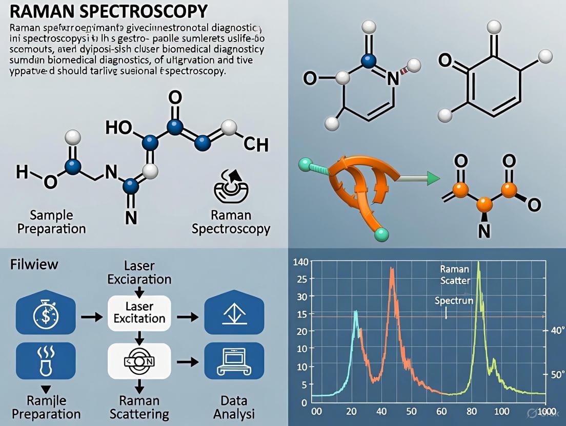
Raman spectroscopy is rapidly evolving into a powerful, non-invasive tool for biomedical diagnostics, offering label-free molecular fingerprinting of tissues and biofluids.
Raman Spectroscopy in Biomedical Diagnostics: From Principles to AI-Enhanced Clinical Applications
Abstract
Raman spectroscopy is rapidly evolving into a powerful, non-invasive tool for biomedical diagnostics, offering label-free molecular fingerprinting of tissues and biofluids. This article explores the foundational principles of Raman spectroscopy and its advanced variants like SERS and SRS. It details methodological applications in cancer diagnosis, neurodegenerative disease detection, and liquid biopsy, emphasizing the transformative role of machine learning in data analysis. The content also addresses key challenges such as signal enhancement and clinical translation, providing a comparative analysis with traditional diagnostic methods. Aimed at researchers and drug development professionals, this review synthesizes current innovations and future trajectories for integrating Raman-based techniques into precision medicine and point-of-care diagnostics.
The Principles and Promise of Raman Spectroscopy in Biomedicine
Raman spectroscopy is a powerful analytical technique that provides a unique biochemical fingerprint of a sample based on the inelastic scattering of light. This method is invaluable in biomedical research for its ability to probe molecular structures and compositions without requiring extrinsic labels or causing significant sample damage [1]. The fundamental process involves exciting molecular vibrational modes using monochromatic laser light and detecting the resulting energy shifts in scattered photons, which correspond directly to specific chemical bonds and functional groups within the sample [2] [3].
The core principle of Raman spectroscopy centers on the Raman effect, first discovered by C.V. Raman in 1928, which occurs when light interacts with matter and undergoes inelastic scattering [1] [4]. This interaction provides chemical specificity that makes Raman spectroscopy particularly valuable for analyzing complex biological systems. When diseases alter the chemical composition of tissues or biofluids, these changes manifest as detectable variations in Raman spectral features—including shifts in intensity, band shape, and peak position [1] [5]. For biomedical researchers and drug development professionals, this capability enables non-invasive investigation of disease mechanisms, diagnostic biomarker discovery, and therapeutic monitoring at the molecular level.
Fundamental Physics of Inelastic Scattering
The Raman Effect and Scattering Processes
The Raman effect originates from the inelastic scattering of photons by molecules. When monochromatic light interacts with a molecule, most photons are elastically scattered (Rayleigh scattering) with unchanged energy. However, approximately 1 in 10 million photons undergoes inelastic (Raman) scattering, resulting in a shift in the photon's energy that provides information about molecular vibrational states [2] [1].
The quantum mechanical description involves transitions to virtual energy states rather than real electronic excited states. When a photon interacts with a molecule, it may promote the system to a short-lived virtual state. As the molecule returns from this virtual state, scattered photons may have different energies than the incident photons. The energy difference between incident and scattered photons corresponds to the vibrational energy levels of the molecule, following the relationship:
E = hν(n + ½)
where h is Planck's constant, ν is the vibrational frequency, and n is the vibrational quantum number [2].
Figure 1: Energy level diagram showing Rayleigh, Stokes Raman, and anti-Stokes Raman scattering processes.
Stokes and Anti-Stokes Scattering
Raman scattering manifests in two primary forms with distinct characteristics:
Stokes Raman scattering occurs when molecules initially in the ground vibrational state (n=0) absorb energy and transition to higher vibrational states (n=1). This process results in scattered photons with lower energy (longer wavelength) than the incident photons. Stokes scattering is more intense than anti-Stokes scattering because, at thermal equilibrium, most molecules populate the ground vibrational state according to Boltzmann distribution [2] [1].
Anti-Stokes Raman scattering occurs when molecules initially in excited vibrational states (n=1) transition to lower energy states. This process produces scattered photons with higher energy (shorter wavelength) than incident photons. Anti-Stokes scattering is inherently weaker than Stokes scattering due to the lower population of excited vibrational states at room temperature [2] [1].
The energy shift in both scattering processes is measured in wavenumbers (cm⁻¹) and calculated using the formula:
Δν̃ = (1/λ₀ - 1/λ₁) × 10⁷
where λ₀ is the excitation wavelength and λ₁ is the Raman scattered wavelength, both in nanometers [4].
Raman Scattering Cross-Section and Selection Rules
The intensity of Raman scattering depends on the Raman scattering cross-section, which is determined by the change in molecular polarizability during vibration. Unlike infrared spectroscopy, which requires a change in the permanent dipole moment, Raman activity depends on how the electron cloud deforms in response to the electric field of light—a property described by the polarizability tensor [3] [4].
The selection rule for Raman spectroscopy states that a vibration is Raman-active if it produces a change in the molecular polarizability. This makes Raman spectroscopy particularly sensitive to symmetric vibrations and non-polar bonds (e.g., C-C, C=C, S-S), whereas infrared spectroscopy is more sensitive to asymmetric vibrations in polar bonds (e.g., C=O, O-H, N-H). This complementary relationship allows researchers to obtain comprehensive vibrational profiles of biological molecules [1] [4].
Molecular Fingerprints in Biomedical Analysis
The Fingerprint Region and Biomolecular Assignments
The Raman "fingerprint region" (500-1800 cm⁻¹) contains the majority of vibrational bands for biological molecules, providing characteristic patterns for identification and quantification. This region is particularly valuable for biomedical diagnostics because it captures overlapping signatures from proteins, lipids, nucleic acids, and carbohydrates that constitute biological systems [1] [3].
Table 1: Characteristic Raman bands of key biomolecules in the fingerprint region
| Biomolecule Class | Raman Shift (cm⁻¹) | Vibrational Assignment | Biomedical Significance |
|---|---|---|---|
| Proteins | 1650-1660 | Amide I (C=O stretch) | Secondary structure analysis |
| 1240-1300 | Amide III (C-N stretch, N-H bend) | Protein conformation changes | |
| 1003 | Phenylalanine ring breathing | Protein content marker | |
| Lipids | 1440-1460 | CH₂ bending | Membrane composition |
| 1650-1680 | C=C stretch | Lipid unsaturation degree | |
| 1730-1750 | C=O stretch | Ester carbonyl groups | |
| Nucleic Acids | 785-795 | Phosphodiester backbone | DNA/RNA content |
| 1095 | O-P-O symmetric stretch | Nucleic acid quantification | |
| 1575-1580 | Guanine, adenine rings | Purine base markers | |
| Carbohydrates | 1045-1060 | C-O, C-C stretches | Glycogen content |
| 1120 | C-O-C glycosidic link | Polysaccharide identification |
The high-wavenumber region (2700-3500 cm⁻¹) also provides valuable information, particularly from C-H stretching vibrations in proteins and lipids, and O-H stretching from water and carbohydrates [5]. The distinct spectral patterns in these regions enable researchers to detect subtle biochemical changes associated with disease states, often before morphological changes become apparent.
Spectral Alterations in Disease States
Disease-induced biochemical changes manifest as quantifiable alterations in Raman spectra rather than the appearance of entirely new peaks. These alterations include:
Intensity Changes: Variations in peak intensity reflect concentration changes of specific biomolecules. For example, decreased lipid-to-protein ratios often indicate membrane alterations in cancer cells [1] [5].
Peak Shifts: Shifts in peak position indicate molecular environment changes, such as protein conformational alterations or stress-induced bond length modifications. Compressive stress shifts peaks to higher frequencies, while tensile stress shifts them to lower frequencies [6].
Bandwidth Changes: Peak broadening often reflects molecular disorder or heterogeneous environments within samples, which can indicate disease progression or treatment effects [6] [5].
These spectral alterations form the basis for disease detection and classification using multivariate statistical analysis and machine learning algorithms.
Advanced Raman Techniques for Enhanced Detection
Surface-Enhanced Raman Spectroscopy (SERS)
Surface-Enhanced Raman Spectroscopy (SERS) dramatically improves detection sensitivity by leveraging plasmonic nanostructures to amplify Raman signals. When laser light excites localized surface plasmon resonance in metallic nanostructures (typically gold, silver, or copper), the electromagnetic field near the metal surface is significantly enhanced, resulting in Raman signal intensification of 10⁸ to 10¹¹ times compared to conventional Raman spectroscopy [2] [3].
SERS enables the detection of biomarkers at clinically relevant low concentrations in complex biological matrices like blood, urine, and saliva. This exceptional sensitivity makes SERS invaluable for early disease diagnosis, particularly for cancer biomarkers, cardiac markers, and neurological disease indicators [2] [5]. The technique has been successfully applied to detect proteins, nucleic acids, and other biomarkers at trace levels, facilitating liquid biopsy approaches for non-invasive diagnostics [7].
Other Enhanced Raman Techniques
Table 2: Comparison of advanced Raman spectroscopy techniques
| Technique | Enhancement Mechanism | Key Advantages | Biomedical Applications |
|---|---|---|---|
| Resonance Raman (RRS) | Electronic transition resonance | Selective enhancement of specific chromophores | Hemoprotein analysis, carotenoid detection |
| Confocal Raman (CRS) | Spatial filtering through pinhole | Depth profiling, 3D imaging | Skin layer analysis, drug penetration studies |
| Spatially Offset Raman (SORS) | Collection from offset positions | Subsurface probing (>2 mm depth) | Bone disease, deep tissue cancer detection |
| Stimulated Raman (SRS) | Nonlinear coherent excitation | Fast imaging, reduced background | Real-time tissue imaging, live-cell analysis |
| Tip-Enhanced Raman (TERS) | Nanoscale plasmonic tip | Nanometer spatial resolution | Single-molecule detection, viral particle analysis |
| Coherent Anti-Stokes Raman (CARS) | Four-wave mixing process | Directional signal, no fluorescence | Lipid droplet imaging, myelin sheath analysis |
These advanced techniques have expanded Raman spectroscopy from a laboratory analytical tool to a versatile method for addressing diverse biomedical challenges, from intraoperative tumor margin assessment to single-cell analysis and drug delivery monitoring [2] [8] [3].
Experimental Protocols for Biomedical Applications
Protocol 1: SERS-Based Protein Biomarker Detection
This protocol details the detection of protein biomarkers from human serum using SERS, applicable for cancer, cardiovascular disease, and neurodegenerative disorder diagnostics [5].
Materials and Reagents:
- SERS substrate (gold or silver nanoparticles)
- Antibody-conjugated Raman reporters
- Phosphate buffered saline (PBS)
- Centrifugal filters (MWCO 10 kDa)
- Serum samples from patients and healthy controls
Procedure:
Sample Preparation
- Centrifuge serum samples at 14,000 × g for 10 minutes to remove particulates
- Dilute clarified serum 1:10 in PBS buffer
- Add 100 µL of diluted serum to antibody-conjugated SERS nanoparticles
Incubation and Capture
- Incubate serum-nanoparticle mixture at room temperature for 30 minutes with gentle agitation
- Transfer mixture to centrifugal filter and centrifuge at 5,000 × g for 5 minutes
- Wash twice with 200 µL PBS to remove unbound components
SERS Measurement
- Resuspend nanoparticle pellet in 50 µL PBS
- Deposit 10 µL onto aluminum-coated slide
- Acquire spectra using 785 nm laser, 10 mW power, 10-second integration
- Collect 10-20 spectra from different spots for statistical robustness
Data Analysis
- Preprocess spectra: subtract background, normalize to internal standard
- Identify characteristic biomarker peaks using reference spectra
- Quantify concentration using calibration curves
- Apply multivariate analysis for classification
Troubleshooting Tips:
- If signal intensity is low, optimize nanoparticle concentration and incubation time
- If background is high, increase wash steps or optimize filter molecular weight cutoff
- For reproducibility issues, standardize nanoparticle batch and laser alignment
Protocol 2: Raman Spectroscopy of Biological Tissues
This protocol describes ex vivo Raman analysis of tissue specimens for cancer diagnosis and margin assessment [1].
Materials and Reagents:
- Cryostat or microtome
- Aluminum-coated glass slides
- Optimal Cutting Temperature (OCT) compound
- Phosphate buffered saline
- Liquid nitrogen for flash freezing
Procedure:
Tissue Preparation
- Flash freeze fresh tissue specimens in liquid nitrogen
- Embed tissue in OCT compound on cryostat chuck
- Section tissue to 10-20 µm thickness at -20°C
- Thaw-mount sections onto aluminum-coated slides
- Rinse with PBS to remove OCT compound
Spectral Acquisition
- Use Raman microscope with 785 nm or 830 nm laser source
- Set laser power to 10-25 mW to prevent tissue damage
- Focus laser spot on tissue section using 20× or 50× objective
- Acquire spectra with 5-10 second integration time
- Collect 30-50 spectra from different tissue regions
- Include background spectra from slide for subtraction
Data Processing
- Subtract fluorescence background using polynomial fitting
- Normalize spectra to internal standard (e.g., phenylalanine 1003 cm⁻¹ peak)
- Perform vector normalization for comparative analysis
- Apply principal component analysis for feature extraction
Validation Methods:
- Compare Raman classification with histopathology results
- Perform immunohistochemistry on adjacent sections for biomarker correlation
- Use cross-validation to assess classification accuracy
Figure 2: Experimental workflow for Raman-based biomedical analysis of liquid and tissue samples.
The Scientist's Toolkit: Essential Research Reagents and Materials
Table 3: Key research reagents and materials for Raman biomedical applications
| Item | Function | Application Examples | Technical Notes |
|---|---|---|---|
| Gold Nanoparticles | SERS substrate, signal enhancement | Biomarker detection, immunoassays | Tunable plasmon resonance (40-100 nm) |
| Raman Reporters | Molecular tags for detection | Multiplexed biomarker assays | 4-MBA, DTNB, rhodamine derivatives |
| Antibody Conjugates | Target-specific capture | Protein biomarker detection | Orientation-specific conjugation |
| Aluminum-Coated Slides | Low background substrates | Tissue section analysis | Reflectance enhancement |
| OCT Compound | Tissue embedding medium | Cryosection preparation | Must be thoroughly washed before analysis |
| Laser Sources | Monochromatic excitation | Spectral acquisition | 785 nm reduces fluorescence in biosamples |
| Notch Filters | Rayleigh rejection | Signal purification | OD > 6 for laser line rejection |
| CCD Detectors | Signal detection | Spectral collection | Cooled to -60°C to reduce dark noise |
| Calibration Standards | Instrument calibration | Wavenumber accuracy | Silicon (520.7 cm⁻¹), neon lamps |
| Chemometric Software | Data analysis | Pattern recognition, classification | PCA, LDA, support vector machines |
Raman spectroscopy provides an exceptional platform for biomedical diagnostics by leveraging the fundamental principles of inelastic scattering and molecular fingerprint analysis. The technique's label-free nature, molecular specificity, and compatibility with aqueous environments make it ideally suited for analyzing complex biological systems. With advanced implementations like SERS pushing detection limits to clinically relevant concentrations, Raman spectroscopy continues to transform capabilities in disease diagnosis, therapeutic monitoring, and fundamental biological research.
The experimental protocols and technical resources outlined in this document provide researchers with practical frameworks for implementing Raman-based approaches in diverse biomedical contexts. As instrumentation advances and data analysis methods become more sophisticated, Raman spectroscopy is poised to play an increasingly central role in precision medicine and pharmaceutical development.
Raman spectroscopy has emerged as a powerful analytical technique in biomedical research, offering a unique combination of non-invasiveness, label-free operation, and excellent compatibility with aqueous samples. This application note details how these core advantages make Raman spectroscopy particularly suitable for analyzing biological systems, from single cells to tissues, without altering their native state. The technique provides a biochemical "fingerprint" based on inelastic light scattering, enabling researchers to probe molecular structures and compositions in their natural physiological environment [9] [1]. The fundamental principle involves shining monochromatic light on a sample and detecting the minutely shifted wavelengths of the scattered light, which correspond to specific vibrational modes of molecular bonds [10]. This process requires no external labels or destructive sample preparation, preserving biological integrity while yielding rich molecular information.
Core Advantages and Supporting Data
The value of Raman spectroscopy for biomedical research rests on three foundational pillars, each supported by robust experimental data and distinct from conventional analytical methods.
Table 1: Core Advantages of Raman Spectroscopy in Biomedical Research
| Advantage | Technical Basis | Research Impact | Comparison to Alternative Techniques |
|---|---|---|---|
| Non-invasiveness | No sample destruction; minimal photodamage with Near-IR lasers [1] [11] | Enables longitudinal studies on living cells (e.g., monitoring cell death) and in vivo diagnostics [11] [12] | Fluorescence microscopy often causes photobleaching and phototoxicity, altering samples [11] |
| Label-Free Detection | Relies on intrinsic molecular vibrations without dyes, stains, or radioactive labels [1] [13] | Reveals true biochemical state without label-induced artifacts; simplifies sample preparation [13] [11] | Immunoassays (ELISA) and fluorescence imaging require specific labels that can be costly and introduce variability [14] |
| Water Compatibility | Weak Raman scattering from water molecules [9] [1] | Allows analysis of cells in physiological buffer, biofluids (e.g., serum, saliva), and hydrated tissues [9] [15] | Infrared (IR) spectroscopy is strongly absorbed by water, complicating the study of aqueous solutions [9] [1] |
Experimental Evidence from Recent Studies
Recent studies quantitatively demonstrate these advantages in practice. For instance, in research on regulated cell death (RCD), Raman microscopy combined with a support vector machine (SVM) correctly classified 73% of all spectra from untreated cells and those undergoing apoptosis, ferroptosis, and necroptosis, all without using any fluorescent labels [11]. This highlights the technique's capability for sensitive, label-free discrimination of subtle biochemical changes.
In another application for diagnosing drug-induced liver injury, confocal Raman imaging identified a distinct spectral peak at 1638 cm⁻¹ in injured liver tissues, which differed from characteristic peaks in controls (1203, 1266, and 1746 cm⁻¹). Machine learning models trained on this label-free data achieved accurate staging of liver injury with an Area Under the Curve (AUC) > 0.95 [15]. Furthermore, the analysis of liquid biopsies showcases the advantage of water compatibility. A study classifying cancer-derived exosomes from blood plasma using Raman spectroscopy achieved an overall accuracy of 93.3%, with high F1-scores for different cancer types (e.g., 98.2% for colon cancer) [7]. This demonstrates high sensitivity in a complex aqueous matrix.
Experimental Protocols
This section provides detailed methodologies for implementing Raman spectroscopy in two key biomedical research applications: analyzing living cells and processing liquid biopsies.
Protocol 1: Label-Free Analysis of Cell Death in Live Cells
This protocol is adapted from a study investigating ferroptosis, apoptosis, and necroptosis [11].
- Objective: To distinguish between different types of regulated cell death (RCD) in mammalian cell lines using label-free Raman spectroscopy and machine learning.
- Materials and Reagents:
- Murine fibroblast cell line (e.g., L929sAhFas).
- Appropriate cell culture medium and reagents.
- Cell death inducers: Anti-Fas antibody (for apoptosis), RSL3 (for ferroptosis), murine TNF (mTNF, for necroptosis).
- Glass-bottom culture dishes suitable for microscopy.
- Instrumentation: A confocal Raman microscope system, typically equipped with a 785 nm or 532 nm laser to minimize cell damage and fluorescence background [11].
- Procedure:
- Cell Culture and Induction: Culture cells in glass-bottom dishes. Divide into experimental groups: Control (untreated), Apoptosis (induced by Anti-Fas antibody), Ferroptosis (induced by RSL3), and Necroptosis (induced by mTNF).
- Raman Data Acquisition:
- Place the dish on the microscope stage.
- Focus the laser on individual cells.
- Acquire Raman spectra from multiple cells per condition. Typical parameters might include a laser power of a few milliwatts (mW) and an integration time of 0.35 seconds per spectrum [15].
- Collect spectra from a defined wavenumber range (e.g., 600–1800 cm⁻¹).
- Data Preprocessing: Perform baseline correction to remove fluorescence background and normalize spectra to a standard peak (e.g., the phenylalanine peak at 1003 cm⁻¹) to account for intensity variations.
- Machine Learning Analysis:
- Input the preprocessed spectra directly into a Support Vector Machine (SVM) classifier.
- Use a nested cross-validation strategy to train the model and evaluate its performance on an independent test set, reporting prediction accuracy.
The workflow for this experimental and analytical process is outlined below.
Protocol 2: Cancer Classification via Exosomes in Liquid Biopsy
This protocol is based on a study classifying exosomes from different cancer cell lines [7].
- Objective: To isolate and classify exosomes from cell culture media or patient biofluids using Raman spectroscopy for non-invasive cancer diagnostics.
- Materials and Reagents:
- Source material: Cell culture supernatant from cancer cell lines (e.g., COLO205 for colon, A375 for skin, LNCaP for prostate) or patient blood plasma.
- Ultracentrifuge and suitable tubes.
- Phosphate-buffered saline (PBS).
- Purification filters (e.g., 0.22 µm).
- Instrumentation: Raman spectrometer, potentially with a Surface-Enhanced Raman Scattering (SERS) substrate to boost sensitivity for low-concentration targets [7] [14].
- Procedure:
- Exosome Isolation:
- Centrifuge cell culture media or plasma at low speed (e.g., 2,000 × g) to remove cells and debris.
- Filter the supernatant through a 0.22 µm filter.
- Ultracentrifuge the filtered supernatant at high speed (e.g., 100,000 × g) for 70-90 minutes to pellet exosomes.
- Resuspend the purified exosome pellet in a small volume of PBS.
- Raman Measurement:
- Deposit the exosome suspension onto a suitable substrate (e.g., aluminum-coated slides or a SERS-active substrate).
- Acquire Raman spectra. Focus on key wavenumber regions critical for exosome composition: 700–900 cm⁻¹, 1000–1200 cm⁻¹, and 2800–3000 cm⁻¹ (lipid and protein regions) [7].
- Data Analysis:
- Use Principal Component Analysis (PCA) to reduce data dimensionality and extract the most significant spectral features.
- Train a Linear Discriminant Analysis (LDA) classifier on the principal components to differentiate exosomes by their cancer cell line of origin.
- Exosome Isolation:
The following diagram illustrates the key steps from sample collection to classification.
The Scientist's Toolkit
Table 2: Essential Research Reagent Solutions for Raman Spectroscopy in Biomedicine
| Item | Function/Description | Application Example |
|---|---|---|
| Iron Oxide Nanoparticles (IONPs) | Engineered nanostructures used as contrast agents; provide magnetic properties and signal enhancement for multimodal imaging [16]. | Targeted drug delivery and sensitive biosensing [16]. |
| Gold Nanoparticles (AuNPs) & SERS Substrates | Plasmonic nanostructures that dramatically amplify the weak Raman signal via localized surface plasmon resonance, enabling single-molecule detection [9] [14]. | Functionalized with antibodies to detect specific disease biomarkers (e.g., cancer, viral infections) at ultra-low concentrations [16] [14]. |
| Specific Cell Death Inducers | Chemical compounds that selectively activate distinct cell death pathways [11]. | RSL3 (ferroptosis inducer), Anti-Fas antibody (apoptosis inducer), and mTNF (necroptosis inducer) for studying RCD mechanisms [11]. |
| Exosome Isolation Kits | Reagents for purifying extracellular vesicles from complex biofluids like blood plasma or cell culture media via ultracentrifugation or precipitation [7]. | Isolation of cancer-derived exosomes for liquid biopsy analysis and cancer classification [7]. |
| Machine Learning Algorithms | Computational tools (e.g., SVM, CNN, PCA-LDA) for analyzing complex spectral data, identifying patterns, and building classification models [10] [11] [15]. | Automated, high-accuracy classification of tissue pathology (e.g., liver injury staging, cancer type identification) [7] [15]. |
The synergistic combination of non-invasiveness, label-free detection, and water compatibility solidifies Raman spectroscopy's role as an indispensable tool in modern biomedical research. These inherent advantages allow scientists to interrogate biological samples—from living cells to complex clinical biofluids—in their native state, providing unbiased molecular fingerprints. When coupled with machine learning for data analysis, this technique transforms into a powerful platform for diagnostic classification, metabolic profiling, and real-time monitoring of disease progression. As standardization improves and computational methods advance, Raman spectroscopy is poised for further integration into mainstream biomedical research and clinical translation, driving innovation in precision medicine [9] [12].
Vibrational spectroscopy, encompassing both Raman and Infrared (IR) spectroscopy, has emerged as a powerful tool in biomedical diagnostics research. These techniques provide label-free, non-destructive analysis of biological samples by probing molecular vibrations, yielding a biochemical "fingerprint" that reflects the total molecular composition of cells and tissues [17]. In the context of biomedical research, particularly in the development of non-invasive diagnostic tools, understanding the complementary nature of Raman and IR spectroscopy is crucial for selecting the appropriate technique for specific applications. Both techniques investigate molecular vibrations but through different physical mechanisms—Raman spectroscopy measures inelastically scattered light resulting from changes in molecular polarizability, while IR spectroscopy measures light absorption due to changes in dipole moment [1]. This fundamental difference in mechanism confers unique advantages and limitations to each technique, making them complementary rather than competitive for comprehensive biomolecular analysis.
The growing importance of vibrational spectroscopy in biomedicine is driven by the need for medical imaging contrast that goes beyond morphological information to include functional differences at cellular and molecular levels. Molecular imaging using vibrational techniques answers this need by providing high spatial resolution with chemical specificity, enabling researchers to characterize biological processes at molecular and cellular levels by localizing and measuring specific molecular targets or biochemical pathways associated with various pathologies [17]. This application note details the complementary nature of these techniques, providing structured comparisons, experimental protocols, and visualization of workflows to guide researchers in leveraging both methods for advanced biomedical diagnostics.
Theoretical Foundation and Technical Comparison
Physical Principles and Selection Rules
The fundamental difference between Raman and IR spectroscopy lies in their underlying physical mechanisms. IR spectroscopy involves the absorption of infrared light when the energy of incident photons matches the energy difference between vibrational states of molecular bonds. This absorption only occurs for chemical bonds with an electric dipole moment that changes during vibration, making these bonds "IR-active" [17]. In contrast, Raman spectroscopy relies on inelastic scattering of monochromatic light, typically from ultraviolet, visible, or near-infrared lasers. During Raman scattering, photons transfer energy to molecules as vibrational energy (Stokes scattering) or receive energy from vibrating molecules (anti-Stokes scattering). The Raman effect occurs due to changes in molecular polarizability during vibration, making bonds with significant electron cloud deformation "Raman-active" [17] [1].
The complementary selection rules of these techniques mean that symmetric molecular vibrations and non-polar functional groups typically yield strong Raman signals but weak IR absorption, while asymmetric vibrations and polar bonds produce strong IR signals but weak Raman scattering. For example, homo-nuclear molecular bonds like C-C, C=C, and S-S are more easily detected with Raman spectroscopy, while heteronuclear functional groups such as C=O, O-H, and N-H are more readily detected with IR spectroscopy [1] [18]. This complementarity enables a more complete biomolecular characterization when both techniques are employed.
Technical Comparison for Biomedical Applications
Table 1: Fundamental Comparison of Raman and IR Spectroscopy Techniques
| Parameter | Raman Spectroscopy | IR Spectroscopy |
|---|---|---|
| Physical Principle | Inelastic scattering of light | Absorption of infrared radiation |
| Measured Interaction | Change in molecular polarizability | Change in dipole moment |
| Sensitivity to | Symmetric vibrations, non-polar bonds | Asymmetric vibrations, polar bonds |
| Water Compatibility | High (minimal water interference) | Low (strong water absorption) |
| Spatial Resolution | High (diffraction-limited, ~0.5-1 μm) | Moderate (~3-10 μm for FT-IR microscopy) |
| Sample Preparation | Minimal (can analyze aqueous solutions directly) | Often requires dehydration or specialized techniques like ATR |
Table 2: Performance Comparison for Biofluid Analysis
| Performance Metric | Raman Spectroscopy | FT-IR Spectroscopy |
|---|---|---|
| Sample Throughput | ~80 samples/day [19] | ~80 samples/day [19] |
| Pathlength for Biofluids | Standard cuvettes (mm range) [19] | Limited pathlength (<100 μm) or ATR [19] |
| Key Spectral Regions | 700-900 cm⁻¹ (lipids), 1000-1200 cm⁻¹ (proteins), 2800-3000 cm⁻¹ (CH-stretching) [7] | 1800-1500 cm⁻¹ (Amide I/II), 1700-1500 cm⁻¹ (proteins), 1240-1080 cm⁻¹ (phosphate) [17] |
| Glucose Quantification (Serum) | Achievable with multivariate analysis [19] | Achievable with multivariate analysis [19] |
The most significant practical difference for biomedical applications is water compatibility. Water exhibits strong absorption in the mid-IR region, making the direct analysis of aqueous biological samples challenging with conventional transmission IR spectroscopy. This limitation can be partially addressed using attenuated total reflectance (ATR) accessories or very short pathlengths (<10 μm) [17] [19]. Conversely, water exhibits weak Raman scattering, allowing direct analysis of cells, tissues, and biofluids with minimal interference, making Raman particularly advantageous for physiological measurements [1].
Experimental Protocols
Protocol 1: Serum Analysis via Raman Spectroscopy for Disease Detection
Principle: This protocol details the detection of Hepatitis C virus (HCV) in serum samples using Near-Infrared Raman Spectroscopy combined with machine learning, demonstrating a non-invasive approach for viral infection diagnosis [20].
Materials and Reagents:
- Serum samples (stored at -80°C until analysis)
- Quartz cuvettes for Raman measurements
- Hellmanex II solution (1%) for cuvette cleaning
- Portable or benchtop Raman spectrometer with 785 nm laser excitation
Procedure:
- Sample Preparation: Thaw frozen serum samples at room temperature. Gently mix to ensure homogeneity. Transfer 70-100 μL to a sterile quartz cuvette [20].
- Instrument Setup: Configure the Raman spectrometer with 785 nm laser excitation, ensuring power at the sample is approximately 200 mW to achieve sufficient signal intensity while minimizing potential photodamage [19] [20].
- Spectral Acquisition: Position the cuvette in the sample holder. Collect Raman spectra in the range of 300-1870 cm⁻¹ with a resolution of 8 cm⁻¹. Acquire spectra over 5 minutes using 12 acquisitions of 25 seconds each to improve signal-to-noise ratio [19].
- Data Preprocessing: Normalize raw spectra and subtract fluorescence background using a fifth-order polynomial fitting algorithm. Apply vector normalization to correct for minor variations in laser power or sample positioning [19].
- Machine Learning Analysis: Employ L1-regularized Logistic Regression for feature selection to identify the most informative wavelengths for disease detection. Integrate these spectral features with clinical data using a Random Forest classifier to enhance diagnostic accuracy [20].
Validation: Compare results with standard clinical methods such as polymerase chain reaction (PCR) for HCV RNA detection. The combined approach of Raman spectroscopy and clinical data has achieved 72.2% accuracy and an AUC-ROC of 0.850 in HCV detection [20].
Protocol 2: Exosome Analysis via Raman Spectroscopy for Cancer Diagnostics
Principle: This protocol describes the classification of cancer cell lines through Raman spectral analysis of exosomes, offering a non-invasive liquid biopsy approach for early cancer detection [7].
Materials and Reagents:
- Cell culture supernatants from cancer cell lines (e.g., COLO205 colon cancer, A375 skin cancer, LNCaP prostate cancer)
- Ultracentrifugation equipment for exosome isolation
- Phosphate buffered saline (PBS) for sample washing
- Quartz cuvettes or specialized flow cells for Raman measurements
Procedure:
- Exosome Isolation: Collect cell culture media and centrifuge at 2,000 × g for 30 minutes to remove cells and debris. Transfer supernatant to ultracentrifuge tubes and centrifuge at 100,000 × g for 70 minutes at 4°C to pellet exosomes. Resuspend exosome pellets in PBS [7].
- Sample Loading: Transfer exosome suspension to quartz cuvettes. For enhanced sensitivity, consider using surface-enhanced Raman spectroscopy (SERS) substrates by mixing exosomes with gold or silver nanoparticles [7] [21].
- Spectral Acquisition: Use a Raman microscope with 785 nm excitation laser. Focus laser beam on the sample using a 20× or 40× objective. Collect spectra in the range of 500-1800 cm⁻¹ with 5-10 seconds integration time per spectrum. Accumulate multiple spectra from different sample spots to account for heterogeneity.
- Data Analysis: Apply principal component analysis (PCA) to extract chemically significant features from Raman spectra. Use linear discriminant analysis (LDA) to classify exosomes based on their cancer cell line origin. Note that key discriminatory regions typically include 700-900 cm⁻¹ (lipids), 1000-1200 cm⁻¹ (proteins), and 2800-3000 cm⁻¹ (CH-stretching modes) [7].
- Lipid Profiling: Analyze specific lipid composition differences, particularly abundance of omega-3 25:5 in prostate and skin cancer exosomes versus glycerophospholipids in colon cancer exosomes [7].
Validation: This approach has achieved 93.3% overall classification accuracy with high F1 scores (98.2% for colon cancer, 91.1% for skin cancer, and 91.0% for prostate cancer) when validated against known cancer cell lines [7].
Protocol 3: Tissue Analysis via FT-IR Spectroscopy for Disease Diagnosis
Principle: This protocol outlines the use of FT-IR spectroscopy for rapid diagnosis of fibromyalgia syndrome (FM) and related rheumatologic disorders using bloodspot samples [22].
Materials and Reagents:
- Blood samples collected via fingerstick or venipuncture
- Silicon sample carriers or IR-transparent windows (e.g., BaF₂, CaF₂)
- Desiccator for sample drying (optional)
- FT-IR spectrometer with ATR accessory
Procedure:
- Sample Preparation: Spot 3 μL of blood or serum onto silicon sample carriers. Allow to dry in ambient air for 30 minutes, forming a thin film of 2-10 μm thickness suitable for transmission measurements [19] [22].
- ATR Alternative: For ATR measurements, place liquid samples directly on the ATR crystal, ensuring complete coverage of the crystal surface. Apply consistent pressure to ensure proper sample-crystal contact.
- Spectral Acquisition: Using an FT-IR spectrometer equipped with a DLaTGS detector, acquire spectra in the range of 500-4000 cm⁻¹ at 4 cm⁻¹ resolution. Average 32 scans to improve signal-to-noise ratio. For each sample, prepare and measure three technical replicates to ensure reproducibility [19].
- Data Preprocessing: Correct absorbance spectra for substrate background and apply vector normalization. Calculate the median of the three pre-processed spectra for each sample to minimize artifacts [19].
- Chemometric Analysis: Employ orthogonal partial least squares discriminant analysis (OPLS-DA) to classify spectra into diagnostic categories. Identify spectral biomarkers, particularly in amide bands (1700-1500 cm⁻¹) and aromatic amino acid regions [22].
Validation: This method has successfully classified fibromyalgia spectra with high sensitivity and specificity (Rcv > 0.93), identifying peptide backbones and aromatic amino acids as potential biomarkers [22].
Workflow Visualization
Essential Research Reagent Solutions
Table 3: Key Research Reagents and Materials for Vibrational Spectroscopy
| Reagent/Material | Function/Application | Technical Notes |
|---|---|---|
| Quartz Cuvettes | Sample holder for Raman measurements of liquids | Low fluorescence background; compatible with visible and NIR lasers |
| ATR Crystals (diamond, ZnSe, Ge) | Enables IR analysis of challenging samples without extensive preparation | Diamond: durable but expensive; ZnSe: good general purpose; Ge: high refractive index for strong absorbers |
| Silicon Sample Carriers | Substrate for dried film FT-IR measurements | IR-transparent; disposable option reduces cross-contamination |
| Hellmanex II Solution (1%) | Cleaning solution for Raman cuvettes and optics | Effective removal of biological residues; critical for maintaining signal quality |
| Gold/Silver Nanoparticles | SERS substrates for signal enhancement | Amplify Raman signals by 10⁴-10⁸ times; enable single-molecule detection |
| Standard Normal Variate (SNV) | Spectral preprocessing algorithm | Corrects for scattering effects in reflectance measurements |
| Principal Component Analysis (PCA) | Dimensionality reduction for spectral data | Identifies most significant spectral patterns; reduces data complexity |
| Linear Discriminant Analysis (LDA) | Classification of spectral data | Maximizes separation between predefined sample classes |
Advanced Applications and Future Perspectives
Integration with Artificial Intelligence
The combination of vibrational spectroscopy with artificial intelligence represents a paradigm shift in biomedical diagnostics. AI and machine learning algorithms significantly enhance the interpretability of complex spectral data by enabling robust classification, pattern recognition, and biomarker discovery [7]. These computational methods can detect subtle spectral differences imperceptible to human analysis, thereby improving diagnostic accuracy and enabling automated, high-throughput analysis [21].
Deep learning approaches applied to Raman spectra have demonstrated remarkable success in biomedical applications. For example, deep learning algorithms applied to colorectal tissue Raman spectra achieved 98.5% accuracy in cancer detection [1]. Similarly, AI-guided analysis of Raman spectra from cancer-derived exosomes has shown 93.3% classification accuracy for different cancer types [7] [23]. The implementation of FAIR (Findable, Accessible, Interoperable, and Reusable) data principles is crucial for developing robust, standardized AI models in vibrational spectroscopy, as publicly accessible and harmonized Raman databases provide large, high-quality datasets for model training and validation [21].
Multimodal Imaging and Integrated Approaches
The complementary nature of Raman and IR spectroscopy makes them ideal partners in multimodal imaging approaches. Combining these vibrational techniques with other imaging modalities creates powerful diagnostic platforms that leverage the strengths of each method. For instance, integrating Raman spectroscopy with photoacoustic imaging, magnetic resonance imaging (MRI), or computed tomography (CT) enables correlation of molecular-level biochemical information with anatomical context [17].
Multimodal probes that combine vibrational imaging with other modalities are emerging as valuable tools in preclinical research. These approaches allow researchers to obtain comprehensive information about disease processes by leveraging the high spatial resolution of optical microscopy with the chemical specificity of vibrational spectroscopies [17]. The development of such multimodal platforms represents a significant advancement toward clinical translation of vibrational spectroscopy techniques, potentially enabling real-time, in-clinic diagnostics for conditions like fibromyalgia and various cancers [22] [7].
Raman and IR spectroscopy offer complementary approaches to biomedical analysis, each with distinct advantages and limitations that make them suitable for different applications within diagnostics and drug development research. Raman spectroscopy excels in analyzing aqueous samples, provides high spatial resolution, and is ideal for detecting symmetric molecular vibrations. In contrast, IR spectroscopy offers strong sensitivity to polar functional groups and has established protocols for dried tissue analysis. The strategic selection between these techniques—or their combined application—should be guided by sample characteristics, target biomolecules, and analytical requirements.
The integration of these vibrational spectroscopy techniques with artificial intelligence and machine learning represents the future of biomedical diagnostics, enabling automated, high-throughput analysis with enhanced accuracy. As open science initiatives and standardized protocols continue to evolve, Raman and IR spectroscopy are poised to play increasingly important roles in personalized medicine, offering non-invasive, real-time diagnostic capabilities that could transform clinical practice and drug development processes.
Raman spectroscopy has emerged as a powerful, label-free technique for biomedical diagnostics, providing a unique biochemical fingerprint of samples based on the inelastic scattering of light. Its non-destructive nature, minimal sample preparation requirements, and high molecular specificity make it particularly valuable for analyzing complex biological tissues and fluids. The core principle enabling this technology involves probing molecular rotational and vibrational states, generating spectral data that reflects the chemical composition and structure of the sample. The instrumentation required to harness these principles spans sophisticated laboratory systems for research and compact, specialized probes for clinical applications, forming a critical technological continuum for biomedical advancement.
Fundamental Principles and System Configurations
Core Principles of Raman Spectroscopy
Raman spectroscopy relies on the inelastic scattering of photons when light interacts with a material. Most scattered light undergoes Rayleigh (elastic) scattering, where the scattered photon energy equals the incident photon energy. Only approximately one in 10⁸ photons undergoes the Raman effect, resulting in scattered light with a different energy. Stokes Raman scattering occurs when the scattered photon has lower energy than the incident photon, while anti-Stokes Raman scattering occurs when it has higher energy. The energy difference between incident and scattered photons corresponds to the vibrational energy of chemical bonds, creating a unique spectral fingerprint for each molecular species [1].
The technique is particularly valuable for biological analysis because it is compatible with physiological measurements due to low interference from water, unlike infrared spectroscopy. Furthermore, it requires no extrinsic labels such as dyes, stains, or radioactive markers, preserving native biological states [1] [9].
Basic Laboratory Instrumentation
A typical laboratory Raman system consists of several key components arranged in a 180° backscatter geometry:
- Excitation Laser: Common wavelengths include argon (488 nm, 514.5 nm), helium-neon (632.8 nm), and near-infrared (785 nm, 830 nm) lasers. NIR lasers are often preferred for biological samples to reduce photodamage and fluorescence background [1].
- Filter System: Blocks elastically scattered laser light (Rayleigh scattering) while transmitting the weaker Raman signal.
- Spectrometer: Disperses the collected light using a diffraction grating.
- Detector: Typically a charge-coupled device (CCD) that captures the dispersed light to generate a spectrum [1].
Such systems can perform spatial mapping by acquiring a Raman spectrum at each point across a sample area, enabling visualization and quantification of different biochemical components [1].
Table 1: Common Laser Wavelengths Used in Raman Spectroscopy and Their Applications
| Laser Wavelength | Type | Key Advantages | Common Biomedical Applications |
|---|---|---|---|
| 488 nm, 514.5 nm | Visible (Argon) | Higher spatial resolution | Ex vivo analysis of tissues and cells |
| 632.8 nm | Visible (He-Ne) | Good balance of resolution and penetration | General purpose laboratory analysis |
| 785 nm, 830 nm | Near-Infrared (NIR) | Reduced fluorescence, less photodamage | In vivo measurements, sensitive biological samples |
Fiber-Optic Probe Designs for Biomedical Applications
The transition from laboratory systems to clinical applications primarily occurs through specialized fiber-optic probes that enable access to internal organs and integration with medical instruments.
Standard Contact Probe Design
Clinical applications typically use fiber-optic probes with small diameters that can navigate body cavities and integrate with routine medical instruments. A basic Raman probe with a "10 around 1" fiber configuration includes:
- Central Excitation Fiber: Transmits laser light to the sample.
- Collection Fibers: Surrounding fibers (e.g., 10 fibers) that collect backscattered Raman signal.
- Internal Lenses: Focus laser light onto the sample and collect the scattered light.
- Filter Systems: Integrated laser line cleanup and edge filters to remove unwanted signals and ensure only Raman scattering reaches the spectrometer [1] [24].
Advanced Probe Designs
Noncontact Fiber-Optic Probe
A novel probe design addresses limitations of conventional probes during noncontact operation, where performance suffers from decreased collection efficiency and larger laser spot size. This design uses a miniature lens at the probe tip to efficiently collect both fingerprint (FP: 500-1800 cm⁻¹) and high-wavenumber (HW: 2800-3600 cm⁻¹) Raman spectra at an offset from target tissue. This design demonstrated a 90% increase in signal intensity and a four-fold improvement in spatial selectivity compared to conventional contact probes. Lenses fabricated from crystalline materials like sapphire and calcium fluoride best preserved the weak Raman signal from tissues [25].
Disposable Submillimeter Probes
For specialized applications like brain biopsy, disposable submillimeter fiber-optic Raman probes have been developed. These ultra-small probes facilitate intracranial measurements while maintaining signal-to-noise ratio and preventing cross-contamination between procedures [26].
Multi-Modal Probes
Advanced probes now combine multiple spectroscopic techniques. One development integrates ATR, Transflection, and Raman spectroscopy at the same measurement point, providing complementary data for improved process control and biomedical diagnostics [27].
Table 2: Comparison of Raman Fiber-Optic Probe Types for Biomedical Applications
| Probe Type | Key Features | Performance Advantages | Representative Applications |
|---|---|---|---|
| Standard Contact Probe | "10 around 1" fiber configuration, integrated filters | Robust design, proven clinical feasibility | In vivo tissue diagnosis during endoscopy [1] [24] |
| Noncontact Probe | Miniature lens at tip, optimized for offset operation | 90% signal increase, 4x spatial selectivity | In vivo tissue interrogation where contact is undesirable [25] |
| Disposable Submillimeter Probe | Diameter <1 mm, single-use | Prevents cross-contamination, accesses confined spaces | Brain biopsy, intracranial measurements [26] |
| Multi-Modal Probe | Combines Raman, ATR, and Transflection | Simultaneous complementary data from same point | Enhanced process control, advanced diagnostics [27] |
Experimental Protocols
Protocol 1: In Vivo Raman Spectroscopy During Surgical Procedures
This protocol establishes a clinical workflow for intraoperative in vivo Raman spectroscopy during head and neck cancer surgery [24].
Pre-Measurement Preparation
- Confirm regulatory approval for clinical investigation (e.g., CE mark, MDR compliance).
- Position the mobile Raman system (on a medical cart) near the surgical field.
- Perform system calibration using reference standards.
- Sterilize the fiber-optic probe according to hospital protocols.
Intraoperative Measurement Procedure
- After tumor exposure, acquire spectra from the tumor site, tumor margins, and healthy control tissue.
- Maintain consistent probe orientation and contact pressure (for contact probes) or distance (for noncontact probes).
- Acquisition parameters: 785 nm excitation laser, spectral range 500-3300 cm⁻¹, integration time 1-5 seconds.
- Record multiple spectra (typically 3-5) from each measurement site to account for heterogeneity.
- Correlate each measurement with precise anatomical location using integrated wide-field camera guidance.
Data Analysis and Interpretation
- Process raw spectra with preprocessing algorithms: subtract background fluorescence, remove cosmic rays, normalize spectra.
- Employ machine learning classifiers (e.g., principal component analysis with linear discriminant analysis) trained on reference databases to differentiate tumor from healthy tissue.
- Provide real-time diagnostic feedback to the surgical team regarding margin status.
Workflow Integration and Timing
- Initial procedures may require >30 minutes, but with experience, measurement time decreases to approximately 2 minutes after 15 patients.
- Integrate spectroscopic measurements seamlessly between surgical steps to avoid prolonging overall procedure time.
Protocol 2: Ex Vivo Analysis of Liquid Biopsy Samples Using Raman Spectroscopy
This protocol details the use of Raman spectroscopy for classifying cancer-derived exosomes from liquid biopsies [7].
Sample Preparation
- Isolate exosomes from blood plasma or other bodily fluids using ultracentrifugation or commercial kits.
- Deposit exosome samples on appropriate substrates (e.g., aluminum-coated slides, SERS substrates).
- For SERS analysis, mix exosomes with colloidal gold or silver nanoparticles to enhance signal.
Spectral Acquisition
- Use a Raman microscope with 785 nm excitation laser to minimize fluorescence.
- Focus laser beam on the sample using a microscope objective (typically 20× or 50×).
- Acquisition parameters: laser power 10-100 mW, integration time 10-60 seconds, spectral range 500-1800 cm⁻¹ (fingerprint region) and 2800-3000 cm⁻¹ (CH-stretching region).
- Collect multiple spectra from different sample spots to ensure representative sampling.
Data Analysis with Machine Learning
- Preprocess spectra: subtract baseline, normalize, and vector-normalize.
- Apply principal component analysis (PCA) to reduce dimensionality and extract chemically significant features.
- Train a linear discriminant analysis (LDA) classifier or other machine learning models (e.g., support vector machines, neural networks) on known samples.
- Validate model performance using cross-validation and independent test sets.
- Identify significant spectral regions (e.g., 700-900 cm⁻¹, 1000-1200 cm⁻¹, 2800-3000 cm⁻¹) that contribute most to classification accuracy.
The Scientist's Toolkit: Essential Research Reagent Solutions
Table 3: Key Research Reagents and Materials for Raman Biomedical Applications
| Item | Function | Application Example |
|---|---|---|
| CE-Marked Raman System | Ensures compliance with medical device regulations for clinical trials | In vivo measurements during human surgery [24] |
| Fiber-Optic Probes | Enables access to internal organs and integration with medical instruments | In vivo tissue diagnosis during colonoscopy [1] |
| SERS Substrates | Enhances Raman signal by several orders of magnitude through plasmonic effects | Detection of viral RNA and proteins in swabs for COVID-19 [9] |
| Exosome Isolation Kits | Purifies extracellular vesicles from bodily fluids for liquid biopsy analysis | Cancer classification from blood plasma [7] |
| Reference Standards | Calibrates instruments and validates spectral accuracy | Daily quality control and system performance verification |
| Machine Learning Software | Analyzes complex spectral data and develops classification models | Differentiating cancerous from healthy tissues [7] [21] |
Advanced Techniques and Future Directions
Enhanced Raman Techniques
Several advanced Raman techniques address limitations of conventional spontaneous Raman spectroscopy:
Surface-Enhanced Raman Spectroscopy (SERS): Utilizes nanostructured metallic surfaces to amplify Raman signals by several orders of magnitude, enabling detection of biomolecules at ultra-low concentrations. Recent advances include nanotags with interior gaps, orthogonal Raman reporters, and near-infrared-II-responsive properties [28].
Tip-Enhanced Raman Spectroscopy (TERS): Combines scanning probe microscopy with Raman spectroscopy to achieve nanoscale spatial resolution, enabling chemical imaging at the molecular level.
Stimulated Raman Scattering (SRS) and Coherent Anti-Stokes Raman Scattering (CARS): Nonlinear techniques that provide significantly stronger signals than spontaneous Raman, enabling real-time imaging of biological tissues with applications in histopathology and tissue diagnostics [21].
Integration with Artificial Intelligence
The combination of Raman spectroscopy with artificial intelligence represents a paradigm shift in biomedical analysis:
- Deep Learning Models: Automate data processing, extract meaningful features, and enable predictive modeling for disease diagnosis.
- Digitalization and FAIR Data Principles: Initiatives to make Raman data Findable, Accessible, Interoperable, and Reusable are critical for developing robust, standardized analytical workflows [21].
- Real-Time Decision Support: AI-powered Raman systems can provide immediate diagnostic feedback during surgical procedures, potentially transforming cancer surgery and clinical diagnostics [23].
The instrumentation for Raman spectroscopy has evolved from sophisticated laboratory systems to specialized fiber-optic probes capable of in vivo clinical measurements. This progression has been essential for translating Raman spectroscopy from a research tool to a clinically applicable technology for biomedical diagnostics. Current developments in noncontact probes, multi-modal systems, and AI-integrated platforms continue to expand the frontiers of what is possible with Raman-based diagnostics. As these technologies mature and become more integrated with clinical workflows, Raman spectroscopy is poised to make significant contributions to personalized medicine, surgical guidance, and early disease detection.
Advanced Techniques and Cutting-Edge Biomedical Applications
Raman spectroscopy has emerged as a powerful analytical technique in biomedical research due to its ability to provide label-free, non-invasive molecular fingerprinting of samples [8]. However, conventional Raman spectroscopy is limited by an inherently weak signal [16]. This technical note details four enhanced Raman modalities—Surface-Enhanced Raman Spectroscopy (SERS), Stimulated Raman Scattering (SRS), Coherent Anti-Stokes Raman Scattering (CARS), and Tip-Enhanced Raman Spectroscopy (TERS)—that overcome this limitation through various signal amplification mechanisms [3]. These techniques offer unprecedented sensitivity and spatial resolution for diverse biomedical applications, from early disease diagnostics to real-time surgical guidance [29].
Enhanced Raman Modalities: Mechanisms and Applications
Core Principles and Enhancement Mechanisms
- Surface-Enhanced Raman Spectroscopy (SERS): SERS amplifies Raman signals by several orders of magnitude when analyte molecules are adsorbed onto nanostructured metallic surfaces, typically gold or silver [30] [31]. The enhancement arises from two primary mechanisms: (1) an electromagnetic effect due to the excitation of localized surface plasmon resonances (LSPRs), which generate intense local electric fields, and (2) a chemical effect involving charge transfer between the metal and the analyte [16]. The strongest enhancements occur at "hot spots," such as the gaps between nanoparticles or at sharp tips [30].
- Stimulated Raman Scattering (SRS): SRS is a coherent, nonlinear process that uses two synchronized laser beams (pump and Stokes) to selectively excite specific molecular vibrations [16] [3]. This stimulation results in a net transfer of energy, causing a measurable loss in the pump beam intensity and a gain in the Stokes beam intensity. SRS provides significantly stronger signals than spontaneous Raman scattering and eliminates non-resonant background interference [16].
- Coherent Anti-Stokes Raman Scattering (CARS): CARS is another coherent, nonlinear technique that utilizes multiple laser beams to generate a coherent anti-Stokes signal at a frequency higher than the incident light [3]. This blue-shifted signal is easily separated from one-photon fluorescence background, making CARS particularly useful for imaging biological tissues [3].
- Tip-Enhanced Raman Spectroscopy (TERS): TERS combines Raman spectroscopy with scanning probe microscopy by using a sharp, metal-coated tip to confine light and generate strong local electromagnetic fields [3]. When the tip is brought close to the sample surface, it acts as a nanoscale light source, enabling Raman mapping with spatial resolution beyond the optical diffraction limit, down to the nanoscale [3].
Quantitative Comparison of Modalities
Table 1: Comparison of Key Parameters for Enhanced Raman Modalities
| Modality | Enhancement Factor | Spatial Resolution | Key Advantage(s) | Primary Limitation(s) |
|---|---|---|---|---|
| SERS | 10$^6$ - 10$^{8}$ (up to single-molecule) [30] [16] | Diffraction-limited (~200-500 nm) [32] | Ultra-high sensitivity, multiplexing capability [31] | Signal reproducibility, substrate dependency [30] [31] |
| SRS | 10$^3$ - 10$^5$ (vs. spontaneous Raman) [16] | Diffraction-limited | Label-free, background-free, quantitative imaging [16] [3] | Requires complex laser systems, photodamage risk [16] |
| CARS | 10$^3$ - 10$^5$ (vs. spontaneous Raman) [3] | Diffraction-limited | Directional signal, inherent background suppression [3] | Non-resonant background, complex signal interpretation [3] |
| TERS | 10$^3$ - 10$^6$ [3] | Nanoscale (< 10 nm) [3] | Supra-molecular spatial resolution, single-molecule sensitivity [3] | Complex instrumentation, slow acquisition, tip fragility [3] |
Biomedical Applications
Table 2: Representative Biomedical Applications of Enhanced Raman Modalities
| Application Area | Modality | Specific Use Case | Performance Summary |
|---|---|---|---|
| Cancer Diagnostics | SERS | Detection of prostate-specific antigen (PSA) in serum [5] | Multiplexed detection of cancer biomarkers at clinically relevant concentrations [5]. |
| Neuroscience | SERS | Detection of Aβ biomarkers for Alzheimer's disease [5] | Identification of amyloid-β monomers and fibrils in cerebrospinal fluid [5]. |
| Intraoperative Guidance | SRS | Label-free imaging of tumor margins [29] | Distinction between healthy and diseased tissue for precise resection [29]. |
| Cell Biology & Imaging | CARS | Live-cell imaging of lipids [3] | Visualization of lipid droplets and myelin sheaths in living systems without labels [3]. |
| Nanoscale Characterization | TERS | Mapping of individual RNA/DNA strands [3] | Analysis of genetic material and protein aggregates with nanoscale resolution [3]. |
| Infectious Disease | SERS | Detection of SARS-CoV-2 antibodies [5] | Identification of IgM/IgG in patient serum for COVID-19 diagnosis [5]. |
Experimental Protocols
Protocol: SERS-Based Detection of Cardiac Biomarkers for Acute Myocardial Infarction
This protocol outlines a SERS-based immunoassay for the multiplexed detection of cardiac troponin I (cTnI), creatine kinase-MB (CK-MB), and myoglobin in human serum, which are critical biomarkers for diagnosing acute myocardial infarction (AMI) [5].
1. Reagents and Materials
- SERS Substrate: Gold nanoparticles (AuNPs), typically 60-100 nm in diameter [30].
- Raman Reporters: Methylene Blue (for CK-MB), NBA (for cTnI and myoglobin), or Rhodamine 6G (for CK-MB) [5].
- Biorecognition Elements: Monoclonal antibodies specific to cTnI, CK-MB, and myoglobin.
- Other: Phosphate Buffered Saline (PBS), ethanolamine blocking solution, human serum samples.
2. Substrate Functionalization and Immunoassay Assembly
- Activation: Centrifuge the AuNP colloid and resuspend in PBS. Activate the surface by adding a linker molecule (e.g., carbodiimide).
- Antibody Conjugation: Incubate the activated AuNPs with the specific monoclonal antibodies for 2 hours at room temperature with gentle shaking.
- Blocking: Add ethanolamine to block any remaining active sites on the AuNP surface to prevent non-specific binding. Incubate for 30 minutes.
- Antigen Capture: Mix the functionalized SERS nanoprobes with the human serum sample. Incubate for 1 hour to allow the target biomarkers (antigens) to bind to their corresponding antibodies.
- Formation of Sandwich Complex (Optional): For a sandwich assay, add a second detection antibody, which is also labeled with a SERS reporter, to form a "sandwich" complex (antibody-antigen-antibody). This step further enhances specificity and signal.
3. SERS Measurement and Data Acquisition
- Washing: Wash the resulting complexes to remove unbound reagents and reduce background signal.
- Instrument Setup: Use a Raman spectrometer equipped with a 785 nm or 633 nm laser to minimize fluorescence background. Set laser power to 1-10 mW at the sample to avoid photodamage.
- Data Collection: Deposit a droplet of the final complex onto an aluminum slide or in a microfluidic well. Acquire SERS spectra with an integration time of 1-10 seconds. Collect multiple spectra from different spots for statistical robustness.
- Control Measurements: Always run control samples (e.g., serum without biomarkers) under identical conditions.
4. Data Analysis
- Pre-process spectra (cosmic ray removal, background subtraction, normalization).
- Identify the characteristic peak of each Raman reporter (e.g., Methylene Blue at 448 cm⁻¹, NBA at 592 cm⁻¹) [5].
- Plot the intensity of these characteristic peaks against biomarker concentration to generate a calibration curve for quantitative analysis.
Protocol: SRS Microscopy for Label-Free Lipid Imaging in Live Cells
This protocol describes using SRS microscopy for visualizing lipid distribution and dynamics in live cells without the need for fluorescent labels [16] [3].
1. Sample Preparation
- Cell Culture: Plate cells (e.g., macrophages or adipocytes) on a glass-bottom culture dish.
- Treatment (Optional): Treat cells with compounds to modulate lipid metabolism (e.g., fatty acids, drugs) as required by the experimental design.
- Mounting: For live-cell imaging, use a stage-top incubator to maintain cells at 37°C and 5% CO₂ during imaging.
2. SRS Microscope Configuration
- Laser Source: A dual-output, mode-locked laser system is required to generate synchronized pump and Stokes beams. The pulse widths are typically on the order of picoseconds.
- Spectral Focusing: Tune the laser wavelengths to target the specific Raman shift of interest. For lipids, the CH₃ stretching band at ~2940 cm⁻¹ is commonly used.
- Microscope Setup: The spatially and temporally overlapped pump and Stokes beams are directed into a laser-scanning microscope and focused onto the sample through a high-numerical-aperture objective lens (e.g., 60x, water immersion).
- Detection: The stimulated Raman loss on the pump beam is detected using a high-speed photodiode and a lock-in amplifier for sensitive demodulation.
3. Image Acquisition
- Alignment: Align the pump and Stokes beams for optimal spatial and temporal overlap using a sample with a known SRS signal (e.g., silicone).
- Parameter Setting: Set the laser powers (typically 1-50 mW at the sample), pixel dwell time (1-10 µs), and image resolution (512x512 or 1024x1024 pixels).
- Acquisition: Acquire SRS images at the Raman shift specific to the biomolecule of interest. Hyperspectral SRS imaging can be performed by scanning the wavelength difference between the pump and Stokes beams.
4. Data Processing and Analysis
- Background Subtraction: Remove any non-resonant background from the images.
- Analysis: Use image analysis software to quantify lipid droplet size, number, and spatial distribution within cells.
Workflow and Signaling Pathways
SERS Immunoassay Workflow
The following diagram illustrates the key steps involved in a SERS-based sandwich immunoassay for biomarker detection.
Coherent Raman Scattering Energy Diagram
The diagram below illustrates the energy-level diagrams for SRS and CARS, highlighting their four-wave-mixing nature.
The Scientist's Toolkit: Key Reagents and Materials
Table 3: Essential Research Reagents and Materials for Enhanced Raman Experiments
| Item Name | Function/Application | Key Considerations |
|---|---|---|
| Gold Nanoparticles (AuNPs) | Plasmonic substrate for SERS [30]. | Biocompatible; size (60-100 nm) and shape (spheres, rods, stars) tune plasmon resonance [30]. |
| Silver Nanoparticles (AgNPs) | Plasmonic substrate for SERS [30]. | Higher enhancement than gold, but less biocompatible [30]. |
| Raman Reporters (e.g., 4-MBA, NBA) | Molecules with strong Raman cross-sections for SERS tagging [5]. | Used to label antibodies or act as a sensing layer; provide distinct fingerprint spectra [5]. |
| Functionalization Linkers | Attach biomolecules (antibodies, aptamers) to nanoparticle surfaces. | Common linkers include carbodiimide (EDC) and NHS chemistry for amine coupling. |
| Specific Antibodies | Biorecognition elements for SERS-based immunoassays [5]. | High specificity and affinity for the target biomarker (e.g., anti-cTnI for cardiac diagnostics) [5]. |
| Picosecond Tunable Lasers | Laser source for SRS and CARS microscopy [16] [3]. | Required for generating synchronized pump and Stokes beams in coherent Raman techniques. |
| Metal-Coated AFM Tips | Nanoscale light source for TERS [3]. | Tip apex curvature (typically < 50 nm) defines the ultimate spatial resolution [3]. |
| Microfluidic Chips | Integrate with SERS for automated, high-throughput analysis [31]. | Enable precise fluid handling, reduce sample volume, and improve assay reproducibility [31]. |
Raman spectroscopy (RS) has emerged as a powerful, non-invasive analytical technique capable of providing detailed molecular fingerprints of biological samples. Its application in oncology is rapidly advancing, offering new paradigms for cancer detection, intraoperative guidance, and liquid biopsy diagnostics. The fundamental principle of RS relies on the inelastic scattering of light, known as the Raman effect, where energy shifts in scattered photons correspond to specific vibrational modes of molecules in the sample. This produces a spectrum rich in biochemical information, enabling discrimination between healthy and malignant tissues based on their intrinsic molecular composition without requiring labels or contrast agents [33] [34].
The growing integration of RS into clinical cancer research addresses several limitations of conventional diagnostic methods. Traditional histopathology is time-consuming and subjective, while imaging techniques like MRI and CT, though excellent for localization, often lack the molecular specificity to fully characterize tumor biochemistry or detect minute malignant foci at surgical margins [33] [35]. RS fulfills an urgent need for rapid, cost-effective, and precise point-of-care diagnostic tools. Furthermore, by leveraging machine learning (ML) algorithms to analyze complex spectral data, RS achieves high classification accuracy for various cancer types, solidifying its role in the future of precision oncology [34] [36].
This application note details the use of Raman spectroscopy across three critical domains of cancer diagnostics: tissue differentiation, surgical guidance, and liquid biopsies. It provides structured experimental protocols, key data, and resource information to facilitate the adoption of these techniques in biomedical research.
Technical Foundations of Raman Spectroscopy
A typical Raman instrument consists of a monochromatic laser light source, a sampling interface (e.g., a microscope or a fiber-optic probe), a spectrometer for dispersing the collected light, and a detector (e.g., a CCD camera) [33]. The resulting Raman spectrum is a plot of intensity versus Raman shift (measured in wavenumbers, cm⁻¹), which serves as a unique molecular fingerprint of the sample. Biological spectra are complex, with contributions from proteins, lipids, nucleic acids, and carbohydrates.
Several advanced Raman techniques have been developed to enhance signal strength, imaging speed, or penetration depth for specific clinical applications. The table below summarizes the key modalities relevant to cancer diagnostics.
Table 1: Key Raman Spectroscopy Modalities for Cancer Diagnostics
| Technique | Key Principle | Benefits for Cancer Diagnostics | Common Clinical Applications |
|---|---|---|---|
| Spontaneous Raman (SpRS) | Inelastic scattering from a single laser beam [33]. | Rich biochemical fingerprint information. | In vitro cell analysis, ex-vivo tissue studies [33]. |
| Confocal Raman Spectroscopy | Incorporates a pinhole to eliminate out-of-focus light [33]. | High axial and lateral resolution (~2 µm). | High-resolution depth sectioning of cells and tissues [33]. |
| Surface-Enhanced Raman Spectroscopy (SERS) | Signal amplification via adsorption on nanotextured metal surfaces [34] [8]. | Dramatic signal enhancement (enables single-molecule detection), reduces fluorescence. | Detection of trace biomarkers, circulating tumor cells (CTCs), extracellular vesicles in liquid biopsies [37] [34] [36]. |
| Stimulated Raman Scattering (SRS) | A nonlinear process using two synchronized pulsed lasers [33] [35]. | High imaging speed, insensitivity to fluorescence. | Real-time histology (e.g., Stimulated Raman Histology), intraoperative imaging [35]. |
| Spatially Offset Raman Spectroscopy (SORS) | Collection of Raman signal from a spatial offset relative to the excitation point [33]. | Probing of biochemical composition at depths up to several millimeters. | Subsurface tumor margin assessment, bone cancer detection [33]. |
| Coherent Anti-Stokes Raman Scattering (CARS) | A nonlinear four-wave mixing process [33]. | High signal intensity, directional signal. | Imaging of lipid-rich structures in cells and tissues [33]. |
The following diagram illustrates the logical relationships and typical use cases for these primary Raman techniques in a diagnostic workflow.
Application Note 1: Tissue Differentiation and Tumor Margin Delineation
Background and Rationale
Precise differentiation between cancerous and healthy tissue is critical for accurate diagnosis and complete surgical resection. RS excels in detecting subtle biochemical changes that occur during carcinogenesis, such as alterations in protein conformation, lipid metabolism, and nucleic acid content [33]. This allows for objective tumor grading and real-time identification of tumor margins directly in the operating room, surpassing the limitations of visual inspection and tactile feedback.
Key Experimental Data and Performance
Research across various cancer types demonstrates the high diagnostic performance of RS. The following table consolidates key quantitative findings from recent studies.
Table 2: Performance of Raman Spectroscopy in Tissue Differentiation and Margin Assessment
| Cancer Type | Study Model | Raman Technique | Key Outcome | Reported Sensitivity/Specificity/Accuracy |
|---|---|---|---|---|
| Brain Tumors | In vivo human [35] | Spontaneous RS | Discrimination of normal brain from cancer tissue. | 90% Accuracy [35] |
| Brain Tumors | Ex vivo human specimens [36] | Raman Imaging | Detection of cancer cells at surgical margins. | 90% Sensitivity, 95% Specificity [36] |
| Gliomas | Fresh tissue samples [35] | Spontaneous RS | Prediction of IDH mutation status. | 91% Sensitivity, 95% Specificity [35] |
| Breast Cancer | Tissue shavings [38] | Multiplexed SERS | Detection of carcinoma via biomarker overexpression. | 89.3% Sensitivity, 92.1% Specificity [38] |
| General Solid Cancers | In vivo targeted biopsy [39] | High Wavenumber RS | Detection of dense cancer (>60% cancer cells). | 80% Sensitivity, 90% Specificity [39] |
Protocol: Intraoperative Brain Tumor Margin Assessment Using a Handheld Raman Probe
Objective: To differentiate between normal brain tissue and glioma in real-time during resection surgery.
Materials and Reagents:
- Sterile, handheld Raman probe (e.g., 671 nm or 785 nm excitation laser) [39]
- Raman spectrometer with real-time spectral display
- Biocompatible polymer sheath for the probe
- Saline solution for tissue irrigation
- Reference samples for calibration (optional)
Procedure:
- System Calibration: Prior to surgery, perform wavelength and intensity calibration of the Raman system using standard reference materials according to manufacturer instructions.
- Sterile Preparation: Cover the Raman probe with a single-use, sterile, low-Raman-signal biocompatible sheath.
- Data Acquisition:
- Bring the probe tip into gentle contact with the tissue region of interest in the surgical cavity.
- Acquire the Raman spectrum with an integration time of typically 0.1 to 1 second [39].
- Repeat measurements at multiple points across the resection bed, including areas that are visually ambiguous.
- Real-Time Analysis:
- The acquired spectrum is processed in real-time (background subtraction, smoothing, normalization).
- A pre-validated machine learning model (e.g., PCA-LDA, SVM) classifies the spectrum as "Normal Brain" or "Cancer" [35].
- The result is displayed to the surgeon via a simple visual interface (e.g., green/red light) within seconds.
- Validation: The surgeon can take a targeted biopsy from measured locations for subsequent histopathological confirmation to further refine the algorithm.
Critical Steps and Troubleshooting:
- Avoid Blood Contamination: Ensure the measurement site is clear of excessive blood, which can absorb laser light and fluoresce, by gentle irrigation and suction.
- Consistent Pressure: Maintain consistent, gentle contact pressure with the tissue to avoid signal variations.
- Laser Safety: Adhere to all laser safety protocols to protect the patient and operating room staff.
Application Note 2: Surgical Guidance and Targeted Biopsy
Background and Rationale
Blind needle biopsies carry a significant risk of sampling error due to tumor heterogeneity and targeting inaccuracies, leading to non-diagnostic samples and the need for repeat procedures. Integrating RS with biopsy systems allows for in situ, molecular-level analysis of tissue prior to harvest, ensuring the sample is taken from a region with a high likelihood of diagnostic yield [39]. This "optical biopsy" approach enhances precision and patient safety.
Protocol: In Vivo Targeted Brain Biopsy Using an Integrated Raman Needle
Objective: To obtain high-quality biopsy samples from brain tumors by confirming the molecular nature of the tissue prior to collection.
Materials and Reagents:
- Commercial core needle biopsy system (e.g., Medtronic, Inc.)
- Custom Raman optical needle (fibers integrated into the outer cannula) [39]
- 671 nm diode laser and spectrometer (HWN region: 2600–3800 cm⁻¹)
- Steerable biopsy introducer and frameless navigation system
- Standard supplies for sterile biopsy procedure
Procedure:
- Trajectory Planning: Preoperative MRI or CT is used to plan a safe trajectory to the target lesion. A frameless stereotactic system is set up [39].
- Needle Insertion: The integrated Raman biopsy needle is inserted along the planned trajectory to the edge of the target.
- In-Situ Spectroscopy:
- The biopsy window is positioned at the area of interest.
- The laser is activated, and a Raman spectrum is acquired through the optical fibers in the needle.
- The spectrum is classified in real-time as "diagnostic" or "non-diagnostic" based on cancer probability [39].
- Informed Sampling:
- If the classification indicates "diagnostic," the surgeon rotates the needle 180° and fires the inner cannula to collect the tissue sample from the exact characterized location.
- If the signal is "non-diagnostic" (e.g., necrotic tissue or healthy brain), the surgeon can advance the needle to a new depth and repeat the measurement without withdrawal.
- Post-Biopsy Analysis: The collected tissue sample is sent for standard histopathological analysis to correlate with the Raman prediction.
The workflow for this integrated biopsy technique is outlined below.
Application Note 3: Liquid Biopsy for Cancer Detection
Background and Rationale
Liquid biopsy offers a minimally invasive means to detect and monitor cancer through the analysis of biomarkers in biofluids like blood. SERS is particularly powerful in this domain due to its extreme sensitivity, allowing for the detection of low-abundance analytes such as circulating tumor DNA (ctDNA), extracellular vesicles (EVs), and specific proteins in serum or plasma [37] [36]. This facilitates early cancer detection, molecular subtyping, and therapy monitoring.
Key Experimental Data and Performance
SERS-based liquid biopsy has shown remarkable success in classifying various cancers. The table below highlights representative studies.
Table 3: Performance of SERS-Based Liquid Biopsy in Cancer Detection
| Cancer Type | Sample Type | Analysis Method | Key Biomarkers/Spectral Bands | Reported Performance |
|---|---|---|---|---|
| Prostate Cancer | Serum [37] | SVM on FT-Raman | 1306 cm⁻¹, 2929 cm⁻¹ (Lipids/Proteins) | Accuracy: 0.94, F1 Score: 0.93 [37] |
| Prostate Cancer | Plasma [37] | PCA-LDA, PLS-SVM | SERS Spectral Profile | PCA-LDA: 96.8% Acc. (vs normal); PLS-SVM: 100% Acc. [37] |
| Multiple Cancers (Pan-cancer) | Serum [36] | SERS with Deep Learning | Deep Learning Features | Accuracy up to 94.75%, AUC ~0.99 [36] |
| Head and Neck Cancer | Saliva, Plasma [36] | SERS | EGFR, PD-1/PD-L1 | Enabled molecular profiling & therapy monitoring [36] |
Protocol: SERS-Based Prostate Cancer Detection from Serum
Objective: To differentiate serum from prostate cancer patients and healthy controls using SERS and machine learning.
Materials and Reagents:
- Blood collection tubes (e.g., serum-separating tubes)
- SERS substrate (e.g., gold or silver nanoparticles)
- Phosphate-buffered saline (PBS)
- Microtiter plates
- Raman spectrometer (e.g., 785 nm excitation)
Procedure:
- Sample Preparation:
- Collect venous blood from patients and healthy controls.
- Allow blood to clot and centrifuge to isolate serum. Aliquot and store at -80°C until use.
- SERS Assay:
- Spectral Acquisition:
- Focus the laser beam on the sample droplet.
- Acquire multiple SERS spectra (e.g., 10-30 spectra) from different spots for each sample using an integration time of 1-10 seconds to account for heterogeneity and ensure representativeness.
- Data Processing and Analysis:
- Pre-process all spectra: subtract background, correct baseline, and normalize (e.g., Vector Normalization).
- Use Principal Component Analysis (PCA) for initial data exploration and dimensionality reduction [37].
- Train a machine learning classifier (e.g., Support Vector Machine - SVM) on a training set of pre-processed spectra [37].
- Validate the model's performance on a blinded test set using metrics like accuracy, sensitivity, and specificity.
Essential Research Reagent Solutions
Successful implementation of Raman-based cancer diagnostics relies on a suite of specialized reagents and materials. The following table lists key solutions for the protocols described.
Table 4: Essential Research Reagent Solutions for Raman-Based Cancer Diagnostics
| Item | Function/Application | Examples & Notes |
|---|---|---|
| Gold/Silver Nanoparticles | SERS substrate for signal enhancement in liquid biopsies [38]. | Spherical colloidal gold (60-100 nm); must be consistent in size and shape for reproducibility. |
| Antibody-Functionalized SERS NPs | Targeted multiplexed imaging of biomarker expression on tissue surfaces [38]. | NPs conjugated to antibodies against EGFR, HER2, ER, etc. Include an isotype control for rationetric quantification. |
| Low-Raman-Background Sheath | Sterile barrier for intraoperative probes to maintain signal fidelity and aseptic technique [39]. | Biocompatible polymer with no carbon-hydrogen bonds to minimize interfering Raman peaks. |
| Calibration Standards | Daily calibration of Raman spectrometer for wavelength and intensity [41]. | Polystyrene, acetaminophen, or neon/argon lamps for absolute wavenumber calibration. |
| Chemometrics Software | Processing and analysis of complex spectral data; building classification models. | Commercial (e.g., GRAMS AI, Analyze IQ) or open-source (e.g., Python with scikit-learn) packages. |
Raman spectroscopy and its advanced derivatives, particularly when integrated with machine learning, are transforming cancer diagnostics. The protocols and data presented herein demonstrate its robust capability to differentiate tissue types intraoperatively, guide surgical resection and biopsy, and detect cancer through liquid biopsy with high sensitivity and specificity. As instrumentation becomes more portable and data analysis more automated, the transition of these techniques from research laboratories to routine clinical practice is imminent, promising to significantly improve patient outcomes in oncology.
Raman spectroscopy is an established analytical technique that leverages the inelastic scattering of light to provide a unique molecular "fingerprint" of a sample. Its inherent advantages, including its non-destructive nature, minimal need for sample preparation, and high chemical specificity, have propelled its application far beyond material science and into the forefront of biomedical diagnostics [1]. While its utility in oncology is widely recognized, this article focuses on its emerging and transformative role in diagnosing and understanding two other critical disease categories: neurodegenerative disorders and infectious diseases. The ability of Raman spectroscopy to probe body fluids and tissues in a label-free manner offers a promising avenue for rapid, sensitive, and multi-analyte detection, which is crucial for early diagnosis and effective treatment monitoring [42] [5]. This application note details specific protocols and data analysis frameworks to guide researchers in applying Raman spectroscopy to these challenging fields.
Application Note: Neurodegenerative Disease Diagnosis
Neurodegenerative diseases (NDs), such as Alzheimer's disease (AD) and Parkinson's disease (PD), are characterized by the progressive loss of neuronal function and the accumulation of misfolded proteins [42]. Current diagnostic methods often rely on clinical symptoms, which manifest after significant, irreversible neuronal damage has occurred. Raman spectroscopy presents a paradigm shift by detecting subtle molecular changes in various biofluids years before clinical onset.
Key Biomarkers and Spectral Signatures
Raman spectroscopy can identify disease-specific shifts in the biochemical composition of biofluids like cerebrospinal fluid (CSF), blood, and saliva. These shifts are often associated with well-established pathological hallmarks.
Table 1: Key Biomarkers and Their Raman Spectral Features in Neurodegenerative Diseases
| Disease | Target Biomarker | Raman Signature (cm⁻¹) & Technique | Clinical Sample | Key Spectral Findings |
|---|---|---|---|---|
| Alzheimer's (AD) | Amyloid-β (Aβ42) monomers & fibrils | 1266 cm⁻¹ / 1245 cm⁻¹ (SERS) [5] | Cerebrospinal Fluid | Spectral shifts differentiate between pathological fibrils and native monomers [5]. |
| Amyloid-β (Aβ), Tau protein | 1073 cm⁻¹ (Aβ), 1327 cm⁻¹ (Tau) (SERS) [5] | Artificial CSF | Concurrent detection of multiple core biomarkers [5]. | |
| Parkinson's (PD) | α-synuclein (α-syn), Phospho-tau (p-tau-181) | 1508 cm⁻¹ (α-syn), 1080 cm⁻¹ (p-tau-181) (SERS) [5] | Murine Serum | Detection of aggregated proteins and related inflammatory markers [5]. |
| General ND | Dopamine | 998 cm⁻¹ (SERS) [5] | Human Cerebrospinal Fluid | Identification of neurotransmitter deficiency [5]. |
The molecular pathogenesis of these diseases, and the points where Raman spectroscopy provides analytical insight, can be visualized in the following pathway.
Experimental Protocol: Detection of AD Biomarkers in Cerespinal Fluid
Objective: To differentiate CSF from Alzheimer's disease patients and healthy controls using label-free Raman spectroscopy combined with machine learning [43].
Workflow: The multi-step process from sample collection to final diagnosis is outlined below.
Step-by-Step Procedure:
- Sample Collection: Collect CSF via lumbar puncture from clinically assessed AD patients and healthy controls. Follow standardized protocols for handling and storage (e.g., immediate freezing at -80°C) to prevent biomarker degradation [43].
- Sample Preparation: Thaw samples on ice. Deposit a small volume (e.g., 2-5 µL) onto a polished aluminum or calcium fluoride substrate and allow it to air-dry under ambient conditions, forming a homogeneous film for analysis.
- Spectral Acquisition:
- Instrument: Use a confocal Raman microspectrometer system.
- Laser: Employ a 785 nm near-infrared laser diode to minimize fluorescence background.
- Settings: Set the laser power at the sample to ~50 mW. Acquire spectra with a 10-second integration time over 3 accumulations. Collect spectra in the fingerprint region (500-1800 cm⁻¹) from at least 30 different random points per sample to account for heterogeneity.
- Data Pre-processing: Process all raw spectra using standard algorithms: apply Savitzky-Golay smoothing (e.g., 2nd polynomial, 9-point window), perform asymmetric least squares (AsLS) or polynomial fitting for baseline correction, and normalize spectra to the intensity of a stable internal reference peak (e.g., the CH deformation band at ~1450 cm⁻¹) [43].
- Machine Learning & Classification:
- Feature Reduction: Use Principal Component Analysis (PCA) to reduce the dimensionality of the spectral data. Follow this with a Genetic Algorithm (GA) to select the most significant wavenumber features that differentiate AD from controls [43].
- Model Training & Validation: Train a Support Vector Machine (SVM) or an Artificial Neural Network (ANN) model using the PCA-GA selected features. Validate the model's performance using leave-one-out cross-validation or a separate, blinded test set. The reported accuracy for this approach in classifying CSF samples can exceed 95% [43].
The Scientist's Toolkit: Reagents & Materials
Table 2: Essential Research Reagents and Materials for Raman-based ND Studies
| Item | Function/Description | Example/Note |
|---|---|---|
| Aluminum Substrate | Provides a low-Raman-background surface for drying biofluid samples. | Preferred over glass due to minimal interference. |
| SERS Substrate | Enhances Raman signal by several orders of magnitude for low-concentration biomarkers. | Gold or silver nanoparticles (e.g., d-Pt@Au TNRs for Aβ42) [5]. |
| Chemometric Software | For spectral pre-processing, multivariate analysis, and machine learning. | Packages capable of PCA, GA, SVM, and ANN algorithms. |
| Certified Biofluid Samples | For method development and validation. | Commercially sourced CSF or serum from biobanks. |
Application Note: Infectious Disease Diagnosis and Management
Infectious diseases, particularly those caused by antibiotic-resistant bacteria, pose a significant global health threat. The rapid identification of pathogens and their antibiotic susceptibility profile is critical for effective treatment. Raman spectroscopy offers a label-free, rapid alternative to time-consuming culture-based methods.
Key Applications and Methodologies
The technology can be applied across various facets of infectious disease diagnostics, as summarized below.
Table 3: Applications of Raman Spectroscopy in Infectious Diseases
| Application | Method | Clinical Sample/Pathogen | Key Performance |
|---|---|---|---|
| Pathogen Identification | Single-cell Raman Spectroscopy | Bacteria in urine, blood [44] [45] | Differentiation of species (e.g., E. coli vs. S. aureus) based on intrinsic biochemical fingerprints. |
| Antibiotic Susceptibility Testing (AST) | Dose-response profiling | Vancomycin-resistant Enterococci (VRE) [44] | Detection of phenotypic resistance within hours, not days. |
| Sepsis Diagnosis | Spectral fingerprinting of blood plasma | Blood plasma from ICU patients [44] | Differentiation between systemic inflammatory response syndrome (SIRS) and sepsis. |
| Viral Infection Detection | SERS-based Immunoassay | SARS-CoV-2 antigens [5] | Detection of viral proteins or host immunoglobulins (IgG/IgM). |
Experimental Protocol: Rapid AST
Objective: To distinguish between antibiotic-sensitive and -resistant bacterial strains by monitoring their phenotypic response to drug exposure [44] [45].
Workflow: The following chart illustrates the streamlined workflow for rapid AST.
Step-by-Step Procedure:
- Sample Preparation:
- Inoculate the bacterial isolate (e.g., Enterococcus faecium) in a suitable liquid medium and incubate until mid-log phase.
- Split the culture into two aliquots. To the test aliquot, add the antibiotic (e.g., vancomycin) at the minimum inhibitory concentration (MIC) and sub-MIC levels. The second aliquot serves as an untreated control.
- Incubate both aliquots for a short duration (2-4 hours).
- Spectral Acquisition:
- Instrument: A confocal Raman microscope equipped with a high-numerical-aperture objective (e.g., 100x) is required for single-cell analysis.
- Settings: Place a droplet of the incubated culture on a quartz slide. For each condition (treated and control), acquire Raman spectra from at least 30 individual bacterial cells. Use a 532 nm or 785 nm laser with a power of ~10-20 mW at the sample and an integration time of 10-30 seconds per spectrum.
- Data Analysis:
- Pre-processing: Subject all single-cell spectra to smoothing, baseline correction, and vector normalization.
- Clustering & Classification: Perform PCA to reduce data dimensionality. Use the principal component scores as input for Linear Discriminant Analysis (LDA) or Hierarchical Cluster Analysis (HCA). A clear separation in the clustering plot between antibiotic-treated and untreated cells indicates susceptibility. Resistant strains will show minimal spectral changes and will cluster with the untreated control, even after exposure to the antibiotic [44].
The Scientist's Toolkit: Reagents & Materials
Table 4: Essential Research Reagents and Materials for Raman-based Infectious Disease Studies
| Item | Function/Description | Example/Note |
|---|---|---|
| Quartz Microscope Slides | Provide a low-background substrate for single-cell analysis. | Superior to glass for UV-vis transparency and low fluorescence. |
| SERS Substrates | For ultra-sensitive detection of low-abundance biomarkers (e.g., viral antigens). | Gold nanoparticles functionalized with specific antibodies. |
| Culture Media & Antibiotics | For bacterial cultivation and AST experiments. | Use clinical-grade antibiotics for relevant results. |
| Reference Spectral Library | A curated database of Raman spectra from known pathogens. | Essential for automated, high-throughput pathogen identification. |
Raman spectroscopy has firmly established its utility as a powerful diagnostic tool beyond the field of oncology. Its ability to provide a rapid, label-free, and information-rich molecular fingerprint of clinical samples makes it uniquely suited to address critical challenges in neurodegenerative and infectious diseases. The protocols outlined herein—for detecting protein aggregates in CSF and for performing rapid AST—demonstrate the practical application of this technology in a research setting. As instrumentation advances toward portability and data analysis becomes more robust through deep learning, the translation of Raman spectroscopy from a research tool to a routine clinical diagnostic platform is increasingly foreseeable. This will ultimately support earlier intervention, personalized treatment strategies, and improved patient outcomes.
Raman spectroscopy has emerged as a powerful, non-invasive analytical technique capable of providing a unique molecular "fingerprint" of biological samples [1]. This technology relies on the inelastic scattering of light, revealing information about the vibrational modes of molecules within a sample. In biomedical diagnostics, Raman spectroscopy offers significant advantages as it is label-free, requires minimal sample preparation, and is compatible with physiological measurements due to low interference from water [1]. The Raman spectrum plots the intensity of Raman scattered radiation as a function of its frequency difference from the incident radiation, typically in wavenumbers (cm⁻¹), with the most biologically informative spectral range falling between 500-1800 cm⁻¹ where characteristic peaks from nucleic acids, proteins, lipids, and other biomolecules appear [1].
The application of machine learning (ML) and deep learning (DL) to Raman spectroscopy has revolutionized its analytical capabilities, enabling researchers to extract subtle, clinically relevant information from complex spectral data that would be impossible to discern through visual inspection alone [46]. These computational approaches have become essential for classifying tissue types, detecting diseases, and identifying significant spectral features that correlate with pathological states. The high-dimensional, multicollinear nature of Raman data makes their deployment and explainability challenging, necessitating sophisticated feature selection and classification algorithms [46]. This integration of AI with Raman spectroscopy is particularly valuable in biomedical contexts such as cancer diagnostics, neurodegenerative disease detection, and infectious disease screening, where rapid, accurate classification can significantly impact patient outcomes [5] [47].
Machine Learning Fundamentals for Spectral Analysis
Data Preprocessing and Feature Selection
Before applying classification algorithms, Raman spectral data must undergo careful preprocessing to ensure optimal model performance. Standard preprocessing steps include cosmic ray removal, smoothing, baseline correction, and normalization [47]. These steps help mitigate artifacts introduced during measurement and enhance the meaningful biological signals within the spectra. Following preprocessing, feature selection becomes crucial for managing the high-dimensional nature of Raman data, which typically contains hundreds to thousands of wavenumber intensity values.
Feature selection methods for Raman spectroscopy can be broadly categorized into filter, wrapper, and embedded methods. Recent advances include explainable AI-based approaches that leverage deep learning models to identify the most diagnostically relevant features [46]. For instance, GradCam with Convolutional Neural Networks (CNNs) and attention scores with Transformers have been successfully employed to extract meaningful features from Raman spectra [46]. These methods consider the highly correlated nature of Raman signals and select features that maintain biological interpretability while maximizing classification performance. Comparative studies have demonstrated that model-based feature selection approaches generally achieve the highest accuracy, with CNNs combined with Random Forest-assigned feature importance performing particularly well when maintaining between 5-20% of original features [46].
Core Machine Learning Algorithms
Several traditional machine learning algorithms have established strong track records for Raman spectral classification:
Support Vector Machines (SVM): Effective for high-dimensional data, SVMs find optimal hyperplanes to separate different classes in the feature space. Both linear and radial basis function (RBF) kernels are commonly employed, with selection dependent on data characteristics [47].
Random Forests: As an ensemble method, Random Forests construct multiple decision trees and aggregate their results, providing robust performance even with noisy spectral data [46].
Linear Discriminant Analysis (LDA): This method finds linear combinations of features that best separate two or more classes, often serving as a reliable baseline classifier [47].
These classical algorithms, when combined with appropriate feature selection, form powerful pipelines for biomedical spectral classification. For example, in gastric cancer detection, stacked ensemble models incorporating multiple algorithms have achieved 90% accuracy, 90% sensitivity, and 97% specificity in distinguishing pathological stages from gastric juice samples [47].
Table 1: Performance Comparison of Machine Learning Classifiers for Raman Spectral Analysis
| Classifier | Best Use Case | Advantages | Limitations |
|---|---|---|---|
| Support Vector Machine (SVM) | High-dimensional data with clear margins | Effective in high-dimensional spaces; Memory efficient | Sensitive to noise; Performance depends on kernel choice |
| Random Forest | Noisy spectral data with multiple classes | Robust to outliers; Provides feature importance | Can overfit with noisy datasets; Less interpretable |
| Linear Discriminant Analysis (LDA) | Multi-class problems with linear separability | Computationally efficient; Probabilistic outputs | Assumes normal distribution and equal covariance |
| K-Nearest Neighbors (KNN) | Small datasets with simple patterns | Simple implementation; No training period | Computationally intensive for large datasets |
| Multilayer Perceptron (MLP) | Complex nonlinear relationships in data | Adapts to complex patterns; Handles nonlinearity | Requires large data; Sensitive to feature scaling |
Deep Learning Approaches for Advanced Spectral Classification
Convolutional Neural Networks (CNNs) for Spectral Analysis
Convolutional Neural Networks have demonstrated remarkable success in analyzing Raman spectra due to their ability to automatically learn relevant spectral features without extensive manual feature engineering [48]. CNNs apply a series of convolutional filters to the input spectra, detecting meaningful patterns at different spectral ranges. The hierarchical structure of CNNs enables them to identify both simple features (individual peaks) and complex patterns (peak combinations and shapes) that correlate with biological states [49]. A key advantage of CNNs is parameter sharing, which reduces the number of parameters and computational complexity compared to fully connected networks [48]. For Raman spectroscopy, 1D CNN architectures are typically employed, processing the spectral data as a one-dimensional signal and applying temporal convolutions across the wavenumber dimension.
Recent research has shown that CNN-based approaches can achieve exceptional performance in challenging classification tasks. For instance, in the qualitative analysis of mixture Raman spectra, CNNs have successfully identified components in complex mixtures without requiring preprocessing steps such as denoising [49]. The CNN architecture excels at recognizing characteristic spectral shapes and patterns that might be overlooked by traditional methods, making it particularly valuable for biomedical applications where spectral differences between healthy and diseased states can be subtle.
Transformer Networks and Attention Mechanisms
Transformer architectures, originally developed for natural language processing, have recently been adapted for Raman spectral analysis [46] [49]. These models utilize attention mechanisms to weigh the importance of different spectral regions when making classification decisions. Unlike CNNs, which process spectra with local filters, Transformers can capture long-range dependencies across the entire spectral range, potentially identifying complex relationships between distant spectral features [49].
The attention mechanism in Transformers allows the model to focus on relevant peaks while suppressing irrelevant or noisy regions, effectively learning which parts of the spectrum are most informative for a given classification task [49]. This capability is particularly valuable for Raman spectroscopy of biological samples, where multiple molecular constituents contribute to the overall spectrum, but only a subset may be relevant for distinguishing pathological states. Visualization of attention weights can provide interpretable insights into the model's decision-making process, highlighting spectral regions that drive classifications.
Advanced Network Architectures
Beyond standard CNNs and Transformers, several specialized deep learning architectures have shown promise for Raman spectral analysis:
Residual Networks (ResNet): These networks address the vanishing gradient problem in deep networks through skip connections that allow gradients to flow more easily during training [48]. ResNet architectures enable the development of substantially deeper models without degradation in performance, potentially capturing more complex spectral patterns.
Autoencoders: As unsupervised learning architectures, autoencoders consist of an encoder that compresses the input spectrum into a latent representation and a decoder that reconstructs the spectrum from this representation [48]. The bottleneck layer of autoencoders provides a natural mechanism for dimensionality reduction, while the reconstructed output can be used for denoising applications.
Generative Adversarial Networks (GANs): GANs comprise a generator that creates synthetic spectra and a discriminator that distinguishes between real and synthetic spectra [48]. These networks can augment limited experimental datasets by generating realistic synthetic spectra, addressing the common challenge of small sample sizes in biomedical Raman studies.
Table 2: Deep Learning Architectures for Raman Spectroscopy
| Architecture | Key Mechanism | Advantages for Raman | Example Applications |
|---|---|---|---|
| Convolutional Neural Network (CNN) | Local filter application across spectral windows | Automatic feature learning; Translation invariance | Cancer tissue classification [46]; Mixture analysis [49] |
| Transformer | Attention mechanisms weighting spectral regions | Global context understanding; Interpretable attention weights | Qualitative analysis of mixtures [49]; Feature selection [46] |
| Residual Network (ResNet) | Skip connections bypassing layers | Enables very deep networks; Avoids vanishing gradients | Not specified in results |
| Autoencoder | Bottleneck encoder-decoder structure | Dimensionality reduction; Denoising applications | Not specified in results |
| Generative Adversarial Network (GAN) | Generator-Discriminator adversarial training | Synthetic data generation; Data augmentation | Not specified in results |
Experimental Protocols for Raman Spectral Classification
Protocol 1: Gastric Cancer Detection via Raman Spectroscopy of Gastric Juice
This protocol outlines the procedure for detecting gastric cancer and Helicobacter pylori infection using Raman spectroscopy of gastric juice samples combined with machine learning classification [47].
Materials and Reagents:
- Sterile containers for gastric juice collection
- Centrifuge capable of 15,000 ×g
- Calcium fluoride (CaF₂) low-background Raman substrates
- WITec alpha 300R confocal Raman microspectrometer or equivalent
Sample Preparation:
- Collect gastric juice samples via suction into sterile containers immediately following endoscope insertion in patients who have fasted for 8-12 hours.
- Centrifuge samples at 1,800 rpm (4°C) for 10 minutes to remove particulate matter.
- Perform a second centrifugation at 15,000 ×g (4°C) for 30 minutes to clarify the supernatant.
- Deposit a 10-µL aliquot of the final gastric juice supernatant onto a CaF₂ substrate and air-dry.
Spectral Acquisition:
- Use a 532 nm laser excitation source with 100× microscope objective.
- Employ a 600 g/mm grating with laser power set to 20 mW.
- Set accumulation time to 3-5 seconds per spectrum.
- Acquire spectra in the range of 400-3200 cm⁻¹.
- Collect an average of three consecutive acquisitions at the same sample position to reduce noise.
- Measure each sample at more than 25 random locations to avoid bias.
Data Preprocessing:
- Remove cosmic rays from raw spectral data.
- Apply smoothing using vector transformation.
- Perform baseline correction using polynomial fitting.
- Normalize spectra by area.
Machine Learning Classification:
- Split data into training (80%) and test (20%) sets using sample-level stratification.
- Apply dimensionality reduction algorithms (t-SNE, PCA, LDA, OPLS-DA) for feature extraction.
- Train multiple classifiers (GRU, MLP, ANN, GB, KNN, RF, LDA, LR, QDA, NB) with 5-fold cross-validation.
- Optimize hyperparameters using GridSearch.
- Evaluate final model performance on the held-out test set.
This approach has demonstrated 90% accuracy in distinguishing pathological stages and 96% accuracy in detecting H. pylori infection [47].
Protocol 2: High-Content Analysis Raman Spectroscopy (HCA-RS) for Cell Viability Assessment
This protocol describes the use of High-Content Analysis Raman Spectroscopy (HCA-RS) for automated cell viability assessment, enabling large-scale sampling of eukaryotic cells under different physiological conditions [50].
Materials and Reagents:
- CaF₂ cover slips as sample carriers
- Appropriate cell culture materials and reagents
- Doxorubicin (DOX) or other chemotherapeutic agents for treatment
- Raman spectrometer with automated stage and bright-field imaging capability
Sample Preparation:
- Culture monocytic THP-1 cells or other relevant cell lines under standard conditions.
- Incubate cells with varying concentrations of DOX (e.g., 0 μM, 0.01 μM, 0.1 μM, 1 μM, 10 μM) for 24 hours.
- Fix cells according to standard protocols at the time point of interest.
HCA-RS Platform Setup:
- Configure automated measurement parameters: laser power (e.g., 96 mW at 532 nm), integration time (e.g., 0.25 seconds), number of image frames per well (e.g., 25 frames covering 800 × 600 μm²).
- Program waiting time (e.g., 5 seconds) for photobleaching if samples exhibit autofluorescence.
- Set up iterative measurement process: bright-field image acquisition, computational cell localization, automated translation of each cell into laser focus, data acquisition, and data saving.
Spectral Acquisition:
- For each well, determine the z-position of the best focal plane using an auto-focus algorithm at multiple locations to compensate for cover slip tilting.
- Capture bright-field images and detect cell positions using a cell detection algorithm.
- Acquire Raman spectra from all detected cells in each field of view before moving to the next position.
- Continue process automatically for all wells containing samples under different physiological conditions.
Data Preprocessing:
- Exclude spectra not containing cellular information (approximately 10% of data typically removed).
- Apply background correction to remove fluorescence contribution.
- Focus analysis on spectral regions of interest: 615-1800 cm⁻¹ (fingerprint region) and 2790-3010 cm⁻¹ (high wavenumber region).
Statistical Modeling and Classification:
- Employ principal component analysis combined with support vector machine (PCA-SVM) to predict cell viability ratios in mixed populations.
- Validate results against standard cell viability assays (e.g., MTT assay).
This HCA-RS approach enables rapid sampling of thousands of cells across multiple experimental conditions, providing statistically robust classification of cell viability states based on Raman spectral signatures [50].
Visualization of Raman Spectroscopy with AI Workflows
Raman Analysis with AI Workflow
This workflow diagram illustrates the comprehensive process from sample preparation to classification result, highlighting key decision points for algorithm selection in feature selection and model training stages.
The Scientist's Toolkit: Essential Research Reagents and Materials
Table 3: Essential Research Reagents and Materials for Raman Spectroscopy
| Item | Function/Purpose | Application Example |
|---|---|---|
| Calcium Fluoride (CaF₂) substrates | Low-background substrate for spectral acquisition | Minimizes interference during Raman measurement of biological samples [47] [50] |
| Gold, Silver, or Copper nanoparticles | Surface enhancement for SERS | Signal amplification for detection of low-concentration analytes [5] [51] |
| Raman reporters (4-MBA, DTNB, NBA) | Generate enhanced Raman signals in SERS | Biomarker detection in clinical samples [5] |
| Chemotherapeutic agents (e.g., Doxorubicin) | Induce measurable biochemical changes in cells | Cell viability studies and drug response assessment [50] |
| Cell culture materials | Maintain eukaryotic cells for experiments | In vitro models for drug testing and disease studies [50] |
| Centrifuge equipment | Clarify biological samples | Preparation of gastric juice supernatant [47] |
Future Perspectives and Challenges
The integration of machine learning and deep learning with Raman spectroscopy continues to face several challenges that represent opportunities for future research. A significant limitation is the interpretability and explainability of complex models, particularly deep learning architectures [46]. While these models often achieve high classification accuracy, understanding their decision-making processes is crucial for clinical adoption, where diagnostic decisions must be transparent and justifiable. Explainable AI approaches, such as those leveraging GradCam and attention scores, represent promising directions for addressing this challenge [46].
Another critical challenge is the requirement for large, annotated datasets to train robust models [48]. Raman spectral datasets, particularly for rare diseases or specific pathological conditions, are often limited in size. Techniques such as transfer learning, data augmentation, and generative models (e.g., GANs) show promise for addressing data scarcity issues [48]. Additionally, instrument-specific variations in spectral acquisition present obstacles to model generalizability. Recent approaches such as Noise Learning (NL) that learn instrument-specific noise patterns offer potential solutions by shifting deep learning from sample-dependent to instrument-dependent implementations [52].
Future advancements will likely focus on developing more efficient models that maintain high performance with fewer parameters, enabling real-time classification in clinical settings. The integration of Raman spectroscopy with other analytical techniques through multimodal learning approaches may further enhance diagnostic capabilities. As these technologies mature, standardized protocols and validation frameworks will be essential for translating research developments into clinically viable diagnostic tools that can significantly impact patient care across a spectrum of diseases.
Overcoming Technical Hurdles and Optimizing Raman Systems
Raman spectroscopy is a powerful analytical technique that provides a unique biochemical fingerprint of a sample based on the inelastic scattering of light. Its non-destructive nature, minimal need for sample preparation, and compatibility with aqueous environments make it particularly attractive for biomedical diagnostics research [1]. However, a significant limitation hindering its broader application is the inherent weakness of the Raman effect; only approximately one in 10^8 photons undergoes inelastic scattering, resulting in very low signal intensity [9] [1]. For researchers and drug development professionals working with complex biological samples like tissues, blood plasma, or cells, this weak signal often translates to an inability to detect low-concentration biomarkers critical for early disease diagnosis.
This Application Note addresses the central challenge of the weak Raman signal by detailing the most current and effective enhancement strategies. We will focus on Surface-Enhanced Raman Spectroscopy (SERS), which utilizes metallic nanostructures to amplify signals by factors as high as 10^14, enabling single-molecule detection [53]. The content herein is structured to provide a practical guide, featuring quantitative comparisons of enhancement techniques, detailed experimental protocols for core SERS methodologies, and visual workflows to integrate these strategies seamlessly into your biomedical research.
Enhancement Mechanisms and Quantitative Comparison
The strategies for enhancing Raman signals can be primarily categorized into two mechanistic approaches: electromagnetic enhancement and chemical enhancement.
- Electromagnetic Enhancement (EM): This is the dominant mechanism in SERS. It occurs when incident light excites the localized surface plasmon resonance (LSPR) of metallic nanostructures (e.g., gold, silver), creating a dramatically enhanced electromagnetic field at their surface, particularly in nanoscale gaps ("hotspots") [54] [14]. Molecules located within these hotspots experience a massive amplification of both the incoming laser field and the outgoing Raman scattered signal.
- Chemical Enhancement (CM): This mechanism involves a charge-transfer complex formed between the analyte molecule and the metal surface. This interaction can lead to an increase in the molecule's polarizability, thereby boosting the Raman scattering cross-section [54]. While typically providing a smaller boost (10-100x) compared to the EM mechanism, it is highly specific to the molecule-substrate interaction.
The table below provides a quantitative comparison of key Raman enhancement techniques relevant to biomedical diagnostics.
Table 1: Quantitative Comparison of Raman Signal Enhancement Techniques
| Enhancement Technique | Key Principle | Typical Enhancement Factor (EF) | Key Advantages | Primary Biomedical Application Examples |
|---|---|---|---|---|
| Surface-Enhanced Raman Spectroscopy (SERS) | Enhancement via plasmonic nanostructures (Au, Ag) [14] | 10$^6$ to 10$^{14}$ [14] [53] | Ultra-high sensitivity, multiplexing capability, tunable substrates | Cancer biomarker detection (e.g., mRNA, proteins), viral detection (e.g., COVID-19, Hepatitis B) [9] [14] |
| Tip-Enhanced Raman Spectroscopy (TERS) | Combines SERS with scanning probe microscopy for nanoscale spatial resolution [9] | Comparable to SERS | Extreme spatial resolution (nanoscale), single-molecule sensitivity | High-resolution imaging of cellular membranes, protein aggregates [9] |
| Coherent Anti-Stokes Raman Scattering (CARS) | A nonlinear technique using multiple laser beams for coherent signal generation [9] | Up to 10$^5$ times stronger than spontaneous Raman [9] | Enables high-speed vibrational imaging, reduced non-resonant background | Real-time imaging of lipid distribution in live cells and tissues [9] |
| Stimulated Raman Scattering (SRS) | Another nonlinear technique based on the stimulated energy transfer [1] | Similar to CARS | Quantitative chemical imaging, no non-resonant background | Label-free imaging of drug distribution and metabolism in cells [1] |
| Resonance Raman (RRS) | Excitation wavelength matches electronic transition of the analyte [9] | Up to 10$^6$ [9] | Selective enhancement of specific chromophores | Study of heme proteins, carotenoids, and labeled biomolecules [9] |
| Multi-Pass Cavity-Enhanced Raman Spectroscopy (MPC-CERS) | Increases interaction path length between laser and sample [55] | 1000-fold increase in signal intensity demonstrated for gases [55] | Simple, robust, strong anti-interference | Monitoring of gaseous metabolites (e.g., methane, ethane) in breath analysis [55] |
Experimental Protocols
This section provides detailed, step-by-step protocols for implementing key SERS methodologies in a biomedical research context.
Protocol 1: Fabrication of a Reproducible SERS Substrate via Self-Assembly
Objective: To create a uniform SERS substrate with controlled nanogaps for reproducible biomarker detection [54] [14].
Materials:
- Gold Nanoparticles (AuNPs): Spherical, 60 nm diameter, citrate-stabilized.
- Raman Reporter Molecule: 4-aminothiophenol (4-ATP).
- Chemical Inducer: Butanol or other dehydrating agent.
- Substrate: Cleaned glass slide or silicon wafer.
- Surface Modifier: (3-Aminopropyl)triethoxysilane (APTES).
- Wash Buffer: Deionized water, ethanol.
Procedure:
- Substrate Functionalization: Immerse the glass slide in a 2% (v/v) APTES solution in ethanol for 30 minutes. Rinse thoroughly with ethanol and dry under a stream of nitrogen. This creates a positively charged amine-terminated surface.
- Raman Reporter Attachment: Incubate the AuNPs with 1 mM 4-ATP for 30 minutes. Centrifuge to remove excess reporter and re-disperse in deionized water. The thiol group of 4-ATP covalently binds to the gold surface.
- Nanoparticle Self-Assembly: Drop-cast the functionalized AuNP solution onto the APTES-treated substrate. Allow adsorption for 2 hours.
- Pre-concentration and Gap Formation: Expose the substrate to butanol vapor in a closed chamber for 15 minutes. This dehydrating agent induces capillary forces, drawing the nanoparticles closer together and forming uniform sub-10 nm gaps, which are critical "hotspots" for SERS enhancement [14].
- Final Rinse: Gently rinse the substrate with deionized water to remove any loosely bound nanoparticles and dry with nitrogen.
- Quality Control: Characterize the substrate using Scanning Electron Microscopy (SEM) to verify the uniformity of the nanoparticle assembly and gap distances.
Protocol 2: SERS-based "Sandwich" Immunoassay for Protein Biomarker Detection
Objective: To detect a specific protein biomarker (e.g., Hepatitis B surface antigen) at ultra-low concentrations from human blood plasma [14].
Materials:
- Capture Antibody: Monoclonal antibody specific to the target antigen.
- SERS Probe: Gold nanorods conjugated with a detection antibody and a Raman reporter (e.g., basic fuchsin).
- Blocking Buffer: Bovine Serum Albumin (BSA) in phosphate-buffered saline (PBS).
- Wash Buffer: PBS with 0.05% Tween 20 (PBST).
- Sample: Human blood plasma spiked with the target antigen.
- SERS Substrate: Gold-coated gallium nitride (GaN) substrate or from Protocol 1.
Procedure:
- Substrate Preparation: Immobilize the capture antibody onto the gold substrate using EDC/NHS coupling chemistry to form a self-assembled monolayer.
- Blocking: Incubate the substrate with 1% BSA for 1 hour to block non-specific binding sites. Wash three times with PBST.
- Antigen Capture: Incubate the antibody-functionalized substrate with the plasma sample for 1 hour. The target antigen will bind to the capture antibody. Wash thoroughly with PBST to remove unbound material.
- SERS Probe Binding: Incubate the substrate with the SERS probe solution for 1 hour. This forms a "sandwich" immune complex: capture antibody - target antigen - detection antibody (on SERS probe).
- Signal Detection: Wash the substrate to remove unbound SERS probes. Place it under a Raman spectrometer (portable or benchtop). Acquire spectra using a 785 nm laser, 10s integration time.
- Quantification: Plot the SERS signal intensity at the characteristic peak of the Raman reporter (e.g., 1178 cm⁻¹ for basic fuchsin) against the antigen concentration to generate a calibration curve. This assay has demonstrated a limit of detection as low as 0.01 IU/mL for Hepatitis B surface antigen [14].
The Scientist's Toolkit: Research Reagent Solutions
Successful implementation of SERS-based diagnostics requires careful selection of materials. The following table details key reagents and their functions.
Table 2: Essential Research Reagents for SERS-Based Biomedical Diagnostics
| Reagent / Material | Function / Role in SERS | Key Considerations for Selection |
|---|---|---|
| Gold Nanoparticles (AuNPs) | Plasmonic core for electromagnetic enhancement; platform for bioconjugation [14]. | Size & Shape: 40-80 nm spheres common; nanorods/stars offer tunable LSPR. Stability: Citrate or CTAB stabilization. Biocompatibility: Preferred over silver for bio-apps. |
| Raman Reporter Molecules | Provides a strong, unique SERS signature for indirect detection [14]. | Binding Group: Thiol (-SH) or amine (-NH₂) for stable Au attachment. Signal: Large Raman scattering cross-section. Examples: 4-ATP, basic fuchsin, TBBT. |
| Capture & Detection Antibodies | Provide high specificity for the target biomarker (antigen) [14]. | Specificity & Affinity: Monoclonal antibodies preferred for high specificity. Conjugation: Must retain activity after attachment to nanoparticle or substrate. |
| EDC / NHS Coupling Chemistry | Activates carboxyl groups for covalent conjugation of antibodies to nanoparticles or substrates [14]. | Freshness: EDC solution is unstable in water; must be prepared fresh. Step-wise: Often requires a two-step protocol for efficiency. |
| Magnetic Beads | Solid-phase support for easy separation and pre-concentration of target analytes from complex mixtures [14]. | Coating: Carboxyl or tosyl-activated surfaces for easy antibody coupling. Size: Uniform, sub-micron beads for efficient magnetic capture. |
| Microfluidic Chip | Integrates SERS detection; automates fluid handling, reduces reagent use, and improves reproducibility [14]. | Design: Channel geometry for efficient mixing and target capture. Material: PDMS or glass, compatible with optical reading. |
Advanced Integration: The Role of Artificial Intelligence
A major frontier in overcoming the challenges of complex SERS data analysis is the integration of Artificial Intelligence (AI). Machine learning (ML) and deep learning (DL) models are now being used to automatically extract meaningful information from SERS spectra, overcoming limitations of traditional chemometric techniques [53] [10].
- Data Preprocessing: DL models, such as convolutional neural networks (CNNs), can be trained to perform baseline correction and remove fluorescence background directly from raw spectra, eliminating the need for manual preprocessing steps [10].
- Classification and Diagnostics: AI algorithms like support vector machines (SVMs) and random forests are highly effective at classifying SERS spectra to differentiate between diseased (e.g., cancerous) and healthy tissues with high accuracy (>90%) [1] [53]. They can also identify specific pathogens or biomarkers in complex mixtures.
- Quantitative Analysis: DL regression models enable the precise quantification of biomarker concentrations from SERS spectra, even in the presence of overlapping peaks or high background noise [10].
In the realm of biomedical diagnostics research, Raman spectroscopy has emerged as a powerful, label-free technique capable of providing a biochemical fingerprint of samples, from tissues to individual cells and exosomes [1] [7]. However, two significant challenges often impede the acquisition of high-quality data: fluorescence interference and sample photodamage [1] [56]. Fluorescence, a broad-band emission from certain molecules, can swamp the inherently weak Raman signal, while photodamage can alter or destroy viable biological samples [1]. The selection of the laser excitation wavelength is a critical parameter that directly influences both these factors and is, therefore, a primary determinant of experimental success [57] [56]. This Application Note provides a structured framework for researchers and drug development professionals to select optimal laser wavelengths, detailing protocols and tools to enhance data quality in biomedical Raman spectroscopy.
Core Principles and Quantitative Comparison
The inherent conflict in laser wavelength selection arises from its opposing effects on fluorescence and photodamage. Shorter wavelengths (e.g., 488 nm, 532 nm) provide a stronger Raman scattering effect but are also more likely to excite fluorescence in biological samples and cause photodamage [56]. Conversely, longer wavelengths (e.g., 785 nm, 830 nm, 1064 nm) significantly reduce fluorescence and photodamage but yield a weaker Raman signal, often necessitating longer acquisition times [1] [56].
The table below summarizes the key characteristics of common lasers used in biomedical Raman spectroscopy to aid in initial selection.
Table 1: Laser Wavelengths for Biomedical Raman Spectroscopy
| Laser Wavelength (nm) | Key Advantages | Key Limitations | Typical Biomedical Applications |
|---|---|---|---|
| 488 nm, 532 nm | High Raman scattering efficiency; High spatial resolution; Good for inorganic materials [56]. | High risk of fluorescence and sample photodamage [56]. | Resonance Raman studies; Imaging of non-fluorescent inorganic materials [57]. |
| 633 nm | Balance between signal strength and reduced fluorescence for some samples [1]. | Can still induce fluorescence in many biological samples [56]. | General laboratory research on low-fluorescence samples. |
| 785 nm | Optimal balance: Significantly reduced fluorescence & photodamage; Suitable for >90% of applications; Compatible with sensitive silicon CCD detectors [1] [56]. | Lower Raman signal strength than visible lasers. | Primary workhorse: Cell and tissue analysis, pharmaceutical analysis, in vivo fiber-optic probes [1] [7]. |
| 830 nm | Further reduction in fluorescence and photodamage compared to 785 nm [1]. | Requires more specialized detectors. | In vivo studies, particularly with SERS nanoprobes [58]. |
| 1064 nm | Minimal fluorescence and photodamage; can analyze highly fluorescent samples (oils, dyes) [56]. | Very weak Raman signal; Often requires FT-Raman instrumentation [56]. | Analysis of highly fluorescent pharmaceuticals, pigments, and biological materials [56]. |
For advanced applications like Surface-Enhanced Raman Spectroscopy (SERS), the use of near-infrared (NIR) lasers (e.g., 785 nm) is standard. The development of NIR-active SERS substrates, such as gold nanostars or nanorods, allows for deep-tissue imaging with minimal background interference by operating in the "tissue optical window" (NIR-I: 650-900 nm and NIR-II: 1000-1700 nm) where tissue absorption and autofluorescence are low [58].
Experimental Protocol: Laser Wavelength Selection and Validation
This protocol provides a step-by-step methodology for selecting and validating the optimal laser wavelength for a new biological sample.
Research Reagent Solutions
Table 2: Essential Materials for Wavelength Selection Experiments
| Item | Function/Explanation |
|---|---|
| Raman Spectrometer | System with multiple laser options (e.g., 532 nm, 785 nm, 1064 nm) for comparative testing [56]. |
| Standard Reference Sample (e.g., Adhesive Tape) | A material with known Raman features and variable fluorescence; used for initial system and wavelength performance validation [56]. |
| SERS Nanoprobes (e.g., P-GERTs) | Ultrabright nanoprobes containing Raman reporter molecules (e.g., 4-NBT) for signal amplification in low-power/high-speed imaging [59]. |
| Cell Culture Media (e.g., DMEM) | For maintaining viability of biological samples (e.g., cells, exosomes) during analysis [7]. |
| Gold Nanoparticles (Au NPs) | SERS substrates; can be conjugated with targeting ligands (e.g., antibodies) for specific biomarker detection [58]. |
Procedure
Sample Preparation:
- Prepare your biological sample (e.g., tissue section, cell pellet, exosome isolate) using standard protocols. For liquid samples like exosomes, ensure they are suspended in a compatible buffer like phosphate-buffered saline (PBS) [7].
- If using SERS nanoprobes, incubate them with the sample according to the manufacturer's or established protocols (e.g., incubating targeted gold nanoparticles with live cells for 30-60 minutes) [58].
Instrument Setup:
- Start with the longest wavelength laser available (e.g., 1064 nm) to minimize the risk of initial fluorescence and photodamage.
- Set the laser power to the minimum possible level that could potentially yield a detectable signal (e.g., 1-10% of maximum power). Use a defocused beam if possible.
- Configure the spectrometer with a low grating density and wide slit to maximize signal throughput for this initial scout scan.
Preliminary Spectral Acquisition:
- Acquire a spectrum with a short integration time (e.g., 1-5 seconds).
- Visually inspect the acquired spectrum. A high, sloping baseline is indicative of significant fluorescence.
Iterative Wavelength Testing:
- If the signal-to-noise ratio is insufficient and no fluorescence is present, gradually increase the laser power or acquisition time. If the sample is tolerant, switch to a shorter wavelength (e.g., from 1064 nm to 785 nm) and repeat steps 2-3.
- The goal is to identify the shortest wavelength that does not produce overwhelming fluorescence for your specific sample. For many biological applications, this will be 785 nm [56].
Photodamage Assessment:
- Once a candidate wavelength is identified, perform a time-series experiment on a representative sample area.
- Collect sequential spectra from the same spot with your chosen parameters. Monitor the spectral features for changes, such as a decrease in Raman band intensities (e.g., loss of lipid signals at 2800-3000 cm⁻¹) or the emergence of new peaks associated with degradation. Such changes indicate photodamage [1].
Parameter Optimization:
- If photodamage is observed, reduce the laser power and compensate by increasing the acquisition time. If no photodamage is seen, the power or acquisition time can be carefully increased to improve the signal-to-noise ratio.
- The final parameters represent the optimal balance for your sample.
Workflow Visualization
The following diagram illustrates the logical decision process for laser wavelength selection.
Advanced Strategies and Techniques
When conventional Raman spectroscopy with a 785 nm laser is insufficient, researchers can employ advanced strategies.
SERS with NIR Lasers: For detecting trace analytes or enabling high-speed bioimaging, SERS is indispensable. Using ultrabright SERS tags (e.g., Gap-Enhanced Raman Tags) with a 785 nm laser allows for high-contrast imaging with low laser power (e.g., 370 μW) and short acquisition times, mitigating photodamage while achieving high sensitivity [59].
Time-Gated Detection: This technique exploits the difference in timing between the instantaneous Raman scattering and the longer-lived fluorescence. Using pulsed lasers and fast detectors, it is possible to collect the Raman signal after the fluorescence has decayed, effectively removing the fluorescence background.
Machine Learning for Fluorescence Subtraction: Computational approaches can be highly effective. Algorithms, including deep learning models, can be trained to identify and subtract the broad fluorescence background from the composite spectrum, revealing the underlying Raman signal [7]. This can sometimes allow the use of shorter-wavelength lasers where they would otherwise be impractical.
Strategic laser wavelength selection is fundamental to overcoming the dual challenges of fluorescence and photodamage in biomedical Raman spectroscopy. While the optimal choice is sample-dependent, the 785 nm laser serves as a versatile and effective starting point for most applications, offering a robust compromise between signal strength and minimal sample interference [56]. For highly fluorescent samples or sensitive live-cell studies, longer wavelengths (830 nm, 1064 nm) or advanced techniques like SERS are recommended. By adhering to the systematic evaluation protocol and leveraging the tools outlined in this document, researchers can reliably acquire high-quality Raman data, thereby advancing discovery and diagnostics in biomedical science.
Raman spectroscopy has become a powerful analytical tool in biomedical diagnostics, capable of providing a unique biochemical fingerprint of samples ranging from tissues to exosomes in liquid biopsies [7] [1]. This non-invasive, label-free technique reveals molecular composition based on inelastic scattering of light, generating detailed vibrational spectra rich in chemical information [9]. However, the high-dimensional, multicollinear nature of Raman data presents significant challenges for analysis and interpretation [46]. The technique generates complex spectra with hundreds to thousands of wavenumbers, often with high correlation between features and substantial noise components.
The integration of machine learning (ML) and deep learning (DL) has revolutionized Raman spectral analysis, enabling automated pattern recognition and classification of subtle spectral changes indicative of disease states [10]. Yet, the "black-box" nature of many complex models creates barriers to clinical adoption, where understanding decision pathways is crucial for physician trust and regulatory approval [46] [60]. Explainable AI (XAI) and strategic feature selection have emerged as essential components for bridging this gap, transforming opaque models into interpretable diagnostic systems that maintain high accuracy while providing biological insights [46] [60].
Theoretical Foundation: Raman Spectroscopy and XAI
Raman Spectroscopy Principles
Raman spectroscopy is based on the inelastic scattering of photons when light interacts with molecular vibrational modes. When incident photons interact with a sample, most are elastically scattered (Rayleigh scattering), while approximately 1 in 10⁸ photons undergo energy shifts corresponding to molecular vibrational frequencies (Raman scattering) [1]. The resulting spectrum plots intensity against Raman shift (cm⁻¹), creating a unique molecular fingerprint of the sample [9].
The Raman effect occurs in two forms: Stokes scattering (lower energy than incident light) and anti-Stokes scattering (higher energy). Biological applications typically analyze the Stokes region between 500-1800 cm⁻¹, where characteristic peaks arise from nucleic acids, proteins, lipids, and carbohydrates [1]. Near-infrared lasers (785 nm, 830 nm) are often preferred for biomedical applications to reduce fluorescence background and minimize sample photodamage [1].
Explainable AI in Spectroscopy
Explainable AI encompasses techniques that make ML model decisions transparent and interpretable to humans. In Raman spectroscopy, XAI methods identify which spectral regions and features most strongly influence classification decisions, connecting data-driven predictions with chemical and biological knowledge [60]. Key XAI approaches include:
- SHapley Additive exPlanations (SHAP): Calculates feature importance by measuring the marginal contribution of each feature across all possible combinations [60]
- Local Interpretable Model-agnostic Explanations (LIME): Approximates complex models with locally interpretable linear models to explain individual predictions [60]
- Gradient-weighted Class Activation Mapping (Grad-CAM): Visualizes important regions in input data by using gradient information flowing into convolutional layers [46]
- Attention Mechanisms: Quantifies feature importance through learned attention weights in transformer architectures [46]
Application Notes: XAI and Feature Selection in Practice
Comparative Performance of Feature Selection Methods
Table 1: Performance comparison of feature selection methods for Raman spectroscopy across three medical datasets
| Feature Selection Method | Base Algorithm | Optimal Feature Retention | Average Accuracy | Key Advantages |
|---|---|---|---|---|
| CNN-based Grad-CAM | Convolutional Neural Network | 10% | Highest average accuracy [46] | Identifies shape recognition patterns; excels with 5-20% feature retention [46] |
| Transformer Attention Scores | Transformer | 10% | Comparable to established methods [46] | Captures correlated peaks across spectral regions [46] |
| Random Forest Feature Importance | Random Forest | 5-20% | High accuracy in mid-range compression [46] | Model-based selection; robust to multicollinearity [46] |
| LinearSVC with L1 Penalization | Support Vector Classifier | 1% | Higher accuracy at extreme compression [46] | Effective for maximal data reduction [46] |
| Ant Colony Optimization (ACO) | Support Vector Machine | ~0.5% (5 features) | 87.7-93.2% [46] | Swarm intelligence approach; biologically relevant features [46] |
Research Reagent Solutions for Raman Spectroscopy Experiments
Table 2: Essential materials and reagents for Raman spectroscopy in biomedical diagnostics
| Item | Function/Role | Application Example |
|---|---|---|
| Gold or Silver Nanoparticles | SERS substrate for signal enhancement [9] [14] | Amplifying Raman signals for low-concentration biomarkers [14] |
| Raman Reporters (e.g., 4,4'-thiobisbenzenethiol, basic fuchsin) | Generate characteristic SERS signatures [14] | Encoding SERS probes for multiplexed detection [14] |
| Functionalization Ligands (e.g., PEG, AHT) | Improve biocompatibility and binding [14] | Creating stable immunoassay surfaces [14] |
| Plasmonic Nanogap Structures | Create electromagnetic hotspots for SERS [14] | Achieving single-molecule sensitivity [14] |
| Antibody-Conjugated SERS Probes | Target-specific biomarker detection [14] | Liquid biopsy applications (exosomes, ctDNA) [7] |
| Microfluidic Chips | Automated sample processing [14] | High-throughput clinical screening [14] |
Biomedical Applications and Performance
Recent research demonstrates the successful implementation of XAI-guided Raman spectroscopy across diverse medical domains:
Cancer Diagnostics: In colorectal cancer detection, deep learning analysis of Raman spectra achieved 98.5% accuracy using ex vivo tissue samples [1]. For in vivo applications during colonoscopy, a fiber-optic Raman probe combined with supervised machine learning reached 91% accuracy in distinguishing colorectal lesions from healthy tissues [1]. Liquid biopsy approaches analyzing cancer-derived exosomes via Raman spectroscopy and linear discriminant analysis achieved 93.3% overall classification accuracy for colon, skin, and prostate cancer cell lines [7].
Mineral Classification: For uranium mineral identification, interpretable ML models successfully classified minerals based on secondary oxyanion chemistry using Raman spectra, providing physically meaningful insights into mineral structure and composition [61]. The approach identified key spectral regions corresponding to known vibrational modes, such as the symmetric stretching of apical vanadyl oxygen in the V₂O₈ unit at 737 cm⁻¹ [61].
Neurodegenerative Disease: Raman spectroscopy has shown potential for diagnosing and evaluating neurodegenerative diseases by detecting protein aggregates and biochemical changes in neural tissues [1].
Experimental Protocols
Protocol 1: XAI-Based Feature Selection for Raman Spectral Classification
Objective: Implement and compare explainable AI feature selection methods to identify diagnostically relevant spectral regions while maintaining classification accuracy.
Materials and Equipment:
- Raman spectrometer (portable or benchtop system)
- Standard reference samples for instrument calibration
- Biological samples (tissues, biofluids, or cells)
- Computer with Python/R and ML libraries (scikit-learn, TensorFlow/PyTorch, SHAP, Captum)
Procedure:
Sample Preparation and Spectral Acquisition
- Prepare samples according to standardized protocols (fresh frozen sections, fixed cells, or liquid samples)
- Acquire Raman spectra using consistent parameters: 785 nm laser wavelength, 10-100x objective, 5-30s integration time, 1-5 accumulations
- Perform instrument calibration using silicon wafer (520.7 cm⁻¹ peak) before each session
Spectral Preprocessing
- Apply cosmic ray removal algorithm (e.g., standard deviation thresholding)
- Perform background subtraction using asymmetric least squares (AsLS) or polynomial fitting
- Conduct vector normalization to minimize intensity variations
- Implement spectral alignment using peak matching or correlation optimization
Feature Selection Implementation
CNN-Grad-CAM Approach:
- Design 1D convolutional neural network with 3-5 convolutional layers
- Train model using Adam optimizer with categorical cross-entropy loss
- Extract feature importance using Grad-CAM by computing gradient of predicted class score with respect to final convolutional layer
- Select top k features based on activation intensity [46]
Transformer Attention Approach:
- Implement transformer encoder with multi-head self-attention mechanism
- Train model with learning rate warmup and decay schedule
- Compute attention scores across spectral positions
- Aggregate attention heads to identify consistently important regions [46]
Model-Based Selection:
- Train Random Forest classifier with 100-500 trees
- Calculate mean decrease in impurity (Gini importance) for each wavenumber
- Select features exceeding importance threshold [46]
Model Training and Evaluation
- Partition data into training (70%), validation (15%), and test (15%) sets
- Implement 5-fold cross-validation with stratified sampling
- Optimize hyperparameters using grid search or Bayesian optimization
- Evaluate performance using accuracy, precision, recall, F1-score, and ROC-AUC
Explanation and Validation
- Generate SHAP force plots for individual predictions and summary plots for global interpretability
- Correlate important spectral regions with known biochemical assignments
- Validate biological relevance through literature comparison and expert consultation
Troubleshooting Tips:
- If model performance is poor with selected features, adjust the percentage of retained features or try alternative selection methods
- If explanations lack consistency, ensure adequate training data and consider ensemble explanation methods
- If computational requirements are excessive, implement feature pre-screening with variance threshold or correlation filtering
Protocol 2: SERS-Based Liquid Biopsy for Cancer Detection
Objective: Detect and classify cancer-specific biomarkers from liquid biopsy samples using surface-enhanced Raman spectroscopy combined with machine learning.
Materials and Equipment:
- SERS substrate (gold nanorods, silver nanoparticles, or patterned plasmonic surfaces)
- Raman reporters (TBBT, basic fuchsin, or other characteristic molecules)
- Functionalization reagents (EDC/NHS coupling chemistry, thiol-modified ligands)
- Microfluidic device or lateral flow platform
- Portable Raman spectrometer with 633 nm or 785 nm excitation
Procedure:
SERS Substrate Preparation
- Synthesize or acquire plasmonic nanoparticles (e.g., gold nanorods, star-shaped nanoparticles)
- Functionalize with Raman reporter molecules (e.g., 1 mM solution in ethanol)
- Conjugate with detection antibodies specific to target biomarkers (e.g., exosomal surface proteins)
- Characterize SERS tags using TEM and UV-Vis spectroscopy
Sample Processing and Assay Assembly
- Isolate exosomes or other biomarkers from blood plasma using ultracentrifugation or commercial kits
- Prepare capture surface by immobilizing specific antibodies on SERS-active substrate
- Assemble sandwich immunoassay: capture antibody - target biomarker - SERS tag
- For microfluidic implementation, optimize flow rates and incubation times
SERS Measurement and Data Acquisition
- Acquire SERS spectra from multiple points across the detection area
- Use consistent measurement parameters: 1-10 mW laser power, 1-10s integration
- Collect reference spectra from control samples
- Compile dataset with balanced class representation
Data Analysis and Machine Learning
- Preprocess spectra: smoothing, background subtraction, normalization
- Extract features using PCA or direct spectral input
- Train classification model (LDA, SVM, or neural network)
- Implement XAI methods to identify discriminatory SERS features
Validation and Clinical Correlation
- Compare results with standard diagnostic methods (histopathology, PCR)
- Analyze receiver operating characteristics (ROC curves)
- Correlate SERS signatures with known biochemical alterations
Troubleshooting Tips:
- If SERS signal is weak, optimize nanoparticle concentration and aggregation state
- If reproducibility is poor, ensure consistent nanoparticle functionalization and assay conditions
- If non-specific binding occurs, incorporate blocking agents (BSA, casein) and optimize washing steps
Workflow Visualization
XAI-Raman Analysis Workflow
The integration of explainable AI and strategic feature selection with Raman spectroscopy represents a paradigm shift in biomedical diagnostics. By transforming complex spectral data into interpretable models, researchers can maintain the high accuracy of advanced machine learning while providing the transparency necessary for clinical adoption. The protocols and applications outlined in this document demonstrate that XAI approaches not only preserve diagnostic performance with dramatically reduced feature sets but also uncover biologically meaningful insights that advance our understanding of disease mechanisms.
As Raman technology continues to evolve with miniaturized systems and enhanced detection modalities, the marriage of spectroscopic intelligence with explainable artificial intelligence will undoubtedly accelerate the transition from research laboratories to clinical settings. Future directions will likely involve standardized XAI platforms, multimodal data integration, and increasingly sophisticated feature selection methods tailored to the unique challenges of biomedical Raman spectroscopy.
Spatially Offset Raman Spectroscopy (SORS) represents a transformative advancement in biomedical diagnostics, enabling non-invasive molecular analysis of subsurface tissues that conventional Raman spectroscopy cannot access. Traditional Raman spectroscopy, while providing highly specific molecular fingerprints, is inherently limited to surface-level analysis due to the intense scattering and absorption properties of biological tissues. This limitation has historically restricted its utility for diagnosing deep-seated pathologies or analyzing tissues obscured by surface layers. SORS overcomes this barrier by strategically collecting inelastically scattered photons from a spatial offset relative to the laser excitation point, leveraging the transverse migration of photons within turbid media to probe deeper subsurface structures [62]. The technique capitalizes on the fundamental principle that photons migrating through deeper tissue layers emerge at the surface further from the excitation point, enabling depth resolution through controlled spatial offset variations.
Within the broader thesis of using Raman spectroscopy for biomedical diagnostics research, SORS addresses a critical technological gap: the need for non-invasive, chemically specific deep-tissue analysis without requiring exogenous contrast agents. This capability is particularly valuable for diagnosing conditions affecting bone, monitoring deep-seated tumors, and assessing tissue margins beneath layers of healthy tissue. Recent research has demonstrated that SORS can successfully retrieve Raman signals from depths up to several millimeters in biological tissues, far exceeding the capabilities of conventional Raman approaches [62] [63]. As biomedical research increasingly focuses on non-invasive diagnostic methodologies, SORS emerges as a pivotal technology bridging the depth gap in vibrational spectroscopy, offering unprecedented opportunities for in vivo molecular diagnostics and therapeutic monitoring.
Technical Principles and Methodological Framework
Fundamental Mechanisms of SORS
SORS operates on the principle of spatially discriminating photon migration pathways in turbid media. When laser light illuminates biological tissue, a small fraction undergoes inelastic Raman scattering, providing chemical information about the molecular constituents. The key innovation of SORS lies in collecting the Raman signal not at the illumination point but at defined lateral offsets (Δs). Photons that have penetrated deeper tissues before being scattered back to the surface tend to emerge at positions farther from the excitation point due to their extended, randomized paths through the medium [62]. By systematically varying the spatial offset, researchers can effectively tune the sampling depth, with larger offsets providing greater sensitivity to deeper layers.
The relationship between spatial offset and sampling depth is governed by the optical properties of the tissue, particularly the reduced scattering coefficient (μs') and absorption coefficient (μa). In conventional backscattering Raman geometry, collected photons primarily originate from superficial layers, as deeper photons are either absorbed or scattered away from the collection path. In contrast, SORS deliberately collects photons that have undergone multiple scattering events, effectively filtering for photons that have sampled deeper regions before detection. This approach enables the separation of Raman signals from overlapping tissue layers, allowing for the isolation of subsurface spectral features that would otherwise be masked by dominant surface contributions [62] [64].
Advanced SORS Modalities
The fundamental SORS concept has evolved into several specialized modalities optimized for specific biomedical applications. Inverse SORS (iSORS) employs an illumination ring with a central collection fiber, improving light collection efficiency and permitting higher laser powers while maintaining depth penetration capabilities [62]. This configuration is particularly advantageous for probing through highly absorbing or thick tissue barriers.
Surface-Enhanced Spatially Offset Raman Spectroscopy (SESORS) represents a powerful convergence of nanotechnology and deep-tissue spectroscopy. By employing plasmonic nanoparticles as contrast agents, SESORS dramatically amplifies the inherently weak Raman signals from deep tissues, enabling detection of specific molecular targets. This approach has shown exceptional promise in preclinical cancer imaging, where functionalized nanoparticles accumulate in tumor regions, allowing their detection through overlying tissues [64]. The integration of surface enhancement with spatial offset principles has pushed detection limits to new frontiers, permitting molecular imaging of deep-seated tumors with high specificity and sensitivity.
Table 1: Comparison of SORS Modalities and Their Characteristics
| Modality | Key Mechanism | Advantages | Typical Applications |
|---|---|---|---|
| Conventional SORS | Collection fibers offset from excitation | Simple implementation, non-invasive | Bone disease diagnosis, layered tissue analysis |
| Inverse SORS (iSORS) | Illumination ring with central collection | Improved light collection, higher permissible laser power | Through-barrier imaging, deep tissue spectroscopy |
| SESORS | SORS with plasmonic nanoparticles | Signal amplification, molecular targeting | Deep tumor imaging, targeted biomarker detection |
Quantitative Depth Sensing and Performance Metrics
Experimental Depth Profiling
Recent research has systematically quantified the relationship between spatial offset and sampling depth in SORS. A comprehensive study utilizing bilayer tissue phantoms established empirical correlation curves between spatial offset and probing depth for various optical properties [62]. The phantoms consisted of a top layer of poly(dimethylsiloxane) polymer (PDMS) with precisely controlled thickness (0.5-3 mm) and optical properties, overlaying a Nylon substrate with distinct Raman signatures. By measuring the relative contributions of both materials across different spatial offsets, researchers developed a predictive model for sampling depth as a function of experimental parameters.
The results demonstrated consistent detection of the underlying Nylon layer through PDMS thicknesses up to 3 mm, with optimal spatial offsets varying according to the top layer's optical properties [62]. In biological tissue validation experiments, the technique successfully detected protein-rich muscle tissue across layers of Intralipid (simulating human adipose tissue optical properties) up to 3 mm thick and through porcine fat layers less than 2 mm. These findings establish clear operational boundaries for SORS in biological imaging and provide a framework for optimizing spatial offset parameters based on target depth and tissue optical properties.
Table 2: SORS Depth Penetration Capabilities in Various Media
| Medium Type | Maximum Demonstrated Detection Depth | Optimal Spatial Offset Range | Key Experimental Factors |
|---|---|---|---|
| PDMS Phantoms | 3 mm | Variable with optical properties | Absorption and scattering coefficients of top layer |
| Intralipid Layers | 3 mm | Offset increases with depth | Concentration, μs' and μa at excitation wavelength |
| Porcine Fat Tissue | <2 mm | Application-specific | Tissue structure, homogeneity, laser wavelength |
| Bone (transcutaneous) | Through murine skin and bone | Multiple offsets for spectral unmixing | Skin pigmentation, bone mineralization level |
Instrumentation and Signal Processing
Advanced SORS instrumentation employs sophisticated optical designs to optimize signal-to-noise ratio and depth resolution. A hyperspectral line-scanning system with automated spatial offset control has demonstrated particular utility for depth-resolved measurements [62]. This system utilizes a line-shaped laser excitation with synchronized detection along a parallel line at variable offsets, enabling rapid acquisition of spatially resolved Raman data. The implementation of telecentric optical designs with low numerical aperture lenses helps reject diffusely scattered photons, maintaining image quality in projection tomography applications [63].
Critical to SORS effectiveness is the processing of acquired spectra to extract depth-specific chemical information. Multivariate analysis techniques, particularly non-negative least squares regression against reference component libraries, have proven effective for decomposing composite SORS spectra into constituent contributions from different tissue layers [63]. This approach enables quantitative mapping of molecular gradients in three dimensions when combined with tomographic reconstruction algorithms. For computational efficiency and improved interpretability, feature selection methods tailored to Raman data have been developed, including Convolutional Neural Networks with GradCam and Transformer models with attention mechanisms, which can identify diagnostically relevant spectral features while maintaining high classification accuracy with reduced dimensionality [46].
Experimental Protocols and Applications
Protocol: SORS Analysis of Bone Tissue
Objective: To perform transcutaneous bone assessment using SORS for non-invasive compositional analysis [65].
Materials and Reagents:
- SORS instrument with adjustable spatial offset capability (785 nm excitation laser recommended)
- Animal model (e.g., SAMP6 or SAMR1 mice for aging studies)
- Anesthesia equipment and appropriate anesthetic agents
- Sterile saline solution and hair removal cream
- Calibration standards for spectrometer wavelength and intensity
Procedure:
- Anesthetize the animal following approved institutional animal care protocols.
- Remove hair from the tibia region using depilatory cream and clean the area with saline solution.
- Position the animal with the tibia perpendicular to the SORS probe, ensuring gentle contact without tissue compression.
- Acquire Raman spectra at multiple spatial offsets (e.g., 0-5 mm increments) with integration times of 1-10 seconds per spectrum.
- Collect reference spectra from exposed bone post-sacrifice for model validation.
- Process spectra using automated algorithms for fluorescence background subtraction and cosmic ray removal.
- Analyze data using partial least squares regression (PLSR) or machine learning models to predict bone mineral density, age, or biomechanical properties based on established calibration sets.
Applications: This protocol enables longitudinal monitoring of bone aging, osteoporosis progression, and treatment efficacy in preclinical models. The non-invasive nature permits repeated measurements in the same subject over time, significantly enhancing statistical power while reducing animal requirements [65].
Protocol: Depth-Controlled Tumor Detection Using SESORS
Objective: To detect deep-seated tumors through overlying tissues using surface-enhanced spatially offset Raman spectroscopy [64].
Materials and Reagents:
- SORS instrument with inverse geometry configuration
- SERS-active nanoparticles (e.g., gold nanostars, nanoshells, or nanorods)
- Functionalized targeting ligands (antibodies, peptides, or aptamers)
- Tumor-bearing animal model
- Physiological monitoring equipment
Procedure:
- Functionalize SERS nanoparticles with targeting ligands specific to tumor biomarkers.
- Administer nanoparticles via intravenous injection and allow appropriate circulation time (typically 24-48 hours) for tumor accumulation.
- Anesthetize the animal and position the tumor region for SORS measurement.
- Acquire SESORS spectra at multiple spatial offsets, with particular attention to offsets optimized for the expected tumor depth.
- Process data using multivariate curve resolution or component regression to isolate nanoparticle-specific spectral signatures from endogenous tissue background.
- Reconstruct spatial distribution of nanoparticles using tomographic algorithms if multiple projection angles are acquired.
- Validate findings through subsequent histology or other reference standards.
Applications: This protocol enables non-invasive detection of deep-seated tumors, assessment of tumor margins during surgery, and monitoring of targeted nanotherapy delivery. The exceptional specificity of SERS signatures allows multiplexed detection of multiple biomarkers simultaneously [64].
Research Reagent Solutions and Essential Materials
Table 3: Essential Research Reagents and Materials for SORS Applications
| Item | Function/Role | Application Examples |
|---|---|---|
| PDMS with TiO2/Ink | Tunable optical phantoms with controlled μs' and μa | Method validation, depth sensing calibration [62] |
| SERS Nanoparticles | Signal amplification agents for low-concentration detection | Deep tumor imaging, molecular targeting [64] |
| Nylon Substrates | Reference material with distinct Raman signature | Phantom studies, depth profiling calibration [62] |
| Intralipid Emulsions | Tissue-simulating phantoms with adjustable scattering | System validation, performance benchmarking [62] |
| Functionalization Ligands | Target-specific binding for nanoparticle localization | Molecular phenotyping, targeted detection [64] |
Visualization of SORS Workflows
SORS Principle and Photon Migration
SORS Photon Migration Pathways: This diagram illustrates how photons sampled at different spatial offsets (Δs) provide information from different tissue depths. Photons detected with no offset (Δs=0) originate predominantly from superficial layers, while those collected at larger offsets (Δs>0) have migrated through deeper tissue regions before detection.
SORS Experimental Workflow
SORS Experimental Data Pipeline: This workflow outlines the key stages in SORS analysis, from sample preparation through to depth-resolved molecular reconstruction. Each stage builds upon the previous to transform raw spectral data into chemically specific depth information.
Future Perspectives and Concluding Remarks
SORS technology continues to evolve toward greater clinical translation, with emerging trends focusing on enhanced portability, real-time processing, and integration with complementary imaging modalities. The development of Raman spectral projection tomography (RSPT) represents a particularly promising advancement, enabling volumetric molecular imaging with optical sub-millimeter spatial resolution in living tissues [63]. This approach successfully balances the molecular specificity of Raman spectroscopy with the practical requirements for mesoscale imaging, opening new possibilities for non-destructive monitoring of tissue-engineered constructs and disease progression.
Machine learning and explainable AI are playing increasingly important roles in advancing SORS applications. Recent research has demonstrated that feature selection methods based on convolutional neural networks and transformer models can effectively identify diagnostically relevant spectral features while maintaining classification accuracy with significantly reduced dimensionality [46]. These approaches enhance both computational efficiency and interpretability—critical factors for clinical adoption. As these computational methods mature, they will increasingly enable real-time diagnostic decision support during SORS examinations.
In conclusion, SORS has fundamentally addressed the depth limitation that long constrained conventional Raman spectroscopy, establishing a robust framework for non-invasive molecular analysis of subsurface tissues. Through continued refinement of instrumentation, contrast agents, and computational analytics, SORS is poised to make substantial contributions to biomedical diagnostics, particularly in areas requiring deep-tissue molecular characterization without surgical intervention. The protocols, parameters, and methodologies outlined in this document provide researchers with a comprehensive foundation for leveraging SORS technology across diverse biomedical applications.
Clinical Validation, Comparative Analysis, and Path to Translation
Raman spectroscopy has emerged as a powerful, label-free optical technique for biomedical diagnostics, providing a unique biochemical "fingerprint" of samples based on inelastic light scattering [1]. Its high molecular specificity, compatibility with aqueous environments, and ability to probe pathological states in tissues and biofluids make it particularly valuable for disease detection [66] [67]. This Application Note provides a structured framework for benchmarking the diagnostic performance—specifically accuracy, sensitivity, and specificity—of Raman spectroscopy within biomedical research. We present consolidated quantitative data, detailed experimental protocols for key applications, and essential resource guides to standardize performance assessment across studies.
Quantitative Performance Benchmarks
The diagnostic capability of Raman spectroscopy has been extensively validated across various disease categories. The following tables summarize key performance metrics reported in recent studies.
Table 1: Performance in Pathogenic Bacterial Identification (Meta-Analysis) [68]
| Bacterial Classification | Pooled Sensitivity (95% CI) | Pooled Specificity (95% CI) | Number of Studies |
|---|---|---|---|
| Overall | 0.94 (0.89 - 0.96) | 0.99 (0.97 - 0.99) | 19 |
| Gram-Positive | 0.96 (0.90 - 0.98) | 0.99 (0.98 - 1.00) | Included in overall |
| Gram-Negative | 0.92 (0.76 - 0.98) | 0.99 (0.98 - 1.00) | Included in overall |
| Acid-Fast | 0.96 (0.84 - 0.99) | 1.00 (0.96 - 1.00) | Included in overall |
| Summary Metric | Value | ||
| Diagnostic Odds Ratio (DOR) | 1209 (95% CI: 367 - 3980) | ||
| Area Under SROC Curve (AUC) | 0.99 (95% CI: 0.98 - 1.00) |
Table 2: Performance in Cancer Detection via SERS-Based Biomarker Assays [5]
| Disease | Biomarker(s) | Reported Performance | Clinical Sample Type |
|---|---|---|---|
| Prostate Cancer | PSA, CEA, α-fetoprotein | High specificity in multiplexed detection [5] | Human serum |
| Lung Cancer | miR-196a-5p, miR-31-5p | Demonstrated in cohort of 120 patients & 30 controls [5] | Not Specified |
| Acute Myocardial Infarction (AMI) | cTnI, CK-MB, Myoglobin | Detection in spiked and clinical serum samples [5] | Human serum |
| Alzheimer's Disease | Aβ, Tau protein | Detection in artificial cerebrospinal fluid [5] | Cerebrospinal fluid |
Table 3: Representative Performance in Broader Clinical Applications [5] [1]
| Disease Category | Example Application | Reported Outcome |
|---|---|---|
| Colorectal Cancer | Ex vivo tissue classification with deep learning | 98.5% Accuracy [1] |
| Colorectal Lesions | In vivo diagnosis during colonoscopy | 91% Accuracy [1] |
| COVID-19 | Saliva analysis with machine learning | >85% Diagnostic Accuracy [9] |
Experimental Protocols
Protocol 1: Probe-Based System Performance Assessment
This protocol details a method for evaluating the signal-to-noise ratio (SNR) of a fiber-optic probe (FOP) Raman system using a homogeneous biological standard (dairy milk), crucial for ensuring system performance before clinical data acquisition [69].
1. Reagents and Equipment
- Biological Standard: Homogenized dairy milk.
- Raman System: Dispersive spectrometer with:
- 785 nm wavelength-stabilized diode laser.
- Fiber-optic probe with laser cleanup and collection filters.
- Deep-depleted, cooled CCD detector.
- Software: For spectral acquisition and data processing.
2. Procedure
- Step 1: System Setup. Connect the FOP to the spectrometer and laser source. Ensure the laser is warmed up and stable.
- Step 2: Data Acquisition. Submerge the distal end of the FOP into the milk sample to eliminate orientation variability. Acquire sequential spectral frames (e.g., 50-100 frames) with a set integration time (e.g., 0.5 s).
- Step 3: Pre-processing. Average the acquired frames to create a mean spectrum. Apply a model-based correction (e.g., using a polynomial fit) to the fluorescent baseline to remove photobleaching artifacts.
- Step 4: SNR Calculation.
- After baseline correction, select a characteristic Raman peak (e.g., at wavenumber ( \tilde{v} )).
- Calculate ( S(\tilde{v}) ), the intensity of the Raman peak.
- Calculate ( \sigma(\tilde{v}) ), the standard deviation of the intensity at ( \tilde{v} ) across the multiple acquired frames.
- Compute the experimental SNR as: ( \text{SNR} = \frac{S(\tilde{v})}{\sigma(\tilde{v})} ) [69].
- Step 5: Validation. Compare the experimental SNR with the theoretical SNR calculated from instrumental noise sources (shot noise, readout noise, dark current) to validate the measurement.
Protocol 2: SERS-Based Pathogen Identification
This protocol outlines the steps for rapid identification of bacterial pathogens, such as those causing urinary tract infections (UTIs), using Surface-Enhanced Raman Spectroscopy (SERS) [70].
1. Reagents and Equipment
- SERS Substrate: Gold nanoparticle-coated sol-gel chips [70].
- Sample Preparation: Centrifuge, buffers for washing.
- Raman Microscope: Portable system with 785 nm excitation, microscopic capability, piezo stage, and cooled CCD.
- Software: Multivariate analysis software (e.g., for Principal Components Analysis - PCA).
2. Procedure
- Step 1: Sample Preparation.
- For urine samples, centrifuge to pellet bacterial cells.
- Wash the pellet multiple times with distilled water to remove interfering media.
- Resuspend the final pellet in a small volume of water.
- Step 2: Substrate Preparation.
- Apply a small volume (e.g., 2-5 µL) of the bacterial suspension onto the SERS substrate.
- Allow it to air dry.
- Step 3: SERS Acquisition.
- Place the substrate on the microscope stage.
- Using the microscope, focus on a cluster of bacterial cells on the substrate.
- Acquire spectra with low laser power (~2 mW) and short integration times (e.g., 10 seconds).
- Step 4: Data Analysis and Identification.
- Pre-process spectra (e.g., vector normalization, Fourier filtering).
- Generate a spectral "barcode" based on the second derivative to highlight key features.
- Use a PCA-based algorithm to compare the unknown spectrum against a pre-established library of SERS spectra from known bacterial species and strains for identification [70].
Workflow Visualization
The following diagram illustrates the logical workflow for developing and validating a Raman spectroscopy-based diagnostic assay, from system preparation to final clinical validation.
Figure 1: Diagnostic Assay Development Workflow. This chart outlines the key stages in developing a Raman-based diagnostic test, highlighting critical benchmarking phases (green nodes) and their connection to specific experimental protocols (dashed box).
The Scientist's Toolkit
Table 4: Essential Research Reagent Solutions for Raman-Based Diagnostics
| Item | Function/Description | Application Example |
|---|---|---|
| SERS Substrates | Nanostructured metal surfaces (e.g., Au sol-gel chips) that enhance Raman signals by 10⁴–10⁸-fold, enabling single-cell detection [70]. | Pathogen identification [70], biomarker detection [5]. |
| Biological Standard (Milk) | A homogeneous, biologically relevant standard for consistently benchmarking system SNR and performance, eliminating probe orientation variables [69]. | System performance validation [69]. |
| Functionalized Nanoparticles | Metal nanoparticles conjugated with antibodies or oligonucleotides for capturing specific biomarkers (e.g., antibodies against cTnI for AMI) [5]. | Multiplexed biomarker detection in serum [5]. |
| Raman Reporters | Molecules (e.g., 4-MBA, DTNB) with strong, unique Raman spectra used to label and detect specific biomarkers in a SERS immunoassay [5]. | Multiplexed detection of cancer biomarkers [5]. |
| Chemometric Software | Software packages for multivariate statistical analysis (PCA, machine learning) to classify spectra and identify disease-specific patterns [5] [1] [67]. | Tissue classification, disease stratification [1] [67]. |
The landscape of biomedical diagnostics is undergoing a profound transformation with the emergence of advanced spectroscopic techniques. Traditional diagnostic methods, including conventional imaging, tissue biopsies, and histopathological analysis, have long served as the cornerstone of clinical decision-making. However, these approaches face inherent limitations in molecular specificity, sensitivity, and operational efficiency. Raman spectroscopy, an analytical technique based on inelastic light scattering, has emerged as a powerful alternative that provides unique insights into the molecular composition of biological samples without requiring extensive processing or external labels [1]. This analytical technique detects vibrational modes of molecules, generating a highly specific biochemical "fingerprint" that reflects the entire molecular composition of cells and tissues [71] [5].
The fundamental distinction between Raman spectroscopy and traditional diagnostics lies in their operational principles and information output. While traditional methods primarily reveal structural and morphological information, Raman spectroscopy provides detailed molecular-level data that can detect pathological changes at their earliest stages [32]. This capability is particularly valuable for diagnosing complex diseases such as cancer, cardiovascular disorders, and neurodegenerative conditions, where early molecular alterations often precede visible structural changes [5] [72]. As the biomedical field increasingly emphasizes personalized medicine and early intervention, Raman spectroscopy offers a promising pathway toward more precise, non-destructive, and real-time diagnostic capabilities.
Technical Foundations: Principles and Mechanisms
Fundamental Physics of Raman Spectroscopy
Raman spectroscopy operates on the principle of inelastic light scattering, which occurs when photons interact with molecular vibrations in a sample. When light interacts with a material, most photons undergo elastic (Rayleigh) scattering, maintaining the same frequency as the incident light. However, approximately 1 in 10⁷ photons experiences inelastic (Raman) scattering, resulting in a shift in frequency that corresponds to the vibrational energy levels of the molecular bonds [1]. This frequency shift, known as the Raman shift, is measured in wavenumbers (cm⁻¹) and provides a unique molecular fingerprint specific to the chemical bonds present in the sample [32].
The Raman effect encompasses two distinct phenomena: Stokes scattering occurs when molecules absorb energy from photons, resulting in scattered light with lower energy and longer wavelength; anti-Stokes scattering happens when molecules already in an excited state transfer energy to photons, producing scattered light with higher energy and shorter wavelength [1] [32]. In biological applications, Stokes scattering is typically measured due to its stronger signal intensity at room temperature. The entire process enables label-free detection of molecular components based on their inherent vibrational signatures, distinguishing Raman spectroscopy from other analytical techniques that require sample modification or external labeling.
Advanced Raman Techniques
Several enhanced Raman techniques have been developed to overcome the inherent weakness of the spontaneous Raman signal and expand its biomedical applications:
Surface-Enhanced Raman Spectroscopy (SERS): This approach utilizes metallic nanostructures (typically gold or silver) to amplify Raman signals by factors up to 10¹⁴-10¹⁵ through plasmonic effects, enabling detection of analytes at extremely low concentrations [71] [5]. SERS is particularly valuable for detecting specific biomarkers in complex biological fluids like blood serum or saliva [37].
Coherent Anti-Stokes Raman Scattering (CARS): As a nonlinear optical technique, CARS provides significantly stronger signals than spontaneous Raman scattering by employing multiple laser beams to coherently drive molecular vibrations [71]. This makes it particularly suitable for imaging abundant molecular species such as lipids in living cells and tissues.
Stimulated Raman Scattering (SRS): This related coherent technique offers improved sensitivity and background-free imaging capabilities, making it valuable for real-time monitoring of drug distribution and metabolic activities in biological systems [1].
These advanced techniques have dramatically expanded the applicability of Raman spectroscopy in biomedical research, enabling everything from single-molecule detection to real-time imaging of living systems.
Comparative Analysis: Raman Spectroscopy Versus Traditional Methods
Technical Performance Metrics
Table 1: Comparative Analysis of Diagnostic Techniques Across Key Parameters
| Diagnostic Technique | Spatial Resolution | Molecular Specificity | Sample Preparation | Label Requirement | Penetration Depth |
|---|---|---|---|---|---|
| Raman Spectroscopy | ~200-500 nm [32] | High (Chemical bond information) | Minimal | Label-free | Limited (μm-mm) |
| Histopathology | ~200 nm | Low (Morphology-based) | Extensive (fixing, sectioning, staining) | Requires staining | N/A (ex vivo) |
| MRI | 100 μm - 1 mm | Low (unless with contrast agents) | Minimal for in vivo | Often requires contrast agents | Unlimited (whole body) |
| CT | 50-200 μm | Low (density-based) | Minimal for in vivo | Often requires contrast agents | Unlimited (whole body) |
| PET | 1-2 mm | High for specific targets | Requires radioactive tracers | Requires radiotracers | Unlimited (whole body) |
| SERS | ~200-500 nm | Very High (Single molecule possible) | Moderate (nanoparticle application) | Uses enhancing nanoparticles | Limited (μm-mm) |
Diagnostic Performance in Clinical Applications
Table 2: Diagnostic Accuracy of Raman Spectroscopy Across Various Diseases
| Disease Category | Sample Type | Raman Technique | Diagnostic Accuracy | Key Biomarkers/Spectral Features |
|---|---|---|---|---|
| Leukemia | Live cells | SERS | Sensitivity: 93%, Specificity: 91% [73] | Molecular fingerprints of malignant cells |
| Liver Cancer | Tissue | Conventional Raman + Deep Learning | 98.5% accuracy [74] | Carotenoids (1003, 1156, 1519 cm⁻¹), nucleic acids, proteins |
| Prostate Cancer | Serum | FT-Raman + SVM | 87% accuracy [37] | 1306 cm⁻¹, 2929 cm⁻¹ (CH-stretching, lipids) |
| Glioblastoma | FFPE Tissue | Conventional Raman + SVM | 70.5% accuracy for tissue classification [75] | Distinct spectral patterns for necrosis, vital tumor, peritumoral zone |
| Colorectal Cancer | Tissue | Conventional Raman + Deep Learning | 98.5% accuracy [1] | Molecular fingerprints of malignant tissue |
| Cardiovascular Disease | Plaque/Blood | Raman Spectroscopy | Precise plaque composition [76] [32] | Lipid cores, inflammatory cells, thin fibrous caps |
Operational and Practical Considerations
The implementation requirements and practical constraints of diagnostic methods vary significantly between Raman spectroscopy and traditional techniques:
Procedure Time: Traditional histopathology typically requires 24-48 hours for sample processing, sectioning, staining, and analysis. In contrast, Raman spectroscopy can provide results within minutes to hours, with some portable systems enabling real-time intraoperative diagnosis [74].
Cost Considerations: While initial setup costs for Raman systems can be substantial, the elimination of stains, labels, and extensive sample processing can reduce long-term operational expenses. Traditional methods have lower initial costs but recurring expenses for reagents and specialized labor.
Specialized Expertise: Traditional histopathology requires highly trained pathologists for interpretation, whereas Raman spectroscopy increasingly utilizes machine learning algorithms for automated classification, potentially reducing subjectivity and expertise barriers [74] [75].
Safety Profiles: Raman spectroscopy using visible or near-infrared light poses minimal health risks, unlike techniques involving ionizing radiation (CT) or radioactive tracers (PET) [32]. This safety profile facilitates repeated measurements and use in vulnerable populations.
Application Notes: Experimental Protocols
Protocol 1: Raman Spectroscopy for Ex Vivo Tissue Diagnosis
This protocol outlines the procedure for distinguishing carcinoma from non-tumorous tissues in liver samples using Raman spectroscopy combined with deep learning [74].
Materials and Reagents
- Fresh or frozen tissue samples (minimum 5mm × 5mm × 2mm)
- Calcium fluoride (CaF₂) slides or other low-background substrates
- Standard microtome for sectioning (if using fixed tissue)
- Xylene and ethanol series (100%, 95%, 70%) for dewaxing (for FFPE tissue)
- Raman system with 532 nm laser excitation
- Computer with deep learning framework (e.g., TensorFlow, PyTorch)
Procedure
- Sample Preparation:
- For fresh tissue: Mount directly on CaF₂ slides and maintain hydration.
- For FFPE tissue: Cut 7μm sections using microtome, mount on CaF₂ slides, and dewax by heating at 60°C for 60 minutes followed by xylene immersion (2 × 15 minutes) and ethanol series (3 × 2 minutes) [75].
Spectral Acquisition:
- Calibrate the Raman spectrometer using silicon reference standard (peak at 520 cm⁻¹).
- Set acquisition parameters: 10s integration time, 30 accumulations, 90 mW laser power [75].
- Collect at least 50 spectra from random points on each tissue sample to account for heterogeneity.
- Ensure spectral range covers 500-2000 cm⁻¹ (fingerprint region) and 2700-3500 cm⁻¹ (high-wavenumber region).
Data Preprocessing:
- Apply cosmic ray removal algorithm to eliminate spurious signals.
- Perform baseline correction using asymmetric least squares or polynomial fitting.
- Normalize spectra to the intensity of the amide I band (1656 cm⁻¹) or total integrated intensity.
- Conduct vector normalization to minimize inter-spectral intensity variations.
Deep Learning Classification:
- Implement VGG-16 based convolutional neural network with 13 one-dimensional convolutional layers, 5 pooling layers, and 3 fully connected layers [74].
- Divide dataset into training (70%), validation (15%), and test (15%) sets.
- Train model for 100 epochs with early stopping patience of 10 epochs.
- Apply data augmentation techniques including spectral shifting and additive noise.
Validation:
- Compare Raman classifications with standard H&E stained sections evaluated by certified pathologists.
- Perform receiver operating characteristic (ROC) analysis to determine optimal classification thresholds.
- Calculate accuracy, sensitivity, specificity, and F1-score for performance evaluation.
Troubleshooting Tips
- High fluorescence background: Switch to near-infrared excitation (785 nm) or implement photobleaching protocol.
- Weak signal intensity: Increase integration time or laser power (while monitoring for sample damage).
- Poor classification accuracy: Expand training dataset size or incorporate data augmentation techniques.
Protocol 2: SERS-Based Liquid Biopsy for Prostate Cancer Detection
This protocol describes the procedure for detecting prostate cancer using serum samples analyzed with Fourier-Transform Raman (FT-Raman) spectroscopy combined with machine learning classification [37].
Materials and Reagents
- Blood collection tubes (serum separator tubes)
- Centrifuge capable of 3000 × g
- Gold nanoparticles (60 nm diameter) with appropriate surface functionalization
- Silicon or quartz substrates for SERS measurement
- Raman system with 785 nm laser excitation
- Principal Component Analysis (PCA) and Support Vector Machine (SVM) software
Procedure
- Sample Collection and Processing:
- Collect whole blood by venipuncture into serum separator tubes.
- Allow samples to clot at room temperature for 30 minutes.
- Centrifuge at 3000 × g for 10 minutes to separate serum.
- Aliquot and store serum at -80°C until analysis.
SERS Substrate Preparation:
- Functionalize gold nanoparticles with appropriate Raman reporter molecules (e.g., 4-MBA, DTNB).
- Incubate functionalized nanoparticles with diluted serum samples (1:10 in PBS) for 30 minutes.
- Deposit nanoparticle-serum complexes on silicon or quartz substrates by drop-casting.
- Allow substrates to air dry under ambient conditions.
Spectral Acquisition:
- Focus laser beam on sample using 20× objective with ~100 μm spot size.
- Set laser power to 50 mW with 10s integration time.
- Collect 10-20 spectra from different spots on each sample to ensure representative sampling.
- Include control samples from healthy individuals in each batch.
Data Analysis:
- Preprocess spectra with baseline correction and vector normalization.
- Perform PCA to reduce dimensionality and identify significant spectral variations.
- Implement SVM classifier with radial basis function kernel.
- Apply leave-one-out cross-validation (LOOCV) to evaluate classifier performance.
- Identify significant biomarker bands (e.g., 1306 cm⁻¹ and 2929 cm⁻¹ for prostate cancer).
Clinical Correlation:
- Correlate spectral features with clinical parameters (PSA levels, MRI PIRADS scores, lymph node status).
- Perform statistical analysis (Student's t-test, ANOVA) to validate significance of spectral differences.
- Generate ROC curves to determine diagnostic power of identified biomarkers.
Troubleshooting Tips
- Inconsistent SERS enhancement: Ensure uniform nanoparticle distribution and size homogeneity.
- High background from substrates: Test alternative substrates or implement more stringent washing protocols.
- Poor machine learning performance: Feature selection may be necessary to reduce dimensionality before classification.
The Scientist's Toolkit: Essential Research Reagents and Materials
Table 3: Key Research Reagent Solutions for Raman Spectroscopy Applications
| Reagent/Material | Function | Application Examples | Technical Notes |
|---|---|---|---|
| Gold Nanoparticles (60 nm) | SERS signal enhancement | Liquid biopsy, biomarker detection [37] | Functionalize with thiolated reporters for optimal performance |
| Calcium Fluoride (CaF₂) Slides | Low-background substrate | Tissue spectroscopy, cell culture analysis [75] | Minimal Raman signal at 321 cm⁻¹ enables clean measurements |
| Raman Reporter Molecules (4-MBA, DTNB) | SERS tag generation | Multiplexed detection, immunoassays [5] | Select reporters with distinct, sharp spectral features |
| Deuterated Solvents | Signal normalization | Quantitative spectroscopy, method validation | Use for calibration and as internal standards |
| Silicon Wafer Standards | Instrument calibration | Daily performance verification, intensity correction | 520 cm⁻¹ peak provides reliable reference point |
| Surface Functionalization Reagents | Nanoparticle modification | Targeted SERS, in vivo applications [71] | Thiol-PEG-acid commonly used for biocompatibility |
Integration Pathways and Implementation Workflows
The successful implementation of Raman spectroscopy in biomedical diagnostics requires systematic integration with existing laboratory workflows and clinical protocols. The following diagram illustrates a comprehensive framework for incorporating Raman technologies across various diagnostic scenarios:
Complementary Diagnostic Framework
Raman spectroscopy should be viewed as a complementary technology rather than a wholesale replacement for traditional diagnostics. An integrated approach leverages the strengths of each method:
Primary Screening: Raman spectroscopy excels at rapid, non-destructive screening of large sample sets, identifying suspicious cases that warrant deeper investigation [73] [37].
Confirmation and Validation: Traditional histopathology remains essential for final diagnosis confirmation, providing the morphological context that complements molecular information from Raman analysis [74] [75].
Intraoperative Guidance: Portable Raman systems enable real-time tissue assessment during surgical procedures, allowing surgeons to identify tumor margins and ensure complete resection [74].
Treatment Monitoring: The non-destructive nature of Raman spectroscopy facilitates repeated measurements of the same sample over time, making it ideal for monitoring treatment response and disease progression [72].
This integrated framework maximizes diagnostic accuracy while minimizing limitations inherent to any single technique, ultimately providing a more comprehensive understanding of disease states.
Raman spectroscopy represents a paradigm shift in biomedical diagnostics, offering unique capabilities for label-free, molecular-specific analysis of biological samples. The technique's ability to detect subtle biochemical alterations before morphological changes become apparent positions it as a powerful tool for early disease detection and intervention. When combined with advanced machine learning algorithms, Raman spectroscopy achieves diagnostic accuracy comparable to or exceeding traditional methods across various applications, including cancer diagnosis, cardiovascular disease assessment, and neurodegenerative disorder detection [74] [73] [37].
The future trajectory of Raman spectroscopy in biomedical diagnostics will likely focus on several key areas: continued development of portable and cost-effective systems for point-of-care testing, enhanced multiplexing capabilities for simultaneous detection of multiple biomarkers, and deeper integration with artificial intelligence for automated interpretation and diagnostic decision support. As these technological advancements mature and validation studies expand, Raman spectroscopy is poised to transition from a research tool to a routine clinical diagnostic modality that complements traditional methods, ultimately enabling more precise, personalized, and proactive healthcare.
For researchers implementing these protocols, successful application requires careful attention to sample preparation consistency, instrument calibration, and validation against established diagnostic standards. The progressive integration of Raman technologies into biomedical research pipelines promises to accelerate drug development, improve diagnostic accuracy, and advance our fundamental understanding of disease mechanisms at the molecular level.
Raman spectroscopy is rapidly evolving from a traditional laboratory technique to a powerful tool for biomedical diagnostics. Its non-destructive, label-free nature and high molecular specificity make it exceptionally suitable for analyzing complex biological samples [1]. However, its transition into routine clinical and point-of-care (POC) settings is heavily influenced by economic factors and workflow efficiency. This document details the cost structures, workflow protocols, and technological advancements that are enhancing the portability and POC potential of Raman spectroscopy, providing a practical guide for researchers and drug development professionals.
Economic Landscape
The economic viability of biomedical Raman spectroscopy is defined by instrument costs, operational expenses, and the growing value of the market, which is driven by innovation and increasing adoption.
Instrumentation Costs and Market Trends
The market for biomedical Raman spectrometers is experiencing robust growth, with its size estimated at $500 million in 2025 and a projected Compound Annual Growth Rate (CAGR) of 8% from 2025 to 2033 [77]. An alternative analysis estimates the market was valued at $800 million in 2023 and is projected to reach $1.5 billion by 2028, exhibiting a CAGR of 13% [77]. This growth is fueled by the rising prevalence of chronic diseases, technological advancements, and increased demand for non-invasive diagnostic tools.
A key economic driver is the development of low-cost, portable systems. Traditional benchtop Raman instruments can cost upwards of tens of thousands of euros, while new portable versions have been demonstrated at a price point of less than five thousand euros [78]. This order-of-magnitude cost reduction significantly lowers the barrier to entry for clinical settings and resource-limited environments.
Table 1: Key Characteristics of the Biomedical Raman Spectrometer Market
| Characteristic | Detail |
|---|---|
| Estimated Market Size (2025) | $500 million [77] |
| Projected CAGR (2025-2033) | 8% [77] |
| Dominant End-User Segment | Hospitals (~60% of revenue) [77] |
| Key Innovation Areas | Miniaturization, portability, AI integration, enhanced sensitivity [77] |
| Cost of Portable Systems | < €5,000 (demonstrated in research) [78] |
Key Market Drivers and Restraints
The expansion of this market is propelled by several factors:
- Rising Prevalence of Chronic Diseases: Conditions like cancer and diabetes necessitate rapid, accurate diagnostic tools [77] [79].
- Technological Advancements: Miniaturization and the development of portable and handheld devices are crucial for point-of-care testing [77] [79].
- Integration of AI and Machine Learning: These technologies enhance data analysis, improving diagnostic accuracy and speed [77] [80].
- Growing R&D Investments: Increased funding in biomedical research fosters innovation and application development [79].
Conversely, market growth faces challenges:
- High Initial Investment: The cost of high-end systems can be prohibitive for smaller clinics [77] [79].
- Complex Data Analysis: Interpreting Raman spectra requires expertise, though AI is mitigating this [79].
- Stringent Regulatory Requirements: Compliance with medical device regulations (e.g., FDA, CE marking) can slow market entry [77].
Workflow and Experimental Protocols
A standardized workflow is critical for obtaining reproducible and reliable Raman spectral data, especially in a POC context. The process can be divided into three main phases: Experimental Design, Preprocessing, and Data Modeling [81].
Key Experimental Protocols
Protocol 1: Sample Size Planning and Experimental Design
- Objective: To estimate the minimal number of samples required to build a statistically meaningful model.
- Procedure: This is achieved by analyzing a learning curve that characterizes a predefined performance metric (e.g., model accuracy) against increasing sample size. The minimal sample size is identified as the point beyond which the metric shows no further significant improvement [81].
- Importance: Inadequate sample size is a common source of error and can lead to non-reproducible results.
Protocol 2: Preprocessing of Raman Spectra Raw spectral data is corrupted by effects from the instrument, environment, and sample. Preprocessing is a closed-loop procedure where steps are adjusted based on the feedback of others [81].
- Spikes Removal: Cosmic rays hitting the detector manifest as narrow, intense bands. They are detected by comparing successive spectra and replaced via interpolation or by using intensities from the successive measurement [81].
- Baseline Correction: Fluorescence appears as a slowly changing baseline and can be corrected using methods like asymmetric least squares smoothing, polynomial fitting, or standard normal variate (SNV) [81].
- Normalization: Spectral intensities are divided by a factor such as the area, maximum, or l2 norm of a selected spectral region to suppress fluctuations in excitation intensity or focusing conditions [81].
Protocol 3: Data Modeling and AI Integration
- Dimension Reduction: Unsupervised (e.g., Principal Component Analysis - PCA) or supervised (e.g., Partial Least Squares - PLS) methods are used to extract useful features and reduce redundant information [81].
- Model Construction: A machine learning model (e.g., neural network, support vector machine) is trained to translate spectral features into high-level information, such as disease status (classification) or analyte concentration (regression) [78] [81].
- Model Evaluation: The model's performance is rigorously evaluated using independent testing data via cross-validation. Performance is measured using metrics like accuracy, sensitivity, specificity for classification, or root-mean-squared error (RMSE) for regression [81].
The Scientist's Toolkit: Essential Research Reagents and Materials
The following table outlines key materials and their functions in Raman-based biomedical research, particularly for POC applications.
Table 2: Essential Research Reagents and Materials for Raman-Based Diagnostics
| Item | Function/Description | Example Application |
|---|---|---|
| Flexible SERS Chips (Paper-based) | Affordable, biodegradable substrate with natural porosity for analyte retention. Simplifies sample pretreatment. [82] | Lateral flow immunoassay strips for detecting antibodies or biomarkers in serum. [82] |
| Flexible SERS Chips (Polymer-based) | Provides high flexibility and conformability for in-situ monitoring; used in wearable sensors. [82] | Wearable patches for continuous molecular monitoring in sweat. [82] |
| Surface-Enhanced Raman Spectroscopy (SERS) Tags | Nanoparticles (e.g., SiO2@Ag) labeled with Raman dyes and conjugated to antibodies. Provide massive signal enhancement for ultra-sensitive detection. [82] | Multiplexed detection of immunoglobulins (IgM/IgG) in infectious disease diagnostics. [82] |
| Calibration Standards | Materials with known, stable Raman peaks (e.g., ethanol, silicon) used for wavenumber and intensity calibration of the spectrometer. [78] [81] | Essential protocol for ensuring spectral comparability across different devices and measurements. [81] |
| Microfluidic Devices | Chip-based platforms that handle small fluid volumes, automate sample preparation, and integrate with SERS detection for high-sensitivity analysis. [31] | Automated, cartridge-based systems for detecting cancer biomarkers or pathogens in liquid biopsies. [31] |
Point-of-Care Potential and Portable Systems
The convergence of portability, cost reduction, and user-friendly data analysis is unlocking the significant POC potential of Raman spectroscopy.
Advances in Portable Instrumentation
Recent research demonstrates the feasibility of low-cost, portable Raman systems for clinical diagnostics. For instance, a study using a portable Raman system based on the OpenRAMAN project acquired high-quality spectra from urine samples for kidney disease diagnostics, with a hardware cost targeted below €5,000 [78]. The miniaturization of optical components and the use of diode lasers have been key to developing these portable and handheld devices [1] [79].
Integration with AI for Automated Diagnosis
At the POC, the operator may not be a spectroscopy expert. AI integration is therefore critical. In the kidney disease study, a neural network classified preprocessed Raman spectra with 99.19% accuracy and 99.21% precision, automating the diagnostic decision and minimizing the need for specialized human interpretation [78]. This combination of portable hardware and intelligent software creates a true POC solution.
Emerging Form Factors: Wearable and Flexible SERS
Beyond handheld devices, the field is advancing towards wearable monitors. Flexible SERS chips made from materials like paper, hydrogels, or polymers can conform to irregular body surfaces [82]. These can be integrated into wearable devices for continuous, in-situ molecular monitoring of biomarkers in sweat or interstitial fluid, moving diagnostics from single-time-point testing to dynamic health tracking [82].
Technical Specifications for POC Applications
Selecting or developing a system for POC use requires careful consideration of key technical parameters that balance performance, cost, and portability.
Table 3: Technical Specification Considerations for POC Raman Systems
| Parameter | Considerations for POC Applications |
|---|---|
| Laser Wavelength | Near-infrared (785 nm, 830 nm) lasers are often preferred to reduce sample photodamage and minimize fluorescence background from biological samples. [1] |
| Resolution | Sufficient resolution to distinguish closely spaced biomolecular peaks (e.g., in the fingerprint region of 500-1800 cm⁻¹) is necessary. [1] |
| Sensitivity | Surface-Enhanced Raman Spectroscopy (SERS) is often employed to boost sensitivity to single-molecule levels, which is required for detecting low-concentration biomarkers. [82] [31] |
| Portability & Size | Systems should be compact, lightweight, and battery-operated for use in diverse settings (clinic, field, ambulance). [78] [77] |
| Data Analysis | Integrated AI/ML algorithms are essential for automated, rapid, and accurate interpretation of results by non-experts. [78] [80] |
| Cost | The total cost of the instrument and any single-use consumables (e.g., SERS chips) must be low enough for decentralized deployment. [78] |
Raman spectroscopy is a non-destructive analytical technique that probes molecular vibrations to provide detailed chemical structure information based on the inelastic scattering of light [83]. In biomedical diagnostics, it generates a unique molecular "fingerprint" of samples ranging from single cells to tissues and biofluids, offering significant potential for label-free, non-invasive clinical diagnostics [84] [83]. The translational pipeline for Raman-based technologies progresses through defined stages, from initial discovery using pre-clinical models to clinical trials and eventual standardization for widespread clinical adoption. This pipeline faces several challenges, including the inherent weakness of the Raman signal, fluorescence background in biological samples, and the critical need for standardized protocols to ensure reproducible and reliable results across different instruments and laboratories [34] [85]. This application note details the experimental workflows, key reagents, and standardization procedures essential for advancing Raman spectroscopy through this translational pathway.
Experimental Protocols
Protocol 1: Sample Preparation and Spectral Acquisition for Blood Plasma Analysis
This protocol is adapted from methods used in clinical hematology studies for analyzing blood plasma to distinguish between diseased and healthy states [83] [86].
Materials & Reagents:
- EDTA or heparin blood collection tubes
- Centrifuge capable of 2000-3000 × g
- Quartz cuvettes or aluminum foil-coated slides
- Phosphate Buffered Saline (PBS)
- Standard Raman spectrometer (e.g., with 785 nm laser excitation)
Procedure:
- Sample Collection: Collect venous blood into anticoagulant-containing tubes.
- Plasma Separation: Centrifuge blood at 2000 × g for 10 minutes. Carefully collect the supernatant (plasma) without disturbing the buffy coat.
- Sample Placement: Deposit 5-10 µL of plasma onto a Raman-compatible substrate (e.g., aluminum foil-coated slide).
- Air-drying: Allow the sample to air-dry at room temperature to form a homogeneous film.
- Spectral Acquisition:
- Instrument Calibration: Perform daily wavenumber and intensity calibration using standard reference materials (e.g., polystyrene, acetaminophen) [85] [81].
- Acquisition Parameters: Set laser power to 50-100 mW (785 nm excitation), integration time of 10-30 seconds, and accumulate 5-10 spectra per sample spot.
- Quality Control: Monitor for cosmic spikes and excessive fluorescence background.
Protocol 2: Confocal Raman Microspectroscopy of Tissue Sections
This protocol enables spatially resolved biochemical analysis of tissue sections, commonly used in cancer research [84].
Materials & Reagents:
- Cryostat or microtome
- Low-autofluorescence glass slides
- Mounting medium (e.g., Tissue-Tek OCT compound)
- Ethanol series (70%, 90%, 100%)
- Xylene (for deparaffinization if using FFPE tissues)
- Confocal Raman microspectrometer system
Procedure:
- Tissue Sectioning: Cut tissue into 5-20 µm thick sections using a cryostat and mount onto low-autofluorescence slides.
- Deparaffinization (if applicable): For formalin-fixed, paraffin-embedded (FFPE) tissues, immerse slides in xylene followed by an ethanol series for rehydration.
- Spectral Mapping:
- Define the region of interest using the optical microscope.
- Set mapping parameters: step size of 1-10 µm, integration time of 0.1-1 second per spectrum.
- Acquire hyperspectral data cube containing spatial and spectral information.
- Data Pre-processing: Apply spike removal, fluorescence baseline correction (e.g., asymmetric least squares smoothing), and vector normalization [81].
Protocol 3: Quantitative Analysis of IV Drugs in Clinical Formulations
This protocol describes a non-invasive method for identifying and quantifying intravenous drugs directly through their commercial containers, enhancing patient safety in hospital workflows [87].
Materials & Reagents:
- Handheld or portable Raman spectrometer
- Commercial IV drug formulations (e.g., piperacillin/tazobactam)
- Standard solutions of active pharmaceutical ingredients (APIs)
- Gold-coated slides (for reference standard measurements)
Procedure:
- Standard Curve Preparation:
- Prepare standard solutions of known concentrations for each API.
- Acquire Raman spectra of standard solutions.
- Select characteristic peaks (e.g., 1003 cm⁻¹ for piperacillin) and plot peak height/area versus concentration to generate a calibration model.
- Non-invasive Sample Analysis:
- Place the commercial drug container (vial or infusion bag) directly in the spectrometer sample holder.
- Acquire spectra through the container wall using a laser spot focused on the drug substance.
- Ensure consistent contact and orientation between the container and probe.
- Quantitative Prediction:
- Apply the pre-developed calibration model to predict API concentration in the unknown formulation.
- For mixtures, use multivariate regression methods like Partial Least Squares (PLS).
Data Analysis and Computational Workflow
The transformation of raw Raman spectral data into biologically meaningful information requires a comprehensive computational workflow. The diagram below illustrates this multi-step process from data acquisition to model interpretation.
Spectral Pre-processing Steps
Raw Raman spectra require extensive pre-processing to remove instrumental artifacts and enhance the chemically relevant information [81]:
- Spikes Removal: Cosmic rays appearing as sharp, intense spikes are detected by comparing successive spectra or screening along the wavenumber axis, then replaced via interpolation or using intensities from successive measurements [81].
- Baseline Correction: Fluorescence background manifests as a slowly changing baseline underlying Raman bands. Correction methods include derivative spectra, sensitive nonlinear iterative peak (SNIP) clipping, asymmetric least squares smoothing, and polynomial fitting [81].
- Normalization: Spectral intensities are adjusted to suppress fluctuations in excitation intensity or focusing conditions by dividing by the area, maximum, or l2 norm of a selected spectral region [81].
Data Modeling and Machine Learning Approaches
Multivariate statistical and machine learning methods are essential for translating spectral signatures into diagnostic information [84] [83]:
- Dimension Reduction: Principal Component Analysis (PCA) is an unsupervised method that identifies components capturing the most variation in unlabeled data. Partial Least Squares (PLS) is a supervised alternative that incorporates response variable information [81] [83].
- Model Construction: Classification models (e.g., Linear Discriminant Analysis, Support Vector Machines) relate spectral signals to categorical variables like disease status. Regression models derive quantitative values such as metabolite concentrations [81] [83].
- Model Evaluation: Performance is assessed using cross-validation frameworks where data is repeatedly split into training and testing sets. Metrics include accuracy, sensitivity, specificity for classification, and root-mean-square error of prediction (RMSEP) for regression [81].
Table 1: Computational Techniques for Raman Spectral Analysis
| Analysis Stage | Method | Function | Typical Application |
|---|---|---|---|
| Pre-processing | SNIP Algorithm | Removes fluorescence background | All biological samples |
| Standard Normal Variate (SNV) | Corrects for light scattering | Powder samples, tissues | |
| Savitzky-Golay Filter | Smoothing while preserving spectral features | Noisy spectra | |
| Dimension Reduction | Principal Component Analysis (PCA) | Unsupervised feature extraction | Exploratory data analysis |
| Partial Least Squares (PLS) | Supervised feature extraction | Quantitative modeling | |
| Classification | Linear Discriminant Analysis (LDA) | Classifies samples into predefined groups | Disease diagnosis |
| Support Vector Machine (SVM) | Non-linear classification | Cell typing | |
| Random Forest (RF) | Ensemble classification with feature importance | Biomarker discovery | |
| Deep Learning | Convolutional Neural Networks (CNN) | Automatic feature extraction from raw spectra | High-accuracy classification |
Standardization and Validation Framework
The transition of Raman spectroscopy from research laboratories to clinical settings requires robust standardization and validation frameworks. Key performance characteristics must be regularly assessed to ensure analytical validity [85].
Performance Assessment and Calibration
- Wavenumber Calibration: Performed using standard materials with known Raman peaks (e.g., polystyrene, acetaminophen, cyclohexane). A calibration function is estimated by aligning measured band positions with theoretical values through polynomial fitting [85] [81].
- Intensity Calibration: The intensity response function is calculated as the ratio between measured and theoretical emission of an intensity standard (e.g., NIST Standard Reference Materials). This response function corrects for system-specific intensity variations [85].
- Resolution Verification: Determined by measuring the full width at half maximum (FWHM) of atomic emission lines or well-defined Raman peaks from standard materials [85].
Table 2: Standard Reference Materials for Raman Spectroscopy Performance Assessment
| Standard Material | Characteristic Peaks (cm⁻¹) | Primary Application | Reference Standard |
|---|---|---|---|
| Polystyrene | 620, 1002, 1032, 1602 | Wavenumber calibration | ASTM E1840 |
| Acetaminophen | 858, 1172, 1322, 1652 | Wavenumber calibration | Cross-laboratory studies |
| Cyclohexane | 802, 1028, 1267, 1444, 2853, 2924 | Wavenumber calibration | ASTM E1840 |
| NIST SRM 2241 | Certified intensity profile | Relative intensity correction | ASTM E2911 (785 nm) |
| Silicon | 520.7 | Wavenumber offset correction | Manufacturer protocols |
| 4-Acetamidophenol | Multiple certified peaks | Wavenumber & intensity | ISO standards under development |
Phantom-Based Testing for Clinical Translation
Tissue-simulating phantoms with biologically relevant absorption, scattering, and Raman spectral properties provide a crucial tool for characterizing system performance under conditions mimicking clinical use [85]. Recent advances include 3D-printed phantoms based on polyacrylate resins that enable assessment of depth sensitivity and illumination-collection geometry effects [85].
The diagram below illustrates the progressive validation stages from basic instrument qualification to clinical implementation, highlighting the critical role of phantom-based testing.
Essential Research Reagent Solutions
Successful implementation of Raman spectroscopy in translational research requires specific reagents and materials. The table below details key solutions and their functions.
Table 3: Essential Research Reagent Solutions for Raman Spectroscopy
| Reagent/Material | Function | Application Examples |
|---|---|---|
| Nanoparticles (Gold/Silver) | Surface-enhanced Raman scattering (SERS) substrates for signal amplification | Trace detection of biomarkers, pathogens |
| Standard Reference Materials | Instrument calibration and performance verification | Polystyrene, acetaminophen, silicon |
| Tissue-Simulating Phantoms | System performance assessment under biologically relevant conditions | 3D-printed polyacrylate phantoms |
| Low-Fluorescence Substrates | Sample mounting with minimal background interference | Aluminum foil-coated slides, quartz cuvettes |
| Cell Culture Media | Maintenance of live cells during Raman measurements | In vitro drug response studies |
| Stabilization Buffers | Preservation of biochemical integrity in samples | Blood plasma analysis, tissue specimens |
Clinical Applications and Case Studies
Hematological Malignancies
Raman spectroscopy has demonstrated particular promise in hematology, where rapid diagnosis is critical for clinical decision-making. Studies have successfully differentiated between various types of leukemias and lymphomas based on their metabolic fingerprints, identified lymphocyte activation status, and evaluated response to treatment [83]. The technology can be applied directly to blood smears or isolated blood cells, requiring minimal sample preparation compared to conventional flow cytometry or genetic analyses [83].
Intravenous Drug Safety
A practical application in hospital workflows involves the non-invasive verification of IV drugs directly through their commercial containers. Researchers have successfully identified and quantified piperacillin and tazobactam in IV formulations with correlation coefficients of 0.953-0.999 for piperacillin and 0.965-0.997 for tazobactam, providing a rapid (≤10 minutes) quality control method to prevent medication errors [87].
Biopharmaceutical Manufacturing
In biopharma workflows, Raman spectroscopy serves as a Process Analytical Technology (PAT) for real-time monitoring of critical process parameters during both upstream (cell culture) and downstream (protein purification) manufacturing processes. This enables continuous quality control and reduces reliance on time-consuming off-line analyses, accelerating bioprocess development and commercialization [88].
The translational pipeline for Raman spectroscopy in biomedical diagnostics extends from fundamental research using pre-clinical models to clinical validation and eventual standardization. Successful navigation of this pipeline requires robust experimental protocols, comprehensive data analysis workflows, and stringent performance validation using standard reference materials and tissue-simulating phantoms. Ongoing efforts by standards organizations to develop consensus standards for Raman instrumentation and methods will further support the clinical translation of this promising technology. As demonstrated by the growing number of clinical applications in hematology, IV drug safety, and biopharmaceutical manufacturing, Raman spectroscopy offers significant potential to enhance diagnostic capabilities and improve patient outcomes when effectively advanced through this translational pathway.
Conclusion
Raman spectroscopy, particularly when augmented by AI and enhanced techniques like SERS, stands at the forefront of a paradigm shift in biomedical diagnostics. Its capacity for non-invasive, label-free, and highly specific molecular analysis offers unparalleled opportunities for early disease detection, real-time surgical guidance, and personalized medicine. Future progress hinges on overcoming challenges related to instrumentation cost, signal standardization, and robust clinical validation through large-scale studies. The convergence of nanotechnology for better probes, explainable AI for trustworthy models, and the development of portable systems will be crucial in translating this powerful technology from research laboratories into routine clinical practice, ultimately enabling more precise and accessible healthcare.
References
- 1. Biomedical Applications of Raman Spectroscopy: A Review [https://www.mdpi.com/2673-7256/5/4/29]
- 2. Molecular Fingerprint Detection Using Raman and Infrared ... [https://pmc.ncbi.nlm.nih.gov/articles/PMC10216688/]
- 3. Raman Techniques: Fundamentals and Frontiers - PMC [https://pmc.ncbi.nlm.nih.gov/articles/PMC6626094/]
- 4. Raman spectroscopy [https://en.wikipedia.org/wiki/Raman_spectroscopy]
- 5. Raman-Based Techniques in Medical Applications for ... [https://pmc.ncbi.nlm.nih.gov/articles/PMC10647591/]
- 6. Stress, Strain, and Raman Spectroscopy [https://www.spectroscopyonline.com/view/stress-strain-and-raman-spectroscopy]
- 7. An Interview with 2025 Emerging Leader in Molecular ... [https://www.spectroscopyonline.com/view/harnessing-raman-spectroscopy-and-machine-learning-for-liquid-biopsy-classification-an-interview-with-2025-emerging-leader-in-molecular-spectroscopy-award-winner-lingyan-shi]
- 8. How Raman Spectroscopy Is Transforming Biomedicine ... [https://www.spectroscopyonline.com/view/how-raman-spectroscopy-is-transforming-biomedicine-and-microbiology]
- 9. Insights of Surface Enhancing Raman Spectroscopy for ... [https://www.sciencedirect.com/science/article/abs/pii/S1046202325001719]
- 10. Advances in deep learning-based applications for Raman ... [https://www.sciencedirect.com/science/article/abs/pii/S0026265X25000463]
- 11. Machine learning with label-free Raman microscopy to ... [https://www.nature.com/articles/s42003-025-07624-9]
- 12. Advancements in Raman Spectroscopy Propel Biomedical ... [https://www.spectroscopyonline.com/view/advancements-in-raman-spectroscopy-propel-biomedical-science-forward]
- 13. Label-free linear and non-linear vibrational spectroscopy for ... [https://pmc.ncbi.nlm.nih.gov/articles/PMC12612856/]
- 14. Current Trends in In Vitro Diagnostics Using Surface ... [https://pmc.ncbi.nlm.nih.gov/articles/PMC12109636/]
- 15. Machine learning-enhanced confocal Raman imaging ... [https://www.thno.org/v15p9663.htm]
- 16. Engineering Nanomaterials for Raman Bioimaging and ... [https://pmc.ncbi.nlm.nih.gov/articles/PMC12573051/]
- 17. Introduction to Infrared and Raman-Based Biomedical ... [https://pmc.ncbi.nlm.nih.gov/articles/PMC7731440/]
- 18. Material analysis demystified: A comparison of Raman and ... [https://www.news-medical.net/whitepaper/20240821/Material-analysis-demystified-A-comparison-of-Raman-and-infrared-spectroscopy.aspx]
- 19. Comparison of mid-infrared and Raman spectroscopy in ... [https://www.spiedigitallibrary.org/journals/Comparison-of-mid-infrared-and-Raman-spectroscopy-in-the-quantitative/volume-10/issue-03/031108/Comparison-of-mid-infrared-and-Raman-spectroscopy-in-the-quantitative/10.1117/1.1911847.full]
- 20. a machine learning approach for HCV detection in serum ... [https://pmc.ncbi.nlm.nih.gov/articles/PMC12183225/]
- 21. Artificial Intelligence-Powered Raman Spectroscopy through ... [https://pmc.ncbi.nlm.nih.gov/articles/PMC12613844/]
- 22. A Review of the Latest Research Applications Using FT-IR ... [https://www.spectroscopyonline.com/view/a-review-of-the-latest-research-applications-using-ft-ir-spectroscopy]
- 23. Artificial intelligence guided Raman spectroscopy in ... [https://www.sciencedirect.com/science/article/pii/S2095177925000887]
- 24. Establishment of a clinical workflow for in vivo Raman ... [https://www.nature.com/articles/s41598-025-08222-9]
- 25. Noncontact Fiber Optic Probe for Clinical Applications of ... [https://pubmed.ncbi.nlm.nih.gov/40736036/]
- 26. Development of disposable submillimeter fiber optic ... [https://www.spiedigitallibrary.org/conference-proceedings-of-spie/13721/137212O/Development-of-disposable-submillimeter-fiber-optic-Raman-probe-for-brain/10.1117/12.3076531.short]
- 27. Colloquium Spectroscopicum Internationale XLIV | 30 July ... [https://artphotonics.com/colloquium-spectroscopicum-internationale-xliv-30-july-2025-ulm-germany/]
- 28. Surface-Enhanced Raman Spectroscopy for Biomedical ... [https://pubmed.ncbi.nlm.nih.gov/39991932/]
- 29. Using Raman Spectroscopy in Biomedical and Biological ... [https://www.spectroscopyonline.com/view/using-raman-spectroscopy-in-biomedical-and-biological-research]
- 30. Surface-enhanced Raman scattering biomedical applications ... [https://pmc.ncbi.nlm.nih.gov/articles/PMC2943889/]
- 31. Emerging biomedical applications of surface-enhanced ... [https://www.sciencedirect.com/science/article/abs/pii/S1386142525005918]
- 32. Discussion on the comparison of Raman spectroscopy and ... [https://link.springer.com/article/10.1007/s10103-025-04315-z]
- 33. Scientific Advances in Cancer Detection Using Raman ... [https://pmc.ncbi.nlm.nih.gov/articles/PMC11996111/]
- 34. Current Trends of Raman Spectroscopy in Clinic Settings [https://pmc.ncbi.nlm.nih.gov/articles/PMC10870035/]
- 35. Current Applications of Raman Spectroscopy in ... [https://pmc.ncbi.nlm.nih.gov/articles/PMC11505268/]
- 36. The Role of Raman Spectroscopy in Modern Cancer ... [https://www.spectroscopyonline.com/view/the-role-of-raman-spectroscopy-in-modern-cancer-research]
- 37. Investigation of importance Raman shifts in liquid biopsy ... [https://www.nature.com/articles/s41598-025-21204-1]
- 38. A Raman topography imaging method toward assisting ... [https://www.nature.com/articles/s44303-024-00006-6]
- 39. A new method using Raman spectroscopy for in vivo ... [https://www.nature.com/articles/s41598-018-20233-3]
- 40. Raman liquid biopsy: a new approach to the multiple sclerosis ... [https://pmc.ncbi.nlm.nih.gov/articles/PMC12040660/]
- 41. Raman Spectroscopy as a Quantitative Tool for Industry [https://www.spectroscopyonline.com/view/raman-spectroscopy-quantitative-tool-industry]
- 42. Applications of Raman spectroscopy in the diagnosis and ... [https://pmc.ncbi.nlm.nih.gov/articles/PMC10869569/]
- 43. Raman spectroscopy and machine learning for biomedical ... [https://www.sciencedirect.com/science/article/abs/pii/S1386142520311677]
- 44. Raman spectroscopy towards clinical application: drug ... [https://www.sciencedirect.com/science/article/abs/pii/S0924857915003635]
- 45. Applications of Raman Spectroscopy in Bacterial Infections [https://www.frontiersin.org/journals/microbiology/articles/10.3389/fmicb.2021.683580/full]
- 46. Explainable AI-Based Feature Selection Approaches for ... [https://pmc.ncbi.nlm.nih.gov/articles/PMC12385318/]
- 47. Raman spectroscopy and machine learning for early ... [https://www.nature.com/articles/s41598-025-20270-9]
- 48. Deep Learning for Raman Spectroscopy: A Review [https://www.mdpi.com/2673-4532/3/3/20]
- 49. Deep learning-based Raman spectroscopy qualitative ... [https://www.sciencedirect.com/science/article/abs/pii/S0039914024005174]
- 50. New perspectives for viability studies with high-content ... [https://www.nature.com/articles/s41598-019-48895-7]
- 51. Insights of surface enhancing Raman spectroscopy for ... [https://pubmed.ncbi.nlm.nih.gov/40812527/]
- 52. Noise learning of instruments for high-contrast, high-resolution ... [https://pmc.ncbi.nlm.nih.gov/articles/PMC10810791/]
- 53. AI Boosts SERS for Next Generation Biomedical ... [https://www.spectroscopyonline.com/view/ai-boosts-sers-for-next-generation-biomedical-breakthroughs]
- 54. Research progress on regulation strategies for surface ... [https://pubs.rsc.org/en/content/articlehtml/2025/ay/d5ay00555h]
- 55. New Raman Spectroscopy Method Boosts Natural Gas ... [https://www.spectroscopyonline.com/view/new-raman-spectroscopy-method-boosts-natural-gas-leak-detection-sensitivity]
- 56. Laser Wavelength Selection In Raman [https://semiengineering.com/laser-wavelength-selection-in-raman/]
- 57. Laser wavelength selection in Raman spectroscopy [https://pubs.rsc.org/en/content/articlelanding/2025/an/d5an00324e]
- 58. In vivo surface-enhanced Raman scattering techniques [https://pmc.ncbi.nlm.nih.gov/articles/PMC11814295/]
- 59. Ultrabright gap-enhanced Raman tags for high-speed ... [https://www.nature.com/articles/s41467-019-11829-y]
- 60. Emerging Applications, Explainable AI, and Future Trends [https://www.spectroscopyonline.com/view/recent-research-in-chemometrics-and-ai-for-spectroscopy-part-ii-emerging-applications-explainable-ai-and-future-trends]
- 61. Interpretable machine learning models classify minerals ... [https://www.nature.com/articles/s41598-025-92686-2]
- 62. Experimental method to assess depth sensing limits of ... [https://pmc.ncbi.nlm.nih.gov/articles/PMC12447185/]
- 63. Label-free 3D molecular imaging of living tissues using ... [https://www.nature.com/articles/s41467-024-51616-y]
- 64. Optimization of Surface-Enhanced Spatially Offset Raman ... [https://opg.optica.org/abstract.cfm?uri=OMP-2025-OM3E.3]
- 65. Predicting bone aging using spatially offset Raman ... [https://pubmed.ncbi.nlm.nih.gov/40050511/]
- 66. Clinical instrumentation and applications of Raman ... [https://pmc.ncbi.nlm.nih.gov/articles/PMC4854574/]
- 67. Raman Spectroscopy for Biomedical Applications [https://www.spectroscopyonline.com/view/raman-spectroscopy-biomedical-applications]
- 68. Diagnosis Accuracy of Raman Spectroscopy in the ... [https://pubmed.ncbi.nlm.nih.gov/40083205/]
- 69. Performance assessment of probe-based Raman ... [https://pmc.ncbi.nlm.nih.gov/articles/PMC10368060/]
- 70. Rapid Bacterial Diagnostics Via Surface-Enhanced Raman ... [https://www.spectroscopyonline.com/view/rapid-bacterial-diagnostics-surface-enhanced-raman-microscopy-0]
- 71. Imaging with Raman Spectroscopy - PMC [https://pmc.ncbi.nlm.nih.gov/articles/PMC2917525/]
- 72. The application of Raman spectroscopy for the diagnosis ... [https://www.frontiersin.org/journals/bioengineering-and-biotechnology/articles/10.3389/fbioe.2024.1385552/full]
- 73. Evaluating the accuracy of Raman spectroscopy in ... [https://www.sciencedirect.com/science/article/pii/S1572100024002977]
- 74. Rapid, label-free histopathological diagnosis of liver ... [https://www.nature.com/articles/s41467-022-35696-2]
- 75. Application of Raman spectroscopy for detection of ... [https://pmc.ncbi.nlm.nih.gov/articles/PMC8331050/]
- 76. Discussion on the comparison of Raman spectroscopy and ... [https://pubmed.ncbi.nlm.nih.gov/39988624/]
- 77. Biomedical Raman Spectrometer Insightful Analysis [https://www.marketreportanalytics.com/reports/biomedical-raman-spectrometer-23215]
- 78. How Portable Raman Spectroscopy Systems Can Lead to ... [https://www.spectroscopyonline.com/view/how-portable-raman-spectroscopy-systems-can-lead-to-affordable-kidney-disease-diagnostics]
- 79. Biomedical Raman Spectrometer 2025-2033 Trends and ... [https://www.datainsightsmarket.com/reports/biomedical-raman-spectrometer-216044]
- 80. Machine learning in point-of-care testing: innovations, ... [https://www.nature.com/articles/s41467-025-58527-6]
- 81. Key Steps in the Workflow to Analyze Raman Spectra [https://www.spectroscopyonline.com/view/key-steps-in-the-workflow-to-analyze-raman-spectra]
- 82. Challenges and Prospects of Personalized Healthcare ... [https://pmc.ncbi.nlm.nih.gov/articles/PMC12327435/]
- 83. Raman Spectroscopy as a Research and Diagnostic Tool ... [https://www.mdpi.com/1422-0067/25/6/3376]
- 84. Confocal Raman microspectroscopy for spatially resolved ... [https://pmc.ncbi.nlm.nih.gov/articles/PMC12463705/]
- 85. Evaluation of standardized performance test methods for ... [https://pmc.ncbi.nlm.nih.gov/articles/PMC8551908/]
- 86. Raman Spectroscopy as a Research and Diagnostic Tool ... [https://pmc.ncbi.nlm.nih.gov/articles/PMC10969935/]
- 87. Analysis of IV Drugs in the Hospital Workflow by Raman ... [https://pmc.ncbi.nlm.nih.gov/articles/PMC8513103/]
- 88. Accelerating Biopharma Workflows with Process Raman ... [https://www.labcompare.com/10-Featured-Articles/615403-Accelerating-Biopharma-Workflows-with-Process-Raman-Spectroscopy/]