Raman vs IR Spectroscopy: A Comprehensive Comparative Analysis for Pharmaceutical and Biomedical Research
This article provides a detailed comparative analysis of Raman and Infrared (IR) spectroscopy, two pivotal vibrational techniques in pharmaceutical and biomedical research.
Raman vs IR Spectroscopy: A Comprehensive Comparative Analysis for Pharmaceutical and Biomedical Research
Abstract
This article provides a detailed comparative analysis of Raman and Infrared (IR) spectroscopy, two pivotal vibrational techniques in pharmaceutical and biomedical research. Tailored for researchers, scientists, and drug development professionals, it explores the fundamental principles, methodological applications, and practical troubleshooting for each technique. By synthesizing foundational knowledge with the latest advancements—including chemometrics, AI integration, and combined O-PTIR systems—this analysis delivers actionable insights for technique selection, method optimization, and validation to enhance drug discovery, quality control, and process analytical technology (PAT).
Understanding the Core Principles: How Raman and IR Spectroscopy Work
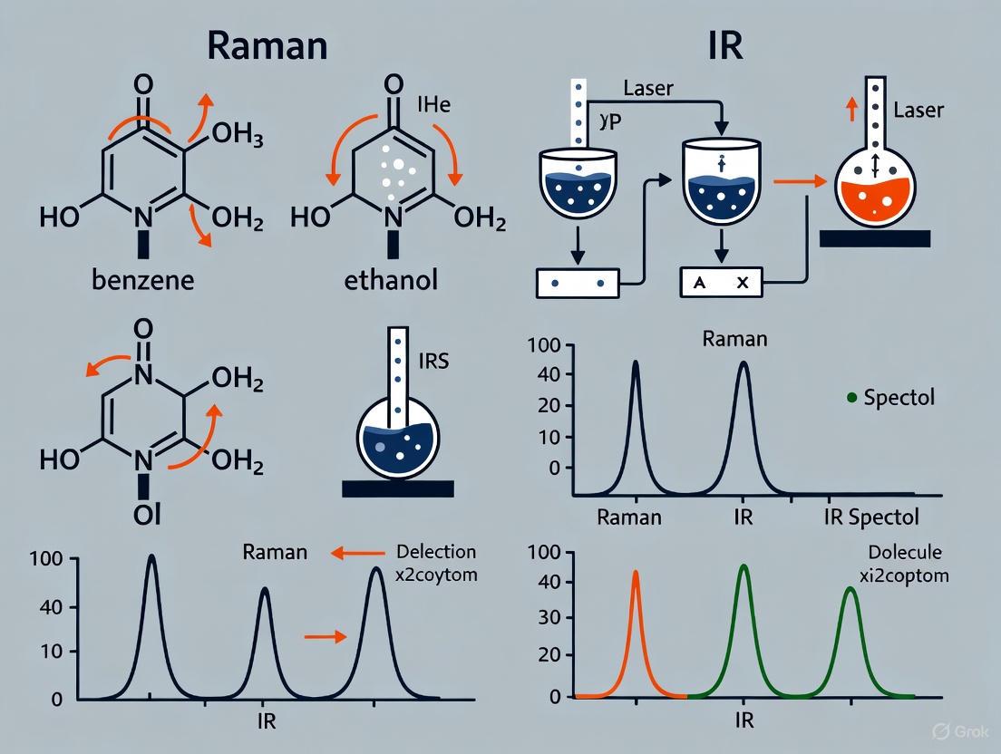
Vibrational spectroscopy is an essential tool for characterizing molecular structures and interactions, with Raman and Infrared (IR) spectroscopy being two of its most prominent techniques. While both methods probe molecular vibrations to generate a unique "fingerprint" for chemical identification, they are founded on fundamentally different physical mechanisms: inelastic light scattering for Raman spectroscopy and direct light absorption for IR spectroscopy [1]. This guide provides a comparative analysis of these core mechanisms, supported by experimental data and protocols, to assist researchers in selecting the appropriate technique for their specific applications in drug development and material science.
Core Principles and Mechanisms
Infrared Spectroscopy: The Mechanism of Light Absorption
In IR spectroscopy, molecules are exposed to infrared light. When the frequency of this incident light matches the natural frequency of a molecular vibration, light is absorbed [2]. This absorption directly promotes the molecule to a higher vibrational energy level [1].
For a vibration to be IR-active, it must cause a change in the dipole moment of the molecule [2] [1]. The dipole moment refers to the separation of positive and negative charges within a molecule. A classic example is the asymmetric stretching vibration of CO₂, which alters the charge distribution and is therefore IR-active [1]. The resulting spectrum is a plot of percent transmittance (or absorbance) against wavenumber, showing bands where energy was absorbed [2].
Raman Spectroscopy: The Mechanism of Inelastic Scattering
Raman spectroscopy, in contrast, relies on a scattering process. A monochromatic laser, typically in the visible or near-infrared range, irradiates the sample [1] [3]. Most of the scattered light is at the same energy as the laser (elastic Rayleigh scattering), but a tiny fraction undergoes inelastic scattering, meaning it emerges with a different energy [4].
This inelastic process involves the molecule being excited to a short-lived "virtual state" before relaxing back to a different vibrational state and emitting a photon. If the molecule ends up in a higher vibrational level, the scattered photon loses energy (Stokes shift). If it ends up in a lower vibrational level, the scattered photon gains energy (anti-Stokes shift) [1] [4]. The energy difference between the incident and scattered photons corresponds to the vibrational energy of the molecule.
For a vibration to be Raman-active, it must cause a change in the polarisability of the molecule—that is, the ease with which its electron cloud can be distorted by an external electric field [2] [1]. The symmetric stretch of CO₂, which changes the electron cloud's shape, is Raman-active [1]. The spectrum presents the intensity of this inelastically scattered light versus its Raman shift (cm⁻¹).
Comparative Workflow: IR Absorption vs. Raman Scattering
The diagram below illustrates the key differences in the fundamental mechanisms of IR absorption and Raman scattering.
Comparative Analysis: Raman vs. IR Spectroscopy
Direct Comparison of Key Parameters
The fundamental differences in mechanism lead to distinct practical advantages and limitations for each technique. The table below summarizes the core differentiating factors.
Table 1: Fundamental and Practical Comparison of Raman and IR Spectroscopy
| Parameter | Raman Spectroscopy | IR Spectroscopy |
|---|---|---|
| Fundamental Process | Inelastic scattering of light [1] | Absorption of infrared light [2] |
| Selection Rule | Change in molecular polarisability [2] [1] | Change in dipole moment [2] [1] |
| Permanent Dipole Required | No [2] | Yes |
| Incident Radiation | Visible or Near-IR (e.g., 800-2500 nm) [2] | Mid-IR (e.g., 2.5 - 50 μm) [2] |
| Water as Solvent | Excellent (weak scatterer) [1] [3] | Poor (strong absorber) [2] [1] |
| Sample Preparation | Minimal; samples can be in glass containers [2] [3] | Can be elaborate; requires IR-transparent materials [2] |
| Key Strength | Analysis of aqueous solutions, covalent bonds [2] | High sensitivity for many functional groups [3] |
| Key Limitation | Fluorescence interference [1] | Strong water absorption [1] |
| Instrument Cost | High [2] [3] | Comparatively inexpensive [2] [3] |
Sensitivity and Performance Data
The performance of each technique can be quantified for specific applications. Recent research on monitoring chlorogenic acid in protein matrices provides comparative experimental data.
Table 2: Experimental Performance Metrics for Phenolic Compound Detection
| Analytical Technique | Target Analytic | Matrix | Limit of Detection (LOD) | Key Experimental Conditions |
|---|---|---|---|---|
| FTIR Spectroscopy | Chlorogenic Acid | Sunflower Meal | 0.75 wt% [5] | Transmission mode, KBr pellet [5] |
| Raman Spectroscopy | Chlorogenic Acid | Bovine Serum Albumin (BSA) | 1.00 wt% [5] | 532 nm laser, 10s accumulation [5] |
| HPLC (Reference Method) | Chlorogenic Acid | Sunflower Meal | Confirmed 5.6 wt% content [5] | Standard chromatographic separation [5] |
Experimental Protocols
Protocol for IR Spectroscopy Analysis of Chlorogenic Acid
This protocol is adapted from a study on monitoring chlorogenic acid in sunflower meal [5].
Step 1: Sample Preparation (KBr Pellet Method)
- Thoroughly dry the sample (e.g., sunflower meal) and potassium bromide (KBr) at about 100°C for several hours to remove water.
- Precisely weigh approximately 2 mg of the sample and 148 mg of KBr [5].
- Mix and grind the materials together using an agate mortar and pestle to create a fine, homogeneous powder.
- Transfer the mixture into a specialized die and compact it into a pellet using a hydraulic press under a pressure of about 200 kPa (2 atm) for 1.5 minutes [5].
Step 2: Data Acquisition
- Place the prepared pellet in the sample holder of an FTIR spectrometer.
- Record the transmission spectrum in the mid-IR range (4,000–400 cm⁻¹) [5].
- Collect a background spectrum using a pure KBr pellet for reference.
Step 3: Data Analysis
- Identify characteristic absorption bands of chlorogenic acid (e.g., C=O stretch, aromatic ring vibrations).
- For quantification, construct a calibration curve using model samples with known concentrations of chlorogenic acid in the protein matrix.
Protocol for Raman Spectroscopy Analysis in a Protein Matrix
This protocol outlines the procedure for detecting chlorogenic acid in a Bovine Serum Albumin (BSA) matrix [5].
Step 1: Sample Preparation for Mapping
- Prepare a series of model samples by mixing and grinding 2, 4, 10, 14, and 20 mg of chlorogenic acid standard with 198, 196, 190, 186, and 180 mg of BSA, respectively, to create a concentration series [5].
- Compact each mixture into a tablet using a pressing mold with a single-axis pressure of approximately 200 kPa for 1.5 minutes [5].
Step 2: Data Acquisition via Mapping
- Place the tablet on a microscope slide under a confocal Raman microscope.
- Use a 532 nm linearly polarized laser for excitation [5].
- Perform mapping on a predefined grid (e.g., 10 × 10 with a step size of 555 μm) to account for sample heterogeneity.
- For each point, acquire the spectrum with an accumulation time of 10 seconds and 2 accumulations [5].
Step 3: Data Analysis
- Process the spectral map to identify the characteristic Raman bands of chlorogenic acid.
- The intensity of key bands can be correlated with concentration to establish a detection limit, which was found to be 1 wt% in the BSA matrix [5].
The Scientist's Toolkit: Essential Research Reagents and Materials
Table 3: Key Reagents and Materials for Vibrational Spectroscopy Experiments
| Item | Function | Application Example |
|---|---|---|
| Potassium Bromide (KBr) | IR-transparent matrix for preparing solid sample pellets for transmission measurements [5]. | FTIR sample preparation (e.g., for sunflower meal) [5]. |
| Bovine Serum Albumin (BSA) | A well-defined protein used as a model protein matrix to simulate complex biological samples [5]. | Preparing calibration standards for analyzing compounds in a protein environment [5]. |
| Chlorogenic Acid Standard | A high-purity (>98%) chemical standard used for calibration and method validation [5]. | Quantifying phenolic compounds in plant-based protein sources [5]. |
| Alkali Metal Salts (e.g., NaCl, AgCl) | Materials used to construct IR-transparent windows for liquid or solid sample holders [2]. | Sample cells for IR spectroscopy, though they can be susceptible to water damage [2]. |
| Quartz or Glass Sample Cells | Containers for holding liquid samples during analysis. | Raman spectroscopy of aqueous solutions, as glass is transparent to visible laser light [2]. |
Raman and IR spectroscopy are powerful, complementary techniques for molecular analysis. The choice between them hinges on the fundamental mechanism best suited to the research problem: Raman spectroscopy excels for aqueous samples, covalent systems, and when minimal sample preparation is critical, leveraging its inelastic scattering mechanism. IR spectroscopy is highly sensitive for detecting functional groups with a dipole moment change and is often more cost-effective, relying on the direct absorption of light.
Emerging technologies, particularly artificial intelligence, are set to enhance the utility of both techniques. For instance, recent advances in AI-driven IR structure elucidation have demonstrated the ability to predict molecular structures from IR spectra with high accuracy, pushing Top-1 identification accuracy to over 63% [6]. This progression towards more powerful, accessible, and intelligent spectroscopic tools promises to further solidify vibrational spectroscopy's role as an indispensable asset in scientific research and drug development.
Molecular vibration techniques, primarily Raman and Infrared (IR) spectroscopy, are cornerstone analytical methods for identifying unknown materials and monitoring chemical reactions across pharmaceutical, material science, and biological research [7] [8]. Both techniques probe molecular vibrational energies to generate a unique "molecular fingerprint" for the sample under investigation [9]. Despite this common goal, Raman and IR spectroscopy are governed by different physical mechanisms and selection rules, making them powerfully complementary [7] [8] [9]. IR spectroscopy measures the absorption of infrared light by molecular bonds that undergo a change in dipole moment during vibration, making it particularly sensitive to polar functional groups [10]. Conversely, Raman spectroscopy relies on the inelastic scattering of light caused by molecular vibrations that induce a change in molecular polarizability, which generally makes it more sensitive to non-polar bonds and symmetric molecular structures [10] [9]. This fundamental difference in mechanism underpins their complementarity and forms the basis for selecting the appropriate technique for specific analytical challenges.
The convergence of these techniques with advanced platforms like scanning probe microscopy has further revolutionized nanoscale chemical analysis, enabling researchers to investigate hierarchical structures in biological materials and advanced nanomaterials with unprecedented resolution [9]. This guide provides a comprehensive comparative analysis of Raman and IR spectroscopy, offering researchers and drug development professionals a detailed framework for technique selection, method implementation, and data interpretation in compound identification.
Fundamental Principles and Technical Comparison
Core Physical Mechanisms
The fundamental distinction between Raman and IR spectroscopy lies in their physical interaction with matter. In IR spectroscopy, when the electric field of the infrared light oscillates at a frequency matching a molecular vibration that causes a change in the dipole moment, energy is absorbed [10]. This absorption is measured as a function of wavelength, producing a spectrum that reveals information about functional groups and bond vibrations, making it highly effective for identifying organic compounds [10]. The resulting spectrum plots absorbance or transmittance against wavenumber (cm⁻¹), with characteristic absorption bands corresponding to specific molecular vibrations.
Raman spectroscopy operates on a fundamentally different principle based on light scattering. When monochromatic light interacts with a molecule, most photons are elastically scattered (Rayleigh scattering). However, approximately one in 10⁷ photons undergoes inelastic scattering, where it gains or loses energy corresponding to molecular vibrational frequencies [9]. This energy shift, known as the Raman effect, provides information about the molecular vibrations via changes in polarizability rather than dipole moment [10] [9]. The Raman spectrum plots scattering intensity against the Raman shift (cm⁻¹), revealing vibrational information complementary to IR spectroscopy.
Performance Characteristics and Selection Criteria
The different physical mechanisms of Raman and IR spectroscopy lead to distinct performance characteristics that guide technique selection for specific applications. The following table summarizes the key technical differences:
Table 1: Fundamental Differences Between Raman and IR Spectroscopy
| Parameter | IR Spectroscopy | Raman Spectroscopy |
|---|---|---|
| Physical Principle | Absorption of IR radiation | Inelastic scattering of visible/NIR light |
| Selection Rule | Change in dipole moment | Change in polarizability |
| Spatial Resolution | Diffraction-limited (several to ~15 μm) [10] | Submicron level (can reach <250 nm) [10] [9] |
| Water Compatibility | Strong water absorption interferes with measurements [10] | Minimal water interference; suitable for aqueous solutions |
| Sample Preparation | Often requires thin sections or ATR crystal contact [10] | Minimal preparation; works in reflection mode [10] |
| Key Limitation | Poor spatial resolution, strong water absorbance [10] | Fluorescence interference, poor spectral sensitivity [10] |
The complementarity of these techniques extends to their sensitivity toward different molecular vibrations. IR spectroscopy excels at detecting polar functional groups such as carbonyls (C=O), hydroxyls (O-H), and amines (N-H) [10]. Raman spectroscopy, meanwhile, is particularly effective for analyzing non-polar bonds including carbon-carbon double bonds (C=C), sulfur-sulfur (S-S) bonds, and symmetric molecular vibrations that may be IR-silent [10] [9]. This complementarity means that in many cases, both techniques are required for complete molecular characterization, particularly for complex samples with diverse chemical functionalities.
Experimental Protocols and Methodologies
Sample Preparation and Measurement Procedures
IR Spectroscopy Protocols typically require specific sample preparation depending on the measurement mode. For transmission FTIR, samples must be thin enough (typically <15 μm) to adhere to the Beer-Lambert law, often requiring compression into KBr pellets for solids or placement between salt plates for liquids [10]. Attenuated Total Reflectance (ATR)-FTIR has become increasingly popular as it often requires minimal preparation—the sample is simply placed in direct contact with a diamond or germanium crystal [10]. However, this contact approach risks sample damage or cross-contamination [10]. Reflection mode in traditional IR microscopy often generates spectral artifacts, limiting its application for certain sample types.
Raman Spectroscopy Protocols generally require less extensive sample preparation, as the technique often works effectively in reflection mode without physical contact [10]. Solid samples can typically be analyzed as-is, while liquids can be contained in glass vials or capillaries. However, Raman measurements are extremely susceptible to fluorescence interference, which can overwhelm the weaker Raman signal [10]. Strategies to mitigate fluorescence include using longer wavelength lasers (e.g., 785 nm or 1064 nm instead of 532 nm), photobleaching samples prior to analysis, or employing surface-enhanced Raman spectroscopy (SERS) to boost signal intensity. The following workflow diagram illustrates the complementary experimental approaches:
Spectral Calibration and Data Processing
Accurate spectral interpretation requires proper instrument calibration and data processing. For Raman spectroscopy, wavelength calibration is typically performed using standard reference materials with known peak positions. A detailed protocol from a DIY Raman spectroscopy initiative for biological research recommends using acetonitrile and neon as calibration standards [11]. The calibration process involves collecting spectra of these standards with the same system configuration and acquisition parameters as the samples of interest, typically using 1,000-10,000 ms exposure for acetonitrile and 1,000 ms for neon, with 0 dB gain and 5 averaged acquisitions [11]. The generated calibration equations then correct all sample data, ensuring accurate peak assignment.
For IR spectroscopy, calibration verification is typically performed using polystyrene films, which exhibit characteristic absorption bands at known wavenumbers. Modern FTIR instruments often include automated validation protocols to ensure spectral accuracy and reproducibility. Data preprocessing for both techniques may include normalization, scatter correction, baseline correction, and noise reduction to enhance spectral quality and facilitate accurate interpretation [12]. Advanced chemometric approaches, including principal component analysis (PCA) and partial least squares (PLS) regression, are increasingly integrated with both techniques to extract meaningful information from complex spectral datasets [12].
Comparative Analysis and Complementary Applications
Pharmaceutical and Biopharmaceutical Applications
The pharmaceutical industry represents a major application area for both Raman and IR spectroscopy, driven by stringent regulatory requirements and the need for precise compound identification. IR spectroscopy is particularly valuable in pharmaceutical analysis for identifying active pharmaceutical ingredients (APIs), excipients, and polymorphic forms through their characteristic functional group vibrations [13] [14]. The technique's sensitivity to polar bonds makes it ideal for quantifying specific functional groups and monitoring chemical reactions in real-time.
Raman spectroscopy offers complementary advantages for pharmaceutical applications, particularly its minimal interference from water, which enables the analysis of hydrates and aqueous formulations without extensive sample preparation [10]. Raman is also highly effective for characterizing crystal polymorphs, which often exhibit distinct Raman spectra despite nearly identical IR profiles. The spatial resolution of Raman microscopy (down to submicron levels) allows for mapping API distribution within solid dosage forms, detecting phase separations, and identifying contaminants in drug products [10] [9].
The growing investment in pharmaceutical research and development—exemplified by the UK's pharmaceutical R&D budget of approximately $49.6 billion (£39.8 billion) for 2022-2025—continues to drive adoption of both spectroscopic techniques throughout drug discovery, development, and quality control processes [14].
Materials Science and Industrial Applications
In materials science, the complementary nature of Raman and IR spectroscopy enables comprehensive characterization of complex material systems. IR spectroscopy excels at identifying organic components in polymers, composites, and coatings through their functional group fingerprints [10]. The development of portable and handheld IR devices has expanded applications to field-based analysis, including environmental monitoring, food safety testing, and industrial quality control [15].
Raman spectroscopy provides unique capabilities for analyzing carbon-based materials, semiconductor structures, and inorganic compounds that may yield weak or complex IR spectra [10]. The technique's superior spatial resolution enables detailed mapping of phase distributions, stress states, and molecular orientation in advanced materials. The non-destructive nature of Raman analysis makes it particularly valuable for analyzing precious samples, cultural heritage artifacts, and forensic evidence.
The market data reflects the growing adoption of these complementary techniques. The Raman spectroscopy research market is projected to grow at a CAGR of 16.05% from 2026 to 2033, reaching $16.02 billion by 2033 [13]. Similarly, the IR spectroscopy market is expected to reach $2.29 billion by 2032, growing at a CAGR of 7.3% from 2025 [16]. The combined NIR and Raman spectroscopy market specifically is projected to grow from $2.05 billion in 2025 to $3.56 billion in 2029 at a CAGR of 14.8% [14].
Table 2: Application-Based Technique Selection Guide
| Application Area | Recommended Technique | Rationale | Key Measurable Parameters |
|---|---|---|---|
| Aqueous Solutions | Raman Spectroscopy | Minimal water interference [10] | Solute concentration, molecular conformation |
| Polymer Characterization | Both Complementary | IR: functional groups; Raman: backbone structure [10] | Crystallinity, orientation, additive distribution |
| Pharmaceutical Polymorphs | Both Complementary | Different sensitivity to molecular packing | Polymorphic identity, distribution, purity |
| Biological Tissues | Both Complementary | IR: protein secondary structure; Raman: non-polar components [9] | Protein/lipid ratio, disease markers, cellular components |
| Microplastics Analysis | AFM-IR (Nanospectroscopy) | Submicron spatial resolution for small particles [10] | Polymer identification, particle size distribution |
| Process Analytical Technology | NIR Spectroscopy | Rapid, non-invasive analysis through containers [14] | Reaction monitoring, blend uniformity, content uniformity |
Advanced Integration and Emerging Trends
Nanoscale and Correlative Spectroscopy
The convergence of Raman and IR spectroscopy with scanning probe microscopy has created powerful new paradigms for nanoscale analysis, overcoming the diffraction limit of conventional optical techniques [9]. Tip-Enhanced Raman Spectroscopy (TERS) combines the chemical specificity of Raman spectroscopy with the spatial resolution of atomic force microscopy (AFM), enabling Raman mapping at the nanoscale [9]. Similarly, Photothermal Induced Resonance (PTIR), also known as AFM-IR, provides IR spectroscopic information at spatial resolutions down to 20 nm, far beyond the conventional diffraction limit of IR microscopy [9].
These nanoscale techniques enable researchers to correlate chemical composition with morphological features at previously inaccessible resolution levels, opening new opportunities in semiconductor characterization, polymer blend analysis, biological membrane studies, and nanopharmaceutical development [9]. The recent development of optical photothermal infrared (O-PTIR) technology represents a significant advancement, enabling simultaneous submicron IR and Raman spectroscopy from the exact same sample location [10]. This simultaneous measurement eliminates uncertainties associated with sequential analysis on different instruments and provides perfectly co-registered complementary datasets.
Artificial Intelligence and Automation
The integration of artificial intelligence (AI) and machine learning with both Raman and IR spectroscopy is transforming spectral analysis across applications. In Raman spectroscopy, AI algorithms enable automated spectral identification, classification of complex biological samples, and real-time decision-making in process analytical technology (PAT) applications [13]. Companies are developing smart spectrometers with AI capabilities to improve accuracy and reduce analysis time, particularly in pharmaceutical and clinical settings [13].
Similarly, IR spectroscopy benefits from AI-enabled spectral libraries that automate compound identification by matching unknown spectra against vast databases in seconds, reducing dependency on manual interpretation and minimizing human error [15]. Predictive modeling algorithms extend this capability by correlating spectral features with material properties, guiding researchers toward novel compound discovery and accelerating development cycles in pharmaceutical and material science applications [15].
The implementation of DIY Raman systems with open-source data analysis pipelines, as demonstrated by Arcadia Science's repository for biological research, further democratizes access to advanced spectroscopic capabilities [11]. These systems combine affordable hardware with standardized calibration protocols and Python-based data processing scripts, making vibrational spectroscopy more accessible to research groups with limited instrumentation budgets [11].
Essential Research Reagents and Materials
Successful implementation of Raman and IR spectroscopic analysis requires specific calibration standards and sampling accessories. The following table details essential research reagents and their applications in vibrational spectroscopy:
Table 3: Essential Research Reagent Solutions for Vibrational Spectroscopy
| Reagent/Accessory | Primary Application | Function | Technical Specifications |
|---|---|---|---|
| Acetonitrile | Raman Calibration Standard | Wavelength calibration reference | Peak positions: 2253 cm⁻¹ (C≡N stretch), 2943 cm⁻¹ (C-H stretch) [11] |
| Neon Lamp | Raman Calibration Standard | Wavelength calibration reference | Multiple emission lines across visible spectrum [11] |
| Polystyrene Film | IR Calibration Standard | Wavelength accuracy verification | Characteristic IR bands: 1601 cm⁻¹, 1493 cm⁻¹, 1028 cm⁻¹ |
| ATR Crystals (Diamond/Ge) | IR Sampling Accessory | Internal reflection element | Diamond: broad IR transmission; Germanium: high refractive index [10] |
| KBr Powder | IR Sample Preparation | Pellet matrix for transmission measurements | Transparent in mid-IR region; forms transparent pellets under pressure |
| Silicon Wafer | Raman Reference Standard | Intensity calibration and background | Characteristic peak at 520 cm⁻¹ for Raman shift verification |
| Tungsten-Halogen Lamp | NIR Light Source | Broadband illumination for NIR spectroscopy | Typical lifespan: 1,000-2,000 hours; blackbody emission profile [14] |
| Quantum Cascade Laser | Advanced IR Source | Tunable, high-brightness IR source for microspectroscopy | Pulse lengths: 10-500 ns; repetition rates: 1-1750 kHz [9] |
Raman and IR spectroscopy provide powerful, complementary approaches for compound identification through molecular fingerprinting. While IR spectroscopy excels at detecting polar functional groups and is well-established for qualitative analysis, Raman spectroscopy offers advantages for aqueous samples, non-polar bonds, and high-resolution mapping. The selection between these techniques should be guided by sample characteristics, information requirements, and analytical constraints rather than treating them as competing alternatives.
Emerging technological integrations—including combined IR-Raman systems, nanoscale spectroscopy techniques, and AI-enhanced data analysis—are pushing the boundaries of vibrational spectroscopy beyond traditional applications. These advancements enable researchers to address increasingly complex analytical challenges in pharmaceutical development, materials characterization, and biological research with unprecedented precision and efficiency. As both techniques continue to evolve through miniaturization, automation, and enhanced computational analysis, their synergistic application will remain fundamental to decoding complex spectral information for compound identification across scientific disciplines.
Vibrational spectroscopy techniques are indispensable tools for material identification and reaction monitoring, with Raman and Infrared (IR) spectroscopy being two of the most prominent methods. While both techniques probe the vibrational energy levels of molecules, they operate under fundamentally different selection rules governed by distinct physical principles. Raman spectroscopy measures the inelastic scattering of light and depends on changes in molecular polarizability during vibrations, whereas IR spectroscopy involves the absorption of light and requires changes in the permanent dipole moment of the molecule [17] [18]. This fundamental difference makes the two techniques highly complementary, often revealing different aspects of molecular structure and dynamics.
The selection rules governing these spectroscopic methods determine which vibrational modes are "active" or "observable" in each technique. Understanding the role of polarizability and dipole moment changes is crucial for researchers and drug development professionals to select the appropriate analytical method for their specific application, interpret spectral data accurately, and gain comprehensive molecular-level insights into their systems of interest [7] [8]. This comparative guide provides an objective analysis of both techniques, supported by experimental data and methodologies relevant to pharmaceutical and materials science applications.
Theoretical Framework: Polarizability vs. Dipole Moment
Molecular Polarizability in Raman Spectroscopy
Polarizability refers to the ease with which the electron cloud of a molecule can be distorted by an external electric field, such as that of an incident photon [18]. Raman activity occurs when a molecular vibration causes a change in this polarizability. The Raman scattering intensity is proportional to the square of the change in polarizability during the vibration, making symmetric vibrations particularly prominent in Raman spectra [18].
During Raman scattering, incident photons interact with the molecule's electron cloud, resulting in energy transfer to or from the molecular vibrations. This inelastic scattering produces shifted frequencies in the scattered light that correspond to the vibrational energies of the molecule. The Raman shift, measured in wavenumbers (cm⁻¹), provides a molecular fingerprint based on polarizability changes during vibrations [18].
Dipole Moment Changes in Infrared Spectroscopy
In contrast, IR spectroscopy detects molecular vibrations that produce a change in the permanent dipole moment of the molecule [18]. When a vibration causes a fluctuation in the molecular dipole moment, it can interact with the oscillating electric field of IR radiation, leading to absorption of specific frequencies. This absorption forms the basis of IR spectroscopy, with the absorption frequency corresponding to the vibrational energy [2].
The requirement for a dipole moment change means IR spectroscopy is particularly sensitive to asymmetric vibrations and functional groups with strong dipole characteristics, such as carbonyl groups, hydroxyl groups, and other heteroatom-containing moieties [18].
Symmetry Considerations and Complementarity
The different selection rules based on polarizability and dipole moment changes make Raman and IR spectroscopy highly complementary techniques. For molecules with a center of symmetry (centrosymmetric molecules), a mutual exclusion principle often applies: vibrational modes that are Raman-active are IR-inactive, and vice versa [18]. For instance, in carbon dioxide (CO₂), the symmetric stretch is Raman-active but IR-inactive, while the asymmetric stretch is IR-active but Raman-inactive [18].
Table 1: Fundamental Differences Between Raman and IR Spectroscopy
| Parameter | Raman Spectroscopy | Infrared Spectroscopy |
|---|---|---|
| Physical Basis | Inelastic scattering of light | Absorption of light |
| Selection Rule | Change in polarizability | Change in dipole moment |
| Symmetric Vibrations | Strongly active | Often inactive |
| Asymmetric Vibrations | Often inactive | Strongly active |
| Centrosymmetric Molecules | Mutual exclusion principle applies | Mutual exclusion principle applies |
| Aqueous Solutions | Well-suited (weak water signal) | Problematic (strong water absorption) |
Experimental Methodologies and Protocols
Raman Spectroscopy Experimental Setup
Modern Raman instrumentation typically consists of three core components: a laser source, sampling optics, and a detector [19]. The choice of laser wavelength is critical, with most pharmaceutical and bioprocessing applications utilizing near-infrared wavelengths (785 nm or 830 nm) to minimize fluorescence interference while maintaining acceptable scattering efficiency [19].
Several sampling configurations are available for different applications:
- Backscattered Raman: Traditional configuration with minimal separation between excitation and collection fibers, primarily collecting signal from superficial layers [19]
- Wide Area Raman: Utilizes a defocused laser beam to illuminate a large area, improving sampling representativeness for both superficial and deep layers [19]
- Spatially Offset Raman Spectroscopy (SORS): Employs larger separation (1-3 mm) between illumination and collection fibers to preferentially collect subsurface signals, even through millimeters of turbid media [19]
- Transmission Raman: Excitation on one side of a sample with collection on the opposite side, providing bulk measurement capability and suppressing fluorescence from superficial layers [19]
For bioprocessing applications, particularly in Chinese hamster ovary (CHO) cell culture monitoring, specialized immersion probes with 785 nm excitation are typically employed to overcome autofluorescence from intracellular NADH and flavins [19].
Infrared Spectroscopy Experimental Protocols
Fourier-transform infrared (FT-IR) spectroscopy has become the standard implementation of IR spectroscopy due to its superior speed and sensitivity compared to dispersive instruments. Sample preparation is more critical for IR spectroscopy, with common techniques including:
- Attenuated Total Reflectance (ATR): Allows direct measurement of solids and liquids with minimal preparation
- Transmission cells: For liquid samples with controlled pathlengths to manage strong solvent absorptions
- KBr pellets: For solid powder analysis, though this technique is being replaced by ATR in many applications
A recent pharmaceutical stability study demonstrated FT-IR coupled with hierarchical cluster analysis in Python for assessing similarity of secondary protein structures in biotherapeutics under varying storage conditions [20].
Data Analysis Approaches
Both Raman and IR spectroscopy data can be analyzed using univariate or multivariate approaches. Univariate analysis focuses on specific band features (area, intensity, center of gravity) and is often reported as band ratios [19]. Multivariate analysis employing chemometric methods like Principal Component Analysis (PCA) or Partial Least Squares (PLS) regression is increasingly common, especially for complex biological or pharmaceutical samples [20] [19].
Machine learning integration has significantly advanced Raman applications, as demonstrated in a 2023 bioprocessing study where hardware automation and machine learning reduced calibration efforts while enabling product quality measurements every 38 seconds [20].
Comparative Analysis: Applications and Performance Data
Pharmaceutical Applications
Table 2: Pharmaceutical Applications of Raman and IR Spectroscopy
| Application | Raman Strength | IR Strength | Experimental Evidence |
|---|---|---|---|
| Polymorph Identification | Excellent for API forms [19] | Limited by sampling depth | Raman identified and quantified polymorph mixtures [19] |
| Protein Structure | Secondary structure in aqueous environments [20] | Secondary structure in solid state [20] | FT-IR with HCA analyzed protein drug stability [20] |
| Process Monitoring | Real-time in situ monitoring [20] [19] | Limited for aqueous systems | Inline Raman monitored 27 cell culture components [20] |
| Quantitative Analysis | Excellent with multivariate calibration [19] | Good for non-aqueous systems | Raman enabled real-time release testing [19] |
Biological and Biopharmaceutical Applications
In bioprocessing and biopharmaceutical applications, Raman spectroscopy has demonstrated significant advantages, particularly for real-time monitoring. A 2024 study showcased Raman's capability for inline monitoring of cell culture processes, establishing models for 27 components with predictive R-squared values (Q²) exceeding 0.8 for most analytes [20]. The methodology successfully identified and eliminated anomalous spectra while demonstrating effectiveness in detecting bacterial contamination through control charts [20].
IR spectroscopy faces limitations in biological systems due to strong water absorption, though it remains valuable for solid-state protein structure analysis. A recent stability study of protein drugs utilized FT-IR with hierarchical cluster analysis to assess secondary structure similarity across different storage conditions, demonstrating maintained stability despite temperature variations [20].
Material Identification and Characterization
For material identification, the complementary nature of Raman and IR spectroscopy is particularly valuable. Symmetric vibrations such as C-C stretching in carbon chains (∼1000 cm⁻¹) and breathing modes in aromatic rings produce strong Raman signals but weak IR signals [18]. Conversely, asymmetric vibrations including C=O stretching (∼1700 cm⁻¹) and O-H stretching (∼3300 cm⁻¹) yield strong IR bands but weak Raman signals [18].
The combination of both techniques provides a comprehensive vibrational profile, as demonstrated in studies of octasulfur, where molecular symmetries and group theory were used to determine allowed vibrational modes for each technique [17] [7] [8].
Research Reagent Solutions and Essential Materials
Table 3: Essential Research Reagents and Materials for Raman and IR Spectroscopy
| Item | Function | Application Notes |
|---|---|---|
| 785 nm Laser | Excitation source for Raman | Reduces fluorescence in biological samples [19] |
| Fiber Optic Probes | In situ sampling | Various configurations (backscatter, SORS, transmission) [19] |
| ATR Crystals (Diamond, ZnSe) | IR sampling without preparation | Enables direct solid and liquid measurement |
| SERS Substrates | Signal enhancement | Gold/silver nanoparticles for trace detection [20] [21] |
| Reference Standards | Instrument calibration | Polystyrene for Raman, polystyrene films for IR |
| Cell Culture Media | Bioprocess monitoring | Requires NIR lasers to avoid autofluorescence [19] |
Advanced Techniques and Emerging Applications
Enhanced Raman Techniques
Several advanced Raman techniques have been developed to address specific analytical challenges:
- Surface-Enhanced Raman Spectroscopy (SERS): Utilizes plasmonic nanostructures to dramatically enhance Raman signals, enabling single-molecule detection and trace analysis [20] [21]
- Tip-Enhanced Raman Spectroscopy (TERS): Combines scanning probe microscopy with Raman spectroscopy for nanoscale spatial resolution [20]
- Spatially Offset Raman Spectroscopy (SORS): Enables subsurface analysis through turbid media and packaging [19]
- Transmission Raman: Provides bulk characterization of pharmaceutical formulations with minimal sampling error [19]
Pharmaceutical Manufacturing and PAT
The adoption of Quality by Design (QbD) principles and Process Analytical Technology (PAT) initiatives has driven Raman spectroscopy integration into pharmaceutical manufacturing [19]. Raman systems are now successfully implemented in real-time release testing, continuous manufacturing, and statistical process control [19]. A 2023 study demonstrated real-time measurement of product aggregation and fragmentation during clinical bioprocessing using hardware automation and machine learning, achieving measurements every 38 seconds [20].
Raman and IR spectroscopy offer complementary approaches to molecular vibrational analysis governed by fundamentally different selection rules. Raman spectroscopy probes changes in molecular polarizability, making it ideal for symmetric vibrations, aqueous solutions, and real-time process monitoring. IR spectroscopy detects changes in dipole moment, excelling for asymmetric vibrations, functional group identification, and solid-state analysis.
The choice between these techniques depends on the specific analytical requirements, sample characteristics, and information needs. For comprehensive molecular characterization, particularly in pharmaceutical development and bioprocessing, the combined application of both techniques often provides the most complete understanding of molecular structure, dynamics, and interactions. As both technologies continue to advance, particularly with integration of machine learning and enhanced sampling methods, their value in research and industrial applications continues to grow.
The comparative analysis of Raman and Infrared (IR) spectroscopy techniques reveals two powerful, yet fundamentally different, approaches to probing molecular vibrations. While both techniques provide characteristic "fingerprints" of molecular structure and composition, their instrumentation configurations differ significantly due to their distinct physical mechanisms. IR spectroscopy measures the absorption of infrared light when molecular bonds undergo a change in dipole moment during vibration. In contrast, Raman spectroscopy relies on the inelastic scattering of light from a monochromatic source, detecting energy shifts resulting from changes in molecular polarizability [1] [22]. This fundamental difference dictates unique requirements for their respective key components—from light sources and wavelength selection systems to detectors and sampling interfaces. This guide provides an objective, data-driven comparison of these instrumental architectures to inform researchers and drug development professionals selecting appropriate characterization tools for specific applications.
Core Instrumentation Comparison
The following table summarizes the key components and their specifications for Raman and IR spectroscopy systems.
Table 1: Instrument Component Comparison: Raman vs. Infrared Spectroscopy
| Component | Raman Spectroscopy | Infrared Spectroscopy |
|---|---|---|
| Light Source | Monochromatic laser (UV, Vis, or NIR). Common types: diode, Nd:YAG, argon-ion [22]. | Broadband infrared source. Common types: globar (silicon carbide), tungsten-halogen, deuterium, synchrotron, or Quantum Cascade Laser (QCL) [23] [22]. |
| Wavelength Range | Typically UV (e.g., 244 nm, 325 nm), Visible (e.g., 532 nm, 633 nm), or NIR (e.g., 785 nm, 1064 nm) lasers [22]. | Mid-infrared (MIR: 4000 - 400 cm⁻¹) is most common for spectroscopy [22]. |
| Spectral Separation | Monochromator or spectrometer with a grating, often coupled to a CCD detector. Requires high-efficiency notch or edge filters to block the intense Rayleigh line [24]. | Interferometer (most common in modern systems), utilizing a Michelson design with a moving mirror for Fourier Transform IR (FTIR). Also, diffraction gratings in dispersive instruments [23]. |
| Detector | Typically a Charge-Coupled Device (CCD) detector, often cooled to reduce noise [24]. For NIR excitation (e.g., 1064 nm), InGaAs detectors are used [7] [8]. | Common detectors include Deuterated Triglycine Sulfate (DTGS) and Mercury Cadmium Telluride (MCT). MCT detectors are faster and more sensitive but require cooling with liquid N₂ [23]. |
| Sampling Interface | Conventional free-space optics or fiber optic probes. Microspectroscopy is achieved by coupling the spectrometer to a standard optical microscope [22]. | Sampling modes include Transmission, Transflection (on reflective substrates), and Attenuated Total Reflection (ATR) using crystals like diamond or ZnSe [23]. |
| Key Advantage | Minimal interference from water, making it suitable for aqueous solutions and biological samples. Excellent for measuring low-frequency vibrations [1]. | Simpler instrument operation and widespread adoption. Direct absorption measurement is generally more sensitive than the weak Raman scattering effect [1]. |
| Key Limitation | Susceptible to fluorescence interference, which can swamp the much weaker Raman signal. The laser can also damage sensitive samples [1]. | Strong absorption by water requires specialized techniques (e.g., ATR) or sample dehydration for aqueous samples. Sample preparation can be more demanding [22]. |
Experimental Protocols and Data Acquisition
Standard Procedure for FTIR Spectroscopy
Fourier Transform Infrared (FTIR) spectroscopy is the dominant modern IR technique due to its speed and sensitivity. The following protocol outlines a standard procedure for analyzing a biological tissue section in transmission mode [23].
- Step 1: Sample Preparation. For transmission measurements, tissue sections are typically cut to a thickness of 5–10 µm and placed on an IR-transparent substrate like barium fluoride (BaF₂). The sample must be thin enough to avoid complete absorption of the IR beam. For hydrated biological samples, the strong water absorption bands necessitate the use of Attenuated Total Reflection (ATR) accessories, which minimize the path length, or careful dehydration of the sample [23] [22].
- Step 2: Instrument Setup and Background Collection. The instrument is configured for the desired sampling mode (transmission, ATR, etc.). A background spectrum (a scan of the environment without the sample) is collected first. This is critical for FTIR as it allows the instrument to subtract the background signal from the sample spectrum later.
- Step 3: Spectral Acquisition. The sample is placed in the beam path. The interferometer in the FTIR instrument scans the moving mirror, collecting an interferogram. This interferogram is then Fourier-transformed to generate a spectrum. Typical parameters include a spectral resolution of 4-8 cm⁻¹ and 64-256 co-added scans to achieve a sufficient signal-to-noise ratio [23].
- Step 4: Data Pre-processing. The raw spectrum undergoes pre-processing, which includes atmospheric correction (removal of CO₂ and H₂O vapor bands), baseline correction, and sometimes smoothing or derivation (e.g., using the Savitzky-Golay algorithm) to enhance spectral features [23].
Standard Procedure for Raman Spectroscopy
Raman spectroscopy measurements often require careful optimization of parameters to maximize signal and minimize fluorescence.
- Step 1: Sample Preparation and Mounting. Raman spectroscopy generally requires minimal sample preparation. Solids, liquids, and powders can be analyzed directly. The sample is placed under the microscope objective or in a sampling compartment. For aqueous samples, Raman is advantageous as water is a weak scatterer [1].
- Step 2: Selection of Excitation Wavelength. The laser wavelength is a critical parameter. While visible lasers (e.g., 532 nm) offer high Raman scattering efficiency, they often induce fluorescence in organic samples. Near-infrared lasers (e.g., 785 nm) are widely used to mitigate this problem, as their lower energy is less likely to cause electronic excitation and subsequent fluorescence [1] [22].
- Step 3: Spectral Acquisition. The laser is focused onto the sample. The scattered light is collected and first passes through a notch or edge filter to block the elastically scattered Rayleigh light. The remaining Raman-shifted light is dispersed by a grating onto a CCD detector. Acquisition times can vary from seconds to minutes, depending on the sample and laser power [24].
- Step 4: Data Processing. The resulting spectrum is processed to remove cosmic rays, apply a calibration, and may include baseline correction to subtract any broad fluorescent background [24].
The workflow for both techniques is summarized in the diagram below.
The Scientist's Toolkit: Essential Research Reagents and Materials
Table 2: Key Research Reagent Solutions for Vibrational Spectroscopy
| Item | Function | Application Notes |
|---|---|---|
| ATR Crystals (Diamond, ZnSe) | Enables Attenuated Total Reflection (ATR) measurement in FTIR by generating an evanescent wave for surface analysis with minimal sample prep [23]. | Diamond is robust but expensive. ZnSe is common for general use but can be attacked by acids. Critical for analyzing aqueous biological samples [23]. |
| IR-Transparent Substrates (BaF₂, CaF₂, Low-E Slides) | Windows for transmission IR measurements. Low-E (low-emissivity) slides are used for transflection measurements, a common mode for tissue analysis [23]. | BaF₂ is water-soluble; CaF₂ is not. Low-E slides are inexpensive but can produce electric field standing wave artifacts that require computational correction [23]. |
| Notch/Edge Filters | Optical filters placed in the Raman collection path to block the intense elastically scattered laser light (Rayleigh scatter) while transmitting the weaker Raman signal [24]. | Essential for detecting the weak Raman signal. The performance of these filters directly impacts the ability to measure low-frequency Raman shifts close to the laser line. |
| Cooled CCD Detectors | The standard detector for Raman spectroscopy. Cooling (e.g., with Peltier or liquid N₂) drastically reduces dark current and readout noise, enabling long exposures for weak signals [24]. | Crucial for achieving a high signal-to-noise ratio in Raman measurements, especially when signal levels are low, such as with biological samples or low laser power. |
| MCT Detectors | A high-sensitivity semiconductor detector for FTIR spectroscopy. Must be cooled with liquid nitrogen for optimal performance [23]. | Offers much higher sensitivity and speed compared to the more common DTGS detector. Ideal for fast imaging or analyzing very small or dilute samples. |
Raman and IR spectroscopy, while both targeting molecular vibrations, are instrumentally distinct techniques that offer complementary information. The choice between them is not a matter of superiority, but of application-specific suitability. IR spectroscopy, with its simpler operation and generally higher sensitivity, is often the first choice for identifying functional groups and analyzing dry or non-aqueous samples. Its instrumentation, particularly with the widespread adoption of FTIR and ATR accessories, is highly standardized and robust. Raman spectroscopy excels where IR faces challenges, particularly in the analysis of aqueous solutions, through glass containers, or when low-frequency vibrations are of interest. However, its instrumentation is more complex, requiring careful selection of laser wavelength and advanced filtering to manage fluorescence and detect its inherently weak signal. For researchers in drug development and material science, understanding the core components and operational principles of these tools is fundamental to selecting the correct technique, designing valid experiments, and accurately interpreting the rich chemical information contained within vibrational spectra.
Practical Applications in Pharmaceutical and Biomedical Analysis
In the realm of drug discovery and development, ensuring the stability and structural integrity of therapeutic proteins is paramount. These biological drugs, used to treat conditions from cancer to autoimmune diseases, are significantly less stable than traditional small-molecule pharmaceuticals [25]. Vibrational spectroscopy, which encompasses both Raman and Infrared (IR) techniques, provides a powerful, label-free approach for analyzing protein structure and monitoring stability under various conditions, such as the stressful lyophilization (freeze-drying) process used to create solid dosage forms [25] [22]. This guide offers a comparative analysis of these two techniques, framing them within the broader context of analytical tools available to researchers and scientists for ensuring the quality, efficacy, and safety of biologic drug products.
Fundamental Principles and a Direct Comparison
Physical Mechanisms and Molecular Sensitivity
The fundamental difference between Raman and IR spectroscopy lies in their underlying physical mechanisms, which dictate the molecular information they provide.
Infrared Spectroscopy measures the absorption of light when the energy of the incident IR photon matches the energy required to excite a molecular bond to a higher vibrational state. For a vibration to be IR-active, it must result in a change in the dipole moment of the molecule [22] [26]. This makes IR highly sensitive to polar functional groups, such as C=O, N-H, and O-H, which is why water (with its strong O-H stretching) creates a significant interference [22].
Raman Spectroscopy is based on an inelastic scattering process. When monochromatic light (usually a laser) interacts with a molecule, a tiny fraction of photons are scattered at energies different from the incident light. This energy shift corresponds to the vibrational energy levels of the molecular bonds. For a vibration to be Raman-active, it must involve a change in the polarizability of the electron cloud around the bond [22] [26]. This makes Raman particularly effective for probing non-polar covalent bonds, such as C-C, C=C, and S-S, which are common in protein backbones and disulfide bridges [26].
Comparative Analysis: Raman vs. Infrared Spectroscopy
The table below summarizes the key operational characteristics, advantages, and limitations of each technique, particularly in the context of protein analysis.
Table 1: Comprehensive comparison of Raman and Infrared spectroscopy for protein analysis.
| Feature | Raman Spectroscopy | Fourier-Transform Infrared (FTIR) Spectroscopy |
|---|---|---|
| Fundamental Basis | Inelastic light scattering; change in polarizability [26] | Absorption of light; change in dipole moment [26] |
| Sample Preparation | Minimal; suitable for aqueous solutions and solids; can use glass containers [25] | Constrained; requires careful control of path length (<10 µm); dehydration often needed for aqueous samples [25] [22] |
| Water Compatibility | Excellent; weak water scattering signal [25] | Poor; strong water absorption obscures protein signals [25] [22] |
| Key Advantages | - Probes hydrophobic regions & S-S bridges [27]- Minimal sample prep [25]- Suitable for aqueous and solid-state analysis [25] | - Rapid measurement [25]- Strong signal for polar groups (C=O, N-H) [22]- Can be used pre- and post-lyophilization [25] |
| Key Limitations | - Fluorescence interference can mask signals [25] [26]- Laser can cause local heating/sample damage [25]- Slower data acquisition (spontaneous Raman) [28] | - Strong water interference [22]- Limited ability to predict solid-state degradation [25]- Only measures global conformation, not tertiary structure [25] |
| Spatial Resolution | High (confocal microscopy possible) | Lower than Raman |
Experimental Protocols for Protein Stability
Combined DLS-Raman Protocol for Unfolding and Aggregation
A powerful approach for studying protein stability involves integrating Raman spectroscopy with Dynamic Light Scattering (DLS). This combination provides simultaneous insights into colloidal stability (size and aggregation via DLS) and conformational stability (structural changes via Raman) from a single sample [27].
Table 2: Key research reagents and solutions for a combined DLS-Raman experiment.
| Item | Function/Description |
|---|---|
| Spectrometer System | Integrated system like Zetasizer Helix, combining a Raman spectrometer and a DLS instrument [27]. |
| Protein Sample | Therapeutic protein (e.g., Lysozyme, Bovine Serum Albumin) at concentrations typically from 0.1 mg/mL to 100 mg/mL [27]. |
| Buffer Solutions | Controlled pH buffers (e.g., citrate buffer) to study pH-dependent stability [27]. |
| Quartz Cuvette | Low-volume cuvette (e.g., 3 mm pathlength, ~120 µL) with high transmission for UV-Vis and Raman signals [27]. |
| Temperature Controller | Precision temperature control unit (e.g., 0°C to 90°C ± 0.1°C) for thermal ramp and isothermal studies [27]. |
Detailed Methodology:
- Sample Preparation: Prepare formulations of the target protein (e.g., Bovine Serum Albumin) at a specific concentration (e.g., 50 mg/mL) in buffers of varying pH [27].
- Loading: Introduce a ~120 µL aliquot of the sample into a quartz cuvette placed in the temperature-controlled compartment of the instrument [27].
- Thermal Ramp Experiment:
- Set the instrument to collect both DLS and Raman data at predefined temperature increments.
- DLS Data: Monitors the Z-average hydrodynamic radius, polydispersity index (PDI), and size distribution. An increase in these parameters indicates the onset of aggregation (Tonset) [27].
- Raman Data: Collects spectra using a 785 nm excitation laser. Monitor specific spectral markers:
- Secondary Structure: Amide I band (1600-1700 cm⁻¹), Amide III band (1200-1350 cm⁻¹) [27].
- Tertiary Structure: Vibrational modes of aromatic side chains (Tryptophan, Tyrosine) around 850 cm⁻¹ and 1550 cm⁻¹. Shifts here indicate changes in the hydrophobic environment, characteristic of unfolding [27].
- Isothermal Incubation Experiment:
- Collect baseline DLS and Raman data at a low temperature.
- Rapidly increase the temperature to just below the melting temperature (Tm) and collect data over a long period (e.g., 7-8 hours) to monitor slow unfolding and aggregation kinetics [27].
- Data Analysis: Correlate the temporal changes in protein size (from DLS) with the structural changes (from Raman) to elucidate the unfolding and aggregation pathway [27].
The workflow below visualizes the structure of this integrated experimental approach.
Key Experimental Data and Interpretation
The combination of DLS and Raman provides a multifaceted view of protein behavior. For instance, in a study of BSA at different pH levels:
- At pH 7.1: Raman spectroscopy showed significant changes in the Amide I band, while DLS showed a concurrent increase in hydrodynamic radius. This close tracking indicates that structural unfolding results in a larger monomer before significant aggregation occurs [27].
- At pH 5.4 (near the isoelectric point, pI): Raman showed minimal structural changes, but DLS detected large size increases (>500 nm). This suggests that at pI, where charge repulsion is minimized, aggregates form freely without major unfolding, a pathway that would be missed by using either technique alone [27].
Advanced Techniques and Clinical Translation
Overcoming Limitations with Advanced Raman
Traditional spontaneous Raman spectroscopy has limitations, including slow imaging speed and low sensitivity (typical detection in the millimolar range) [28]. To address these, advanced techniques have been developed:
- Stimulated Raman Scattering (SRS) and Coherent Anti-Stokes Raman Scattering (CARS): These are forms of coherent Raman scattering (CRS) microscopy. They offer orders of magnitude faster imaging speeds than spontaneous Raman, enabling high-resolution, high-throughput chemical imaging of living cells and tissues. This is crucial for longitudinal tracking of drug uptake, distribution, and response within complex biological models like organoids [28]. SRS, in particular, is free from a non-resonant background, provides linear concentration quantification, and is highly compatible with biological imaging [28].
Comparison with Other Imaging Modalities
While vibrational imaging is powerful, it is one of several tools available. The table below compares it with other common molecular imaging modalities.
Table 3: Comparison of Raman/IR imaging with other key molecular imaging techniques.
| Modality | Key Principle | Key Advantages | Key Limitations |
|---|---|---|---|
| Raman/IR Imaging | Molecular vibrations [22] | Label-free, chemical specificity, simultaneous multi-analyte detection [22] [28] | Low sensitivity (Raman), water interference (IR), limited depth penetration [28] |
| Fluorescence Imaging | Light emission from excited fluorophores | Extremely high sensitivity (single molecule), high spatial/temporal resolution [28] | Requires labeling, which can alter drug properties; photobleaching [28] |
| Mass Spectrometry Imaging (MSI) | Ionization & mass detection of molecules | Unparalleled specificity & multiplex capability; can detect drugs, metabolites, lipids [28] | Destructive; complex sample prep; difficult 3D imaging; quantification challenges [28] |
| Magnetic Resonance Imaging (MRI) | Radio waves & magnetic fields on nuclei | Excellent soft-tissue contrast; deep penetration; non-invasive [22] | Low sensitivity; often requires contrast agents; low spatial resolution vs. optical [22] |
| Photoacoustic Imaging (PAI) | Light absorption & ultrasound detection | Good depth penetration; combines optical contrast & ultrasound resolution [22] | Often requires exogenous contrast agents for molecular imaging [22] |
Raman and IR spectroscopy are complementary, powerful techniques for analyzing protein structure and stability in drug development. IR spectroscopy excels in rapidly assessing global secondary structure and is sensitive to polar bonds, while Raman spectroscopy offers superior compatibility with aqueous samples, minimal preparation, and provides unique insights into tertiary structure and hydrophobic domains. The combination of Raman with techniques like DLS delivers a more complete picture of protein behavior under stress. Furthermore, advanced forms of Raman, such as SRS microscopy, are overcoming historical limitations and opening new frontiers for label-free drug imaging in physiologically relevant, complex models. The choice between these techniques, or the decision to use them in concert, depends on the specific protein attribute of interest, the sample environment, and the desired throughput, underscoring their collective value in the scientist's toolkit for ensuring the development of stable and effective biopharmaceuticals.
Quality Control and Raw Material Identification
In the demanding environments of pharmaceutical development and manufacturing, the rapid and accurate identification of raw materials is a critical quality control (QC) checkpoint. Vibrational spectroscopy techniques, namely Raman and Infrared (IR) spectroscopy, have become cornerstone methods for this non-destructive analysis. Both techniques probe molecular vibrations to generate a unique "fingerprint" for a substance, allowing for the verification of a material's identity against a known standard [5] [26]. This is essential for ensuring that every batch of raw material entering the production process meets the required specifications, thereby safeguarding product efficacy and safety.
While both techniques serve the same fundamental purpose, their underlying principles and practical applications differ significantly. The choice between Raman and IR spectroscopy can impact the speed, cost, and success of QC protocols. This guide provides an objective comparison of their performance, supported by experimental data, to help researchers and scientists select the most appropriate technique for their specific raw material identification needs.
Raman and IR spectroscopy are complementary vibrational techniques, but they operate on different physical principles. IR spectroscopy measures the absorption of infrared light by a molecule. For a vibration to be IR-active, it must cause a change in the molecule's dipole moment [26] [1]. This makes IR highly sensitive to polar functional groups like C=O, O-H, and N-H.
In contrast, Raman spectroscopy is a scattering technique. It involves irradiating a sample with monochromatic light (usually a laser) and detecting the inelastically scattered light. For a vibration to be Raman-active, there must be a change in the molecule's polarizability—that is, the ease with which its electron cloud can be distorted [26] [1]. This makes Raman particularly sensitive to homo-nuclear covalent bonds (e.g., C-C, C=C, S-S) and symmetric vibrations.
The following diagram illustrates the fundamental energy transitions that differentiate these two techniques.
The table below provides a high-level summary of the core characteristics of each technique.
Table 1: Fundamental Comparison of Raman and IR Spectroscopy
| Feature | Raman Spectroscopy | Infrared (IR) Spectroscopy |
|---|---|---|
| Underlying Phenomenon | Inelastic scattering of light [3] | Absorption of light [3] |
| Physical Principle | Change in molecular polarizability [26] [1] | Change in dipole moment [26] [1] |
| Sensitive to | Homo-nuclear bonds (C-C, C=C, S-S), symmetric vibrations [26] | Polar bonds (O-H, C=O, N-H), asymmetric vibrations [26] |
| Typical Spectrum | Sharp peaks on a flat background [3] | Broad absorbance bands [3] |
| Complementary Nature | Best for covalent bond characterization | Best for ionic and functional group characterization [3] |
Performance Comparison for Raw Material Identification
Selecting the right technique requires a practical understanding of its advantages and limitations in a QC setting. The following table summarizes the key performance metrics critical for raw material identification (RMID).
Table 2: Performance Comparison for Pharmaceutical RMID
| Parameter | Raman Spectroscopy | IR Spectroscopy |
|---|---|---|
| Sample Preparation | Minimal to none; can analyze through glass/plastic containers [29] [3] | Often required; constraints on sample thickness and uniformity [26] |
| Water Compatibility | Excellent (water is a weak scatterer) [3] [1] | Poor (strong absorption by water) [3] [1] |
| Fluorescence Interference | A significant problem; can obscure the Raman signal [29] [1] | Not an issue [26] [1] |
| Sensitivity | Generally less sensitive than IR; can require enhancement techniques (e.g., SERS) [3] | Generally more sensitive for most functional groups [1] |
| Quantitative LOD Example | ~1.0 wt% for chlorogenic acid in protein matrix [5] | ~0.75 wt% for chlorogenic acid in protein matrix [5] |
| Portability | Excellent; many robust handheld systems available [29] [30] | Good; handheld FTIR systems are available [3] |
| Cost | Higher (due to lasers and sensitive detectors) [3] | Lower [3] |
Key Experimental Findings
- Detection of Impurities: A 2025 study on monitoring chlorogenic acid in sunflower meal protein isolates demonstrated that while both techniques are viable, FTIR offered a slightly better limit of detection (LOD) of 0.75 wt% compared to Raman's LOD of 1.0 wt% [5]. This highlights IR's potential for superior sensitivity in certain quantitative applications.
- Portability and In-Situ Analysis: Portable Raman instruments have been successfully deployed for in-situ identification of raw materials in warehouses, testing samples directly through plastic bags or glass vials without any preparation [29]. This minimizes sampling error and increases operational efficiency.
- Material Variability and Fluorescence: A key challenge in Raman-based RMID is fluorescence, which can vary between different batches or vendors of the same material. For instance, microcrystalline cellulose (a weak Raman scatterer) can produce false negatives if the fluorescence background of a new batch differs from the reference library [29]. This necessitates robust library management and can sometimes be mitigated by using longer-wavelength lasers (e.g., 785 nm or 1064 nm) [29].
Experimental Protocols for RMID
This section outlines standardized methodologies for developing identification methods using both techniques, based on cited research.
Protocol for Raman Spectroscopy Identification
This protocol is adapted from studies on pharmaceutical raw materials and plant-based protein matrices [5] [29].
1. Sample Presentation:
- For powders, present the sample in a glass vial or a low-density polyethylene (LDPE) bag. The technique can often measure directly through the container wall [29].
- As an alternative, powders can be compacted into a tablet using a hydraulic press with low pressure (e.g., ~200 kPa) to form a uniform surface for analysis [5].
2. Instrument Setup:
- Laser Wavelength: Select an excitation wavelength to minimize fluorescence. A 785 nm laser is a common starting point for organic compounds. If fluorescence persists, a 1064 nm laser can be tested [29].
- Microscope Objective: Use a ×50 objective for a good balance of spatial resolution and light gathering [5].
- Signal Acquisition: Set an integration time (e.g., 10 ms to 10 s) and number of accumulations (e.g., 1 to 10) to achieve an adequate signal-to-noise ratio without damaging the sample. Laser power should be optimized to prevent sample degradation [5].
3. Data Collection & Analysis:
- Collect spectra from multiple points on the sample (e.g., a 10 × 10 grid mapping) to account for heterogeneity [5].
- Compare the unknown sample's spectrum against a validated reference spectral library using correlation algorithms. The match is typically reported as a "hit quality" or p-value [29].
Protocol for FTIR Spectroscopy Identification
This protocol is based on the analysis of chlorogenic acid and other pharmaceutical compounds [5] [31].
1. Sample Preparation (Transmission Mode):
- Grind approximately 1-2 mg of the sample with 100-150 mg of an infrared-transparent matrix, such as potassium bromide (KBr) [5].
- Use a hydraulic press to compress the mixture into a uniform, translucent pellet under pressure (e.g., ~200 kPa) [5].
2. Instrument Setup:
- Use a Fourier Transform Infrared (FTIR) spectrometer in transmission mode.
- Scan across the standard mid-infrared range (4,000–400 cm⁻¹) [5].
- Collect a background spectrum using a pure KBr pellet before analyzing the sample.
3. Data Collection & Analysis:
- Acquire the spectrum of the sample pellet.
- Preprocess the spectra (e.g., baseline correction, normalization) as needed.
- For identification, use chemometric tools like correlation, principal component analysis (PCA), or support vector machine (SVM) to compare the sample spectrum against a pre-built library [31].
The workflow below summarizes the key steps for both techniques, highlighting their differences in sample handling.
The Scientist's Toolkit: Essential Research Reagents and Materials
Successful implementation of spectroscopic RMID relies on a set of key materials and reagents. The following table details these essentials.
Table 3: Essential Research Reagents and Materials for Spectroscopic RMID
| Item | Function | Example Use Case |
|---|---|---|
| Potassium Bromide (KBr) | An infrared-transparent matrix used to dilute samples for FTIR analysis to avoid signal saturation [5]. | Preparing pellets for transmission FTIR spectroscopy of solid raw materials [5]. |
| Chlorogenic Acid Standard | A high-purity chemical standard used for calibration and method development. | Quantifying phenolic compound impurities in plant-based protein sources like sunflower meal [5]. |
| Bovine Serum Albumin (BSA) | A model protein used to create a simulated sample matrix for method development. | Preparing calibration curves for impurities (e.g., chlorogenic acid) in a protein-based raw material [5]. |
| Reference Materials | Certified raw materials (APIs, excipients) from qualified vendors. | Building and validating the spectral reference library for identity testing [29]. |
| Low-Density Polyethylene (LDPE) Bags/Glass Vials | Standardized containers that are largely transparent to Raman laser and NIR light. | Enabling non-destructive, in-situ analysis of samples without removing them from their packaging [29] [31]. |
| Support Vector Machine (SVM) Software | A powerful chemometric tool for classifying complex spectral data. | Managing large-scale spectral libraries and ensuring model transferability between different instruments [31]. |
Both Raman and IR spectroscopy are powerful, non-destructive techniques that are firmly established in the modern QC laboratory for raw material identification. The choice is not a matter of which technique is universally superior, but which is more appropriate for the specific sample and application context.
- Choose Raman spectroscopy when dealing with aqueous samples, when minimal sample preparation is a priority, when analysis through packaging is required, and when targeting symmetric covalent bonds and lattice vibrations (e.g., polymorphism).
- Choose IR spectroscopy when cost is a primary concern, when analyzing non-aqueous samples, when fluorescence is anticipated to be a problem, and when high sensitivity to polar functional groups is needed.
For the most comprehensive material characterization, particularly when dealing with novel or complex substances, employing Raman and IR as complementary techniques provides the fullest picture of molecular structure and composition, ensuring the highest level of confidence in raw material quality [3] [1].
Process Analytical Technology (PAT) and Real-Time Bioprocess Monitoring
Process Analytical Technology (PAT) is a system for designing, analyzing, and controlling manufacturing through timely measurements of critical quality and performance attributes of raw and in-process materials. The goal is to ensure final product quality, with a focus on building quality into products rather than testing it in after production [19]. In biopharmaceutical manufacturing, where living cells are used to produce complex drugs, even small variations in process parameters like pH, temperature, or nutrient levels can significantly impact yield and product quality [32]. Real-time monitoring is therefore crucial for detecting process deviations early, allowing for prompt corrective actions and reducing the risk of costly batch failures [33].
Vibrational spectroscopy techniques, particularly Raman and Infrared (IR) spectroscopy, have emerged as powerful PAT tools for providing molecular-level information about bioprocesses. Both techniques probe the vibrational energy levels of molecules, generating unique "molecular fingerprints" that can identify chemical composition and structure [19] [3]. However, their underlying physical principles and selection rules differ, making them complementary for various applications in real-time bioprocess monitoring. This guide provides an objective comparison of their performance, supported by experimental data and protocols.
Fundamental Principles and Selection Rules
The fundamental difference between the two techniques lies in their physical mechanisms. IR spectroscopy measures the direct absorption of infrared light by a molecule, which occurs when the radiation's frequency matches a natural vibrational frequency of the molecule and the vibration causes a change in the molecule's dipole moment [34] [1]. In contrast, Raman spectroscopy is a scattering technique. It involves irradiating a sample with a monochromatic laser and detecting the inelastically scattered light, which has shifted in energy due to interactions with molecular vibrations. A vibration is Raman active if it induces a change in the polarizability (the deformability) of the molecule's electron cloud [34] [1].
The following diagram illustrates the fundamental principles and selection rules that govern whether a molecular vibration is active in IR or Raman spectroscopy.
For molecules with a center of symmetry (centrosymmetric), the Rule of Mutual Exclusion often applies. This rule states that no vibration can be both IR and Raman active; the vibrational modes are mutually exclusive [34]. This complementarity makes the techniques powerful when used together for full molecular characterization.
Technical Comparison: Raman vs. IR Spectroscopy
The different physical principles of Raman and IR spectroscopy lead to distinct practical advantages and limitations in a bioprocessing environment. The following table summarizes these key performance differentiators.
Table 1: Performance Comparison of Raman and IR Spectroscopy for Bioprocess Monitoring
| Aspect | Raman Spectroscopy | Infrared (IR) Spectroscopy |
|---|---|---|
| Fundamental Principle | Inelastic scattering of monochromatic light [3] | Absorption of infrared radiation [3] |
| Sample Preparation | Minimal; samples can be analyzed in glass vials or through sight glasses [3] | Often requires specific sampling accessories (e.g., ATR crystal); can be more involved [1] |
| Aqueous Samples | Excellent compatibility. Water is a weak Raman scatterer, causing minimal interference [32] [3] [1] | Major interference. Water absorbs IR radiation strongly, complicating analysis [3] [1] |
| Sensitivity & Fluorescence | Can suffer from fluorescence, which can swamp the weaker Raman signal [19] [1]. Generally less sensitive than IR without enhancement techniques [3] | No fluorescence interference. IR spectroscopy is generally a more sensitive technique for direct absorption [3] [1] |
| Sampling Flexibility | High. Fiber optic probes enable remote, in-line, and on-line monitoring [19] [32] | More limited. Fiber optics are available but less common and can be expensive [3] |
| Cost & Accessibility | Generally more expensive due to high-powered lasers and sensitive detectors [3] | Lower cost; instruments are simpler and more common in quality control labs [1] |
PAT Implementation and Experimental Protocols
The choice between Raman and IR spectroscopy significantly influences how a PAT method is implemented on the manufacturing floor. Their inherent strengths dictate their suitability for different monitoring setups.
PAT Deployment Configurations
Raman spectroscopy's compatibility with fiber optics allows for versatile deployment [32]:
- In-line: A probe is inserted directly into the bioreactor or process stream, enabling continuous, real-time monitoring without removing the sample [32] [33].
- On-line: A portion of the product is diverted through a flow cell where the Raman measurement occurs, after which the sample can be returned to the stream or sent to waste [32].
- At-line/Off-line: Compact or handheld Raman analyzers can be used for measurements near the production line or in a remote lab [32].
IR spectroscopy is more commonly used in at-line or off-line configurations due to challenges with water interference and fiber-optic integration, though handheld FTIR systems are available for some applications [3].
Experimental Protocol: In-line Glucose Monitoring in a Bioreactor
The following workflow details a standard protocol for developing a Raman spectroscopy-based method for monitoring glucose in a mammalian cell culture, a critical application in biomanufacturing [32] [33].
Key Considerations:
- Model Robustness: As demonstrated in one study, data collected from the same process across different global sites can be combined to create a robust "global" calibration model for glucose, enhancing predictive accuracy and transferability [32].
- Validation: The correctness of Raman endpoint detection can be cross-verified using a second independent technique, such as NIR spectroscopy, which provides higher certainty in the results [35].
Essential Research Reagent Solutions
Implementing a spectroscopic PAT strategy requires specific reagents and hardware. The following table outlines essential solutions for setting up these monitoring systems.
Table 2: Key Research Reagent Solutions for Spectroscopic PAT
| Item | Function | Key Considerations |
|---|---|---|
| Raman Spectrometer | Provides laser excitation and detects inelastically scattered photons. | Systems with 785 nm or 830 nm lasers minimize fluorescence in biological samples [19]. |
| IR Spectrometer | Provides infrared radiation and measures absorption. | FTIR systems are common. Attenuated Total Reflectance (ATR) accessories simplify solid and liquid sampling [1]. |
| Fiber Optic Probe (Raman) | Enables remote, in-line sampling within bioreactors or process streams. | Immersion probes are used for in-line monitoring in tanks, while flow cells are used for on-line diverted streams [19] [33]. |
| Calibration Standards | For verifying wavelength accuracy and instrument performance. | Common standards include polystyrene for Raman and a polystyrene film for IR [1]. |
| Chemometric Software | For developing quantitative models linking spectral data to analyte concentrations. | Uses algorithms like Partial Least Squares (PLS) regression for multivariate calibration [32] [33]. |
Raman and IR spectroscopy are both powerful PAT tools for real-time bioprocess monitoring, yet they serve different niches based on their core principles. Raman spectroscopy excels in aqueous environments like bioreactors, offering superior sampling flexibility through fiber optics for in-line monitoring of critical process parameters such as glucose, lactate, and amino acids [32] [3] [33]. Its minimal sample preparation and ability to analyze samples through glass are significant operational advantages.
IR spectroscopy, being a more sensitive and established technique, is highly effective for material identification and quality control of raw materials and final products, particularly in dry or non-aqueous environments [3] [1]. Its lower cost and operational simplicity make it a staple in many quality control laboratories.
The decision between the two is not a matter of which is universally better, but which is more appropriate for the specific analytical challenge. For real-time, in-line monitoring of aqueous bioprocesses, Raman spectroscopy is often the preferred choice. However, for many at-line material verification and quality control tasks, IR spectroscopy remains a robust and cost-effective solution. In an ideal PAT framework, their complementary nature can be leveraged to gain a comprehensive understanding of the entire manufacturing process from start to finish.
Quantitative Analysis and Trace-Contaminant Detection using SERS
Surface-Enhanced Raman Spectroscopy (SERS) has emerged as a powerful analytical technique for quantitative analysis and trace-level contaminant detection, offering exceptional sensitivity and molecular specificity. Within the broader framework of vibrational spectroscopy, SERS occupies a unique position, complementing traditional infrared (IR) absorption spectroscopy through its fundamentally different detection mechanism [1]. While IR spectroscopy relies on the absorption of infrared light by molecules undergoing a change in dipole moment, Raman spectroscopy—and by extension SERS—depends on the inelastic scattering of light by molecules with a change in polarizability [1]. This fundamental difference in selection rules makes the two techniques highly complementary, with each exhibiting particular strengths for different molecular systems and analytical challenges.
The exceptional capability of SERS stems from the dramatic signal enhancement achieved when analyte molecules are adsorbed onto or in close proximity to nanostructured metallic surfaces, primarily silver and gold [36]. This enhancement, which can reach factors of 10^6 to 10^8 under optimized conditions, transforms conventional Raman spectroscopy from a technique with inherently weak signals to one capable of detecting single molecules and quantifying contaminants at parts-per-billion (ppb) levels [36] [37]. For researchers and drug development professionals, this sensitivity, combined with the technique's minimal sample preparation requirements and compatibility with aqueous systems, makes SERS an invaluable tool for applications ranging from pharmaceutical quality control to environmental monitoring and medical diagnostics [38].
Fundamental Principles and Enhancement Mechanisms
Theoretical Basis of SERS
The remarkable sensitivity of SERS originates from two primary enhancement mechanisms: electromagnetic enhancement and chemical enhancement. The electromagnetic effect, which typically accounts for the majority of the signal enhancement (10^4 to 10^7), arises from the localized surface plasmon resonance (LSPR) exhibited by nanostructured noble metals [36]. When incident light matches the frequency of the collective oscillation of conduction electrons in these nanostructures, enormous electromagnetic fields are generated at specific "hot spots"—typically in nanoscale gaps between particles or at sharp tips—resulting in dramatically enhanced Raman scattering from nearby molecules [36].
The chemical enhancement mechanism (typically contributing factors of 10 to 10^3) involves charge transfer between the analyte molecule and the metal surface, which alters the polarizability of the molecule and thus increases its Raman scattering cross-section [36]. The overall SERS intensity for a vibrational mode can be expressed as:
[ I{SERS} \propto I0(\upsilon0 - \upsilonk)^4N A \Omega Q Tm T0 \cdot G_{SERS} ]
Where (I0) is the incident laser intensity, ((\upsilon0 - \upsilonk)) is the Raman shift, (N) is the number of adsorbed molecules, (A) is the illuminated area, (\Omega) is the collection solid angle, (Q Tm T0) represents instrumental efficiency factors, and (G{SERS}) is the total enhancement factor [36].
Comparison with Infrared Absorption Spectroscopy
The complementary nature of SERS and IR spectroscopy arises from their different selection rules, making each technique preferentially sensitive to different types of molecular vibrations [1] [39]. The table below summarizes their key characteristics:
Table 1: Fundamental Comparison Between SERS and IR Spectroscopy
| Characteristic | SERS | IR Spectroscopy |
|---|---|---|
| Physical Principle | Inelastic scattering | Absorption |
| Selection Rule | Change in polarizability | Change in dipole moment |
| Water Compatibility | Excellent (weak scatterer) | Problematic (strong absorber) |
| Sensitivity | Extremely high (single molecule) | Moderate |
| Spatial Resolution | Diffraction-limited (∼0.5-1 μm) | Diffraction-limited (∼3-10 μm) |
| Quantitative Capability | Good with proper standardization | Excellent |
| Fluorescence Interference | Potential issue | Not affected |
| Sample Preparation | Minimal (for colloidal substrates) | Often requires specific protocols |
For symmetric vibrations and non-polar functional groups (e.g., C-C, C=C, S-S), SERS typically provides stronger signals, while IR spectroscopy is more sensitive to asymmetric vibrations and polar bonds (e.g., C=O, O-H, N-H) [1] [39]. This complementary relationship means that together, the two techniques can provide a more complete vibrational profile of complex samples than either could alone.
Quantitative Performance and Detection Limits
Analytical Figures of Merit
The quantitative capabilities of SERS have improved significantly with advances in substrate engineering and analytical methodologies. The key to reliable quantification lies in controlling substrate homogeneity and implementing robust internal standardization protocols. The SERS Performance Factor (SPF) has been recently proposed as a standardized parameter for evaluating and comparing substrate performance [37]:
[ SPF = \frac{\Delta I{SERS}/\Delta C{SERS}}{\Delta I{Raman}/\Delta C{Raman}} ]
Where (\Delta I{SERS}/\Delta C{SERS}) is the slope of the SERS intensity versus concentration plot, and (\Delta I{Raman}/\Delta C{Raman}) is the corresponding slope for normal Raman measurements [37]. This approach provides a practical operational parameter that incorporates both the electromagnetic enhancement of the substrate and the interfacial interactions between probe molecules and the substrate.
Detection Limit Comparison with IR Techniques
The exceptional sensitivity of SERS becomes particularly evident when comparing detection limits with those achievable by IR spectroscopy, even in its most advanced implementations:
Table 2: Comparison of Typical Detection Limits for SERS and IR Spectroscopy
| Technique | Typical Detection Limits | Optimal Reported Performance | Representative Applications |
|---|---|---|---|
| SERS | 10^-9 - 10^-12 M (single molecule detection) | Parts-per-billion (ppb) to parts-per-trillion (ppt) for specific analytes | Drug metabolite monitoring, environmental contaminants, food adulterants |
| Macro-ATR-FTIR | 0.1 - 1.0% (1000-10,000 ppm) | ~1700 ppm for pharmaceutical formulations | Bulk material identification, quality control of major components |
| Micro-ATR-FTIR Imaging | 0.01 - 0.1% (100-1000 ppm) | 35 ppm (theoretical), 220 ppm (empirical) for pharmaceutical mixtures | Heterogeneous sample analysis, counterfeit drug detection |
The theoretical detection limit for micro-ATR-FTIR imaging under diffraction-limited conditions can reach 35 ppm, though in practice, this is often limited to 220 ppm or higher for real-world samples with particle size limitations [40]. In contrast, SERS regularly achieves detection limits that are several orders of magnitude lower, with demonstrated applications in detecting trace contaminants, biomarkers, and pathogens at clinically and environmentally relevant concentrations [41] [38].
Experimental Protocols for SERS Analysis
Substrate Preparation and Selection
The choice and preparation of SERS-active substrates represent a critical step in method development. The most common approaches include:
Colloidal Nanoparticle Suspensions: Citrate-reduced silver or gold nanoparticles (typically 20-60 nm) are among the most widely used SERS substrates due to their ease of preparation and high enhancement factors [37]. Protocol: 1) Prepare 1 mM HAuCl₄ or AgNO₃ solution; 2) Heat to boiling with vigorous stirring; 3) Rapidly add sodium citrate solution (1% w/v); 4) Continue heating and stirring for 10-15 minutes; 5) Cool to room temperature. The resulting colloids can be characterized by UV-Vis spectroscopy (λmax ~530 nm for Au, ~420 nm for Ag) and transmission electron microscopy for size distribution.
Solid-State Substrates: These include lithographically patterned surfaces, immobilized nanoparticles on solid supports, and commercially available SERS substrates. Protocol for silicon wafer-based substrates: 1) Clean silicon wafers with piranha solution (Caution: highly corrosive); 2) Functionalize with amine or thiol groups; 3) Immerse in colloidal nanoparticle solution for 12-24 hours; 4) Rinse thoroughly with deionized water and dry under nitrogen [36].
Membrane-Based Substrates: Nanoparticles embedded in or filtered onto membranes provide convenient handling for certain applications. Protocol: 1) Filter nanoparticle solution through appropriate membrane (e.g., alumina, polycarbonate); 2) Dry and store in desiccator [41].
Sample Preparation and Measurement
Standardized sample preparation is essential for quantitative SERS analysis:
Liquid Samples: 1) Mix analyte solution with colloidal nanoparticles in optimized ratio (typically 1:1 to 1:4 v/v); 2) Add aggregation agent if required (e.g., MgSO₄, NaCl, HNO₃ at mM concentrations); 3) Incubate for predetermined time (5-30 minutes) to allow for adsorption and aggregation; 4) Transfer to appropriate measurement cuvette or substrate [37].
Solid Samples: 1) Extract analytes using appropriate solvent; 2) Concentrate if necessary; 3) Follow liquid sample protocol; or 1) Directly deposit solid sample onto SERS substrate; 2) Apply minimal pressure to ensure contact; 3) Add minimal solvent if necessary to facilitate adsorption [42].
Complex Matrices: 1) Implement sample clean-up steps (e.g., centrifugation, filtration, solid-phase extraction) to reduce interference; 2) Use standard addition or internal standards to correct for matrix effects [38].
Instrumental Parameters and Data Acquisition
Optimal spectral acquisition requires careful parameter selection: 1) Select laser wavelength to balance scattering efficiency (ν⁴ dependence), resonance effects, and fluorescence minimization (typically 532 nm, 633 nm, or 785 nm); 2) Adjust laser power to maximize signal while avoiding sample degradation (typically 0.1-10 mW at sample); 3) Set integration time to achieve adequate signal-to-noise without saturation (typically 1-10 seconds, with multiple accumulations); 4) Calibrate spectrometer wavelength using silicon or toluene standards; 5) Collect representative blank spectra for background subtraction [43].
SERS Experimental Workflow: From substrate selection to result interpretation
Research Reagent Solutions and Essential Materials
Successful implementation of SERS methodologies requires specific reagents and materials optimized for enhanced Raman signal acquisition. The following table details essential components for SERS-based analysis:
Table 3: Essential Research Reagents and Materials for SERS Applications
| Reagent/Material | Function | Application Notes |
|---|---|---|
| Gold Nanoparticles (20-60 nm) | Primary SERS substrate | High enhancement, biocompatible, tunable LSPR from visible to NIR |
| Silver Nanoparticles (30-80 nm) | High enhancement substrate | Superior enhancement to gold but more susceptible to oxidation |
| Raman Reporters | Signal amplification in multiplexed detection | Molecules with high cross-sections (e.g., malachite green, crystal violet) |
| Aggregation Agents | Control interparticle distance for "hot spot" generation | Salts (NaCl, MgSO₄) or pH adjustment to optimize enhancement |
| Internal Standards | Signal normalization for quantification | Isotope-labeled analogs or molecules with distinct Raman bands |
| Functionalization Agents | Surface modification for specific adsorption | Thiols, silanes, or polymers for targeted analyte capture |
| Solid SERS Substrates | Ready-to-use platforms | Commercial chips with reproducible enhancement factors |
The selection of appropriate nanoparticles represents perhaps the most critical consideration, with gold nanoparticles offering superior stability and biocompatibility, while silver nanoparticles provide higher enhancement factors but require more careful handling to prevent oxidation [36] [37]. For quantitative applications, internal standards are essential to correct for variations in substrate enhancement, laser fluctuations, and matrix effects.
Applications in Trace-Contaminant Detection
Simultaneous Multi-Contaminant Detection
SERS has demonstrated exceptional capability for the simultaneous detection of multiple contaminants, a significant advantage over many conventional analytical techniques. Two primary strategies have been developed for this purpose:
Label-Free Detection: Direct detection based on the intrinsic Raman fingerprints of target analytes. This approach is particularly valuable for unknown screening and has been successfully applied to complex matrices including food products, environmental samples, and biological fluids [41]. Key advantages include minimal sample preparation and the ability to detect unanticipated contaminants.
Labeled Detection: Utilizes Raman reporter molecules for highly sensitive and specific detection, typically divided into SERS encoding detection and spatial isolation detection [41]. This approach enables multiplexed analysis through unique spectral signatures and is particularly valuable for pathogen detection, biomarker screening, and pharmaceutical analysis.
SERS Detection Strategies for Trace Contaminants
Sector-Specific Applications
The exceptional sensitivity of SERS has led to its adoption across diverse fields requiring trace-level detection:
Pharmaceutical Industry: SERS enables drug quality control by detecting trace impurities and degradation products at levels undetectable by conventional techniques [38]. It facilitates formulation analysis to ensure proper active ingredient concentrations and monitors drug metabolism through detection of low-concentration metabolites, guiding therapeutic optimization [38].
Environmental Monitoring: SERS-based sensors detect heavy metals (e.g., lead, mercury) and organic pollutants (e.g., pesticides, polycyclic aromatic hydrocarbons) in water systems at parts-per-billion levels, significantly below regulatory limits [38]. Real-time air quality monitoring and soil contamination assessment represent additional applications where SERS sensitivity provides distinct advantages over traditional methods.
Food Safety and Authenticity: The technique identifies unauthorized additives, contaminants, and pathogens throughout the food production chain [41] [38]. SERS enables food authentication through origin verification and detection of economically motivated adulteration, while also monitoring freshness indicators and spoilage products to ensure product quality.
Healthcare Diagnostics: SERS revolutionizes disease detection through identification of cancer biomarkers at stages earlier than conventional methods [38]. The technique facilitates rapid pathogen identification for infectious disease diagnosis and enables personalized treatment plans through biomarker monitoring and therapeutic response assessment.
Future Perspectives and Development Trends
The evolution of SERS technology continues with several promising directions enhancing its quantitative capabilities and practical implementation. The development of stable, intelligent substrates with reproducible enhancement factors represents a key research focus, addressing current challenges in method validation and transferability [41]. Advanced nanomaterials including metal-organic frameworks (MOFs), core-shell structures, and hybrid materials show particular promise for creating next-generation SERS substrates with tailored properties [36].
Improvements in analytical algorithms, including machine learning and multivariate analysis techniques, are increasingly addressing challenges related to spectral interpretation, background subtraction, and quantitative modeling [41] [39]. These computational advances complement hardware innovations in portable and handheld SERS instruments, which are expanding applications for field-based analysis and point-of-care testing [38] [39].
The creation of portable, on-site detection platforms represents another significant trend, with miniaturized spectrometers and integrated sample processing systems enabling real-time decision-making in environmental monitoring, food safety inspection, and clinical diagnostics [41]. As these technological advancements converge, SERS is poised to transition from primarily a research technique to a widely deployed analytical tool for quantitative analysis across scientific, industrial, and regulatory domains.
Excipient Interaction Mapping and Formulation Optimization
In modern pharmaceutical development, excipients are far from inert fillers; they are functional components that can significantly influence the stability, bioavailability, and efficacy of active pharmaceutical ingredients (APIs). Excipient interaction mapping has therefore emerged as a critical discipline, enabling the rational design of stable and effective drug formulations. Unforeseen interactions between APIs and excipients can lead to physical instability or chemical degradation, resulting in loss of potency, formation of potentially toxic impurities, and ultimately, product failure [44]. A proactive approach to mapping these interactions allows formulators to identify and mitigate compatibility issues early in the development process, saving considerable time and resources.
Vibrational spectroscopy techniques, namely Raman and Infrared (IR) spectroscopy, are powerful tools in the formulator's arsenal for detecting and characterizing these interactions. Both techniques provide a molecular fingerprint of a sample, but they operate on different physical principles and are sensitive to different types of molecular vibrations. This guide provides a comparative analysis of Raman and IR spectroscopy, offering experimental data and protocols to help researchers select the optimal technique for excipient compatibility studies and formulation optimization.
Comparative Analysis: Raman vs. Infrared Spectroscopy
Fundamental Principles and Selection Rules
While both Raman and IR spectroscopy are vibrational techniques used to identify molecular components and study chemical interactions, their fundamental mechanisms differ, making them complementary.
- Infrared (IR) Spectroscopy measures the absorption of infrared light by a molecule. For a vibration to be IR-active, it must result in a change in the dipole moment of the molecule. IR spectroscopy is highly effective for identifying polar functional groups (e.g., O-H, C=O, N-H) [3].
- Raman Spectroscopy measures the inelastic scattering of monochromatic light, usually from a laser in the visible or near-infrared range. For a vibration to be Raman-active, there must be a change in the polarizability of the electron cloud around a molecule during the vibration. Raman is particularly sensitive to homonuclear covalent bonds (e.g., S-S, C=C, C-C) and symmetric vibrations [7] [8].
This difference in selection rules means that some molecular vibrations that are strong in IR may be weak in Raman, and vice versa. For a comprehensive molecular understanding, especially when dealing with unknown excipient interactions, using both techniques can provide a fuller picture [3].
Direct Comparison of Advantages and Disadvantages
The table below summarizes the practical advantages and disadvantages of each technique, crucial for selecting the right tool for formulation studies.
Table 1: Comparative advantages and disadvantages of Raman and IR spectroscopy.
| Feature | Raman Spectroscopy | Infrared (IR) Spectroscopy |
|---|---|---|
| Sample Preparation | Minimal; can analyze samples through glass or plastic vials [3] | Can require more preparation (e.g., KBr pellets for solids, ATR crystal contact) |
| Aqueous Samples | Excellent for analyzing aqueous solutions as water is a weak scatterer [3] | Problematic due to strong water absorption, requiring specialized cells and pathlength control |
| Detection Sensitivity | Inherently less sensitive, but enhanced by techniques like Surface-Enhanced Raman Spectroscopy (SERS) [3] | Generally a highly sensitive and established technique |
| Cost & Accessibility | Typically more expensive due to high-powered lasers and sensitive detectors [3] | Generally more cost-effective and widely available |
| Sample Damage | Potential for sample heating or destruction due to intense laser radiation [3] | Generally non-destructive, especially with FTIR-ATR |
| Spectral Information | Provides irregular absorbance lines [3] | Shows a scattered Rayleigh line and Stokes/anti-Stokes lines [3] |
| Quantitative Performance | Achieved a root mean square error of prediction (RMSEP) of 17.1 mg/dL for glucose in serum [45] | Achieved an RMSEP of 14.7 mg/dL for glucose in serum [45] |
Experimental Data and Protocol for Excipient Compatibility
Key Experimental Protocol: Drug-Excipient Compatibility Screening
A standard protocol for assessing drug-excipient compatibility using spectroscopic and other analytical techniques is outlined below. This protocol is foundational for identifying physical and chemical incompatibilities before formal stability studies begin.
Table 2: Key research reagents and materials for excipient compatibility studies.
| Reagent/Material | Function in the Experiment |
|---|---|
| Binary Mixtures | 1:1 (w/w) physical mixtures of API and individual excipient [46] |
| Control Samples | Pure API and pure excipients for baseline comparisons [46] |
| Sealed Vials | To store mixtures under controlled humidity (e.g., 5-20% w/w water) [46] |
| HPLC/UPLC System | To separate and quantify the API and any degradation products [46] [47] |
| Differential Scanning Calorimeter (DSC) | To detect thermal events (melting point shifts, decomposition) indicating interactions [46] |
| Spectrometer (FTIR/Raman) | To identify changes in molecular vibrations and functional groups [46] |
Step-by-Step Methodology:
- Sample Preparation: Prepare binary mixtures (typically 1:1 ratio by weight) of the API with each excipient under consideration. Include controls of the pure API and pure excipients. Seal the mixtures in vials, often with controlled moisture content (e.g., 5% w/w) to simulate humidity stress [46].
- Stress Conditions: Store the samples under accelerated stability conditions. Common stress protocols include elevated temperatures (e.g., 40°C, 60°C), high relative humidity (e.g., 75% RH), and exposure to intense light. Samples are typically monitored over 1 to 12 weeks [46] [47].
- Analysis and Detection:
- Visual Inspection: Check regularly for physical changes such as discoloration, caking, liquefaction, or odor development [46] [44].
- Chromatography (HPLC/HPTLC): Analyze samples to quantify the percentage of API remaining and to detect and identify any new degradation products. The formation of new peaks is a clear indicator of chemical incompatibility [46] [48].
- Thermal Analysis (DSC): Subject the mixtures to a controlled temperature program. The disappearance of an API melting peak, a significant shift in its melting onset, or the appearance of new exothermic/endothermic events can signal an interaction [46]. For example, a study showed the complete disappearance of the ofloxacin melting peak in a mixture with lactose, flagging a severe incompatibility [46].
- Vibrational Spectroscopy (FTIR/Raman): Collect spectra of the initial and stressed mixtures. Compare them to the spectra of the pure components. The appearance of new peaks, shifts in existing peak positions, or changes in peak intensity can indicate molecular-level interactions, such as hydrogen bonding, complex formation, or the emergence of new covalent bonds [46] [48].
Experimental Workflow for Technique Selection
The following diagram illustrates a logical workflow for choosing between Raman and IR spectroscopy based on sample properties and study goals, integrating the comparative factors from Table 1.
Advanced and Integrated Approaches
Statistical Design and Machine Learning
Modern formulation development is increasingly leveraging advanced statistical and computational methods to efficiently map complex excipient interactions.
- Hall Experimental Design: A study on Nanostructured Lipid Carriers (NLCs) utilized a Hall-2 experimental design to screen a large number of excipients (solid lipids, oils, surfactants) with a minimal number of experimental runs. This approach allowed for the systematic evaluation of both main effects and two-factor interactions between excipients on critical quality attributes like particle size and entrapment efficiency, leading to an optimized formulation [49].
- Machine Learning (ML): ML models are now being developed to predict drug-excipient compatibility, potentially reducing the need for extensive laboratory testing. One such model, using mol2vec molecular descriptors, demonstrated remarkable accuracy (0.98) in predicting incompatible drug-excipient pairs [48]. Another tool, DE-INTERACT, uses binary fingerprints of molecules to rapidly forecast incompatibilities [48]. These models represent a shift towards in-silico pre-screening in formulation science.
Integrated Problem-Solving Strategies
When an incompatibility is identified, a strategic approach is required to resolve it. The following diagram outlines a systematic process for mitigating stability issues rooted in drug-excipient interactions.
Common mitigation strategies include [47] [44]:
- Excipient Selection: Replacing an incompatible excipient with a more suitable one (e.g., using dextrose instead of lactose for amine-containing APIs to avoid Maillard reactions).
- Stabilizers: Incorporating buffers to control micro-environmental pH, or adding antioxidants (e.g., BHT) and chelating agents (e.g., EDTA) to inhibit oxidation.
- Advanced Techniques: Employing processes like lyophilization (freeze-drying) for moisture-sensitive drugs or microencapsulation to create a protective barrier around the API.
- Protective Packaging: Using desiccants, inert gas purging (nitrogen), or light-resistant containers to shield the product from environmental stressors.
Raman and IR spectroscopy are both indispensable techniques for excipient interaction mapping and formulation optimization. The choice between them is not a matter of which is universally superior, but which is most appropriate for the specific sample and analytical question at hand. IR spectroscopy often holds an advantage in cost and sensitivity for detecting polar functional groups, while Raman spectroscopy excels in analyzing aqueous samples and requires minimal preparation. A comparative study on serum analysis concluded that with identical quantification procedures, both techniques can deliver similar and clinically relevant accuracies [45].
A modern, robust formulation strategy integrates these spectroscopic tools with systematic experimental design, advanced computational predictions, and logical mitigation workflows. This integrated approach enables scientists to efficiently navigate the complexity of excipient interactions, de-risk formulation development, and accelerate the delivery of stable and effective drug products to patients.
Overcoming Challenges: Fluorescence, Water Interference, and Data Complexity
In the field of analytical science, Raman and Infrared (IR) spectroscopy stand as two pillars for molecular fingerprinting. Each technique probes the vibrational states of molecules, providing invaluable chemical and structural information for research and drug development. However, their widespread application is often hindered by two significant and complementary limitations: fluorescence interference in Raman spectroscopy and strong water absorption in IR spectroscopy. Fluorescence, which can be tens of thousands of times stronger than the Raman signal, often swamps the desired spectral information [50]. Conversely, the intense absorption of IR radiation by water complicates the analysis of biological and aqueous samples, which are ubiquitous in the life sciences [51] [50]. This guide provides a comparative analysis of these limitations, supported by experimental data and detailed protocols, to equip researchers with strategies to overcome these challenges effectively.
Core Limitation Comparison
The following table summarizes the fundamental characteristics of the two primary limitations discussed in this guide.
Table 1: Core Limitations of Raman and IR Spectroscopy
| Feature | Fluorescence in Raman Spectroscopy | Water Absorption in IR Spectroscopy |
|---|---|---|
| Physical Origin | Emission from electronic transitions in impurities or the sample itself, typically lasting nanoseconds to milliseconds [50]. | Excitation of fundamental O-H stretching and bending vibrations in water molecules [50]. |
| Spectral Manifestation | A broad, sloping baseline that obscures weaker Raman peaks [52]. | Strong, broad absorption bands, particularly in the mid-IR region, which can mask analyte signals [51] [50]. |
| Impact on Analysis | Can completely obscure the Raman spectrum, making identification and quantification impossible. | Limits pathlengths for liquid samples (often to microns), requires sophisticated sampling techniques, and complicates the study of hydration-dependent processes [51] [50]. |
| Common Triggers | Trace organics, dyes, impurities, or the sample itself (e.g., in biological specimens) [50]. | Aqueous solvents, moisture in the sample environment, and hydrated biological materials. |
Overcoming Fluorescence in Raman Spectroscopy
Experimental Strategies and Protocols
Several experimental approaches can mitigate fluorescence interference, each with its own workflow.
Strategy 1: Photobleaching Prolonged exposure to the laser beam can cause fluorescent impurities to degrade, reducing the fluorescence background.
- Protocol: Focus the laser on the sample and continuously irradiate for a period (seconds to minutes) while monitoring the spectrum. The Raman peaks, which are stable, will become more prominent as the fluorescent background decays.
Strategy 2: Wavelength Shifting Using a near-infrared (NIR) or ultraviolet (UV) laser source can avoid the electronic excitation that leads to fluorescence.
- Protocol: For samples that fluoresce with a standard 785 nm laser, switch to an NIR laser (e.g., 1064 nm) for Fourier-Transform Raman (FT-Raman) spectroscopy. This often moves the excitation energy below the threshold required to excite electronic transitions [3].
Strategy 3: Advanced Raman Techniques Surface-Enhanced Raman Spectroscopy (SERS) and Time-Gated Raman Spectroscopy offer more powerful solutions.
- SERS Protocol:
- Time-Gated Protocol:
- Use a pulsed laser and a gated detector.
- The short-lived Raman scattering is collected immediately after the laser pulse.
- The detector is closed before the long-lived fluorescence is emitted, effectively filtering it out.
Data Processing Workflow
When fluorescence cannot be prevented experimentally, computational baseline correction is essential. The workflow below outlines a standard procedure for processing a raw spectrum affected by fluorescence.
Figure 1: Raman spectral analysis workflow for fluorescence correction.
Key Pre-processing Steps [52] [53]:
- Spike Removal: Cosmic rays hitting the detector appear as sharp, intense lines. They are detected by comparing successive spectra or screening for abnormal intensity changes and are replaced via interpolation [52].
- Baseline Correction: This is the critical step for fluorescence removal. Techniques include:
- Asymmetric Least Squares (AsLS): Fits a smooth baseline underneath the Raman peaks.
- Polynomial Fitting: Fits a low-order polynomial to the regions of the spectrum devoid of Raman peaks.
- Derivative Spectra: Calculating the first or second derivative of the spectrum eliminates slow-varying baseline contributions [52] [53].
- Normalization: The spectrum is divided by its norm, area, or a specific peak intensity to correct for fluctuations in laser power or sampling conditions [52].
Overcoming Water Absorption in IR Spectroscopy
Experimental Strategies and Protocols
The strong absorption of water in IR spectroscopy has been addressed through innovative sampling techniques and instrumental adjustments.
Strategy 1: Attenuated Total Reflectance (ATR) ATR is the most common method for analyzing aqueous samples. It allows measurement through direct contact with the sample, minimizing the effective pathlength.
- Protocol:
- Place a drop of the liquid sample onto the ATR crystal (e.g., diamond, ZnSe).
- Ensure good contact between the sample and the crystal.
- The IR beam undergoes total internal reflection within the crystal, generating an evanescent wave that probes only a few microns into the sample, thus mitigating the strong water absorption [51] [50].
Strategy 2: Transmission with Ultra-Short Pathlengths For traditional transmission measurements, the pathlength must be drastically reduced.
- Protocol:
- Use a sealed liquid cell with precisely spaced windows (e.g., CaF₂) and a spacer defining a pathlength of a few micrometers (e.g., 4-10 µm).
- Fill the cell with the sample, ensuring no bubbles are present.
- Acquire the spectrum, using a background spectrum of pure water or an empty cell for ratioing [50].
Strategy 3: Spectral Processing and Multivariate Analysis Near-IR (NIR) spectroscopy, which probes overtones and combination bands, suffers less from water absorption.
- Protocol:
IR Spectral Analysis Workflow
The analysis of IR spectra, particularly from aqueous or complex biological samples, requires a robust pre-processing pipeline to handle baselines and scattering effects.
Table 2: Key Steps in IR Spectral Pre-Processing [53]
| Step | Function | Common Methods |
|---|---|---|
| Exclusion (Cleaning) | Removes spectral outliers and poor-quality data. | Signal-to-Noise Ratio (SNR) thresholding, Principal Component Analysis (PCA). |
| Filtering | Reduces high-frequency noise and enhances spectral features. | Savitzky-Golay smoothing, Derivative filters, Fourier Self-Deconvolution (FSD). |
| Baseline Correction | Removes slow, varying baseline shifts from scattering. | Polynomial fitting, Asymmetric Least Squares, Multiplicative Scatter Correction (MSC). |
| Normalization | Corrects for pathlength and concentration variations. | Vector normalization, Min-Max normalization, Standard Normal Variate (SNV). |
The Scientist's Toolkit: Essential Research Reagents & Materials
Table 3: Key Reagents and Materials for Mitigating Spectroscopy Limitations
| Item | Function | Application Context |
|---|---|---|
| Gold/Silver Nanoparticles | Plasmonic substrate for signal enhancement in SERS. | Raman spectroscopy of low-concentration analytes to overcome fluorescence and sensitivity limits [51]. |
| ATR Crystals (Diamond, ZnSe) | Enables internal reflection spectroscopy with minimal sample preparation. | IR analysis of strongly absorbing samples like aqueous solutions, gels, and solids [51] [50]. |
| Short Pathlength Cells (CaF₂ Windows) | Limits the effective sample volume to reduce total absorption. | Transmission IR spectroscopy of liquids [50]. |
| NIR Lasers (1064 nm) | Low-energy excitation source to avoid electronic transitions. | FT-Raman systems to minimize sample fluorescence [3]. |
| Calibration Standards (Polystyrene) | Verifies the wavenumber and intensity accuracy of the spectrometer. | Essential for ensuring data reproducibility in both Raman and IR [52]. |
Fluorescence in Raman and water absorption in IR represent significant, yet manageable, challenges in vibrational spectroscopy. The optimal choice between these techniques often depends on the sample matrix: Raman spectroscopy is superior for aqueous solutions due to water's weak scattering, provided fluorescence can be controlled. In contrast, IR spectroscopy is highly effective for solid and non-aqueous samples. The strategies outlined herein—from advanced hardware techniques like SERS and ATR to sophisticated data processing workflows—provide a clear path for researchers to overcome these limitations. A complementary approach, using both techniques to interrogate the same sample, often yields the most comprehensive molecular understanding, as their selection rules differ, providing orthogonal yet complementary information [3].
Sample Preparation Strategies for Solids, Liquids, and Aqueous Solutions
In the field of analytical chemistry, particularly within pharmaceutical research and drug development, vibrational spectroscopy techniques serve as cornerstone methods for material identification and characterization. Mid-infrared (Mid-IR) and Raman spectroscopy have emerged as two powerful, non-destructive analytical techniques that provide molecular fingerprint information for a wide range of samples [54]. While both techniques probe molecular vibrations, they operate on fundamentally different principles, making them complementary rather than competitive. IR spectroscopy measures the absorption of infrared light by vibrating molecules that undergo a change in their dipole moment, whereas Raman spectroscopy relies on the inelastic scattering of light from molecules that experience a change in polarizability during vibration [3].
The selection between these techniques for specific analytical challenges often hinges on the nature of the sample matrix and the required preparation protocols. Sample preparation represents a critical step in the analytical workflow, significantly influencing the quality, reliability, and interpretability of the resulting spectral data. This comparative guide examines the sample preparation strategies for solids, liquids, and aqueous solutions across both techniques, providing researchers with practical methodologies to optimize their analytical outcomes while minimizing preparation artifacts and experimental variability.
Fundamental Principles and Comparative Basis
Core Physical Principles
The fundamental difference between IR and Raman spectroscopy lies in their underlying physical mechanisms. Infrared spectroscopy depends on the absorption of infrared radiation when the frequency of the incident light matches the natural vibrational frequency of molecular bonds. For a vibration to be IR-active, it must result in a change in the dipole moment of the molecule. In practice, this is observed as absorbance bands in the IR spectrum corresponding to specific molecular vibrations [3]. Conventional FTIR spectrometers typically cover the mid-infrared region (4000-400 cm⁻¹), which encompasses the fingerprint region highly diagnostic for molecular identification.
In contrast, Raman spectroscopy is based on the inelastic scattering of monochromatic light, usually from a laser in the visible, near-infrared, or near-ultraviolet range. When photons interact with molecules, a tiny fraction (approximately 1 in 10⁷ photons) undergoes Raman scattering, where energy is exchanged between the photon and the molecule. For a vibration to be Raman-active, it must involve a change in the polarizability of the electron cloud around the molecule. The resulting Raman spectrum displays shifts in wavelength (Raman shifts) corresponding to the vibrational energies of the molecular bonds, typically reported in wavenumbers (cm⁻¹) [55] [3].
Practical Implications for Sample Preparation
These fundamental differences create distinct practical implications for sample preparation. IR spectroscopy often requires careful consideration of pathlength and solvent compatibility, particularly because common solvents like water exhibit strong IR absorption that can obscure regions of interest [3]. Raman spectroscopy, meanwhile, faces challenges with fluorescence interference and potential sample degradation from laser heating but generally offers greater flexibility for aqueous samples and minimal sample preparation requirements [3].
The complementary selection rules mean that some molecular vibrations strong in IR may be weak in Raman, and vice versa. For instance, homonuclear diatomic molecules like O₂ and N₂ are Raman-active but IR-inactive, while ionic functional groups often show strong IR signals but weak Raman signals. This complementarity makes the techniques valuable partners in comprehensive molecular characterization [3].
Comparative Sample Preparation Methods
Solid Samples
Solid samples represent one of the most common sample types in pharmaceutical analysis, ranging from active pharmaceutical ingredients (APIs) to final dosage forms. The preparation methods vary significantly between IR and Raman techniques due to their different measurement principles.
For IR spectroscopy, the most prevalent preparation techniques include:
- Potassium Bromide (KBr) Pellet Method: Approximately 1-2 mg of finely ground sample is mixed with 100-200 mg of dry KBr powder, then compressed under high pressure (8-10 tons) to form a transparent pellet. This method provides excellent spectral quality but requires careful moisture control and thorough cleaning of dies to prevent cross-contamination.
- Diffuse Reflectance Infrared Fourier Transform (DRIFT): Samples are typically diluted with KBr or KCl (1-5% concentration) and placed in a DRIFT cup without further compression. This technique is particularly valuable for powdered materials and surface analysis, requiring minimal preparation while maintaining sample integrity.
- Attenuated Total Reflectance (ATR): The solid sample is directly placed on the ATR crystal (diamond, ZnSe, or Ge) and constant pressure is applied to ensure optimal contact. ATR-FTIR has revolutionized solid sample analysis by eliminating extensive preparation, allowing for direct measurement of tablets, powders, and polymers [54].
For Raman spectroscopy, solid sample preparation is notably simpler:
- Direct Analysis: Most solid samples can be analyzed directly by placing them under the Raman microscope or laser path. No dilution or compression is typically required, though sample orientation and laser focus require optimization.
- Glass Slide Mounting: Powders and solids can be placed on a standard microscope slide or in a capillary tube for analysis. This approach is particularly useful for heterogeneous samples that may require mapping.
- Compression for Homogeneity: For quantitative analysis of powder mixtures, gentle compression into a pellet without diluent can improve homogeneity and spectral reproducibility.
The key advantage of Raman for solid samples lies in its minimal preparation requirements and compatibility with various packaging materials, including glass and plastic containers, enabling non-destructive analysis through packaging [3].
Liquid Samples
Liquid samples, including organic solvents, solutions, and liquid formulations, require distinct preparation approaches for IR and Raman spectroscopy.
IR spectroscopy methods for liquids include:
- Transmission Cell Technique: Liquid samples are placed between two IR-transparent windows (KBr, NaCl, or CaF₂) separated by a precise pathlength spacer (typically 0.015-0.1 mm). This method provides high-quality spectra but requires careful cell assembly and pathlength selection to avoid saturation effects.
- ATR Liquid Analysis: A drop of liquid is placed directly on the ATR crystal, forming a thin film for measurement. This approach has largely replaced transmission cells for routine analysis due to its simplicity and minimal cleaning requirements. Diamond ATR crystals are particularly valuable for their chemical inertness and durability [54].
Raman spectroscopy for liquid samples involves:
- Direct Analysis in Containers: Many liquid samples can be analyzed directly in their original containers, including vials, bottles, and well plates, provided the container material is Raman-inactive (e.g., glass). This facilitates rapid screening and process monitoring.
- Capillary Tube Sampling: Small volumes (microliters) can be analyzed using glass capillary tubes, which are ideal for precious or limited-quantity samples.
- Flow-Through Cells: For process analytical technology (PAT) applications, flow-through cells with quartz or sapphire windows enable real-time monitoring of chemical reactions and manufacturing processes [55].
A significant advantage of Raman for liquid analysis is its compatibility with aqueous solutions, as water is a weak Raman scatterer but a strong IR absorber. This makes Raman ideal for biological and pharmaceutical samples in aqueous matrices [3].
Aqueous Solutions
The analysis of aqueous solutions presents one of the most striking contrasts between IR and Raman spectroscopy, with significant implications for sample preparation.
For IR spectroscopy, water's intense IR absorption creates substantial analytical challenges:
- Very Short Pathlengths: Transmission cells with pathlengths of 15-25 microns are required to avoid complete absorption of the IR signal, requiring precise cell construction and alignment.
- Differential Spectroscopy: Reference spectra of pure water are often subtracted from sample spectra to reveal solute signals, though this approach can introduce spectral artifacts if not carefully implemented.
- ATR Techniques: ATR-FTIR minimizes pathlength issues through the evanescent wave phenomenon, typically probing only 0.5-5 microns into the sample. This has made ATR the preferred method for aqueous samples in IR spectroscopy [54].
For Raman spectroscopy, aqueous solutions require minimal special preparation:
- Direct Analysis: Water exhibits very weak Raman scattering, enabling direct analysis of aqueous solutions without pathlength restrictions or complex background subtraction.
- No Special Cells: Standard glass vials, cuvettes, or well plates can be used without modification, simplifying high-throughput analysis.
- Compatibility with Biological Matrices: The minimal interference from water makes Raman ideal for analyzing proteins, cell cultures, and other biological samples in their native aqueous environments [3].
Table 1: Comparative Sample Preparation Requirements for Different Sample Types
| Sample Type | IR Spectroscopy Preparation | Raman Spectroscopy Preparation |
|---|---|---|
| Solids | KBr pellets, DRIFT, ATR with pressure application | Direct analysis, glass slide mounting, minimal preparation |
| Liquids (Organic) | Transmission cells with precise pathlength, ATR with liquid cell | Direct analysis in container, capillary tubes, flow cells |
| Aqueous Solutions | Very short pathlength cells (15-25µm), ATR with water subtraction | Direct analysis without special considerations |
| Polymer Films | Direct transmission, ATR with pressure | Direct analysis, no special requirements |
| Biological Tissues | Microtomed sections, ATR with pressure | Direct analysis, frozen sections for mapping |
Table 2: Advantages and Limitations in Sample Preparation
| Parameter | IR Spectroscopy | Raman Spectroscopy |
|---|---|---|
| Aqueous Sample Compatibility | Poor (strong water absorption) | Excellent (weak water scattering) |
| Preparation Time | Moderate to extensive | Minimal for most samples |
| Container Requirements | Special IR-transparent materials needed | Standard glass/plastic containers suitable |
| Pathlength Control | Critical for liquids | Not critical |
| Risk of Sample Alteration | Moderate (pressure, dilution) | Low (unless laser heating occurs) |
| Sensitivity to Moisture | High (especially for KBr) | Low |
Experimental Protocols and Methodologies
Protocol for Solid Sample Analysis Using IR Spectroscopy (KBr Pellet Method)
The KBr pellet method remains a standard technique for solid sample analysis in IR spectroscopy, particularly when high-quality spectra are required for molecular identification.
Materials and Equipment:
- Hydraulic press capable of applying 8-10 tons of pressure
- KBr pellet die set (typically 13 mm diameter)
- High-purity, dry potassium bromide powder
- Mortar and pestle or ball mill for grinding
- Vacuum pump (optional but recommended)
- Desiccator for storage of hygroscopic materials
Step-by-Step Procedure:
- Sample Preparation: Grind approximately 1-2 mg of the solid sample to a fine powder using an agate mortar and pestle. The particle size should be reduced below 5 µm to minimize scattering losses.
- Matrix Mixing: Combine the ground sample with 100-200 mg of dry KBr powder (creating 1% sample concentration) and mix thoroughly using a mortar and pestle or mechanical mixer. All operations should be conducted in a low-humidity environment or glove box to prevent moisture absorption.
- Pellet Formation: Transfer the mixture to a die set and apply approximately 8 tons of pressure for 1-2 minutes under vacuum (if available) to form a transparent pellet. The vacuum helps remove trapped air and moisture, improving pellet clarity.
- Spectral Acquisition: Mount the pellet in the IR spectrometer sample holder and collect the spectrum against a pure KBr pellet background reference. Typical parameters include 4 cm⁻¹ resolution and 32 scans to achieve adequate signal-to-noise ratio.
- Post-Analysis Cleaning: Thoroughly clean all equipment with appropriate solvents to prevent cross-contamination between samples.
Critical Considerations: The success of this method depends heavily on maintaining moisture control, achieving uniform particle size, and ensuring homogeneous sample distribution within the KBr matrix. For hygroscopic samples, the entire process should be conducted in a dry atmosphere [54].
Protocol for Aqueous Sample Analysis Using Raman Spectroscopy
Raman spectroscopy offers straightforward analysis of aqueous solutions with minimal preparation, making it ideal for biological and pharmaceutical applications.
Materials and Equipment:
- Raman spectrometer with 785 nm or 1064 nm laser excitation (to minimize fluorescence)
- Standard glass vials, quartz cuvettes, or multi-well plates
- Pipettes for sample transfer
- Optional: Capillary tubes for small-volume samples
Step-by-Step Procedure:
- Sample Loading: Transfer 0.5-2 mL of the aqueous solution into a clean glass vial or cuvette. For limited samples, volumes as small as 10 µL can be analyzed using capillary tubes or specialized micro-sampling accessories.
- Instrument Alignment: Place the sample in the spectrometer and optimize the laser focus position within the solution to maximize signal intensity while avoiding container walls that may contribute to the spectral background.
- Spectral Acquisition: Collect Raman spectra using appropriate parameters (typically 4 cm⁻¹ resolution, 10-30 seconds integration time, and 2-5 accumulations). The 785 nm laser excitation is preferred for aqueous samples as it provides a balance between Raman scattering efficiency and fluorescence minimization.
- Data Processing: Apply basic preprocessing steps including cosmic ray removal, background subtraction (if necessary), and vector normalization to enhance spectral quality.
Critical Considerations: Although water is a weak Raman scatterer, its signal may still dominate at certain regions, particularly in the O-H stretching region around 3400 cm⁻¹. For samples with low analyte concentrations, signal enhancement techniques such as Surface-Enhanced Raman Spectroscopy (SERS) may be employed to improve detection sensitivity [55] [3].
Experimental Data and Performance Comparison
Quantitative Analytical Performance
Direct comparisons of IR and Raman spectroscopy for quantitative analysis reveal their respective strengths and limitations across different sample types and analytical contexts.
Table 3: Quantitative Performance Comparison for Serum Analysis [45]
| Analyte | Mean Concentration (mg/dL) | IR Prediction Error (RMSEP, mg/dL) | Raman Prediction Error (RMSEP, mg/dL) |
|---|---|---|---|
| Glucose | 154.0 | 14.7 | 17.1 |
| Total Protein | - | Comparable accuracy | Comparable accuracy |
| Cholesterol | - | Comparable accuracy | Comparable accuracy |
| Triglycerides | - | Comparable accuracy | Comparable accuracy |
| Urea | - | Comparable accuracy | Comparable accuracy |
A clinical study comparing mid-infrared and Raman spectroscopy for serum analysis demonstrated that both techniques deliver similar accuracies for quantifying key biochemical analytes when using identical multivariate quantification procedures [45]. The researchers noted that vibrational spectroscopy-based quantification appears to be fundamentally limited to accuracies in the 0.1 mmol/L range, regardless of the specific technique employed.
Analysis Time and Throughput Considerations
The sample preparation requirements directly impact analytical throughput and operational efficiency in pharmaceutical and clinical settings.
Table 4: Time Investment Comparison for Different Sample Types
| Sample Type | Typical IR Preparation Time | Typical Raman Preparation Time |
|---|---|---|
| Solid Powder | 10-15 minutes (KBr pellet) | 1-2 minutes (direct analysis) |
| Tablet Formulation | 2-5 minutes (ATR) | 1-2 minutes (direct analysis) |
| Organic Solution | 3-5 minutes (cell assembly) | 1 minute (direct transfer) |
| Aqueous Solution | 5-10 minutes (optimization needed) | 1 minute (direct transfer) |
| Biological Tissue | 10-20 minutes (sectioning) | 2-5 minutes (mounting) |
Raman spectroscopy consistently requires less preparation time across all sample types, particularly for aqueous solutions and solids that need minimal processing. This throughput advantage makes Raman particularly valuable for high-throughput screening applications and process analytical technology (PAT) implementations in pharmaceutical manufacturing [55].
The Scientist's Toolkit: Essential Research Reagents and Materials
Successful implementation of sample preparation strategies requires specific materials and reagents optimized for each spectroscopic technique.
Table 5: Essential Materials for Sample Preparation in Vibrational Spectroscopy
| Material/Reagent | Primary Function | Application Notes |
|---|---|---|
| Potassium Bromide (KBr) | IR-transparent matrix for solid samples | Must be spectroscopic grade, stored dry; used for pellet preparation |
| ATR Crystals (Diamond, ZnSe) | Internal reflection element for ATR-FTIR | Diamond offers durability; ZnSe provides broader spectral range but is more fragile |
| IR-Transparent Windows (KBr, CaF₂, BaF₂) | Transmission cell construction | Selection depends on spectral range and solubility concerns; KBr avoids for aqueous samples |
| Laser Sources (785 nm, 1064 nm) | Excitation for Raman spectroscopy | 785 nm balances signal and fluorescence; 1064 nm minimizes fluorescence |
| Quartz Cuvettes | Sample holders for Raman spectroscopy | Superior transmission in UV-Vis range; minimal background signal |
| Capillary Tubes | Micro-sampling for Raman | Enable analysis of limited volume samples (μL range) |
Decision Framework and Workflow Integration
The choice between IR and Raman spectroscopy for specific applications depends on multiple factors, including sample characteristics, analytical requirements, and available resources. The following decision pathway provides a systematic approach to technique selection:
Diagram 1: Technique Selection Workflow for Sample Analysis
This decision pathway highlights how sample properties directly influence the optimal choice of analytical technique. Aqueous solutions strongly favor Raman spectroscopy due to water's weak scattering, while fluorescent samples typically require IR spectroscopy to avoid interference. For comprehensive molecular characterization, both techniques often provide complementary information that enhances analytical confidence.
Emerging Trends and Advanced Applications
AI-Enhanced Spectroscopy
The integration of artificial intelligence with vibrational spectroscopy represents a revolutionary advancement in pharmaceutical analysis. Deep learning algorithms including convolutional neural networks (CNNs), long short-term memory networks (LSTM), and transformer models are dramatically improving the interpretation of complex spectral data from both IR and Raman techniques [56] [57]. These AI approaches automatically identify intricate patterns in noisy data, reducing the need for manual feature extraction and enabling more accurate identification of chemical components even in challenging sample matrices.
AI-guided Raman spectroscopy is particularly transformative for pharmaceutical quality control, where it enables real-time monitoring of chemical compositions, detection of contaminants, and assurance of product consistency across manufacturing batches. These capabilities are vital for meeting stringent regulatory standards while reducing time-to-market for new therapies [56]. Similarly, in clinical diagnostics, AI-enhanced Raman techniques facilitate early disease detection through high-resolution component mapping that identifies biochemical markers at previously undetectable levels.
Open-Source Spectral Data
The development of comprehensive, open-source spectral databases is addressing a critical challenge in vibrational spectroscopy - the scarcity of accessible reference data. Recent initiatives have produced structured datasets containing thousands of Raman spectra for chemical compounds commonly used in pharmaceutical development [55]. These resources enable more robust calibration models and facilitate the training of machine learning algorithms for automated substance identification.
For example, one recently published open-source dataset includes 3,510 samples spanning 32 compounds relevant to active pharmaceutical ingredient (API) development, acquired using standardized experimental configurations with 785 nm excitation and 1 cm⁻¹ spectral resolution [55]. Such community resources are particularly valuable for method development and validation, allowing researchers to compare their results against high-quality reference data without proprietary restrictions.
The selection between IR and Raman spectroscopy for sample analysis requires careful consideration of multiple factors, including sample physical state, chemical composition, and analytical objectives. IR spectroscopy offers well-established protocols and sensitivity for ionic species and polar functional groups but faces limitations with aqueous samples and requires more extensive preparation for solids. Raman spectroscopy provides distinct advantages for aqueous solutions, requires minimal sample preparation, and enables analysis through packaging, though it may face challenges with fluorescent compounds and represents a greater initial instrumentation investment.
The most effective analytical strategies often combine both techniques in a complementary approach, leveraging their different selection rules to obtain comprehensive molecular information. With ongoing advancements in AI integration and the growing availability of open-source spectral databases, both techniques continue to evolve as powerful tools for pharmaceutical research, quality control, and clinical diagnostics. By understanding the specific sample preparation requirements and performance characteristics of each method, researchers can optimize their analytical workflows to achieve efficient, reliable characterization of diverse sample types across the drug development pipeline.
Vibrational spectroscopy, encompassing both Raman and Infrared (IR) techniques, serves as a cornerstone for material identification and reaction monitoring in scientific research and drug development. These techniques probe molecular vibrations to generate unique spectral fingerprints for chemical substances. However, measured spectra are invariably contaminated by instrumental noise and baseline artifacts, which can obscure critical spectral information and compromise qualitative and quantitative analysis. The inherent weakness of the Raman scattering effect, typically 10⁻⁸ of the incident radiation intensity, makes it particularly susceptible to factors like fluorescence background and Gaussian noise [58]. In IR absorption spectroscopy, baseline artifacts arising from instrument misalignment, light scattering, temperature fluctuations, or optical fouling similarly lead to measurement inaccuracies [59].
The necessity for robust preprocessing is universally acknowledged as a critical first step in the chemometric workflow. For decades, classical algorithms have formed the backbone of spectral processing. However, the selection of optimal algorithms and their parameters has largely depended on operator expertise, creating a bottleneck for high-throughput analysis, especially with large datasets like those from hyperspectral Raman imaging [60]. Recently, deep learning has emerged as a transformative solution, demonstrating significant potential to overcome the limitations of traditional methods by automating feature learning and reducing parameter dependence [58] [60]. This guide provides a comparative analysis of contemporary baseline correction and noise reduction algorithms for Raman and IR spectroscopy, offering researchers a structured framework for selecting and implementing these advanced processing tools.
Comparative Analysis of Noise Reduction Algorithms
Noise reduction is a fundamental preprocessing step aimed at improving the signal-to-noise ratio (SNR) without distorting the underlying spectral features, particularly the intensity, shape, and position of characteristic Raman or IR peaks. The performance of various denoising algorithms can be evaluated based on their ability to suppress noise while preserving these critical features.
Traditional and Machine Learning-Based Denoising
Table 1: Comparison of Noise Reduction Algorithms for Vibrational Spectroscopy.
| Algorithm | Principle | Key Parameters | Strengths | Weaknesses | Suitability |
|---|---|---|---|---|---|
| Savitzky-Golay (SG) Filter [58] | Local polynomial smoothing | Window size, polynomial order | Simple, fast; good peak preservation. | Smears sharp peaks with improper parameters. | Raman & IR; low-to-medium noise. |
| Wavelet Threshold Denoising (WTD) [58] | Multi-resolution thresholding | Wavelet type, decomposition level, threshold rule | Effective for non-stationary noise. | Complex parameter optimization; can distort peaks. | Raman & IR; complex noise profiles. |
| Convolutional Denoising Autoencoder (CDAE) [58] | Deep learning using convolutional neural networks (CNNs) | Network architecture, loss function | High performance; minimal peak distortion; automated. | Requires large training dataset; computational cost. | Raman (esp. high fluorescence). |
| Backpropagation (BP) Neural Network [58] | Fully-connected deep learning | Number of layers/nodes, learning rate | Simplifies parameter optimization. | May be less effective than CNN for spectral features. | Raman & IR; smaller datasets. |
Experimental Protocol for Deep Learning-Based Denoising
A state-of-the-art denoising protocol for Raman spectroscopy employs a Convolutional Denoising Autoencoder (CDAE). The model's novelty lies in adding two extra convolutional layers within the bottleneck, enhancing its ability to learn and preserve spectral features [58].
- Data Preparation: A dataset of clean Raman spectra is collected. Corrupted input (
x̃) is generated by adding synthetic noise (e.g., Gaussian) to the clean spectra (x). - Model Architecture:
- Encoder: Comprises two convolutional layers and two pooling layers for feature extraction and noise reduction.
- Bottleneck: Contains two additional convolutional layers to enhance feature learning without excessive compression.
- Decoder: Utilizes convolutional and upsampling layers to reconstruct the denoised output (
z) to the size of the input data.
- Training: The model is trained to minimize the Mean Square Error (MSE) between the reconstructed output (
z) and the original clean input (x). The loss function is:L(x,z) = (1/N) * ∑(x(i) - z(i))²[58]. - Validation: Performance is validated using both simulated spectra and experimental spectra measured with a Raman spectrometer system, comparing metrics like SNR and MSE against traditional methods.
Figure 1: Workflow of a Convolutional Denoising Autoencoder (CDAE) for Raman spectra, featuring enhanced bottleneck layers for superior noise reduction and feature preservation [58].
Comparative Analysis of Baseline Correction Algorithms
Baseline artifacts, often caused by fluorescence in Raman or scattering effects in IR, introduce a slow-varying background that hinders accurate peak analysis. Effective baseline correction is crucial for subsequent quantitative and qualitative analysis.
Classical and AI-Driven Correction Methods
Table 2: Comparison of Baseline Correction Algorithms for Vibrational Spectroscopy.
| Algorithm | Principle | Key Parameters | Strengths | Weaknesses | Suitability |
|---|---|---|---|---|---|
| Polynomial Fitting [59] [61] | Fits a polynomial to baseline points | Polynomial order, baseline point selection | Intuitive, widely available. | Over-fitting/under-fitting; can reduce peak intensity. | IR & Raman; simple baselines. |
| Asymmetric Least Squares (AsLS) [60] | Iteratively reweighted penalized least squares | Smoothness parameter, asymmetry parameter | Effective for complex, smooth baselines. | Computationally intensive; parameter sensitive. | Raman (fluorescence). |
| Time-Domain m-FID [59] | Molecular Free Induction Decay analysis in time domain | Truncation point of the FID signal | Superior for complex baselines with low noise. | Performance degrades with high noise. | IR spectroscopy. |
| Convolutional Autoencoder (CAE+) [58] | Deep learning for baseline feature capture | Network architecture, comparison function | Preserves peak intensity; handles complex shapes. | Requires training data; "black box" nature. | Raman spectroscopy. |
Experimental Protocol for Baseline Correction
The choice between frequency-domain and time-domain approaches for IR spectroscopy is application-dependent. A recent study compared frequency-domain polynomial fitting (ninth-order polynomial) and time-domain molecular free induction decay (m-FID) for correcting IR absorption spectra of mixtures containing up to 464 components [59].
- Frequency-Domain Protocol:
- Method: A ninth-order polynomial is used to fit and subtract the baseline artifacts directly from the absorption spectrum.
- Evaluation: This approach demonstrated superior performance and stability in high-noise environments and when spectral resolution was varied through peak broadening.
- Time-Domain Protocol:
- Method: The acquired IR absorption spectrum is transformed into the time domain. The early portion of the molecular free induction decay (m-FID) signal, which corresponds to baseline artifacts, is discarded or filtered. The processed signal is then transformed back to the frequency domain to yield the baseline-corrected spectrum.
- Evaluation: This method generally yielded better results for complex baselines under low-noise conditions [59].
For Raman spectroscopy, a Convolutional Autoencoder (CAE+) model has been developed. This model is based on a standard convolutional autoencoder but incorporates a comparison function after the decoder specifically designed for effective baseline correction and preservation of Raman peak intensities [58].
Figure 2: Decision workflow for selecting a baseline correction method in IR spectroscopy, based on noise level and baseline complexity [59].
The Scientist's Toolkit: Essential Research Reagents and Materials
Successful implementation of advanced spectral processing, particularly deep learning models, relies on access to high-quality, well-curated data and computational resources.
Table 3: Essential Resources for Developing and Applying Advanced Spectral Processing Algorithms.
| Resource Category | Item | Function & Application Notes |
|---|---|---|
| Reference Datasets | NIST SRD 35 [62] | Commercial dataset of 5,228 IR spectra; used for training and validating ML models for functional group identification. |
| Open-Access Repositories (e.g., Chemotion) [62] | Provide real-world IR data from various laboratories; valuable for benchmarking and improving model generalizability. | |
| Software & Libraries | Deep Learning Frameworks (e.g., TensorFlow, PyTorch) | Essential for building and training custom models like CDAE and CAE+. |
| Traditional Chemometrics Software (e.g., PLS Toolbox) | Provide implementations of classical algorithms (SG, AsLS, PLS) for baseline comparison. | |
| Computational Hardware | GPU-Accelerated Workstations | Drastically reduce training time for deep learning models, enabling rapid iteration and hyperparameter tuning. |
The evolution of baseline correction and noise reduction algorithms from classical, parameter-sensitive methods to sophisticated, data-driven deep learning models marks a significant advancement in vibrational spectroscopy. For the researcher, the optimal choice is not universal but hinges on the specific technique and analytical context.
For Raman spectroscopy, deep learning approaches like the Convolutional Denoising Autoencoder (CDAE) and the baseline correction CAE+ model demonstrate superior performance in noise suppression and, critically, in preserving the intensity and shape of Raman peaks, which is paramount for accurate biochemical analysis [58]. For IR spectroscopy, the choice is nuanced: the time-domain m-FID method is powerful for complex baselines in low-noise scenarios, whereas frequency-domain polynomial fitting proves more robust and stable in high-noise environments or with varying spectral resolutions [59].
While deep learning offers remarkable automation and performance, it comes with costs including high computational demand, the need for extensive training data, and limited interpretability—the "black box" problem [60]. In contrast, traditional multivariate approaches retain value due to their simplicity, interpretability, and lower computational footprint. The future of spectral processing lies in leveraging the strengths of both paradigms, selecting the right tool based on the application's requirements for accuracy, throughput, and interpretability.
Leveraging Chemometrics and AI for Enhanced Data Interpretation
In the comparative analysis of Raman and Infrared (IR) spectroscopy, a transformative shift is occurring through the integration of chemometrics and artificial intelligence (AI). These computational techniques are revolutionizing how researchers extract meaningful information from complex spectral data, moving beyond traditional manual interpretation to automated, intelligent analysis systems. While both Raman and IR spectroscopy provide molecular fingerprints by probing vibrational energy levels, they generate complementary data based on different physical principles—IR absorption depends on changes in dipole moment during molecular vibration, while Raman scattering relies on changes in molecular polarizability [63] [64]. This fundamental difference makes the combined application of both techniques particularly powerful, especially when enhanced by AI-driven data fusion approaches that can uncover patterns and relationships invisible to conventional analysis methods.
The adoption of AI in spectroscopy is driven by several converging factors: the growing complexity of analytical challenges in pharmaceutical research and material science, the need for real-time monitoring in industrial processes, and the expanding capabilities of machine learning algorithms to handle high-dimensional spectral data. As the global market for Raman spectroscopy research demonstrates robust growth—projected to increase from $6.56 billion in 2025 to $16.02 billion by 2033 at a CAGR of 16.05%—and the IR spectroscopy market expected to reach $2.29 billion by 2032, the economic impetus for advanced data interpretation solutions continues to strengthen [13] [16]. This growth is paralleled by technological advancements in both instrumentation and computational power, creating a fertile environment for AI and chemometrics to redefine the possibilities of spectroscopic analysis across research and industrial applications.
Fundamental Techniques Comparison: Raman vs. IR Spectroscopy
Understanding the distinct characteristics of Raman and IR spectroscopy is essential for effectively applying chemometrics and AI to the data they generate. These complementary techniques probe molecular vibrations through different physical mechanisms, resulting in unique strengths and limitations that influence how their data should be processed and interpreted.
Table 1: Fundamental Comparison of Raman and IR Spectroscopy Techniques
| Parameter | Raman Spectroscopy | Infrared (IR) Spectroscopy |
|---|---|---|
| Physical Principle | Inelastic light scattering measuring change in polarizability | Absorption of IR radiation measuring change in dipole moment |
| Spatial Resolution | Submicron level (can reach ~1 µm) | Diffraction-limited (several to ~15 µm) [63] |
| Water Compatibility | Excellent (weak scatterer) | Poor (strong absorbance) [64] |
| Sample Preparation | Minimal, often works in reflection mode | Typically requires thin samples for transmission or ATR crystal contact [63] |
| Key Strength | Excellent for non-polar bonds, aqueous solutions, inorganic molecules | Superior for polar functional groups, organic molecules |
| Primary Limitation | Fluorescence interference, poor spectral sensitivity | Strong water absorbance, spatial resolution limits |
| Spectral Range | 4000 cm⁻¹ to ~100 cm⁻¹ | Requires multiple instruments for full vibrational range [64] |
The selection rules governing these techniques reveal their complementary nature. IR spectroscopy is particularly sensitive to asymmetric vibrations and polar functional groups such as hydroxyl (-OH), carbonyl (C=O), and amine (N-H) groups, where vibrational motions produce significant changes in the molecular dipole moment. In contrast, Raman spectroscopy excels at detecting symmetric vibrations and non-polar bonds including carbon-carbon backbone stretches, sulfur-sulfur bonds, and aromatic ring vibrations, where the electron cloud distortion during vibration creates significant polarizability changes [63] [64] [8]. This fundamental complementarity means that combined Raman-IR analysis, especially when enhanced by AI, provides a more complete molecular picture than either technique alone.
Recent technological advancements are further bridging the historical limitations of both techniques. Optical Photothermal Infrared (O-PTIR) technology now enables simultaneous submicron IR and Raman spectroscopy, overcoming the traditional diffraction limit of IR analysis by detecting photothermal effects with a visible probe beam [63]. This innovation allows researchers to collect IR and Raman data from the exact same sample point at the same spatial resolution, eliminating positioning errors and providing perfectly co-registered chemical information. Similarly, the miniaturization of spectroscopic instruments has reached the point of wearable vibrational spectroscopy, with devices no longer confined to laboratory benches but integrated into industrial equipment and even human bodies for continuous, real-time monitoring [12].
AI and Chemometrics Workflow in Spectroscopy
The application of AI and chemometrics in spectroscopic data analysis follows a structured workflow that transforms raw spectral data into actionable chemical insights. This process involves multiple stages of computational treatment, each with specific methodologies tailored to handle the unique characteristics of Raman and IR spectral data.
Data Preprocessing Foundations
The initial and crucial step in spectroscopic analysis involves data preprocessing to transform raw spectral data into clean, reliable inputs for chemometric modeling. As highlighted in recent research, proper preprocessing is essential for improving accuracy, reproducibility, and interpretability in both IR and Raman spectroscopic analysis [12]. For FT-IR ATR spectra, key preprocessing methods include:
- Normalization: Adjusts for variations in sample thickness or concentration by scaling spectra to a common reference, typically using standard normal variate (SNV) or vector normalization techniques.
- Baseline Correction: Removes background effects and fluorescence interference (particularly critical in Raman spectroscopy) through algorithms such as asymmetric least squares or polynomial fitting.
- Scatter Correction: Compensates for light scattering effects in particulate samples using methods like multiplicative signal correction (MSC) or extended multiplicative signal correction (EMSC).
- Centering and Scaling: Centers data by subtracting the mean spectrum and applies scaling (e.g., unit variance, Pareto) to balance the influence of variables with different intensities.
The selection of preprocessing techniques must be tailored to both the spectroscopic method (Raman vs. IR) and the specific sample matrix, as inappropriate preprocessing can introduce artifacts or remove chemically meaningful information [12]. For pharmaceutical applications, validated preprocessing workflows are particularly important to meet regulatory requirements for method validation.
AI Integration for Spectral Interpretation
Artificial intelligence has dramatically advanced spectral interpretation through several key applications:
AI-Enabled Spectral Libraries automate compound identification by matching unknown spectra against vast databases in seconds, significantly reducing dependency on manual interpretation and minimizing human error in critical analyses [15]. These systems employ sophisticated similarity algorithms that account for variations in instrument response and measurement conditions, providing confidence metrics for identification matches.
Predictive Modeling Algorithms extend beyond simple identification by correlating spectral features with material properties and performance characteristics. For pharmaceutical development, these models can predict critical quality attributes of drug formulations directly from spectral data, guiding researchers toward optimized formulations and accelerating time-to-market [15]. The integration of machine learning with Raman spectroscopy has proven particularly valuable for monitoring protein structures in biopharmaceuticals, differentiating α-helix and β-sheet configurations that are essential for therapeutic efficacy [13] [63].
Deep Learning Applications represent the cutting edge of spectroscopic analysis, with convolutional neural networks (CNNs) being applied to raw spectral data for complex pattern recognition tasks. A recent study from Heilongjiang Bayi Agricultural University demonstrated successful implementation of deep learning with near-infrared spectroscopy for rapid, non-destructive detection of illicit food additives [12]. Similarly, research into Explainable AI (XAI) methods is addressing the "black box" nature of complex models, providing interpretable insights into which spectral features drive specific predictions [12].
Experimental Protocols and Methodologies
Simultaneous IR-Raman Analysis Protocol
The emergence of simultaneous IR-Raman systems represents a significant advancement for comprehensive material characterization. The following protocol outlines the methodology for integrated analysis using Optical Photothermal Infrared (O-PTIR) technology:
Sample Preparation Protocol:
- Sample Mounting: Mount samples on standard glass slides or low-e slides optimized for IR reflection measurements. Avoid using IR-absorbing substrates when possible.
- Thickness Consideration: For transmission measurements, ensure sample thickness is appropriate for the spectral range of interest (typically 5-20 μm for traditional IR, but flexible for O-PTIR).
- Reference Standards: Include calibration standards such as acetonitrile and neon in the measurement set when quantitative analysis is required [11].
- Environmental Control: For hydrated biological samples, use controlled humidity chambers to prevent dehydration during measurement.
Instrumentation Parameters:
- Excitation Sources: Configure IR source (tunable quantum cascade laser) and visible probe laser according to manufacturer specifications.
- Spatial Alignment: Verify co-alignment of IR excitation and visible probe beams using standardized reference samples.
- Spectral Calibration: Perform daily wavelength calibration using known spectral standards to maintain data integrity [11].
- Resolution Settings: Set spectral resolution to 4 cm⁻¹ for general screening or 2 cm⁻¹ for detailed structural analysis.
Data Acquisition Procedure:
- Region Selection: Identify regions of interest using the visible imaging capability of the system.
- Simultaneous Collection: Acquire IR and Raman spectra from the exact same spatial location using the integrated collection mode.
- Parameter Optimization: Adjust acquisition time (typically 1-10 seconds per spectrum) and laser power to maximize signal-to-noise while avoiding sample damage.
- Quality Verification: Monitor spectral quality in real-time using built-in quality metrics (peak intensity, signal-to-noise ratio, baseline stability).
This methodology enables the collection of perfectly co-registered IR and Raman spectra, eliminating positioning uncertainties and facilitating direct correlation of complementary chemical information [63].
AI-Driven Pharmaceutical Analysis Experiment
Objective: To demonstrate the application of AI-enhanced Raman spectroscopy for quality control in pharmaceutical manufacturing, specifically for monitoring active pharmaceutical ingredient (API) distribution in solid dosage forms.
Materials and Equipment:
- Raman spectrometer system (portable or benchtop) with 785 nm or 532 nm excitation laser
- Pharmaceutical tablet samples with known API concentration gradient
- Reference standards for calibration
- Computer workstation with AI/chemometrics software (Python with scikit-learn, TensorFlow, or commercial alternatives)
Experimental Workflow:
- Spectral Library Development:
- Collect reference spectra from pure API and excipients
- Acquire training set spectra from tablets with known API concentration (20-100 samples)
- Apply standardized preprocessing: Savitzky-Golay smoothing, baseline correction, vector normalization
Model Training Phase:
- Extract spectral features using Principal Component Analysis (PCA)
- Train Partial Least Squares Regression (PLSR) model to predict API concentration
- Validate model using cross-validation (k-fold or leave-one-out)
- Establish acceptance criteria: R² > 0.95, RMSEP < 2% of target concentration
Production Monitoring Application:
- Acquire spectra from unknown production samples
- Apply trained model to predict API concentration in real-time
- Generate statistical process control charts for quality monitoring
- Flag outliers exceeding ±3σ control limits for further investigation
This protocol demonstrates how AI-enhanced spectroscopy enables real-time quality control in pharmaceutical manufacturing, significantly reducing analysis time compared to traditional chromatographic methods while providing spatial distribution information not available through destructive testing [13] [64].
Essential Research Reagent Solutions
Table 2: Key Research Reagents and Materials for AI-Enhanced Spectroscopy
| Reagent/Material | Function | Application Notes |
|---|---|---|
| Acetonitrile | Raman calibration standard | Provides characteristic peaks at 2253 cm⁻¹ (C≡N stretch) and 918 cm⁻¹ (C-C stretch) for wavelength calibration [11] |
| Neon Lamp | Wavelength calibration | Emission lines provide absolute wavelength reference across spectral range |
| Silicon Wafer | Background reference | Provides low fluorescence background for Raman measurements |
| Polystyrene | Spectral validation | Standard reference material with well-characterized IR and Raman peaks |
| ATR Crystals (Diamond, Germanium) | IR sampling interface | Enables contact-based IR measurements; diamond for general use, Germanium for low-frequency range |
| Metal Nanoparticles (Au, Ag) | SERS enhancement | Enhance Raman signals by 10⁶-10⁸ times for trace analysis [64] |
| NIST Traceable Standards | Method validation | Certified reference materials for regulatory-compliant analyses |
| Low-E Slides | IR-compatible substrates | Enable reflection-mode IR measurements of micro-samples |
The selection and proper use of these reference materials is critical for generating high-quality, reproducible spectral data suitable for AI analysis. Calibration standards like acetonitrile and neon are particularly important for maintaining data integrity across multiple instruments and measurement sessions [11]. For surface-enhanced Raman scattering (SERS) applications, consistent preparation of nanoparticle substrates is essential for obtaining quantitative results, as nanostructural regularity significantly impacts enhancement reproducibility [64].
Comparative Performance Analysis
Table 3: Quantitative Performance Metrics of AI-Enhanced Spectroscopy Techniques
| Performance Metric | Traditional Raman | AI-Enhanced Raman | Traditional IR | AI-Enhanced IR |
|---|---|---|---|---|
| Identification Accuracy | 85-92% | 96-99% [15] | 88-94% | 97-99.5% [15] |
| Analysis Time | Minutes to hours | Seconds to minutes [14] | Minutes | Seconds to minutes |
| Quantitation R² | 0.82-0.95 | 0.96-0.99 [13] | 0.85-0.96 | 0.97-0.995 |
| Detection Limits | μM-mM range | nM-μM range with SERS [64] | μM range | Comparable with advanced algorithms |
| Reproducibility (%RSD) | 5-15% | 2-8% [64] | 4-12% | 2-7% |
| Multi-component Resolution | Moderate | Excellent [14] | Moderate-High | Excellent |
The integration of AI and chemometrics provides dramatic improvements across all performance metrics, with particularly significant gains in analysis speed, detection limits, and multi-component resolution. The implementation of time-gated Raman systems like the PicoRaman M3 demonstrates how technological innovations combined with advanced data processing can achieve measurement speeds 10-40 times faster than conventional systems while maintaining excellent specificity and resistance to ambient interference [14]. Similarly, AI-enhanced IR spectroscopy benefits from predictive modeling algorithms that correlate spectral features with material properties, enabling more accurate quantification in complex mixtures [15].
For pharmaceutical applications, the combination of Raman spectroscopy and AI has proven particularly valuable for real-time monitoring of critical process parameters, with studies demonstrating successful application in tracking API concentration, polymorphism, and blend homogeneity during manufacturing [13] [64]. The non-destructive nature of Raman analysis allows for continuous monitoring without compromising product quality or requiring sample withdrawal, making it ideal for Process Analytical Technology (PAT) initiatives in regulated industries.
Integrated Data Analysis Framework
The true power of AI in spectroscopic analysis emerges when combining data from multiple techniques into an integrated analytical framework. This approach, known as data fusion, leverages the complementary strengths of Raman and IR spectroscopy to build more comprehensive chemical models than possible with either technique alone.
The data fusion framework illustrated above enables researchers to overcome the inherent limitations of individual spectroscopic techniques by leveraging their complementary nature. Multi-block PLS algorithms can identify latent variables that capture the covariance between Raman and IR spectral features, revealing underlying chemical phenomena that manifest differently in each technique. Deep neural networks automatically learn hierarchical representations from the combined data, often discovering subtle spectral patterns that escape conventional univariate analysis. Support vector machines provide robust classification even with high-dimensional, multimodal data, making them particularly valuable for authentication and quality verification applications.
This integrated approach is especially powerful for complex analytical challenges in pharmaceutical development, where comprehensive molecular understanding is essential. For example, characterizing protein therapeutic formulations requires information about both secondary structure (best probed by IR) and aromatic amino acid environments (best probed by Raman) [63] [64]. Similarly, in polymer science, the combination of Raman and IR data provides insights into both chemical composition and structural organization that directly influence material performance. The continued development of explainable AI methods ensures that these complex models provide not only accurate predictions but also chemically interpretable insights that researchers can use to guide further experimentation and development [12].
In the comparative analysis of Raman and Infrared (IR) spectroscopy, two of the most pivotal technical considerations are the spatial resolution achievable and the influence of sample preparation, particularly how a sample is packed or presented. These physical sample effects directly determine the quality, reproducibility, and quantitative accuracy of the acquired vibrational spectra. Spatial resolution dictates the smallest volume from which a chemical signature can be obtained, making it critical for analyzing heterogeneous materials. Meanwhile, variations in packing density—the uniformity and compactness of a solid sample—can alter scattering, absorption, and path length, leading to significant spectral artefacts. For researchers in drug development and materials science, where samples are often diverse and non-ideal, understanding how to mitigate these challenges through the choice of spectroscopic technique and corresponding methodology is essential. This guide provides an objective, data-driven comparison of how Raman and IR spectroscopy manage these physical sample effects, empowering scientists to select the optimal approach for their specific analytical needs.
Fundamental Techniques and Sample Interaction
Core Principles and Selection Rules
While both Raman and IR spectroscopy probe molecular vibrations, they operate on fundamentally different physical principles, which leads to their complementary nature. IR spectroscopy is an absorption technique. It measures the frequencies of infrared light absorbed by a molecule when the energy of the photons matches the energy required to excite a molecular vibration. A critical requirement for a vibration to be IR-active is that it must involve a change in the dipole moment of the molecule [3] [65]. This makes IR exceptionally sensitive to polar functional groups such as O-H, N-H, and C=O.
In contrast, Raman spectroscopy is a scattering technique. It relies on the inelastic scattering of monochromatic light (usually a laser). The energy shift in the scattered photons provides information about the vibrational modes of the molecule. For a vibration to be Raman-active, it must involve a change in the polarizability of the electron cloud around the molecule [3] [39]. This makes Raman particularly strong for analyzing non-polar bonds and symmetric vibrations, such as those in C-C, C=C, S-S, and ring structures.
This difference in selection rules is encapsulated by the Mutual Exclusion Principle, which states that for molecules with a center of symmetry, vibrations that are Raman-active are IR-inactive and vice versa [66]. In practice, for complex molecules without perfect symmetry, many vibrations can be observed with both techniques, but with vastly different intensities.
Visualizing the Mechanisms
The following diagram illustrates the fundamental mechanisms of each technique and their relationship to spatial resolution.
Diagram 1: Fundamental mechanisms of IR and Raman spectroscopy and their inherent spatial resolution limits.
Comparative Analysis: Spatial Resolution and Packing Density
Spatial Resolution
Spatial resolution is a defining differentiator between Raman and IR microscopy. As shown in Diagram 1, the resolution is fundamentally limited by the diffraction of light, which is dependent on the wavelength of the light used for analysis.
Raman spectroscopy typically employs lasers in the visible or near-infrared range (e.g., 532 nm, 785 nm). The shorter wavelengths of this light enable a superior spatial resolution, typically on the order of 0.5 to 1 micrometer [67] [63]. This allows for the detailed chemical mapping of fine structures, such as individual cells, polymer domains, or pharmaceutical granules.
Traditional IR spectroscopy (especially FTIR) uses light in the mid-infrared range (e.g., 2.5 µm to 25 µm). The longer wavelengths result in a diffraction-limited spatial resolution of ~3-20 µm [67] [63]. This makes it challenging to probe fine microstructures, as the measurement volume is significantly larger, potentially leading to spectral data that represents an average of multiple components.
Table 1: Spatial Resolution and Sample Preparation Comparison
| Feature | Raman Spectroscopy | Traditional IR Spectroscopy |
|---|---|---|
| Typical Spatial Resolution | 0.5 - 1 µm [67] [63] | 3 - 20 µm [67] [63] |
| Governing Factor | Wavelength of visible/NIR laser | Wavelength of mid-IR light |
| Sample Packing & Form | Minimal preparation; works through glass/quartz [3] [66] | Critical; often requires KBr pellets or ATR contact [5] [63] |
| Particle Size Effect | Lower sensitivity; less affected by scattering from large particles [39] | High sensitivity; can cause scattering artefacts and baseline shifts [68] |
| Aqueous Samples | Excellent compatibility; weak water scattering [3] [39] | Challenging; strong water absorption obscures analyte peaks [3] [67] |
Mitigating Packing Density Effects
The physical packing of powdered samples is a major source of spectral variance, primarily due to the scattering of light, which is influenced by particle size, shape, and compactness.
Raman Spectroscopy is generally more robust against variations in packing density. As a scattering technique that often uses a reflection mode, it does not require a specific sample thickness. The laser can focus through air or glass onto the sample, and the scattered signal is collected back from the focal volume. This means that sample preparation is often minimal, and spectra can be acquired directly from powders in glass vials or on slides with minimal compression [66]. This significantly reduces artefacts stemming from inconsistent packing.
IR Spectroscopy is far more susceptible to packing density effects. In transmission mode, samples must be prepared as thin, uniform sections or compressed into pellets with a transparent matrix like KBr. Inhomogeneities in these pellets or variations in thickness lead to scattering losses and baseline distortions, adversely affecting both qualitative and quantitative analysis [68]. The Attenuated Total Reflectance (ATR) accessory, which is now the most common sampling method, mitigates this by pressing the sample into direct, uniform contact with a crystal. However, this pressure can potentially alter the sample, and the requirement for good contact itself introduces a variable related to sample hardness and morphology [63].
Advanced Nanoscale Techniques
To overcome the diffraction limit, both techniques have been integrated with scanning probe microscopy, giving rise to nanoscale spectroscopy.
- Tip-Enhanced Raman Spectroscopy (TERS) combines Raman spectroscopy with the sharp tip of an atomic force microscope (AFM). The tip acts as an optical antenna, confining the light and creating a highly localized "hot spot," enabling Raman mapping with spatial resolution well below 100 nm [9] [69].
- AFM-IR (Photothermal IR Spectroscopy) uses a pulsed, tunable IR laser to excite the sample. The subsequent rapid thermal expansion is detected by an AFM tip. This provides IR chemical analysis and mapping with a spatial resolution of ~20 nm, breaking the far-field diffraction limit [9].
Table 2: Advanced Nanoscale Spectroscopy Techniques
| Technique | Principle | Best Spatial Resolution | Key Advantage |
|---|---|---|---|
| TERS | Plasmonic enhancement at an AFM/SFM tip [9] [69] | < 10 nm [9] | Correlates Raman chemical data with topographical data at the nanoscale. |
| AFM-IR | Detection of photothermal sample expansion [9] | ~20 nm [9] | Provides IR absorption spectra at a spatial resolution independent of IR wavelength. |
Experimental Protocols for Mitigating Sample Effects
Protocol: Quantitative Analysis in Protein Matrices using FTIR
This protocol, adapted from a study on chlorogenic acid in sunflower meal, demonstrates how to achieve reproducible quantitative results with FTIR, directly addressing packing density challenges [5].
- Objective: To determine the concentration of an active compound (e.g., chlorogenic acid) within a complex, heterogeneous protein matrix.
- Materials:
- Spectrometer: FTIR spectrometer with a transmission mode (e.g., Perkin Elmer Spectrum 3) [5].
- Pressing Kit: Hydraulic press and a 9-mm die for pellet formation.
- Matrix Material: Potassium Bromide (KBr), spectroscopy grade.
- Standards: High-purity (>98%) analyte standard (e.g., chlorogenic acid) and a protein matrix (e.g., Bovine Serum Albumin).
- Method:
- Prepare Calibration Curve Standards: Accurately weigh 2, 4, 10, 14, and 20 mg of chlorogenic acid. Mix each with the appropriate amount of BSA to create a total mass of 200 mg, resulting in standards of 1%, 2%, 5%, 7%, and 10% (w/w) concentration [5].
- Homogenize and Pelletize: Thoroughly grind each standard mixture with ~150 mg of dry KBr. Transfer the homogeneous powder to the die and compact under a hydraulic press at ~200 kPa pressure for 1.5 minutes to form a translucent pellet [5].
- Data Acquisition: Place the pellet in the spectrometer. Acquire transmission spectra in the range of 4,000–400 cm⁻¹. For each concentration, collect multiple spectra from different pellet spots to account for residual heterogeneity.
- Data Analysis: Identify a characteristic absorption band for the analyte. Plot the peak area or height against the known concentration for each standard to generate a calibration curve. The limit of detection (LOD) for this method has been demonstrated to be as low as 0.75 wt% [5].
Protocol: Spatial Mapping of Blends using Raman Microscopy
This protocol leverages Raman's high spatial resolution to characterize the distribution of components in a heterogeneous mixture, such as a pharmaceutical blend.
- Objective: To map the spatial distribution of an Active Pharmaceutical Ingredient (API) within a solid dosage form or blend.
- Materials:
- Instrument: Confocal Raman microscope equipped with a 532 nm or 785 nm laser and a motorized microscope stage [5].
- Sample Substrate: Glass microscope slide or well plate.
- Method:
- Sample Presentation: For a powder blend, a small, representative amount can be lightly pressed onto a slide to create a flat surface. For a tablet, a flat, cross-sectional surface is ideal.
- Define Mapping Grid: Using the microscope software, define a rectangular grid over the region of interest (e.g., 10 x 10 points) with a step size (e.g., 1-5 µm) that matches or exceeds the spatial resolution of the system [5].
- Spectral Acquisition: At each point on the grid, acquire a full Raman spectrum using a short acquisition time (e.g., 1-10 seconds). The confocal aperture ensures the signal is primarily collected from the laser's focal volume, providing depth resolution [5].
- Data Analysis: Use chemometric software to generate chemical maps. This involves identifying a unique Raman band for the API and plotting its intensity at every pixel in the grid. The resulting map visually reveals API-rich and API-poor regions, quantifying blend homogeneity.
Visualizing the Experimental Workflow
The following diagram outlines the key decision points and workflows for selecting and applying these techniques to mitigate physical sample effects.
Diagram 2: Decision workflow for selecting between Raman and IR spectroscopy based on sample physical properties and analytical goals.
The Scientist's Toolkit: Key Reagents and Materials
Table 3: Essential Research Reagents and Materials for Raman and IR Spectroscopy
| Item | Function | Common Examples / Specifications |
|---|---|---|
| KBr (Potassium Bromide) | IR-transparent matrix for forming pellets for transmission FTIR analysis [5]. | Spectroscopy grade, desiccated [5]. |
| BSA (Bovine Serum Albumin) | A well-defined protein used to create model protein matrix systems for calibration curves [5]. | ≥98% purity [5]. |
| ATR Crystals | Enables direct IR analysis of solids and liquids via internal reflection, minimizing packing effects [67] [63]. | Diamond (durable), Germanium (high refractive index). |
| Hydraulic Press & Die | Applies uniform pressure to create KBr pellets for transmission IR or to compact powders for Raman analysis [5]. | 9-13 mm diameter dies, capable of ~200 kPa pressure [5]. |
| Quartz/Glass Slides | Sample substrate for Raman analysis; transparent to visible and NIR laser light [66]. | -- |
| High-Purity Standards | Certified reference materials for building quantitative calibration models [5]. | e.g., Chlorogenic acid (≥98%) [5]. |
Head-to-Head Comparison: Selecting the Right Technique for Your Analysis
Direct Comparison of Sensitivity, Specificity, and Speed
Vibrational spectroscopy, encompassing both Raman and Infrared (IR) techniques, has become a cornerstone of modern analytical science, offering non-destructive, label-free analysis of diverse samples [22]. For researchers and drug development professionals, selecting the appropriate technique is crucial for project success, necessitating a clear understanding of their comparative performance. This guide provides a direct, data-driven comparison of Raman and IR spectroscopy, focusing on the critical parameters of sensitivity, specificity, and speed. The objective is to furnish scientists with the empirical evidence and practical protocols needed to make an informed choice tailored to their specific analytical challenges, from clinical diagnostics to pharmaceutical quality control [70].
The fundamental difference between these techniques lies in their underlying physical mechanisms. IR spectroscopy measures the absorption of infrared light by molecular bonds that undergo a change in dipole moment during vibration. In contrast, Raman spectroscopy relies on the inelastic scattering of light, resulting from a change in the polarizability of a molecular bond [22]. This fundamental distinction dictates their selection rules, making them complementary; bonds that are strong in IR may be weak in Raman and vice versa. The following sections will quantify how these differences translate into practical performance metrics, supported by experimental data and detailed methodologies.
Performance Comparison: Sensitivity, Specificity, and Speed
The following tables summarize experimental data from direct comparisons and individual studies to provide a clear overview of the performance characteristics of Raman and IR spectroscopy.
Table 1: Direct Quantitative Comparison in Serum Analysis [45]
| Performance Metric | Mid-Infrared Spectroscopy | Raman Spectroscopy |
|---|---|---|
| Analyte | Glucose (Mean Concentration: 154 mg/dl) | Glucose (Mean Concentration: 154 mg/dl) |
| Sensitivity (Quantitative) | Root Mean Square Error of Prediction (RMSEP): 14.7 mg/dl | Root Mean Square Error of Prediction (RMSEP): 17.1 mg/dl |
| Key Finding | Achieves slightly better accuracy for quantifying key serum analytes under identical conditions. | Delivers comparable accuracy for the quantification of serum analytes. |
| Overall Conclusion | Using an identical sample set and mathematical procedures, both techniques delivered similar accuracies, with a practical quantification limit in the range of 0.1 mmol/L. |
Table 2: Application-Based Performance in Various Fields
| Application Context | Technique | Sensitivity / Specificity Performance | Analysis Speed |
|---|---|---|---|
| SARS-CoV-2 Detection [71] | ATR-FTIR | Sensitivity: 95%, Specificity: 89% | < 2 minutes (including algorithm analysis) |
| Chlorogenic Acid in Protein Matrices [5] | FTIR | Limit of Detection (LOD): 0.75 wt% | Rapid, non-destructive (minutes) |
| Chlorogenic Acid in Protein Matrices [5] | Raman | Limit of Detection (LOD): 1.0 wt% | Rapid, non-destructive (minutes) |
| General Technique Limitation [45] | Vibrational Spectroscopy (Both) | Quantification accuracy appears limited to ~0.1 mmol/L range. | High-speed, reagent-free |
Experimental Protocols for Key Comparisons
To ensure the validity and reproducibility of the data presented, this section details the methodologies from key studies that directly compared or rigorously evaluated these techniques.
Protocol 1: Quantitative Analysis of Serum
This clinical trial provides a direct, head-to-head comparison of mid-infrared and Raman spectroscopy for analyzing human serum, a complex biological fluid highly relevant to drug development [45].
- 1. Sample Preparation: Serum samples were obtained from 247 blood donors. The samples were used without complex pre-processing, highlighting the reagent-free nature of vibrational spectroscopy.
- 2. Data Acquisition:
- Mid-Infrared Spectroscopy: Spectra of the serum samples were collected in the mid-infrared range.
- Raman Spectroscopy: Spectra of the identical serum samples were collected using a Raman spectrometer. The study ensured comparable sample throughput for both techniques.
- 3. Data Analysis: Partial least squares (PLS), a multivariate regression analysis, was applied to the spectral data from both techniques. The model was trained to quantify concentrations of total protein, cholesterol, lipoproteins, triglycerides, glucose, urea, and uric acid.
- 4. Validation: The model's accuracy was validated using root mean square error of prediction (RMSEP), a standard metric for quantifying the accuracy of a predictive model. The slightly lower RMSEP for mid-infrared spectroscopy (14.7 mg/dl vs. 17.1 mg/dl for glucose) indicates its marginally superior performance in this specific application [45].
Protocol 2: Ultrafast Diagnostic Screening for SARS-CoV-2
This study demonstrates the high speed and specificity achievable with ATR-FTIR spectroscopy for viral detection, a critical capability in rapid diagnostics [71].
- 1. Sample Collection: Pharyngeal swabs were collected from participants and placed in saline.
- 2. Sample Preparation (for contrived samples): Saliva samples were spiked with inactivated, γ-irradiated SARS-CoV-2 virus particles at known concentrations, down to 1582 copies/mL, to establish sensitivity.
- 3. Data Acquisition: ATR-FTIR spectra were acquired directly from the swab samples. The ATR accessory allows for minimal sample preparation and rapid analysis.
- 4. Data Analysis and Validation: A genetic algorithm-linear discriminant analysis (GA-LDA) was used to develop a classification model. The model was trained and validated using a set of samples with known status (111 negatives and 70 positives as determined by RT-qPCR). The model's performance was then tested on a blind sample set, achieving 95% sensitivity and 89% specificity, with results generated in under 2 minutes [71].
Protocol 3: Analysis of Plant-Based Protein Composition
This research compares the limits of detection for both techniques in a pharmaceutical and food industry context, analyzing active ingredients in a complex plant matrix [5].
- 1. Sample Preparation:
- Model samples were created by mixing and grinding a chlorogenic acid (CGA) standard with a bovine serum albumin (BSA) matrix to create concentrations from 1% to 10%.
- Tablets were formed using a pressure mold with single-axis pressure.
- Real-world samples were prepared from sunflower meal (SFM).
- 2. Data Acquisition:
- Raman Spectroscopy: Spectra were recorded using a confocal Raman microscope with 532 nm laser excitation. Mapping of the tablets was performed on a 10x10 grid.
- FTIR Spectroscopy: Spectra were recorded in transmission mode. Samples were prepared by mixing with KBr and compacting into pellets.
- 3. Data Analysis: Calibration curves were plotted based on the spectral data. The limit of detection (LOD) was calculated for each technique, with FTIR achieving a lower LOD (0.75 wt%) compared to Raman (1.0 wt%) for CGA in the BSA matrix [5].
Workflow and Decision Pathway
The following diagrams illustrate the general workflows for both techniques and a logical pathway for selecting the most appropriate method based on analytical needs.
Diagram 1: Generalized Raman Spectroscopy Workflow. The process involves laser excitation of the sample, collection of the inelastically scattered light, and sophisticated spectral processing for analysis [5] [68].
Diagram 2: Generalized FTIR Spectroscopy Workflow. The process involves passing infrared light through the sample, measuring wavelength-specific absorption, and applying the Fourier transform to generate an interpretable spectrum [71] [70].
Diagram 3: Technique Selection Decision Pathway. This flowchart guides users in selecting between Raman and IR based on their sample properties and analytical requirements [45] [5] [22].
The Scientist's Toolkit: Essential Research Reagents and Materials
Table 3: Essential Materials for Vibrational Spectroscopy Experiments
| Item | Function / Application |
|---|---|
| Bovine Serum Albumin (BSA) [5] | Used as a purified protein matrix to create model samples for method development and calibration, as in the chlorogenic acid study. |
| Potassium Bromide (KBr) [5] | An infrared-transparent salt used to prepare solid pellets for transmission FTIR analysis of solid samples. |
| Chlorogenic Acid Standard [5] | A high-purity (>98%) chemical standard used for calibration and validation of methods designed to detect phenolic compounds. |
| Attenuated Total Reflection (ATR) Crystal [71] [70] | A key accessory in FTIR that allows for direct analysis of liquids, pastes, and solids with minimal sample preparation by measuring the evanescent wave. |
| Genetic Algorithm-Linear Discriminant Analysis (GA-LDA) [71] | A chemometric algorithm used to identify the most relevant spectral features for classification, crucial for achieving high specificity in complex samples like biofluids. |
| Partial Least Squares (PLS) Regression [45] [68] | A core multivariate data analysis method used to build quantitative models that correlate spectral data with analyte concentrations. |
| Phosphate Buffered Saline (PBS) [71] | Used to suspend and dilute biological samples like pharyngeal swabs, maintaining a stable pH and ionic environment for consistent spectral analysis. |
Vibrational spectroscopy, encompassing both Raman and Infrared (IR) techniques, serves as a cornerstone for molecular analysis across scientific disciplines. These methods provide "molecular fingerprints" of samples, enabling researchers to identify chemical compositions, monitor reactions, and characterize materials [72]. While both techniques probe molecular vibrations, they are founded on fundamentally different physical principles, making their performance highly dependent on the sample's nature, particularly its aqueous or non-aqueous state [63] [65]. For researchers in drug development and material science, selecting the appropriate technique is critical for obtaining accurate and reliable data.
Fourier Transform Infrared (FTIR) spectroscopy measures the absorption of infrared light by molecular bonds. When IR light interacts with a sample, specific wavelengths are absorbed, causing bonds to vibrate. These absorption events correspond to the sample's functional groups and molecular structure [72]. The technique is exceptionally sensitive to polar bonds (e.g., O-H, C=O, N-H) that undergo a change in dipole moment during vibration [65]. In contrast, Raman spectroscopy is based on the inelastic scattering of monochromatic laser light. It measures the energy shift that occurs when light interacts with molecular vibrations, which provides information about the chemical structure [72]. Raman scattering is most intense for non-polar bonds (e.g., C-C, C=C, S-S) whose electron clouds are easily polarized during vibration [65]. This fundamental difference in mechanism is the origin of their complementarity and their divergent performance with aqueous and non-aqueous samples.
Fundamental Principles and Technical Comparison
The underlying physics of Raman and IR spectroscopy can be visualized through a Jablonski diagram, which illustrates the energy transitions involved. Infrared absorption occurs when a photon's energy directly matches the energy required to excite a molecular vibration from its ground state to a higher vibrational state. Raman scattering, however, involves the molecule being excited to a short-lived "virtual state" before returning to a different vibrational state, emitting light at a shifted energy (Raman shift) [65]. The majority of scattered light is at the same energy (Rayleigh scattering), but a tiny fraction undergoes this energy shift, which is what Raman spectroscopy detects.
The following workflow provides a structured approach for selecting the optimal technique based on sample properties:
The core technical differences between the two techniques are systematically compared in the table below:
Table 1: Fundamental Comparison of Raman and IR Spectroscopy
| Aspect | IR Spectroscopy | Raman Spectroscopy |
|---|---|---|
| Primary Principle | Absorption of infrared light [72] | Inelastic scattering of laser light [72] |
| Molecular Requirement | Change in dipole moment [63] [65] | Change in polarizability [63] [65] |
| Best for Bond Types | Polar bonds (O-H, C=O, N-H) [72] | Non-polar bonds (C=C, C-C, S-S) [72] |
| Water Compatibility | Strong water absorption interferes [72] [73] | Weak Raman signal from water [72] |
| Spatial Resolution | Diffraction-limited (~3-15 μm) [63] | Can achieve submicron resolution [63] |
| Typical Sample Prep | Often requires ATR accessory or thin films [63] [74] | Minimal preparation; can analyze through glass [72] |
| Key Limitation | Strong water absorbance [72] [74] | Fluorescence interference [63] [72] |
Analysis of Aqueous Samples
Capabilities and Challenges
The analysis of aqueous solutions presents unique challenges and opportunities. Water is a strong absorber of infrared radiation, particularly in the bending vibration region around 1640 cm⁻¹, which heavily overlaps with the crucial amide I band of proteins (1600-1700 cm⁻¹) [74]. This strong absorption necessitates the use of very short path lengths (often 4-10 μm) in transmission-mode FTIR to avoid complete signal attenuation, making experiment assembly difficult and prone to irreproducibility [74]. The Attenuated Total Reflectance (ATR) sampling mode has significantly improved the feasibility of IR for aqueous solutions. ATR-FTIR operates by measuring the evanescent wave that penetrates a short distance (typically 0.5-2 μm) into the sample in contact with a high-refractive-index crystal, such as ZnSe or diamond [75] [74]. However, the resulting spectrum is influenced by the wavelength-dependent refractive index of the sample, requiring careful data processing to transform it into a standard transmission-like spectrum for quantitative analysis [74].
Raman spectroscopy holds a natural advantage for aqueous samples because liquid water exhibits only weak Raman scattering [72]. This allows researchers to study analytes in their native aqueous environment without significant spectral interference from the solvent. Consequently, Raman is ideally suited for in-situ monitoring of biological processes, analyzing protein structures in buffer solutions, and investigating chemical reactions in water [72]. This capability is invaluable for drug development professionals studying biopharmaceutical formulations, where maintaining the protein's native conformation is essential.
Experimental Protocols for Aqueous Solutions
ATR-FTIR Protocol for Aqueous Proteins This protocol is adapted from studies on protein secondary structure in aqueous solutions [74].
- Instrument Setup: Use an FTIR spectrometer equipped with a single-reflection ATR accessory containing a ZnSe crystal. A triglycine sulfate (TGS) detector is suitable. The spectrometer sample compartment is often left open for this setup.
- Background Collection: Collect a background spectrum (e.g., 64 scans at 4 cm⁻¹ resolution) of the clean, dry ATR crystal immediately before sample measurement.
- Sample Preparation: Prepare protein solutions at concentrations around 10 mg/mL in ultrapure water. No further preparation is needed.
- Data Acquisition: Apply 5 μL of the aqueous protein sample directly onto the ATR crystal. Collect the sample spectrum using the same parameters (64 scans, 4 cm⁻¹ resolution from 3000 to 800 cm⁻¹).
- Data Processing:
- Perform automatic background subtraction using the previously collected reference.
- A critical step is the subtraction of the liquid water spectrum. Use the flat 2125 cm⁻¹ region (a combination band of water with no protein signal) as an internal standard to scale and subtract the water contribution accurately.
- For quantitative secondary structure analysis, apply a correction algorithm to transform the ATR spectrum into a standard transmission spectrum shape, accounting for the wavelength-dependent penetration depth.
Raman Spectroscopy Protocol for Aqueous Solutions
- Instrument Setup: Use a Raman spectrometer with a laser source, typically at 785 nm (NIR) to minimize fluorescence, which is a common interference in biological samples [65].
- Sample Loading: The aqueous sample can be analyzed in a variety of containers: in a cuvette, a glass vial, or even through plastic packaging, as glass and many plastics are weak Raman scatterers [72]. This enables non-invasive analysis.
- Data Acquisition: Focus the laser beam onto the sample. Collection times can vary from seconds to minutes, depending on the concentration of the analyte and the sensitivity of the instrument. The weak signal of water will not dominate the spectrum.
- Data Analysis: Identify the Raman shifts (peaks) specific to the analyte. The spectrum will be dominated by the solute, with minimal interference from the aqueous solvent.
Analysis of Non-Aqueous Samples
Capabilities and Material Suitability
For non-aqueous samples, including solids, powders, oils, and organic solvents, the choice between Raman and IR spectroscopy is primarily dictated by the chemical nature of the analyte. FTIR spectroscopy excels in characterizing organic compounds and materials rich in polar functional groups. It is widely used for identifying polymers, pharmaceuticals, and inorganic salts containing groups like carbonate (CO₃²⁻) and sulfate (SO₄²⁻) [76] [72]. For instance, the carbonate anion in calcite produces a distinct IR peak at about 1070 cm⁻¹ [76]. ATR-FTIR has become a dominant sampling method for solids and powders due to its minimal sample preparation requirements, making it indispensable in forensic analysis of falsified drugs and quality control of pharmaceuticals [75].
Raman spectroscopy is particularly powerful for analyzing symmetric molecular structures and inorganic materials. It is highly effective for characterizing carbon-based materials (e.g., graphite, graphene, carbon nanotubes), detecting sulfur-sulfur (S-S) bonds, and identifying crystalline phases in minerals [76] [72]. A key application is differentiating polymorphs, which are different crystalline forms of the same substance. For example, the two common crystalline phases of titanium dioxide, rutile and anatase, have distinctly different Raman spectra, allowing for easy identification [76]. This is crucial in pharmaceuticals, as polymorphs can have different efficacies and stabilities.
Experimental Protocols for Solid/Powder Samples
ATR-FTIR Protocol for Powdered Solids (e.g., Pharmaceutical Tablets)
- Instrument Setup: Use an FTIR spectrometer with an ATR accessory featuring a durable diamond crystal.
- Sample Preparation: For a tablet, a small fragment can be placed directly on the crystal. For loose powders, a small amount is placed on the crystal and compacted to ensure good optical contact. No grinding or pressing is typically required.
- Data Acquisition: Apply consistent pressure to the sample using the instrument's pressure clamp to ensure good contact with the crystal. Collect the spectrum (often 16-32 scans at 4-8 cm⁻¹ resolution).
- Data Analysis: Compare the collected spectrum to reference spectral libraries of active pharmaceutical ingredients (APIs) and excipients (e.g., lactose, starch) for identification. This is a standard method for detecting falsified medicines [75].
Raman Spectroscopy Protocol for Minerals and Crystals
- Instrument Setup: A Raman spectrometer in a backscattering configuration is commonly used for mineral analysis [76].
- Sample Preparation: Place the mineral or crystal sample under the microscope objective. No preparation is required, making the technique truly non-destructive [76].
- Data Acquisition: Focus the laser spot on the region of interest. The measurement times are short (seconds to minutes) due to the high degree of crystallinity, which gives strong, narrow Raman bands [76].
- Data Analysis: Identify the "spectral fingerprint" by comparing the peak positions and shapes to standard mineral spectra. For example, quartz shows a characteristic peak at ~500 cm⁻¹, which is absent in amorphous silica glass [76].
Comparative Performance and Data Presentation
The practical performance of Raman and IR spectroscopy for different sample types is summarized in the table below, which integrates experimental observations from the cited literature.
Table 2: Experimental Performance Summary for Aqueous and Non-Aqueous Samples
| Sample Type | IR Spectroscopy Performance & Data | Raman Spectroscopy Performance & Data |
|---|---|---|
| Aqueous Solutions (e.g., Proteins) | Challenging. Requires ATR mode and careful water subtraction [74]. Protein amide I band (1600-1700 cm⁻¹) is obscured by strong water bending mode [74]. | Excellent. Weak water signal allows clear detection of solute. Ideal for in-situ monitoring of biological molecules and reactions in water [72]. |
| Pharmaceutical Powders/Tablets | Excellent. Direct identification of APIs and excipients via ATR-FTIR [75]. Identifies polar functional groups (e.g., N-H stretch in sildenafil citrate) [75]. | Good. Can detect APIs and polymorphs [72]. Susceptible to fluorescence from tablet fillers and dyes. |
| Minerals & Inorganics | Good. Identifies anions like carbonate (e.g., calcite at ~1070 cm⁻¹) and sulfate (e.g., barite at ~950 cm⁻¹) [76]. | Excellent. Highly sensitive to crystal structure. Differentiates polymorphs (e.g., rutile vs. anatase TiO₂) [76]. Strong signal from crystalline materials [76]. |
| Polymers & Plastics | Excellent. Identifies functional groups, additives, and degradation products (e.g., C=O formation) [72]. | Excellent. Provides insights into backbone structure, crystallinity, and stress-strain effects on chains (e.g., C-C backbone) [72]. |
Essential Research Reagent Solutions
The following table details key materials and accessories required for conducting the experiments described in this guide.
Table 3: Essential Research Reagents and Materials for Vibrational Spectroscopy
| Item | Function & Application |
|---|---|
| ATR Crystals (Diamond, ZnSe) | High-refractive-index crystals for ATR-FTIR sampling. Diamond is durable for hard solids; ZnSe is common for liquids and soft materials [75] [74]. |
| Short Path-length Cells (CaF₂ Windows) | Demountable liquid cells with spacers (e.g., 4 μm) for transmission FTIR of aqueous solutions, required to avoid total signal absorption [74]. |
| NIR Lasers (785 nm) | Laser excitation sources for Raman spectroscopy that minimize fluorescence interference, especially critical for biological and complex organic samples [65]. |
| Reference Spectral Libraries | Databases of known IR and Raman spectra for unknown material identification (e.g., for APIs, excipients, minerals) [76] [75]. |
| Protein Standards (e.g., Hemoglobin, Lysozyme) | Stable proteins with known secondary structure (α-helical, β-sheet) used for calibration and validation of spectroscopic methods in aqueous solution [74]. |
Raman and IR spectroscopy are powerful, complementary techniques for analyzing both aqueous and non-aqueous samples. The choice between them is not a matter of which is superior, but which is most appropriate for the specific analytical question. IR spectroscopy (particularly ATR-FTIR) is the tool of choice for identifying organic compounds and polar functional groups, especially when working with solid dosages or when water interference can be effectively managed. Raman spectroscopy is unequivocally superior for analyzing aqueous solutions, non-polar bonds, crystalline structures, and for applications requiring in-situ analysis through packaging. For the most comprehensive molecular understanding, particularly when characterizing complex, multi-component systems in drug development, a combined approach utilizing both techniques provides an unparalleled level of insight, ensuring no critical chemical information is overlooked.
Vibrational spectroscopy, encompassing both Raman and Infrared (IR) spectroscopy, serves as a cornerstone analytical technique in pharmaceutical research and development. These techniques enable researchers to identify unknown materials, monitor reactions, and characterize both small-molecule drugs and complex biological products through their distinct vibrational energy level fingerprints [7]. While both methods probe molecular vibrations to extract chemical and structural information, they operate on different physical principles and selection rules, making them complementary rather than competitive tools [7] [17].
The fundamental difference between these techniques lies in their underlying mechanisms. Infrared spectroscopy measures the direct absorption of infrared light by molecules when the photon energy matches the energy required to excite a vibrational transition, primarily involving a change in the dipole moment [7]. In contrast, Raman spectroscopy relies on the inelastic scattering of light when photons interact with molecules, involving a temporary energy exchange that reveals information about vibrational modes with changing polarizability [7] [17]. This fundamental distinction translates to practical differences in sampling requirements, interference susceptibility, and application suitability across pharmaceutical dosage forms and biological products.
This guide provides an objective comparison of Raman and IR spectroscopy performance through specific case studies involving pharmaceutical tablet analysis and biologics characterization, presenting experimental data, detailed methodologies, and practical implementation frameworks to inform technique selection for specific analytical challenges.
Technical Comparison: Fundamental Principles and Selection Rules
Molecular Interaction Mechanisms
The complementary nature of Raman and IR spectroscopy arises from their distinct molecular interaction mechanisms governed by different selection rules. Infrared absorption requires a change in the dipole moment during molecular vibration, making it particularly sensitive to polar functional groups such as hydroxyls, amines, and carbonyls [7] [17]. This explains why IR spectroscopy excels at detecting and quantifying water in biological samples and identifying key functional groups in active pharmaceutical ingredients (APIs).
Raman scattering, conversely, depends on a change in the molecular polarizability during vibration, making it especially effective for analyzing symmetric molecular vibrations, non-polar bonds, and aromatic ring systems [7]. This fundamental distinction means that some vibrational modes that are strong in IR may be weak in Raman and vice versa, allowing researchers to extract different but complementary information from the same sample. Molecular symmetries and character tables derived from group theory provide the theoretical framework for determining allowed vibrational modes for each technique [7].
Comparative Technique Advantages and Limitations
Table 1: Direct comparison of Raman and IR spectroscopy technical characteristics
| Parameter | Raman Spectroscopy | Infrared Spectroscopy |
|---|---|---|
| Fundamental Principle | Inelastic light scattering | Direct absorption measurement |
| Water Compatibility | Minimal interference, suitable for aqueous solutions | Strong absorption, problematic for aqueous samples |
| Sample Preparation | Minimal; glass containers can be used | Often requires specialized cells (ATR, transmission) |
| Spatial Resolution | High (<1 μm with microscopes) | Limited by diffraction (~10 μm with ATR) |
| Information Depth | Surface and subsurface (with SORS) | Surface-predominant (ATR) or bulk (transmission) |
| Key Strength | Symmetric bonds, non-polar groups, crystalline forms | Polar functional groups, water content, identity testing |
| Primary Limitation | Fluorescence interference, sample heating | Water interference, limited spatial resolution |
Raman spectroscopy offers significant advantages for biological materials analysis because it typically does not suffer from interference from water molecules and requires minimal sample preparation, allowing biochemical and structural information to be obtained without labeling [77]. Infrared spectroscopy, particularly Fourier-Transform IR (FTIR), provides exceptional sensitivity for polar functional groups and established identity testing methods, but faces challenges with aqueous samples due to strong water absorption bands [7].
Case Study 1: Pharmaceutical Tablet Analysis
Experimental Protocol for Tablet Characterization
A comprehensive study directly compared Near Infrared (NIR) and Raman spectroscopic methods for fast characterization of fixed-dose combination tablets containing amlodipine and valsartan [78]. The experimental methodology followed a rigorous validation approach:
Sample Preparation: Pharmaceutical tablets with varying compositions of amlodipine and valsartan were compressed under controlled conditions to ensure consistent physical properties. Tablets were analyzed intact without destruction for both NIR and Raman measurements [78].
Instrumentation Parameters: For API content analysis, NIR-transmittance and Raman-reflectance methods were employed. For tensile strength assessment, Raman spectra were recorded in reflectance configuration while NIR spectra were acquired in both reflectance and transmittance modes [78].
Data Acquisition: Multiple spectra were collected from different tablet positions to account for potential heterogeneity. Spectral preprocessing included Standard Normal Variate (SNV), Multiplicative Scatter Correction (MSC), and Savitzky-Golay derivative with SNV (SD+SNV) applied to specific spectral regions to enhance signal-to-noise ratio and remove scattering effects [78].
Multivariate Modeling: Partial Least Squares (PLS) regression models were built by correlating pre-processed spectral data with reference values for API content and tensile strength. Models were validated using cross-validation and independent test sets to ensure predictive accuracy [78].
Quantitative Results and Comparative Performance
Table 2: Experimental results from comparative study of NIR vs. Raman for tablet analysis [78]
| Analytical Parameter | Spectroscopic Technique | Performance Metric | Result |
|---|---|---|---|
| API Content (Amlodipine) | NIR-transmittance | Predictive Accuracy | Highly predictive models |
| API Content (Amlodipine) | Raman-reflectance | Predictive Accuracy | Highly predictive models |
| API Content (Valsartan) | NIR-transmittance | Predictive Accuracy | Highly predictive models |
| API Content (Valsartan) | Raman-reflectance | Predictive Accuracy | Highly predictive models |
| Tensile Strength | Raman-reflectance | Predictive Accuracy | Highly predictive models |
| Tensile Strength | NIR-reflectance | Predictive Accuracy | Acceptable models |
| Tensile Strength | NIR-transmittance | Predictive Accuracy | Poor performance |
The study demonstrated that both NIR and Raman methods produced highly predictive models for API content determination, with no statistically significant difference in analytical performance based on Bland and Altman analysis [78]. For tensile strength assessment, Raman reflectance and NIR reflectance provided acceptable models, while NIR-transmittance failed to generate reliable predictions, highlighting the technique-dependent suitability for specific tablet attributes.
The research concluded that both spectroscopic methods could be used in association to confirm each other's results for at-line characterization of pharmaceutical products, providing manufacturers with complementary process analytical technology (PAT) tools for real-time quality monitoring [78].
Figure 1: Experimental workflow for comparative pharmaceutical tablet analysis using Raman and NIR spectroscopy.
Case Study 2: Biologics Characterization
Experimental Protocol for Biological Products Analysis
The application of vibrational spectroscopy for biologics characterization requires specialized methodologies to handle the complexity and sensitivity of biological molecules:
Sample Preparation for Biologics: Biological materials including formalin-fixed and fresh frozen mammalian tissues, fixed cells, and biofluids require specific preparation protocols. For Raman analysis, biological samples typically do not require extensive preparation, and biochemical information can be obtained without labeling [77]. Infrared analysis of biological products may require careful sample handling to minimize water interference and maintain native protein structures.
Instrumentation and Acquisition Parameters: Microspectrometers are employed for both techniques to enable spatial mapping of biological components. Raman measurements for biological materials utilize near-infrared lasers (typically 785 nm or 1064 nm) to reduce fluorescence interference while providing sufficient penetration depth [77]. FTIR microscopy employs mercury-cadmium-telluride (MCT) detectors cooled with liquid nitrogen for optimal sensitivity in the mid-infrared region.
Spectral Processing and Analysis: Biological spectra require robust preprocessing including cosmic ray removal (Raman), baseline correction, vector normalization, and in some cases, resonance background subtraction. Multivariate analysis techniques such as principal component analysis (PCA) and linear discriminant analysis (LDA) are employed to extract meaningful biological information from complex spectral datasets [79] [77].
Application in Protein Structure and Therapeutic Monitoring
Both infrared and Raman spectroscopy are increasingly applied in biomolecular structure characterization of biological products, gaining acceptance alongside traditional bioanalytical techniques [79]. These methods provide valuable information about protein secondary structure, post-translational modifications, and higher-order structure comparability.
Raman spectroscopy has demonstrated particular utility in characterizing monoclonal antibodies, B-cell-depleting agents, and other complex biologics used in treatments for conditions such as myasthenia gravis [80]. The technique can monitor protein conformation, aggregation, and interactions without extensive sample preparation or labeling. Infrared spectroscopy provides complementary information about amide I and II bands that are sensitive to protein secondary structure changes during manufacturing and storage.
Advanced applications include the use of vibrational spectroscopy for the assessment of comparability following manufacturing process changes for biological products, providing a detailed fingerprint of product quality attributes that may not be detected by conventional bioanalytical methods [79].
Figure 2: Biologics characterization workflow using Raman and IR microspectroscopy.
Research Reagent Solutions and Essential Materials
Successful implementation of Raman and IR spectroscopy for pharmaceutical and biologics characterization requires specific research reagents and instrumentation components. The following table details essential solutions for conducting these analyses:
Table 3: Research reagent solutions for pharmaceutical and biologics characterization
| Item | Function | Application Examples |
|---|---|---|
| High-Resolution Spectrometers | Spectral acquisition with precise wavelength detection | Avantes systems with PDA, CMOS, CCD, back-thinned CCD, and InGaAs detectors [7] [17] |
| Specialized Light Sources | Provide excitation for Raman or illumination for IR | Tungsten-halogen, deuterium, LED, and xenon calibration sources [7] |
| Fiber-Optic Sampling Accessories | Enable flexible light transmission and sampling | Reflection probes, liquid holders, and surface analysis accessories [17] |
| Microspectroscopy Systems | Enable spatial mapping of components in tablets and tissues | HORIBA Raman microscope with mapping stage [81] |
| Multivariate Analysis Software | Process spectral data and build predictive models | PLS regression packages for API quantification [78] |
| Reference Materials | Validate method performance and instrument calibration | USP standards for pharmaceutical applications [78] |
| ATR Accessories | Enable minimal sample preparation for IR spectroscopy | Diamond, germanium, and silicon ATR crystals for different sample types [7] |
Technical Guidelines for Method Selection
Decision Framework for Pharmaceutical Applications
Selecting between Raman and IR spectroscopy requires careful consideration of analytical goals, sample properties, and operational constraints:
Choose Raman Spectroscopy When:
- Analyzing aqueous formulations or hydrated biological samples [77]
- High spatial resolution mapping of component distribution is required [81]
- Minimal sample preparation is desirable [77]
- Information about crystalline forms or polymorphism is needed [7]
- Measuring through container walls or packaging is advantageous
Choose Infrared Spectroscopy When:
- Analyzing non-aqueous organic solutions or solid dosage forms
- Strong signals from polar functional groups (C=O, N-H, O-H) are diagnostic
- Established compendial methods (USP, PhEur) exist for the application
- Routine identity testing of raw materials or finished products
- Operating budget constraints limit instrument options
For comprehensive characterization, particularly of complex formulations like multilayer tablets or biotherapeutic products, a combined approach utilizing both techniques provides the most complete chemical and structural information [78] [81].
Implementation Considerations for Biologics
The characterization of biological products presents unique challenges that influence technique selection:
Raman Advantages for Biologics:
- Compatibility with physiological conditions and aqueous buffers [77]
- Ability to monitor protein conformation without isotopic labeling [79]
- Minimal interference from water vibrations [77]
- Suitability for in-situ analysis of live cells and tissues [77]
IR Advantages for Biologics:
- Sensitivity to protein secondary structure through amide I and II bands [79]
- Rapid assessment of sample homogeneity and gross structural changes
- Higher throughput for routine analysis of multiple samples
- Established methods for protein quantification and structural assessment
Advanced techniques including spatially offset Raman spectroscopy (SORS) enable subsurface probing of calcifications and deeper tissue layers, while FTIR imaging provides rapid assessment of protein distribution in tissue sections [77].
Raman and infrared spectroscopy offer complementary capabilities for pharmaceutical tablet analysis and biologics characterization, with selection dependent on specific analytical requirements rather than inherent technical superiority. For solid dosage forms, Raman spectroscopy provides superior spatial resolution for component distribution mapping, while both techniques demonstrate comparable performance for API content quantification [78]. For biological products, Raman spectroscopy offers distinct advantages for aqueous samples and minimal sample preparation scenarios [77], while infrared spectroscopy remains valuable for protein secondary structure assessment and higher-throughput applications [79].
The convergence of these techniques with advanced multivariate analysis, microspectroscopy, and mapping capabilities continues to expand their utility throughout pharmaceutical development and quality control. Implementation of both technologies in complementary roles provides the most comprehensive analytical framework for addressing complex characterization challenges across the pharmaceutical and biopharmaceutical industry.
Evaluating Cost, Operational Complexity, and Regulatory Compliance
In the fields of pharmaceutical development and material science, selecting the appropriate analytical technique is a critical decision that impacts research efficiency, data reliability, and regulatory success. Infrared (IR) and Raman spectroscopy, the two principal vibrational spectroscopy techniques, provide complementary molecular "fingerprint" information yet differ significantly in their operational and economic profiles [8] [3]. This guide provides an objective comparison of Fourier Transform Infrared (FT-IR) and Raman spectroscopy, focusing on the critical decision factors of cost, operational complexity, and regulatory compliance to inform researchers and scientists in their method selection process.
Technical Comparison: Mechanisms and Data Output
While both techniques probe molecular vibrations, their underlying physical principles differ, leading to distinct spectral outputs and selection rules.
- Infrared (IR) Spectroscopy measures the absorption of infrared light by molecules. A transition is IR-active when the vibration causes a change in the dipole moment of the molecule [3] [82]. Polar bonds such as C=O, O–H, and N–H are typically strong IR absorbers.
- Raman Spectroscopy is based on the inelastic scattering of monochromatic light, usually from a laser. A transition is Raman-active when the vibration causes a change in the polarizability of the electron cloud around the molecule [3] [83]. It is particularly effective for analyzing covalent bonds and symmetric vibrations.
The following diagram illustrates the fundamental workflow and logical decision points in the operational use of both techniques.
Core Comparison: Cost, Operation, and Compliance
Economic Factors: Acquisition and Operational Costs
The total cost of ownership varies considerably between the two techniques, influencing budget planning for research and quality control laboratories.
Table 1: Cost and Instrumentation Comparison
| Factor | FT-IR Spectroscopy | Raman Spectroscopy |
|---|---|---|
| Typical Instrument Cost | Generally more affordable [3] | Higher initial cost (USD $60,000-$90,000 for advanced systems) [84] |
| Key Cost Drivers | Simpler optical design, robust components | High-powered lasers, sensitive detectors, advanced amplification [3] [84] |
| Operational Costs | Lower; minimal consumables (e.g., KBr) | Higher; potential for laser replacement, specialized maintenance [84] |
| Market Trend | Established, cost-stable technology | Growing market (CAGR of 7.7%); increasing adoption of lower-cost portable/handheld systems [84] |
Operational Complexity and Sample Handling
Ease of use and sample preparation requirements directly impact analytical throughput and the required technical expertise.
Table 2: Operational and Sampling Considerations
| Factor | FT-IR Spectroscopy | Raman Spectroscopy |
|---|---|---|
| Sample Preparation | Can be extensive; may require KBr pellets, thin films, or careful homogenization [85] | Minimal; can analyze solids, liquids, and gases directly with little to no preparation [85] [3] |
| Sampling Flexibility | Multiple modes (ATR, transmission, DRIFTS) [82] | High flexibility; fiber-optic probes for inline/online monitoring, through-container analysis [83] |
| Sensitivity to Interference | Strong water absorption limits analysis of aqueous solutions [3] | Fluorescence from samples or impurities can overwhelm the Raman signal [85] [3] |
| Sample Damage Risk | Generally low risk of sample damage [85] | Risk of thermal damage or decomposition due to intense laser radiation [3] |
Regulatory Compliance and Standards
Both techniques are well-established in regulated environments, but they reference different standards and pharmacopoeial methods.
Table 3: Regulatory Compliance Landscape
| Factor | FT-IR Spectroscopy | Raman Spectroscopy |
|---|---|---|
| Governing Standards | ASTM E275-01, Pharmacopoeial monographs (e.g., USP, EP) [86] | ASTM standards, Pharmacopoeial guidelines (e.g., USP <1119>) [86] [83] |
| Wavelength Calibration | Well-defined solid and gas standards (e.g., NIST SRM 2035, Polystyrene) [86] | Relies on traceable standards; NIST SRM 2034 is sometimes recommended but not certified for NIR [86] |
| Validation and Model Transfer | Established procedures for qualitative and quantitative method validation [82] | Model transferability is critical; hardware with uniform design ensures reproducible cross-instrument performance [83] |
| Industry Adoption in GxP | Long history of use in pharmaceutical QC for raw material identity testing [82] | Growing adoption as a Process Analytical Technology (PAT) for real-time monitoring in cGMP manufacturing [83] |
Experimental Data and Methodologies
Case Study: Monitoring Chlorogenic Acid in Sunflower Meal
A 2025 study directly compared FT-IR and Raman spectroscopy for the rapid, non-destructive monitoring of chlorogenic acid in a protein matrix, providing excellent experimental data for this comparison [5].
Experimental Protocol:
- Sample Preparation: For FT-IR, 2 mg of the standard (chlorogenic, caffeic, or quinic acid) was mixed with 148 mg of KBr and pressed into a pellet under ~200 kPa pressure. For Raman, model samples were prepared by mixing and grinding chlorogenic acid with Bovine Serum Albumin (BSA) or sunflower meal, which were then compacted into tablets.
- Instrumentation: FT-IR spectra were recorded in transmission mode using a Perkin Elmer Spectrum 3 spectrometer (4000–400 cm⁻¹). Raman spectra were acquired using a Horiba LabRAM HR Evolution confocal microscope with 532 nm laser excitation.
- Data Collection: Raman mapping was performed on a 10x10 grid with a 555 μm step size, with a 10-second accumulation time and 2 accumulations per point.
- Quantification: Calibration curves were plotted for both techniques to determine the Limit of Detection (LOD) for chlorogenic acid.
Results: The study concluded that FT-IR was more sensitive for this specific application, achieving an LOD of 0.75 wt% for chlorogenic acid in sunflower meal. Raman spectroscopy showed the "principal possibility" of analysis, with an LOD of 1.0 wt% [5].
Workflow for Industrial Raman Spectroscopy
The implementation of Raman in an industrial setting follows a rigorous workflow to ensure robustness and compliance, as illustrated below.
The Scientist's Toolkit: Essential Research Reagents and Materials
Table 4: Key Reagent Solutions for Spectroscopy
| Item | Function | Application Context |
|---|---|---|
| Potassium Bromide (KBr) | Matrix for creating transparent pellets for transmission FT-IR analysis. | FT-IR sample preparation for solid powders [5]. |
| Internal Reflection Element (IRE) | Crystal (e.g., diamond, ZnSe) used in ATR-FT-IR to guide IR beam through sample interface. | Enables direct analysis of solids, liquids, and gels with minimal preparation [82]. |
| Chlorogenic Acid Standard | High-purity analytical standard used for calibration curve generation. | Quantitative determination of phenolic compounds in plant and protein matrices [5]. |
| NIST Traceable Wavelength Standards | Certified reference materials (e.g., SRM 2035, SRM 2065) for calibrating spectrometer wavelength scale. | Mandatory for instrument qualification and regulatory compliance in both IR and NIR spectroscopy [86]. |
| Bovine Serum Albumin (BSA) | Well-characterized protein used to create a uniform model matrix for method development. | Studying interactions between small molecules (e.g., chlorogenic acid) and proteins [5]. |
The choice between Raman and IR spectroscopy involves a careful balance of technical, economic, and regulatory factors. FT-IR spectroscopy generally offers a lower-cost, highly sensitive solution for identifying functional groups, especially with well-prepared samples, and has a long-established history in regulatory frameworks. In contrast, Raman spectroscopy, despite its higher initial investment, provides superior sampling flexibility, minimal sample preparation, and robust capabilities for in-line process monitoring, making it a powerful Process Analytical Technology (PAT). For comprehensive material identification and reaction monitoring, these techniques are highly complementary. The optimal choice is dictated by the specific sample properties, analytical requirements, and the operational and regulatory context of the laboratory.
Vibrational spectroscopy, encompassing both infrared (IR) and Raman techniques, has long been a cornerstone of molecular analysis in research and industrial settings. These complementary techniques probe molecular vibrations to provide unique chemical fingerprints, enabling identification, quantification, and structural characterization of diverse materials. While traditional Fourier-transform infrared (FTIR) and conventional Raman spectroscopy have served as laboratory workhorses for decades, recent technological advances are fundamentally reshaping the analytical landscape. Two particularly significant trends are emerging: the development of highly sophisticated laboratory instruments offering unprecedented spatial resolution and multimodal capabilities, and the miniaturization of robust field-deployable systems that bring analytical power directly to the sample. Optical photothermal infrared (O-PTIR) spectroscopy represents the cutting edge of the former trend, achieving sub-diffraction-limited spatial resolution by innovatively combining IR excitation with a visible probe laser [87]. Simultaneously, handheld Raman and FTIR spectrometers exemplify the latter trend, offering increasingly powerful performance in compact, portable formats for use at the point of need [88] [89]. This comparative analysis examines the technical capabilities, applications, and experimental approaches of these advanced systems, framing them within the broader context of vibrational spectroscopy's evolution and its growing impact on scientific research and drug development.
Comparative Analysis of Advanced Vibrational Spectroscopy Techniques
The following table summarizes the key performance characteristics and application suitability of O-PTIR, handheld Raman, and handheld FTIR spectroscopic systems.
Table 1: Technical Comparison of Advanced Vibrational Spectroscopy Techniques
| Parameter | O-PTIR Spectroscopy | Handheld Raman Spectroscopy | Handheld FTIR Spectroscopy |
|---|---|---|---|
| Spatial Resolution | <500 nm (sub-diffraction limit) [87] | >25 μm (diffraction-limited at excitation wavelength) | ~10-20 μm (diffraction-limited in mid-IR) |
| Spectral Quality | Transmission-like spectra in reflection mode; artifact-free [87] | Good for identification; can be affected by fluorescence [88] [90] | Laboratory-grade performance; ATR and reflectance modes [91] |
| Key Advantage | Sub-micron IR + simultaneous Raman & fluorescence | Portability, speed, and minimal sample prep | Lab-grade performance in a field-deployable package |
| Fluorescence Mitigation | Unaffected by fluorescence [87] | SSE, dual lasers (e.g., 785 nm & 852 nm) [90] | Not applicable (IR absorption technique) |
| Primary Applications | Single-cell biology, microplastics, pharmaceutical impurities, materials science [92] [87] | Raw material verification, narcotics ID, pharmaceutical QC, art restoration [90] | Surface contamination, polymer/composite analysis, forensics, hazardous material ID [91] [89] |
| Sample Preparation | Minimal; works on native samples [87] | Minimal; often through packaging [90] | Minimal for ATR; external reflectance for surfaces [89] |
| Portability | Benchtop system | Fully handheld, battery-operated | Portable trolley or handheld designs [91] |
O-PTIR Spectroscopy: Breaking the Diffraction Limit
Technology and Principles
Optical photothermal infrared (O-PTIR) spectroscopy represents a paradigm shift in infrared microspectroscopy. It overcomes the fundamental diffraction limit of conventional IR microscopy, which typically restricts spatial resolution to ~10 μm at 1000 cm⁻¹ [87]. O-PTIR employs a pump-probe mechanism: a pulsed, tunable mid-IR laser (pump) excites molecular vibrations, and a co-axial, continuous-wave visible laser (probe) detects the resulting photothermal effect—localized thermal expansion and refractive index changes—in the sample. Critically, the spatial resolution is determined by the wavelength of the visible probe beam (e.g., 532 nm), enabling resolution down to ~400 nm, which is independent of the IR wavelength [87]. This allows for chemical imaging and spectroscopy at a scale previously inaccessible to IR techniques. Furthermore, this platform inherently enables simultaneous IR and Raman measurement from the exact same sub-micron volume, as the visible probe laser also acts as the source for Raman scattering [92] [87]. This multimodality provides complementary vibrational information in a single measurement.
Experimental Protocol and Applications
A typical experiment for analyzing microplastics or biological cells using a system like the mIRage involves specific steps and reagents [92] [5] [93]. The following workflow outlines the key stages for a multimodal O-PTIR and Raman analysis.
Table 2: Key Research Reagents for O-PTIR and Related Vibrational Spectroscopy
| Reagent/Material | Function in Experimental Protocol |
|---|---|
| Chlorogenic Acid | Model phenolic compound used for method development and calibration in plant/protein matrix analysis [5]. |
| Bovine Serum Albumin (BSA) | Common protein matrix for creating standardized model samples to simulate biological conditions [5]. |
| Potassium Bromide (KBr) | Used for preparing pellets in traditional FTIR transmission spectroscopy [5]. |
| Deionized Water | High-purity water (≥18.2 MΩ·cm) for preparing aqueous solutions and standards to avoid spectral interference [5]. |
| Silicon or Gold Substrates | Optically suitable substrates for reflection-mode O-PTIR measurements, especially for low-emitting samples. |
| Fatty Acids, Alcohols, Esters | Standard lipid compounds used to validate and compare spectroscopic responses across different techniques [93]. |
Handheld Spectrometers: Laboratory Performance in the Field
Technology and Implementation
The drive to miniaturize Raman and FTIR spectrometers without sacrificing critical performance has led to sophisticated handheld systems. Portable Raman spectrometers have seen remarkable size reduction, enabled by transmission gratings, advanced electronics, and tight optical coupling. Modern handheld Raman instruments can be as small as a pack of playing cards, with some "SERS reader" modules measuring just 6.3 × 3.9 × 1.7 cm and weighing 63 g [88]. A key challenge for field-portable Raman is mitigating fluorescence from real-world samples. Technologies to address this include Sequentially Shifted Excitation (SSE), which uses controlled laser temperature variation to sample the fluorescence background, and dual-laser excitation (e.g., 785 nm and 852 nm in the BRAVO spectrometer) [90]. Some systems are now classified as Laser Class 1, requiring no special safety precautions, which dramatically simplifies their use by non-specialists [90].
Handheld FTIR systems, such as the MOBILE-IR II, bring true laboratory-grade performance to the field. These systems often feature ruggedized designs (e.g., IP65 protection against dust and water), internal batteries, and thermoelectrically cooled MCT detectors that eliminate the need for liquid nitrogen [91]. The sampling interface is critical; many systems offer interchangeable attenuated total reflectance (ATR) crystals (including heatable diamond) and external reflectance adapters for analyzing a wide range of surfaces and sample types with minimal to no preparation [91] [89].
Experimental Protocol for Field Analysis
The methodology for using handheld systems is designed for simplicity and robustness. The following workflow is typical for raw material verification or unknown substance identification in a pharmaceutical or forensic context.
Comparative Experimental Data and Case Studies
Direct Technique Comparison in Environmental Analysis
A 2024 study provides a direct, data-driven comparison of vibrational spectroscopic probes, including ATR-FTIR, O-PTIR, and micro-Raman, for analyzing lipids in environmental samples like sea spray aerosols [93]. The key finding was that infrared methods (ATR-FTIR and O-PTIR) could clearly differentiate lipid structures—such as fatty acids, alcohols, and esters—and determine the protonation state of fatty acids. In contrast, micro-Raman spectroscopy struggled to differentiate these lipid classes. The study also demonstrated a high correlation between ATR-FTIR and O-PTIR spectra, validating O-PTIR's spectral fidelity. The combination of O-PTIR and Raman on the same platform was shown to provide superior chemical insights into complex environmental samples than either technique alone [93].
Application-Specific Performance
Table 3: Summary of Experimental Results from Cited Studies
| Application Area | Technique Used | Key Experimental Outcome | Source |
|---|---|---|---|
| Sunflower Meal Analysis | FTIR & Raman | FTIR achieved LOD of 0.75 wt% for chlorogenic acid in protein matrix; Raman LOD was 1 wt%. Demonstrated rapid, non-destructive monitoring without extraction. | [5] |
| Composite Material Analysis | Handheld FTIR (Exoscan) | Detected oxidative degradation (growth of carbonyl species) in graphite composites heated from 300–500°F, correlating with material weakening. | [89] |
| Microplastics Identification | O-PTIR with Raman | Achieved chemical identification of sub-micron and even 200 nm plastic particles in biological tissues and 3D cell cultures, generating FTIR-like library-matchable spectra. | [92] [87] |
| Lipid Characterization | ATR-FTIR, O-PTIR, micro-Raman | IR methods successfully differentiated lipid classes (acids, alcohols, esters); Raman showed poor differentiation capability. | [93] |
| Pharmaceutical Raw Material Verification | Handheld Raman (BRAVO) | Enabled non-destructive verification through sealed packaging using SSE for fluorescence mitigation and Class 1 laser safety. | [90] |
The Integrated Future: Synergy of High Resolution and Portability
The future of vibrational spectroscopy lies not in choosing between high-resolution lab systems and portable field instruments, but in leveraging their synergistic capabilities. O-PTIR technology, with its sub-micron resolution and multimodal correlation, is poised to become the laboratory tool of choice for the most challenging analytical problems where fine spatial heterogeneity is critical, such as in drug particle characterization, single-cell analysis, and nanoplastic identification [92] [87]. Concurrently, the proliferation of handheld Raman and FTIR spectrometers will continue to decentralize analytical science, moving quality control and initial screening from centralized labs to production lines, warehouses, and the field [88] [91] [89].
A powerful emerging trend is the integration of artificial intelligence (AI) with both portable and advanced spectroscopic tools. AI-driven data analysis is enhancing the interpretation of complex spectra, enabling mixture analysis, and improving the detection limits for trace components [94]. This is particularly valuable for translating the rich data generated by techniques like O-PTIR and field-portable instruments into actionable insights more rapidly and accurately. As these technologies continue to evolve and converge, they will collectively expand the boundaries of what is possible in molecular analysis, driving innovation across drug development, materials science, and environmental monitoring.
Conclusion
Raman and IR spectroscopy are highly complementary techniques, each with distinct strengths that make them indispensable in modern laboratories. Raman excels in aqueous environments and offers high specificity for symmetric molecular vibrations, while IR is highly sensitive to polar functional groups. The choice between them depends on specific sample properties and analytical goals. Future directions point toward the increased integration of AI and machine learning for data analysis, the wider adoption of combined systems like O-PTIR for simultaneous, co-located measurement, and the miniaturization of devices for point-of-care diagnostics and field-based analysis. These advancements will further solidify the role of vibrational spectroscopy in accelerating biomedical research and ensuring pharmaceutical quality.
References
- 1. Infrared or Raman Spectroscopy? - Edinburgh Instruments [https://www.edinst.com/resource/infrared-or-raman-spectroscopy/]
- 2. Difference in raman and spectroscopy | PPTX ir spectroscopy [https://www.slideshare.net/slideshow/difference-in-raman-spectroscopy-and-ir-spectroscopy/238621739]
- 3. Versus IR - The Advantages and Disadvantages Raman [https://www.azooptics.com/Article.aspx?ArticleID=1291]
- 4. sciencedirect.com/topics/physics-and-astronomy/ inelastic - scattering [https://www.sciencedirect.com/topics/physics-and-astronomy/inelastic-scattering]
- 5. Frontiers | Raman and IR spectroscopy as a promising approach to... [https://www.frontiersin.org/journals/chemistry/articles/10.3389/fchem.2025.1543663/full]
- 6. Setting new benchmarks in AI-driven infrared structure elucidation - PMC [https://pmc.ncbi.nlm.nih.gov/articles/PMC12203481/]
- 7. Material analysis demystified: A comparison of Raman and infrared ... [https://www.news-medical.net/whitepaper/20240821/Material-analysis-demystified-A-comparison-of-Raman-and-infrared-spectroscopy.aspx]
- 8. Analysis of Comparative and Raman in... Infrared Spectroscopy [https://www.azolifesciences.com/whitepaper/20240815/Comparative-Analysis-of-Raman-and-Infrared-Spectroscopy-in-Material-Identification.aspx]
- 9. Infrared and Raman chemical imaging and spectroscopy at the nanoscale - PMC [https://pmc.ncbi.nlm.nih.gov/articles/PMC7675782/]
- 10. Traditional IR vs Raman Spectroscopy — and how to carry out both simultaneously [https://www.photothermal.com/ir-vs-raman-spectroscopy-simultaneous-analysis/]
- 11. GitHub - Arcadia-Science/ -diyraman-bio: Repository associated... [https://github.com/Arcadia-Science/2025-diyraman-bio]
- 12. Spectroscopy Online [https://www.spectroscopyonline.com/]
- 13. Market Trends by Region Raman : Japan Spectroscopy Research 2025 [https://www.linkedin.com/pulse/raman-spectroscopy-research-market-trends-region-2025-japan-vpquf]
- 14. NIR And Raman Global Market Report Spectroscopy 2025 [https://www.researchandmarkets.com/reports/6104632/nir-raman-spectroscopy-global-market-report]
- 15. IR Spectroscopy Market Size & Share 2025-2030 [https://www.360iresearch.com/library/intelligence/ir-spectroscopy]
- 16. Global IR Spectroscopy Market Deep Research 2025-2032 | Agilent Technologies, Inc., Bruker Corporation [https://www.openpr.com/news/3906885/global-ir-spectroscopy-market-deep-research-2025-2032-agilent]
- 17. . Raman : A vs for... Infrared Spectroscopy Comparative Analysis [https://www.azonano.com/article.aspx?ArticleID=6785]
- 18. and molecular symmetry | Molecular Physics... Raman spectroscopy [https://fiveable.me/molecular-physics/unit-8/raman-spectroscopy-molecular-symmetry/study-guide/9xK104iLwQyAaFex]
- 19. Raman spectroscopy as a process analytical technology for pharmaceutical manufacturing and bioprocessing | Analytical and Bioanalytical Chemistry [https://link.springer.com/article/10.1007/s00216-016-9824-1]
- 20. A Review of the Latest Spectroscopic Research in Pharmaceutical and Biopharmaceutical Applications | Spectroscopy Online [https://www.spectroscopyonline.com/view/review-latest-spectroscopic-research-pharmaceutical-and-biopharmaceutical-applications]
- 21. Current Trends of Raman Spectroscopy in Clinic Settings: Opportunities and Challenges - PMC [https://pmc.ncbi.nlm.nih.gov/articles/PMC10870035/]
- 22. Introduction to Infrared and Raman-Based Biomedical Molecular Imaging and Comparison with Other Modalities - PMC [https://pmc.ncbi.nlm.nih.gov/articles/PMC7731440/]
- 23. Using Fourier transform IR spectroscopy to analyze biological materials - PMC [https://pmc.ncbi.nlm.nih.gov/articles/PMC4480339/]
- 24. High-throughput computation of Raman spectra from first principles [https://www.nature.com/articles/s41597-023-01988-5?error=cookies_not_supported&code=4c128989-3df5-4218-8be0-63d2c3971432]
- 25. FTIR, NIR and Raman – advantages and disadvantages for protein ... [https://www.linkedin.com/pulse/ftir-nir-raman-advantages-disadvantages-protein-characterization-]
- 26. Comparison Of Raman And FTIR Spectroscopy: Advantages And Limitations - Analysis Solutions [https://gatewayanalytical.com/comparison-of-raman-and-ftir-spectroscopy-advantages-and-limitations/]
- 27. The Determination of Protein and Structure Stability [https://www.azom.com/article.aspx?ArticleID=11169]
- 28. Innovative Approaches for Drug : Quantifying Discovery ... Drug [https://pmc.ncbi.nlm.nih.gov/articles/PMC11108735/]
- 29. Developing Portable Raman Spectroscopy Methods for Identification of Raw Materials Used in Pharmaceutical Development and Manufacturing [https://www.spectroscopyonline.com/view/developing-portable-raman-spectroscopy-methods-identification-raw-materials-used-pharmaceutical-deve]
- 30. NIR and Raman Market Size and Share Report 2034 Spectroscopy [https://www.thebusinessresearchcompany.com/report/nir-and-raman-spectroscopy-global-market-report]
- 31. Pharmaceutical Raw Material Identification Using Miniature Near-Infrared (MicroNIR) Spectroscopy and Supervised Pattern Recognition Using Support Vector Machine - PMC [https://www.ncbi.nlm.nih.gov/pmc/articles/PMC4871175/]
- 32. Streamlines Raman in... Spectroscopy Process Monitoring [https://www.americanpharmaceuticalreview.com/Featured-Articles/592742-Raman-Spectroscopy-Streamlines-Process-Monitoring-in-Biopharmaceutical-Manufacturing/]
- 33. Application of Raman as Spectroscopy Analytical... Process [https://www.thermofisher.com/blog/behindthebench/application-of-raman-spectroscopy-as-process-analytical-technology-pat-in-biopharmaceutical-manufacturing/]
- 34. 1.13: Selection Rules for IR and Raman Spectroscopy - Chemistry LibreTexts [https://chem.libretexts.org/Bookshelves/Inorganic_Chemistry/Supplemental_Modules_and_Websites_(Inorganic_Chemistry)/Advanced_Inorganic_Chemistry_(Wikibook)/01:_Chapters/1.13:_Selection_Rules_for_IR_and_Raman_Spectroscopy]
- 35. Raman spectroscopy as a process analytical technology (PAT) tool for the in-line monitoring and understanding of a powder blending process - PubMed [https://pubmed.ncbi.nlm.nih.gov/18799281/]
- 36. Advances of surface - enhanced and Raman : from... IR spectroscopies [https://www.nature.com/articles/s41377-021-00599-2?error=cookies_not_supported]
- 37. Factor: A Convenient SERS for the... Performance Parameter [https://pubmed.ncbi.nlm.nih.gov/39440964/]
- 38. Unlocking the Power of SERS Spectroscopy: Transforming Industries... [https://www.linkedin.com/pulse/unlocking-power-sers-spectroscopy-transforming-precision-gautam-ubnec]
- 39. IR vs Raman Spectroscopy | Separation Science [https://www.sepscience.com/ir-vs-raman-spectroscopy-9516]
- 40. Approximating the detection limit of an infrared spectroscopic imaging microscope operating in an attenuated total reflection (ATR) modality: theoretical and empirical results for an instrument using a linear array detector and a 1.5 millimeter germanium hemisphere internal reflection element - PubMed [https://pubmed.ncbi.nlm.nih.gov/25588210/]
- 41. Simultaneous Detection of Food Contaminants Using... [https://pubmed.ncbi.nlm.nih.gov/40941098/]
- 42. On the definition of Raman spectroscopic detection limits for the analysis of biomarkers in solid matrices - ScienceDirect [https://www.sciencedirect.com/science/article/abs/pii/S0032063311003485]
- 43. Raman spectroscopy - Wikipedia [https://en.wikipedia.org/wiki/Raman_spectroscopy]
- 44. Managing Excipient Interactions [https://www.pharmtech.com/view/managing-excipient-interactions]
- 45. of mid- Comparison and infrared in the... Raman spectroscopy [https://pubmed.ncbi.nlm.nih.gov/16229633/]
- 46. Drug-Excipient Compatibility Study in Pharmaceutical Formulation [https://www.m-pharmainfo.com/2025/05/drug-excipient-compatibility-study-in.html]
- 47. Strategies for Resolving Stability Issues in Drug Formulations | Pharmaguideline [https://www.pharmaguideline.com/2025/04/strategies-for-resolving-stability-issues.html]
- 48. Towards safer and efficient formulations: Machine learning approaches to predict drug-excipient compatibility - ScienceDirect [https://www.sciencedirect.com/science/article/abs/pii/S0378517324001182?via=ihub%3D]
- 49. Excipient-excipient interactions in the development of nanocarriers: an innovative statistical approach for formulation decisions | Scientific Reports [https://www.nature.com/articles/s41598-019-47270-w]
- 50. for In-Line Raman Quality Monitoring... Spectroscopy Water [https://pmc.ncbi.nlm.nih.gov/articles/PMC4208224/]
- 51. Advancements in Molecular Spectroscopy ... | Spectroscopy Online [https://www.spectroscopyonline.com/view/advancements-molecular-spectroscopy-instrumentation-and-applications]
- 52. Key Steps in the Workflow to Analyze Raman Spectra | Spectroscopy Online [https://www.spectroscopyonline.com/view/key-steps-in-the-workflow-to-analyze-raman-spectra]
- 53. Exploring the Steps of Infrared (IR) Spectral Analysis: Pre-Processing, (Classical) Data Modelling, and Deep Learning - PMC [https://pmc.ncbi.nlm.nih.gov/articles/PMC10574384/]
- 54. Pharmaceutical applications of Mid-IR and Raman spectroscopy - PubMed [https://pubmed.ncbi.nlm.nih.gov/15885850/]
- 55. Open-source Raman spectra of chemical compounds for active pharmaceutical ingredient development - PMC [https://pmc.ncbi.nlm.nih.gov/articles/PMC11933687/]
- 56. AI-Powered Raman Spectroscopy Signals New Era for Drug Development and Disease Diagnosis [https://www.spectroscopyonline.com/view/ai-powered-raman-spectroscopy-signals-new-era-for-drug-development-and-disease-diagnosis]
- 57. Artificial intelligence guided Raman spectroscopy in biomedicine: Applications and prospects - ScienceDirect [https://www.sciencedirect.com/science/article/pii/S2095177925000887]
- 58. Denoising and Baseline Methods for Correction ... Raman [https://pmc.ncbi.nlm.nih.gov/articles/PMC11125329/]
- 59. Getting to the Bottom of Baseline: New Study Compares Approaches to Correcting Infrared Spectroscopy Artifacts | Spectroscopy Online [https://www.spectroscopyonline.com/view/new-study-compares-approaches-to-correcting-infrared-spectroscopy-artifacts]
- 60. Advances in deep learning-based applications for Raman spectroscopy analysis: A mini-review of the progress and challenges - ScienceDirect [https://www.sciencedirect.com/science/article/abs/pii/S0026265X25000463]
- 61. Frontiers | Raman spectroscopy for agricultural applications [https://www.frontiersin.org/journals/plant-science/articles/10.3389/fpls.2025.1607036/full]
- 62. Infrared spectrum analysis of organic molecules with neural networks using standard reference data sets in combination with real-world data | Journal of Cheminformatics | Full Text [https://jcheminf.biomedcentral.com/articles/10.1186/s13321-025-00960-2]
- 63. Traditional IR vs Raman Spectroscopy — and how to carry out both ... [https://www.photothermal.com/technical-article/ir-vs-raman-spectroscopy-simultaneous-analysis/]
- 64. Raman spectroscopy in the analysis of food and pharmaceutical nanomaterials - PMC [https://pmc.ncbi.nlm.nih.gov/articles/PMC9359147/]
- 65. An Introduction to IR and Raman : Harnessing the... Spectroscopy [https://www.triprinceton.org/post/ir-and-raman-spectroscopy-harnessing-the-power-of-light-in-molecular-analysis]
- 66. of Advantages over raman | PPTX infrared spectroscopy [https://www.slideshare.net/slideshow/advantages-of-raman-over-infrared-spectroscopy/32850919]
- 67. Fourier Transform Infrared (FTIR) vs. Raman ... Spectroscopy [https://www.labx.com/resources/fourier-transform-infrared-ftir-versus-raman-spectroscopy/177]
- 68. Review of multidimensional data processing approaches for Raman ... [https://epjtechniquesandinstrumentation.springeropen.com/articles/10.1140/epjti/s40485-015-0018-6]
- 69. Advances of surface-enhanced Raman and IR spectroscopies: from nano/microstructures to macro-optical design - PMC [https://pmc.ncbi.nlm.nih.gov/articles/PMC8338991/]
- 70. A Review of the Latest Research Applications Using FT-IR Spectroscopy | Spectroscopy Online [https://www.spectroscopyonline.com/view/a-review-of-the-latest-research-applications-using-ft-ir-spectroscopy]
- 71. Ultrarapid On-Site Detection of SARS-CoV-2 Infection Using Simple ATR-FTIR Spectroscopy and an Analysis Algorithm: High Sensitivity and Specificity - PubMed [https://pubmed.ncbi.nlm.nih.gov/33481583/]
- 72. FTIR vs Raman : Which Technique Suits... | Lab Manager Spectroscopy [https://www.labmanager.com/ftir-vs-raman-spectroscopy-which-technique-suits-your-analysis-33682]
- 73. Infrared Spectroscopy in Aqueous Solutions: Capabilities and Challenges - PubMed [https://pubmed.ncbi.nlm.nih.gov/35212600/]
- 74. Infrared absorbance spectroscopy of aqueous proteins: Comparison of transmission and ATR data collection and analysis for secondary structure fitting - PMC [https://www.ncbi.nlm.nih.gov/pmc/articles/PMC6099411/]
- 75. Frontiers | The usefulness of infrared spectroscopy and X-ray powder diffraction in the analysis of falsified, illegal, and medicinal products [https://www.frontiersin.org/journals/chemistry/articles/10.3389/fchem.2025.1536209/full]
- 76. of Minerals, Crystals and inorganic Salts... Raman Spectroscopy [https://physicsopenlab.org/2022/05/11/raman-spectroscopy-of-minerals-crystals-and-inorganic-salts/]
- 77. Using Raman to spectroscopy materials characterize biological [https://www.nature.com/articles/nprot.2016.036?error=cookies_not_supported&code=1745fd25-cb20-44c6-935f-961307fb0af8]
- 78. Development, validation and comparison of near infrared and Raman ... [https://pubmed.ncbi.nlm.nih.gov/28340729/]
- 79. and Infrared of Raman ... Spectroscopy Characterization Biological [https://www.nist.gov/publications/infrared-and-raman-spectroscopy-characterization-biological-products]
- 80. Comparative efficacy and acceptability of novel biologics in the treatment of myasthenia gravis: systematic review and network meta-analysis of randomized trials | Systematic Reviews | Full Text [https://systematicreviewsjournal.biomedcentral.com/articles/10.1186/s13643-025-02859-3]
- 81. and mapping of active Characterization ... - HORIBA pharmaceutical [https://www.horiba.com/gbr/scientific/applications/pharmaceutical/pages/characterization-and-mapping-of-active-pharmaceutical-ingredients-and-excipients-in-a-tablet-using-raman-and-ir-spectroscopy/]
- 82. FT-IR Spectroscopy Mini-Tutorial: Principles, Practice, and Applications Across Disciplines | Spectroscopy Online [https://www.spectroscopyonline.com/view/ft-ir-spectroscopy-mini-tutorial-principles-practice-and-applications-across-disciplines]
- 83. Making Industrial Raman Spectroscopy Practical | Spectroscopy Online [https://www.spectroscopyonline.com/view/making-industrial-raman-spectroscopy-practical]
- 84. Global Raman Market Size & Forecast, Spectroscopy -2032 2025 [https://www.coherentmarketinsights.com/industry-reports/global-raman-spectroscopy-market]
- 85. vs. Raman : When and Why to Use... - Chemical CEO IR Spectroscopy [https://chemicalceo.com/analytical-techniques/raman-vs-ir/]
- 86. Wavelength Standards for the Near-Infrared Spectral Region | Spectroscopy Online [https://www.spectroscopyonline.com/view/wavelength-standards-near-infrared-spectral-region]
- 87. An Introduction to Optical Photothermal Infrared ( O - PTIR ) Spectroscopy [https://www.spectroscopyonline.com/view/an-introduction-to-optical-photothermal-infrared-o-ptir-spectroscopy]
- 88. Portable Raman Spectrometers: How Small Can They Get? | Spectroscopy Online [https://www.spectroscopyonline.com/view/portable-raman-spectrometers-how-small-can-they-get-]
- 89. Handheld FT-IR Spectrometers: Bringing the Spectrometer to the Sample | Spectroscopy Online [https://www.spectroscopyonline.com/view/handheld-ft-ir-spectrometers-bringing-spectrometer-sample]
- 90. BRAVO Handheld Raman Spectrometer | Bruker [https://www.bruker.com/en/products-and-solutions/raman-spectroscopy/raman-spectrometers/bravo-handheld-raman-spectrometer.html]
- 91. MOBILE-IR II FT-IR Spectrometer | Bruker [https://www.bruker.com/en/products-and-solutions/infrared-and-raman/ft-ir-routine-spectrometer/mobile-ir-ii-ft-ir-spectrometer.html]
- 92. Multimodal Photothermal O - PTIR & Raman Spectroscopy & Microscopy [https://www.photothermal.com/]
- 93. of Different Vibrational Comparison Probes... Spectroscopic [https://pubmed.ncbi.nlm.nih.gov/39991033/]
- 94. Pittcon 2025 : Preview the Symposium on AI... | Spectroscopy Online [https://www.spectroscopyonline.com/view/pittcon-2025-preview-the-symposium-on-ai-and-vibrational-spectroscopy]