Spectroscopic Method Validation in 2025: A Guide to Parameters, Regulations, and Advanced Applications for Drug Development
This article provides a comprehensive guide for researchers, scientists, and drug development professionals on validating spectroscopic methods in line with 2025 regulatory trends, including ICH Q2(R2) and Q14.
Spectroscopic Method Validation in 2025: A Guide to Parameters, Regulations, and Advanced Applications for Drug Development
Abstract
This article provides a comprehensive guide for researchers, scientists, and drug development professionals on validating spectroscopic methods in line with 2025 regulatory trends, including ICH Q2(R2) and Q14. It covers foundational principles, from accuracy and precision to specificity, and explores their application across modern techniques like NIR, MIR, and Raman spectroscopy. The content details troubleshooting common issues, optimizing methods using Quality-by-Design (QbD) and Artificial Intelligence (AI), and offers a practical framework for risk-based validation and cross-technique comparison to ensure regulatory compliance and analytical excellence.
Core Principles and Regulatory Landscape of Spectroscopic Method Validation
In the field of analytical science, particularly for spectroscopic techniques, the reliability of any method hinges on its properly validated performance characteristics. For researchers, scientists, and drug development professionals, demonstrating that an analytical procedure is fit for purpose is a regulatory and scientific necessity. This guide provides a detailed comparison of four fundamental validation parameters—Accuracy, Precision, Specificity, and Linearity—by framing them within the context of spectroscopic analysis. We will explore their definitions, methodologies for determination, and performance across different spectroscopic techniques, supported by experimental data and standardized protocols.
Parameter Definitions and Core Concepts
The following table summarizes the core objectives and foundational concepts for each key validation parameter.
Table 1: Core Definitions of Key Validation Parameters
| Parameter | Core Definition | Primary Objective in Validation |
|---|---|---|
| Accuracy | The closeness of agreement between a test result and the accepted true value [1] [2]. | To ensure the method provides results that are unbiased and close to the true analyte concentration [3]. |
| Precision | The closeness of agreement (degree of scatter) between a series of measurements obtained from multiple sampling of the same homogeneous sample [1]. | To quantify the random error and ensure the method produces reproducible results under specified conditions [4]. |
| Specificity | The ability to assess unequivocally the analyte in the presence of components that may be expected to be present [5] [3]. | To demonstrate that the method can accurately measure the analyte despite potential interferents like impurities, degradants, or matrix components [1]. |
| Linearity | The ability of the method to obtain test results that are directly proportional to the concentration of the analyte in a given range [5] [1]. | To establish that the method provides an accurate and precise proportional response across the intended working range [3]. |
Experimental Protocols for Determination
A robust validation requires carefully designed experiments. Below are standard protocols for determining each parameter, applicable to spectroscopic techniques such as UV-Vis, FT-IR, and Raman spectroscopy.
Accuracy
The most common protocol for determining accuracy is the spike recovery method [2].
- Protocol: A known amount of a pure reference standard of the analyte is added (spiked) into a known volume of the sample matrix. The analysis is then performed on the spiked sample using the validated method. The recovery is calculated as a percentage of the measured amount versus the theoretically added amount [1] [2].
- Standard Design: Accuracy should be established across the specified range of the procedure. According to ICH guidelines, this is typically demonstrated using a minimum of 9 determinations over a minimum of 3 concentration levels (e.g., 80%, 100%, and 120% of the test concentration) with 3 replicates each [1] [3].
- Data Interpretation: The mean recovery percentage is calculated. Acceptance criteria vary by industry and analyte but are often set within 98-102% for the drug substance at the target level [1].
Precision
Precision is investigated at multiple levels, with repeatability being the most fundamental.
- Protocol (Repeatability): Multiple samplings are taken from a single, homogeneous sample solution. Each sample is processed and analyzed independently using the same method, by the same analyst, on the same instrument, over a short time interval [1].
- Standard Design: A minimum of 6 determinations at 100% of the test concentration or a minimum of 9 determinations covering the specified range (e.g., 3 concentrations/3 replicates each) is required [1].
- Data Interpretation: Precision is expressed statistically as the standard deviation (SD) and the relative standard deviation (RSD%) of the measured results. A lower RSD indicates higher precision [1].
Specificity
For identity tests or assays in spectroscopy, specificity is demonstrated by showing that the analyte signal is unique.
- Protocol: The spectra of the following are compared: a) the pure analyte, b) a placebo or sample matrix without the analyte, and c) a sample containing the analyte plus potential interferents (impurities, degradants, excipients) [5] [6].
- Application in Spectroscopy: In techniques like Raman spectroscopy, the method's specificity is confirmed if the analyte's characteristic spectral peak(s) (e.g., a key vibration band) can be clearly identified and are unobscured by peaks from the placebo or other components [6]. For quantitative methods, it must be shown that interferents do not contribute to the quantitative measurement of the analyte.
- Data Interpretation: The method is considered specific if the signal from the analyte is resolved and unaffected by the presence of other components.
Linearity
Linearity is established by preparing and analyzing a series of standard solutions at different concentration levels.
- Protocol: A series of standard solutions are prepared, typically from a single stock solution by serial dilution or from separate weighings. The solutions are analyzed, and the instrumental response (e.g., absorbance in UV-Vis, peak area in chromatography) is recorded for each [1] [3].
- Standard Design: A minimum of 5 concentration levels is recommended to demonstrate linearity [1] [3]. The range should bracket the expected concentration of samples, for example, from 80% to 120% of the test concentration for an assay [3].
- Data Interpretation: The response is plotted against the concentration, and a regression line is calculated using the least-squares method. The correlation coefficient (R²), y-intercept, and slope of the regression line are reported. An R² value > 0.995 is often expected for a strong linear relationship, though >0.95 may be acceptable in some contexts [1]. The visual inspection of the plot is also critical [5].
Comparative Performance in Spectroscopic Techniques
Different analytical techniques present unique advantages and challenges for meeting validation parameters. The following table compares these aspects for common spectroscopic methods.
Table 2: Comparison of Validation Parameter Performance Across Spectroscopic Techniques
| Technique | Accuracy & Precision | Specificity | Linearity | Key Considerations & Supporting Data |
|---|---|---|---|---|
| FT-IR Spectroscopy | High precision due to Fellgett's and Jacquinot's advantages [7]. Accuracy can be high with proper calibration. | High for molecular structures due to unique "fingerprint" regions [7]. | Demonstrable over defined ranges; requires careful baseline correction [7]. | ✓ Application Example: Quantification of protein secondary structure showed >90% reproducibility [7].✓ Pitfall: Overlapping spectral bands in complex mixtures can challenge specificity without chemometrics. |
| Raman Spectroscopy | Can achieve high precision and accuracy, as demonstrated in pharmaceutical validation studies [6]. | High; can analyze aqueous solutions directly (water is a weak scatterer) and offers high chemical specificity [6]. | Linearity validated for paracetamol in the range of 7.0-13.0 mg/mL with a high correlation coefficient [6]. | ✓ Application Example: Paracetamol determination in a liquid product showed no significant difference from HPLC reference methods in t- and F-tests [6].✓ Advantage: Minimal sample preparation reduces errors and improves precision. |
| UV-Vis Spectroscopy | Generally high precision. Accuracy can be compromised in complex matrices without separation. | Low to Moderate; measures chromophores, so any compound with similar absorption can interfere. | Generally excellent linearity over a wide range, obeying the Beer-Lambert law. | ✓ Pitfall: Lacks inherent specificity for mixtures, often requiring separation techniques or derivative spectroscopy to resolve overlaps. |
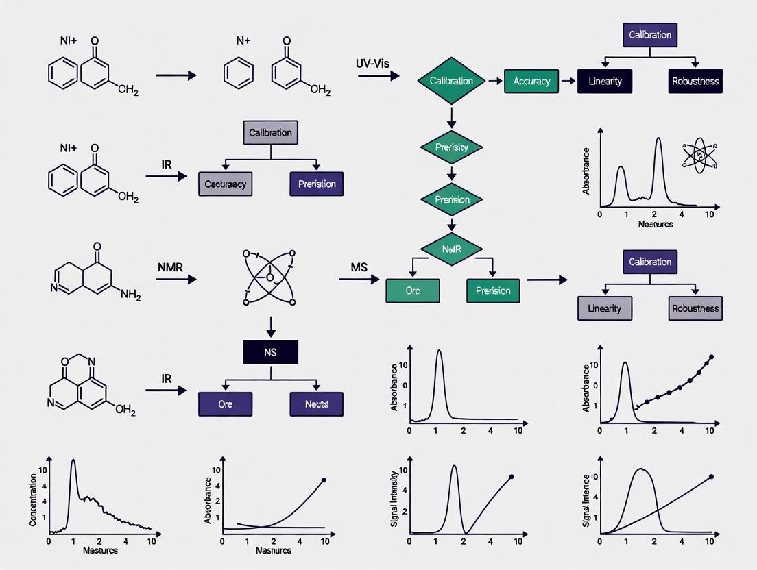
Logical Workflow for Method Validation
The process of validating a spectroscopic method follows a logical sequence, where earlier parameters often form the foundation for subsequent ones. The following diagram visualizes this workflow and the core objectives of each parameter.
The Scientist's Toolkit: Essential Research Reagent Solutions
The following reagents and materials are fundamental for conducting the validation experiments described in this guide.
Table 3: Essential Research Reagents and Materials for Validation Studies
| Item | Function in Validation | Critical Application Note |
|---|---|---|
| Certified Reference Standard | Serves as the benchmark for identity, calibration (linearity), and accuracy (recovery) experiments [2]. | The purity of the standard must be well-characterized and certified, as inaccuracies here propagate through all quantitative results [2]. |
| Placebo Matrix | Used in specificity testing to demonstrate that the signal is from the analyte and not from excipients or the sample matrix [6]. | For a drug product, this is a mixture of all non-active ingredients. It is critical for proving the method's selectivity. |
| Sample Matrix for Spiking | The actual material (e.g., drug substance, placebo, biological fluid) used in spike recovery experiments to determine accuracy [1] [2]. | The matrix should be as representative as possible of the real test samples to accurately assess matrix effects. |
| High-Purity Solvents & Reagents | Used for preparing mobile phases, standard solutions, and sample dilutions. | Impurities can contribute to baseline noise, affect linearity, and lead to inaccurate quantification, especially at low analyte levels. |
| Standardized Validation Protocols | Documents (e.g., ICH Q2(R2)) providing the formal framework, experimental designs, and acceptance criteria for validation [1] [3]. | Ensures the validation study meets regulatory standards and scientific rigor, facilitating reproducibility and reliability. |
The rigorous validation of analytical methods is a cornerstone of reliable scientific research and drug development. As demonstrated, Accuracy, Precision, Specificity, and Linearity are distinct yet interconnected parameters that collectively define the performance of a spectroscopic technique. While FT-IR and Raman spectroscopy often exhibit high inherent specificity due to their molecular "fingerprinting" capabilities, all techniques require systematic evaluation through standardized protocols. The choice of technique and the stringency of validation must always align with the method's intended purpose. By adhering to the detailed experimental protocols and comparative insights provided in this guide, scientists can ensure their spectroscopic methods generate data that is trustworthy, reproducible, and fit for regulatory submission.
In the pharmaceutical industry, demonstrating that analytical methods are reliable and fit for their intended purpose is a fundamental regulatory requirement. The regulatory framework for analytical procedures has recently evolved with the introduction of new and revised guidelines. The International Council for Harmonisation (ICH) finalized two key documents: ICH Q2(R2) on the validation of analytical procedures and ICH Q14 on analytical procedure development. These guidelines, adopted in late 2023, represent a significant shift towards a more holistic, science- and risk-based lifecycle approach [8].
Alongside these ICH guidelines, regional regulatory bodies like the U.S. Food and Drug Administration (FDA) and the European Medicines Agency (EMA) have their own expectations and guidance documents. Understanding the synergies and differences between the ICH, FDA, and EMA frameworks is crucial for researchers, scientists, and drug development professionals to ensure regulatory compliance and robust analytical practices. This guide provides a comparative analysis of these frameworks, placing them in the context of modern spectroscopic techniques and method validation.
Comparative Analysis of ICH Q2(R2), Q14, FDA, and EMA Guidelines
The following table summarizes the core focus, lifecycle view, and key tools or concepts endorsed by each regulatory guideline and agency concerning analytical procedures.
| Aspect | ICH Q2(R2) | ICH Q14 | FDA | EMA |
|---|---|---|---|---|
| Core Focus | Validation of analytical procedures; definitions and methodology for validation tests [9]. | Science- and risk-based approaches for analytical procedure development and lifecycle management [10]. | Enforcement of validation standards; has released specific guidance on topics like Bioanalytical Method Validation (BMV) for Biomarkers [11]. | Adherence to GMP; detailed requirements outlined in Annex 15; has a reflection paper on method transfer and 3Rs (Replacement, Reduction, Refinement) [12]. |
| Lifecycle Approach | Integrated with Q14; emphasizes that validation is a lifecycle activity [8]. | Explicitly outlines an Analytical Procedure Lifecycle from development through post-approval changes [10] [8]. | Supports a lifecycle model, evident in process validation guidance (3 stages) and the adoption of ICH Q12/Q14 principles for post-approval changes [13] [10]. | Supports a lifecycle approach, emphasizing ongoing process verification and product quality reviews [13]. |
| Key Tools/Concepts | Validation parameters (Accuracy, Precision, Specificity, etc.); application to multivariate or complex procedures [14]. | Analytical Target Profile (ATP); Enhanced Approach; Established Conditions (ECs); Change Management Protocols [10] [8]. | Recommends ICH M10 as a starting point for biomarker BMV, despite its stated non-applicability; encourages a Context of Use (COU)-driven strategy [11]. | Encourages use of a Validation Master Plan (VMP); more flexible on batch numbers for validation, relying on scientific justification [13]. |
Experimental Protocols for Analytical Procedure Validation
Protocol for Validation Based on ICH Q2(R2)
A robust validation protocol for a spectroscopic technique must demonstrate that the method meets predefined criteria for its intended use, as outlined in ICH Q2(R2). The following workflow details the key experiments and their logical sequence.
Figure 1. Sequential Workflow for Analytical Method Validation based on ICH Q2(R2).
Step-by-Step Methodology:
- Define the Analytical Target Profile (ATP): Before experimentation, draft an ATP. This is a foundational concept from ICH Q14 that precedes validation. The ATP is a predefined objective that articulates the procedure's intended purpose, specifying the analyte(s), the matrix, and the required performance criteria (e.g., target precision and accuracy) that the method must demonstrate upon validation [8].
- Specificity/Selectivity: Using a spectrophotometer or spectrometer, analyze a blank sample (e.g., placebo or sample matrix) and a sample spiked with the target analyte. The method should be able to unequivocally assess the analyte in the presence of components like impurities, degradation products, or matrix interferences that may be expected to be present [9].
- Linearity and Range: Prepare a minimum of 5 concentrations of the analyte across the specified range (e.g., 50-150% of the target concentration). Analyze each concentration in triplicate. Plot signal response (e.g., absorbance, peak area) versus concentration and calculate the correlation coefficient, y-intercept, and slope of the regression line using statistical software. The range is the interval between the upper and lower concentration levels for which linearity, accuracy, and precision have been demonstrated [9].
- Accuracy: Typically assessed using a spike/recovery experiment. Analyze samples of the matrix spiked with known quantities of the analyte at multiple levels (e.g., 50%, 100%, 150%) within the range. The mean value of the recovered analyte is compared to the true added value, and the percentage recovery is calculated [9].
- Precision:
- Repeatability: Inject a homogeneous sample at 100% of the test concentration at least 6 times. Calculate the relative standard deviation (RSD) of the measurements.
- Intermediate Precision: Perform the analysis on different days, with different analysts, or using different instruments. The combined RSD from the repeatability and intermediate precision studies demonstrates the method's reliability within a single laboratory [9].
- Detection Limit (LOD) & Quantitation Limit (LOQ): Based on signal-to-noise ratio, standard deviation of the response, and slope of the calibration curve. For spectroscopic techniques, a signal-to-noise ratio of 3:1 is generally accepted for LOD, and 10:1 for LOQ. Alternatively, LOD and LOQ can be calculated based on the standard deviation of the response and the slope of the calibration curve [9].
- Robustness: Deliberately introduce small, deliberate variations in method parameters (e.g., wavelength, temperature, flow rate, sample preparation time) to evaluate the method's capacity to remain unaffected by these changes. An experimental design (e.g., Design of Experiments, DoE) is often employed for this purpose.
Protocol for Managing Post-Approval Changes under ICH Q14
ICH Q14 provides a framework for managing changes to analytical procedures throughout their lifecycle. The following diagram outlines the science- and risk-based process for implementing a post-approval change.
Figure 2. ICH Q14 Workflow for Managing Post-Approval Changes to Analytical Procedures.
Step-by-Step Methodology:
- Risk Assessment: Initiate the process by conducting a risk assessment to evaluate the potential impact of the proposed change. Factors considered include the complexity of the test, the extent of the modification, and the procedure's relevance to product quality and Critical Quality Attributes (CQAs). This assessment classifies the change as high-, medium-, or low-risk [10].
- Bridging Studies: Design and execute bridging studies to directly compare the performance of the new or modified analytical procedure against the existing one. These studies use representative samples and are tailored to the nature of the change. The goal is to build a scientific understanding of the revised procedure [10].
- Confirm Performance Meets ATP: Conduct appropriate validation studies to confirm that the modified procedure continues to meet the performance criteria defined in the Analytical Target Profile (ATP). This ensures the method remains fit for its intended purpose. Update the procedure description, including parameters for system suitability and the analytical control strategy if needed [10].
- Assess Regulatory Impact: Determine the regulatory reporting category for the change (e.g., prior approval, notification, or do-and-report). This assessment is guided by tools like Established Conditions (ECs) and Post-Approval Change Management Protocols (PACMPs), which may have been agreed upon with regulatory authorities beforehand to streamline the process [10].
- Implement Change: After completing the necessary regulatory actions for each region, the change can be implemented in the Quality Control laboratory [10].
The Scientist's Toolkit: Essential Research Reagent Solutions
The following table details key materials and reagents essential for conducting validation experiments for spectroscopic techniques.
| Item Name | Function & Role in Validation |
|---|---|
| High-Purity Reference Standard | Serves as the benchmark for identifying and quantifying the analyte. Its purity is critical for accurate calibration, and for determining linearity, accuracy, and limits of detection [9]. |
| Placebo Matrix | Contains all components of the sample except the active analyte. It is used in specificity/selectivity experiments to prove the method does not generate interfering signals from excipients or the sample matrix itself [9]. |
| Forced Degradation Samples | Samples of the drug substance or product stressed under conditions (e.g., heat, light, acid/base) to generate degradants. Analysis of these samples is key to demonstrating the method's specificity and stability-indicating properties [9]. |
| Surrogate Matrix | Used in biomarker bioanalysis when a true blank matrix is unavailable. It allows for the preparation of calibration standards and is crucial for demonstrating accuracy and precision via spike/recovery experiments [11]. |
| System Suitability Standards | A prepared reference solution used to verify that the entire analytical system (instrument, reagents, columns) is performing adequately at the time of testing. It is a routine control for precision and reproducibility [10]. |
The regulatory framework for analytical procedures is converging towards an integrated lifecycle approach, as championed by ICH Q2(R2) and Q14. While regional agencies like the FDA and EMA align with these harmonized principles, nuances remain in their implementation focus and existing guidance documents. For spectroscopic techniques, a successful validation strategy begins with a well-defined ATP and employs a risk-based approach to both initial validation and subsequent lifecycle management. Mastering this framework enables scientists to develop more robust and reliable methods, streamline post-approval changes, and ultimately ensure the consistent quality and safety of pharmaceutical products.
The Role of Data Integrity and ALCOA+ Principles in Spectroscopy
In the field of spectroscopic analysis, where data forms the fundamental basis for critical decisions in drug development and material characterization, data integrity is not merely a regulatory requirement but a scientific necessity. The ALCOA+ framework provides a structured set of principles—Attributable, Legible, Contemporaneous, Original, Accurate, Complete, Consistent, Enduring, and Available—that ensure the reliability and trustworthiness of spectroscopic data throughout its entire lifecycle [15] [16]. For researchers and scientists working with spectroscopic techniques, adhering to these principles is paramount for producing valid, reproducible results that withstand regulatory scrutiny [17] [18].
The connection between data integrity and spectroscopy has intensified with the increasing adoption of electronic data systems and automated spectroscopic platforms. Regulatory agencies including the FDA, EMA, and WHO explicitly expect implementation of ALCOA+ principles in Good Manufacturing Practice (GMP) environments where spectroscopic data supports product quality assessments [16]. This article explores the practical application of ALCOA+ in spectroscopy, comparing implementation across software platforms and providing methodological guidance for researchers seeking to validate their spectroscopic methods within this rigorous framework.
Understanding ALCOA+ Principles in Spectroscopic Context
The ALCOA+ framework forms the cornerstone of modern data integrity practices in regulated scientific environments, with each principle addressing specific aspects of data reliability essential for spectroscopic analysis [15] [16].
Core ALCOA Principles
- Attributable: Data must be traceable to the person who generated it, the specific instrument used, and the time of creation [15] [18]. In spectroscopy, this means every spectrum or measurement must be linked to the analyst, the spectrometer, and the exact date and time of acquisition [18].
- Legible: Data must remain readable and accessible throughout its required retention period [15] [16]. For spectroscopic data, this involves ensuring that spectral files remain interpretable by software systems despite technology upgrades and that human-readable forms are preserved [18].
- Contemporaneous: Documentation must occur at the time the activity is performed [15] [16]. In spectroscopic workflows, this prohibits back-dating entries or documenting measurements after the analysis has been completed [18] [16].
- Original: The first recorded observation or a "verified true copy" must be preserved [15] [16]. For spectroscopy, this means retaining the raw spectral data before any processing or transformation, maintaining its authentic form [18].
- Accurate: Data must be free from errors, with any modifications documented and justified [15] [16]. In spectroscopic analysis, this requires that instruments are properly calibrated and that any data processing does not distort the original signal [18].
Extended ALCOA+ Principles
- Complete: All spectroscopic data must be included, with no omissions [15] [16]. This encompasses the entire dataset from multiple analyses, including failed runs or outliers that might otherwise be excluded [16].
- Consistent: Data should follow a standardized sequence of events with documented date and time stamps [15] [16]. In spectroscopy, this means applying uniform procedures across all analyses and maintaining chronological records of all activities [18] [16].
- Enduring: Data must be preserved for the required retention period, typically the product's shelf life plus several years in pharmaceutical applications [15] [16].
- Available: Data must be accessible for review, inspection, and copying throughout the retention period [15] [16]. For spectroscopic data, this means ensuring that spectra and associated metadata can be retrieved long after the initial analysis [18].
Table 1: ALCOA+ Principles and Their Implementation in Spectroscopy
| Principle | Definition | Spectroscopy Implementation Examples |
|---|---|---|
| Attributable | Data traceable to source | Secure user logins, instrument identifiers, electronic signatures [15] [18] |
| Legible | Permanently readable | Export to PDF/CSV formats, maintained backups, non-erasable formats [18] |
| Contemporaneous | Recorded in real-time | Automated time-stamping, immediate database storage [18] |
| Original | First recording or certified copy | Raw spectral data preservation, audit trails [18] |
| Accurate | Error-free with documented edits | Validation checks, documented calibrations, change control [18] |
| Complete | All data included with no omissions | Protected storage, prevention of data deletion, sequence integrity [15] [16] |
| Consistent | Chronological with protected sequence | Consistent workflows, time-stamped entries, standardized procedures [18] [16] |
| Enduring | Long-term preservation | Durable storage media, regular backups, migration plans [15] |
| Available | Accessible when needed | Searchable databases, retrieval systems, organized archives [15] [18] |
Implementing ALCOA+ in Spectroscopic Workflows: A Comparative Analysis
Successful implementation of ALCOA+ principles in spectroscopy requires both technical solutions and organizational culture. The following comparative analysis examines how different software platforms address these requirements and how they integrate into broader spectroscopic workflows.
Software Solutions for ALCOA+ Compliance in Spectroscopy
Specialized software plays a crucial role in enabling ALCOA+ compliance for spectroscopic systems by providing technical controls that enforce data integrity principles [18].
Table 2: Comparison of Software Features Supporting ALCOA+ in Spectroscopy
| Software Platform | ALCOA+ Features | Spectroscopy Applications | Compliance Standards |
|---|---|---|---|
| Vision Air | Secure user authentication, automated timestamping, SQL database storage, audit trails, two-level signing for configuration changes [18] | NIR spectroscopy, quantitative analysis, method development [18] | FDA 21 CFR Part 11, GMP/GLP [18] |
| AuditSafe | Secure logins for attribution, export of human-readable audit trails, project timestamping, validation capabilities [15] | Data collection and analysis in life sciences, pharmaceutical production [15] | FDA guidelines, global regulatory standards [15] |
| Thermo Fisher Spectroscopy Solutions | OQ (Operational Qualification) automation, audit trails, access controls, electronic signatures [17] | FTIR spectroscopy, pharmacopoeia testing, identity and purity algorithms [17] | 21 CFR Part 11, pharmacopoeia standards [17] |
| SpectraFit | Output file-locking system, collection of input data/results/initial model in single file, open-source validation [19] | XAS spectral analysis, peak fitting, quantitative composition analysis [19] | Transparency and reproducibility standards [19] |
Workflow Integration of ALCOA+ Principles
The following diagram illustrates how ALCOA+ principles integrate into a typical spectroscopic analysis workflow, from sample preparation to final reporting:
Experimental Protocol for Validating ALCOA+ Implementation in Spectroscopy
To systematically assess the implementation of ALCOA+ principles in spectroscopic systems, the following experimental protocol can be employed:
Objective: Verify and validate the proper implementation of ALCOA+ principles in a spectroscopic analysis system.
Materials and Equipment:
- Spectrometer with associated data acquisition software
- Certified reference materials for system suitability testing
- Secure database or electronic notebook system
- Access to system audit trails and metadata
Methodology:
- System Configuration and Security Assessment
- Create and verify unique user accounts for all analysts
- Configure instrument identifiers and method-specific parameters
- Validate electronic signature capabilities if applicable
- Verify access controls to prevent unauthorized data modification
Data Generation and Recording Process
- Analyze a series of certified reference materials
- Document all analyses in real-time during acquisition
- Intentionally introduce and correct errors to test documentation procedures
- Generate data across multiple sessions and analysts
Data Processing and Modification Tracking
- Apply data processing techniques (baseline correction, smoothing, etc.)
- Make authorized modifications to processing parameters
- Document all processing steps and parameter changes
- Generate reports at various processing stages
Data Storage, Retrieval and Archive Testing
- Store completed analyses in primary and backup systems
- Retrieve data after specified time intervals (24 hours, 1 week, 1 month)
- Verify data integrity through checksum validation
- Generate and verify certified true copies of original data
Acceptance Criteria:
- All data must be traceable to specific analyst and instrument
- No data points should be lost or unaccounted for in the final report
- All modifications must be documented with timestamp and justification
- Data must be retrievable and readable after specified storage periods
Advanced Applications: AI and Machine Learning in Spectroscopic Data Integrity
The emergence of artificial intelligence (AI) and machine learning (ML) in spectroscopic analysis introduces both opportunities and challenges for data integrity [20]. These advanced computational methods can enhance the reliability and interpretation of spectroscopic data when properly implemented within the ALCOA+ framework.
AI-Enhanced Spectral Analysis: Modern spectroscopic techniques generate high-dimensional data that creates pressing needs for automated and intelligent analysis beyond traditional expert-based workflows [20]. Machine learning approaches, collectively termed Spectroscopy Machine Learning (SpectraML), are being applied to both forward tasks (molecule-to-spectrum prediction) and inverse tasks (spectrum-to-molecule inference) while maintaining data integrity standards [20].
Data Quality Requirements for AI Applications: The implementation of AI in spectroscopic analysis heightens the importance of ALCOA+ principles, particularly:
- Complete datasets for training and validation
- Consistent data formatting and preprocessing
- Original data preservation before feature extraction
- Accurate labeling and annotation of training data
Open-source tools like SpectraFit demonstrate how AI-assisted analysis can be integrated with data integrity safeguards through features like output file-locking systems that collect input data, results data, and the initial fitting model in a single file to promote transparency and reproducibility [19].
Implementing robust data integrity practices in spectroscopic research requires both technical solutions and methodological approaches. The following toolkit outlines essential resources for maintaining ALCOA+ compliance:
Table 3: Research Reagent Solutions for ALCOA+-Compliant Spectroscopy
| Tool Category | Specific Solutions | Function in ALCOA+ Implementation |
|---|---|---|
| Spectroscopy Software | Vision Air, AuditSafe, Thermo Fisher OQ Packages, SpectraFit [15] [17] [18] | Provide technical controls for user attribution, audit trails, data protection, and automated compliance [15] [18] |
| Data Management Systems | SQL Databases, Electronic Lab Notebooks (ELNs), Laboratory Information Management Systems (LIMS) [18] | Ensure data endurance, availability, and completeness through secure storage and retrieval mechanisms [18] |
| Quality Assurance Tools | Automated Backup Systems, Checksum Validators, Audit Trail Reviewers [18] [16] | Verify data consistency, accuracy, and completeness throughout data lifecycle [18] [16] |
| Reference Materials | Certified Reference Materials, System Suitability Standards [17] | Establish accuracy and reliability of spectroscopic measurements through instrument qualification [17] |
| Documentation Systems | Electronic Signatures, Version Control Systems, Template Libraries [18] [16] | Support attributable, contemporaneous, and consistent documentation practices [18] [16] |
The implementation of ALCOA+ principles in spectroscopy represents a fundamental requirement rather than an optional enhancement for researchers and drug development professionals. As regulatory scrutiny of electronic data intensifies, the integration of these data integrity principles into spectroscopic method validation provides both compliance benefits and scientific advantages through enhanced data reliability and reproducibility [17] [18] [16].
The comparative analysis presented demonstrates that while software solutions vary in their specific implementation approaches, the core ALCOA+ requirements remain consistent across platforms and spectroscopic techniques. Successful adoption requires a holistic approach combining technical solutions with personnel training, organizational culture, and robust quality systems [16]. As spectroscopic technologies continue to evolve with increasing incorporation of AI and automation, the foundational principles of ALCOA+ will remain essential for ensuring the trustworthiness of spectroscopic data in research and regulated environments.
Establishing Linearity, Range, and Robustness for Spectroscopic Assays
In pharmaceutical development and analytical research, the reliability of spectroscopic data is paramount. Establishing method validity is a formal prerequisite for generating results that meet regulatory standards and support critical decisions in drug development. This guide focuses on three interconnected validation parameters—linearity, range, and robustness—which collectively ensure that an analytical method performs as intended in a reliable and reproducible manner.
Linearity defines the ability of a method to obtain results that are directly proportional to the concentration of the analyte within a given range [21]. The range is the interval between the upper and lower concentration levels of analyte for which demonstrated linearity, precision, and accuracy are achieved [21]. Robustness, on the other hand, evaluates the capacity of a method to remain unaffected by small, deliberate variations in procedural parameters, indicating its reliability during normal use [22]. This guide objectively compares the performance of different spectroscopic techniques against these critical validation parameters, providing a framework for scientists to select and optimize the most appropriate assay for their specific needs.
Conceptual Foundations and Definitions
A clear understanding of terminology is crucial for proper method validation. The linear range, or linear dynamic range, is specifically defined as the concentration interval over which the analytical signal is directly proportional to the concentration of the analyte [21]. This differs from the broader term dynamic range, which may encompass concentrations where a response is observed, but the relationship may be non-linear. Finally, the working range is the span of concentrations where the method delivers results with an acceptable level of uncertainty, and it can be wider than the strictly linear range [21].
For a method to be considered linear, it must demonstrate this proportional relationship, typically confirmed through a series of calibration standards. A well-established linear range should adequately cover the intended application, often spanning from 50-150% or 0-150% of the expected target analyte concentration [21]. It is vital to recognize that linearity can be technique- and compound-dependent. For instance, the linear range for LC-MS instruments is often fairly narrow, but can be extended using strategies such as isotopically labeled internal standards (ILIS), sample dilution, or instrumental modifications like lowering the flow rate in an ESI source to reduce charge competition [21].
Experimental Protocols for Validation
Protocol for Establishing Linearity and Range
The following detailed protocol is applicable to various spectroscopic techniques, including UV-Vis, to generate a calibration model.
1. Preparation of Standard Solutions:
- Prepare a stock solution of the analyte reference standard with high accuracy.
- Serially dilute the stock solution to obtain a minimum of five to six concentration levels. The range should cover 50-150% or 0-150% of the expected sample concentration [21] [23].
2. Instrumental Analysis:
- Analyze each standard solution in triplicate to account for instrumental variability.
- Maintain consistent instrumental parameters (e.g., slit width, path length, integration time) throughout the analysis.
- For UV-Vis, measure the absorbance at the wavelength of maximum absorption (λmax) to maximize sensitivity and minimize errors [24].
3. Data Analysis and Assessment:
- Plot the mean analytical response (e.g., absorbance, peak area) against the concentration of the standard solutions.
- Calculate the regression line using the least-squares method (y = mx + c), where
yis the response,mis the slope,xis the concentration, andcis the y-intercept. - Determine the correlation coefficient (R²), sum of squared residuals, and y-intercept to evaluate linearity. A high R² value (e.g., >0.999 in a validated UV method for deferiprone) indicates strong linearity [23].
- The range is validated by demonstrating that the method meets acceptable criteria for linearity, precision, and accuracy across the specified concentration interval.
Protocol for Assessing Robustness
Robustness testing evaluates the method's resilience to deliberate, small changes in operational parameters.
1. Experimental Design:
- Identify critical method parameters that could influence the results. These are technique-specific:
- Use a structured approach, such as a full factorial or Plackett-Burman design, to efficiently test the impact of varying these parameters simultaneously.
2. Execution and Analysis:
- Perform the assay under the varied conditions while analyzing a system suitability sample or reference standard at a fixed concentration.
- Monitor the effect of these variations on critical performance attributes, such as retention time, signal intensity, spectral shape, and quantitative result (assay value).
- The method is considered robust if the quantitative results remain within predefined acceptance criteria (e.g., ±1-2% of the value obtained under standard conditions) and performance attributes show minimal deviation despite the introduced variations.
Comparative Performance Data of Spectroscopic Techniques
The following tables synthesize experimental data and characteristics from various spectroscopic methods, highlighting their performance in terms of linearity, range, and robustness.
Table 1: Comparative Linearity and Range of Spectroscopic Techniques
| Technique | Typical Linear Range (Order of Magnitude) | Example Correlation Coefficient (R²) | Key Factors Influencing Linearity |
|---|---|---|---|
| UV-Vis Spectroscopy | 1-2 (e.g., 2-12 μg/mL for Deferiprone [23]) | ≥ 0.999 [23] | Deviations from Beer-Lambert law at high concentration, stray light, instrumental noise. |
| Fluorescence Spectroscopy | 3-4 | > 0.99 (assay dependent) | Inner-filter effect, self-quenching, photobleaching, concentration saturation. |
| LC-MS | 2 (can be extended with ILIS) | > 0.99 (assay dependent) | Charge competition in the ion source (especially ESI), detector saturation. [21] |
| NIR Spectroscopy | Requires multivariate calibration (PLS, etc.) | Model dependent (e.g., R² > 0.95 for robust models) | Scattering effects, complex baseline offsets, weak and overlapping absorption bands. [25] |
Table 2: Robustness Considerations Across Spectroscopic Techniques
| Technique | Critical Parameters to Test for Robustness | Common Vulnerabilities & Mitigation Strategies |
|---|---|---|
| UV-Vis Spectroscopy | Wavelength accuracy, pH of solvent, temperature, source lamp aging. | Vulnerability: Solvent/ matrix effects. Mitigation: Use matched solvent/blanks and standardize sample preparation. [24] [23] |
| Fluorescence Spectroscopy | Excitation/Emission bandwidths, temperature, solvent viscosity, sample turbidity, quenchers. | Vulnerability: Inner-filter effects, photo-bleaching. Mitigation: Use narrow cuvettes, dilute samples, and minimize exposure. [22] |
| Vibrational Spectroscopy (IR, Raman) | Laser power/flux, sampling depth/pressure, calibration model stability. | Vulnerability: Fluorescence background (Raman), water vapor (IR). Mitigation: Use 1064 nm lasers (Raman), purge optics with dry air (IR). [26] [25] |
| Hyphenated Techniques (e.g., LC-MS) | Mobile phase composition/buffer concentration, flow rate, ion source temperature, interface parameters. | Vulnerability: Ion suppression, column degradation. Mitigation: Use stable isotope internal standards, guard columns. [21] |
Advanced Topics: Managing Nonlinearity and Enhancing Robustness
Addressing Nonlinear Effects
Real-world spectroscopic data often deviates from ideal linear behavior due to chemical, physical, and instrumental factors [25]. Identifying and managing these nonlinearities is essential for accurate quantification, especially when using multivariate calibration models.
Common sources of nonlinearity include:
- Chemical Effects: Spectral band saturation at high concentrations and molecular interactions like hydrogen bonding.
- Physical Effects: Light scattering and path length variations in diffuse reflectance measurements.
- Instrumental Effects: Detector nonlinearity, stray light, and wavelength misalignments [25].
When linear models like classical Partial Least Squares (PLS) are insufficient, several advanced calibration methods can be employed:
- Polynomial Regression: A simple extension that includes higher-order terms, but it can overfit high-dimensional spectral data.
- Kernel Partial Least Squares (K-PLS): Maps data into a higher-dimensional feature space where linear relations can be applied, effectively capturing complex nonlinearities.
- Artificial Neural Networks (ANNs): Highly flexible models suitable for large, high-dimensional datasets like hyperspectral images, though they require significant data and can be prone to overfitting [25].
Data Preprocessing for Robust Models
Robust analytical methods rely on high-quality data. Spectral preprocessing is a critical step to mitigate unwanted variance and enhance the reliability of the analytical signal, particularly for robustness. A systematic preprocessing pipeline includes:
- Cosmic Ray/Spike Removal: Identifies and removes sharp, spurious signals.
- Baseline Correction: Suppresses low-frequency drifts caused by instrumental or sample matrix effects.
- Scattering Correction: Corrects for multiplicative scattering effects (e.g., using Multiplicative Scatter Correction).
- Normalization: Minimizes systematic errors from sample-to-sample variations.
- Smoothing and Filtering: Reduces high-frequency random noise [27].
Implementing these steps before regression or model building significantly improves the accuracy, precision, and transferability of spectroscopic methods.
The Scientist's Toolkit: Essential Reagents and Materials
Table 3: Key Research Reagent Solutions for Spectroscopic Assay Validation
| Item | Function in Validation | Application Notes |
|---|---|---|
| Analytical Reference Standard | Serves as the primary material for preparing calibration standards to establish linearity and range. | Must be of high and well-defined purity (e.g., pharmacopeial grade). Its concentration is the basis for all quantitative measurements. |
| Isotopically Labeled Internal Standard (ILIS) | Compensates for analyte loss during preparation and signal variation in the instrument, widening the linear dynamic range in techniques like LC-MS. | Used in mass spectrometry; should be structurally analogous to the analyte but distinguishable by mass. [21] |
| High-Purity Solvents | Dissolve analytes and standards without introducing interfering spectral signals or contaminants. | UV-Vis grade solvents are essential for low UV absorbance. Water purity is critical (e.g., from a system like Milli-Q). [26] |
| Buffer Solutions | Control the pH of the sample matrix, which can critically affect spectral shape, intensity, and stability, thereby testing robustness. | Required for analytes with ionizable groups. Buffer type and concentration should be specified and controlled. |
| Certified Reference Materials (CRMs) | Provide an independent, matrix-matched control to verify method accuracy and precision across the validated range. | Used for final method verification; traceable to international standards. |
Workflow and Decision Pathways
The following diagram illustrates the logical workflow for establishing and troubleshooting the linearity and range of a spectroscopic assay.
Assay Linearity Establishment Workflow
The rigorous establishment of linearity, range, and robustness is non-negotiable for developing trustworthy spectroscopic methods in drug development and analytical research. As demonstrated, the performance of different spectroscopic techniques varies significantly, with factors like dynamic range, susceptibility to matrix effects, and optimal calibration strategies being highly technique-specific.
The field continues to evolve, with future advancements pointing toward wider adoption of hybrid physical-statistical models that integrate fundamental spectroscopic theory with machine learning for greater interpretability and generalization. Furthermore, the development of transferable nonlinear models that maintain accuracy across different instruments and the application of Explainable AI (XAI) to complex models like neural networks will be crucial for meeting regulatory demands and enhancing scientist trust in predictive outcomes [25]. By adhering to structured experimental protocols and leveraging advanced data processing tools, scientists can ensure their spectroscopic assays are not only valid but also robust and fit-for-purpose in the modern laboratory.
Implementing Validation for Modern Spectroscopic Techniques
Validation Strategies for UV-Vis, NIR, and MIR Spectroscopy
Method validation is a critical process that establishes documented evidence providing a high degree of assurance that a specific spectroscopic technique will consistently produce results meeting predetermined analytical method requirements. For researchers and drug development professionals, implementing robust validation strategies for ultraviolet-visible (UV-Vis), near-infrared (NIR), and mid-infrared (MIR) spectroscopy ensures data integrity, regulatory compliance, and reliable decision-making throughout the product development lifecycle. Each technique possesses unique characteristics dictated by its underlying physical principles—electronic transitions for UV-Vis-NIR, and molecular vibrations for MIR—which directly influence the appropriate validation approach. This guide systematically compares validation parameters across these spectroscopic techniques, providing experimental protocols and performance data to support method development in regulated environments.
Fundamental Principles and Instrumentation
Technical Basis of Spectroscopic Techniques
The validation of any spectroscopic method must begin with understanding its fundamental principles and how they influence performance characteristics. UV-Vis-NIR spectroscopy measures the absorption of electromagnetic radiation in the 175-3300 nm range, primarily resulting from electronic transitions in molecules. These transitions occur when valence electrons are excited to higher energy states, with UV-Vis regions (175-800 nm) covering π→π* and n→π* transitions in organic molecules, while NIR (800-3300 nm) encompasses weaker overtones and combination bands of fundamental molecular vibrations [28] [29]. In contrast, MIR spectroscopy, particularly Fourier-transform infrared (FT-IR), probes fundamental molecular vibrations in the 4000-400 cm⁻¹ range (approximately 2500-25000 nm), providing detailed molecular fingerprint information through absorption of IR light by molecules undergoing vibrational transitions between quantized energy states [7].
Instrumentation Comparisons
The core instrumentation differences between these techniques significantly impact validation strategies. UV-Vis-NIR instruments typically employ dispersive designs with monochromators containing diffraction gratings that separate wavelengths spatially, while modern FT-IR and FT-NIR instruments utilize interferometers with moving mirrors to create interferograms that are mathematically transformed into spectra using Fourier transformation [30] [7]. Key performance advantages of FT instruments include Fellgett's (multiplex) advantage for improved signal-to-noise ratio through simultaneous measurement of all wavelengths, Jacquinot's (throughput) advantage for higher energy throughput with fewer optical slits, and Connes' advantage for superior wavelength calibration precision derived from an internal laser reference [7].
Figure 1: Comprehensive Workflow for Spectroscopic Method Validation
Critical Validation Parameters Comparison
Performance Characteristics Across Techniques
The validation of spectroscopic methods requires demonstration of several key performance parameters that vary significantly across UV-Vis, NIR, and MIR techniques due to their different physical principles and instrumentation. Specificity, the ability to measure analyte response in the presence of potential interferents, is typically highest in MIR spectroscopy due to its detailed molecular fingerprinting capabilities, followed by NIR with its complex overtone patterns, while UV-Vis may suffer from spectral overlaps in complex mixtures [7] [29]. Linear dynamic range is generally widest in UV-Vis spectroscopy (typically 2-4 absorbance units), while NIR and MIR exhibit narrower linear ranges due to deviations from Beer-Lambert law at higher concentrations, particularly for fundamental vibrations in MIR [29].
Table 1: Comparison of Key Validation Parameters Across Spectroscopic Techniques
| Validation Parameter | UV-Vis Spectroscopy | NIR Spectroscopy | MIR Spectroscopy (FT-IR) |
|---|---|---|---|
| Typical Wavelength Range | 175-800 nm [31] [29] | 800-3300 nm [31] [29] | 2500-25000 nm (4000-400 cm⁻¹) [7] |
| Primary Transitions | Electronic transitions [28] | Overtone/combination vibrations [28] | Fundamental molecular vibrations [7] |
| Specificity | Moderate (potential spectral overlaps) [29] | High (complex overtone patterns) [30] | Very high (molecular fingerprint region) [7] |
| Linear Dynamic Range | 2-4 AU (wide) [29] | 1-3 AU (moderate) [30] | 0.5-2 AU (narrower) [7] |
| Typical LOD (Absorbance) | 0.001-0.01 AU [31] | 0.005-0.05 AU [30] | 0.01-0.1 AU [7] |
| Precision (RSD) | 0.1-1% [31] | 0.5-2% [30] | 0.3-1.5% [7] |
| Sample Preparation Needs | Minimal (dilution) [29] | Minimal to moderate [30] | Variable (ATR, KBr pellets, etc.) [7] |
| Primary Regulatory Applications | Quantitative analysis in dissolution, content uniformity [31] | Raw material ID, polymorph screening, process monitoring [26] | Compound identification, polymorph characterization [7] |
Accuracy, Precision, and Robustness
Accuracy and precision validation approaches differ substantially across techniques. UV-Vis methods typically demonstrate excellent accuracy (98-102% recovery) and precision (RSD 0.1-1%) for quantitative analysis of single components in simple matrices, validated against certified reference materials [31] [29]. For NIR methods, accuracy validation must account for the multivariate nature of the technique, with typical RMSEP (Root Mean Square Error of Prediction) values of 0.1-0.5% for major components in pharmaceuticals when validated against primary reference methods, with precision RSD ranging from 0.5-2% depending on the sampling technique [30]. MIR accuracy varies significantly with sampling technique, with ATR-FTIR typically showing 95-105% recovery for quantitative analysis when proper calibration models are used, and precision RSD of 0.3-1.5% [7].
Robustness testing evaluates method resilience to deliberate variations in method parameters. For UV-Vis, this includes testing wavelength accuracy (±1 nm), bandwidth, and sampling pathlength variations [31]. NIR method robustness must evaluate spectral pretreatment variations, temperature effects, and sample presentation consistency due to light scattering effects [30]. MIR robustness testing focuses on ATR crystal pressure consistency, sample homogeneity, and environmental humidity control due to water vapor interference [7].
Experimental Protocols for Validation
Specificity and Selectivity Protocols
Specificity validation experimentally demonstrates that the analytical method can unequivocally assess the analyte in the presence of potential interferents. For UV-Vis methods, specificity is established by comparing analyte spectra with placebo mixtures, stressed samples, and related compounds, requiring baseline separation of analyte peak from interfering peaks [29]. A typical protocol involves preparing solutions of analyte, placebo, and synthetic mixtures, scanning from 200-400 nm or wider range as needed, and demonstrating that placebo components show no interference at the analyte λmax [31].
For NIR specificity validation, the multivariate nature requires different approaches. Using a minimum of 20-30 representative samples spanning expected variability, collect spectra in appropriate mode (diffuse reflectance for solids, transmission for liquids). Apply chemometric tools like PCA (Principal Component Analysis) to demonstrate clustering of acceptable materials and separation from unacceptable materials, with statistical distance metrics such as Mahalanobis distance establishing classification boundaries [32].
MIR specificity validation in FT-IR leverages the fingerprint region (1500-500 cm⁻¹) where molecules show unique absorption patterns. Using ATR or transmission sampling, collect spectra of reference standards, potential contaminants, and degraded samples. Specificity is confirmed when the analyte spectrum shows unique absorption bands not present in interferents, or through spectral library matching with match scores exceeding predefined thresholds (typically >0.95 for pure compound identification) [7].
Linearity and Range Determination
Linearity establishes that the analytical method produces results directly proportional to analyte concentration within a specified range. For UV-Vis validation, prepare a minimum of 5 concentrations spanning the expected range (typically 50-150% of target concentration) in triplicate. Plot absorbance versus concentration and calculate correlation coefficient (r > 0.999), y-intercept (not significantly different from zero), and residual sum of squares [29]. A typical UV-Vis linearity experiment for drug substance assay might use concentrations of 50, 75, 100, 125, and 150 μg/mL with 1 cm pathlength, expecting r² ≥ 0.998 [31].
NIR linearity validation follows different principles due to frequent use of multivariate calibration. Prepare 20-30 samples with concentration variation spanning expected range using appropriate experimental design. Develop PLS (Partial Least Squares) or PCR (Principal Component Regression) models with full cross-validation, reporting RMSECV (Root Mean Square Error of Cross-Validation) and R² for the calibration model. For a pharmaceutical blend analysis, RMSECV values <0.5% for API concentration typically demonstrate acceptable linearity [32].
MIR linearity using ATR-FTIR requires special consideration of the Beer-Lambert law limitations at higher concentrations due to reflection/absorption complexities. Prepare standard mixtures with 5-7 concentration levels in appropriate matrix, ensuring uniform contact with ATR crystal. Use peak height or area of specific vibrational bands, expecting linear r² > 0.995 for quantitative applications. Pathlength correction factors may be necessary for accurate quantification [7].
Precision and Accuracy Experiments
Precision validation demonstrates the degree of agreement among individual test results under prescribed conditions, while accuracy establishes agreement between test results and accepted reference values.
Table 2: Experimental Protocols for Precision and Accuracy Validation
| Validation Type | UV-Vis Protocol | NIR Protocol | MIR (FT-IR) Protocol |
|---|---|---|---|
| Repeatability (Intra-day) | 6 determinations at 100% concentration, RSD ≤ 1% [31] | 10 spectra of single sample, repositioning between scans, RSD ≤ 2% [30] | 10 measurements of single preparation with repositioning, RSD ≤ 1.5% [7] |
| Intermediate Precision (Inter-day) | 6 determinations each on 2 different days, by 2 analysts, with different instruments; RSD ≤ 2% [31] | 3 preparations each on 3 days, different operators, instrument; compare RMSEP [32] | 3 preparations analyzed over 3 days with different sample positioning; RSD ≤ 2.5% [7] |
| Accuracy Recovery | 9 determinations over 3 concentration levels (80%, 100%, 120%) with 98-102% recovery [29] | 20-30 validation set samples spanning concentration range, RMSEP < 0.5% of range [32] | Standard addition method with 3 spike levels, 95-105% recovery [7] |
| Sample Preparation | Dilution in appropriate solvent, minimal preparation [29] | Representative sampling, consistent presentation geometry [30] | Uniform contact with ATR crystal or consistent pellet preparation [7] |
Detection and Quantitation Limits
Experimental Determination Approaches
Limit of detection (LOD) and quantitation (LOQ) establish the lowest levels of analyte that can be reliably detected or quantified, with approaches varying significantly across techniques. For UV-Vis, LOD and LOQ are typically determined based on signal-to-noise ratio (S/N) of 3:1 for LOD and 10:1 for LOQ, or using standard deviation of the response and slope of the calibration curve (LOD = 3.3σ/S, LOQ = 10σ/S) [31]. A typical UV-Vis method might achieve LOD of 0.001-0.01 AU, corresponding to low μg/mL concentrations for compounds with high molar absorptivity [29].
NIR spectroscopy, dealing with weak overtone bands, generally has higher detection limits. LOD/LOQ determination requires multivariate approaches, typically based on the RMSEP of cross-validated calibration models. The LOD is often estimated as 3 times the standard error of the residual variance in the response, while LOQ is estimated as 10 times this value. Practical LOQ values for major components in pharmaceuticals typically range from 0.1-0.5% w/w using diffuse reflectance NIR [30].
MIR FT-IR detection limits depend strongly on sampling technique. For ATR-FTIR, LOD is typically determined by analyzing progressively diluted samples until the characteristic absorption bands become indistinguishable from background noise (S/N < 3). Using modern FT-IR instruments with high-sensitivity detectors, LOD values for organic compounds typically range from 0.1-1% with ATR sampling, potentially reaching ppm levels with transmission cells or specialized techniques like photoacoustic detection [7].
Advanced Validation Considerations
Data Processing and Chemometrics
Modern spectroscopic validation, particularly for NIR and MIR, increasingly relies on sophisticated data processing and chemometrics, requiring validation of both instrumental and mathematical procedures. Spectral preprocessing techniques must be validated for their intended purpose, including derivatives (Savitzky-Golay) for resolution enhancement, standard normal variate (SNV) and multiplicative scatter correction (MSC) for scatter effects, and normalization for pathlength variations [27]. Each preprocessing method introduces specific artifacts that impact validation parameters; for example, derivative operations improve specificity but may degrade signal-to-noise ratio, requiring optimization of derivative order and window size [27].
Multivariate calibration models require comprehensive validation including determination of optimal number of latent variables to avoid overfitting, outlier detection methods (leverage, residuals, and influence measures), and rigorous external validation with independent sample sets. For NIR methods, the ratio of performance to deviation (RPD), calculated as the ratio of standard deviation of reference data to SECV (Standard Error of Cross-Validation), should exceed 3 for acceptable quantitative screening applications and 5 for quality control purposes [32]. Model transferability between instruments must be validated using techniques like direct standardization or piecewise direct standardization when methods are deployed across multiple systems [30].
Regulatory and Compendial Requirements
Validation strategies must align with regulatory expectations from FDA, EMA, and compendial requirements (USP, Ph. Eur.). USP general chapters <857> (UV-Vis Spectroscopy), <1856> (NIR Spectroscopy), and <851> (Spectrophotometry and Light-Scattering) provide specific validation guidance [31]. For NIR methods, the FDA's Process Analytical Technology (PAT) guidance encourages rigorous multivariate validation approaches including real-time release testing applications [26]. Regulatory submissions should include comprehensive validation data packages demonstrating all relevant validation parameters with appropriate statistical analysis, plus ongoing monitoring procedures for method maintenance including periodic performance qualification and model updating strategies for handling raw material and process changes [31] [7].
Figure 2: Advanced Validation Workflow Incorporating Data Processing
Essential Research Reagent Solutions
Successful validation of spectroscopic methods requires appropriate reference materials and reagents with documented purity and traceability. The selection of suitable materials forms the foundation for accurate method characterization and should be carefully considered during validation planning.
Table 3: Essential Research Reagents and Materials for Spectroscopic Validation
| Reagent/Material | Technical Function | Validation Application | Technical Specifications |
|---|---|---|---|
| Certified Reference Materials (CRMs) | Primary calibration standards with documented purity and traceability [31] | Accuracy determination, method calibration | Purity ≥ 99.5%, uncertainty ≤ 0.5%, traceable to national standards |
| Holmium Oxide Filter | Wavelength accuracy verification [31] | UV-Vis/NIR wavelength calibration | Characteristic peaks at 241.5, 287.5, 361.5, 486.0, and 536.5 nm with ±0.5 nm tolerance |
| Polystyrene Standard | Wavelength and resolution validation [30] | NIR/FT-IR performance qualification | Characteristic peaks at 906.0, 1028.3, 1601.0, 2929.8 cm⁻¹ with ±1.0 cm⁻¹ tolerance |
| NIST Traceable Neutral Density Filters | Photometric accuracy verification [31] | Absorbance/transmittance accuracy validation | Specific absorbance values at multiple wavelengths with ±0.01 AU uncertainty |
| Spectroscopic Solvents (HPLC Grade) | Sample preparation and dilution [29] | Sample and standard preparation | UV-Vis transparency with cutoff below 200 nm, low fluorescent impurities |
| ATR Cleaning Solutions | Crystal surface maintenance [7] | FT-IR/ATR sampling reproducibility | Isopropanol/water mixtures, non-abrasive, residue-free, compatible with crystal material |
| Background Reference Materials | Spectral background correction [7] | Daily instrument qualification | Spectralon for NIR, KBr pellets for MIR, appropriate solvent for UV-Vis |
Validation strategies for UV-Vis, NIR, and MIR spectroscopy must be tailored to each technique's fundamental principles, instrumentation, and application requirements. UV-Vis methods excel in quantitative applications with straightforward validation approaches based on univariate calibration, while NIR and MIR techniques require sophisticated multivariate validation strategies incorporating chemometric model validation. Contemporary validation approaches must address both instrumental performance and data processing algorithms, particularly for NIR and MIR methods deployed in PAT environments. Successful validation requires thorough understanding of regulatory expectations, appropriate statistical approaches, and scientifically sound experimental designs that challenge method capabilities under realistic conditions. As spectroscopic technologies continue evolving with miniaturization, increased automation, and enhanced computational power, validation approaches must similarly advance to ensure data integrity while facilitating innovation in pharmaceutical development and manufacturing.
Multi-attribute methods (MAM) represent a paradigm shift in the analytical characterization of complex products, from biopharmaceuticals to natural products. For biologics, MAM is a liquid chromatography-mass spectrometry (LC-MS)-based peptide mapping method that enables direct identification, monitoring, and quantification of multiple product quality attributes (PQAs) at the amino acid level in a single, streamlined workflow [33] [34]. This approach provides a more informative and efficient alternative to conventional chromatographic and electrophoretic assays that typically monitor only one or a few attributes separately [35].
The core innovation of MAM lies in its two-phase workflow: an initial discovery phase to identify quality attributes for monitoring and create a targeted library, followed by a monitoring phase that uses this library for routine analysis while employing differential analysis for new peak detection (NPD) [33]. This dual capability allows for both targeted quantification of specific attributes and untargeted detection of impurities or modifications [34]. As regulatory agencies increasingly emphasize Quality by Design (QbD) principles, MAM has gained prominence for its ability to provide comprehensive product understanding throughout the development lifecycle [36] [35].
MAM for Biopharmaceuticals Characterization
Core Principles and Workflow
The MAM workflow for biopharmaceuticals involves several critical steps designed to ensure comprehensive characterization of therapeutic proteins such as monoclonal antibodies (mAbs). The process begins with proteolytic digestion of the protein sample using enzymes like trypsin to generate peptides, followed by reversed-phase chromatographic separation and high-resolution LC-MS analysis [33] [37]. This workflow enables primary sequence verification, detection and quantitation of post-translational modifications (PTMs), and identification of impurities [33].
A key differentiator of MAM from traditional peptide mapping is its data analysis approach, which includes both targeted attribute quantification (TAQ) of specific critical quality attributes (CQAs) and new peak detection (NPD) through differential analysis between test samples and reference standards [34] [35]. The NPD function is particularly valuable for detecting unexpected product variants or impurities that might not be included in targeted monitoring [34].
Experimental Protocol for MAM Implementation
Implementing a robust MAM requires careful optimization of each step in the workflow:
Sample Preparation: The protein therapeutic must be digested into peptides using highly specific proteases. Trypsin is most commonly used as it produces peptides in the optimal size range (∼4–45 amino acid residues) for mass spectrometric analysis [36]. This critical step requires 100% sequence coverage, high reproducibility, and minimal process-induced modifications (e.g., deamidation) [36]. Protocols typically take 90–120 minutes [33]. Use of immobilized trypsin kits (e.g., SMART Digest Kits) can enhance reproducibility and compatibility with automation [36].
Chromatographic Separation: Peptides are separated using reversed-phase ultra-high-pressure liquid chromatography (UHPLC) systems, which provide exceptional robustness, high gradient precision, and improved reproducibility [36]. Columns with 1.5 µm solid core particles (e.g., Accucore, Hypersil GOLD) deliver sharp peaks, maximal peak capacities, and remarkably low retention time variations essential for reliable batch-to-batch analysis [36].
Mass Spectrometric Detection: Separated peptides are analyzed using high-resolution accurate mass (HRAM) MS instrumentation [36] [37]. The high mass accuracy and resolution enable confident peptide identification and modification monitoring without the need for full chromatographic separation of all species [36].
Data Processing: Specialized software is used for automated peptide identification, relative quantification of targeted PTMs, and new peak detection [37]. For NPD, appropriate detection thresholds must be established to balance sensitivity against false positives [35].
Table 1: Key Research Reagent Solutions for MAM Workflows
| Reagent/Equipment | Function | Examples/Characteristics |
|---|---|---|
| Proteolytic Enzymes | Protein digestion into peptides | Trypsin (most common), Lys-C, Glu-C, AspN; immobilized formats enhance reproducibility |
| UHPLC System | Peptide separation | High-pressure capability (>1000 bar), high gradient precision, minimal carryover |
| HRAM Mass Spectrometer | Peptide detection & identification | High resolution (>30,000) and mass accuracy (<5 ppm); Q-TOF commonly used |
| Chromatography Columns | Peptide separation | C18 stationary phase with 1.5-2µm particles; provides sharp peaks and stable retention |
| Data Analysis Software | Attribute quantification & NPD | Targeted processing of attribute lists; differential analysis for impurity detection |
Application Scope and Replaceable Methods
MAM has the potential to replace multiple conventional analytical methods used in quality control of biopharmaceuticals, providing site-specific information with greater specificity and often superior sensitivity [34] [35]. The following table summarizes the conventional methods that can be consolidated through MAM implementation:
Table 2: Conventional QC Methods and Their MAM Replaceable Capabilities
| Conventional Method | Attributes Monitored | MAM Capability |
|---|---|---|
| Ion-Exchange Chromatography (IEC) | Charge variants (deamidation, oxidation, C-terminal lysine) | Yes – site-specific quantification [34] |
| Hydrophilic Interaction LC (HILIC) | Glycosylation profiles | Yes – site-specific glycan identification [35] [37] |
| Reduced CE-SDS | Fragments, cleaved variants | Potential – depending on fragment sequence [34] |
| Peptide Mapping (LC-UV) | Identity, sequence variant | Yes – with enhanced specificity [34] |
| ELISA | Host Cell Proteins (HCPs) | Potential – though challenging for low-level HCPs [34] |
MAM for Natural Products
Characterization Challenges and Analytical Solutions
Natural products present unique characterization challenges due to their inherent complexity, variability in composition based on source and extraction methods, and the presence of multiple active constituents [38] [39]. Unlike biologics with defined amino acid sequences, natural products such as botanicals, herbal remedies, and dietary supplements are complex mixtures where insufficient assessment of identity and chemical composition hinders reproducible research [38].
The principles of multi-attribute approaches are increasingly being applied to natural products research to address these challenges. For natural products, the focus shifts to characterizing multiple marker compounds or biologically active components rather than specific amino acid modifications [39]. This requires rigorous method validation and the use of matrix-based reference materials to ensure analytical measurements are accurate, precise, and sensitive [38].
Emerging Technologies and Applications
Recent advances in analytical technologies are enabling more comprehensive characterization of natural products:
Multi-Attribute Raman Spectroscopy (MARS): This novel approach combines Raman spectroscopy with multivariate data analysis to measure multiple product quality attributes without sample preparation [40]. MARS allows for high-throughput, nondestructive analysis of formulated products and has demonstrated capability for monitoring both protein purity-related and formulation-related attributes through generic multi-product models [40].
Integrated (Q)STR and In Vitro Approaches: For toxicity assessment, quantitative structure-toxicity relationship ((Q)STR) models integrated with in vitro assays provide a comprehensive approach to predict the toxicity of natural product components [39]. This methodology has been applied to predict acute toxicity using LD50 data from natural product databases, helping prioritize compounds for further development [39].
Single-Cell Multiomics and Network Pharmacology: Advanced technologies including single-cell multiomics and network pharmacology are being deployed to elucidate the mechanisms of action of natural products, particularly those used in traditional Chinese medicine [41]. These approaches help identify molecular targets and understand complex interactions between multiple active components.
Experimental Framework for Natural Product MAM
A robust analytical framework for natural products should include:
Comprehensive Characterization: Initial thorough characterization of the natural product using LC-MS, NMR, and other orthogonal techniques to identify major and minor constituents [38].
Reference Material Development: Establishment of well-characterized reference materials that represent the chemical complexity of the natural product [38].
Method Validation: Rigorous validation of analytical methods demonstrating they are reproducible and appropriate for the specific sample matrix (plant material, phytochemical extract, etc.) [38].
Multi-Attribute Monitoring: Implementation of monitoring protocols for multiple critical constituents that correlate with product quality, efficacy, and safety [39].
Comparative Performance Analysis
MAM Platform Comparisons
Different analytical platforms offer distinct advantages for multi-attribute analysis depending on the application requirements:
Table 3: Comparison of Multi-Attribute Method Platforms
| Platform/Technique | Attributes Monitored | Sample Preparation | Throughput | Key Applications |
|---|---|---|---|---|
| LC-MS MAM | Site-specific PTMs, sequence variants, oxidation, deamidation, glycosylation | Extensive (digestion required) | Moderate (1-few days) [33] | Biotherapeutic characterization, cGMP testing [33] [34] |
| Raman Spectroscopy (MARS) | Protein concentration, osmolality, formulation additives | Minimal (non-destructive) | High (single scan) [40] | Formulated mAb therapeutics, real-time release testing [40] |
| (Q)STR + In Vitro | Acute toxicity, hepatotoxicity, cytotoxicity | Variable | High (in silico) Moderate (in vitro) | Natural product safety screening [39] |
Performance Metrics and Experimental Data
Studies have demonstrated the performance of MAM in comparison to conventional methods:
Glycan Analysis Comparison: When comparing MAM to conventional HILIC for glycan analysis, MAM performed similarly in identifying and quantifying major glycan species while providing additional site-specific information for monoclonal antibodies [35].
Attribute Monitoring Precision: In a study implementing MAM using a QTOF platform, the method demonstrated capability to monitor numerous PQAs including glycosylation profiles, methionine oxidation, tryptophan dioxidation, asparagine deamidation, N-terminal pyro-Glu, and glycation with sufficient precision for quality control applications [37].
New Peak Detection Sensitivity: The MAM Consortium Interlaboratory Study established performance metrics for NPD, highlighting its sensitivity in detecting low-level impurities that might be missed by conventional purity methods [33].
Visualizing MAM Workflows and Pathways
LC-MS MAM Workflow Diagram
LC-MS MAM Workflow
Method Selection Decision Pathway
Method Selection Pathway
Multi-attribute methods represent a significant advancement in analytical science for both biologics and natural products. For biopharmaceuticals, LC-MS-based MAM provides a comprehensive approach to monitor critical quality attributes directly at the molecular level, enabling better process understanding and control while potentially replacing multiple conventional methods [33] [34] [35]. For natural products, adapted multi-attribute approaches address the challenges of characterizing complex mixtures through rigorous method validation, reference materials, and emerging technologies like Raman spectroscopy and integrated in silico-in vitro frameworks [38] [40] [39].
The implementation of MAM aligns with regulatory priorities around Quality by Design and enhances control strategies throughout product lifecycles [36] [35]. As these methodologies continue to evolve, they promise to further transform the characterization and quality assessment of complex products, ultimately contributing to the development of safer and more effective therapeutics.
Leveraging Hyphenated Techniques like LC-MS and Real-Time Release Testing (RTRT)
Liquid Chromatography-Mass Spectrometry (LC-MS) represents a cornerstone hyphenated technique in modern pharmaceutical analysis, combining the physical separation capabilities of liquid chromatography with the mass analysis capabilities of mass spectrometry [42]. This powerful synergy allows for the precise separation, identification, and quantification of compounds in complex mixtures, making it indispensable for drug development. Real-Time Release Testing (RTRT) represents a paradigm shift in pharmaceutical quality control, moving away from traditional end-product testing toward a continuous monitoring approach based on Process Analytical Technology (PAT) frameworks [43] [44]. RTRT leverages in-line, on-line, or at-line measurements to ensure product quality during manufacturing, enabling the release of products based on process data rather than discrete laboratory testing.
The integration of advanced analytical techniques like LC-MS into RTRT strategies provides unprecedented capabilities for ensuring drug quality, safety, and efficacy while streamlining manufacturing processes. This guide explores the performance characteristics of various LC-MS systems and their applications within method validation and RTRT frameworks, providing researchers and drug development professionals with objective comparisons to inform their analytical strategies.
LC-MS Technology Comparison: Performance Characteristics
The landscape of LC-MS instrumentation offers diverse platforms tailored to specific application needs, from routine quality control to advanced research applications. The following comparison examines key systems and their performance characteristics.
Orbitrap LC-MS System Portfolio
Thermo Fisher Scientific's Orbitrap portfolio demonstrates the range of available high-resolution, accurate-mass (HRAM) systems suitable for different analytical challenges [45].
Table 1: Comparison of Select Orbitrap LC-MS Systems for Pharmaceutical Applications
| Model | Resolving Power @ m/z 200 | Mass Range (m/z) | Scan Speed (Hz) | Ideal Applications |
|---|---|---|---|---|
| Q Exactive Plus MS | 140,000 (up to 280,000 with BioPharma option) | 50-6,000 (up to 8,000 with option) | 12 | Forensic Toxicology, Clinical Research, Biopharma Development, Metabolomics, Lipidomics |
| Orbitrap Exploris 120 MS | 120,000 | 40-3,000 | 22 | Food & Environmental Safety Testing, Targeted/Semi-targeted Metabolomics, Pharmaceuticals |
| Orbitrap Exploris 240 MS | 240,000 | 40-6,000 (up to 8,000 with option) | Up to 22 | Forensic Toxicology, Sport Anti-Doping, Extractables & Leachables, Lipidomics |
| Orbitrap Exploris 480 MS | 480,000 | 40-6,000 (up to 8,000 with option) | Up to 40 | Quantitative Proteomics, Protein Identification, Biopharma R&D |
| Q Exactive UHMR | 200,000 @ m/z 400 | 350-80,000 | Up to 12 | Proteomics, Structural Biology, Protein Characterization |
Tribrid Mass Spectrometer Systems
For advanced research applications, Tribrid Orbitrap systems offer sophisticated capabilities for structural elucidation and multi-omics studies [45].
Table 2: Comparison of Tribrid Orbitrap Mass Spectrometers
| Model | Key Applications | Advanced Data-Dependent Experiments | Optional Upgrades |
|---|---|---|---|
| Orbitrap IQ-X Tribrid MS | Small molecule characterization, Metabolomics, Drug development Met ID | Universal Method, Product Ion & Neutral Loss Triggered-MSn, Intelligent MSn with Real-Time Library Search | FAIMS Pro Duo Interface |
| Orbitrap Fusion Lumos Tribrid MS | Multi-omics, TMT, Intact/top-down proteomics | SPS MS3, Universal Method, Isolation offset, Quantitation/Confirmation Acquisition | EASY-ETD HD, UVPD Laser, High Mass Range (up to m/z 8000) |
| Orbitrap Ascend Tribrid MS | Multi-omics, Biotherapeutics, New modalities | Real-Time Search & Real-Time Library Search, Universal Method, SureQuant | Proton Transfer Charge Reduction (PTCR), 1M Resolution |
Ionization Modes and Technical Considerations
LC-MS analysis can be conducted in either positive or negative ion mode, with each approach having distinct advantages for different compound classes [46]. Positive ion mode charges analytes through protonation, making it ideal for basic compounds, while negative ion mode charges through deprotonation, suited for acidic analytes. The ability to rapidly switch between polarities (e.g., 1.4 Hz for the Orbitrap Exploris 120 MS) enables comprehensive analysis of chemically diverse compounds in a single run [45] [46].
Technical considerations for LC-MS implementation include [46]:
- Mobile phase requirements: Volatile buffers at low concentrations to prevent ion suppression
- Flow rate optimization: Lower flow rates (typically 1 mL/min or less) for improved sensitivity
- System maintenance: Regular source cleaning, roughing pump oil changes every 6 months, and electron multiplier replacement every 2-3 years
- Environmental controls: Clean environment with adequate exhaust to prevent contamination
Method Validation Parameters for LC-MS and Spectroscopic Techniques
Method validation is essential for demonstrating the reliability of analytical methods, with increasing complexity for sophisticated techniques like LC-MS [47]. For LC-MS/MS methods, eight essential characteristics must be validated [48]:
Table 3: Essential Validation Parameters for LC-MS/MS Methods
| Parameter | Definition | Assessment Approach |
|---|---|---|
| Accuracy | Difference between measured and true value | Compare measured concentration to known standard solution |
| Precision | Agreement between multiple measurements of same sample | Calculate variability of repeated results |
| Specificity | Ability to measure target analyte without interference | Analyze samples with analyte plus potentially interfering substances |
| Quantification Limit | Lowest concentration that can be reliably measured | Analyze decreasing concentrations until signal-to-noise reaches 20:1 |
| Linearity | Results proportional to analyte concentration over defined range | Plot response against concentration across range |
| Recovery | Ability to accurately measure analyte after sample preparation | Compare measured value to expected value after spiking |
| Matrix Effect | Interference from sample matrix on ionization/detection | Extract multiple matrix lots spiked with known concentrations |
| Stability | Analyte stability under storage and processing conditions | Analyze samples at different time intervals and temperatures |
For spectroscopic techniques used in pharmaceutical QA/QC, similar validation parameters apply within regulatory frameworks such as ICH Q2(R1) [44]. UV-Vis spectroscopy is routinely validated for concentration determination, content uniformity testing, and dissolution studies, while IR spectroscopy is primarily validated for identity confirmation and raw material verification. NMR spectroscopy requires validation for structural elucidation, impurity profiling, and quantitative NMR (qNMR) applications [44].
Experimental Protocols and Workflows
LC-MS Untargeted Metabolomics Workflow
A comprehensive workflow for untargeted LC-MS analysis involves multiple stages from sample preparation to statistical analysis and annotation [49]:
- Sample Preparation: Proper collection, storage, and extraction to maintain metabolite integrity
- Chromatographic Separation: Optimization of LC conditions for compound resolution
- Mass Spectrometry Analysis: Data acquisition in full scan or data-dependent modes
- Data Preprocessing: Peak picking, retention time alignment, and quantification using tools like XCMS [50]
- Statistical Analysis: Univariate and multivariate methods to identify significantly altered features [50]
- Metabolite Annotation: Using databases (METLIN, HMDB, KEGG) and MS/MS confirmation [50]
For data processing, XCMS software provides algorithms for peak picking, retention time alignment, and gap filling, while tools like CAMERA help filter redundancy by annotating isotopes and adducts [50] [49].
Figure 1: LC-MS Untargeted Analysis Workflow
RTRT Implementation with UV/Vis Spectroscopy
A recent study demonstrated RTRT for pharmaceutical tablets using UV/Vis diffuse reflectance spectroscopy with CIELAB color space transformation [43]:
Experimental Protocol:
- Materials: Five formulations varying in particle size and deformation behavior
- Tableting: Rotary tablet press with compression forces from 3-18 kN
- In-line Monitoring: UV/Vis probe implemented in ejection position
- Measurement: CIELAB color space parameters (L, a, b, C, h°)
- Correlation Analysis: Color parameters vs. porosity and tensile strength
Results: Linear relationships were observed between chroma value (C*) and both porosity and tensile strength across all formulations, enabling real-time monitoring of these critical quality attributes [43].
SARS-CoV-2 Detection via Peptide Immunoaffinity LC-MS
A novel application demonstrating the specificity of LC-MS involved detecting SARS-CoV-2 proteins using peptide immunoaffinity enrichment combined with LC-MS [51]:
Experimental Protocol:
- Sample Collection: Combined throat/nasopharynx/saliva samples in PBS
- Protein Digestion: Trypsin digestion to generate signature peptides
- Peptide Enrichment: SISCAPA (Stable Isotope Standards and Capture by Anti-Peptide Antibodies) immunoaffinity enrichment
- LC-MS Analysis: Multiple reaction monitoring (MRM) for quantification
- Data Analysis: TargetLynx XS processing with quantifier to qualifier ion ratio threshold (30%)
Results: The method showed 100% negative percent agreement and 95% positive percent agreement with RT-PCR for samples with Ct ≤ 30, demonstrating clinical utility with the advantage of detecting actual viral proteins rather than genetic material [51].
Figure 2: Real-Time Release Testing Implementation
The Scientist's Toolkit: Essential Research Reagent Solutions
Successful implementation of LC-MS and RTRT requires specific reagents and materials optimized for each technique.
Table 4: Essential Research Reagents and Materials for LC-MS and RTRT
| Item | Function | Technical Specifications |
|---|---|---|
| Volatile Buffers (e.g., ammonium acetate, formate) | Mobile phase additives for LC-MS compatibility | Low concentration (typically <50 mM) to prevent ion suppression [46] |
| Stable Isotope Labeled (SIL) Internal Standards | Precise quantification in complex matrices | Isotopic purity >99% for accurate quantification [51] |
| Anti-Peptide Antibodies (SISCAPA) | Immunoaffinity enrichment of target peptides | High specificity and affinity for target peptides [51] |
| FlexMix Calibration Solution | Mass accuracy calibration for Orbitrap systems | Enables <1 ppm mass accuracy for up to 5 days with EASY-IC source [45] |
| CIELAB Color Standards | Calibration of UV/Vis systems for color space analysis | Certified reference materials for instrument qualification [43] |
| Deuterated Solvents (for NMR) | Solvent for NMR analysis without interference | Deuterium purity >99.8% for minimal background interference [44] |
| ATR-FTIR Crystals (diamond, ZnSe) | Sample presentation for IR spectroscopy | Chemically inert surfaces with specific refractive indices [44] |
Comparative Performance Data and Applications
Application-Based System Selection
Different analytical questions require specific LC-MS system capabilities. The following comparison highlights how system specifications align with application requirements:
Table 5: Application-Based LC-MS System Selection Guide
| Application Area | Recommended System Type | Critical Performance Parameters | Data Output |
|---|---|---|---|
| Targeted Metabolomics | Q Exactive Plus MS | Resolving power: 140,000, Scan speed: 12 Hz | Confident compound identification and quantification [45] |
| Proteomics | Orbitrap Exploris 480 MS | Resolving power: 480,000, Scan speed: Up to 40 Hz | Comprehensive protein identification and quantification [45] |
| Clinical Toxicology | Orbitrap Exploris 120 MS | Scan speed: 22 Hz, Polarity switching: 1.4 Hz | Rapid screening and confirmation of diverse compounds [45] |
| Biopharma Characterization | Q Exactive UHMR | Mass range: m/z 350-80,000 | Intact protein analysis and structural characterization [45] |
Validation Performance Standards
For regulatory acceptance, analytical methods must demonstrate consistent performance against predefined criteria [48]:
- Accuracy: Typically within ±15% of nominal values (±20% at LLOQ)
- Precision: Coefficient of variation ≤15% (≤20% at LLOQ)
- Linearity: Correlation coefficient (r) ≥0.99 across calibrated range
- Specificity: No interference ≥20% of LLOQ for analyte and ≥5% for internal standard
The integration of advanced hyphenated techniques like LC-MS within Real-Time Release Testing frameworks represents the future of pharmaceutical quality control. LC-MS systems offer a range of capabilities, from the high-throughput screening performance of the Orbitrap Exploris 120 MS to the advanced structural characterization capabilities of Tribrid systems, with selection dependent on specific application requirements [45].
Successful implementation requires thorough method validation addressing the eight essential characteristics [48], proper workflow execution [50] [49], and strategic application of these technologies within quality-by-design frameworks. As demonstrated by emerging applications in viral detection [51] and real-time tablet monitoring [43], the combination of sophisticated separation science, mass spectrometry, and innovative data analysis approaches continues to expand the possibilities for ensuring drug quality and safety throughout the manufacturing process.
For researchers and drug development professionals, understanding the comparative performance of available systems and their validation requirements is essential for selecting the right analytical tools for specific challenges in pharmaceutical development and quality control.
This case study provides a comprehensive performance validation of handheld Near-Infrared (NIR) spectroscopy for raw material identification in pharmaceutical manufacturing. Through systematic comparison with Raman spectroscopy and laboratory-grade NIR instruments, we demonstrate that handheld NIR devices deliver excellent detection capabilities for specific brand identification of medicines through primary packaging, with Matthews's correlation coefficients generally close to one [52] [53]. The implementation of advanced machine learning frameworks further enhances classification accuracy, addressing traditional challenges with small-sample analysis and establishing handheld NIR as a robust, compliant solution for rapid, on-site raw material verification [54] [55].
The global expansion of pharmaceutical markets has been accompanied by a concerning increase in substandard and falsified drugs, particularly affecting emerging markets [52]. These counterfeit products represent a critical threat to patient safety and brand integrity, creating an urgent need for reliable, rapid screening technologies at various points in the supply chain [56]. Traditional analytical methods, while accurate, are time-consuming, require sample preparation, and must be performed in laboratory settings, causing delays in raw material release and production workflows.
Handheld NIR spectrometers have emerged as a powerful Process Analytical Technology (PAT) tool for non-destructive analysis that can be deployed directly in warehouses and production areas [55] [56]. Unlike mid-infrared spectroscopy, NIR offers deeper sample penetration, minimal sample preparation, and the ability to analyze materials through packaging, making it ideally suited for pharmaceutical raw material identification [57]. This case study systematically validates the performance of handheld NIR technology against established analytical techniques, providing experimental data and methodologies to support its adoption in regulated pharmaceutical environments.
Performance Comparison: Handheld NIR vs. Alternative Techniques
Handheld NIR vs. Handheld Raman Spectroscopy
A critical study directly compared the qualitative performances of handheld NIR and Raman spectrophotometers for detecting falsified pharmaceutical products, utilizing three groups of drug samples (artemether-lumefantrine, paracetamol, and ibuprofen) in tablet or capsule forms [52] [53]. The analytical performances were statistically compared using three methods: hierarchical clustering algorithm (HCA), data-driven soft independent modelling of class analogy (DD-SIMCA), and hit quality index (HQI).
Table 1: Performance Comparison of Handheld NIR vs. Raman Spectroscopy
| Performance Metric | Handheld NIR | Handheld Raman |
|---|---|---|
| Detection Ability | Excellent (Matthews's correlation coefficients ≈1) [52] | Less effective for specific product identification [53] |
| Sensitivity to Physical State | More sensitive to physical state of samples [52] | Less sensitive to physical state of samples [52] |
| Fluorescence Interference | Not affected by fluorescence | Suffers from autofluorescence phenomenon [52] [53] |
| API Signal Masking | Not subject to API masking | Signal of highly dosed API may mask low-dosed compounds [52] |
| Spectral Interpretation | Requires chemometrics for interpretation | Allows visual interpretation of spectral signature (presence/absence of API) [53] |
The overall results demonstrate superior detection abilities for NIR systems based on Matthews's correlation coefficients, which were generally close to one [52] [53]. While Raman systems are less sensitive to the physical state of samples, they suffer from autofluorescence phenomenon, and the signal of highly dosed active pharmaceutical ingredients (e.g., paracetamol or lumefantrine) may mask the signal of low-dosed and weaker Raman active compounds (e.g., artemether) [52].
Handheld NIR vs. Laboratory NIR Systems
A performance comparison of low-cost NIR spectrometers to a conventional laboratory spectrometer was conducted for rapid biomass compositional analysis, providing insights relevant to pharmaceutical applications [58]. The study compared a Foss XDS laboratory spectrometer with two NIR spectrometer prototypes (Texas Instruments NIRSCAN Nano EVM and InnoSpectra NIR-M-R2) by collecting reflectance spectra of 270 well-characterized herbaceous biomass samples.
Table 2: Performance Comparison of Laboratory vs. Handheld NIR Spectrometers
| Performance Characteristic | Laboratory NIR (Foss XDS) | Handheld NIR Prototypes |
|---|---|---|
| Wavelength Range | 400-2500 nm [58] | 900-1700 nm [58] |
| Spectral Resolution | 0.5 nm [58] | ~10.5 nm [58] |
| Prediction Model Performance | Slightly better RMSECV and R²_cv [58] | Statistically comparable when wavelength matched [58] |
| Portability | Laboratory-bound | Portable for on-site use |
| Analysis Time | ~1 minute per sample [58] | ~55 seconds per sample [58] |
When the spectra from the Foss XDS spectrometer were truncated to match the wavelength range of the two prototype units (900-1700 nm), the resulting model was not statistically significantly different from the models from either prototype, demonstrating that handheld units can deliver comparable performance within their operational range [58].
Experimental Protocols and Methodologies
Sample Preparation and Spectral Acquisition
For pharmaceutical raw material identification, proper sample preparation and spectral acquisition are critical for obtaining reliable results. The following methodology is adapted from validated approaches used in performance comparisons:
Sample Preparation:
- For powder blends, use a gravimetric approach to prepare calibration samples with API concentrations ranging between 67-133% w/w of the target dose [55].
- Pack samples into appropriate cells with an optical glass window for reflectance measurements [58].
- For repeatability assessment, collect multiple measurements per sample (typically 3-5) with repositioning of the sample cell between scans [58] [55].
Spectral Acquisition:
- Enable spectrometer lamps 30 minutes prior to collecting spectra to ensure source stability [58].
- Collect spectra in reflectance mode with appropriate white reference measurements [58].
- For handheld units, use an external white reference (e.g., Calibrated Diffuse Reflectance Target) scanned before sample analysis and re-scanned periodically during extended sessions [58].
- Average multiple scans (typically 32) to improve signal-to-noise ratio [58].
Data Analysis and Chemometric Methods
Preprocessing Techniques:
- Apply standard normal variate (SNV) or multiplicative scatter correction (MSC) to reduce physical artefacts [55].
- Use Savitzky-Golay derivatization (first or second derivative) to enhance spectral resolution and remove baseline effects [55].
- Implement normalization (min-max normalization) to standardize spectral intensity [55].
Multivariate Analysis:
- Employ Principal Component Analysis (PCA) for exploratory data analysis and outlier detection [59].
- Utilize Partial Least Squares (PLS) for regression models or Partial Least Squares-Discriminant Analysis (PLS-DA) for classification tasks [59].
- For complex datasets, machine learning approaches such as convolutional neural networks (CNN) with self-supervised learning (SSL) frameworks can significantly enhance classification accuracy, particularly with small sample sizes [54].
Model Validation and Lifecycle Management
To ensure ongoing method reliability, implement robust validation protocols:
- Use hold-out and k-fold cross-validation to assess model performance [55].
- Apply bias-variance decomposition (BVD) to investigate bias in NIR results [55].
- Implement bootstrapping-based cross-validation for model lifecycle management, with established acceptance criteria for model generalizability and transferability [55].
Advanced Applications and Machine Learning Integration
Self-Supervised Learning for Small-Sample Analysis
A groundbreaking approach to overcoming traditional limitations in NIR spectroscopy involves the integration of convolutional neural networks (CNN) with self-supervised learning (SSL) frameworks [54]. This methodology addresses the challenge of limited labeled data, which is common in pharmaceutical applications where sample preparation and labeling are costly and time-consuming.
The SSL framework operates in two distinct stages:
- Pre-training: The model utilizes pseudo-labeled data to learn intrinsic spectral features without requiring human-labeled samples, setting optimal initial parameters.
- Fine-tuning: These parameters are then optimized using a smaller set of labeled data, significantly reducing preprocessing requirements while enhancing classification accuracy [54].
Validation across multiple datasets demonstrates remarkable results:
- Tea Dataset: 99.12% classification accuracy
- Mango Dataset: 97.83% accuracy for four mango varieties
- Tablet Dataset: 98.14% accuracy in categorizing pharmaceutical samples by active substance concentration
- Coal Dataset: 99.89% accuracy across varied coal types and acquisition conditions [54]
When tested with only 5% of labeled data, the SSL model outperformed traditional machine learning methods by a substantial margin, demonstrating particular value for pharmaceutical applications where reference samples may be limited [54].
Machine Learning for Physical Artefact Mitigation
NIR spectra of powder blends often contain overlapping physical and chemical information, creating challenges for accurate raw material identification. Machine learning-enabled NIR spectroscopy provides sophisticated data analytics to deconvolute chemical information from physical effects [55].
Key approaches include:
- Clustered regression (non-parametric and linear) to overcome bias induced by physical artefacts [55].
- Data quality metrics (DQM) and bias-variance decomposition (BVD) to identify and quantify sources of variability [55].
- Implementation of a workflow integrating machine learning with NIR spectral analysis to establish blend homogeneity with low mean absolute error and standard deviation [55].
This approach has demonstrated the ability to achieve NIR-based blend homogeneity with interval estimates of 0.674 (mean) ± 0.218 (standard deviation) w/w, with bootstrapping-based cross-validation showing mean absolute error of ±1.5-3.5% w/w for model transferability and generalizability [55].
Essential Research Reagent Solutions
Successful implementation of handheld NIR validation requires specific materials and computational tools. The following table details key research reagent solutions and their functions:
Table 3: Essential Research Reagent Solutions for Handheld NIR Validation
| Item | Function | Application Notes |
|---|---|---|
| Calibration Samples | API-excipient mixtures with known concentration gradients for model development [55] | Prepare using gravimetric approach (67-133% w/w of target dose) [55] |
| White Reference | Calibrated diffuse reflectance target for instrument calibration [58] | Rescan every 120 minutes during extended sessions [58] |
| Chemometrics Software | MATLAB with custom scripts for multivariate analysis [59] | Enables PCA, PLS, and PLS-DA implementation [59] |
| Machine Learning Framework | CNN-based self-supervised learning for small-sample analysis [54] | Reduces labeled data requirements while maintaining accuracy [54] |
| Data Quality Metrics | Tools for assessing applicability of pre-processing procedures [55] | Identifies heteroscedasticity, non-normality, multicollinearity [55] |
This validation study demonstrates that handheld NIR spectroscopy represents a robust, reliable technology for raw material identification in pharmaceutical manufacturing environments. The technology demonstrates excellent detection capabilities compared to Raman alternatives, with statistical validation showing Matthews's correlation coefficients generally close to one [52] [53]. When properly validated with comprehensive chemometric protocols and supported by advanced machine learning approaches, handheld NIR devices provide regulatory-compliant solutions that align with current good manufacturing practices (cGMP) and 21 CFR Part 11 requirements [56].
The integration of self-supervised learning frameworks specifically addresses the challenge of small-sample analysis, achieving classification accuracies exceeding 98% across multiple pharmaceutical-relevant datasets [54]. Furthermore, machine learning approaches successfully mitigate biases induced by physical artefacts in powder blends, establishing blend homogeneity with low mean absolute error [55]. These advancements, combined with the portability, minimal sample preparation requirements, and non-destructive nature of handheld NIR technology, position it as an invaluable tool for enhancing supply chain security and combating the global proliferation of falsified medicines [52] [56].
Solving Common Challenges and Enhancing Method Performance
Addressing Signal-to-Noise, Baseline Drift, and Matrix Interference
In spectroscopic analysis, the reliability of quantitative and qualitative results is fundamentally dependent on the integrity of the signal. Signal-to-noise ratio (SNR), baseline drift, and matrix interference represent three pervasive challenges that can compromise data accuracy, particularly in complex matrices encountered in pharmaceutical and biological research. These phenomena introduce systematic errors that affect detection limits, quantification accuracy, and ultimately, method validation parameters essential for regulatory compliance.
Baseline drift manifests as low-frequency signal variations that distort the true analytical signal, while matrix effects cause unexpected suppression or enhancement of target analyte signals due to co-eluting components. Simultaneously, inadequate signal-to-noise ratios obscure detection limits and reduce measurement precision. Addressing these interconnected issues requires a systematic approach encompassing instrumental optimization, sophisticated algorithmic correction, and appropriate sample preparation protocols. This guide examines current methodologies for identifying, quantifying, and correcting these critical analytical challenges to ensure data integrity in spectroscopic analysis.
Understanding and Correcting Baseline Drift
Origins and Impact on Data Quality
Baseline drift is classified as a type of long-term noise represented by a continuous upward or downward trend in the spectral signal, often taking a curved rather than strictly linear form [60]. In chromatographic studies, this drift primarily stems from temperature fluctuations, solvent programming effects, and detector instability [60]. Similarly, in spectroscopic techniques like FTIR, thermal expansion or mechanical disturbances can misalign optical components, leading to baseline deviations [61].
The consequences of uncorrected baseline drift are substantial for quantitative analysis. A drifting baseline introduces systematic errors in the determination of peak height and peak area—critical parameters for accurate quantification [60]. When an artificial baseline is drawn beneath a peak on a drifting baseline, the resulting measurements will be either greater or smaller than actual values depending on whether the true baseline has a convex or concave shape [60]. This distortion compounds over time, progressively compromising the reliability of quantitative results [61].
Correction Methodologies and Algorithms
Multiple mathematical approaches have been developed to address baseline drift, each with distinct advantages and limitations. The table below summarizes prominent baseline correction methods:
Table: Comparison of Baseline Correction Methods
| Method | Core Mechanism | Advantages | Limitations |
|---|---|---|---|
| Polynomial Fitting | Fits polynomial function to baseline points | Simple, fast, effective for smooth baselines | Struggles with complex or noisy baselines; sensitive to degree selection [62] [27] |
| Penalized Least Squares (asPLS, airPLS) | Balances fidelity and smoothness with penalty function | Fast, avoids peak detection, adaptive to various baseline types [63] | Requires parameter optimization (λ, p) [63] |
| Wavelet Transform | Decomposes signal into frequency components | Effective for noisy data, preserves spectral features [60] | Computationally intensive; requires selection of wavelet basis and decomposition level [60] [62] |
| Morphological Operations | Uses erosion/dilation with structural element | Maintains spectral peaks/troughs (geometric integrity) [27] | Structural element width must match peak dimensions [27] |
| Iterative Polynomial Fitting (ModPoly) | Iterative polynomial fitting with minimum value selection | No physical assumptions, handles complex baselines | Tends to overestimate baseline in wide peak regions [63] |
Recent algorithmic advances focus on parameter automation to enhance usability and reproducibility. The extended range Penalized Least Squares (erPLS) method automatically selects the optimal smoothness parameter (λ) by linearly expanding the spectrum ends and adding a Gaussian peak to the extended range [63]. The algorithm then determines the optimal λ by minimizing the root-mean-square error (RMSE) in the extended range, enabling automated processing without manual parameter tuning [63].
Experimental Protocol: Baseline Correction Using Penalized Least Squares
Data Acquisition: Collect spectral data using appropriate instrument parameters (resolution, scanning range, integration time) [63].
Initial Assessment: Visually inspect the raw spectrum to identify baseline drift patterns (linear, curved, or complex) [61].
Algorithm Selection: Choose an appropriate baseline correction algorithm based on drift complexity. For automated processing, implement erPLS as follows [63]:
- Define the wavenumber range (Ω) for linear fitting (typically 1/20 of spectral length)
- Set Gaussian peak width (W) to 1/5 of spectral length and height (H) to maximum ordinate value
- Perform linear fit on the extended range
- Apply asPLS algorithm with varying λ values to determine optimal parameter minimizing RMSE
- Subtract the fitted baseline from the original signal
Validation: Verify correction efficacy by ensuring the corrected baseline centers around zero across the spectral range without distorting genuine analytical peaks [60].
Optimizing Signal-to-Noise Ratio
Fundamental Concepts and Measurement Standards
Signal-to-noise ratio (SNR) quantifies the strength of an analytical signal relative to background fluctuations, directly impacting detection limits, measurement precision, and quantification accuracy [64]. In fluorescence spectroscopy, the water Raman test has emerged as an industry standard for comparing instrument sensitivity, utilizing the Raman vibrational band of pure water excited at 350 nm with emission scanned from 365 to 450 nm [64].
Two primary methodologies exist for SNR calculation:
FSD (First Standard Deviation) Method: Appropriate for photon counting detection systems, this approach calculates SNR as (Peak Signal - Background Signal) / √(Background Signal) [64].
RMS Method: Preferred for analog detection systems, this method divides the difference between peak and background signals by the root mean square (RMS) noise value derived from kinetic measurements [64].
Consistent application of the same calculation method is essential when comparing different instrumental systems, as varying methodologies and experimental conditions can significantly influence reported SNR values [64].
Instrumental Factors Affecting SNR
Multiple hardware configurations and operational parameters directly impact achievable SNR:
Table: Instrumental Parameters Affecting Signal-to-Noise Ratio
| Parameter | Effect on SNR | Optimization Strategy |
|---|---|---|
| Slit Size/Bandpass | Doubling slit from 5nm to 10nm can triple SNR by increasing throughput [64] | Use narrowest slits providing adequate signal intensity |
| Integration Time | Longer integration increases signal collection; SNR improves proportionally to √time [64] | Balance analysis speed with required sensitivity |
| Detector Type | Cooled PMTs reduce dark counts; specific PMTs optimized for different wavelength ranges [64] | Select detector matched to analytical wavelength range |
| Optical Filters | Proper filters reduce stray light, improving SNR [64] | Implement filters to block non-analyte wavelengths |
| Source Stability | Fluctuations in lamp intensity directly affect baseline noise [61] | Allow sufficient warm-up time; replace aging sources |
Computational Enhancement Approaches
Beyond instrumental optimization, digital processing techniques can significantly improve SNR:
- Smoothing Algorithms: Savitzky-Golay filtering preserves spectral features while reducing high-frequency noise [27]
- Wavelet Denoising: Effectively separates signal from noise in frequency space, particularly beneficial for preserving weak spectral features [62]
- Ensemble Averaging: Multiple spectral acquisitions averaged to improve SNR by √N, where N is the number of scans [27]
Each method presents trade-offs between noise reduction and spectral fidelity, requiring careful implementation to avoid introducing artifacts or distorting legitimate analytical signals [27].
Managing Matrix Interference
Defining Matrix Effects
Matrix effects occur when sample components other than the target analyte interfere with the analytical measurement, predominantly in mass spectrometry through ion suppression or enhancement in the ionization source [65] [66]. The International Union of Pure and Applied Chemistry (IUPAC) defines matrix effect as "the combined effect of all components of the sample other than the analyte on the measurement of the quantity" [67].
In LC-MS analysis, matrix effects typically arise when compounds co-eluting with the analyte interfere with the ionization process through various mechanisms: less-volatile compounds may affect droplet formation efficiency; charged species might neutralize analyte ions; or high-viscosity interferents can increase droplet surface tension [66]. The distinction between "matrix interference" (when the causative component is identified) and "matrix effect" (when the cause remains unknown) is important for troubleshooting [67].
Detection and Quantification Methods
Several experimental approaches exist for detecting and quantifying matrix effects:
Post-Extraction Spike Method: Compares analyte signal in neat mobile phase versus spiked blank matrix extract; signal differences indicate matrix effects [66].
Post-Column Infusion: Continuously infuses analyte during chromatographic separation of blank matrix; signal variations reveal ionization suppression/enhancement regions [66].
Matrix Effect Calculation: Quantifies matrix effect (ME) using the formula: ME (%) = (Matrix Spike Recovery / Laboratory Control Sample Recovery) × 100 [67]. Values >100% indicate signal enhancement, while values <100% indicate suppression.
Statistical assessment using F-tests (Fcalc = s²MS/MSD / s²LCS) can identify significant matrix effects by comparing variability in matrix spike/matrix spike duplicate (MS/MSD) recoveries versus laboratory control samples (LCS) [67].
Mitigation Strategies and Correction Techniques
Effective management of matrix effects employs multiple complementary approaches:
Table: Strategies for Addressing Matrix Effects
| Strategy | Mechanism | Effectiveness | Limitations |
|---|---|---|---|
| Sample Dilution | Reduces concentration of interfering compounds | Simple and effective when sensitivity permits [66] | Limited by analyte detection limits |
| Enhanced Sample Cleanup | Removes interfering compounds prior to analysis | Targeted approach for specific interferents [66] | May not remove structurally similar compounds |
| Chromatographic Optimization | Separates analytes from interferents | Resolves many matrix effect issues [66] | Time-consuming; mobile phase additives may cause suppression |
| Stable Isotope-Labeled Internal Standards (SIL-IS) | Compensates for ionization variability | Gold standard for correction; co-elutes with analyte [66] | Expensive; not always commercially available |
| Matrix-Matched Calibration | Matches standard and sample matrices | Compensates for consistent matrix effects [67] | Requires blank matrix; difficult to match exactly |
| Standard Addition | Adds standards directly to sample matrix | Accounts for matrix effects without blank matrix [66] | Labor-intensive for large sample sets |
The following workflow outlines a systematic approach to addressing matrix effects:
For laboratories working with endogenous analytes or without access to stable isotope-labeled standards, the standard addition method offers a viable alternative. This technique involves adding known amounts of analyte to the sample matrix and measuring the response increase to calculate the original concentration, effectively accounting for matrix effects without requiring blank matrix [66]. Similarly, structural analogues that co-elute with the target analyte can serve as internal standards when SIL-IS are unavailable, though with potentially lower accuracy [66].
Integrated Workflow for Comprehensive Method Validation
Research Reagent Solutions for Method Development
Table: Essential Research Reagents for Addressing Analytical Challenges
| Reagent/Solution | Primary Function | Application Context |
|---|---|---|
| Ultrapure Water | Standard for SNR verification via water Raman test | Instrument sensitivity validation [64] |
| Stable Isotope-Labeled Standards | Internal standards for matrix effect correction | Quantitative LC-MS/MS analysis [66] |
| Structural Analog Compounds | Alternative internal standards | When SIL-IS are unavailable or cost-prohibitive [66] |
| Certified Reference Materials | Method validation and accuracy verification | Quality control and standardization across laboratories |
| Matrix-Matched Calibrators | Compensation for consistent matrix effects | Environmental and biological sample analysis [67] |
| Blank Matrix Samples | Assessment of matrix effects | Method development and validation [66] |
Strategic Approach for Method Validation
Effective management of SNR, baseline drift, and matrix interference requires an integrated approach throughout method development and validation:
Initial Assessment Phase:
- Characterize baseline stability using blank injections
- Determine inherent method SNR using reference standards
- Identify potential matrix effects through post-column infusion or post-extraction spiking
Optimization Phase:
- Implement instrumental parameters to maximize SNR without sacrificing resolution
- Select and validate appropriate baseline correction algorithm
- Incorporate sample preparation techniques to minimize matrix interference
Validation Phase:
- Verify method performance with matrix-matched quality controls
- Establish system suitability criteria based on SNR and baseline stability
- Document correction factors for residual matrix effects
Routine Monitoring:
- Track SNR degradation as early warning of instrumental issues
- Monitor matrix spike recoveries for each sample batch
- Periodically reassess baseline correction parameters
This comprehensive approach ensures that analytical methods produce reliable, accurate data capable of withstanding regulatory scrutiny while maintaining robustness across diverse sample matrices.
Signal-to-noise ratio, baseline drift, and matrix interference represent interconnected challenges that demand systematic attention during spectroscopic method development and validation. Effective management requires both instrumental optimization and sophisticated data processing approaches tailored to specific analytical requirements.
Baseline correction algorithms, particularly automated methods based on penalized least squares, provide robust solutions for removing low-frequency drift without distorting analytical signals. Signal-to-noise optimization combines hardware configuration with digital processing to enhance detection capabilities. Matrix effect mitigation employs sample preparation, chromatographic separation, and standardized correction protocols to ensure quantification accuracy.
A comprehensive understanding of these phenomena, coupled with implementation of the strategies outlined in this guide, enables researchers to develop more robust analytical methods, improve data quality, and strengthen method validation parameters across diverse spectroscopic applications in pharmaceutical and biomedical research.
Optimizing Methods with Quality-by-Design (QbD) and Design of Experiments (DoE)
Quality by Design (QbD) represents a systematic, risk-based approach to analytical and process development that begins with predefined objectives and emphasizes product and process understanding and control based on sound science and quality risk management [68]. In the pharmaceutical industry, QbD has emerged as a transformative framework that shifts quality assurance from traditional retrospective testing (Quality by Test) to a proactive methodology where quality is built into the product and process design [68] [69]. This approach aligns with regulatory expectations and has been formalized through International Council for Harmonization (ICH) guidelines Q8, Q9, and Q10 [68].
The application of QbD to analytical method development ensures that methods are robust, reproducible, and fit for their intended purpose throughout their lifecycle. When combined with Design of Experiments (DoE), a statistical methodology for systematically investigating the effects of multiple variables, QbD provides a powerful toolkit for developing and optimizing spectroscopic and chromatographic methods [68] [70]. This guide compares the performance of various spectroscopic techniques when developed and optimized using QbD and DoE principles, providing researchers with experimental data and protocols for implementation.
Fundamental Principles of QbD and DoE
Core Elements of Pharmaceutical QbD
Implementation of QbD in analytical method development involves several key elements that provide a structured framework for ensuring method robustness and reliability [68]:
- Quality Target Product Profile (QTPP): A prospective summary of the quality characteristics of a method that should be achieved to ensure the quality of the drug product.
- Critical Quality Attributes (CQAs): Physical, chemical, biological, or microbiological properties or characteristics that should be within an appropriate limit, range, or distribution to ensure the desired product quality.
- Critical Method Attributes (CMAs) and Critical Process Parameters (CPPs): Key variables of the method that significantly impact the CQAs.
- Design Space: The multidimensional combination and interaction of input variables and process parameters that have been demonstrated to provide assurance of quality.
- Control Strategy: A planned set of controls, derived from current product and process understanding, that ensures method performance and maintains the method within the design space.
The Role of Design of Experiments (DoE) in QbD
DoE provides the statistical foundation for establishing the relationship between CMAs/CPPs and CQAs [68]. Rather than the traditional one-factor-at-a-time approach, DoE allows for the simultaneous evaluation of multiple factors and their interactions, leading to more efficient and comprehensive method understanding. Common DoE approaches include:
- Screening Designs (e.g., Plackett-Burman, fractional factorial) to identify significant factors
- Response Surface Methodology (e.g., central composite design, Box-Behnken) to model relationships and locate optima
- Mixture Designs for optimizing compositional factors
The systematic implementation of QbD in method development follows a defined sequence: defining objectives and QTPP, identifying CQAs, determining CMAs and CPPs, conducting DoE studies to establish relationships and design space, and finally implementing control strategies [68].
Comparative Analysis of Spectroscopic Techniques Through a QbD Lens
Technique Selection and QTPP Considerations
Different spectroscopic techniques offer distinct advantages and limitations for pharmaceutical analysis. The selection of an appropriate technique should be guided by the QTPP, which includes factors such as intended use, required sensitivity, specificity, sample throughput, and regulatory requirements [44]. The table below compares major spectroscopic techniques across key attributes relevant to QbD implementation.
Table 1: Comparison of Spectroscopic Techniques for Pharmaceutical Analysis
| Technique | Key QTPP Attributes | Critical Quality Attributes (CQAs) | Common CMAs/CPPs | QbD Implementation Complexity |
|---|---|---|---|---|
| FTIR [7] [44] | Structural information, functional group identification, polymorph detection | Spectral resolution, peak position accuracy, signal-to-noise ratio | Sample preparation method, scanning resolution, number of scans, apodization function | Medium |
| NIR [71] | Rapid analysis, minimal sample preparation, suitability for PAT | Wavelength accuracy, photometric precision, model robustness | Sample presentation, spectral range, data preprocessing, chemometric model parameters | High (requires multivariate calibration) |
| Raman [71] | Structural information, specificity in aqueous matrices, spatial resolution | Spectral resolution, laser power stability, fluorescence background | Laser wavelength and power, integration time, sampling geometry | Medium-High |
| UV-Vis [44] | Quantification, sensitivity for chromophores, routine analysis | Wavelength accuracy, photometric accuracy, stray light | Sample clarity, pathlength, dilution factors, integration time | Low |
| NMR [44] [72] | Structural elucidation, quantification, impurity profiling | Spectral resolution, signal-to-noise ratio, chemical shift accuracy | Solvent choice, pulse sequences, acquisition time, relaxation delay | High |
Experimental Data: QbD-Optimized Method Performance
The following table summarizes experimental data from studies implementing QbD and DoE for spectroscopic method development, demonstrating the enhanced method performance achievable through this systematic approach.
Table 2: Performance Comparison of QbD-Optimized Spectroscopic Methods
| Technique | Application | DoE Approach | Key Optimized Parameters | Method Performance | Reference |
|---|---|---|---|---|---|
| FTIR-ATR [7] | Protein secondary structure quantification | Full factorial design | Number of scans, resolution, apodization function | >90% reproducibility in replicate spectra, sensitivity to conformational changes | Jiang et al. |
| NIR [71] | Content uniformity of tablets | PLS regression with experimental design | Spectral preprocessing, wavelength selection, number of latent variables | RMSEP reduced by 40% compared to traditional approach, real-time release capability | Markl et al. |
| Raman [71] | Polymorph characterization | Central composite design | Laser power, integration time, sample positioning | Improved signal-to-noise by 60%, better differentiation of polymorphic forms | Calvo et al. |
| UV-Vis [44] | API concentration in dissolution testing | Response surface methodology | Wavelength selection, sampling interval, smoothing parameters | RSD <2.0%, improved accuracy in dissolution profile | USP <1225> |
QbD-Driven Experimental Protocols for Spectroscopic Methods
Generic Workflow for QbD-Based Spectroscopic Method Development
Detailed Protocol: QbD-Based FTIR Method for Polymorph Detection
4.2.1 Define ATP and QTPP
- Analytical Target Profile (ATP): Quantitatively distinguish between polymorphic forms A and B of API X with sufficient sensitivity to detect ≥5% of minor polymorph.
- QTPP: The method must provide specificity for polymorph identification, precision (RSD <5%), and robustness across different instruments and operators [7].
4.2.2 Risk Assessment and CQA Identification
- Critical Quality Attributes: Spectral resolution, signal-to-noise ratio, absorbance linearity, and wave number accuracy.
- Critical Method Parameters: Sample preparation technique, number of scans, spectral resolution, apodization function, and pressure applied during ATR measurement.
- Risk Assessment Tool: Employ Failure Mode and Effects Analysis (FMEA) to prioritize high-risk factors for experimental investigation [70].
4.2.3 DoE Screening Phase
- Experimental Design: Plackett-Burman screening design for 7 factors in 12 runs.
- Factors: Sample preparation method (2 levels), number of scans (16-64), resolution (2-8 cm⁻¹), apodization (3 functions), pressure (2 levels), data processing algorithm (2 types), and environmental humidity control (2 levels).
- Responses: Signal-to-noise ratio, spectral resolution, and discrimination power between polymorphs.
- Statistical Analysis: ANOVA to identify significant factors (p < 0.05) affecting CQAs.
4.2.4 DoE Optimization Phase
- Experimental Design: Central Composite Design (CCD) for the 3-4 significant factors identified in the screening phase.
- Factors and Ranges: Number of scans (32-64), resolution (4-8 cm⁻¹), apodization function (selected from screening).
- Modeling: Response surface methodology to establish mathematical relationships between factors and responses.
- Design Space: Establish the multidimensional region where method performance meets QTPP requirements.
4.2.5 Design Space Verification and Validation
- Verification: Challenge the method at edges of the design space to verify robustness.
- Validation: Perform full method validation according to ICH Q2(R1) guidelines, including specificity, accuracy, precision, linearity, range, and robustness [73].
- System Suitability: Establish system suitability tests (SST) to ensure ongoing method performance.
Essential Research Reagent Solutions for QbD-Optimized Spectroscopy
Successful implementation of QbD for spectroscopic methods requires specific reagents, reference materials, and analytical tools. The following table details essential research solutions for this field.
Table 3: Essential Research Reagent Solutions for QbD-Optimized Spectroscopy
| Category | Specific Items | Function in QbD Implementation | Quality Requirements |
|---|---|---|---|
| Reference Standards [73] | USP/EP/JP reference standards, certified reference materials | Method calibration, system suitability testing, accuracy determination | Certified purity, traceable documentation, stability data |
| Spectroscopic Accessories [7] | ATR crystals (diamond, ZnSe), transmission cells, diffuse reflectance accessories | Sample presentation optimization, reproducibility enhancement | Material compatibility, optical quality, durability |
| Chemometric Tools [71] | PLS, PCA, SIMCA software packages, multivariate calibration tools | Data processing, design space establishment, method control | Validation according to USP <1039>, algorithm transparency |
| Validation Kits [73] | Wavelength accuracy standards, photometric accuracy standards, resolution standards | Method performance verification, design space boundary testing | NIST-traceable certification, stability, compatibility |
| QbD Documentation [68] | Electronic laboratory notebooks, method lifecycle management software | Documentation of risk assessments, DoE studies, design space | 21 CFR Part 11 compliance, audit trail functionality |
Regulatory and Validation Considerations
Method Validation in QbD Framework
Method validation remains essential in QbD but shifts from a one-time exercise to an ongoing process throughout the method lifecycle [73]. Key validation parameters for spectroscopic methods include:
- Specificity: Ability to assess unequivocally the analyte in the presence of components that may be expected to be present [73].
- Accuracy: The closeness of agreement between the value which is accepted as a conventional true value or an accepted reference value and the value found [73].
- Precision: The closeness of agreement between a series of measurements obtained from multiple sampling of the same homogeneous sample under the prescribed conditions [73].
- Linearity: The ability to obtain test results that are directly proportional to the concentration of analyte in the sample within a given range.
- Range: The interval between the upper and lower concentration of analyte in the sample for which it has been demonstrated that the analytical procedure has a suitable level of precision, accuracy, and linearity.
- Robustness: A measure of the method's capacity to remain unaffected by small, deliberate variations in method parameters and provides an indication of its reliability during normal usage.
For QbD-based methods, validation should demonstrate that the method performs as expected throughout the design space, not just at nominal conditions [73].
Regulatory Framework and Compliance
Regulatory bodies including FDA, EMA, and ICH have incorporated QbD principles into their guidelines [69]. ICH Q8 (Pharmaceutical Development), Q9 (Quality Risk Management), and Q10 (Pharmaceutical Quality System) provide the foundation for QbD implementation [68]. For analytical methods, ICH Q2(R1) provides validation requirements, while USP chapters <1225> and <1039> offer additional guidance for spectroscopic and chemometric methods [73] [71].
The regulatory relief offered by QbD comes from the established design space, within which changes do not require regulatory notification or approval [69]. This flexibility allows for continuous improvement without submitting regulatory supplements, representing a significant business benefit alongside the technical advantages of more robust methods.
The integration of Quality-by-Design principles with Design of Experiments represents a paradigm shift in spectroscopic method development and optimization. This systematic approach moves beyond traditional univariate optimization to establish a comprehensive understanding of method performance based on multivariate relationships. The comparative data presented in this guide demonstrates that QbD-optimized methods consistently outperform those developed using traditional approaches, with improvements in robustness, reproducibility, and reliability.
For researchers and pharmaceutical professionals, adopting QbD and DoE methodologies requires initial investment in training and planning but yields significant returns through reduced method failures, easier tech transfers, and regulatory flexibility. As regulatory agencies continue to emphasize science-based and risk-based approaches, QbD implementation for spectroscopic and analytical methods will increasingly become the standard for pharmaceutical development and quality control.
Utilizing AI and Machine Learning for Parameter Optimization and Predictive Maintenance
The validation of analytical methods for spectroscopic techniques is a cornerstone of research and development in the pharmaceutical and life sciences industries. The critical parameters of these methods—such as sensitivity, specificity, and robustness—directly impact the reliability of data for drug development and quality control. Traditional approaches to establishing and maintaining these parameters are often manual, time-consuming, and based on fixed schedules, which can lead to inefficiencies and unforeseen instrumental errors. The integration of Artificial Intelligence (AI) and Machine Learning (ML) presents a paradigm shift, enabling intelligent parameter optimization and proactive predictive maintenance. This guide objectively compares the performance of AI-driven approaches against conventional methods, providing researchers and scientists with experimental data and protocols to validate these advanced techniques within their own method validation frameworks.
AI-Driven Parameter Optimization for Spectroscopic Methods
Parameter optimization in spectroscopy involves calibrating a multitude of settings to achieve the best possible analytical performance. AI transforms this process from a manual, one-variable-at-a-time exercise into an automated, multivariate search for an optimal configuration.
Core Optimization Techniques
Several AI model optimization techniques are particularly suited for tuning spectroscopic parameters:
- Hyperparameter Tuning: This involves optimizing the settings that govern the AI/ML models themselves used for spectral analysis. Bayesian Optimization has emerged as a superior method, using probabilistic models to guide the search for optimal hyperparameters far more efficiently than traditional grid or random search [74]. It requires fewer evaluations, reducing computational costs and time [75] [74].
- Pruning: This technique removes unnecessary parameters or connections within a neural network, creating a smaller, faster model [75]. This is valuable for deploying efficient spectral analysis models on equipment with limited computational resources.
- Quantization: This process reduces the numerical precision of a model's parameters (e.g., from 32-bit floating-point to 8-bit integers) [75]. The result is a significantly smaller model size with minimal impact on accuracy, ideal for integration into spectrometer firmware or edge-computing devices.
Performance Comparison of Optimization Algorithms
The table below summarizes experimental data comparing the performance of different optimization algorithms on a model tuning task for a spectral calibration problem with 12 hyperparameters [74].
Table 1: Comparative Performance of Hyperparameter Optimization Methods
| Optimization Method | Number of Evaluations | Total Time (Hours) | Final Model Performance (Score) |
|---|---|---|---|
| Grid Search | 324 | 97.2 | 0.872 |
| Random Search | 150 | 45.0 | 0.879 |
| Bayesian Optimization (Basic) | 75 | 22.5 | 0.891 |
| Bayesian Optimization (Advanced) | 52 | 15.6 | 0.897 |
Supporting Experimental Data: A case study on fine-tuning a large language model (conceptually similar to complex spectral models) using advanced Bayesian optimization demonstrated a 42% reduction in training time and a 3.7% improvement in task performance, while using 68% fewer total GPU hours [74].
Experimental Protocol: Bayesian Hyperparameter Optimization
Objective: To optimize the hyperparameters of a convolutional neural network (CNN) used for classifying spectral data (e.g., identifying material composition from IR spectra).
Methodology:
- Define Search Space: Establish the hyperparameters and their value ranges:
- Learning Rate: Log-uniform between 1e-5 and 1e-1
- Batch Size: Choice of [16, 32, 64, 128, 256]
- Number of Hidden Units: Qlograndint from 64 to 2048
- Dropout Rate: Uniform between 0.0 and 0.5 [74].
- Set Objective Function: The function will train a CNN with a given set of hyperparameters and return a validation accuracy score on a held-out spectral dataset.
- Initialize Optimizer: Use a framework like Ray Tune with a BoTorch search algorithm, configured to maximize the validation score [74].
- Run Optimization: Execute a defined number of trials (e.g., 100). Each trial trains the model with a unique hyperparameter combination suggested by the Bayesian optimizer.
- Validate: Select the best-performing hyperparameter set and evaluate it on a completely independent test set of spectra to report final performance.
Workflow Visualization: AI for Spectral Model Optimization
The following diagram illustrates the iterative workflow for optimizing a spectral analysis model using Bayesian methods.
Diagram 1: Workflow for optimizing spectral analysis models using Bayesian methods.
AI-Enhanced Predictive Maintenance for Spectroscopic Instruments
Predictive maintenance (PdM) uses data and analytics to predict equipment failures before they occur, shifting from reactive or fixed-schedule maintenance to a condition-based approach.
How AI-Driven Predictive Maintenance Works
- Data Collection: Sensors integrated into spectrometers (e.g., monitoring laser intensity, detector temperature, voltage stability) continuously collect real-time data [76] [77]. Historical maintenance records are also incorporated.
- Real-Time Analysis & Predictive Insights: AI algorithms process the incoming data streams. Machine learning models, trained on historical failure data, forecast equipment performance trends and identify anomalies indicative of potential faults [78] [76] [79].
- Decision-Making & Action: The system generates alerts for maintenance teams and can recommend specific interventions, such as cleaning optics or replacing a degrading light source, scheduling them proactively to minimize disruption [76] [79].
Performance Comparison: Predictive vs. Traditional Maintenance
The table below compiles key performance metrics from industry case studies and research, demonstrating the impact of AI-driven predictive maintenance.
Table 2: Comparative Impact of Predictive Maintenance Strategies
| Performance Metric | Preventive Maintenance | AI-Predictive Maintenance | Source / Context |
|---|---|---|---|
| Reduction in Unplanned Downtime | Baseline | 50% - 70% | [78] [79] |
| Increase in Mean Time Between Failures (MTBF) | Baseline | Up to 69% | Lighthouse factory case study [78] |
| Reduction in Maintenance Costs | Baseline | 10% - 45% | [78] [76] |
| Improvement in Operational Productivity | Baseline | 25% | Deloitte study [79] |
Supporting Experimental Data:
- A Johnson & Johnson pharmaceutical facility implemented predictive maintenance alongside other Pharma 4.0 technologies, resulting in a 50% reduction in unplanned downtime [78].
- Mondelez India achieved a 69% increase in Mean Time Between Failures after deploying a predictive maintenance model [78].
- Unilever Brazil reported a 45% decrease in maintenance costs following the implementation of predictive maintenance [78].
Key Machine Learning Models for Predictive Maintenance
Table 3: Machine Learning Models and Their Applications in Predictive Maintenance
| ML Model Type | Examples | Application in Spectroscopic Instrument Maintenance |
|---|---|---|
| Supervised Learning | Random Forest, Support Vector Machines (SVM), Neural Networks [76] [79] | Classifying sensor data into "normal" vs "impending failure" states; predicting Remaining Useful Life (RUL) of critical components like lasers or pumps. |
| Unsupervised Learning | K-Means Clustering, Autoencoders [76] [79] | Anomaly detection by identifying unusual patterns in sensor readings that deviate from normal operational clusters, without needing pre-labeled failure data. |
| Reinforcement Learning | Q-Learning, Deep Q-Networks (DQN) [76] | Optimizing maintenance scheduling decisions in complex, dynamic environments with multiple instruments and constraints. |
Experimental Protocol: Vibration Anomaly Detection for Spectrometer Pumps
Objective: To detect early-stage failures in the cooling or vacuum pumps of a spectrometer using vibration analysis.
Methodology:
- Data Acquisition: Install tri-axial accelerometer sensors on pump housings. Collect vibration data (frequency and amplitude) over time, under both normal and fault-induced conditions (e.g., imbalance, bearing wear). The data should be labeled accordingly [77].
- Data Preprocessing: Clean the data to remove noise. Perform feature engineering to extract relevant features from the vibration signals, such as Root Mean Square (RMS), Kurtosis, and frequency domain features via Fast Fourier Transform (FFT) [77].
- Model Training: Train a supervised learning model, such as a Random Forest classifier or a Support Vector Machine (SVM), on the labeled dataset of features to classify the pump's state as "normal" or "faulty" [80] [76].
- Model Validation & Deployment: Validate the model using a hold-out test set, reporting metrics like accuracy, precision, and recall. Once validated, deploy the model to analyze real-time sensor data and trigger alerts when a fault condition is predicted.
System Architecture Visualization: Predictive Maintenance for Spectrometers
The following diagram outlines the key components and data flow in an AI-based predictive maintenance system for spectroscopic instruments.
Diagram 2: AI-driven predictive maintenance system architecture for spectrometers.
Table 4: Essential Tools and Reagents for AI-Enhanced Spectroscopy
| Item / Solution | Function / Application | Relevance to Method Validation |
|---|---|---|
| Bayesian Optimization Frameworks (e.g., Ray Tune, BoTorch) [74] | Automates the hyperparameter tuning process for machine learning models used in spectral analysis. | Ensures chemometric models (e.g., PLS, SVM) are optimally calibrated, directly impacting method robustness and transferability. |
| Model Optimization Tools (e.g., TensorRT, ONNX Runtime) [75] [81] | Converts and optimizes trained models for efficient deployment on various hardware, including embedded systems in spectrometers. | Enables real-time, in-line spectral analysis for process analytical technology (PAT), validating method performance in a production environment. |
| IoT Vibration & Temperature Sensors [78] [77] | Collects real-time physical data from instrumentation to monitor health and performance. | Provides the critical data stream needed for predictive maintenance models, ensuring the instrument itself remains a validated component of the analytical process. |
| Open-Source Spectral Datasets [82] [83] | Provides standardized, high-quality data for training, benchmarking, and validating AI models for spectroscopic applications. | Serves as a reference for testing and comparing new AI-driven analytical methods, a key part of method verification. |
| Digital Twin Technology [82] [77] | Creates a virtual replica of a physical spectrometer for simulation, monitoring, and predictive analysis. | Allows for "what-if" scenarios and failure mode analysis without disrupting the physical instrument, supporting rigorous method risk assessment. |
In the pharmaceutical and biotech industries, the Analytical Procedure Life Cycle (APLC) is an essential framework for ensuring the ongoing quality, accuracy, and reliability of analytical methods, including spectroscopic techniques [84]. This structured approach moves beyond initial method validation to encompass continuous monitoring and strategic revalidation, ensuring methods remain robust amidst evolving conditions. For researchers and drug development professionals, effective APLC management is critical for regulatory compliance and operational excellence. A 2022 survey conducted by BioPhorum, which included 91 participants, revealed a significant knowledge gap, finding that more than forty percent of companies do not know how to address method robustness after initial validation, underscoring the importance of a structured lifecycle approach [84].
This guide compares key protocols for the ongoing monitoring and revalidation stages of the APLC, providing a data-driven comparison to inform laboratory practice.
Ongoing Monitoring: Procedures and Quantitative Comparison
Once an analytical method is validated, continuous monitoring is crucial for maintaining its performance. Key components of this stage include System Suitability Testing (SST), trend analysis, and periodic review [84].
Core Monitoring Procedures
- System Suitability Testing (SST): Routine SST checks are performed before sample analysis to verify that the analytical system is operating as expected. Proactive SST can decrease related method deviations by 50% [84].
- Trend Analysis: This involves using control charts and other statistical tools to monitor fluctuations in method performance. Implementing trend analysis can reduce method variability by 35% [84].
- Periodic Review: A scheduled review of method performance helps identify gradual changes or drift. According to the ICH Q12 guidelines, this practice can help reduce the need for full revalidation [84].
Experimental Protocol for Monitoring
A standard protocol for ongoing monitoring involves the following steps [84]:
- Define Control Points: Identify critical method parameters and performance attributes (e.g., peak retention time, signal-to-noise ratio) to monitor.
- Establish Frequency: Determine the schedule for SST and data collection for trend analysis (e.g., with every analytical run).
- Collect Data: Systematically record data from control samples and system suitability standards.
- Analyze Trends: Use statistical process control (SPC) charts to visualize data and identify trends that deviate from established control limits.
- Review and Act: Conduct periodic reviews of the collected data and trends. Investigate any out-of-trend (OOT) or out-of-specification (OOS) results and implement corrective actions.
Table 1: Comparative Effectiveness of Ongoing Monitoring Procedures
| Monitoring Procedure | Primary Function | Key Performance Metric | Reported Impact on Method Performance |
|---|---|---|---|
| System Suitability Testing (SST) | Verifies analytical system performance before use | Failure rate of pre-analysis checks | Reduces method deviations by 50% [84] |
| Statistical Trend Analysis | Monitors long-term method performance stability | Variability of key parameters (e.g., peak area) | Reduces method variability by 35% [84] |
| Scheduled Periodic Review | Formally assesses method performance over time | Frequency of required revalidation | Reduces laboratory performance variability by 25% [84] |
Revalidation: Managing Change and Quantitative Evidence
Changes in the laboratory environment are inevitable and necessitate a robust change management and revalidation process. A 2021 Deloitte survey revealed that 65% of firms experienced performance issues due to poor change management [84].
The Revalidation Workflow
The process for managing changes and determining the scope of revalidation follows a logical pathway to ensure method integrity.
Diagram 1: Change Management and Revalidation Workflow
Key Revalidation Procedures
- Impact Assessment: A comprehensive evaluation of how a change (e.g., new reagent source, updated instrument software) may affect method accuracy, precision, and robustness. Thorough assessments can reduce revalidation time by 20% [84].
- Documentation: Meticulous documentation of the change, its justification, the assessment, and the revalidation results is critical. Companies with strong documentation practices have 30% lower revalidation costs [84].
- Revalidation Execution: Based on the impact assessment, either a full or partial revalidation is performed. The FDA notes that approximately 50% of method updates require only partial revalidation, which conserves resources [84].
Experimental Comparison of Spectroscopic Techniques
The choice of spectroscopic technique significantly impacts the initial method development and ongoing monitoring strategy. A 2025 comparative study evaluated Raman and FT-IR spectroscopy for on-site drug testing, providing a clear comparison of their performance characteristics [85].
Table 2: Performance Comparison of Raman vs. FT-IR Spectroscopy for Drug Seizure Analysis
| Sample Type | Raman Sensitivity | FT-IR Sensitivity | Key Experimental Findings |
|---|---|---|---|
| Powders & Crystals | 100% | >95% | Raman is highly effective through packaging [85]. |
| Tablets ('Ecstasy') | 41% | >95% | FT-IR is superior for complex, formulated tablets [85]. |
| Liquids | 67% | >95% | FT-IR demonstrates greater reliability for liquid samples [85]. |
| All Samples (Overall) | Not Specified | >95% | FT-IR provided consistently high performance across sample types [85]. |
Experimental Protocol for Technique Comparison [85]:
- Sample Acquisition: 166 seized samples (90 tablets, 53 powders, 16 crystals, 7 liquids) were obtained at a dance festival.
- On-Site Analysis: Samples were first measured through packaging using Raman spectroscopy. Subsequently, homogenized samples were analyzed using FT-IR spectroscopy.
- Dose Estimation (FT-IR): A chemometric model was applied to the FT-IR spectra of MDMA tablets to estimate dosage, which ranged from 52 mg to 336 mg of MDMA hydrochloride.
- Laboratory Confirmation: All on-site results were confirmed post-festival in a forensic laboratory using gas chromatography coupled with mass spectrometry (GC-MS) and flame ionization detection (GC-FID).
The Scientist's Toolkit: Essential Research Reagents & Materials
Successful implementation of APLC, particularly for spectroscopic methods, relies on several key reagents and materials.
Table 3: Essential Research Reagents and Materials for Spectroscopic APLC
| Item | Function in Monitoring & Revalidation |
|---|---|
| System Suitability Test Standards | Certified reference materials used to verify instrument performance and method precision before sample analysis [84]. |
| Chemometric Software | Enables advanced data analysis for trend monitoring, baseline correction, and building quantitative models (e.g., for dose estimation with FT-IR) [27] [85]. |
| Control Charting Software | Facilitates the creation of statistical process control (SPC) charts for visualizing method performance trends over time [84]. |
| Ultrapure Water Purification System | Provides high-purity water essential for sample preparation, buffer creation, and mobile phases, minimizing background interference [26]. |
A proactive and data-driven approach to the Analytical Procedure Life Cycle is non-negotiable in modern pharmaceutical and biotech research. As the data demonstrates, structured ongoing monitoring using SST and trend analysis significantly reduces variability and deviations [84]. Furthermore, a strategic change management and revalidation process, informed by rigorous impact assessment, minimizes costs and prevents method failures [84]. The comparative data on Raman and FT-IR spectroscopy also highlights that the choice of analytical technique is context-dependent, and understanding their performance profiles is crucial for developing robust methods [85]. Organizations that integrate these practices can expect enhanced regulatory compliance, a 40% reduction in method failure rates, and more efficient operations throughout a method's lifetime [84].
Risk-Based Validation, Cross-Technique Comparison, and Compliance
Developing a Risk-Based Validation Strategy for Spectroscopic Methods
In the pharmaceutical industry, the validation of analytical procedures is a regulatory requirement to ensure the reliability, accuracy, and reproducibility of test methods used in quality control. A risk-based validation strategy determines the amount of qualification and validation work necessary to demonstrate that analytical instruments and computerized laboratory systems are fit for their intended purpose [86]. This approach has gained significant traction following the publication of the Food and Drug Administration's "Good Manufacturing Practices (GMPs) for the 21st Century" and the International Conference on Harmonization (ICH) Q9 guideline on Quality Risk Management [86].
The evolution of regulatory guidelines, particularly the recent ICH Q2(R2) revision, emphasizes a science-based and risk-based approach to analytical procedure validation. This revised guideline expands its scope to include validation principles for analytical use of spectroscopic data (e.g., NIR, Raman, NMR, or MS), some of which often require multivariate statistical analyses [87]. The guideline applies to new or revised analytical procedures used for release and stability testing of commercial drug substances and products, and can also be applied to other analytical procedures used as part of the control strategy following a risk-based approach [87].
Regulatory Framework and Instrument Classification
Integrated Risk Assessment Model
A fundamental component of risk-based validation is the integrated risk assessment model that combines analytical instrument qualification (AIQ) with computerized system validation (CSV). This model uses a hierarchical approach with six decision points comprising simple, closed (yes/no) questions to determine the appropriate level of qualification and validation required [86]. The process begins with describing the instrument or system and defining its intended use, followed by assessing GMP relevance, software complexity, and specific functionalities.
The United States Pharmacopeia (USP) General Chapter <1058> provides an implicit risk assessment by classifying analytical instrumentation into three groups. Group A includes standard laboratory apparatus with no measurement capability or calibration requirement (e.g., magnetic stirrers, vortex mixers). Group B includes instruments requiring qualification but not additional validation (e.g., pH meters, balances). Group C includes systems requiring both qualification and validation due to their complexity and data generation capabilities (e.g., HPLC, NIR, Raman spectrometers) [86].
Enhanced Classification for Spectroscopic Systems
The simplistic A-B-C classification has been enhanced to address the software pervasiveness in modern spectroscopic systems. Group B instruments are now subdivided into:
- B1: Instruments with firmware performing predefined functions
- B2: Instruments with software allowing user-defined programs or calculations
- B3: Instruments creating electronic records requiring verification per 21 CFR 211.68(b) [86]
Similarly, Group C systems are subdivided into:
- C1: Systems with configurable applications
- C2: Systems with configurable applications and electronic records
- C3: Customized systems or those with complex configurations [86]
This refined classification provides greater granularity for determining appropriate validation activities for different types of spectroscopic systems used in pharmaceutical analysis.
Validation Parameters for Spectroscopic Methods
Core Validation Criteria
For spectroscopic methods used in pharmaceutical analysis, key validation parameters must be demonstrated to establish method suitability. According to ICH guidelines, these parameters include:
- Specificity: The ability to assess unequivocally the analyte in the presence of components that may be expected to be present [73]
- Accuracy: The closeness of agreement between the value accepted as a true value and the value found [73]
- Precision: The closeness of agreement between a series of measurements (repeatability, intermediate precision, reproducibility) [73]
- Linearity/Working Range: The ability to obtain test results proportional to the concentration of analyte [87]
- Robustness: The capacity to remain unaffected by small variations in method parameters [88]
The revised ICH Q2(R2) guideline has updated some terminology, replacing "Linearity" with "Working Range," which consists of "Suitability of calibration model" and "Lower Range Limit verification" [87].
Application to Different Spectroscopic Techniques
Different spectroscopic techniques present unique validation considerations. Ultraviolet-Visible (UV-Vis) spectroscopy is commonly used with HPLC-UV systems for pharmaceutical analysis, with detection typically based on chromophores such as nitriles, acetylenes, alkenes, ketones, and other functional groups with characteristic absorption [89]. Near-Infrared (NIR) spectroscopy employs overtones and combination bands of fundamental molecular vibrations and often requires chemometric modeling due to overlapping spectral features [89]. Raman spectroscopy provides complementary information to IR spectroscopy and is particularly valuable for aqueous samples as water is a weak scatterer [89].
Table 1: Key Validation Parameters for Different Spectroscopic Techniques
| Validation Parameter | UV-Vis Spectroscopy | NIR Spectroscopy | Raman Spectroscopy |
|---|---|---|---|
| Specificity | High for compounds with distinct chromophores | Requires chemometrics for overlapping bands | High for specific molecular vibrations |
| Working Range | Typically 5-60 μg/mL [88] | Wide range, requires multivariate calibration | Dependent on laser intensity and sampling |
| Accuracy | Verified through recovery studies (80-120%) [73] | Verified through PLS models and reference methods | Matrix-dependent, requires standard validation |
| Precision | System precision RSD <2.0% [73] | Dependent on sampling technique and homogeneity | Sensitive to positioning and focus |
| Robustness | Sensitive to pH, solvent composition | Sensitive to moisture, temperature, physical properties | Sensitive to fluorescence, sample positioning |
Risk Assessment Methodology
Integrated AIQ-CSV Risk Assessment Workflow
The integrated risk assessment for spectroscopic methods follows a logical workflow that systematically evaluates the intended use and regulatory impact of each system. The following diagram illustrates this decision-making process:
This integrated risk assessment flowchart provides a systematic approach for classifying analytical instruments and determining appropriate validation activities based on intended use, GMP relevance, and system complexity [86].
Phase-Appropriate Validation Approach
The validation strategy should be phase-appropriate throughout the drug development lifecycle. Early-phase methods (Phase 1) may require only cursory validation to verify "scientific soundness," while late-phase methods (Phase 3) require full validation in compliance with ICH guidelines with an approved validation protocol and predetermined method performance acceptance criteria [73]. This risk-based approach ensures efficient resource allocation while maintaining data integrity and regulatory compliance.
Experimental Protocols and Methodologies
Spectroscopic Method Development Protocol
The development of validated spectroscopic methods follows a structured approach. For example, in the development of a UV-Vis spectroscopic method for L-Ornithine L-Aspartate, researchers employed alkaline potassium permanganate to oxidize the compound at room temperature (30 ± 2°C) and monitored the reaction using spectrophotometry at 610 nm [88]. Two methodological approaches were used:
Initial Rate Method: Aliquots of 0.05% L-ornithine-L-aspartate were pipetted into standard flasks, followed by addition of potassium permanganate and sodium hydroxide solutions. The initial reaction rate was determined from the tangent slope of the absorbance-time plot at different concentrations [88].
Fixed-Time Method: Absorbance measurements were taken at a fixed time (10 minutes) and compared with a reagent blank. The calibration curve was generated by comparing absorbance with the initial concentration of the drug substance [88].
Both methods demonstrated excellent linearity over the concentration range of 5-60 μg/mL, with the initial rate method following the regression equation log A = 2.566 + 0.988 log C, and the fixed-time method following A = -3.34 × 10⁻² + 0.02833 C [88].
Chemometric Methods for Spectroscopic Analysis
Modern spectroscopic analysis increasingly relies on chemometric methods to extract meaningful information from complex spectral data. The process typically involves:
- Exploratory Data Analysis: Using Principal Component Analysis (PCA) to identify patterns, trends, and outliers in multivariate spectral data [90].
- Classification Models: Applying techniques like PLS-DA (Partial Least Squares Discriminant Analysis) to develop predictive models for sample authentication [91].
- Quantitative Calibration: Building multivariate calibration models to correlate spectral features with analyte concentrations.
Table 2: Comparison of Spectroscopic Techniques for Authentication Applications
| Parameter | Near Infrared (NIR) | Handheld NIR (hNIR) | Mid Infrared (MIR) |
|---|---|---|---|
| Accuracy for Geographic Origin | >93% [91] | Lower sensitivity for geographic distinctions [91] | >93% [91] |
| Accuracy for Cultivar | High | Effective for cultivar distinction [91] | High |
| Advantages | Fast, non-destructive, minimal sample preparation | Portable, field-deployable | High specificity, rich spectral information |
| Limitations | Overlapping bands require chemometrics | Reduced sensitivity compared to benchtop | Sample presentation challenges |
| Typical Applications | Raw material identification, quality control | Field testing, supply chain verification | Structural characterization, identity testing |
The application of PCA to mid-infrared spectroscopic data demonstrates how this technique can effectively separate samples based on their composition. In one study, PCA successfully distinguished between ketoprofen and ibuprofen tablets, with the first principal component accounting for approximately 90% of the variance [90].
Essential Research Reagents and Materials
The implementation of validated spectroscopic methods requires specific reagents and materials to ensure method reliability and reproducibility. The following table details essential research reagent solutions for spectroscopic pharmaceutical analysis:
Table 3: Essential Research Reagent Solutions for Spectroscopic Analysis
| Reagent/Material | Specification | Function in Analysis | Example Application |
|---|---|---|---|
| Potassium Permanganate | 0.9 × 10⁻³ M, GR Grade [88] | Oxidizing agent for spectroscopic determination | L-Ornithine L-Aspartate quantification at 610 nm [88] |
| Sodium Hydroxide | 1.0 M, GR Grade [88] | Provides alkaline medium for reaction | Optimization of reaction conditions for drug compounds |
| Reference Standards | Pharmacopeial grade (e.g., USP, EP) | Calibration and method validation | System suitability testing, quantitative calibration |
| Placebo Mixture | Matching formulation without API | Specificity demonstration | Method selectivity verification for drug products |
| Mobile Phase Components | HPLC grade with specified pH | Chromatographic separation when coupled with spectroscopy | HPLC-UV method development for stability testing |
| Forced Degradation Samples | Stressed under controlled conditions | Specificity and stability-indicating property demonstration | Validation of stability-indicating methods [73] |
Implementation Strategy and Compliance
Analytical Procedure Lifecycle Approach
The modern approach to spectroscopic method validation embraces the analytical procedure lifecycle concept as outlined in ICH Q14, which complements the validation principles in ICH Q2(R2). This lifecycle approach consists of three stages:
- Procedure Design: Based on enhanced understanding of the procedure through prior knowledge, experiments, or risk assessment.
- Procedure Performance Qualification: Demonstrating that the procedure meets appropriate acceptance criteria for intended use.
- Continued Procedure Performance Verification: Ongoing monitoring to ensure the procedure remains in a state of control [87].
This approach allows for using suitable data derived from development studies as part of validation data, promoting science-based and risk-based decision making throughout the method lifecycle.
Compliance with Updated Regulatory Standards
The recent ICH Q2(R2) revision introduces important updates for spectroscopic method validation:
- Platform Procedures: When an established platform analytical procedure is used for a new purpose, reduced validation testing is possible when scientifically justified [87].
- Multivariate Methods: Explicit inclusion of validation principles for spectroscopic techniques requiring multivariate statistical analysis [87].
- Working Range: Replacement of "Linearity" with "Working Range" consisting of "Suitability of calibration model" and "Lower Range Limit verification" [87].
These updates reflect the evolving landscape of analytical technologies and emphasize a risk-based approach to method validation that focuses on the intended use of the analytical procedure.
A well-designed risk-based validation strategy for spectroscopic methods is essential for modern pharmaceutical analysis. By implementing a science-based, phase-appropriate approach that integrates instrument qualification with computerized system validation, organizations can ensure regulatory compliance while optimizing resource allocation. The updated ICH Q2(R2) guideline and complementary ICH Q14 provide a forward-thinking framework that accommodates both traditional and advanced spectroscopic techniques, including those requiring multivariate analysis.
The successful implementation of this strategy requires careful consideration of instrument classification, method validation parameters, chemometric tools, and lifecycle management. By adopting this comprehensive approach, pharmaceutical scientists can develop robust, reliable spectroscopic methods that ensure product quality while maintaining regulatory compliance throughout the product lifecycle.
In the pharmaceutical sciences, the choice of an analytical technique is pivotal to the success of quality control, drug development, and research. Among the most critical techniques are chromatography, which separates mixtures into individual components, and spectroscopy, which probes the interaction between matter and electromagnetic radiation to identify substances. The selection between these methods is not merely a matter of preference but must be guided by the specific analytical question, required performance parameters, and the context of use, such as in a quality control lab or for point-of-care analysis.
Framed within the broader thesis on method validation parameters for spectroscopic techniques, this guide provides an objective comparison of these two foundational technologies. We will summarize key experimental data, detail representative methodologies, and analyze both techniques through the lens of validation parameters such as specificity, accuracy, and precision to offer drug development professionals a clear framework for instrument selection.
Performance Comparison: Key Experimental Data
The following tables consolidate quantitative findings from comparative studies, highlighting the performance of spectroscopic and chromatographic methods in specific, real-world applications.
Table 1: Comparison of HPLC vs. Portable FT-IR for Quantifying Amoxicillin API [92]
| Performance Parameter | HPLC (Reference Method) | Portable FT-IR | Application Context |
|---|---|---|---|
| Agreement with Reference | Reference Method | Good agreement with HPLC | Quality assurance of amoxicillin capsules in developing countries |
| API Quantification | Standard Pharmacopeia Protocol | Reliably identified substandard capsules (API <90%) | Analysis of 290 capsules from Haiti, Ghana, Sierra Leone, India, etc. |
| Key Finding | -- | 13 substandard capsules identified; 4 contained <80% API | Suitable for point-of-care use where sophisticated labs are unavailable |
API: Active Pharmaceutical Ingredient
Table 2: Comparison of HPLC vs. Raman Spectroscopy for Quality Control of Fluorouracil [93]
| Performance Parameter | HPLC | Raman Spectroscopy (RS) | Application Context |
|---|---|---|---|
| Analytical Performance | Excellent (Trueness, Precision, Accuracy) | Excellent (Trueness, Precision, Accuracy) | Quality control of fluorouracil in elastomeric portable pumps |
| Correlation | Reference Method | Strong correlation with HPLC (p-value <1x10⁻¹⁵) | Quantification across 7.5-50 mg/mL range |
| Key Advantages | -- | 1. Non-intrusive (no dilution)2. No consumables/waste3. Fast response (<2 min)4. Enhanced operator safety | Complex therapeutic objects (drug + device combination) |
Detailed Experimental Protocols
To illustrate the practical implementation of these techniques, we detail the methodologies from the cited comparative studies.
This protocol was designed to validate a portable Fourier Transform Infrared (FT-IR) spectrometer for intercepting substandard antibiotics in resource-limited settings.
- 1. Sample Collection: Randomly collect amoxicillin capsules from various supply chains, including countries like Haiti, Ghana, and Sierra Leone. Canadian samples were used as controls.
- 2. Sample Preparation: For FT-IR analysis, prepare samples in a manner suitable for direct measurement, often with minimal processing. For HPLC, follow the established pharmacopeia sample preparation protocol, which typically involves dissolving the capsule content in a suitable solvent, filtration, and dilution.
- 3. Analysis:
- HPLC Method: Perform analysis using the compendial liquid chromatography protocol. The method should be validated for specificity, accuracy, and precision. The active pharmaceutical ingredient (API) is quantified by comparing the peak area of the analyte to a calibration curve.
- FT-IR Method: Using a portable FT-IR spectrometer, acquire the infrared spectrum of the sample. Quantify the amoxicillin content by correlating specific absorption bands to a pre-established calibration model.
- 4. Data Comparison: Calculate the total API percentage for each sample from both techniques. Compare the results from the portable FT-IR to the HPLC reference method. Statistically evaluate the agreement, for instance, by determining if the FT-IR can reliably identify outliers where the total API falls outside the acceptable range of 90-110%.
This protocol demonstrates the use of Raman Spectroscopy (RS) for the non-intrusive quality control of a complex therapeutic object—a drug infused in a medical device.
- 1. Sample Preparation: Fill elastomeric portable infusion pumps with 5-fluorouracil solution in different solubilizing phases (isotonic sodium or 5% dextrose) across a concentration range of 7.5 to 50 mg/mL.
- 2. HPLC Analysis:
- Chromatography: Inject a sample from the pump into the HPLC system. Use a suitable reversed-phase column and a mobile phase optimized to separate fluorouracil from any potential degradants.
- Detection & Quantification: Use a UV or PDA detector. Quantify the fluorouracil concentration by comparing the peak area to a validated calibration curve.
- 3. Raman Spectroscopy Analysis:
- Spectral Acquisition: Place the entire portable pump directly into the Raman spectrometer. No dilution, intrusion, or sample preparation is needed.
- Spectral Range: Focus on the spectral interval between 700 and 1400 cm⁻¹, which was optimized to capture the drug's fingerprint while accounting for signal from the pump matrix and packaging.
- Quantification: Use a pre-validated model to quantify the fluorouracil concentration directly from the Raman spectrum.
- 4. Statistical Correlation: Analyze the results from both methods using non-parametric correlation tests (e.g., Spearman and Kendall tests). The high correlation (e.g., p-value <1x10⁻¹⁵) confirms that RS can perform as well as the reference HPLC method under these conditions.
Analytical Method Validation Framework
For any analytical technique used in a regulated environment, establishing that it is "suitable for its intended purpose" through method validation is a fundamental requirement [94] [73]. The International Council for Harmonisation (ICH) guideline Q2(R1) outlines the key validation parameters. The table below compares how chromatography and spectroscopy address these parameters, which is central to the thesis of validating spectroscopic methods.
Table 3: Comparison of Key Method Validation Parameters [94] [73]
| Validation Parameter | Chromatography (e.g., HPLC) | Spectroscopy (e.g., FT-IR, Raman) |
|---|---|---|
| Specificity | Physically separates the API from impurities, degradants, and excipients. Proven via resolution of peaks in a chromatogram [73]. | Discriminates based on unique molecular vibrations. Requires a selective spectral range and may use chemometrics; proven by differentiating API from matrix [92] [93]. |
| Accuracy | Assessed by spiking known amounts of API/impurities into a placebo and determining recovery (e.g., 80-120% for assay) [73]. | Assessed by comparing results to a reference method (e.g., HPLC) or using validated calibration models. Demonstrates closeness to the true value [92] [93]. |
| Precision (Repeatability) | Measured by multiple injections of a homogeneous sample; RSD for peak area is typically <2.0% for assay [73]. | Measured by repeated analysis of the same sample. Performance depends on instrument stability and sample presentation. |
| Linearity & Range | Demonstrated by a linear response (peak area) of the analyte over a specified range (e.g., from reporting threshold to 120% of specification) [73]. | Requires a linear relationship between spectral response (e.g., absorbance) and concentration. The range must be demonstrated for the intended use [92]. |
The Scientist's Toolkit: Essential Research Reagent Solutions
The following table lists key materials and reagents essential for conducting the experiments described in this guide.
Table 4: Essential Reagents and Materials for Featured Experiments
| Item Name | Function/Application | Experimental Context |
|---|---|---|
| Reverse-Phase HPLC Column (e.g., C18) | The stationary phase for separating analytes based on hydrophobicity. | Used in the amoxicillin [92] and fluorouracil [93] HPLC methods. |
| Portable FT-IR Spectrometer | A field-deployable instrument for rapid, non-destructive identification and quantification of chemical compounds. | Used for point-of-care quality assurance of amoxicillin capsules [92]. |
| Raman Spectrometer | Provides a molecular fingerprint based on inelastic light scattering; ideal for non-intrusive analysis. | Used for direct quality control of fluorouracil inside portable infusion pumps [93]. |
| Authentic API Reference Standards | Highly purified substances used to prepare calibration curves and verify method accuracy and identity. | Critical for accurate quantification in both HPLC and spectroscopic methods [73]. |
| Chromatography Data System (CDS) | Software for instrument control, data acquisition, processing, and reporting in compliance with 21 CFR Part 11. | Essential for all chromatographic analyses in a regulated lab [94]. |
| Open-Source Analysis Software (e.g., Appia) | Free software for processing and visualizing chromatographic data from multiple manufacturers, simplifying collaboration [95]. | An alternative to proprietary manufacturer software for analyzing chromatography data. |
Visualizing the Technique Selection Workflow
The following diagram illustrates a logical decision pathway for selecting between spectroscopy and chromatography based on analytical goals and sample characteristics.
This comparative analysis demonstrates that both spectroscopy and chromatography are powerful techniques, yet they serve distinct purposes. Chromatography, particularly HPLC, remains the gold standard for quantitatively analyzing complex mixtures with high specificity, especially for structurally similar compounds and low-level impurities in drug substances and products [96] [73]. Its strengths lie in its physical separation power and well-established, robust validation protocols.
Spectroscopy, including FT-IR and Raman, offers compelling advantages in speed, portability, and non-destructive analysis. As evidenced by the experimental data, spectroscopic methods can achieve performance comparable to HPLC for specific quantitative applications, such as API quantification [92] and quality control of complex objects [93]. Their suitability for point-of-care use and minimal sample preparation makes them invaluable for rapid screening and in-field analysis.
The choice between them is not a question of which is universally better, but which is more fit-for-purpose. For developing a stability-indicating method for a new drug substance, HPLC is indispensable. For rapidly screening drug quality in a remote clinic, a portable FT-IR spectrometer is transformative. Furthermore, the combination of these techniques in hyphenated systems like LC-MS continues to push the boundaries of analytical science, offering the separation power of chromatography with the detailed molecular identification of spectroscopy [96] [97].
In the field of pharmaceutical development, spectroscopic techniques such as UV-Vis, NIR, and Raman spectroscopy play a critical role in analyzing drug substances and products. The reliability of data generated by these techniques depends on rigorous method validation to ensure accuracy, precision, and reproducibility. The International Council for Harmonisation (ICH) and the World Health Organization (WHO) provide the foundational standards for this validation process, creating a harmonized framework for regulatory compliance across global markets. For spectroscopic methods, this involves demonstrating that analytical procedures are suitable for their intended use, providing evidence that the method consistently delivers reliable results that can be trusted for critical decision-making in drug development and quality control.
The ICH guidelines, particularly the Q-series, provide detailed technical requirements for pharmaceutical products. The upcoming implementation of ICH Q2(R2) in 2025 represents a significant advancement, as it explicitly encompasses validation principles for modern spectroscopic techniques like NIR, Raman, and NMR, which often require multivariate statistical analyses for data interpretation [98]. Similarly, WHO standards emphasize quality assurance systems that ensure analytical methods produce consistent, reliable results. Compliance with these standards is not merely a regulatory formality but a fundamental component of pharmaceutical quality systems that protect patient safety and ensure product efficacy.
Comprehensive Comparison of ICH and WHO Standards
ICH Guidelines for Analytical Method Validation
The ICH framework provides specifically dedicated guidelines for analytical method validation, with ICH Q2(R1) currently serving as the primary reference. A revised version, ICH Q2(R2), is scheduled for implementation in 2025, extending its scope to include advanced spectroscopic techniques [99] [98]. The core validation parameters required by ICH are comprehensively outlined in the table below:
Table 1: Key ICH Method Validation Parameters and Requirements
| Validation Parameter | Technical Definition | Experimental Approach | Acceptance Criteria Example |
|---|---|---|---|
| Accuracy | Closeness between measured value and accepted reference value [100] | Analysis of samples with known concentrations (e.g., certified reference materials) [100] | Recovery of 98-102% for API in drug product |
| Precision (Repeatability, Intermediate Precision) | Agreement among repeated measurements from multiple sampling [100] [98] | Multiple measurements of homogeneous samples by same analyst (repeatability) and different analysts/days (intermediate precision) [100] | RSD ≤ 1.0% for API assay |
| Specificity | Ability to measure analyte accurately in presence of potential interferents [101] [100] | Compare analytical response of pure analyte vs. analyte with interferents (e.g., excipients, impurities) [101] | Peak purity match ≥ 990 for HPLC-UV methods |
| Linearity | Ability to obtain results proportional to analyte concentration [100] [98] | Analyze minimum of 5 concentrations across specified range [100] | Correlation coefficient (r) ≥ 0.998 |
| Range | Interval between upper and lower concentration with demonstrated accuracy, precision, and linearity [100] | Established from linearity studies based on intended application | Typically 80-120% of test concentration for assay |
| Detection Limit (LOD) | Lowest concentration that can be detected [100] [98] | Signal-to-noise ratio (typically 3:1) or based on standard deviation of blank [100] | Visual or statistical determination of lowest detectable signal |
| Quantitation Limit (LOQ) | Lowest concentration that can be quantified with acceptable accuracy and precision [100] [98] | Signal-to-noise ratio (typically 10:1) or based on standard deviation and slope of calibration curve [100] | RSD ≤ 5% and accuracy 80-120% at LOQ |
| Robustness | Capacity to remain unaffected by small, deliberate method parameter variations [100] | Purposeful variations in parameters (temperature, pH, mobile phase composition) [100] | Consistent results within specified tolerances |
ICH guidelines adopt a risk-based approach to method validation, emphasizing "fitness for purpose" rather than one-size-fits-all requirements [98]. The extent of validation depends on the method's application, with more rigorous requirements for methods used in batch release testing compared to those used for in-process controls.
WHO Quality Standards and Requirements
While the search results do not provide specific WHO validation guidelines comparable to the detailed ICH parameters, WHO standards align with ICH principles in emphasizing quality risk management and method suitability [102]. WHO has formally adopted ICH Q9 on Quality Risk Management, publishing its own QRM guideline (WHO TRS 981, Annex 2) that covers risk management from development through packaging [102]. This alignment creates a harmonized framework where compliance with ICH standards generally satisfies WHO expectations, though regional variations may exist in implementation and documentation requirements.
WHO standards particularly emphasize supply chain transparency and vendor qualification – principles that are also embedded in ICH Q7 for Active Pharmaceutical Ingredients [102]. For spectroscopic methods, this translates to requirements for proper documentation of reference standards, instrument qualification, and supplier audits to ensure data integrity throughout the method lifecycle.
Experimental Protocols for Spectroscopic Method Validation
Sample Preparation and Standardization
Proper sample preparation is fundamental for reliable spectroscopic analysis. For UV-Vis determination of API concentration in tablets, the protocol typically involves:
- Standard Solution Preparation: Accurately weigh 10 mg of reference standard into 100 mL volumetric flask. Dissolve with solvent and dilute to volume to create 100 μg/mL stock solution [101] [100].
- Sample Preparation: Grind not less than 20 tablets to homogeneous powder. Accurately weigh powder equivalent to 10 mg API into 100 mL volumetric flask. Extract with solvent using 30 minutes sonication, dilute to volume, and filter.
- Dilution Series for Linearity: Prepare minimum 5 concentrations spanning 50-150% of target concentration (e.g., 50, 75, 100, 125, 150 μg/mL) from stock solution [100].
- Placebo Preparation: Prepare placebo mixture containing all excipients except API using same procedure as sample preparation to assess interference.
All measurements should be performed in triplicate to assess variability, with appropriate blank solutions (solvent only) measured before each sample series to establish baseline [27].
Specificity and Interference Testing
For spectroscopic methods, specificity demonstrates the ability to quantify the analyte accurately in the presence of other components. The experimental protocol includes:
- Analyte Standard: Measure spectrum of pure analyte standard at target concentration.
- Placebo Mixture: Measure spectrum of placebo mixture prepared without API.
- Forced Degradation Samples: Expose sample to stress conditions (acid, base, oxidation, heat, light) and measure spectra to demonstrate degradation products do not interfere [101].
- Comparison: Overlay spectra to confirm no significant interference at analytical wavelength.
Specificity is demonstrated when the placebo and degradation product spectra show no significant absorption at the analyte's maximum wavelength (λmax), typically with absorbance < 0.05 at λmax for placebo [101].
Precision and Accuracy Studies
Precision and accuracy are validated through a multi-tiered experimental approach:
Table 2: Experimental Design for Precision and Accuracy Validation
| Validation Tier | Experimental Approach | Sample Types | Statistical Evaluation |
|---|---|---|---|
| Repeatability | Six replicate preparations at 100% test concentration by same analyst, same equipment, same day [100] | Homogeneous sample from single batch | Calculate mean, standard deviation, and %RSD (typically ≤ 1.0%) |
| Intermediate Precision | Six replicate preparations at 100% test concentration by different analysts, different days, different instruments [100] | Homogeneous sample from single batch | Compare results between conditions; %RSD typically ≤ 2.0% |
| Accuracy | Nine determinations over minimum of three concentration levels (80%, 100%, 120%) with three replicates each [100] | Samples with known concentrations (spiked placebo) | Calculate percent recovery (typically 98-102%) and confidence intervals |
Documentation and Reporting Frameworks
Analytical Method Validation Report
Comprehensive documentation is essential for demonstrating regulatory compliance. The method validation report should include:
- Method Summary: Detailed description of analytical procedure including instrumentation, parameters, reagents, and sample preparation.
- Scope and Purpose: Clearly state intended use of method and analytical requirements.
- Experimental Design: Detailed protocols for each validation parameter assessment.
- Results and Data Analysis: Raw data and statistical treatment for each validation parameter.
- Conclusion: Statement of method fitness for purpose with signed approval by relevant stakeholders.
The report must include all raw data, instrument printouts, and spectra to allow reconstruction of the validation study if needed during regulatory inspections [100] [98].
Quality Risk Management Integration
Both ICH and WHO standards emphasize integrating Quality Risk Management (QRM) throughout the method lifecycle. ICH Q9 provides a systematic framework for identifying, assessing, and controlling potential risks to method performance [102]. For spectroscopic methods, this involves:
Diagram: QRM Process for Analytical Methods
Common risk assessment tools include Failure Mode and Effects Analysis (FMEA) which systematically evaluates potential failure modes in the analytical method, their causes, and effects. For a spectroscopic method, this might include:
- Sample Preparation Risks: Incomplete extraction, stability issues, contamination
- Instrumental Risks: Wavelength drift, light source degradation, detector variability
- Data Processing Risks: Improper baseline correction, peak integration errors
Risk controls are implemented through method safeguards such as system suitability tests, control charts, and preventative maintenance schedules [102].
Advanced Spectroscopic Techniques and Emerging Standards
Multivariate Spectroscopy and Chemometrics
Advanced spectroscopic techniques like NIR and Raman often employ chemometrics - the application of mathematical and statistical techniques to chemical data [101]. These methods require specialized validation approaches addressed in the upcoming ICH Q2(R2) guideline:
- Multivariate Calibration Validation: For methods using Partial Least Squares (PLS) or Principal Component Regression (PCR), validation must demonstrate model robustness across expected sample variability.
- Model Maintenance: Procedures for ongoing model performance monitoring and updating as new data becomes available.
- Data Preprocessing Validation: Demonstration that spectral preprocessing techniques (derivatives, smoothing, normalization) do not introduce artifacts or bias [27].
Table 3: Essential Research Reagent Solutions for Spectroscopic Analysis
| Reagent/Material | Technical Specification | Function in Analysis | Quality Control Requirements |
|---|---|---|---|
| Certified Reference Standards | ≥ 99.5% purity with certificate of analysis | Primary calibration and method accuracy verification | Storage conditions documented; expiration date monitoring |
| HPLC-Grade Solvents | Low UV absorbance; specified spectral grade | Sample preparation and dilution to minimize background interference | Lot-to-lot consistency testing for absorbance specifications |
| Spectroscopic Cells/Cuvettes | Matched pathlength (±0.5%); specified transmission range | Contain samples during spectral measurement | Regular cleaning validation; transmission verification |
| Neutral Density Filters | Certified absorbance values at specified wavelengths | Instrument performance qualification | Calibration traceable to national standards |
| Wavelength Standards | Certified emission/absorption wavelengths (e.g., holmium oxide) | Wavelength accuracy verification | Storage protected from light and moisture |
Implementing Continuous Verification and Lifecycle Management
Modern regulatory thinking emphasizes an analytical method lifecycle approach rather than one-time validation. This includes:
- Method Performance Monitoring: Ongoing verification through quality control samples, system suitability tests, and control charts.
- Change Management: Formal assessment of any proposed changes to method parameters with appropriate revalidation [98].
- Periodic Review: Scheduled reassessment of method performance to identify drift or emerging issues.
Revalidation is required when changes occur that may impact method performance, including changes to product formulation, analytical instrumentation, or manufacturing process [98]. The extent of revalidation should be based on risk assessment, focusing on parameters most likely to be affected by the change.
Ensuring compliance with WHO and ICH standards for spectroscopic method validation requires a systematic, science-based approach with comprehensive documentation. The upcoming ICH Q2(R2) guideline provides enhanced guidance for modern spectroscopic techniques, emphasizing method lifecycle management and risk-based validation strategies. By implementing robust experimental protocols, maintaining detailed documentation, and integrating quality risk management principles, researchers can ensure their spectroscopic methods generate reliable, regulatory-compliant data that supports drug development and manufacturing across global markets. As regulatory frameworks continue to evolve, maintaining a proactive approach to method validation and knowledge management remains essential for sustainable compliance.
Method Transfer Protocols and Ensuring Cross-Laboratory Consistency
The successful transfer of analytical methods, particularly spectroscopic techniques, is a critical pillar in ensuring data integrity and product quality across different laboratories and manufacturing sites. Within regulated industries such as pharmaceuticals, consistent analytical results are not merely a scientific goal but a regulatory imperative, forming the bedrock of quality control and product release. The process of method transfer validates that an analytical procedure performs as reliably and accurately in a receiving laboratory as it does in the originating one, establishing cross-laboratory consistency. This guide objectively compares the performance of various spectroscopic techniques in this context, framed within the broader thesis of method validation, to provide researchers and drug development professionals with a clear framework for ensuring analytical robustness.
Theoretical Foundations of Spectroscopic Method Transfer
Core Principles of Analytical Method Transfer
At its core, analytical method transfer is the formal, documented process that qualifies a receiving laboratory to use a validated analytical method that originated in a transferring laboratory. The fundamental principle is to ensure that the method will produce comparable results within the receiving laboratory's environment, with its unique instrumentation, reagents, and analysts. The process demonstrates that the method's key performance characteristics—such as accuracy, precision, specificity, and robustness—are maintained post-transfer. This is especially crucial for spectroscopic methods, where instrument response, environmental conditions, and sample handling can significantly influence the final result [103].
The Role of Method Validation Parameters
Method transfer is intrinsically linked to the original validation parameters of the analytical procedure. According to regulatory guidelines, a method must be validated before transfer, providing a benchmark for comparison. The key validation parameters assessed during transfer include:
- Accuracy: The closeness of agreement between a measured value and a true value.
- Precision: The degree of agreement among individual test results when the procedure is applied repeatedly to multiple samplings.
- Specificity: The ability to assess the analyte unequivocally in the presence of other components.
- Linearity and Range: The ability to obtain results proportional to the concentration of the analyte, across a specified range.
- Robustness: A measure of the method's capacity to remain unaffected by small, deliberate variations in method parameters.
During transfer, the receiving laboratory's performance is measured against the predefined acceptance criteria for these parameters, which are often derived from the original validation data [104].
Comparative Analysis of Spectroscopic Techniques
The suitability of a spectroscopic technique for successful transfer often depends on its inherent robustness, sensitivity to environmental variables, and the complexity of its data analysis. The table below summarizes the performance of common spectroscopic techniques against key transferability criteria.
Table 1: Comparison of Spectroscopic Techniques for Cross-Laboratory Transfer
| Technique | Typical Application in Pharma | Key Advantages for Transfer | Key Transferability Challenges | Common Acceptance Criteria |
|---|---|---|---|---|
| UV-Vis Spectroscopy [89] [104] | Quantification of APIs, dissolution testing [105]. | Simple operation; high reproducibility; easily transferable protocols. | Sensitivity to sample clarity/cuvette positioning; limited specificity for complex mixtures. | Absorbance accuracy; wavelength accuracy; linearity (R² > 0.99). |
| Atomic Absorption (AAS) [106] | Trace metal analysis in drug substances and water. | High selectivity for metals; well-established, standardized methods. | Graphite furnace requires skilled operation; single-element analysis is slow. | Detection limit; calibration curve linearity; recovery rates (90-110%). |
| FTIR Spectroscopy [72] [89] | Raw material identity testing, polymorph screening. | Provides unique molecular fingerprint; minimal sample preparation. | Sensitive to moisture (KBr pellets); pressure in ATR crystals affects intensity. | Spectral match to reference; peak position tolerance (± 1 cm⁻¹). |
| Near-Infrared (NIR) Spectroscopy [89] [107] | Raw material identification, moisture content analysis. | Non-destructive; requires no sample prep; suitable for online monitoring. | Dependent on robust chemometric models which are sensitive to instrument differences. | Prediction error vs. reference method (e.g., RMSEP) [107]. |
| Raman Spectroscopy [72] [89] [107] | API polymorph identification; reaction monitoring. | Minimal interference from water; compatible with glass containers. | Fluorescence quenching; sensitive to sample heating by laser. | Peak intensity and position reproducibility. |
Experimental Data on Vibrational Spectroscopy Performance
A direct comparison of vibrational spectroscopic techniques for a specific quantitative application—measuring water content in a Natural Deep Eutectic Solvent (NADES)—demonstrates the practical performance differences relevant to method transfer. The following data, derived from a controlled study, highlights how the choice of technique impacts quantitative accuracy [107].
Table 2: Quantitative Performance of Vibrational Spectroscopic Techniques for Water Determination
| Technique | RMSECV (% w/w) | RMSEP (% w/w) | Mean Relative Error (%) | Key Observation for Transfer |
|---|---|---|---|---|
| ATR-IR | 0.27 | 0.27 | 2.59% | Highest accuracy; results are readily transferable due to common instrument availability. |
| NIRS (Benchtop) | 0.35 | 0.56 | 5.13% | Good performance; models may require adjustment for different instruments. |
| NIRS (Handheld) | 0.36 | 0.68 | 6.23% | Slightly lower performance than benchtop; highlights need for device-specific validation. |
| Raman Spectroscopy | 0.43 | 0.67 | 6.75% | Accurate but less so than ATR-IR; offers potential for in-situ analysis. |
Abbreviations: RMSECV, Root Mean Square Error of Cross-Validation; RMSEP, Root Mean Square Error of Prediction.
This experimental data underscores that while all techniques are viable, ATR-IR spectroscopy delivered the most accurate and precise results for this application, suggesting a potentially smoother transfer process. The performance difference between benchtop and handheld NIRS instruments further emphasizes that the specific instrument model must be considered a critical variable during method transfer and protocol development [107].
Essential Protocols for Spectroscopic Method Transfer
Standardized Workflow for Method Transfer
A rigorous, standardized protocol is fundamental to ensuring consistency. The following workflow outlines the key stages in a successful spectroscopic method transfer, from planning through to closure.
Diagram 1: Method Transfer Workflow
Detailed Experimental Methodology
The following protocol, adaptable to various spectroscopic techniques, ensures a comprehensive transfer process.
1. Pre-Transfer Agreement and Protocol Development: The transferring and receiving laboratories jointly develop a detailed transfer protocol. This document must define the objective and scope, clearly state the responsibilities of each laboratory, and specify the acceptance criteria for all key experiments. It should list the specific samples (including blinded or spiked samples), reference standards, and reagents to be used. Crucially, it must detail the statistical methods (e.g., t-tests, F-tests, equivalence testing) that will be used to compare the data from the two labs [104] [103].
2. Training and Knowledge Transfer: This is a critical, often underestimated step. Analysts from the receiving laboratory must undergo hands-on training with the method, preferably at the transferring laboratory. This includes detailed instruction on sample preparation (e.g., extraction times, sonication, filtration), instrument operation (specific settings, sequence programming), and data processing (integration parameters, baseline correction). Comprehensive documentation, including the analytical procedure, validation report, and known quirks or pitfalls, must be transferred [103].
3. Instrument Qualification and System Suitability: The receiving laboratory must demonstrate that their instrument is qualified (Installation, Operational, and Performance Qualification) and capable of executing the method. A system suitability test (SST) is performed before analysis to verify that the entire system—instrument, reagents, columns, and analyst—is performing as required. For a UV-Vis method, this might involve checking the absorbance and wavelength accuracy of a standard; for chromatography, it could be evaluating peak symmetry and resolution [106] [104].
4. Joint Experimental Execution: Both laboratories analyze a pre-defined set of samples. This typically includes:
- Accuracy/Recovery Studies: Analysis of samples spiked with known quantities of analyte to confirm recovery rates fall within acceptance criteria (e.g., 90-110%).
- Precision Studies: Repeated analysis (e.g., n=6) of a homogeneous sample to determine repeatability and intermediate precision (different days, different analysts).
- Specificity/Linearity Studies: Demonstration that the method can accurately quantify the analyte in the presence of other sample components, across the specified range.
5. Data Analysis and Performance Assessment: Data from both laboratories is compiled and statistically compared against the pre-defined acceptance criteria. Techniques like comparative statistics (t-test for means, F-test for variances) or equivalence testing (e.g., two-one-sided t-tests) are employed. For techniques relying on multivariate models like NIRS, the transfer of the calibration model itself is the focus, often requiring techniques like Piecewise Direct Standardization (PDS) to correct for spectral differences between instruments [103].
6. Documentation and Final Report: A final report summarizes all activities, presents raw and processed data, provides the statistical analysis, and states a formal conclusion on whether the transfer was successful. Any deviations from the protocol and their justifications are documented. This report serves as the auditable record of the successful transfer [104].
The Scientist's Toolkit: Essential Reagents and Materials
Successful method transfer relies on the consistent quality of all materials involved. The table below details key reagent solutions and materials critical for spectroscopic analyses.
Table 3: Essential Research Reagent Solutions for Spectroscopic Method Transfer
| Item Name | Function & Importance in Method Transfer | Common Examples / Specifications |
|---|---|---|
| Certified Reference Standards | Provides the benchmark for accuracy and calibration. Consistency is non-negotiable for transfer. | USP/EP/BP reference standards; NIST-traceable certified materials. |
| HPLC-Grade Solvents | Ensures low UV absorbance and minimal impurity interference, critical for baseline stability. | Methanol, Acetonitrile, Water specified for HPLC or LC-MS. |
| System Suitability Test Mixtures | Verifies instrument performance meets method requirements before analysis begins. | Toluene/benzene for UV wavelength accuracy; caffeine for chromatographic systems. |
| Stable Control Samples | A homogeneous, well-characterized sample used to demonstrate precision and system performance over time. | In-house prepared drug product or substance with known analyte concentration. |
| ATR-IR Crystal Cleaning Solvents | Prevents cross-contamination and ensures consistent contact for reproducible IR spectra. | High-purity solvents like methanol or isopropanol, depending on sample solubility [107]. |
| Atomic Spectroscopy Stock Standards | Used to prepare calibration standards for trace metal analysis by AAS or ICP. | Single-element or multi-element standards from certified suppliers [106]. |
Ensuring cross-laboratory consistency through robust method transfer protocols is a multidisciplinary endeavor that blends rigorous science with meticulous documentation. As the comparative data shows, the choice of spectroscopic technique inherently influences the transfer strategy, with methods like ATR-IR offering high transferability for specific applications, while chemometrics-dependent techniques like NIRS require a focus on model robustness. The universal foundation for success, however, lies in a structured, collaborative approach centered on a detailed experimental protocol, comprehensive training, and a statistical demonstration of equivalence. By adhering to these principles, researchers and drug development professionals can ensure that their analytical methods remain pillars of quality and reliability, regardless of where they are deployed.
Conclusion
The validation of spectroscopic methods is a dynamic field, increasingly driven by technological integration, regulatory harmonization, and a lifecycle approach. Mastering core parameters and adapting them to advanced techniques like handheld NIR and MAM is crucial for efficiency and compliance. The adoption of QbD, AI, and risk-based strategies is no longer optional but essential for robust, future-proof methods. As the industry moves towards real-time release testing and personalized medicine, validated spectroscopic methods will be the cornerstone of agile, quality-driven drug development, requiring continuous innovation and skilled talent development to meet future challenges.
References
- 1. Performance Characteristics In Analytical Method Validation [https://gmpinsiders.com/performance-characteristics-method-validation/]
- 2. Accuracy, Precision, and Reliability of Chemical Measurements in Natural Products Research - PMC [https://pmc.ncbi.nlm.nih.gov/articles/PMC3026088/]
- 3. Bioanalytical method validation: An updated review - PMC [https://pmc.ncbi.nlm.nih.gov/articles/PMC3658022/]
- 4. Mastering Precision in Lab Settings [https://www.numberanalytics.com/blog/mastering-precision-in-lab-settings]
- 5. | PPTX Validation parameters [https://www.slideshare.net/slideshow/validation-parameters-63372770/63372770]
- 6. study for paracetamol determination in liquid products... Validation [https://ijpsr.com/bft-article/validation-study-for-paracetamol-determination-in-liquid-products-paracerol-using-raman-spectroscopy/?view=fulltext]
- 7. FT-IR Spectroscopy Mini-Tutorial: Principles, Practice, and Applications Across Disciplines | Spectroscopy Online [https://www.spectroscopyonline.com/view/ft-ir-spectroscopy-mini-tutorial-principles-practice-and-applications-across-disciplines]
- 8. BioPhorum publishes Guidance for ICH 2(R2) and Q ... Q 14 [https://www.gmp-compliance.org/gmp-news/biophorum-publishes-guidance-for-ich-q2r2-and-q14-implementation]
- 9. ( ICH ) Validation of analytical procedures - Scientific Q 2 R 2 guideline [https://www.ema.europa.eu/en/ich-q2r2-validation-analytical-procedures-scientific-guideline]
- 10. Streamlining Post-Approval Changes Industry Insights on ICH Q 14 [https://ispe.org/pharmaceutical-engineering/september-october-2025/streamlining-post-approval-changes-industry]
- 11. FDA Guidance for Industry on Bioanalytical Method Validation (BMV) for Biomarkers [https://labs.iqvia.com/blog/fda-guidance-bioanalytical-method-validation-biomarkers]
- 12. EMA publishes Document on the Validation of analytical Methods - ECA Academy [https://www.gmp-compliance.org/gmp-news/ema-publishes-document-on-the-validation-of-analytical-methods]
- 13. EMA vs FDA Expectations on Process Validation | Pharmaguideline [https://www.pharmaguideline.com/2025/07/ema-vs-fda-expectations-on-process-validation.html]
- 14. publishes Training Materials on ICH ( Q ) and Q14 - ECA Academy 2 R 2 [https://www.gmp-compliance.org/gmp-news/ich-publishes-training-materials-on-q2r2-and-q14]
- 15. in Life Sciences and Pharmaceutical... - TotalLab ALCOA Principles [https://totallab.com/resources/alcoa-principles/]
- 16. , ALCOA + and ALCOA ++ ALCOA | Pharmaguideline Principles [https://www.pharmaguideline.com/2018/12/alcoa-to-alcoa-plus-for-data-integrity.html]
- 17. How Data Factors into Compliance for Integrity Solution Spectroscopy [https://www.thermofisher.com/blog/behindthebench/how-data-integrity-factors-into-compliance-for-spectroscopy-solution/]
- 18. Near-Infrared Spectroscopy , Data Integrity Requirements [https://www.azom.com/article.aspx?ArticleID=17363]
- 19. Introducing SpectraFit: An Open-Source Tool for Interactive Spectral Analysis - PMC [https://pmc.ncbi.nlm.nih.gov/articles/PMC11155667/]
- 20. [2502.09897] Artificial Intelligence in Spectroscopy: Advancing Chemistry from Prediction to Generation and Beyond [https://arxiv.org/abs/2502.09897]
- 21. 3.3. Estimating the linear – Validation of liquid chromatography... range [https://sisu.ut.ee/lcms_method_validation/33-estimating-linear-range/]
- 22. Spectroscopic Advances in Real Time Monitoring of Pharmaceutical Bioprocesses: A Review of Vibrational and Fluorescence Techniques [https://www.mdpi.com/2813-446X/3/2/12]
- 23. (PDF) a validated UV spectroscopic method development for... assay [https://www.academia.edu/40236381/A_VALIDATED_UV_SPECTROSCOPIC_ASSAY_METHOD_DEVELOPMENT_FOR_THE_ESTIMATION_OF_DEFERIPRONE_IN_BULK_AND_ITS_FORMULATION]
- 24. Uv-visible spectrophotometric method and development of... validation [https://ijpsr.com/bft-article/uv-visible-spectrophotometric-method-development-and-validation-of-assay-of-paracetamol-tablet-formulation/?view=fulltext]
- 25. Beyond Linearity : Identifying and Managing... | Spectroscopy Online [https://www.spectroscopyonline.com/view/beyond-linearity-identifying-and-managing-nonlinear-effects-in-spectroscopic-data]
- 26. 2025 Review of Spectroscopic Instrumentation [https://www.spectroscopyonline.com/view/2025-review-of-spectroscopic-instrumentation]
- 27. A review on spectral data preprocessing techniques for machine learning and quantitative analysis - PMC [https://pmc.ncbi.nlm.nih.gov/articles/PMC12221524/]
- 28. Application of UV - Vis - NIR to Gemology Spectroscopy [https://www.gia.edu/gems-gemology/winter-2024-uv-vis-nir-spectroscopy]
- 29. / UV / VIS | Ultraviolet-Visible | EAG Laboratories % NIR Spectroscopy [https://www.eag.com/techniques/spectroscopy/uv-vis-spectroscopy/]
- 30. A Technology Comparison of Near-Infrared Spectroscopy [https://www.azom.com/article.aspx?ArticleID=14371]
- 31. - UV - Vis Spectrophotometers NIR : Shimadzu Scientific... Comparison [https://www.ssi.shimadzu.com/products/molecular-spectroscopy/uv-vis/comparison/index.html]
- 32. Chemometric technique performances in predicting forest soil chemical... [https://iforest.sisef.org/contents/?id=ifor1495-008]
- 33. Comprehensive multi - attribute workflow... | method Protocols Nature [https://www.nature.com/articles/s41596-022-00785-5?error=cookies_not_supported]
- 34. Mass spectrometry-based multi-attribute method in protein therapeutics... [https://pmc.ncbi.nlm.nih.gov/articles/PMC10114992/]
- 35. The Multi-Attribute Method ( MAM ) for the Characterization of... [https://www.chromatographyonline.com/view/the-multi-attribute-method-mam-for-the-characterization-of-biopharmaceuticals]
- 36. Biopharmaceutical Multi-Attribute Method | Thermo Fisher Scientific - RU [https://www.thermofisher.com/ru/ru/home/industrial/pharma-biopharma/pharma-biopharma-learning-center/biopharmaceutical-characterization-information/multi-attribute-method.html]
- 37. Multi attribute method implementation using a High Resolution Mass Spectrometry platform: From sample preparation to batch analysis | PLOS One [https://journals.plos.org/plosone/article?id=10.1371/journal.pone.0262711]
- 38. The Importance of Reference Materials and Method Validation for Advancing Research on the Health Effects of Dietary Supplements and Other Natural Products - PubMed [https://pubmed.ncbi.nlm.nih.gov/34970578/]
- 39. Enhancing toxicity prediction for natural products in herbal medicine and dietary supplements: Integrating (Q)STR models and in vitro assays - ScienceDirect [https://www.sciencedirect.com/science/article/abs/pii/S0041008X24004198]
- 40. - Multi Raman attribute (MARS) for monitoring spectroscopy ... product [https://pubmed.ncbi.nlm.nih.gov/34965193/]
- 41. New opportunities and challenges of natural products research: When target identification meets single-cell multiomics - PMC [https://pmc.ncbi.nlm.nih.gov/articles/PMC9643300/]
- 42. Liquid chromatography–mass spectrometry - Wikipedia [https://en.wikipedia.org/wiki/Liquid_chromatography%E2%80%93mass_spectrometry]
- 43. A Step Towards Real-Time Release Testing of Pharmaceutical Tablets: Utilization of CIELAB Color Space [https://www.mdpi.com/1999-4923/17/3/311]
- 44. Spectroscopic Methods in Pharma QA/QC: UV-Vis, IR, and NMR Applications | Lab Manager [https://www.labmanager.com/spectroscopic-methods-in-pharma-qa-qc-uv-vis-ir-and-nmr-applications-34005]
- 45. Orbitrap LC - MS Chart | Thermo Fisher Scientific - DE Comparison [https://www.thermofisher.com/de/de/home/industrial/mass-spectrometry/liquid-chromatography-mass-spectrometry-lc-ms/lc-ms-systems/orbitrap-lc-ms/orbitrap-lc-ms-comparison-chart.html]
- 46. Positive and Negative Mode in Mass Spectroscopy : LC - MS / MS Method [https://www.nebiolab.com/positive-and-negative-mode-in-mass-spectroscopy/]
- 47. 1 Validation : General – Validation of liquid chromatography mass... [https://sisu.ut.ee/lcms_method_validation/validation-general/]
- 48. 8 Essential Characteristics of LC - MS / MS - Resolian Method Validation [https://www.resolian.com/bioanalytics/8-essential-characteristics-of-lc-ms-ms-method-validation/]
- 49. Hands-on: Mass spectrometry: LC - MS analysis / Mass spectrometry... [https://training.galaxyproject.org/training-material/topics/metabolomics/tutorials/lcms/tutorial.html]
- 50. A Guideline to Univariate Statistical Analysis for LC / MS -Based... [https://pmc.ncbi.nlm.nih.gov/articles/PMC3901240/]
- 51. Rapid and sensitive detection of SARS-CoV-2 infection using... [https://elifesciences.org/articles/70843]
- 52. (PDF) Comparing the qualitative performances of handheld and... NIR [https://www.academia.edu/110624720/Comparing_the_qualitative_performances_of_handheld_NIR_and_Raman_spectrophotometers_for_the_detection_of_falsified_pharmaceutical_products]
- 53. the qualitative Comparing of performances and... handheld NIR [https://pubmed.ncbi.nlm.nih.gov/31171209/]
- 54. The Marriage of Near-Infrared Spectroscopy with AI: The Small-Sample Breakthrough | Spectroscopy Online [https://www.spectroscopyonline.com/view/the-marriage-of-near-infrared-spectroscopy-with-ai-the-small-sample-breakthrough]
- 55. Machine Learning-Enabled NIR Spectroscopy in Assessing Powder Blend Uniformity: Clear-Up Disparities and Biases Induced by Physical Artefacts | AAPS PharmSciTech [https://link.springer.com/article/10.1208/s12249-022-02403-9]
- 56. for NIR in Pharmaceutical... Spectroscopy Raw Material Identification [https://www.thermofisher.com/hk/zt/home/industrial/spectroscopy-elemental-isotope-analysis/portable-analysis-material-id/portable-pharmaceutical-qa-qc-manufacturing-solutions/nir-spectroscopy-raw-material-identification-pharmaceutical-drug-manufacturing-faqs.html]
- 57. NIR vs IR: What is the difference? | Metrohm [https://www.metrohm.com/en/discover/blog/2024/nir-vs-ir.html]
- 58. A Performance Comparison of Low-Cost Near-Infrared (NIR) Spectrometers to a Conventional Laboratory Spectrometer for Rapid Biomass Compositional Analysis | BioEnergy Research [https://link.springer.com/article/10.1007/s12155-020-10135-6]
- 59. Exploring NIR spectroscopy data: A practical chemometric tutorial for analyzing freeze-dried pharmaceutical formulations - ScienceDirect [https://www.sciencedirect.com/science/article/pii/S0169743924002314]
- 60. Baseline Drift - an overview | ScienceDirect Topics [https://www.sciencedirect.com/topics/computer-science/baseline-drift]
- 61. How to Troubleshoot a Spectrum That Looks Wrong [https://www.azooptics.com/Article.aspx?ArticleID=2811]
- 62. The Art of Baseline Correction in Spectroscopy [https://www.numberanalytics.com/blog/baseline-correction-spectroscopy-techniques-best-practices]
- 63. An Automatic Baseline Correction Method Based on the Penalized Least Squares Method - PMC [https://pmc.ncbi.nlm.nih.gov/articles/PMC7181009/]
- 64. How to Calculate Signal - HORIBA to Noise Ratio [https://www.horiba.com/fra/scientific/technologies/fluorescence-spectroscopy/how-to-calculate-signal-to-noise-ratio/]
- 65. Matrix Effect - an overview | ScienceDirect Topics [https://www.sciencedirect.com/topics/engineering/matrix-effect]
- 66. Strategies for the Detection and Elimination of Matrix Effects in Quantitative LC–MS Analysis | LCGC International [https://www.chromatographyonline.com/view/strategies-detection-and-elimination-matrix-effects-quantitative-lc-ms-analysis]
- 67. A Look at Matrix Effects | LCGC International [https://www.chromatographyonline.com/view/look-matrix-effects]
- 68. Application of quality in the current drug development - PMC by design [https://pmc.ncbi.nlm.nih.gov/articles/PMC7032183/]
- 69. approach: Regulatory need - Arabian Journal of... Quality by design [https://arabjchem.org/quality-by-design-approach-regulatory-need/]
- 70. : Unlocking Precision in Pharma... Spectroscopy Quality by Design [https://www.americanpharmaceuticalreview.com/Specialty/Spectroscopy/Featured-Articles/609722-Quality-by-Design-Unlocking-Precision-in-Pharma-Formulation/]
- 71. Application of chemometrics using direct spectroscopic methods as a QC tool in pharmaceutical industry and their validation - ScienceDirect [https://www.sciencedirect.com/science/article/abs/pii/S1871512521000182]
- 72. 16- Spectroscopic Techniques [https://scienceinfo.com/16-spectroscopic-techniques/]
- 73. Validation of Stability-Indicating HPLC Methods for Pharmaceuticals: Overview, Methodologies, and Case Studies | LCGC International [https://www.chromatographyonline.com/view/validation-of-stability-indicating-hplc-methods-for-pharmaceuticals-overview-methodologies-and-case-studies]
- 74. Agent Hyperparameter Tuning: AI 's Best Practices... | Markaicode 2025 [https://markaicode.com/ai-agent-hyperparameter-tuning-bayesian-optimization-scale/]
- 75. Model AI for Enhanced Performance in Optimization Techniques 2025 [https://www.netguru.com/blog/ai-model-optimization]
- 76. Innovations in Machine ... - IntexSoft Learning Predictive Maintenance [https://intexsoft.com/blog/machine-learning-innovations-in-predictive-maintenance-strategies-and-case-studies-for-2025/]
- 77. Artificial Intelligence for Predictive Maintenance Applications: Key Components, Trustworthiness, and Future Trends [https://www.mdpi.com/2076-3417/14/2/898]
- 78. with Predictive in Maintenance Machine Learning 2025 [https://scw.ai/blog/predictive-maintenance-with-machine-learning/]
- 79. with Predictive ... | SPD Technology Maintenance Machine Learning [https://spd.tech/machine-learning/predictive-maintenance/]
- 80. Recent Research in Chemometrics and AI for Spectroscopy, Part I: Foundations, Definitions, and the Integration of Artificial Intelligence in Chemometric Analysis | Spectroscopy Online [https://www.spectroscopyonline.com/view/recent-research-in-chemometrics-and-ai-for-spectroscopy-part-i-foundations-definitions-and-the-integration-of-artificial-intelligence-in-chemometric-analysis]
- 81. in AI : Smarter Optimization and Tools 2025 Techniques [https://www.rolustech.com/blog/ai-optimization-2025]
- 82. AI-empowered spectroscopic gas sensing towards real-time food system monitoring and predictive quality control - ScienceDirect [https://www.sciencedirect.com/science/article/abs/pii/S0924224425003851]
- 83. Machine Learning Advances Spectroscopic Imaging in Biomedical Research [https://www.spectroscopyonline.com/view/machine-learning-advances-spectroscopic-imaging-in-biomedical-research]
- 84. of Analytical Lifecycle - Global Leading... Management Procedures [https://glceurope.com/blog/lifecycle-management-of-analytical-procedures]
- 85. Evaluation of spectroscopic techniques for on-site drug testing of festival seizures - PubMed [https://pubmed.ncbi.nlm.nih.gov/39103284/]
- 86. An Integrated Risk Assessment for Analytical... | Spectroscopy Online [https://www.spectroscopyonline.com/view/integrated-risk-assessment-analytical-instruments-and-computerized-laboratory-systems]
- 87. ICH Q2(R2) Validation of Analytical Procedures: An... | QbD Group [https://www.qbdgroup.com/en/blog/ich-q2r2-validation-of-analytical-procedures-an-overview-of-the-revised-guideline/]
- 88. Development and Validation of Spectroscopic for... Method [https://medvixpublications.org/article/development-and-validation-of-spectroscopic-method-for-determination-of-l-ornithine-l-aspartate-in-commercial-dosage-form]
- 89. The Essentials of Analytical Spectroscopy: Theory and Applications | Spectroscopy Online [https://www.spectroscopyonline.com/view/the-essentials-of-analytical-spectroscopy-theory-and-applications]
- 90. Frontiers | Chemometric Methods for Spectroscopy-Based Pharmaceutical Analysis [https://www.frontiersin.org/journals/chemistry/articles/10.3389/fchem.2018.00576/full]
- 91. analysis of Comparative for rapid... spectroscopic methods [https://pubmed.ncbi.nlm.nih.gov/39531898/]
- 92. Toward Point-of-Care Drug Quality Assurance in Developing ... [https://pubmed.ncbi.nlm.nih.gov/29893196/]
- 93. of Raman Comparison . high spectroscopy liquid... vs performance [https://pubmed.ncbi.nlm.nih.gov/24463044/]
- 94. Analytical Method Validation: Back to Basics, Part I | LCGC International [https://www.chromatographyonline.com/view/analytical-method-validation-back-basics-part-i]
- 95. Appia: Simpler chromatography analysis and visualization - PMC [https://pmc.ncbi.nlm.nih.gov/articles/PMC9844859/]
- 96. Analytical Strategies in the Development of Generic Drug Products... [https://www.chromatographyonline.com/view/analytical-strategies-development-generic-drug-products-role-chromatography-and-mass-spectrometry-0]
- 97. Frontiers | Innovative applications and future perspectives of... [https://www.frontiersin.org/journals/pharmacology/articles/10.3389/fphar.2025.1529468/full]
- 98. Analytical Method & Its Role In Lab Testing | GMP Insiders Validation [https://gmpinsiders.com/analytical-method-validation/]
- 99. International Council for Harmonisation ( ICH ) - Guidelines - Canada.ca [https://www.canada.ca/en/health-canada/services/drugs-health-products/drug-products/applications-submissions/guidance-documents/international-council-harmonisation/guidelines.html]
- 100. and verification | Analytical Chemistry... | Fiveable Method validation [https://fiveable.me/analytical-chemistry/unit-11/method-validation-verification/study-guide/4TDcQU2AjXaBX0OY]
- 101. Unlocking UV-Vis Spectroscopy Secrets [https://www.numberanalytics.com/blog/unlocking-uv-vis-spectroscopy-secrets]
- 102. Guide to ICH Q7, Q8, & Q9: GMP, QbD, and QRM Standards [https://intuitionlabs.ai/articles/ich-q7-q8-q9-guide]
- 103. US6441388B1 - Methods and apparatus for spectroscopic calibration... [https://patents.google.com/patent/US6441388B1/en]
- 104. Mastering UV-Vis Spectroscopy Techniques [https://www.numberanalytics.com/blog/mastering-uv-vis-spectroscopy-techniques]
- 105. Spectroscopic Technique - an overview | ScienceDirect Topics [https://www.sciencedirect.com/topics/mathematics/spectroscopic-technique]
- 106. Atomic Absorption Spectrophotometry (AAS): Principles... [https://www.sepscience.com/atomic-absorption-spectrophotometry-aas-principles-instrumentation-and-comparisons-with-other-as-techniques-12134]
- 107. Comparison of Vibrational Spectroscopic Techniques for Quantification of Water in Natural Deep Eutectic Solvents - PMC [https://www.ncbi.nlm.nih.gov/pmc/articles/PMC9370017/]