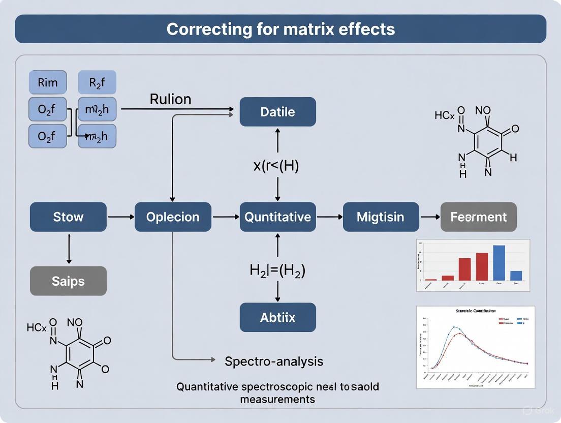
Strategies for Correcting Matrix Effects in Quantitative Spectroscopic Measurements: From Fundamentals to Advanced Applications in Biomedical Research
Matrix effects, the suppression or enhancement of analyte signal by co-eluting sample components, represent a critical challenge in quantitative spectroscopic analysis, particularly in LC-MS/MS and imaging techniques used in drug...
Strategies for Correcting Matrix Effects in Quantitative Spectroscopic Measurements: From Fundamentals to Advanced Applications in Biomedical Research
Abstract
Matrix effects, the suppression or enhancement of analyte signal by co-eluting sample components, represent a critical challenge in quantitative spectroscopic analysis, particularly in LC-MS/MS and imaging techniques used in drug development. This article provides a comprehensive guide for researchers and scientists, covering the fundamental origins of matrix effects, proven methodological corrections, practical troubleshooting, and rigorous validation protocols. By synthesizing current best practices and emerging strategies—including standard addition, stable isotope-labeled internal standards, and advanced chemometric modeling—this resource aims to equip professionals with the knowledge to develop robust, accurate, and reliable bioanalytical methods, ultimately enhancing the quality and translatability of preclinical and clinical data.
Understanding Matrix Effects: Fundamental Concepts and Impact on Data Integrity
FAQs: Understanding Matrix Effects
What is a matrix effect in spectroscopic and LC-MS analysis?
A matrix effect is the combined effect of all components of the sample other than the analyte on the measurement of the quantity. In mass spectrometry, this most frequently manifests as ion suppression or enhancement, where co-eluting compounds from the sample matrix interfere with the ionization efficiency of the target analyte [1] [2]. According to IUPAC, it is defined as the combined effect of all components of the sample other than the analyte on the measurement of the quantity [1] [3].
What are the practical consequences of matrix effects in quantitative analysis?
Matrix effects can lead to several analytical problems, including:
- Erroneous quantitative results [4]
- Reduced sensitivity and detection capability [1]
- Poor method repeatability and accuracy [1]
- False negative or false positive diagnostics [1]
- Nonlinearity of response (signal vs. concentration) [1]
Which compounds typically cause matrix effects?
Common matrix effect culprits include:
- Endogenous species: ionic species (salts), polar compounds (phenols, arylsulfonates), carbohydrates, amines, urea, lipids, and peptides [1]
- Phospholipids in plasma are particularly problematic [2]
- Exogenous compounds: anticoagulants, dosing vehicles, stabilizers, and co-medications [4]
- Reagents: mobile phase additives, ion-pairing agents, buffers, and organic acids [1]
How do ionization sources (ESI vs. APCI) differ in susceptibility to matrix effects?
Electrospray Ionization (ESI) is generally more susceptible to matrix effects compared to Atmospheric Pressure Chemical Ionization (APCI). Many authors have observed that matrix effects are lower in APCI, and signal enhancement in APCI has been observed, particularly with high percentages of organic modifier in the mobile phase [1]. Switching from ESI to APCI is a recognized strategy to mitigate matrix effects [4].
Troubleshooting Guides: Detection and Assessment
Guide 1: Post-Column Infusion for Qualitative Assessment
Purpose: To identify regions of ionization suppression or enhancement throughout the chromatographic run [5] [4].
Experimental Protocol:
- Setup: Connect a syringe pump to the system between the HPLC column outlet and the MS inlet.
- Infusion: Continuously introduce a dilute solution of the analyte of interest at a constant flow rate.
- Injection: Inject a blank sample extract (a processed sample without the analyte) onto the LC column.
- Monitoring: Observe the ion chromatogram for the infused analyte. A constant signal is expected. Any significant deviation (dip or peak) indicates a region where matrix components eluting from the column cause ion suppression or enhancement [5] [4].
Interpretation and Solution:
- A constant signal indicates no significant matrix effect.
- A dip in the signal indicates ion suppression from co-eluting matrix components.
- A peak in the signal indicates ion enhancement.
- Solution: Modify chromatographic conditions (e.g., gradient, column) to shift the analyte's retention time away from the suppression/enhancement regions [4].
Guide 2: Post-Extraction Spiking for Quantitative Assessment
Purpose: To quantitatively measure the Matrix Factor (MF) and assess the extent of ion suppression/enhancement [6] [4].
Experimental Protocol:
- Prepare Neat Solutions: Prepare analyte solutions in neat mobile phase at low and high concentrations.
- Prepare Post-Extraction Spiked Samples: Process a blank matrix through the entire sample preparation procedure. Spike the same concentrations of analyte into the final extracted blank matrix.
- Analysis: Analyze both the neat solutions and the post-extraction spiked samples using the LC-MS method.
- Calculation: Calculate the Matrix Factor (MF) using the formula: MF = Peak Area (Post-extraction spiked sample) / Peak Area (Neat solution) [4].
Interpretation and Solution:
- MF ≈ 1: No significant matrix effect.
- MF < 1: Signal suppression.
- MF > 1: Signal enhancement.
- Solution: An IS-normalized MF (MFanalyte / MFIS) close to 1 indicates the internal standard effectively compensates for the matrix effect. If not, optimize sample cleanup or chromatographic separation [4].
Experimental Data and Methodologies
| Method | Type of Information | Key Outcome | Advantages | Limitations |
|---|---|---|---|---|
| Post-Column Infusion [5] [4] | Qualitative | Identifies regions of suppression/enhancement in the chromatogram | Excellent for method development; reveals problematic retention times | Does not provide quantitative data; requires additional hardware [6] |
| Post-Extraction Spiking [6] [4] | Quantitative | Calculates Matrix Factor (MF) for signal suppression/enhancement | "Golden standard" for quantitative assessment; required by regulatory guidance [4] | Requires a blank matrix; tedious for multiple analyte concentrations [6] |
| Pre-Extraction Spiking [4] | Qualitative (Performance) | Evaluates accuracy and precision of QCs in different matrix lots | Confirms method robustness as per ICH M10 guidance [4] | Does not provide scale of matrix effect, only its impact on performance [4] |
Detailed Protocol: Standard Addition Method
The standard addition method is a powerful technique to compensate for matrix effects, especially for endogenous analytes or when a blank matrix is unavailable [6].
Procedure:
- Aliquot Preparation: Split the sample extract into several equal aliquots (e.g., 4-5).
- Spiking: Spike increasing known concentrations of the target analyte into each aliquot, except one (the unspiked sample).
- Analysis: Analyze all aliquots using the LC-MS method.
- Calibration Plot: Plot the measured peak area (or peak height) against the concentration of the added analyte.
- Calculation: Extrapolate the line backwards until it intersects the x-axis. The absolute value of the x-intercept represents the original concentration of the analyte in the sample [6].
Use Case: This method was successfully applied in the determination of biogenic amines in cheese, where it revealed significant signal enhancement that was corrected to obtain accurate results [1].
Research Reagent Solutions
Table 2: Essential Reagents and Materials for Mitigating Matrix Effects
| Reagent/Material | Function/Purpose | Application Example |
|---|---|---|
| Stable Isotope-Labeled Internal Standard (SIL-IS) [4] [7] | Compensates for matrix effects by experiencing the same ionization suppression/enhancement as the analyte. Ideal due to nearly identical chemical and chromatographic properties. | Quantification of drugs in plasma; IROA TruQuant workflow for metabolomics uses a 13C-labeled IS library to correct for ion suppression [7]. |
| Structural Analog Internal Standard | A co-eluting compound with similar chemical structure to the analyte can be used as an IS when SIL-IS is unavailable or too expensive. | Cimetidine was investigated as a co-eluting IS for creatinine quantification in urine as an alternative to creatinine-d3 [6]. |
| Phospholipid-Removal SPE Sorbents | Selectively removes phospholipids from biological samples (e.g., plasma), which are a major source of matrix effects in ESI [2]. | Sample clean-up prior to LC-MS analysis of pharmaceuticals in plasma to reduce ion suppression in the early to mid-part of the chromatogram. |
| High-Purity Mobile Phase Additives | Using volatile additives (e.g., formic acid, ammonium acetate/formate) at the lowest effective concentration minimizes source contamination and signal suppression [1]. | Mobile phase for HILIC separation of biogenic amines used ammonium formate and formic acid [1]. |
Signaling Pathways and Workflows
Diagram 1: Matrix effect origin and mitigation pathway. The diagram illustrates how matrix components lead to signal suppression or enhancement, resulting in erroneous quantification, and outlines the primary pathways for correction.
Diagram 2: Matrix effect troubleshooting workflow. This decision tree guides the analyst through the sequential steps of qualitatively assessing and then quantitatively measuring matrix effects to determine the appropriate mitigation strategy.
Electrospray Ionization Mass Spectrometry (ESI-MS) has become an indispensable technique in modern analytical laboratories, particularly for the analysis of biological macromolecules and pharmaceuticals. Its soft ionization capability allows for the transfer of intact ions from the solution phase to the gas phase with minimal fragmentation. However, the accuracy and precision of quantitative measurements using ESI-MS are fundamentally challenged by matrix effects—a phenomenon where the ionization efficiency of target analytes is altered by the presence of co-eluting substances. This technical guide addresses the key mechanisms underlying matrix effects, focusing on ion competition, physical-chemical interactions, and solvent parameters, while providing practical troubleshooting methodologies for researchers and drug development professionals working to correct for these effects in quantitative spectroscopic measurements.
Fundamental Mechanisms of Matrix Effects
Ion Competition: The Charge Competition Model
In electrospray ionization, the number of charges available for analyte ionization is finite, leading to direct competition between analyte molecules and co-eluting matrix components for these limited charges [8] [9]. This charge competition occurs throughout the ESI process and follows these principles:
- Limited Charge Availability: The electrospray process generates a fixed amount of excess charge (either positive or negative depending on mode), creating an inherent "ESI capacity" for ionizing molecules [9].
- Concentration-Dependent Suppression: At low analyte concentrations relative to matrix components, the MS response is typically linear. As concentrations increase toward the ESI capacity limit, response plateaus and suppression effects become pronounced [9].
- Compound-Specific Affinities: Different compounds exhibit varying affinities for available charges based on their physicochemical properties, particularly their surface activity and gas-phase basicity/acidity [10] [8].
Theoretical models predict that charge competition becomes significant when the total number of analyte and matrix molecules approaches the available charge in the electrospray, establishing a fundamental limitation on the linear dynamic range of ESI-MS measurements [9].
Physical-Chemical Interactions in the ESI Process
Matrix effects manifest through specific physical-chemical interactions during the electrospray process, which occurs through three sequential steps [11]:
- Dispersal of charged droplets: A fine spray of charged droplets is generated at the ESI tip under high voltage.
- Solvent evaporation: Droplets shrink through solvent evaporation, increasing surface charge density.
- Ion ejection: Gas-phase ions are released via either Coulomb fission or ion evaporation mechanisms.
Matrix components interfere with this process through multiple mechanisms:
- Droplet Formation Interference: Less-volatile compounds increase solution viscosity and surface tension, reducing the efficiency of charged droplet formation and subsequent ion release [6] [8].
- Gas-Phase Neutralization: Co-eluting compounds in the gas phase can neutralize analyte ions through proton transfer reactions or charge exchange [8].
- Surface Competition: Matrix components with high surface activity preferentially occupy droplet surfaces, preventing analytes from reaching optimal positions for ion emission [10].
These interference mechanisms are visualized in the following diagram of the ESI process and matrix effect points:
Solvent and Additive Effects on Ionization Efficiency
Solvent composition and additives significantly influence ESI response by altering solution properties and ionization dynamics [12]. The following table summarizes key solvent parameters and their demonstrated effects on glucose response as a model analyte:
Table 1: Solvent Parameter Effects on ESI-MS Response of Glucose [12]
| Solvent Parameter | Effect on Positive Ion Mode | Effect on Negative Ion Mode | Optimal Conditions for Glucose |
|---|---|---|---|
| Organic Modifier | Methanol: Higher signal intensityAcetonitrile: Severe ionization suppression | Acetonitrile: Higher signal intensity with specific additives | Positive mode: Methanol:WaterNegative mode: Acetonitrile:Water |
| Additive Type | Ammonium trifluoroacetate: Good response across wide pH range | Ammonium formate or lithium fluoride: Highest signal intensities | Additive choice is critical for sensitivity |
| Additive Concentration | Effective across wide concentration ranges | Varies with additive type | Must be optimized for each analyte |
| pH Effects | Effective across wide pH ranges with proper additives | Specific pH optima depending on additive | pH 5-9 effective with ammonium trifluoroacetate in positive mode |
The mechanisms behind these solvent effects include:
- Surface Tension Modulation: Organic solvents reduce surface tension, facilitating droplet formation and fission [10].
- Conductivity Enhancement: Additives like ammonium salts increase solution conductivity, promoting efficient droplet charging and fission [10].
- Adduct Formation: Cations (H+, Na+, NH4+) or anions (Cl-, formate) form gas-phase adducts with neutral analytes, enabling their ionization [13].
- Proton Transfer Efficiency: Solution pH affects the availability of protons for protonation reactions in positive ion mode [11].
Experimental Assessment of Matrix Effects
Post-Extraction Addition Method
This quantitative approach assesses matrix effects by comparing analyte response in neat solution versus spiked biological matrix [14] [8].
Protocol:
- Prepare blank matrix samples (plasma, urine, etc.) from at least six different sources
- Extract samples using your standard preparation procedure
- Spike known concentrations of target analytes into the post-extraction samples
- Prepare equivalent concentration standards in neat mobile phase
- Analyze all samples and calculate matrix effect (ME) using: ME (%) = (Peak Area post-extraction spike / Peak Area neat solution) × 100%
- Interpretation: ME < 100% indicates ion suppression; ME > 100% indicates ion enhancement
Acceptance Criteria: The precision of ME across different matrix sources should be ≤15% for validation acceptance [14].
Post-Column Infusion Method
This qualitative technique identifies regions of ionization suppression/enhancement throughout the chromatographic run [14].
Protocol:
- Configure LC-MS system with a tee-connector between HPLC column and MS source
- Infuse a constant flow of analyte solution post-column using a syringe pump
- Inject a blank matrix extract onto the chromatographic system
- Monitor the signal response of the infused analyte throughout the chromatographic run
- Identify retention time windows where signal suppression/enhancement occurs
- Modify chromatographic conditions to elute analytes away from suppression regions
This method provides a visual profile of matrix effects across the entire chromatogram but does not provide quantitative assessment [14].
Troubleshooting Guide: FAQs on Matrix Effects
Table 2: Frequently Asked Questions on ESI Matrix Effects
| Question | Root Cause | Solutions & Troubleshooting Steps |
|---|---|---|
| Why do I observe ion suppression in my method? | Co-eluting matrix components competing for available charge [8] | 1. Improve chromatographic separation2. Optimize sample cleanup3. Dilute and inject4. Switch to APCI ionization if possible |
| How can I improve my method's linear dynamic range? | Limited ESI charge capacity [9] | 1. Reduce flow rate to improve ionization efficiency2. Use nanospray at low nL/min flow rates3. Ensure sample concentrations are within linear range4. Use internal standard correction |
| Which biological matrices cause the most severe effects? | Phospholipids in plasma; salts and metabolites in urine [8] | 1. Use phospholipid removal plates for plasma2. Dilute urine samples when possible3. Employ extensive chromatographic separation for complex matrices |
| How does ionization mode affect matrix effects? | Different competition mechanisms in positive vs. negative mode [8] | 1. Negative mode generally less susceptible to suppression2. Choose mode based on analyte properties3. Test both modes during method development |
| Why do I get different matrix effects with different solvents? | Solvent properties affect droplet formation and evaporation [12] | 1. Optimize organic modifier type and percentage2. Use volatile additives like ammonium formate3. Avoid non-volatile buffers and salts |
Research Reagent Solutions for Managing Matrix Effects
Table 3: Essential Reagents for Matrix Effect Investigation and Compensation
| Reagent Category | Specific Examples | Function & Application | Considerations |
|---|---|---|---|
| Internal Standards | Stable isotope-labeled analogs (SIL-IS) [6] | Compensates for ionization suppression/enhancement through identical retention and ionization | Gold standard but expensive; may not be available for all analytes |
| Alternative Standards | Structural analogues or homologs [6] | Cost-effective alternative when SIL-IS unavailable | Must demonstrate similar matrix effects to analyte |
| Sample Cleanup | Phospholipid removal plates, SPE cartridges [14] | Remove specific matrix components causing interference | Can add time and cost to sample preparation |
| Mobile Phase Additives | Ammonium formate, ammonium acetate, formic acid [12] | Enhance ionization efficiency and chromatographic separation | Must be volatile to avoid source contamination |
| Matrix Effect Assessment | Blank matrix from multiple sources [14] | Evaluate variability and magnitude of matrix effects | Requires 6+ different matrix lots for proper validation |
Methodologies for Compensation and Correction
Standard Addition Method
The standard addition method effectively compensates for matrix effects without requiring blank matrix [6].
Protocol:
- Divide the unknown sample into multiple aliquots
- Spike increasing known concentrations of analyte standards into each aliquot
- Analyze all samples and plot peak area versus spiked concentration
- Extrapolate the line to the x-axis to determine original concentration
- The slope of the standard addition curve reflects the matrix effect magnitude
This approach is particularly valuable for endogenous compounds where blank matrix is unavailable, though it increases analytical time and sample consumption [6].
Chromatographic Method Optimization
Effective chromatographic separation represents the most direct approach to minimizing matrix effects by physically separating analytes from interfering components [14].
Optimization Strategies:
- Gradient Elution: Develop steep gradients to elute analytes in regions free of matrix interference
- Column Selection: Use specialized columns (e.g., HILIC) to alter selectivity and separate analytes from interferences
- Retention Time Adjustment: Modify pH, temperature, or mobile phase composition to shift analyte retention away from suppression zones identified by post-column infusion
- Cycle Time Extension: Increase run time to allow late-eluting interferences to clear before next injection
Matrix effects stemming from ion competition, physical-chemical interactions, and solvent parameters represent significant challenges in quantitative ESI-MS analyses. Understanding these fundamental mechanisms enables researchers to develop robust analytical methods that deliver accurate and precise results. Through systematic assessment using post-extraction addition or post-column infusion methods, followed by implementation of appropriate compensation strategies including stable isotope internal standards, chromatographic optimization, and sample preparation improvements, the impact of matrix effects can be effectively managed. This comprehensive approach ensures the generation of reliable quantitative data essential for drug development, clinical research, and spectroscopic measurement applications.
Frequently Asked Questions (FAQs)
Q1: Why are phospholipids a major source of matrix effect in LC-MS/MS bioanalysis? A1: Phospholipids are endogenous, surface-active compounds that can co-elute with analytes, causing ion suppression or enhancement by competing for charge and droplet space during the electrospray ionization process. Their elution profile is highly dependent on the chromatographic conditions.
Q2: How can high salt concentrations in biological samples impact my analysis? A2: High concentrations of salts (e.g., from PBS dosing vehicles or sample preparation) can:
- Cause severe ion suppression in the ESI source.
- Lead to salt adduct formation ([M+Na]+, [M+K]+), reducing the intensity of the target [M+H]+ ion.
- Accumulate on the LC column and MS source, requiring frequent maintenance and leading to signal instability.
Q3: What is the difference between an ISMF and a CVB, and when should I use each? A3:
- Ion Suppression Mass Spectrometry Factor (ISMF): A post-column infusion method used to identify regions of ion suppression/enhancement across a chromatographic run. It is ideal for method development to pinpoint problematic elution times.
- Calculated Variance of the Internal Standard (CVB): A quantitative measure that uses the internal standard's response to assess matrix effects in actual study samples. It is used for method validation and monitoring during sample analysis to ensure data quality.
Q4: My analyte is a metabolite of an endogenous compound. How can I accurately quantify it? A4: Quantifying metabolites against an endogenous background requires a surrogate matrix (e.g., stripped matrix, artificial cerebrospinal fluid) for calibration standards or a standard addition method to account for the inherent baseline level.
Q5: What are the key considerations when a dosing vehicle like PEG-400 is used in vivo? A5: PEG-400 and similar vehicles can be difficult to remove during sample preparation and can cause significant, variable matrix effects. It is critical to:
- Use a stable isotope-labeled internal standard (SIL-IS) to correct for these effects.
- Prepare calibration standards and QCs in the same matrix containing the expected concentration of the vehicle to mimic study samples.
- Ensure the chromatographic method separates the analyte from the vehicle's elution front.
Troubleshooting Guide
Problem: High and variable ion suppression in plasma samples.
- Symptoms: Low and erratic analyte response, poor precision, failure of CVB criteria.
- Potential Causes & Solutions:
- Cause 1: Inefficient removal of phospholipids during sample preparation.
- Solution: Switch from protein precipitation (PPT) to a more selective technique like solid-phase extraction (SPE) with a mixed-mode or phospholipid removal plate. See Protocol 1 below.
- Cause 2: Co-elution of the analyte with the phospholipid region.
- Solution: Optimize the chromatographic gradient to shift the analyte's retention time away from the typical phospholipid elution window (often between 1-3 minutes in reversed-phase LC).
- Cause 3: High salt content from the sample or dosing vehicle.
- Solution: Implement a more thorough wash step in the sample preparation (e.g., in SPE). Dilute the sample if possible, and ensure the LC gradient includes a strong wash to elute salts.
- Cause 1: Inefficient removal of phospholipids during sample preparation.
Problem: Inaccurate quantification of a drug metabolite.
- Symptoms: Calibration curves are non-linear, QCs are inaccurate, results do not align with pharmacokinetic expectations.
- Potential Causes & Solutions:
- Cause 1: In-source fragmentation of a conjugate (e.g., glucuronide) back to the parent metabolite.
- Solution: Optimize MS source parameters (cone voltage, collision energy) to minimize in-source fragmentation. Use chromatographic separation to resolve the conjugate from the metabolite.
- Cause 2: The metabolite is unstable in the biological matrix.
- Solution: Identify optimal collection and storage conditions (e.g., immediate acidification, use of specific enzyme inhibitors, storage at -80°C).
- Cause 1: In-source fragmentation of a conjugate (e.g., glucuronide) back to the parent metabolite.
Problem: Poor reproducibility in samples from a study using a PEG-400 vehicle.
- Symptoms: High CV% in study samples, but QCs prepared in clean matrix are acceptable.
- Potential Causes & Solutions:
- Cause: Variable matrix effect from residual PEG-400.
- Solution: Ensure the calibration standards and QCs are prepared in a matrix containing a representative concentration of PEG-400. The use of a SIL-IS is mandatory. Consider diluting samples to reduce the vehicle's absolute concentration.
- Cause: Variable matrix effect from residual PEG-400.
Data Presentation
Table 1: Comparison of Sample Preparation Techniques for Mitigating Matrix Effects
| Technique | Phospholipid Removal Efficiency | Salt Removal Efficiency | Complexity | Typical Matrix Effect (ISMF, %) |
|---|---|---|---|---|
| Protein Precipitation (PPT) | Low (<20%) | Low | Low | -40% to +20% |
| Liquid-Liquid Extraction (LLE) | Medium-High (50-90%) | High | Medium | -15% to +10% |
| Solid-Phase Extraction (SPE) - C18 | Medium (60-80%) | High | Medium-High | -10% to +10% |
| SPE - Phospholipid Removal | Very High (>95%) | High | Medium | -5% to +5% |
Table 2: Common Dosing Vehicles and Their Potential Impact on LC-MS/MS Analysis
| Dosing Vehicle | Typical Use | Primary Matrix Effect Concern | Recommended Mitigation Strategy |
|---|---|---|---|
| Polyethylene Glycol (PEG-400) | Poorly soluble compounds | Severe ion suppression, viscous samples | SIL-IS, matrix-matched standards, dilution |
| Tween 80 | Emulsion formulations | Ion suppression, source contamination | LLE or SPE, intensive source cleaning |
| Dimethyl Sulfoxide (DMSO) | In vitro studies / stock solutions | Alters retention time, high background | Keep injection volume low (< 2 µL) |
| Saline / Phosphate Buffered Saline (PBS) | Soluble compounds | Ion suppression from salts | Dilution, SPE with wash steps |
Experimental Protocols
Protocol 1: Solid-Phase Extraction for Comprehensive Phospholipid Removal
- Conditioning: Load 1 mL of methanol to a dedicated phospholipid removal SPE plate (e.g., Ostro Pass-Through). Follow with 1 mL of water. Do not let the sorbent dry.
- Sample Loading: Acidify 100 µL of plasma sample with an equal volume of 1% formic acid in water. Vortex and load onto the conditioned plate.
- Washing: Apply a gentle vacuum. Wash the plate with 1 mL of 1% formic acid in water, followed by 1 mL of 5 mM ammonium acetate in methanol.
- Elution: Elute the analytes into a collection plate using 1 mL of a solution of methanol:acetonitrile (50:50, v/v) with 0.1% formic acid.
- Evaporation & Reconstitution: Evaporate the eluent to dryness under a gentle stream of nitrogen at 40°C. Reconstitute the dry residue in 100 µL of initial mobile phase for LC-MS/MS analysis.
Protocol 2: Post-Column Infusion for ISMF Assessment
- Setup: Connect a T-union between the LC column outlet and the MS source. A syringe pump is connected to the second port of the union.
- Preparation: Prepare a solution of the analyte of interest at a concentration that provides a stable, baseline signal when infused directly into the MS.
- Infusion: Start the syringe pump to infuse the analyte solution at a constant flow rate (e.g., 10 µL/min).
- Chromatography: Inject a blank matrix extract (prepared using your sample prep method) onto the LC system and run the intended chromatographic method.
- Data Analysis: Monitor the analyte signal from the infusion. A dip in the baseline signal indicates ion suppression; a peak indicates ion enhancement. The resulting chromatogram is an "ion suppression map."
Visualization
Diagram 1: Matrix Effect Assessment Workflow
Diagram 2: Phospholipid Ion Suppression Mechanism
Diagram 3: SPE vs PPT Phospholipid Removal
The Scientist's Toolkit
Table 3: Essential Research Reagent Solutions for Mitigating Matrix Effects
| Reagent / Material | Primary Function | Application Note |
|---|---|---|
| Stable Isotope-Labeled Internal Standard (SIL-IS) | Corrects for variability in ionization efficiency and recovery during sample preparation. | The gold standard for quantitative LC-MS/MS. Should be added to the sample at the earliest possible step. |
| Phospholipid Removal SPE Plates | Selectively binds and retains phospholipids while allowing analytes to pass through or elute separately. | Critical for clean extracts from plasma/serum. Superior to traditional C18 or PPT for this specific purpose. |
| Stripped/Charcoal-Treated Serum | A surrogate matrix with depleted levels of endogenous phospholipids and metabolites. | Used for preparing calibration standards when analyzing compounds with high endogenous background. |
| Ammonium Formate / Acetate | Volatile salts used in mobile phases. They improve chromatographic separation and do not cause source contamination. | Preferred over non-volatile salts (e.g., phosphate) for LC-MS/MS. |
| Formic Acid | A volatile acid used to modify mobile phase pH and promote [M+H]+ ion formation in positive ESI mode. | Helps with peak shape and ionization efficiency. |
A Technical Support Center for Spectroscopic Measurements
Frequently Asked Questions (FAQs)
What is a matrix effect, and why is it a problem in my quantitative analysis? A matrix effect is the combined influence of all components of a sample other than the target analyte on the measurement of the quantity [15]. In practical terms, it means that the sample matrix (e.g., blood, urine, soil extract, or rock) can alter the signal from your analyte, leading to inaccurate results. The core problem is that it can cause bias, making your results either higher or lower than the true value [15]. This compromises the accuracy, precision, and sensitivity of your method, and is a major contributor to poor reproducibility between labs and experiments [8] [16] [17].
How can I quickly check if my method is suffering from matrix effects? A common and effective strategy is the post-column infusion test for techniques like LC-MS [6] [5]. In this setup, a constant flow of your analyte is infused into the LC eluent while a blank sample extract is injected. A variation in the baseline signal of the analyte indicates regions of ionization suppression or enhancement caused by co-eluting matrix components [5]. Alternatively, you can use a post-extraction spike test, where you compare the detector response for an analyte in a neat solution to its response in a blank matrix that has been spiked with the same amount of analyte after extraction [8] [6]. A difference in response indicates a matrix effect.
What is the single best way to correct for matrix effects? The most effective and widely recommended correction technique is the use of a stable isotope-labeled internal standard (SIL-IS) [18] [6]. Because the SIL-IS is chemically nearly identical to the analyte, it experiences the same matrix effects during sample preparation, chromatography, and ionization. By using the ratio of the analyte signal to the internal standard signal for quantification, the matrix effect is effectively canceled out [6] [5]. However, these standards can be expensive or unavailable for some analytes.
Are some detection techniques more prone to matrix effects than others? Yes, the susceptibility to matrix effects varies significantly by detection principle. In general, electrospray ionization (ESI) in mass spectrometry is highly susceptible to ion suppression [8] [18]. Atmospheric pressure chemical ionization (APCI) is generally less susceptible [8]. Techniques like fluorescence detection can suffer from fluorescence quenching, and UV/Vis absorbance can be affected by solvatochromism, where the solvent matrix alters the absorptivity [5]. Understanding the inherent vulnerabilities of your detection method is the first step in managing matrix effects.
Troubleshooting Guides
Problem: Inconsistent calibration and inaccurate quantification. Potential Cause: Multiplicative matrix effects that change the slope of your calibration curve [15]. This happens when matrix components alter the detector's fundamental response to the analyte.
Solutions:
- Use Internal Standardization: Implement a stable isotope-labeled internal standard (SIL-IS) [6] [5]. If that is not available, a co-eluting structural analogue can sometimes be used as a surrogate [6].
- Apply Standard Addition: Use the method of standard addition to your sample. This method accounts for the matrix because the calibration is performed directly in the sample itself, eliminating the need for a perfectly matrix-matched blank [6].
- Employ Matrix-Matched Calibration: Prepare your calibration standards in a matrix that is as similar as possible to your sample matrix [18] [15]. This can be challenging as it requires a blank matrix and may not be feasible for all sample types.
Problem: Loss of sensitivity and high detection limits. Potential Cause: Ion suppression in mass spectrometry or similar signal suppression in other techniques, often from co-eluting compounds competing for ionization or affecting droplet formation [8] [6].
Solutions:
- Improve Sample Cleanup: Optimize your sample preparation (e.g., solid-phase extraction, liquid-liquid extraction) to remove more of the interfering matrix components before analysis [8] [6].
- Enhance Chromatographic Separation: Modify your chromatographic method (e.g., mobile phase, gradient, column) to separate the analyte peak from the region where matrix interferences elute, as identified by a post-column infusion experiment [6] [5].
- Dilute the Sample: If the method sensitivity allows, simply diluting the sample can reduce the concentration of interfering matrix components below the threshold where they cause significant effects [6].
Problem: Poor reproducibility and high variability in quality control samples. Potential Cause: Variable matrix effects from sample to sample, making it difficult to obtain consistent recovery from matrix spike (MS) samples compared to laboratory control samples (LCS) [15].
Solutions:
- Benchmark Your Method's Matrix Effect: Calculate the Matrix Effect (ME) as a percentage:
ME (%) = (MS Recovery / LCS Recovery) x 100[15]. An ME of 100% indicates no effect, while values above or below indicate enhancement or suppression. Tracking this helps quantify the problem. - Implement a Robust Internal Standard: A good internal standard corrects for not only matrix effects but also for variability in sample preparation and injection volume, directly improving reproducibility [5].
- Control Sample-Related Variables: Ensure consistent sample handling to prevent issues like evaporation, freeze-thaw degradation, or mislabeling, which can all contribute to irreproducible results [5].
Experimental Protocols for Detecting Matrix Effects
Protocol 1: Post-Column Infusion for Qualitative Assessment
This method helps you visually identify regions of ionization suppression or enhancement in your chromatographic run [6] [5].
- Setup: Connect a syringe pump containing a solution of your analyte to a T-union between the HPLC column outlet and the MS ion source.
- Infusion: Start the LC flow and the syringe pump, infusing the analyte at a constant rate to produce a stable background signal.
- Injection: Inject a blank extract of your sample matrix (one that has undergone the full sample preparation procedure).
- Detection: Monitor the signal of the infused analyte. A dip in the signal indicates ion suppression caused by co-eluting matrix components. A peak would indicate ion enhancement.
- Application: Use this information to adjust your chromatographic method so that your analyte elutes in a "clean" region with minimal suppression/enhancement.
The workflow below illustrates this setup and the expected outcome.
Protocol 2: Post-Extraction Spike for Quantitative Assessment
This method provides a numerical value for the matrix effect by comparing signal responses [8] [6].
- Prepare Solutions:
- Solution A (Neat): Prepare the analyte at a known concentration in a pure, matrix-free solvent.
- Solution B (Spiked): Take a blank sample matrix extract and spike it with the same known concentration of analyte.
- Analyze: Analyze both solutions using your LC-MS method.
- Calculate: Compare the peak areas.
- Matrix Effect (ME%) = (Peak Area of Solution B / Peak Area of Solution A) × 100%
- An ME% of 85-115% is often considered acceptable. Values below 85% indicate suppression, and above 115% indicate enhancement.
Data Presentation: Comparative Susceptibility and Strategies
The table below summarizes the susceptibility of different analytical techniques to matrix effects and recommends primary mitigation strategies.
Table 1: Matrix Effects Across Analytical Techniques
| Analytical Technique | Primary Mechanism of Matrix Effect | Susceptibility | Recommended Correction/Mitigation Strategy |
|---|---|---|---|
| LC-ESI-MS/MS | Ion suppression/enhancement from co-eluting compounds competing for charge [8] [6] | High (ESI is particularly vulnerable) [8] [18] | Stable isotope-labeled internal standard (SIL-IS) [18] [6] |
| LC-APCI-MS/MS | Competition for charge in the gas phase [8] | Moderate (Generally less than ESI) [8] [18] | Internal standard, improved chromatography |
| GC-EI-MS | Ionization occurs in gas phase under vacuum [18] | Low | Matrix-matched calibration, internal standard |
| UV/Vis Spectrophotometry | Solvatochromism (matrix alters absorptivity) [5] | Variable | Standard addition method [6] |
| Fluorescence Detection | Fluorescence quenching [5] | Variable | Standard addition, improved sample cleanup |
| Energy Dispersive XRF | Absorption/enhancement effects between elements [19] | High | Fundamental parameter method, empirical coefficients [19] |
The Scientist's Toolkit: Key Research Reagent Solutions
Table 2: Essential Materials for Managing Matrix Effects
| Item | Function/Benefit | Key Consideration |
|---|---|---|
| Stable Isotope-Labeled Internal Standard (SIL-IS) | Gold standard for correction; chemically identical to analyte and co-elutes, perfectly compensating for matrix effects and preparation losses [18] [6]. | Can be expensive or unavailable for novel analytes [6]. |
| Structural Analogue Internal Standard | A co-eluting compound with similar structure and properties can serve as a cheaper, though less perfect, alternative to SIL-IS [6]. | Must be carefully selected to ensure it behaves similarly to the analyte. |
| High-Purity Solvents & Reagents | Minimizes the introduction of exogenous impurities that can contribute to baseline noise and matrix effects [17]. | Essential for maintaining low background and consistent analyte response. |
| Certified Reference Materials (CRMs) | Provides a matrix-matched standard with a known analyte concentration, crucial for validating method accuracy and calibration [19]. | Acts as a "ground truth" for your quantitative method. |
| Solid-Phase Extraction (SPE) Cartridges | Selectively retains the analyte or removes interfering matrix components during sample preparation, directly reducing matrix effects [8] [6]. | The sorbent chemistry must be optimized for your specific analyte and matrix. |
Decision Pathway for Matrix Effect Correction
The following diagram provides a logical workflow to guide your strategy for addressing matrix effects in your research.
Frequently Asked Questions
1. What is the fundamental difference between a matrix effect and an analyte effect? A matrix effect is caused by co-eluting endogenous substances from the sample matrix (such as salts, phospholipids, or metabolites) that interfere with the ionization of your target analyte [20] [1]. An analyte effect is caused by a co-eluting analyte (another compound in your sample, which could be a drug or metabolite) interfering with the ionization of your target analyte [20]. Both can lead to signal suppression or enhancement, compromising quantitative accuracy.
2. Why is electrospray ionization (ESI) particularly prone to these effects? ESI is more vulnerable than other ionization sources (like APCI) because the ionization occurs in the liquid phase. Co-eluting substances compete with the analyte for the limited available charge on the electrospray droplets' surface, leading to suppression or enhancement [20] [21] [1].
3. Beyond inaccurate quantification, what other unusual symptoms can matrix effects cause? In rare but documented cases, matrix effects can break fundamental chromatographic rules. One study showed that matrix components can significantly alter the retention time of analytes and even cause a single compound to yield two separate LC-peaks [21].
4. Can I completely eliminate matrix and analyte effects? It is often challenging to eliminate them entirely. The focus is typically on reducing their impact through better sample cleanup and chromatographic separation, and compensating for them during data processing, most effectively by using a stable isotope-labeled internal standard (SIL-IS) [6] [22].
Troubleshooting Guides
Guide 1: Diagnosing Signal Suppression/Enhancement
Symptom: Inconsistent quantification, poor reproducibility, or a sudden loss of sensitivity for an established method.
| Investigation Step | What to Look For | Potential Outcome |
|---|---|---|
| Post-column Infusion [5] | Regions of signal dip (suppression) or rise (enhancement) in the baseline. | Identifies retention time windows affected by matrix. |
| Post-extraction Spike [6] [23] | Difference in analyte response in neat solution vs. spiked pre-extracted matrix. | Quantifies the overall matrix effect (e.g., 40% signal suppression). |
| Compare Calibration Slopes [5] [22] | Different slopes for calibration curves in neat solvent vs. matrix. | Confirms a matrix-dependent change in detector response. |
Guide 2: Resolving Co-elution Issues
Symptom: Overlapping peaks or confirmed ion suppression in the retention time zone of your analyte.
| Solution Strategy | Specific Action | Key Consideration |
|---|---|---|
| Chromatographic Optimization | Adjust gradient, mobile phase pH, or column type to shift retention times [20] [24]. | Even a small shift can move the analyte away from a suppression zone [20]. |
| Sample Preparation Enhancement | Switch from PPT to SPE or LLE to remove more matrix interferents [20] [22]. | More selective cleanup reduces the concentration of interferents [22]. |
| Sample Dilution | Dilute the sample before injection [6] [25]. | Only feasible for assays with high sensitivity. |
Experimental Protocols for Detection and Quantification
Protocol 1: The Post-Column Infusion Method (Qualitative Assessment)
This method helps you visually map the regions of ion suppression/enhancement in your chromatographic run [5].
- Setup: Connect a syringe pump and a tee-union between your HPLC column outlet and the MS ion source.
- Infusion: Continuously infuse a solution of your analyte(s) at a constant rate through the syringe pump into the post-column eluent.
- Injection: Inject a blank, pre-extracted sample matrix (e.g., blank plasma extract) onto the LC column and start the chromatographic method.
- Detection: Monitor the MRM or signal for the infused analyte. A steady signal indicates no matrix effects. A dip in the signal indicates ion suppression; a rise indicates ion enhancement.
The diagram below illustrates this workflow and the expected output.
Protocol 2: The Pre-Spike & Post-Spike Method (Quantitative Assessment)
This method quantitatively determines both extraction recovery and the matrix effect [23].
Prepare Samples: For at least three concentration levels (Low, Mid, High), prepare the following sets in triplicate:
- Pre-Spiked Samples: Spike the analyte into the blank biological matrix before extraction. Then, perform the full sample preparation.
- Post-Spiked Samples: First, extract the blank biological matrix. Then, spike the analyte into the final extract after extraction.
- Neat Solutions: Prepare analyte standards in pure reconstitution solvent (no matrix).
Analyze and Calculate: Analyze all samples by LC-MS/MS and use the average peak areas to calculate:
| Parameter | Calculation Formula | Interpretation |
|---|---|---|
| % Matrix Effect (ME) | [1 - (Avg. Post-Spike Area / Avg. Neat Area)] × 100 |
>0: Suppression; <0: Enhancement |
| % Recovery (RE) | (Avg. Pre-Spike Area / Avg. Post-Spike Area) × 100 |
Efficiency of extraction |
| % Process Efficiency (PE) | (Avg. Pre-Spike Area / Avg. Neat Area) × 100 |
Overall method efficiency |
The following diagram outlines the experimental workflow for this protocol.
The Scientist's Toolkit: Essential Reagents & Materials
| Item | Function in Mitigating Effects | Example from Literature |
|---|---|---|
| Stable Isotope-Labeled Internal Standard (SIL-IS) | The gold standard for compensation. Co-elutes with the analyte and experiences nearly identical matrix/analyte effects, allowing for perfect correction [6] [22]. | Creatinine-d3 for creatinine analysis [6]. |
| Structural Analog Internal Standard | A less ideal but sometimes used alternative to SIL-IS. Must have very similar physicochemical properties and co-elute with the analyte to be effective [6]. | Cimetidine was investigated as an IS for creatinine [6]. |
| Selective Solid-Phase Extraction (SPE) | Removes interfering phospholipids and other endogenous compounds more effectively than protein precipitation, thereby reducing the source of matrix effects [20] [22]. | Used for clean-up of plasma samples for vitamin E analysis [22]. |
| Liquid-Liquid Extraction (LLE) | An alternative sample cleanup technique that can selectively transfer the analyte to a clean solvent, leaving many matrix interferents behind [22]. | Optimized for extraction of vitamin E from human plasma [22]. |
| Phospholipid Removal Cartridges | Specialized products designed to specifically remove phospholipids, a major class of matrix interferents in plasma and serum [20]. | -- |
Proven Correction Strategies: From Standard Addition to Advanced Internal Standardization
Stable Isotope-Labeled Internal Standards (SIL-IS) are a cornerstone of modern quantitative analysis, renowned for their ability to correct for matrix effects and experimental variability. This guide provides troubleshooting and best practices for leveraging SIL-IS to achieve reliable results in your research.
FAQs on SIL-IS Fundamentals and Best Practices
1. What is the primary advantage of using a SIL-IS over other internal standards?
The primary advantage is its nearly identical chemical and physical behavior to the target analyte. A SIL-IS is the analyte itself but with one or several atoms replaced by stable isotopes (e.g., ²H, ¹³C, ¹⁵N). This means it tracks the analyte perfectly through sample preparation, extraction, and chromatography, and most importantly, it experiences the same ion suppression or enhancement from co-eluting matrix components during mass spectrometric detection. This allows it to accurately correct for matrix effects, a common source of inaccuracy in LC-MS/MS [26].
2. When should a SIL-IS not be used?
While rare, there are specific scenarios where a SIL-IS might be problematic:
- Severe Deuterium Isotope Effect: For ²H-labeled standards, a significant retention time shift compared to the analyte can occur. This means the analyte and its SIL-IS may not co-elute perfectly, leading to them experiencing different matrix effects and compromising accurate correction [27].
- Instability of the Label: Deuterium labels positioned on exchangeable sites (e.g., on heteroatoms like oxygen or nitrogen, or alpha to a carbonyl group) can undergo hydrogen-deuterium exchange with protons from the solvent or matrix. This alters the mass of the SIL-IS and invalidates the quantification [28].
- Impurity in the Standard: If the SIL-IS contains even a small amount of the unlabeled analyte, it will lead to artificially high concentration readings for the target compound [27].
3. What are the key design considerations for an effective SIL-IS?
When selecting or designing a SIL-IS, several factors are critical for optimal performance [28] [26]:
- Stable Label Position: Isotope labels should be placed on non-exchangeable sites. Using ¹³C or ¹⁵N is often preferred over ²H as they are not susceptible to H/D exchange.
- Adequate Mass Difference: A mass difference of at least 3-5 Da from the analyte is recommended to prevent overlap of isotopic peaks in the mass spectrum and avoid cross-talk.
- High Isotopic Purity: The SIL-IS must be free from significant amounts of the unlabeled analyte to prevent interference.
- Label on a Key Fragment: For MS/MS methods, the label should be on the part of the molecule that produces the fragment ion used for quantification.
4. How can I compensate for matrix effects if a SIL-IS is not available?
If a specific SIL-IS is unavailable, researchers can employ several strategies, though they are generally less ideal:
- Structural Analogue Internal Standard: Use a compound with very similar chemical structure, hydrophobicity, and ionization properties [26].
- Sample Dilution: Diluting the sample can reduce the concentration of matrix components below the level that causes significant effects [29] [30].
- Improved Sample Clean-up: Techniques like solid-phase extraction (SPE) can remove more matrix interferences [27] [30].
- Matrix-Matched Calibration: Using calibration standards prepared in a matrix that is free of the analyte but matches the composition of the sample as closely as possible [3] [30].
Troubleshooting Guide: Common SIL-IS Issues and Solutions
| Problem | Potential Cause | Solution |
|---|---|---|
| Inaccurate Quantification | Analyte and SIL-IS do not co-elute due to deuterium isotope effect [27]. | Use a ¹³C/¹⁵N-labeled IS instead of a ²H-labeled one. |
| SIL-IS is unstable, undergoing H/D exchange [28]. | Ensure labels are on non-exchangeable positions; use ¹³C/¹⁵N labels. | |
| The SIL-IS is impure and contains unlabeled analyte [27]. | Source a new batch of SIL-IS with higher isotopic purity. | |
| High Variability in IS Response | Inconsistent addition of the IS volume across samples [26]. | Check pipette calibration and technique; use an automated liquid handler. |
| Partial clogging of the autosampler needle [26]. | Inspect and clean the autosampler needle and injector. | |
| Severe and variable matrix effects that the SIL-IS cannot fully compensate [27] [29]. | Improve chromatographic separation or sample clean-up to reduce matrix components. | |
| Signal Suppression in Calibration | The concentration of the SIL-IS is too high, causing it to suppress its own signal and that of the analyte [27]. | Lower the concentration of the spiked SIL-IS. |
| The selected SIL-IS does not perfectly match the analyte's ionization characteristics. | If possible, use a different SIL-IS or a structural analogue that co-elutes more precisely. |
Research Reagent Solutions
The table below lists key reagents and materials essential for working with SIL-IS.
| Reagent / Material | Function in SIL-IS Workflow |
|---|---|
| ¹³C, ¹⁵N-labeled Growth Media | Used for metabolic labeling of microorganisms to biologically generate SIL-IS for compounds like modified nucleosides in RNA [31]. |
| l-Methionine-methyl-D3 | A deuterated methyl group donor used in yeast cultures to generate methyl-labeled metabolites and biomolecules for SILIS production [31]. |
| Isotopically Labeled Building Blocks | Chemically synthesized compounds (e.g., urea-¹³C,¹⁵N₂) used in the de novo synthesis of SIL-IS, ensuring specific and stable label incorporation [28]. |
| Mixed Internal Standard Mix (ISMix) | A pre-prepared cocktail of multiple isotopically labeled compounds covering a range of polarities, used for non-target screening or multi-analyte methods when analyte-specific SIL-IS are not available [29]. |
Experimental Workflow for Bio-Generation of SIL-IS
For certain complex molecules, like Phase II drug metabolites, chemical synthesis of a SIL-IS can be difficult and costly. The following protocol outlines a method for the bio-generation of these standards using in vitro systems [32].
Bio-Generation of SIL-IS for Phase II Metabolites
Principle: This generic method synthesizes stable isotope-labeled glucuronide or glutathione conjugates using in vitro biotransformation systems (e.g., liver microsomes for glucuronidation) with either a stable isotope-labeled parent drug or a labeled conjugation co-factor (e.g., UDP-glucuronic acid) [32].
Materials:
- Stable isotope-labeled parent drug OR labeled co-factor (e.g., UDP-glucuronic acid)
- In vitro incubation system (e.g., liver microsomes, recombinant enzymes)
- Co-factors (NADPH, UDPGA, etc.)
- Incubation buffer (e.g., phosphate buffer)
- Liquid Chromatography-Mass Spectrometry (LC-MS/MS) system
- Solid-Phase Extraction (SPE) cartridges for purification
Procedure:
- Preparation: Obtain the stable isotope-labeled version of the parent drug or the necessary labeled co-factor.
- Incubation: Set up the in vitro biotransformation system containing the appropriate buffer, enzyme source (e.g., liver microsomes for glucuronidation), and co-factors. Initiate the reaction by adding the SIL precursor (parent drug or co-factor).
- Reaction Monitoring: Allow the incubation to proceed and monitor the formation of the desired SIL metabolite using LC-MS/MS.
- Purification: Once the reaction is complete, stop the incubation. Purify the generated SIL metabolite from the incubation mixture, typically using techniques like solid-phase extraction (SPE) or preparative LC.
- Characterization: Confirm the identity and assess the purity of the bio-generated SIL-IS using LC-MS/MS. Verify the accurate mass and ensure the absence of significant impurities.
- Application: The characterized SIL metabolite can now be used as an internal standard for absolute and relative quantitation of the unlabeled metabolite in plasma or other biological samples [32].
This approach can save significant time and cost compared to the de novo chemical synthesis of complex metabolite standards.
The Standard Addition Method is a fundamental technique in analytical chemistry designed to overcome the challenge of matrix effects, which occur when other components in a sample alter the instrument's response to the target analyte, leading to inaccurate concentration measurements [33]. This method is particularly crucial when analyzing complex samples such as biological fluids, environmental samples, pharmaceuticals, and food products, where the sample composition is unpredictable or highly variable [33] [5].
Unlike traditional calibration curves prepared in pure solvent, the standard addition method involves adding known quantities of the analyte directly to the sample itself [34]. This ensures that the standards and the unknown experience identical matrix effects, thereby compensating for signal suppression or enhancement and enabling a more accurate determination of the original analyte concentration [33] [35]. The core principle is that by measuring how the signal changes with each addition, one can extrapolate back to find the concentration of the analyte in the original, unspiked sample [33].
Detailed Protocols & Methodologies
Basic Successive Standard Addition Protocol
This is the most commonly employed protocol, suitable for a wide range of techniques including atomic spectroscopy and chromatography [33] [34].
Workflow: Basic Successive Standard Addition
Step-by-Step Procedure:
- Preparation of Test Solutions: Pipette equal volumes ((Vx)) of the sample with unknown concentration ((Cx)) into a series of volumetric flasks (e.g., 5 flasks) [33].
- Spiking: Add increasing, known volumes ((Vs)) of a standard solution with a known, high concentration ((Cs)) to each flask. For example, add 0, 1, 2, 3, and 4 mL of the standard. One flask should receive no standard, serving as the "blank" for the sample itself [33] [34].
- Dilution: Dilute all solutions to the same final volume with an appropriate solvent. This ensures that the total volume is constant across all measurements [34].
- Measurement: Analyze each solution using your instrument (e.g., ICP-MS, HPLC-MS, AAS) and record the analytical signal (S) for each (e.g., peak area, intensity) [33] [35].
- Data Plotting: Plot the measured signal (S) on the y-axis against the concentration of the added analyte ((C{added})) on the x-axis. (C{added}) is calculated as ((Vs \times Cs) / V_{total}) for each solution [33].
- Calculating the Unknown Concentration ((Cx)):
- Perform a linear regression on the data points to obtain an equation in the form (S = m \times C{added} + b).
- The x-intercept (where (S = 0)) corresponds to the concentration of the analyte in the sample. The absolute value of the x-intercept is (Cx) [33].
- The concentration in the original, undiluted sample can be calculated using the formula: [ Cx = -\frac{b}{m} \times \frac{V{total}}{Vx} ]
Adaptation for Single Particle ICP-MS (SP-ICP-MS)
For characterizing nanoparticles in complex matrices, a novel on-line standard addition approach has been developed [36].
Workflow: On-line Standard Addition for SP-ICP-MS
Key Adaptations:
- On-line Spiking: A T-piece connector is used to continuously merge the sample suspension with a stream of ionic or nanoparticle standards. The standard inlet is smaller to ensure minimal dilution of the sample [36].
- Mixed Histograms: This process generates a single data stream containing signals from both the sample nanoparticles and the added standards, which may overlap [36].
- Signal Deconvolution: Advanced data processing algorithms are required to separate the overlapping signal populations, allowing for accurate determination of nanoparticle size and concentration despite the matrix [36].
Researcher's Toolkit: Essential Reagents and Materials
Table 1: Key Research Reagents and Materials for Standard Addition Experiments
| Item | Function / Description | Example / Specification |
|---|---|---|
| Certified Reference Material (CRM) | High-purity standard with known analyte concentration ((C_s)) for spiking; ensures accuracy and traceability [35]. | NIST-traceable ICP/MS multi-element standard [35]. |
| Internal Standard | Compound added to all samples to correct for instrument drift and variability; different from standard addition but often used complementarily [5]. | Isotopically labelled analog of the analyte (e.g., 13C-toluene) [5]. |
| Matrix-Matched Blank | A sample free of the analyte but with the same background matrix; used to assess and quantify the matrix effect itself [37]. | Extract of organically grown strawberries for pesticide analysis [37]. |
| Sample Diluent/Solvent | High-purity solvent used to dilute samples and standards to a constant final volume without introducing interference [33]. | HPLC-grade water, acetonitrile, or mobile phase-compatible solvent [5]. |
Troubleshooting Common Issues (FAQs)
FAQ 1: My standard addition curve is not linear. What could be the cause?
Non-linearity can arise from several factors:
- Excessive Matrix Effects: The matrix may be so complex that it causes non-linear signal suppression or enhancement, especially at different analyte concentrations. This violates the method's fundamental assumption [5] [14].
- Presence of Translational Matrix Effects: These are interferences that affect the baseline or background signal (the y-intercept) without changing the slope of the curve proportionally. Standard addition cannot correct for these, and they can cause bias [34].
- Chemical Interactions: The added analyte may interact chemically with the matrix (e.g., forming complexes), changing its properties and thus the instrument response [38].
- Instrument Saturation: The detector may be operating outside its linear dynamic range at higher spike concentrations.
FAQ 2: Standard addition is time-consuming. When is it absolutely necessary?
Standard addition is essential in the following scenarios [33] [35] [34]:
- When the sample matrix is unknown or highly variable.
- When you lack a matrix-matched blank to create a traditional calibration curve.
- When method validation (e.g., via post-extraction spiking or post-column infusion) has confirmed a significant matrix effect (>15-20% signal suppression/enhancement) [37] [14].
- For one-off analyses where developing a fully validated, matrix-matched calibration method is not cost-effective.
FAQ 3: I am getting inconsistent results between replicates. How can I improve precision?
Poor precision often stems from procedural errors:
- Pipetting Inaccuracy: This is a major source of error. Use calibrated, high-quality pipettes and ensure proper technique, especially when handling small volumes [33].
- Insufficient Mixing: After spiking and dilution, ensure solutions are thoroughly mixed to achieve homogeneity.
- Sample Inhomogeneity: The original sample itself may not be uniform. Ensure the sample is well-homogenized before aliquoting [5].
- Instrument Instability: Allow the instrument to stabilize before analysis and use internal standards if available to correct for minor signal fluctuations [5].
FAQ 4: How do I calculate the error or uncertainty in the determined concentration?
The standard deviation of the unknown concentration ((sx)) can be estimated from the linear regression data using the following formula, which accounts for the uncertainty in the slope ((m)), y-intercept ((b)), and the spread of the data points [34]: [ sx = \frac{sy}{|m|} \sqrt{\frac{1}{n} + \frac{\bar{y}^2}{m^2 \sum (xi - \bar{x})^2}} ] Where:
- (s_y) is the standard error of the regression.
- (m) is the absolute value of the slope.
- (n) is the number of standard addition solutions.
- (\bar{y}) is the mean of the measured signals.
- (x_i) are the individual added concentrations.
- (\bar{x}) is the mean of the added concentrations.
FAQ 5: Can standard addition correct for all types of matrix effects?
No. It is most effective for correcting proportional matrix effects that influence the slope of the calibration curve. It cannot correct for:
- Translational (Additive) Effects: A constant background signal (e.g., from an interfering species that co-elutes with the analyte) will bias the result [34].
- Spectral Interferences: If a matrix component produces a signal that directly overlaps with the analyte signal, standard addition will not eliminate this interference [34]. These interferences must be addressed chromatographically, spectrally, or through sample preparation prior to analysis.
In the context of research on correcting for matrix effects in quantitative spectroscopic measurements, achieving high chromatographic resolution is a fundamental prerequisite. Matrix effects, where co-eluted compounds interfere with analyte ionization, detrimentally affect accuracy, reproducibility, and sensitivity in techniques like LC-MS [6]. The primary manifestation of this problem is the co-elution of interferents with your target analytes. This co-elution can cause either suppression or enhancement of the analyte signal, leading to unreliable quantitative data [6]. This guide provides targeted troubleshooting and methodologies to optimize your separations, minimize co-elution, and thereby produce more robust and accurate quantitative results.
Understanding Interferences: Spectral Overlaps and Matrix Effects
In spectrochemical analysis, interferences that affect quantitative measurements are broadly classified into two types, each requiring a different correction strategy [39].
- Line Overlap: This occurs when spectral lines of two or more elements are too close to be resolved by the spectrometer. The interference is straightforward: it always adds to the measured signal, causing a parallel shift in the calibration curve. The correction always involves subtracting the contribution of the interfering element [39].
- Matrix Effects: These are more complex, causing a change in the slope of the calibration curve. The correction can be either positive or negative and involves a multiplicative factor. In LC-MS, matrix effects occur when compounds co-eluting with the analyte interfere with the ionization process in the MS detector [6] [39].
Table 1: Types of Interferences and Their Mathematical Corrections
| Interference Type | Effect on Calibration | Correction Equation | Example |
|---|---|---|---|
| Spectral Line Overlap [39] | Parallel shift; always increases signal | ( Ci = A0 + A1(Ii - hC_j) ) | Carbon line at C I 193.07 nm overlapped by Aluminum line at Al II 193.1 nm in OES [39]. |
| Matrix Effect [6] [39] | Slope change; can suppress or enhance signal | ( Ci = A0 + A1Ii (1 \pm kC_j) ) | Co-elution of less-volatile compounds in LC-MS reducing formation of protonated analyte ions [6]. |
The Optimization Toolkit: A Systematic Approach to Improve Resolution
Optimizing chromatographic resolution requires a systematic, step-by-step approach, changing only one parameter at a time to assess its effectiveness [40]. The following workflow outlines the key parameters to investigate.
Sample and Mobile Phase Preparation
The process begins before the sample is injected. Proper preparation is crucial.
- Sample Preparation: Implement filtration or specific extraction techniques to remove particulates and impurities from your sample. This prevents column clogging and reduces the introduction of potential interferents [40].
- Mobile Phase Composition: The aqueous/organic solvent ratio, buffer pH, and ionic strength profoundly impact analyte retention and selectivity [40]. For example, in Ion-Pairing RPLC of oligonucleotides, hexafluoromethylisopropanol provided superior chromatographic resolution compared to other agents [41]. Using acetonitrile as the organic modifier can provide better peak capacity and lower back pressure than methanol [41].
- Interference Chromatography: A novel approach involves adding "interfering agents" like citrate or EDTA to the sample and mobile phase. These agents modify molecular interactions with the chromatographic matrix, dramatically improving the separation of host cell proteins from viruses during purification [42].
Column and Hardware Configuration
- Column Selection: The stationary phase is critical. Consider columns with smaller particle sizes (e.g., sub-2 µm for UHPLC) and solid-core particles, which increase efficiency and resolution [40] [41]. The pore size should be appropriate for your analyte; for instance, 100 Å pores are optimal for small oligonucleotides [41]. Longer columns can improve resolution but increase backpressure and analysis time [40].
- Column Temperature: Higher temperatures allow faster flow rates and quicker analysis but may lower resolution and cause degradation. Lower temperatures generally improve resolution and retention but extend run times. Always operate within the specified limits of your column and sample [40].
- System Configuration: To mitigate thermal mismatches, incorporate an active column preheater, which can lead to narrower peaks and prevent peak splitting [41]. Ensure all connecting capillaries have the minimal possible internal diameter and length to reduce extra-column volume, which can broaden peaks [43].
Instrument Method Parameters
Fine-tuning the instrument method is key to finalizing the separation.
- Flow Rate: Lower flow rates generally decrease the retention factor, making peaks narrower and improving response. Higher flow rates can broaden peaks, reducing resolution, but will shorten the run time [40].
- Injection Volume: Overloading the column with too much sample (mass overload) causes peak fronting, decreases retention time, and negatively impacts resolution. A general rule is to inject 1-2% of the total column volume for sample concentrations of 1 µg/µL [40].
- Detector Settings:
- Wavelength: For UV-Vis detectors, select the optimal wavelength that gives the highest absorption for your analyte to minimize interference and maximize sensitivity [40].
- Data Acquisition Rate: Ensure you have a sufficient number of data points per peak—a minimum of 20, but ideally 30-40—for optimal peak integration and resolution [40].
Table 2: Key Research Reagent Solutions for Chromatographic Optimization
| Reagent/Material | Function in Optimization | Application Example |
|---|---|---|
| Solid-Core Particles [40] [41] | Increases chromatographic efficiency and resolution; allows high resolution at faster flow rates. | 1.7 µm core-shell particles provided maximum resolving power for small oligonucleotides (15-35 mers) [41]. |
| Hexafluoromethylisopropanol (HFIP) [41] | Ion-pairing agent that improves chromatographic resolution for certain analytes. | Provided superior chromatographic resolution for oligonucleotides in IP-RPLC-MS [41]. |
| Citrate Interference Agent [42] | Modifies molecular interactions with the chromatographic matrix to improve selectivity and impurity clearance. | Dramatically improved host cell protein removal during purification of Newcastle disease virus using anion exchange chromatography [42]. |
| High-Purity Silica (Type B) Columns [43] | Minimizes interaction of basic compounds with acidic silanol groups on the silica surface, reducing peak tailing. | Recommended for analyzing basic compounds to achieve symmetric peaks and better resolution [43]. |
Troubleshooting Guide: Resolving Common Peak Shape Issues
Even with a good method, issues can arise. Here is a quick-reference FAQ for common problems related to resolution and co-elution.
FAQ: How do I fix peak tailing?
- Cause: For basic compounds, this is often due to interaction with silanol groups on the stationary phase. Other causes include a blocked frit, active sites on the column, or a flow path with too much extra-column volume [44] [43].
- Solution: Use a high-purity silica (Type B) or a polar-embedded column. Add a competing base like triethylamine to the mobile phase. Check for column blockages and replace the column if necessary. Use short, narrow-bore capillary connections [43].
FAQ: What causes broad peaks and how can I resolve them?
- Causes: A detector cell with too large a volume, a long detector response time, high longitudinal dispersion, or an overly long retention time in isocratic mode [43]. Contamination or a column that is losing its packing integrity can also be the culprit [44] [43].
- Solutions: Ensure the flow cell volume is less than 1/10 of the smallest peak volume. Set the detector response time to be less than 1/4 of the narrowest peak's width at half-height. For long isocratic retention, switch to a gradient or a stronger mobile phase [43].
FAQ: Why do I see peak fronting and what can I do?
- Causes: The most common cause is column overload—you are injecting too much mass of the analyte. Other reasons include the sample being dissolved in a solvent stronger than the mobile phase, or a blocked frit [44] [43].
- Solutions: Reduce the injection volume or dilute your sample. Ensure the sample is dissolved in the starting mobile phase or a weaker solvent. Replace the column inlet frit or the entire column if the problem persists [43].
FAQ: My resolution is low and peaks are co-eluting. Where should I start?
- Start with the mobile phase: Prepare a fresh mobile phase and check its pH and composition. Consider adjusting the organic solvent ratio, buffer pH, or ionic strength to improve selectivity [40] [44].
- Check the column: Ensure the column is not contaminated or degraded. Replace the guard column if one is in use. Consider switching to a column with a different stationary phase chemistry, a smaller particle size, or a longer length [40] [44].
- Adjust the temperature: Lowering the column temperature can increase retention and improve resolution, though it extends run time [40].
Advanced Strategies: Correcting for Unavoidable Matrix Effects
Despite optimal chromatographic separation, some matrix effects may persist. For these scenarios, advanced calibration techniques are required to obtain accurate quantitative data.
- Standard Addition Method: This method involves spiking the sample with known concentrations of the analyte. It is particularly useful for compensating matrix effects for endogenous analytes where a blank matrix is not available [6]. While widely used in atomic spectroscopy, it is less documented but applicable to LC-MS.
- Stable Isotope-Labeled Internal Standards (SIL-IS): This is considered the "gold standard" for correcting matrix effects in LC-MS. The SIL-IS is virtually identical to the analyte in chemical behavior and retention time, allowing it to experience the same matrix-induced ionization effects, thus providing a reliable correction factor [6].
- Post-Extraction Addition & Dilution: The post-extraction spike method evaluates matrix effects by comparing the analyte signal in neat solvent to its signal in a blank matrix sample spiked post-extraction [6]. Simply diluting the sample can also reduce matrix effects, though this is only feasible for assays with high sensitivity [6] [25]. One study used a "dilute-and-shoot" approach to find an optimal enrichment factor for wastewater analysis [25].
Achieving high chromatographic resolution is not an isolated goal but a foundational element for reliable quantitative analysis, especially in research focused on correcting for matrix effects. By systematically optimizing your method from sample preparation to detector settings, you can minimize the co-elution of interferents that lead to inaccurate results. When chromatographic resolution reaches its practical limit, mathematical and calibration-based corrections provide a necessary safety net. Employing the strategies and troubleshooting guides outlined in this technical center will empower researchers to produce data of the highest quality and reliability.
In quantitative spectroscopic and mass spectrometric analyses, matrix effects are a paramount concern, detrimentally affecting the accuracy, reproducibility, and sensitivity of measurements [6]. These effects occur when compounds co-eluting with the analyte interfere with the ionization process, leading to ion suppression or enhancement [6]. Among the most common and challenging interferents are phospholipids, which are ubiquitous in biological samples such as blood plasma, serum, and tissues [45]. Their amphiphilic nature and tendency to elute over a wide range in reversed-phase chromatography can cause significant and variable matrix effects, particularly in Liquid Chromatography-Mass Spectrometry (LC-MS) [45]. This guide details advanced cleanup strategies to remove phospholipids and other interferents, thereby correcting for matrix effects and ensuring the reliability of quantitative data.
Detection and Assessment of Matrix Effects
Before implementing cleanup procedures, it is crucial to detect and assess the presence and severity of matrix effects. The following table summarizes the primary techniques used for this purpose.
Table 1: Methods for Detecting and Assessing Matrix Effects
| Method | Principle | Advantages | Limitations |
|---|---|---|---|
| Post-Extraction Spike [6] | Compares the signal response of an analyte spiked into a blank matrix extract to its response in neat mobile phase. | Quantitative assessment of matrix effect magnitude. | Requires a true blank matrix, which is unavailable for endogenous analytes [6]. |
| Post-Column Infusion [6] | A constant flow of analyte is infused into the LC eluent while a blank matrix extract is injected. Ionization suppression/enhancement is observed as a signal drift. | Qualitative; identifies regions of ionization interference in the chromatogram. | Time-consuming, requires additional hardware, not ideal for multi-analyte methods [6]. |
| Dilute-and-Shoot [25] | The sample is progressively diluted to determine if the matrix effect decreases. | Simple and effective for samples with sufficient analyte concentration. | Reduces sensitivity; may not eliminate matrix effects entirely [6]. |
The following workflow outlines a systematic approach for evaluating phospholipid-mediated matrix effects in your sample preparation process:
Advanced Cleanup Techniques for Phospholipid Removal
Several sample preparation techniques can be employed to remove phospholipids effectively. The choice of method depends on the sample type, required sensitivity, and available resources.
Table 2: Comparison of Advanced Cleanup Techniques for Phospholipid Removal
| Technique | Mechanism | Best For | Phospholipid Removal Efficiency |
|---|---|---|---|
| Liquid-Liquid Extraction (LLE) [45] | Partitioning of lipids into organic phase (e.g., chloroform) vs. proteins/polar interferents in aqueous phase. | High-throughput, robust class-level separation. | Good for major classes, but can be less selective. |
| Solid-Phase Extraction (SPE) [45] | Selective retention and elution of lipid classes based on polarity using tailored stationary phases (e.g., silica). | High selectivity, cleaner extracts, better reproducibility for LC-MS/MS. | Excellent; specific protocols can target phospholipids. |
| Dilution [6] | Simple reduction of matrix component concentration. | Samples with very high analyte concentration and sensitivity. | Low; reduces but does not eliminate effects. |
| SPE with Phospholipid Removal Plates | Specialized sorbents designed to selectively bind phospholipids while allowing analytes to pass through. | High-throughput bioanalysis where phospholipids are the primary interferent. | Excellent and specific. |
Detailed Protocol: Phospholipid Removal via Solid-Phase Extraction (SPE)
SPE often provides cleaner extracts and better reproducibility compared to LLE, making it advantageous for LC-MS/MS applications where low background noise is critical [45].
Materials and Reagents:
- SPE Cartridges: Silica-based or specialized phospholipid removal plates (e.g., 30 mg, 1 mL capacity).
- Solvents: HPLC-grade methanol, acetonitrile, chloroform, and water.
- Samples: Biological sample (e.g., plasma, serum) pre-processed (e.g., protein precipitation).
- Internal Standards: Deuterated phospholipid analogs or other suitable IS added at the beginning [45].
Procedure:
- Conditioning: Condition the SPE sorbent with 1 mL of methanol, followed by 1 mL of water or a weak solvent matching your sample matrix.
- Sample Loading: Load your prepared sample (e.g., protein-precipitated plasma supernatant) onto the conditioned cartridge. Allow it to pass through slowly under gentle vacuum or positive pressure.
- Washing: Wash the cartridge with 1-2 mL of a weak wash solvent (e.g., 5-10% methanol in water) to remove polar impurities and salts while retaining phospholipids and your analyte (if using reverse-phase SPE for analyte retention).
- Elution of Analyte: If using a method where the analyte is retained, elute your target analytes with an appropriate organic solvent (e.g., methanol or acetonitrile). Collect this eluate.
- Elution of Phospholipids (if necessary): In methods designed specifically to remove phospholipids, they may be retained on the sorbent while your analyte flows through. If they need to be cleaned off the cartridge, a strong solvent like chloroform or 2% formic acid in acetonitrile can be used.
- Evaporation and Reconstitution: Evaporate the collected analyte fraction to dryness under a gentle stream of nitrogen. Reconstitute the dried extract in an appropriate volume of the initial mobile phase for LC-MS analysis.
Detailed Protocol: Phospholipid Extraction via Liquid-Liquid Extraction (LLE)
Classical solvent systems like the Folch or Bligh-Dyer methods are effective for broad lipid extraction and can be adapted to isolate phospholipids [45].
Materials and Reagents:
- Chloroform, Methanol, Water: HPLC grade.
- Internal Standards: Add at the beginning of extraction [45].
- Centrifuge Tubes: Glass tubes with Teflon-lined caps.
Bligh-Dyer Method Procedure:
- Homogenize: To a sample (e.g., 1 volume of plasma), add 2 volumes of methanol and 1 volume of chloroform. Vortex vigorously to form a monophasic solution.
- Partition: Add 1 volume of chloroform and 1 volume of water. Vortex thoroughly. This will create a biphasic mixture: a lower organic phase (chloroform, containing lipids) and an upper aqueous phase (methanol/water, containing proteins and polar interferents).
- Centrifuge: Centrifuge at low speed (e.g., 1000 × g) for 10 minutes to achieve clear phase separation.
- Collection: Carefully collect the lower organic layer, which contains the extracted phospholipids and other non-polar lipids.
- Washing (Optional): To purify further, wash the collected organic phase with a fresh upper phase solution (prepared from chloroform:methanol:water in specific ratios) to remove any residual water-soluble contaminants.
- Evaporation: Evaporate the chloroform fraction under nitrogen and reconstitute in a suitable solvent for analysis.
The Scientist's Toolkit: Essential Research Reagent Solutions
Table 3: Key Reagents for Phospholipid Analysis and Matrix Effect Correction
| Reagent / Material | Function / Application | Key Considerations |
|---|---|---|
| Deuterated Internal Standards (e.g., DPPC-d₉) [46] [45] | Corrects for variability in extraction, ionization, and matrix effects; enables absolute quantification. | Should be added at the very beginning of sample preparation; must be absent in the native sample [45]. |
| Stable Isotope-Labeled Analytes (SIL-IS) [6] | Considered the gold standard for compensating matrix effects in LC-MS; co-elutes with the analyte. | Expensive and not always commercially available for all analytes [6]. |
| Chloroform & Methanol [45] | Primary solvents for LLE (Folch, Bligh-Dyer methods) to efficiently partition phospholipids into the organic phase. | High purity (HPLC-grade) is critical to prevent contamination. Chloroform requires careful handling. |
| Silica-based SPE Sorbents [45] | Stationary phase for selective retention of phospholipids based on headgroup polarity. | Enables class-level separation and provides cleaner extracts than LLE for complex matrices. |
| Formic Acid [6] | Common mobile phase additive in LC-MS to improve ionization efficiency and chromatographic peak shape. | Can suppress signal for some analytes; purity is critical to avoid metal ion contamination [47]. |
| Matrix-Free LDI Surfaces (e.g., NALDI) [46] | Eliminates the need for a chemical matrix, reducing background interference and signal suppression for small molecule analysis (e.g., lipids). | Useful for quantitative analysis of small molecules like phospholipids where traditional MALDI matrices cause interference [46]. |
FAQs and Troubleshooting Guide
Q1: My LC-MS analysis shows significant signal suppression in mid-retention times, which I suspect is from phospholipids. How can I confirm this? A: The post-column infusion method is an excellent qualitative tool for this [6]. Infuse a constant flow of your analyte into the MS while injecting a prepared blank matrix extract. A dip in the baseline at specific retention times indicates ionization suppression. Phospholipids often cause broad regions of suppression in the mid-to-late portion of reversed-phase chromatograms. Alternatively, monitoring specific precursor ion scans (e.g., m/z 184 for phosphatidylcholines) can directly trace phospholipid elution.
Q2: I cannot find a stable isotope-labeled internal standard for my analyte. What are my options for correcting matrix effects? A: Several viable alternatives exist:
- Co-eluting Structural Analogue: Use a structurally similar compound that elutes at the same time as your analyte as an internal standard [6]. While not perfect, it can effectively correct for ionization suppression if it experiences the same matrix effects.
- Standard Addition Method: This technique involves adding known amounts of the analyte to aliquots of your sample [6]. It does not require a blank matrix and is particularly useful for endogenous compounds. However, it is more sample- and time-intensive.
- Echo-Peak Technique: This method involves injecting a standard shortly after the sample, but it does not fully compensate for matrix effects as the peaks do not co-elute exactly [6].
Q3: Despite using SPE, I still get inconsistent results between sample replicates. What could be going wrong? A: Inconsistent replicates often point to technical issues during sample preparation:
- Particle Size and Homogeneity: Ensure your sample is homogeneous before loading onto the SPE cartridge. Grinding or thorough mixing may be necessary for solid tissues [48].
- Cuvette and Handling: If using spectrophotometry, always use the same cuvette for blank and sample, place it in the holder with the same orientation, and ensure it is clean and free of scratches [49].
- Contamination: Check for cross-contamination between samples during the SPE process. Ensure all containers and SPE manifolds are properly cleaned [48].
- Solvent Evaporation: Incomplete or inconsistent drying during the evaporation/reconstitution step can lead to significant variability.
Q4: How can I reduce matrix effects originating from the mobile phase or instrument itself? A: Matrix effects can also stem from the analytical system:
- Mobile Phase Purity: Use MS-grade solvents and high-purity additives to minimize chemical noise [47].
- System Cleanliness: Flush the LC system regularly, especially after analyzing dirty samples. For oligonucleotide analysis, flushing with 0.1% formic acid helps remove alkali metal ions that cause adducts [47]. This principle applies broadly.
- Sample Dilution: If sensitivity allows, simply diluting the sample can reduce the concentration of interferents and mitigate matrix effects [6] [25].
Matrix effects, characterized by ion suppression or enhancement, represent a significant challenge in quantitative Liquid Chromatography-Mass Spectrometry (LC-MS) analysis, detrimentally affecting method accuracy, reproducibility, and sensitivity [6]. These effects occur when compounds co-eluting with the analyte interfere with the ionization process in the mass spectrometer interface [6]. For researchers and scientists focused on correcting for matrix effects in quantitative measurements, selecting the appropriate ionization source is a critical methodological decision. This guide explores the strategic transition from the widely used Electrospray Ionization (ESI) to Atmospheric Pressure Chemical Ionization (APCI) as a means to mitigate matrix-related issues.
Electrospray Ionization (ESI) is a soft ionization technique that generates ions directly from a solution by creating a fine spray of charged droplets under a high electrical field. As the solvent evaporates, the charged droplets undergo Coulombic fission, eventually releasing analyte ions into the gas phase [50] [11]. This mechanism is particularly effective for polar, thermally labile, and high molecular weight compounds such as proteins and peptides, and often produces multiply charged ions [50] [51].
Atmospheric Pressure Chemical Ionization (APCI) also operates at atmospheric pressure but utilizes a different mechanism. The LC effluent is first nebulized into a fine mist and vaporized in a heated tube (typically 400–550°C). The resulting gas-phase molecules are then ionized through chemical reactions initiated by a corona discharge needle, which creates a plasma of solvent-derived reagent ions that subsequently protonate or deposit charge onto the analyte molecules [52] [53].
The table below summarizes the fundamental differences between these two ionization techniques:
Table 1: Fundamental Comparison of ESI and APCI Characteristics
| Characteristic | Electrospray Ionization (ESI) | Atmospheric Pressure Chemical Ionization (APCI) |
|---|---|---|
| Ionization Mechanism | Ion formation from charged liquid droplets [50] [11] | Gas-phase chemical ionization after thermal vaporization [52] [53] |
| Primary Ionization Site | In solution/liquid phase [52] | In the gas phase [52] |
| Typical Analyte Polarity | Polar to ionic compounds [53] | Polar to moderately non-polar compounds [53] |
| Molecular Weight Suitability | Best for large molecules (e.g., proteins, peptides) [50] [51] | Best for small to medium molecules (typically < 1500 Da) [53] |
| Predominant Ion Type | Often multiply charged ions [50] | Primarily singly charged ions [53] |
| Thermal Stability Requirement | Suitable for thermally labile compounds [50] | Requires some thermal stability [52] |
Quantitative Evidence: APCI for Reduced Matrix Effects
Direct comparative studies provide compelling evidence for the advantage of APCI in situations where matrix effects are a primary concern.
A study analyzing levonorgestrel in human plasma found that while ESI provided superior sensitivity (LLOQ of 0.25 ng/mL vs. 1 ng/mL for APCI), the "APCI source appeared to be slightly less liable to matrix effect than ESI source" [54].
A more comprehensive multi-residue analysis of biocides, UV-filters, and benzothiazoles in environmental samples like wastewater and activated sludge yielded clear results. The study concluded that "ion suppression using ESI was identified to be more severe compared to APCI for the majority of the investigated compounds," and that "APCI is generally less susceptible to matrix effects than ESI" [55]. The following table quantifies these findings for a selection of compounds:
Table 2: Comparison of Matrix Effects (Ion Suppression) in ESI vs. APCI for Selected Environmental Analytes [55]
| Analyte | Matrix Effect in Treated Wastewater (ESI) | Matrix Effect in Treated Wastewater (APCI) |
|---|---|---|
| Climbazole | -42% (Suppression) | -9% (Slight Suppression) |
| Triclosan | -32% (Suppression) | +3% (Minimal Effect) |
| Benzophenone-1 (BZP-1) | -25% (Suppression) | +1% (Minimal Effect) |
| Benzothiazole | -2% (Minimal Effect) | +4% (Minimal Enhancement) |
| Mecoprop | -11% (Suppression) | +5% (Minimal Enhancement) |
The data demonstrates that APCI consistently exhibited significantly less ion suppression across various compound classes. The fundamental reason for this improved performance is that in APCI, the analyte is already in the gas phase before ionization, making the process less susceptible to interference from non-volatile matrix components that can affect droplet formation and desolvation in ESI [52] [55].
Decision Workflow and Experimental Protocol for Source Comparison
When to Consider Switching from ESI to APCI
The following decision pathway can help researchers determine if investigating APCI is appropriate for their specific application.
Experimental Protocol: Direct Comparison of ESI and APCI
To empirically determine the best ionization source for your application, follow this standardized comparison protocol.
Objective: To evaluate and compare the extent of matrix effects for a target analyte using ESI and APCI sources.
Materials and Reagents:
- Mass Spectrometer: LC-MS/MS system equipped with interchangeable ESI and APCI probes.
- Mobile Phase: HPLC-grade solvents (e.g., methanol, acetonitrile, water) and volatile additives (e.g., formic acid, ammonium acetate).
- Analytical Standards: Pure reference standard of the target analyte and a stable isotope-labeled internal standard (SIL-IS), if available.
- Matrix: Blank matrix representative of the study samples (e.g., human plasma, urine, wastewater, tissue homogenate).
Procedure:
- Sample Preparation:
- Prepare a set of post-extraction spiked samples. Extract blank matrix with your chosen method (e.g., protein precipitation, solid-phase extraction, liquid-liquid extraction).
- Spike the extracted blank matrix with a known concentration of the analyte (e.g., at the QC medium level). These are your "post-extraction spike" samples.
- Prepare neat standard solutions in mobile phase at the same concentration.
LC-MS/MS Analysis:
- ESI Analysis: Configure the instrument with the ESI source. Use typical ESI-optimized parameters (e.g., lower flow rate ~0.2 mL/min, higher vaporizer temperature might not be applicable). Inject the post-extraction spike and the neat standard in triplicate.
- APCI Analysis: Reconfigure the instrument with the APCI source. Adjust method parameters for APCI: increase the flow rate (e.g., 0.5-1.0 mL/min) [54], set the vaporizer/nebulizer temperature appropriately (e.g., 350-500°C) [52] [56], and optimize the corona needle current. Re-inject the same set of samples in triplicate.
Data Analysis:
- Calculate the Matrix Effect (ME) for each source using the following formula [6]:
ME (%) = (Peak Area of Post-extraction Spike / Peak Area of Neat Standard) × 100% - An ME of 100% indicates no matrix effect. <100% indicates ion suppression, and >100% indicates ion enhancement.
- Compare the ME values obtained from the ESI and APCI analyses. A value closer to 100% and with lower variability indicates a source less susceptible to matrix effects.
- Calculate the Matrix Effect (ME) for each source using the following formula [6]:
The Scientist's Toolkit: Essential Reagents and Materials
Table 3: Key Research Reagent Solutions for Method Development
| Item | Function/Description | Example Use Case |
|---|---|---|
| Stable Isotope-Labeled Internal Standard (SIL-IS) | The gold standard for compensating for matrix effects; corrects for analyte recovery and ionization variability [6]. | Quantification of drugs in plasma (e.g., Creatinine-d3 for creatinine analysis) [6]. |
| Structural Analog Internal Standard | A co-eluting compound with similar structure and properties to the analyte; a more affordable alternative to SIL-IS for compensating matrix effects [6]. | Using cimetidine as an IS for creatinine in urine analysis when SIL-IS is unavailable [6]. |
| Solid Phase Extraction (SPE) Cartridges | For sample clean-up; removes interfering matrix components prior to LC-MS analysis, thereby reducing potential ion suppression [55]. | HLB cartridges for multi-residue extraction of biocides and UV-filters from wastewater [55]. |
| Volatile Mobile Phase Additives | Acids or buffers that are compatible with MS; facilitate ionization and improve chromatographic separation without causing source contamination or signal suppression. | Formic acid (0.01-0.1%) or ammonium formate buffer for positive ion mode analysis in both ESI and APCI [54]. |
| Liquid-Liquid Extraction (LLE) Solvents | Organic solvents used to selectively partition analytes away from the biological or environmental matrix. | Cyclohexane for the extraction of levonorgestrel from human plasma [54]. |
Frequently Asked Questions (FAQs)
Q1: Can APCI completely eliminate matrix effects in my analysis? A1: No, APCI cannot completely eliminate matrix effects, but it can significantly reduce them compared to ESI for many compounds, particularly those that are less polar [55]. Matrix effects are inherent to atmospheric pressure ionization techniques. The most reliable approach is to use APCI in combination with effective sample clean-up and a stable isotope-labeled internal standard for optimal compensation [6] [55].
Q2: My analyte is a large protein. Should I consider switching to APCI? A2: No. APCI is generally suitable for small to medium-sized molecules with molecular weights typically under 1500 Da [53]. ESI is the preferred technique for large biomolecules like proteins and peptides because it can generate multiply charged ions, effectively extending the mass range of the analyzer and is gentle enough to prevent decomposition [50] [51].
Q3: What are the key instrument parameters I need to optimize when switching to an APCI method? A3: The most critical parameters to optimize in APCI are:
- Vaporizer/Nebulizer Temperature: This must be high enough to rapidly and completely vaporize the LC effluent (typically 400-550°C) [52] [53].
- Corona Discharge Current/Voltage: This controls the plasma that generates the primary reagent ions [56].
- Source Geometry and Gas Flows: The position of the needle and column, as well as the make-up gas flow and its humidity, can significantly impact sensitivity and repeatability [56].
Q4: I am using a normal-phase HPLC method with non-polar solvents. Is APCI a viable option? A4: Yes, one of the advantages of APCI over ESI is its broader solvent compatibility, including the ability to handle a wider range of non-polar solvents [53]. This makes it an excellent choice for coupling normal-phase chromatography with mass spectrometry.
For scientists and drug development professionals seeking to correct for matrix effects in quantitative LC-MS analyses, Atmospheric Pressure Chemical Ionization offers a powerful alternative to the more commonly used Electrospray Ionization. Empirical evidence demonstrates that APCI is generally less susceptible to ion suppression, particularly for small to medium-sized, thermally stable molecules ranging from polar to moderately non-polar [55] [53].
The decision to switch from ESI to APCI should be guided by the physicochemical properties of the analyte and supported by empirical data from a post-extraction spike experiment. While APCI may sometimes come at a slight cost to ultimate sensitivity, the benefit of improved assay robustness and reproducibility in complex matrices often outweighs this drawback. Integrating APCI into the analytical toolkit provides a strategic pathway toward more reliable and accurate quantitative measurements.
Defining Matrix Effects and Their Impact on Quantitative Analysis
What is a matrix effect in quantitative spectroscopic and mass spectrometric analysis?
A matrix effect refers to the phenomenon where components in a sample other than the target analyte (the sample matrix) alter the analytical signal, leading to either suppression or enhancement of the measured response [57]. In mass spectrometry, this predominantly occurs when co-eluting matrix components interfere with the ionization efficiency of the analyte in the ion source [5] [37] [8]. For spectroscopic techniques like LIBS, matrix effects arise from differences in the sample's physical or chemical properties, such as thermal conductivity or absorption coefficient, which influence the laser-sample interaction and the resulting plasma emission [38].
Why are matrix effects a critical problem in research and drug development?
Matrix effects compromise the accuracy, precision, and sensitivity of quantitative measurements [8]. This can lead to:
- Inaccurate Concentration Estimates: The reported concentration of an analyte does not reflect its true value in the sample, potentially leading to incorrect scientific conclusions or decisions in drug development [57].
- Reduced Precision and Sensitivity: Signal variability and suppression lower the detection capability for your analyte [57].
- Risk of False Results: In severe cases, matrix interference can lead to both false positives and false negatives [57].
Accurately correcting for matrix effects is therefore not just a technical detail but a fundamental requirement for generating reliable data in biomonitoring, pharmacokinetic studies, and quality control [8].
Detection and Quantification of Matrix Effects
How can I quickly determine if my method is susceptible to matrix effects?
A common and effective approach is the post-column infusion assay [5]. In this setup, a constant solution of the analyte is infused into the LC eluent via a T-connector between the column outlet and the MS inlet. A blank sample extract is then injected and chromatographed. If the matrix contains components that cause ionization suppression or enhancement, the signal of the infused analyte will drop or rise at the specific retention times where those interferences elute, creating a "dip" or "peak" in the chromatogram [5]. This method provides a qualitative map of ionization suppression/enhancement regions.
What are the standard methods for quantifying the magnitude of matrix effects?
The most common quantitative method is the post-extraction spike method [37]. It involves comparing the analytical signal of an analyte spiked into a blank matrix sample after extraction with the signal of the same analyte in a pure, matrix-free solvent [37]. The matrix effect (ME) is calculated as follows:
ME (%) = (Signal of analyte in matrix / Signal of analyte in neat solution) × 100% [57]
A result of 100% indicates no matrix effect. Values below 100% signal ion suppression, and values above 100% signal ion enhancement [37] [57]. This assessment can be done at a single concentration or across a calibration range to show the effect is not concentration-dependent [57].
Table 1: Interpreting Matrix Effect Quantification
| ME Value | Interpretation | Impact on Quantitation |
|---|---|---|
| < 100% | Ion Suppression | Underestimation of analyte concentration |
| ≈ 100% | No Significant Effect | Accurate quantitation is possible |
| > 100% | Ion Enhancement | Overestimation of analyte concentration |
Troubleshooting Guide: Mitigating Matrix Effects
How can I use sample preparation to reduce matrix effects?
The goal of sample preparation is to remove interfering matrix components while efficiently recovering the analyte.
- Selective Extraction: Techniques like Solid-Phase Extraction (SPE) and liquid-liquid extraction (LLE) can selectively isolate the analyte from many interfering substances, such as salts, phospholipids, and carbohydrates [57].
- Protein Precipitation: While simple, protein precipitation often leaves behind many interfering compounds that can cause matrix effects and is generally less effective than SPE or LLE for this specific purpose [6].
What chromatographic strategies can help minimize co-elution of interferents?
Since matrix effects require the interfering compound and the analyte to co-elute, chromatographic separation is a key mitigation tool.
- Improve Chromatographic Resolution: Optimize the LC method (gradient, column temperature, mobile phase) to increase the separation between the analyte peak and peaks of matrix interferents [6]. This can shift the analyte's retention time away from regions of high suppression or enhancement identified by the post-column infusion experiment [5].
- Use Alternative Mobile Phase Additives: Some buffers or additives can suppress ionization. Experiment with different volatile additives (e.g., formic acid, ammonium acetate) to find conditions that minimize this effect [6].
When is sample dilution an effective and practical strategy?
Sample dilution is a straightforward and effective strategy if the sensitivity of your assay is high enough to accommodate it [6] [29]. By diluting the sample, you reduce the absolute concentration of both the analyte and the interfering matrix components, thereby diminishing the magnitude of the matrix effect [6]. This approach is particularly useful in non-targeted screening where other correction methods are difficult to apply [29].
The following workflow outlines a systematic approach to diagnosing and mitigating matrix effects:
Calibration Strategies to Correct for Matrix Effects
What is matrix-matched calibration and how is it performed?
Matrix-matched calibration involves preparing calibration standards in a blank matrix that is representative of the sample matrix [58]. This ensures that the calibration curve experiences the same matrix effects as the actual samples, thus correcting for them during quantitation. The CLSI recommends calibration curves composed of a blank and at least six to eight calibration standards, spaced logarithmically across the range of interest [58].
A robust serial dilution for a matrix-matched curve can be designed to minimize pipetting error propagation. Instead of one continuous dilution, multiple primary standards (e.g., Points A-E) are individually prepared, and subsequent points are created by diluting these primary standards [58].
What is the gold standard for correcting matrix effects in LC-MS?
The use of a stable isotope-labeled internal standard (SIL-IS) is considered the most effective correction method [6]. A SIL-IS is chemically identical to the analyte but differs in mass due to isotopic substitution (e.g., ²H, ¹³C, ¹⁵N). It is added to every sample at the same concentration.
- Because the SIL-IS co-elutes with the analyte and has nearly identical chemical properties, it experiences the same matrix effects and ionization behavior.
- Quantitation is then based on the ratio of the analyte signal to the internal standard signal, which corrects for variations caused by matrix suppression/enhancement and instrumental drift [5] [6].
Are there alternatives if a stable isotope-labeled standard is not available?
Yes, though they are generally less ideal.
- Structural Analogues: A compound with a similar structure that co-elutes with the analyte can be used as an internal standard, though its ability to perfectly mimic the analyte's matrix effect is limited [6].
- Standard Addition: The analyte is spiked at several increasing concentrations into the sample itself. The sample's original concentration is determined by extrapolating the calibration line back to the x-axis. This method is accurate but requires a separate calibration for each individual sample, making it labor-intensive [6].
Table 2: Comparison of Matrix Effect Correction Methods
| Method | Principle | Advantages | Limitations |
|---|---|---|---|
| Stable Isotope-Labeled IS | Uses deuterated/13C-labeled analog as internal standard | Gold standard; corrects for both ME and recovery | Expensive; not always commercially available [6] |
| Matrix-Matched Calibration | Calibrators prepared in blank sample matrix | Directly accounts for average matrix effect | Requires large volume of blank matrix; cannot account for individual sample variability [58] [6] |
| Standard Addition | Analyte is spiked at different levels into the sample itself | Highly accurate; no blank matrix needed | Extremely time-consuming for large batches; not practical for every sample [6] |
| Sample Dilution | Reduces concentration of interferents | Simple, effective if sensitivity allows | Limited by assay sensitivity; may not fully eliminate strong ME [6] [29] |
Advanced and Emerging Strategies
What novel strategies are being developed for complex samples like urban runoff?
For highly variable samples, a one-size-fits-all internal standard correction may fail. The Individual Sample-Matched Internal Standard (IS-MIS) strategy has been developed to address this. Instead of using a single, pooled sample to assign internal standards, the IS-MIS method analyzes each individual sample at multiple dilutions to directly observe and correct for sample-specific matrix effects. Although it requires more analysis time, it significantly improves accuracy and reliability in heterogeneous sample sets [29].
How are matrix effects being tackled in techniques like LIBS?
In Laser-Induced Breakdown Spectroscopy (LIBS), matrix effects are addressed by quantifying and modeling the laser-sample interaction itself. One innovative approach involves using a depth-of-focus imaging system to perform a high-precision 3D reconstruction of the laser ablation crater morphology. The calculated ablation volume, which reflects the energy coupling efficiency, is then integrated into a multivariate nonlinear calibration model. This method directly links the physical matrix effect to a measurable quantity (crater volume), significantly improving quantitative accuracy [38].
The Scientist's Toolkit: Essential Reagents and Materials
Table 3: Key Research Reagent Solutions for Matrix Effect Management
| Item | Function in Mitigating Matrix Effects |
|---|---|
| Stable Isotope-Labeled Analytes | Serves as the ideal internal standard to correct for ionization suppression/enhancement and variable sample recovery [6]. |
| Blank Matrix (e.g., plasma, urine) | Essential for preparing matrix-matched calibration standards and for use in post-extraction spike experiments to quantify matrix effects [58] [37]. |
| Solid-Phase Extraction (SPE) Cartridges | Used for selective sample clean-up to remove phospholipids, salts, and other interfering compounds that cause matrix effects [6] [57]. |
| LC Columns (e.g., C18, HILIC) | Different stationary phases provide the chromatographic resolution needed to separate analytes from co-eluting matrix interferents [6]. |
| Mobile Phase Additives (MS-grade) | High-purity acids (e.g., formic acid) and buffers (e.g., ammonium formate) are used to optimize separation and minimize background ionization suppression [6]. |
Troubleshooting and Optimization: A Practical Guide for Robust Method Development
FAQs: Core Concepts and Troubleshooting
1. What is the fundamental difference between Post-Column Infusion and Post-Extraction Spiking?
Post-column infusion and post-extraction spiking are both used to diagnose matrix effects, but they answer different questions. Post-column infusion provides a qualitative, real-time map of ionization suppression/enhancement across the entire chromatogram. In contrast, post-extraction spiking offers a quantitative measure of the matrix effect at the analyte's specific retention time [6] [8] [5].
The table below summarizes their key characteristics:
| Feature | Post-Column Infusion | Post-Extraction Spiking |
|---|---|---|
| Primary Purpose | Qualitative profiling of matrix effects | Quantitative assessment of matrix effects |
| Information Gained | Identifies regions of ion suppression/enhancement throughout the chromatographic run [59] [5]. | Calculates the percentage of signal suppression/enhancement for the analyte at its retention time [6] [8]. |
| Methodology | Continuous infusion of analyte into column eluent during blank matrix injection [59] [8]. | Comparison of analyte response in neat solution vs. analyte spiked into a blank matrix extract [6]. |
| Output | Matrix effect profile (chromatogram) [59]. | Percentage Matrix Effect (%ME) [6]. |
| Best Used For | Method development, evaluating sample cleanup, identifying unexpected ionizable interferences [59]. | Method validation, quantitatively justifying the use of an internal standard [6]. |
2. During method development, my Post-Column Infusion shows severe ion suppression. What are my first steps to resolve this?
Significant suppression observed via post-column infusion indicates co-eluting matrix components are interfering with ionization. Your initial mitigation strategies should focus on sample preparation and chromatography:
- Enhance Sample Cleanup: A common cause of suppression, especially in late-eluting regions in reversed-phase LC, is phospholipids from biological matrices. If you are using only protein precipitation, consider adding a selective cleanup step, such as phospholipid removal cartridges, which have been shown to effectively reduce suppression areas [59].
- Improve Chromatographic Separation: Optimize the gradient to shift the analyte's retention time away from the major suppression zones identified in the profile. Even a small shift in retention time can sometimes avoid a co-eluting interference [6] [5].
- Employ a Stable Isotope-Labeled Internal Standard (SIL-IS): This is the gold standard for compensating for residual matrix effects during quantification. A SIL-IS co-elutes with the analyte and experiences nearly identical suppression, correcting for it in the final quantitative result [6] [60].
3. When using Post-Extraction Spiking, when is a matrix effect considered significant?
A matrix effect is generally considered significant and analytically relevant if the calculated %ME falls outside the range of 85-115%, or if the relative standard deviation (RSD) of the %ME across different matrix lots exceeds 15% [8]. Consistent signal suppression (%ME < 85%) or enhancement (%ME > 115%) indicates that the matrix is substantially altering the analyte's ionization efficiency, which can compromise the accuracy and precision of your results if not properly corrected.
Experimental Protocols
Protocol 1: Performing Post-Column Infusion to Generate Matrix Effect Profiles
Principle: A solution of analytical standards is infused post-column while a blank matrix extract is injected. The resulting chromatogram shows deviations from a stable baseline, visually mapping regions of ion suppression (dips) or enhancement (peaks) [59] [5].
Materials:
- LC-MS/MS system with a post-column infusion tee.
- Syringe pump or auxiliary LC pump for infusion.
- Blank matrix (e.g., plasma, urine) extract.
- Post-column infusion standard solution.
Step-by-Step Procedure:
- Prepare Infusion Solution: Select a set of compounds that cover a broad polarity range and are relevant to your analytical scope. Isotopically labeled versions of your analytes are ideal. Prepare a mixed standard solution at a concentration that provides a clear, stable signal without saturating the detector. Example concentrations from a bioanalysis study were: 0.025 mg/L atenolol-d7, 0.125 mg/L caffeine-d3, and 0.25 mg/L diclofenac-13C6 [59].
- Setup Infusion Line: Connect the infusion pump to a post-column tee positioned between the HPLC column outlet and the MS ion source. Use a low-dead-volume connection.
- Establish Baseline: Infuse the standard solution at a constant flow rate (e.g., 10 µL/min) and inject a pure solvent (e.g., mobile phase). The resulting chromatogram should be a nearly flat line, representing the baseline response without matrix [59].
- Inject Blank Matrix: While continuing the infusion, inject the prepared blank matrix extract using your standard LC-MS method.
- Data Analysis: Extract the ion chromatograms for the infused standards. Overlay the chromatogram from the blank matrix injection over the solvent baseline. Any significant deviation (typically > 20%) indicates a matrix effect zone [59] [5].
Post-Column Infusion Workflow: The process involves establishing a baseline with solvent and then comparing it to the signal obtained during a blank matrix injection to identify suppression/enhancement regions.
Protocol 2: Quantifying Matrix Effect via Post-Extraction Spiking
Principle: The analyte is added to a blank matrix extract after the sample preparation is complete. The response of this post-extraction spiked sample is compared to the response of the same amount of analyte in a pure solvent. The difference quantifies the matrix effect at the analyte's retention time [6] [8].
Materials:
- Blank matrix from at least 6 different sources.
- Analyte stock solutions.
- Appropriate solvents and vials.
Step-by-Step Procedure:
- Prepare Sample Set:
- Set A (Neat Standards): Prepare at least 5 calibration standards of the analyte in a pure solvent (e.g., mobile phase).
- Set B (Post-Extraction Spiked): Take aliquots of a blank matrix extract from at least 6 different sources. After the sample preparation (e.g., extraction, evaporation, reconstitution) is fully complete, spike each one with the same 5 analyte concentration levels as in Set A.
- LC-MS Analysis: Analyze all samples (Set A and Set B) in the same batch.
- Calculation: For each concentration level, calculate the percentage Matrix Effect (%ME).
- Assessment: The precision of the %ME across the different matrix lots (expressed as RSD) should also be calculated. An RSD > 15% indicates high variability and a potential problem [8].
Post-Extraction Spiking Workflow: This quantitative method compares analyte response in solvent versus matrix to calculate the precise extent of matrix effect.
The Scientist's Toolkit: Key Research Reagent Solutions
The table below lists essential reagents and materials for implementing these diagnostic techniques.
| Reagent/Material | Function & Importance |
|---|---|
| Stable Isotope-Labeled Internal Standards (SIL-IS) | Ideal for post-column infusion as they are chemically identical to analytes but chromatographically distinguishable. Crucial for compensating for matrix effects during quantification [59] [60]. |
| Structural Analogues (as IS) | A more affordable alternative to SIL-IS for post-column infusion, provided they have similar physicochemical properties and ionization behavior to the target analytes [59] [6]. |
| Phospholipid Removal Cartridges | Specialized solid-phase extraction sorbents used during sample prep to remove phospholipids, a major source of ion suppression in biological matrices [59]. |
| Blank Matrix from ≥6 Sources | Essential for both techniques. Used to create post-extraction spikes and to account for biological variability in matrix effect assessment, ensuring method robustness [8]. |
| Post-Column Infusion Tee | A low-dead-volume "T" or "Y" connector that allows the infusion stream to mix with the column eluent before entering the mass spectrometer ion source [59] [5]. |
## Frequently Asked Questions (FAQs)
1. What is a Matrix Effect and why is it a problem in LC-MS/MS bioanalysis? In quantitative Liquid Chromatography with tandem Mass Spectrometry (LC-MS/MS) bioanalysis, the matrix effect refers to the suppression or enhancement of the analyte signal caused by co-eluting components present in the biological sample. These components can be endogenous (e.g., phospholipids, salts, metabolites) or exogenous (e.g., anticoagulants, dosing vehicles) [4] [8]. This effect leads to erroneous concentration measurements, compromising the accuracy, precision, and sensitivity of the bioanalytical method, which is critical for making reliable decisions in drug development [61] [8].
2. What is the difference between Matrix Factor (MF) and IS-Normalized MF? The Matrix Factor (MF) is a quantitative measure of the absolute matrix effect on the analyte. The IS-Normalized MF measures the matrix effect on the analyte relative to the internal standard (IS). Using a stable isotope-labeled (SIL) IS that co-elutes with the analyte is the best practice, as it experiences a nearly identical matrix effect, making the IS-normalized MF close to 1 and indicating effective compensation [4] [61].
- Matrix Factor (MF):
MF = Response (Post-spiked sample) / Response (Neat solution)MF < 1indicates signal suppression.MF > 1indicates signal enhancement.MF ≈ 1indicates no significant matrix effect [4].
- IS-Normalized MF:
IS-norm MF = MF (Analyte) / MF (IS)[4] [61].
3. My calibration standards and QCs are acceptable, but I'm concerned about incurred samples. What should I do? The matrix in incurred samples is more complex than in blank matrix used for standards and QCs, potentially containing metabolites and co-administered drugs [4]. It is highly recommended to monitor the Internal Standard responses during sample analysis. For samples with abnormal IS responses, repeat the analysis with a dilution. If the IS response normalizes and the analyte concentration from the diluted sample is within ±20% of the original value, the sample-specific matrix effect is considered to have no impact [4].
4. What are the acceptance criteria for matrix effect assessments during method validation? According to regulatory guidelines, the matrix effect should be evaluated by analyzing quality control (QC) samples at low and high concentrations prepared in at least six different lots of matrix [4]. The results must demonstrate that the matrix effect, if present, does not impact method performance. The acceptance criteria are a bias within ±15% and a coefficient of variation (CV) ≤15% for the QC results in each individual matrix source [4] [61].
5. What is the best practice for assessing matrix effect during method development? A combination of qualitative and quantitative assessments is most effective [4].
- Post-column infusion helps visually identify regions of ion suppression/enhancement across the chromatogram [4] [5].
- Post-extraction spiking is the "golden standard" for quantitatively determining the Matrix Factor (MF) and IS-normalized MF, allowing for a lot-to-lot variability assessment [4] [61].
## Troubleshooting Guides
### Guide 1: High Absolute Matrix Factor (MF)
Problem: The calculated absolute MF for your analyte is outside the ideal range of 0.75–1.25, indicating significant signal suppression or enhancement [4].
Investigation and Resolution Steps:
| Step | Action & Investigation | Potential Corrective Measures |
|---|---|---|
| 1 | Confirm the result. Repeat the post-extraction spiking experiment to rule out preparation error. | - |
| 2 | Review chromatography. Check if the analyte co-elutes with known interferences, like phospholipids. Use a post-column infusion experiment to identify regions of suppression/enhancement [4] [5]. | Modify the LC method (e.g., gradient, column) to improve separation and shift the analyte's retention time away from interferences [4] [5]. |
| 3 | Evaluate sample clean-up. The current extraction method (e.g., protein precipitation) may be insufficient. | Implement a more selective sample preparation technique, such as solid-phase extraction (SPE) or liquid-liquid extraction (LLE), to remove more matrix components [4] [5]. |
| 4 | Consider ionization source. Electrospray Ionization (ESI) is highly susceptible to matrix effects [8]. | If other mitigations fail, switch the ionization source to Atmospheric-Pressure Chemical Ionization (APCI), which is generally less prone to matrix effects [4] [8]. |
### Guide 2: High Variability in IS-Normalized MF
Problem: The IS-normalized MF is close to 1.0 on average, but shows high variability (%CV) across different matrix lots.
Investigation and Resolution Steps:
| Step | Action & Investigation | Potential Corrective Measures |
|---|---|---|
| 1 | Verify Internal Standard suitability. A stable isotope-labeled (SIL) IS is the best choice because it co-elutes with the analyte and tracks its behavior perfectly. An analogue IS might not co-elute precisely [4]. | Switch to a stable isotope-labeled (SIL) internal standard if an analogue IS is being used [4]. |
| 2 | Check IS retention time. Ensure the retention times of the analyte and IS are identical. Even a small shift can cause the IS to experience a different matrix environment. | Optimize the LC method to achieve perfect co-elution of the analyte and its SIL IS. |
| 3 | Re-assess sample preparation. Inconsistent extraction recovery for the analyte or IS across different matrix lots can contribute to variability. | Re-optimize the sample preparation protocol to ensure consistent and high recovery for both the analyte and IS. |
## Experimental Protocols
### Protocol 1: Quantitative Assessment of Matrix Effect using Post-Extraction Spiking
This protocol is based on the method established by Matuszewski et al. and is widely used for regulatory compliance [4] [61].
1. Objective: To quantitatively determine the absolute and IS-normalized Matrix Factors for an analyte in a bioanalytical method.
2. Materials and Reagents:
- Blank biological matrix from at least six different sources.
- Analyte stock solution at known concentration.
- Internal Standard (IS) stock solution.
- Appropriate solvents for preparing neat solutions.
- All materials for your sample preparation procedure.
3. Procedure:
1. Prepare post-extracted samples:
- Extract blank matrix from each of the six different lots using your standard sample preparation method.
- After extraction, spike the analyte and IS at known concentrations (e.g., at Low and High QC levels) into the extracted blank samples.
2. Prepare neat solutions:
- Prepare neat solutions of the analyte and IS in a suitable solvent (not matrix) at the same concentrations as in step 3.1.
3. Analyze samples:
- Analyze the post-extracted samples (from step 3.1) and the neat solutions (from step 3.2) using the LC-MS/MS method.
4. Data Analysis:
- For each matrix lot and each concentration level, record the peak responses (e.g., area) for the analyte and IS in both the post-extracted sample (A) and the neat solution (B).
- Calculate Absolute MF: MF_analyte = A_analyte / B_analylete
- Calculate IS-normalized MF: IS-norm MF = MF_analyte / MF_IS
4. Acceptance Criteria:
- The precision (CV%) of the IS-normalized MF across the six matrix lots should be ≤ 15% [4] [61].
- The absolute MF should ideally be between 0.75 and 1.25 and show no concentration dependency [4].
### Protocol 2: Qualitative Assessment of Matrix Effect using Post-Column Infusion
This method helps visually identify chromatographic regions affected by matrix effects [4] [5].
1. Objective: To identify regions of ion suppression or enhancement throughout an LC-MS/MS chromatogram.
2. Materials and Reagents:
- Blank biological matrix.
- Concentrated solution of the analyte for continuous infusion.
- Syringe pump.
3. Procedure: 1. Set up infusion: Connect a syringe pump containing a solution of your analyte to a T-connector between the HPLC column outlet and the MS inlet. Start a continuous infusion of the analyte at a constant rate. 2. Inject and run: Inject an extracted blank matrix sample and run the LC gradient as normal. The MS monitors the signal of the infused analyte. 3. Analyze data: Observe the resulting chromatogram. A steady signal indicates no matrix effect. A dip in the signal indicates ion suppression, while a peak indicates ion enhancement, at that specific retention time.
## Data Presentation
### Table 1: Comparison of Matrix Effect Assessment Methods
| Method | Type of Information | Key Output(s) | Best Used For |
|---|---|---|---|
| Post-Column Infusion [4] [5] | Qualitative / Visual | Chromatogram showing regions of ion suppression/enhancement. | Method development & troubleshooting. Quickly identifying problematic retention times. |
| Post-Extraction Spiking [4] [61] | Quantitative | Matrix Factor (MF), IS-normalized MF, and their precision (CV%). | Method validation & robust quantification. Providing numerical data for regulatory submissions. |
| Pre-extraction Spiking (QC Analysis) [4] | Qualitative (Performance-based) | Accuracy and precision of QCs in different matrix lots. | Confirming consistency of the matrix effect, once assessed. |
### Table 2: Example Dataset from a Comparative Study of MF Calculation Methods
This table summarizes data from a study comparing calculation methods, showing that the IS-normalized MF and IS-norm relative ME yield similar results. The CV(%) of IS-normalized MF was on average 0.5% higher, making it the more conservative approach [61].
| Analyte | Concentration Level | Instrument | IS Type | CV of IS-norm MF (%) | CV of IS-norm Relative ME (%) | Difference (CVMF - CVME) |
|---|---|---|---|---|---|---|
| Lapatinib | Low | MS/MS | SIL | 4.5 | 4.2 | +0.3 |
| Sunitinib | Low | MS/MS | SIL | 6.1 | 5.5 | +0.6 |
| Azithromycin | High | MS | Deuterated | 2.8 | 2.1 | +0.7 |
| Exemestane | Low | MS/MS | SIL | 8.9 | 9.9 | -1.0 |
| Genistein | High | MS | Analog | 3.3 | 2.9 | +0.4 |
| Mean Difference (across 27 datasets) | +0.5 |
## The Scientist's Toolkit: Key Research Reagent Solutions
| Reagent / Material | Function & Importance in MF Assessment |
|---|---|
| Stable Isotope-Labeled Internal Standard (SIL IS) | The gold standard for compensating matrix effects. Co-elutes with the analyte, experiences the same matrix effect, and ensures the IS-normalized MF is close to 1 [4] [61]. |
| Multiple Lots of Blank Biological Matrix | Essential for evaluating the variability of the matrix effect. Using at least six different lots is standard practice to ensure method robustness across a diverse population [4]. |
| Phospholipid Monitoring Solutions | Used to identify if phospholipids are a major source of matrix effect. Helps in troubleshooting and optimizing chromatographic separation [4]. |
| Post-Column Infusion Setup (Syringe Pump, T-connector) | Enables the qualitative post-column infusion experiment, which is a critical diagnostic tool during method development to visually pinpoint ion suppression/enhancement [4] [5]. |
In quantitative bioanalysis, particularly in support of preclinical and clinical drug development, the reliability of data generated from incurred samples (study samples containing the administered drug and its metabolites) is paramount. One of the most significant challenges in techniques like LC-MS/MS is the matrix effect, where co-eluting components from the biological matrix can suppress or enhance the ionization of the analyte, leading to erroneous results. Monitoring the response of the internal standard (IS) is a critical practice that serves as a key indicator for detecting such issues and ensuring the accuracy of reported concentrations from incurred samples. This guide provides troubleshooting advice and best practices for using this essential quality control parameter.
Internal Standard Response: Troubleshooting FAQs
1. Why is monitoring internal standard response so critical in incurred sample analysis?
The internal standard is a known compound added at a fixed concentration to all samples, including calibration standards, quality controls (QCs), and incurred study samples. Its primary role is to compensate for variability in the analytical process, such as inconsistencies in sample preparation, injection volume, or detector sensitivity [62] [63]. In the context of incurred samples, which can have a much more complex matrix than spiked QCs due to the presence of metabolites and co-administered drugs, the IS response acts as a canary in the coal mine [4]. An abnormal IS response in a study sample can be the first sign of a sample-specific issue, such as a matrix effect, that was not present in your validation samples.
2. What are the established acceptance criteria for internal standard responses?
While specific criteria can be method-dependent, a common rule of thumb in regulated bioanalysis is to expect IS responses in study samples to be within 50% to 150% of the average IS response observed in the calibration standards and QCs within the same batch [63] [64]. Samples with IS responses outside this pre-defined range are typically flagged as "IS outliers" and should be investigated. It is critical to note that the precision of the replicate readings for the internal standard is also vital; a relative standard deviation (RSD) greater than 3-5% should be investigated [65].
3. My internal standard response is consistently low across all samples. What could be the cause?
A systematic low IS response across an entire batch often points to an issue with the instrument or the IS solution itself.
- Instrument Sensitivity Drop: The mass spectrometer may be experiencing a temporary loss of sensitivity. This can often be diagnosed by reviewing the IS response over the course of the run; a gradual drift is a typical indicator [62].
- Preparation of IS Solution: The internal standard working solution may have been prepared incorrectly or may have degraded [63].
- Sample Introduction System: Check for clogs or issues with the LC system's autosampler, injector, or tubing.
4. Only a few of my incurred samples show abnormal internal standard responses. What does this indicate?
Isolated IS outliers among incurred samples are a strong indicator of subject-specific or sample-specific matrix effects [63] [4]. Unlike QCs, which are prepared in a controlled, pooled matrix, each incurred sample is unique. The abnormal IS response suggests that in those specific samples, co-eluting components are differently affecting the ionization of the IS. This is a major reason why monitoring IS response is mandated for incurred sample analysis.
5. How can I investigate a sample with an abnormal internal standard response?
The following workflow is recommended for investigating IS outliers [63]:
- Re-injection: Re-inject the sample and observe the IS response and the peak area ratio (analyte/IS). If the IS response becomes normal upon re-injection and the calculated concentration does not change significantly, the initial issue was likely due to an injection anomaly.
- Dilution Test: If the IS response remains abnormal, dilute the sample with control matrix (if sensitivity allows) and re-analyze. If the IS response normalizes after dilution, it confirms a matrix effect was present in the original, undiluted sample [4].
- Investigate Sample Integrity: For samples with consistently low or no IS response upon repeat analysis, investigate potential sample integrity issues. In one case study, low IS responses were traced back to incorrect sample pH during processing, which led to degradation [63].
6. I am using a stable-isotope labeled internal standard. Can I still experience matrix effect issues?
Yes. While a stable-isotope labeled (SIL) IS is considered the gold standard because it co-elutes with the analyte and perfectly mimics its chemical behavior, it is still possible for the IS to be affected by matrix effects. The critical point is that a good SIL-IS will be affected to the same degree as the analyte. Therefore, the peak area ratio (analyte/IS) remains accurate even in the presence of a matrix effect, as both are suppressed or enhanced equally [6] [4]. The problem arises if the IS does not perfectly track the analyte, which can happen with structural analog internal standards [63].
Experimental Protocols for Matrix Effect Assessment
A robust bioanalytical method proactively investigates matrix effects during development and validation. The following are standard protocols for this assessment.
Protocol 1: Post-Column Infusion for Qualitative Assessment
This method helps identify regions of ionization suppression or enhancement throughout the chromatographic run [6] [4].
- Principle: A constant solution of the analyte is infused post-column while a blank matrix extract is injected. The MS signal is monitored for disturbances.
- Procedure:
- Connect a syringe pump containing a neat solution of the analyte to a T-union between the HPLC column outlet and the MS ion source.
- Start a continuous infusion of the analyte at a constant flow rate.
- Inject a processed blank matrix sample (e.g., extracted plasma) onto the LC system.
- Monitor the ion chromatogram of the infused analyte. A stable signal indicates no matrix effect. A depression in the signal indicates ion suppression; an increase indicates ion enhancement.
- Application: This is a excellent troubleshooting tool to visualize which retention times are affected by matrix interferences, allowing you to modify the chromatography to shift the analyte away from these regions.
Protocol 2: Post-Extraction Spiking for Quantitative Assessment
This is the "golden standard" method for quantitatively measuring the Matrix Factor (MF) [6] [4].
- Principle: The signal response of an analyte spiked into a blank matrix extract is compared to its response in a neat solution.
- Procedure:
- Prepare a set of blank matrix samples from at least six different sources.
- Process these blanks through your sample preparation procedure.
- After extraction, spike the analyte at a low and a high concentration into the blank extracts (Sample Set A).
- Prepare equivalent concentrations of the analyte in neat solution (mobile phase or reconstitution solution) (Sample Set B).
- Analyze all samples and calculate the absolute Matrix Factor (MF) for each matrix lot and concentration: MF = Peak Response in Post-Extracted Spiked Sample (Set A) / Peak Response in Neat Solution (Set B) An MF of 1 indicates no matrix effect; <1 indicates suppression; >1 indicates enhancement.
- To assess the effectiveness of your IS, also spike the IS into both sets and calculate the IS-normalized MF: IS-normalized MF = MF (Analyte) / MF (IS) An IS-normalized MF close to 1.0 indicates the IS is effectively compensating for the matrix effect [4].
The table below summarizes how to interpret the Matrix Factor and the role of the internal standard.
Table 1: Interpretation of Matrix Factor (MF) Calculations
| Metric | Result | Interpretation | Action |
|---|---|---|---|
| Absolute MF | ~1.0 | No significant matrix effect. | Method is robust. |
| <0.75 or >1.25 | Significant signal suppression or enhancement. | Optimize sample cleanup or chromatography. Consider switching ionization modes (e.g., ESI to APCI) [4]. | |
| IS-Normalized MF | ~1.0 | Matrix effect is effectively compensated by the internal standard. | Method is suitable, even with an absolute matrix effect. |
| <0.85 or >1.15 | Internal standard is not adequately tracking the analyte through the matrix effect. | Re-evaluate IS choice (prefer SIL-IS). Method may not be reliable [4]. |
Research Reagent Solutions
The following table lists key reagents and materials essential for developing and applying robust bioanalytical methods with effective internal standardization.
Table 2: Essential Research Reagents for Internal Standard and Matrix Effect Studies
| Reagent / Material | Function & Importance |
|---|---|
| Stable Isotope-Labeled Internal Standard (SIL-IS) | The ideal IS (e.g., deuterated, ¹³C, ¹⁵N). It co-elutes with the analyte and mimics its physico-chemical and ionization properties, providing the best compensation for matrix effects [62] [63] [4]. |
| Structural Analog Internal Standard | An alternative when a SIL-IS is unavailable. It should be structurally and chemically similar to the analyte, but may not track the analyte perfectly in the presence of matrix effects, leading to inaccuracies [6] [63]. |
| Control (Blank) Matrix | Essential for preparing calibration standards and QCs. It should be from the same species and type (e.g., human K2EDTA plasma) as the incurred samples. Multiple lots (≥6) are needed to assess matrix variability [4] [64]. |
| Phospholipid Standards | Used to monitor and identify the source of matrix effects, as phospholipids are a major cause of ion suppression in ESI-LC-MS/MS [4]. |
| Ionization Buffer (e.g., Cs, Li salts) | Primarily for ICP-based techniques. An excess of an easily ionized element is added to all solutions to minimize the effect of variable matrices on analyte ionization [65]. |
Workflow Diagram: Internal Standard Response Monitoring
The following diagram illustrates the logical decision process for monitoring and investigating internal standard responses in incurred sample analysis.
FAQs: Understanding Sample Heterogeneity and Its Impacts
Q1: What is the fundamental difference between chemical and physical heterogeneity in spectroscopic analysis?
Sample heterogeneity refers to the spatial non-uniformity of a sample's composition or physical structure and is a fundamental obstacle in quantitative and qualitative spectroscopic analysis [66]. It manifests in two primary forms:
- Chemical Heterogeneity: This involves the uneven distribution of molecular or elemental species throughout a sample. It is common in pharmaceutical tablets, powdered foods, and composite polymers, and leads to composite spectra where the signal is a superposition of the spectra of its constituents [66].
- Physical Heterogeneity: This encompasses differences in a sample's physical properties, such as particle size, shape, surface roughness, and packing density. These factors introduce additive and multiplicative distortions in spectra by altering how light scatters and interacts with the material [66] [67].
Q2: Why is sample heterogeneity considered an "unsolved problem" in spectroscopy?
Despite decades of research, no universal solution exists because heterogeneity is complex, multidimensional, and sample-dependent [66]. Most correction techniques reduce the symptoms rather than eliminate the root cause. The inherent disconnect between the scale of a spectroscopic measurement and the spatial complexity of real-world materials means the problem remains a central focus of ongoing research in spectroscopy and chemometrics [66].
Q3: What are the practical consequences of ignoring matrix effects in quantitative LC-MS?
In Liquid Chromatography-Mass Spectrometry (LC-MS), matrix effects occur when compounds co-eluting with the analyte interfere with the ionization process, causing ionization suppression or enhancement [6] [68]. This detrimentally affects the method's accuracy, reproducibility, and sensitivity. It can severely impact key validation parameters such as precision, accuracy, linearity, and limits of quantification [68].
Q4: What is the principle behind using stable isotope-labeled internal standards to correct for matrix effects?
Stable isotope-labeled internal standards (SIL-IS) are considered the "gold standard" for compensating matrix effects in LC-MS [6] [68]. Because the labeled standard is chemically identical to the analyte but has a different mass, it co-elutes with the analyte and experiences nearly identical ionization suppression or enhancement. The ratio of the analyte response to the internal standard response remains constant, thereby correcting for the matrix effect [6].
Troubleshooting Guides
Guide 1: Diagnosing and Correcting for Physical Heterogeneity in Absorption Spectroscopy
Physical heterogeneity causes multiplicative light scattering, which can mask the chemical information in a spectrum [67]. The following workflow outlines a systematic approach to diagnose and correct for these effects.
Problem: Physical heterogeneity (e.g., varying particle size, packing density) is causing significant baseline shifts and light scattering effects, which degrade the performance of your quantitative calibration model [66] [67].
Step-by-Step Solution:
- Improve Sample Presentation: Before applying computational corrections, ensure your sample presentation is as consistent as possible. For powdered samples, this means controlling packing pressure and using consistent particle size where feasible [66].
- Apply Spectral Preprocessing: Use empirical techniques to correct the spectra. The most common methods are:
- Standard Normal Variate (SNV): This technique centers and scales each spectrum individually to remove multiplicative and additive effects. It is applied on a spectrum-by-spectrum basis, making it useful when a reference spectrum is not appropriate [66] [67].
- Multiplicative Scatter Correction (MSC): This method adjusts each spectrum using a linear regression against a reference spectrum (typically the mean of the dataset) to remove baseline offsets and multiplicative scatter. It is closely linked to the physics of light scattering [66] [67].
- Derivative Spectroscopy (e.g., Savitzky-Golay): Computing first or second derivatives of spectra can minimize broad baseline trends and constant offsets. A smoothing filter is often applied to prevent the amplification of high-frequency noise [66].
- Evaluate Model Performance: Rebuild your quantitative model (e.g., PLS regression) using the corrected spectra and evaluate its performance on a separate validation set. A significant reduction in prediction error (e.g., Root Mean Square Error of Prediction) indicates the correction was successful.
- Escalate to Advanced Methods: If preprocessing does not yield sufficient improvement, consider more advanced strategies such as hyperspectral imaging to spatially resolve heterogeneity or adaptive averaging, which involves collecting and averaging spectra from multiple points on the sample to better represent its global composition [66].
Guide 2: Mitigating Matrix Effects in Quantitative LC-MS Analysis
Matrix effects in LC-MS lead to ion suppression or enhancement, compromising quantitative accuracy [6] [68]. The following table and protocol help in systematically addressing this issue.
Summary of Strategies to Mitigate Matrix Effects in LC-MS
| Strategy Category | Specific Method | Key Principle | Best Use Case |
|---|---|---|---|
| Minimization | Sample Clean-up | Selectively remove interfering compounds during extraction [68]. | When a selective extraction method (e.g., SPE, MIP) is available. |
| Chromatographic Optimization | Improve separation to avoid co-elution of analyte and interferents [6] [68]. | First-line strategy during method development. | |
| Dilute-and-Shoot | Reduce the concentration of interferents [6]. | When method sensitivity is sufficiently high. | |
| Compensation | Stable Isotope-Labeled IS (SIL-IS) | Use a chemically identical, co-eluting internal standard [6] [68]. | The gold standard when standards are available and affordable. |
| Standard Addition | Add known amounts of analyte to the sample itself [6]. | Ideal for endogenous analytes or when a blank matrix is unavailable. | |
| Matrix-Matched Calibration | Prepare calibration standards in a blank matrix [68]. | When a consistent and representative blank matrix is available. |
Experimental Protocol: Using the Post-Extraction Spike Method to Quantify Matrix Effects
This method provides a quantitative assessment of matrix effects [68].
- Prepare Solutions:
- Solution A (Neat Standard): Prepare the analyte at a known concentration in a neat mobile phase or solvent.
- Solution B (Spiked Matrix): Take a blank matrix sample (e.g., plasma, urine) through the entire extraction and preparation process. After extraction, spike the same amount of analyte as in Solution A into this prepared blank matrix.
- LC-MS Analysis: Inject Solution A and Solution B into the LC-MS system under identical analytical conditions.
- Calculate Matrix Effect (ME): Compare the peak areas of the analyte from both solutions.
- Formula:
ME (%) = (Peak Area of Solution B / Peak Area of Solution A) × 100% - Interpretation: An ME of 100% indicates no matrix effect. Values <100% indicate ion suppression, and values >100% indicate ion enhancement. A significant deviation (e.g., <85% or >115%) warrants corrective action [68].
- Formula:
Essential Research Reagent Solutions
Key Materials for Correcting Heterogeneity and Matrix Effects
| Reagent / Material | Function in Analysis | Application Note |
|---|---|---|
| Stable Isotope-Labeled Internal Standards (SIL-IS) | Compensates for matrix effects by mirroring the analyte's behavior during ionization in LC-MS [6] [68]. | The most effective compensation method, but can be expensive and is not available for all analytes. |
| Structural Analogue Internal Standard | A co-eluting compound with similar chemical structure can serve as a more affordable alternative to SIL-IS for correcting matrix effects [6]. | Must be carefully selected to ensure it experiences the same matrix effects as the analyte. |
| High-Purity Solvents & Mobile Phase Additives | Minimizes background noise and source contamination, which can exacerbate matrix effects and baseline noise [6] [69]. | Use LC-MS grade solvents to reduce signal suppression from trace impurities. |
| Appropriate Cuvettes | Ensures accurate light transmission in spectroscopic measurements. Quartz is required for UV range measurements [49] [69]. | Using the wrong cuvette type (e.g., plastic for UV) will lead to inaccurate absorbance readings. |
Advanced Methodologies & Workflows
Workflow 1: Comprehensive Strategy for Heterogeneous Solid Sample Analysis
This integrated workflow combines sampling and modeling strategies to achieve accurate quantitative analysis of challenging solid mixtures, as explored in recent research [66] [70].
Key Steps:
- Representative Sampling: Collect spectra from multiple points on the sample to account for spatial variation. For highly heterogeneous materials, hyperspectral imaging (HSI) is a powerful tool as it captures both spatial and spectral information, allowing for the identification of pure component spectra (endmembers) and their distribution [66].
- Advanced Algorithmic Correction: Move beyond basic preprocessing. Research focuses on physical model-based techniques and smart modeling methodologies that explicitly estimate and correct for multiplicative light scattering parameters, often by solving optimization problems [67] [70].
- Model Building and Validation: Build a calibration model (e.g., Partial Least Squares - PLS) using the corrected spectral data. Its robustness must be rigorously tested on a separate set of samples that were not used in the model calibration process [66] [67].
Workflow 2: Systematic Approach to Managing LC-MS Matrix Effects
This decision tree guides the selection of the most efficient strategy to manage matrix effects based on the requirements of your LC-MS assay.
FAQs: Addressing Common Method Development Challenges
Q1: What is a systematic approach to developing and optimizing an LC-MS method to minimize matrix effects?
A systematic approach involves using Design of Experiments (DOE) to efficiently characterize multiple parameters simultaneously. Rather than changing one factor at a time, DOE allows you to understand interaction effects and build a robust design space for your method. The recommended workflow, known as COLMeD (Comprehensive Optimization of LC-MS Metabolomics Methods using Design of Experiments), involves iterative rounds of tuning LC and MS conditions guided by multivariate statistical analysis. This approach has been shown to significantly improve metabolite response and coverage without compromising the separation of critical analyte pairs [71].
Q2: How can I quantitatively evaluate and correct for matrix effects in my quantitative LC-MS analysis?
Matrix effects occur when co-eluting components alter the ionization efficiency of your analyte, leading to signal suppression or enhancement [72]. To evaluate them quantitatively, use the following approach [72]:
- A: Analyze a neat standard solution.
- B: Analyze a blank matrix extract spiked with the analyte after extraction.
- C: Analyze a blank matrix extract spiked with the analyte before extraction.
Calculate the Matrix Effect (ME), Recovery (RE), and Process Efficiency (PE) using these formulas:
- ME (%) = (B / A) × 100
- RE (%) = (C / B) × 100
- PE (%) = (C / A) × 100
A Matrix Factor (MF = B/A) of 1 indicates no effect, <1 indicates suppression, and >1 indicates enhancement [72].
Q3: What are the key LC and MS parameters I should focus on during method optimization?
For LC optimization, critically evaluate the column chemistry, mobile phase composition (including pH and buffer concentration), and gradient profile [73] [71]. For MS optimization, key parameters include the ionization mode (ESI, APCI, or APPI), source voltages, gas temperatures and flows, and for MS/MS methods, collision energies [73] [74]. Software tools like MassHunter Optimizer can automate the optimization of MRM transitions and ion source parameters in a structured workflow [74].
Q4: How do I validate that my optimized method is fit for its intended purpose?
Method validation should follow established guidelines (e.g., ICH Q2(R1)) and include experiments to estimate different types of analytical error [75]:
- Precision (Random Error): Perform a replication experiment (e.g., ≥20 determinations at multiple concentration levels).
- Accuracy (Systematic Error): Conduct a comparison of methods experiment (e.g., ≥40 patient samples analyzed by both new and established methods).
- Linearity/Reportable Range: Analyze a minimum of 5 specimens with known values in triplicate.
- Specificity: Perform interference and recovery experiments with common interferents like phospholipids [72].
The acceptability of the observed errors is judged by comparing them to pre-defined quality requirements or allowable total error (TEa) [75].
Troubleshooting Guides
Problem 1: Significant Ion Suppression in ESI
Potential Causes:
- Co-elution of phospholipids or other matrix components from biological samples [72].
- Inadequate chromatographic separation.
- Non-volatile compounds in the sample impacting droplet formation and desolvation in the ESI source [72].
Solutions:
- Improve Sample Clean-up: Utilize selective extraction techniques (e.g., phospholipid removal plates) to reduce matrix interferents [72].
- Enhance Chromatography: Optimize the gradient to separate analytes from known matrix components. Phospholipids often elute in specific regions of the chromatogram [72].
- Consider Alternative Ionization: If suppression persists, switch to APCI or APPI, which are generally less susceptible to certain matrix effects because ionization occurs in the gas phase, not from charged droplets [72].
- Use a Stable Isotope-Labeled Internal Standard (SIL-IS): This is the most effective way to correct for residual matrix effects, as the IS experiences the same suppression/enhancement as the analyte [72].
Problem 2: Poor Method Robustness and Reproducibility
Potential Causes:
- Inadequately characterized method parameters and their interactions.
- Uncontrolled critical factors (e.g., mobile phase pH, buffer concentration, temperature).
Solutions:
- Implement DOE: Use a screening design (e.g., D-optimal) to identify critical factors and a subsequent response surface methodology to find a robust operating window [76].
- Define a Method Design Space: Through DOE, establish a multidimensional combination of input variables (e.g., pH, gradient time, column temperature) proven to assure method quality. Operating within this space ensures robustness, and changes within it do not require re-validation [76].
Problem 3: Inconsistent Instrument Response
Potential Causes:
- Sub-optimal ion source parameters.
- Carryover from previous samples.
- Instrument performance drift.
Solutions:
- Systematic Source Optimization: Use an iterative infusion approach. For ESI, tune voltages (capillary, fragmentor), gas flows (nebulizer, drying), and temperatures to find a "maximum plateau" where small changes do not significantly impact response [73].
- Leverage Workflow Intelligence: Modern instruments offer automated features. Use intelligent reflex workflows for carryover detection (which automatically injects blanks) or for automatically re-injecting samples that are above the calibration range with a lower volume [74].
Experimental Protocols & Data Presentation
Purpose: To determine the extent of ion suppression/enhancement and extraction efficiency.
Procedure:
- Prepare three sets of samples:
- Set A (Neat Standard): Prepare your analytes in a neat solution (e.g., mobile phase) at a known concentration.
- Set B (Post-Extraction Spiked): Take several lots (at least 6) of blank matrix (e.g., plasma), process them through your entire sample preparation workflow. After extraction, spike them with the same concentration of analyte as Set A.
- Set C (Pre-Extraction Spiked): Spike the same lots of blank matrix with the analyte before the extraction process, then carry out the full sample preparation.
- Analyze all samples (A, B, C) using the developed LC-MS method.
- Calculate ME, RE, and PE for each lot of matrix using the formulas provided in FAQ A2.
Expected Outcomes: The matrix effect (ME) should be consistent across different lots of matrix. High variability indicates a "relative matrix effect" that can compromise the reliability of results [72].
Purpose: To efficiently optimize multiple LC and MS parameters for maximum response and coverage.
Procedure:
- Define Purpose and Range: Identify the goal (e.g., improve signal for polar metabolites) and the analytical range (concentrations, sample types).
- Risk Assessment & Factor Selection: Identify 3-8 critical factors via risk assessment (e.g., for HILIC-MS: buffer concentration, mobile phase pH, gradient time, column temperature, sheath gas temperature, fragmentor voltage) [71] [76].
- Design Experimental Matrix: Use statistical software to create a custom design (e.g., D-optimal) requiring typically ≤20 injections per round [71].
- Run Experiments & Analyze Data: Execute the experimental design and use multivariate statistical analysis (e.g., Partial Least Squares regression) to model the relationship between factors and responses (e.g., peak area, resolution) [71].
- Iterate and Confirm: Based on the model, run a new round of DoE in the predicted optimal region. Finally, perform confirmation experiments to validate the improved method performance [71] [76].
| Parameter | Influence on Analysis | Optimization Tip |
|---|---|---|
| Ionization Mode | Fundamental selectivity and sensitivity for different compound classes. | Use ESI for polar/ionizable compounds; APCI for less polar, low-MW compounds [73]. |
| Capillary Voltage | Influents droplet charging and ion formation. | Tune via infusion; set on a maximum plateau, not necessarily the absolute maximum [73]. |
| Collision Energy (CE) | Controls fragmentation in MS/MS for SRM transitions. | Optimize for each compound to retain 10-15% of the parent ion [73]. |
| Gas Temperatures & Flows | Affects desolvation and ion transmission. | Optimize drying and sheath gas parameters to achieve stable spray and efficient desolvation [74]. |
Table 2: Essential Reagents and Materials for LC-MS Method Development
| Item | Function | Example from Literature |
|---|---|---|
| Ammonium Formate/Acetate | Volatile buffer salts for mobile phases to maintain pH and assist ionization. | Used at 2-20 mM concentrations in HILIC and RPLC methods [73] [71]. |
| Optima-grade Acetonitrile/Methanol | High-purity LC-MS solvents for mobile phases and standard preparation to minimize background noise [71]. | Used in mobile phase preparation for polar metabolomics on HILIC columns [71]. |
| Stable Isotope-Labeled Internal Standards | Correct for variability in sample preparation, ionization, and matrix effects [72]. | Critical for reliable quantitation in bioanalysis to account for matrix effects [72]. |
| Phospholipid Removal Plates | Solid-phase extraction plates designed to selectively remove phospholipids, a major cause of matrix effects in bioanalysis [72]. | Recommended for sample clean-up from biological matrices like plasma [72]. |
Workflow Diagrams
Systematic Method Optimization Workflow
Matrix Effect Evaluation Protocol
Validation, Comparison, and Emerging Strategies for Regulatory Compliance
Frequently Asked Questions (FAQs)
1. What is matrix effect and why is it a critical parameter in LC-MS bioanalysis?
Matrix effect refers to the alteration of analyte ionization efficiency in the mass spectrometer due to co-eluting components from the biological sample. These components can originate from the biological matrix itself (e.g., phospholipids, proteins, salts) or from exogenous sources (e.g., anticoagulants, dosing vehicles, co-medications) [4]. This interference causes ion suppression or enhancement, leading to erroneous concentration measurements that can compromise data accuracy and precision in preclinical and clinical studies [4] [5]. It is considered one of the key parameters of an LC-MS bioanalytical method because if not properly assessed and mitigated, it can result in suboptimal method performance, including poor accuracy, precision, nonlinearity, and reduced sensitivity [4].
2. How does ICH M10 recommend assessing matrix effect during method validation?
ICH M10 stipulates that matrix effect should be evaluated by demonstrating the accuracy and precision of quality control (QC) samples prepared in at least six different individual sources/lots of blank matrix, as well as in potentially interfering matrices like hemolyzed or lipemic samples [4] [77]. For each individual matrix source, the results for low and high QC concentrations must demonstrate a bias within ±15% of the nominal concentration and a precision (CV) of ≤15% [4] [77]. This approach qualitatively demonstrates that any matrix effect present is consistent and does not impact the method's performance [4].
3. What are the best-practice experimental techniques for investigating matrix effect during method development?
While ICH M10 focuses on the validation outcome, best practices during method development involve several techniques, summarized in the table below.
Table 1: Techniques for Matrix Effect Assessment during Method Development
| Technique | Description | Primary Use | Key Outcome |
|---|---|---|---|
| Post-column Infusion [4] | A constant flow of analyte is infused into the MS post-column while a blank matrix extract is chromatographically separated. | Qualitative investigation & troubleshooting. | Identifies regions of ion suppression/enhancement throughout the chromatographic run. |
| Post-extraction Spiking [4] [77] | Compares the MS response of an analyte spiked into a processed blank matrix extract to its response in a neat solution. | Quantitative assessment. | Calculates the Matrix Factor (MF), quantifying the extent of suppression (MF<1) or enhancement (MF>1). |
| Pre-extraction Spiking [4] | Evaluates the accuracy and precision of QCs spiked into blank matrix before the sample preparation process. | Validation (as per ICH M10) & qualitative assessment of consistency. | Confirms that the entire method, from sample prep to analysis, is not adversely affected by the matrix. |
4. How is the Matrix Factor (MF) calculated and interpreted?
The Matrix Factor is calculated quantitatively via the post-extraction spiking method [4] [37]. It is the ratio of the analyte response in the presence of matrix to the analyte response in a neat solution: MF = Peak Area (Post-extraction spiked sample) / Peak Area (Neat solution) [4]. An MF of 1.0 indicates no matrix effect. An MF <1.0 indicates signal suppression, and an MF >1.0 indicates signal enhancement [4]. For a robust method, the absolute MF for the target analyte should ideally be between 0.75 and 1.25 and be non-concentration dependent [4].
5. What is the recommended approach for compensating for matrix effect?
The most effective strategy for compensating for matrix effect is the use of a suitable Internal Standard (IS) [4] [5]. A stable isotope-labeled (SIL) IS (e.g., ¹³C-, ¹⁵N-labeled) is considered the gold standard because it co-elutes with the analyte and experiences virtually identical matrix effects [4]. The effectiveness is demonstrated by the IS-normalized MF (MF analyte / MF IS), which should be close to 1.0, indicating perfect compensation [4]. Even with a good IS, efforts should be made during method development to reduce the absolute matrix effect through optimized sample cleanup and chromatographic separation [4].
6. What should I do if I encounter matrix effect in my incurred study samples?
Incurred samples can have more complex matrices than blank QC samples due to metabolites and co-administered drugs [4]. It is critical to monitor the IS response during sample analysis. For samples with abnormal IS responses, it is recommended to perform a repeat analysis with a higher dilution factor [4]. If the IS response normalizes in the diluted sample and the re-calculated analyte concentration is within ±20% of the original value, the sample-specific matrix effect is considered to have no impact [4]. For studies where matrix effect is anticipated (e.g., with certain dosing vehicles), pre-dilution of study samples is a recommended proactive measure [4].
Experimental Protocols for Matrix Effect Assessment
Protocol 1: Post-column Infusion for Qualitative Assessment
This method is ideal for initial method development to visually identify chromatographic regions affected by matrix effects [4].
Workflow: The following diagram illustrates the post-column infusion setup and the expected output for identifying matrix effects.
Materials:
- LC-MS/MS system with electrospray ionization (ESI) source.
- Syringe pump.
- T-union or mixing tee.
- Blank biological matrix extract (e.g., plasma from at least 6 different lots).
- Neat solution of the analyte at a constant concentration.
Procedure:
- Connect the syringe pump loaded with the analyte neat solution to the T-union.
- Connect the outlet of the LC column to the second inlet of the T-union.
- Connect the outlet of the T-union to the ion source of the mass spectrometer.
- Start a continuous infusion of the analyte solution at a low, constant flow rate (e.g., 10 µL/min).
- Inject the blank matrix extract onto the LC column and start the chromatographic method.
- Monitor the ion chromatogram of the analyte. A stable signal indicates no matrix effect. A depression (dip) in the signal indicates ion suppression, while a signal increase indicates ion enhancement at that retention time [4].
Protocol 2: Quantitative Matrix Factor Determination
This protocol provides a numerical value for the matrix effect as recommended by regulatory best practices [4] [77].
Workflow: The experiment involves preparing and analyzing three different sample sets to isolate and calculate the matrix effect, recovery, and process efficiency.
Table 2: Sample Sets for Quantitative Matrix Effect and Recovery Evaluation
| Sample Set | Description | Represents |
|---|---|---|
| Set 1 | Analyte and Internal Standard spiked into a neat solution (e.g., mobile phase). | Reference response without matrix [77]. |
| Set 2 | Analyte and Internal Standard spiked into a post-extraction blank matrix supernatant (after protein precipitation, extraction, etc.). | Response showing Matrix Effect [4] [77]. |
| Set 3 | Analyte and Internal Standard spiked into the original blank matrix and carried through the entire sample preparation process. | Response showing overall Process Efficiency [77]. |
Procedure:
- Prepare a minimum of six different lots of blank matrix.
- For each lot, prepare the three sample sets (Set 1, Set 2, Set 3) at least in duplicate at low and high QC concentrations.
- Process the samples as per the method. Note that Set 2 is created after the extraction step.
- Analyze all samples in a single batch.
- Calculate the following parameters for each matrix lot and concentration:
- Absolute Matrix Factor (MF) = Peak Area (Set 2) / Peak Area (Set 1)
- Recovery (RE) = Peak Area (Set 2) / Peak Area (Set 3)
- Process Efficiency (PE) = Peak Area (Set 3) / Peak Area (Set 1) [77]
- Calculate the IS-normalized MF by dividing the absolute MF of the analyte by the absolute MF of the IS. This value should be close to 1.0 [4].
The Scientist's Toolkit: Key Reagent Solutions
Table 3: Essential Materials for Matrix Effect Evaluation and Mitigation
| Reagent / Material | Function in Matrix Effect Evaluation |
|---|---|
| Individual Matrix Lots (≥6 lots) [4] [77] | To assess inter-individual variability and ensure method robustness across a population. |
| Stable Isotope-Labeled Internal Standard (SIL-IS) [4] [78] | The best practice for compensating for matrix effect. It tracks the analyte closely through sample prep and ionization. |
| Hemolyzed and Lipemic Matrix [4] [79] | To challenge the method with abnormal matrices that may be encountered in patient populations. |
| Alternative Ionization Source (e.g., APCI) [4] | A mitigation strategy. Atmospheric Pressure Chemical Ionization (APCI) is generally less susceptible to matrix effects than ESI. |
| Phospholipid Monitoring Solutions [4] | Used to identify if observed matrix effects are specifically caused by endogenous phospholipids. |
| Charcoal-Stripped Matrix [78] | A type of surrogate matrix sometimes used in quantifying endogenous compounds to create an "analyte-free" background. |
FAQs: Understanding and Addressing Matrix Effects
What are matrix effects and why are they a problem in quantitative analysis?
Matrix effects refer to the phenomenon where components in a sample (the "matrix"), other than the analyte of interest, alter the detector's response to that analyte. This leads to either signal suppression or enhancement, compromising the accuracy, precision, and sensitivity of quantitative measurements [5] [6]. The fundamental problem is that the matrix the analyte is detected in can significantly influence the analytical signal, making it difficult to determine the true analyte concentration. These effects are a major concern in techniques like Liquid Chromatography-Mass Spectrometry (LC-MS), where co-eluting compounds can interfere with the ionization process [6] [77].
What are the most common techniques to correct for matrix effects?
The most widely used techniques to correct for matrix effects are:
- Stable Isotope-Labeled Internal Standard (SIL-IS) Method: Adding a known amount of a stable isotope-labeled version of the analyte [6] [77].
- Standard Addition Method (SAM): Adding known quantities of the native analyte to the sample at multiple concentration levels [6] [80].
- Post-Column Infusion of Standards (PCIS): Continuously infusing a standard into the LC effluent post-column to monitor and correct for ionization variability [81] [82].
- Matrix-Matched Calibration: Preparing calibration standards in a matrix that closely resembles the sample matrix [6].
Troubleshooting Guides: Implementing Correction Techniques
Guide 1: Implementing the Stable Isotope-Labeled Internal Standard (SIL-IS) Method
The SIL-IS method is considered the "gold standard" for compensating matrix effects in LC-MS due to the nearly identical chemical behavior of the labeled standard and the native analyte [6].
Workflow Overview:
- Spike: A known, constant amount of the SIL-IS is added to every sample, calibration standard, and quality control (QC) sample.
- Process: Samples are processed through the entire analytical procedure.
- Analyze: The detector response is measured for both the analyte and the SIL-IS.
- Calculate: Quantitation is based on the ratio of the analyte signal to the SIL-IS signal, plotted against the ratio of their concentrations [5] [77].
Pros and Cons:
- High Effectiveness: Excellent compensation for both sample preparation losses and ionization matrix effects [5] [6].
- Improved Precision: Corrects for variability in injection volume and instrument response [5].
- Cost and Availability: Standards are expensive and may not be commercially available for all analytes [6] [80].
- Not Always Feasible: Requires a well-characterized and pure SIL-IS that does not occur naturally in samples.
Guide 2: Implementing the Standard Addition Method (SAM)
SAM is a powerful technique for complex matrices where a blank matrix is unavailable or the matrix effect is severe and variable [6] [80].
- Workflow Overview:
- Aliquot: Split the sample into several equal aliquots.
- Spike: Spike all but one aliquot with increasing, known concentrations of the native analyte.
- Analyze: Measure the detector response for all aliquots.
- Plot and Extrapolate: Plot the signal response against the added concentration. The absolute value of the x-intercept (where response=0) corresponds to the original analyte concentration in the sample.
- Pros and Cons:
- No Blank Matrix Needed: Ideal for endogenous analytes or rare matrices [6] [80].
- Accounts for Matrix Effects: Directly measures the analyte in its specific matrix environment.
- High Sample Consumption: Requires multiple analyses per sample.
- Labor-Intensive: Significantly increases sample preparation time and complexity [6].
Guide 3: Implementing Post-Column Infusion of Standards (PCIS)
PCIS is an emerging technique that uses a constant infusion of a reference standard to monitor and correct for ionization suppression/enhancement throughout the chromatographic run [81] [82].
- Workflow Overview:
- Infuse: A standard solution is infused at a constant rate into the mobile phase between the column outlet and the MS ion source.
- Analyze: The sample is injected and analyzed via the normal LC-MS workflow.
- Correct: The signal for each analyte is normalized to the simultaneously acquired signal of the post-column infused standard to correct for fluctuations in ionization efficiency [81].
- Pros and Cons:
- No SIL-IS Required: Can be a cost-effective alternative when SIL-IS are unavailable [81].
- Real-Time Correction: Corrects for temporal changes in ionization efficiency.
- Complex Setup: Requires modification of the LC-MS interface with additional hardware (pump, tee-connector) [81] [82].
- Standard Selection: The correction is most effective if the PCIS compound's ionization behavior closely matches that of the analytes [81].
The table below provides a direct comparison of the key correction techniques to aid in method selection.
Table 1: Comparison of Matrix Effect Correction Techniques
| Technique | Key Principle | Best For | Major Advantages | Major Limitations |
|---|---|---|---|---|
| Stable Isotope-Labeled Internal Standard (SIL-IS) [5] [6] [77] | Normalization of analyte response using a chemically identical, labeled standard. | High-precision bioanalysis (e.g., pharmacokinetics); methods requiring the highest accuracy. | - Excellent compensation for both preparation and ionization variability- High precision and accuracy | - High cost- Limited commercial availability |
| Standard Addition Method (SAM) [6] [80] | Quantification by extrapolation of signal after adding known amounts of analyte to the sample itself. | Complex, unique, or variable matrices where a blank is unavailable (e.g., tissue, urine). | - Accounts for sample-specific matrix effects- Does not require a blank matrix | - Very high sample consumption- Labor-intensive and low throughput |
| Post-Column Infusion of Standards (PCIS) [81] [82] | Real-time correction using a standard infused post-column to monitor ionization efficiency. | Situations where SIL-IS are not available or for multi-analyte methods where one PCIS can correct for multiple analytes. | - Does not require labeled analogs- Corrects for temporal ionization instability | - Requires hardware modification- Less specific correction than SIL-IS |
| Matrix-Matched Calibration [6] | Calibration standards prepared in a matrix that mimics the sample. | Analyses where a consistent and reproducible blank matrix can be sourced. | - Conceptually simple | - Difficult to find a true "blank" matrix- Cannot account for individual sample-to-sample variation |
Experimental Protocol: Systematic Assessment of Matrix Effects, Recovery, and Process Efficiency
This integrated protocol, based on the approach by Matuszewski et al. and detailed in contemporary studies, allows for a comprehensive evaluation of matrix effects, recovery, and process efficiency in a single experiment [77].
Aim: To systematically determine the absolute and relative matrix effect, extraction recovery, and overall process efficiency for an LC-MS/MS method.
Experimental Design: Three sets of samples are prepared using multiple lots of a blank matrix (e.g., different sources of plasma or cerebrospinal fluid).
- Set 1 (Neat Solution): Standards spiked into a pure mobile phase or solvent. This set represents the ideal detector response.
- Set 2 (Post-Extraction Spiked): Blank matrix is taken through the entire sample preparation workflow. After extraction, the analyte and Internal Standard (IS) are spiked into the resulting extract. This set assesses the Matrix Effect (ME).
- Set 3 (Pre-Extraction Spiked): Analyte and IS are spiked into the blank matrix before it is taken through the entire sample preparation workflow. This set assesses the overall Process Efficiency (PE).
Calculations:
- Matrix Effect (ME):
ME (%) = (Mean Peak Area of Set 2 / Mean Peak Area of Set 1) × 100%. An ME of 100% indicates no matrix effect; <100% indicates suppression; >100% indicates enhancement. - Recovery (RE):
RE (%) = (Mean Peak Area of Set 3 / Mean Peak Area of Set 2) × 100%. This measures the efficiency of the extraction process. - Process Efficiency (PE):
PE (%) = (Mean Peak Area of Set 3 / Mean Peak Area of Set 1) × 100%. This reflects the combined impact of matrix effect and recovery.
The following workflow diagram illustrates the experimental setup for this assessment:
The Scientist's Toolkit: Essential Research Reagents and Materials
Table 2: Key Reagents and Materials for Matrix Effect Assessment and Correction
| Item | Function in Experiment | Example from Literature |
|---|---|---|
| Stable Isotope-Labeled Internal Standard (SIL-IS) | Compensates for analyte loss during preparation and ionization suppression/enhancement during MS detection. | Creatinine-d3 used for LC-MS/MS analysis of creatinine in urine [6]. |
| Structural Analog Internal Standard | A co-eluting compound with similar chemical structure and properties to the analyte, used as a cheaper alternative to SIL-IS for correction. | Cimetidine investigated as an internal standard for creatinine assay [6]. |
| Post-Column Infusion Standard | A compound continuously infused post-column to monitor and correct for real-time fluctuations in ionization efficiency across the chromatogram. | Arachidonoyl-2'-fluoroethylamide used for endocannabinoid analysis [81]. |
| Matrix Lots (from multiple sources) | Used to assess the variability of the matrix effect (relative matrix effect) between different individuals or sample sources. | Multiple lots of human cerebrospinal fluid (CSF) used to validate a glucosylceramide assay [77]. |
| Quality Control (QC) Samples | Prepared at low, medium, and high concentrations in the matrix to monitor the accuracy and precision of the method during validation and routine analysis. | Used in the validation of a UPLC-QDa method for tazarotene in skin [83]. |
FAQ: Troubleshooting Matrix Effects
What are the primary indicators of a significant matrix effect in my LC-MS/MS data?
Several key indicators can signal the presence of a significant matrix effect in your liquid chromatography-tandem mass spectrometry (LC-MS/MS) data. A primary sign is a consistent bias in accuracy and precision data for quality control (QC) samples, where results fall outside the acceptable criteria (typically ±15% bias and ≤15% coefficient of variation) [4]. You might also observe inconsistent calibration curves or a non-linear response across the concentration range [84]. Furthermore, abnormal internal standard (IS) responses in incurred samples, even when calibration standards and QCs meet acceptance criteria, can indicate subject-specific matrix effects from more complex matrix components in study samples [4].
How can I determine if the matrix effect I'm observing is due to my sample preparation technique?
The choice of sample preparation technique has a major influence on the presence and extent of matrix effects. Protein precipitation (PPT), while simple and fast, is widely recognized as the most prone to matrix effects because it is less selective and leaves behind many interfering compounds, such as phospholipids [85] [8]. If you suspect your sample preparation is the issue, compare the matrix factor (MF) from PPT with that from a cleaner technique like solid-phase extraction (SPE) or liquid-liquid extraction. A significant improvement (MF closer to 1 and lower variability) with a more selective technique confirms that your sample cleanup is insufficient [84].
My method passes validation with acceptable MF values, but I see high variability in incurred samples. What could be wrong?
This is a common scenario. During validation, calibration standards and QCs are prepared in a controlled, "clean" matrix. However, incurred samples from dosed subjects contain a much more complex mixture of components, including drug metabolites, co-administered drugs, their metabolites, and subject-specific endogenous components [4]. These can cause matrix effects not observed during validation. To troubleshoot, monitor the IS response across all samples. For samples with abnormal IS responses, perform a repeat analysis with a dilution. If the IS response normalizes after dilution and the analyte concentration is within ±20% of the original value, it indicates a sample-specific matrix effect that was compensated for by the IS [4].
Does the order in which I analyze my samples during matrix effect assessment influence the results?
Yes, recent research indicates that the order of analysis can influence the results of matrix effect assessment. One study found that an interleaved scheme (alternating neat standards with post-extraction spiked matrix samples) was more sensitive in detecting matrix effect variability (%RSD~MF~ > 15%) compared to a block scheme (analyzing all neat standards first, followed by all matrix samples) [85]. The interleaved scheme generally produced a higher %RSD~MF~, making it a more rigorous testing approach. It is recommended that the scheme used for matrix effect testing be clearly reported in methods to ensure experimental reproducibility [85].
Experimental Protocols for Robustness Assessment
Protocol for Quantitative Matrix Effect Assessment via Post-Extraction Spiking
This protocol is the "golden standard" for quantitatively assessing matrix effect as per regulatory guidance [4] [37].
- Collect Matrix Lots: Obtain at least six different lots of the blank biological matrix (e.g., plasma). Additionally, include lots with abnormal properties: two lipemic and two hemolyzed plasma lots [4] [85].
- Prepare Solutions:
- Post-extraction Spiked Samples: Process each of the six (or more) blank matrix lots through your sample preparation procedure. After extraction, spike a known concentration of the analyte (and IS) into the cleaned matrix extract. Prepare at least three replicates each at low and high QC concentrations [4].
- Neat Solutions: Prepare neat standard solutions of the analyte (and IS) in mobile phase or reconstitution solvent at the same concentrations as the spiked samples. These represent the "clean" signal without matrix.
- Analysis: Analyze all post-extraction spiked samples and neat solutions in the same batch. An interleaved order of analysis is recommended for more sensitive detection of variability [85].
- Calculation: For each lot and concentration, calculate the Matrix Factor (MF).
- Acceptance Criteria: The precision of the MF (expressed as %RSD) across the different matrix lots should be ≤ 15% for the method to be considered free from variable matrix effects [4] [85]. The absolute MF value should ideally be between 0.75 and 1.25, and the IS-normalized MF should be close to 1.0 [4].
Protocol for Qualitative Matrix Effect Assessment via Post-Column Infusion
This method helps visualize regions of ion suppression/enhancement throughout the chromatographic run [4] [5].
- Setup: Connect a syringe pump containing a neat solution of your analyte to a T-union between the HPLC column outlet and the MS inlet.
- Infusion: Start a constant flow of the analyte solution into the MS via the syringe pump, creating a steady, continuous signal.
- Injection: While the analyte is being infused, inject a processed blank matrix extract (a sample that has gone through the entire sample preparation protocol) onto the LC column.
- Monitoring: Observe the ion chromatogram for the infused analyte. Any dips (suppression) or peaks (enhancement) in the steady signal indicate the retention times at which matrix components eluting from the column are affecting ionization [4] [5].
- Application: Use this information to modify your chromatographic method (e.g., shift analyte retention time) or sample preparation to move the analyte away from the problematic regions.
Diagram: This workflow visualizes the post-column infusion setup for qualitative matrix effect assessment.
Summarized Quantitative Data
Table 1: Summary of Matrix Effect Assessment Methods and Acceptance Criteria
| Assessment Method | Measurement Type | Key Output | Recommended Acceptance Criteria | Primary Reference |
|---|---|---|---|---|
| Post-Extraction Spiking | Quantitative | Matrix Factor (MF), IS-normalized MF | %RSD of MF across ≥6 matrix lots ≤ 15%; IS-normalized MF ≈ 1.0 | [4] [85] |
| Pre-Extraction Spiking (as per ICH M10) | Qualitative (Performance) | Accuracy & Precision of QCs | Bias within ±15%, CV ≤ 15% in each individual matrix source | [4] |
| Standard Line Slopes (Relative ME) | Quantitative | %CV of Calibration Curve Slopes | CV of slopes in different biofluid lots ≤ 3-5% indicates method is free from relative matrix effect | [84] |
Table 2: Impact of Sample Preparation and Ionization Source on Matrix Effects
| Factor | Effect on Matrix Suppression | Practical Recommendation |
|---|---|---|
| Sample Prep: Protein Precipitation | Highest | Use for simplicity but be aware of limitations; follow with vigorous ME assessment. |
| Sample Prep: Solid-Phase Extraction | Lower | Invest in selective sorbents to remove phospholipids and other interferences. |
| Ionization: Electrospray (ESI) | More susceptible | If ME is severe, consider switching to APCI if analyte properties allow. |
| Ionization: APCI | Less susceptible | Often shows reduced matrix effects as ionization occurs in the gas phase. |
| Ionization Polarity: Positive Mode | More susceptible | Consider negative mode if analyte chemistry permits for potentially higher specificity. |
The Scientist's Toolkit: Key Research Reagent Solutions
Table 3: Essential Materials and Reagents for Matrix Effect Evaluation
| Item | Function in Matrix Effect Assessment | Technical Notes |
|---|---|---|
| Different Lots of Blank Matrix | To assess lot-to-lot variability and "relative matrix effect." | Use at least 6 normal lots, plus lipemic and hemolyzed lots to cover physiological variability. |
| Stable Isotope-Labeled Internal Standard (SIL-IS) | The best IS for compensating matrix effects via the IS-normalized MF. | Co-elutes with analyte, ensuring nearly identical matrix effect and accurate compensation. |
| Structural Analog Internal Standard | A cost-effective alternative to SIL-IS for signal correction. | Must have very similar extraction recovery and chromatographic retention to the analyte. |
| Phospholipid Monitoring MRM Transitions | To identify if phospholipids are the primary source of ion suppression. | Allows for correlation of suppression regions with phospholipid elution profiles. |
| Certified Reference Materials (CRMs) | For techniques like XRF, used to build mathematical models for matrix correction. | Provide known values to establish correction factors for absorption/enhancement effects. |
Visualizing Mitigation Strategies
Diagram: This decision tree outlines a systematic strategy for investigating and mitigating matrix effects during method development.
Frequently Asked Questions (FAQs)
Q1: What are the fundamental differences between MCR-ALS and local modeling approaches for handling matrix effects?
A1: MCR-ALS and local modeling are both powerful for managing matrix effects, but their core philosophies and mechanisms differ.
MCR-ALS (Multivariate Curve Resolution - Alternating Least Squares) is a bilinear factor analysis model that mathematically decomposes a mixed instrumental response (e.g., a spectrum) into the pure contributions of its underlying chemical components. It provides the concentration profiles and pure spectra for each species without requiring prior complete knowledge of the system [86] [87]. Its strength lies in its physical interpretability, as it resolves the chemical constituents directly. A key application is resolving severely overlapping spectral signals from multiple analytes in complex mixtures like pharmaceuticals or biological tissues [86] [88].
Local Modeling strategies, in contrast, do not attempt to resolve pure components. Instead, they build a specific calibration model for each unknown (query) sample using only a subset of calibration samples that are "similar" to it [3] [89]. The core idea is to handle nonlinearities and matrix variations by focusing on a local region of the data space where the relationship between the signal and the property of interest is approximately linear. These methods rely on various similarity criteria—such as Euclidean distance, spectral angle, or correlation in the principal component space—to select the most relevant calibration subset for each prediction [89].
The table below summarizes the key distinctions:
Table 1: Core Differences Between MCR-ALS and Local Modeling
| Feature | MCR-ALS | Local Modeling |
|---|---|---|
| Primary Goal | Resolve pure concentration and spectral profiles [87] | Predict properties accurately despite sample variability [89] |
| Core Principle | Bilinear decomposition of data matrix [3] [87] | Localized calibration based on sample similarity [3] [89] |
| Handling Matrix Effects | Physically resolves components, revealing the source of interference [86] | Statistically minimizes impact by using matrix-matched samples [3] |
| Output | Chemically meaningful profiles (spectra and concentrations) [88] | A predicted value (e.g., concentration) for the query sample [89] |
| Interpretability | High (white- to gray-box) [88] | Lower (black-box, as model is sample-specific) |
Q2: When should I choose MCR-ALS over a PLS or PCR model for quantitative analysis?
A2: The choice depends on the complexity of your sample and the goals of your analysis.
Choose MCR-ALS when:
- You need to identify and quantify unknown interferents in addition to your target analytes [86] [87].
- The sample matrix is complex and unknown, and you require chemically meaningful results (pure spectra) for interpretation [88].
- You are dealing with severely overlapping spectra where traditional regression models fail [86].
Choose PLS (Partial Least Squares) or PCR (Principal Component Regression) when:
- The chemical composition of your calibration standards is well-defined and representative of future unknown samples.
- Your primary goal is fast and accurate prediction, and the interpretation of pure components is not necessary [86] [89].
- The system is predominantly linear, and global models are sufficient.
Q3: My calibration model performs well on standards but fails on real samples. Is this a matrix effect, and how can local modeling help?
A3: Yes, this is a classic symptom of the matrix effect, where components in the real sample not present in the calibration standards alter the analytical signal [3].
Local modeling directly addresses this by abandoning the "one-model-fits-all" approach. Instead, for each real (query) sample, it selects a small subset of calibration samples that are most spectrally similar, effectively ensuring the local calibration model is built with a matrix-matched set [3] [89]. This strategy reduces prediction errors by focusing on the local data structure that is most relevant to the sample being analyzed, thereby minimizing the influence of the foreign matrix.
Q4: What are the essential steps and constraints for a successful MCR-ALS analysis?
A4: A robust MCR-ALS analysis follows a defined workflow and relies heavily on the application of constraints.
- Data Preparation: Organize your spectral data into a matrix ( D ) (samples × wavelengths) [87].
- Estimate Number of Components: Use techniques like Principal Component Analysis (PCA) to estimate the number of contributing chemical species [87].
- Initialization: Provide initial estimates of either the concentration profiles (( C )) or the spectral profiles (( S^T )). These can be from prior knowledge or generated by methods like SIMPLISMA or by using pure variable detection methods [87].
- Apply Constraints & ALS Optimization: This is the core step. The algorithm alternates between solving for ( C ) and ( S^T ) in the bilinear model ( D = C S^T + E ) while applying constraints. Common and critical constraints include:
- Non-negativity: Concentrations and spectral intensities are positive [86].
- Unimodality: Concentration profiles have a single maximum (e.g., in chromatography).
- Closure: The sum of concentrations of certain components is constant.
- Validation: Assess the quality of the resolved profiles by examining the lack of fit, the explained variance, and by comparing with known reference spectra or standards if available [86].
Table 2: Key Constraints in MCR-ALS and Their Functions
| Constraint | Function | Typical Application |
|---|---|---|
| Non-negativity | Forces concentrations and spectra to have only positive values. Resolves physically meaningless results [86]. | Almost all spectroscopic and chromatographic data. |
| Unimodality | Forces a concentration profile to have only one maximum. | Chromatographic elution profiles. |
| Equality | Forces certain values in a profile to be equal to known values. | When a pure spectrum for a component is known. |
| Closure | Forces the sum of concentrations of some components to be constant. | Systems with mass or mole balance. |
The workflow for MCR-ALS analysis can be visualized as follows:
Troubleshooting Guides
Problem: Poor Resolution or Physically Meaningless Profiles in MCR-ALS
Possible Causes and Solutions:
Cause 1: Incorrect number of components.
- Solution: Re-examine the PCA results of your data matrix ( D ). Look at the scree plot of explained variance or use validation methods to determine the optimal number of significant components. Underestimating will merge components; overestimating will split one component into several non-meaningful ones [87].
Cause 2: Inappropriate or insufficient constraints.
- Solution: Constraints are essential to find the correct solution. Apply non-negativity to both concentration and spectral profiles as a baseline [86]. For process data or chromatography, apply unimodality. If you have known pure spectra, use the equality constraint to anchor the solution.
Cause 3: Poor initialization.
- Solution: If the initial estimates are too far from the true solution, the algorithm may converge to a local minimum. Try different initialization methods, such as pure variable detection (e.g., SIMPLISMA), or use prior knowledge to provide better initial estimates [87].
Problem: High Prediction Error in Local Modeling
Possible Causes and Solutions:
Cause 1: Suboptimal similarity criterion.
- Solution: The standard Euclidean distance might not be optimal. Experiment with other metrics like Mahalanobis distance in the principal component space or correlation-based measures [89]. For complex data, advanced methods like Supervised Locality Preserving Projections (SLPP) that use both X (spectral) and Y (property) information can better select a linearly related calibration subset [89].
Cause 2: Inappropriate local model or subset size.
- Solution: The size (k) of the local calibration subset is critical. A size that is too small leads to a model with high variance, while one that is too large reintroduces nonlinearity and matrix effects. Optimize k through cross-validation. Furthermore, ensure that the local model itself (often PLS) is built with the correct number of latent variables for that specific subset [89].
The following diagram illustrates the decision process for implementing and troubleshooting a local modeling strategy:
The Scientist's Toolkit: Essential Research Reagents & Materials
Table 3: Key Reagents and Software for Chemometric Experiments
| Item | Function / Application | Example from Literature |
|---|---|---|
| Methanol (HPLC Grade) | Common solvent for preparing stock and working standard solutions of analytes. | Used to prepare solutions of Paracetamol, Chlorpheniramine, Caffeine, and Ascorbic Acid [86]. |
| Paramagnetic Ion Standards | Model systems for validating methods like MCR-ALS on complex spectroscopic data (e.g., EPR). | Vanadyl sulfate (VOSO₄·5H₂O), Cupric sulfate (CuSO₄), Manganese acetate (Mn(CH₃COO)₂·4H₂O) [87]. |
| Certified Reference Materials | High-purity analytes for preparing calibration standards with known accuracy. | Pharmacopeial standards (e.g., British Pharmacopeia, USP) for drug compounds like Paracetamol and Caffeine [86]. |
| MATLAB with Toolboxes | Premier computational environment for implementing advanced chemometric algorithms. | Used with PLS Toolbox, MCR-ALS Toolbox, and Neural Network Toolbox for model development and data analysis [86] [89]. |
| MCR-ALS Toolbox | Free, dedicated software for implementing MCR-ALS analysis. | Available at www.mcrals.info; used for resolving spectral mixtures in pharmaceutical and material science [86] [87]. |
Frequently Asked Questions (FAQs)
Q1: What is the primary advantage of using standard addition over internal standards for quantification in MALDI-MSI? The primary advantage is its ability to account for site-specific matrix effects without requiring isotopically labeled standards, which can be costly or commercially unavailable. The standard addition method (qSA) is especially valuable when analyzing multiple endogenous metabolites or drugs for which labeled standards are not accessible [80].
Q2: My calibration curve in a qSA experiment shows significant non-linearity. What could be the cause? Non-linearity often stems from saturation effects at high added concentrations or changes in the extraction efficiency of the analyte from the tissue microenvironment. Ensure that the range of added standard concentrations is within the linear dynamic range of your instrument and that the solvent composition does not drastically alter tissue properties [80] [90].
Q3: How can I obtain a standard mixture for quantifying numerous endogenous analytes cost-effectively? A cost-effective strategy is to use an extracted tissue homogenate. For instance, a rat brain extract (RBE) can serve as a source of many endogenous molecules. This extract, sometimes cleaned up via liquid-liquid extraction to remove signal-suppressing lipids, can be spiked into the solvent at different dilutions to create a multi-analyte standard mixture [80].
Q4: What are the critical sample preparation steps to ensure reliable qSA in MALDI-MSI? Key steps include:
- Proper Tissue Handling: Snap-freeze tissues immediately after collection to preserve spatial integrity and prevent analyte delocalization [91].
- Section Thickness and Flatness: For heterogeneous tissues like bone, using thin sections (e.g., 5 µm) and a contactless spin-flattening technique (centrifugation at 7000g) can minimize cracks and ensure surface uniformity, which is crucial for consistent laser focus [92].
- Uniform Matrix Deposition: Employ automated sprayers or sublimation to achieve a homogeneous matrix coating, which reduces spot-to-spot variability [91].
Troubleshooting Guide
The following table outlines common experimental problems, their potential causes, and recommended solutions.
| Problem | Potential Causes | Recommended Solutions |
|---|---|---|
| High pixel-to-pixel variation in calculated concentrations | Inconsistent matrix crystallization; Tissue heterogeneity; Uneven solvent extraction (in liquid extraction-based MSI). | Use automated matrix deposition systems; Normalize spectra using total ion current (TIC) or a background ion; Ensure stable and reproducible solvent flow rates [91]. |
| Poor sensitivity and weak signal for the analyte | Inefficient analyte extraction/co-desorption; Suboptimal matrix selection; Severe ion suppression. | Switch to a matrix with higher efficiency for your analyte class (e.g., NEDC for lipids and metals); Incorporate a washing step to remove interfering salts/lipids; Acidify the matrix solution (e.g., with 5% formic acid) to enhance ionization [93] [94] [92]. |
| Inaccurate extrapolation of the standard addition curve | The added standard does not perfectly mimic the endogenous analyte's behavior; Limited number of standard addition points; Incorrect background subtraction. | Validate the qSA method against a reference method using isotopically labeled internal standards (if available); Use at least 4-5 different standard concentrations for the calibration curve; Ensure the signal from the tissue itself is accurately measured before standard addition [80]. |
| Physical damage or delocalization on tissue section | Improper sectioning of hard tissues; Physical handling during mounting. | For rigid tissues (e.g., bone), use cryofilm support during sectioning and employ lyophilization to minimize cracking; Use a contactless spin-flattening technique to achieve surface uniformity without causing delocalization [92]. |
Experimental Protocol: Standard Addition Workflow for PA Nano-DESI MSI
This protocol details a specific methodology for implementing standard addition in a pneumatically assisted nanospray desorption electrospray ionization (PA nano-DESI) MSI experiment, as described in the literature [80].
Principle
Known quantities of the target analyte(s) are added to the extraction solvent. This solvent is then used to analyze the tissue in a line-scan fashion. The signal increase is measured, and the original analyte concentration in the tissue is determined by extrapolating the calibration curve to zero signal [80].
Materials
- Solvent Delivery System: Syringe pump.
- PA Nano-DESI Probe: Fused silica capillaries assembled in a 3D-printed cassette.
- Mass Spectrometer: High-mass-resolution instrument.
- Solvent: Methanol/water (9:1, v/v) with 0.1% formic acid.
- Standard Solutions: Prepare a series of solutions with increasing concentrations of the target analytical standard or a diluted tissue extract (e.g., Rat Brain Extract).
Procedure
- Tissue Preparation: Section fresh-frozen tissue (e.g., 12 µm thickness for mouse brain) using a cryostat and thaw-mount onto glass slides [80].
- Solvent Spiking: Prepare the standard addition series by spiking the primary solvent with increasing concentrations of your standard. A cocktail of internal standards can also be added for method validation.
- MSI Data Acquisition:
- Move the sample stage at a constant speed (e.g., 0.04 mm/s) under the PA nano-DESI probe.
- Acquire mass spectra along line scans on the tissue.
- Alternate the solvent between the different standard-spiked solutions for consecutive line scans, spacing lines appropriately (e.g., 150 µm offset).
- Data Processing:
- For each pixel, plot the measured analyte signal intensity against the concentration of the standard added to the solvent.
- Perform a linear regression for the data from each pixel.
- Extrapolate the fitted line to find the x-intercept. The absolute value of this intercept represents the endogenous concentration of the analyte in that specific pixel [80].
Workflow Diagram
The following diagram illustrates the logical workflow and data processing pipeline for a standard addition experiment in MALDI-MSI.
The Scientist's Toolkit: Key Reagents and Materials
This table lists essential materials used in quantitative MALDI-MSI with standard addition.
| Item | Function/Benefit |
|---|---|
| N-(1-naphthyl) ethylenediamine dihydrochloride (NEDC) | A MALDI matrix providing excellent coverage for lipids, small metabolites, and endogenous metals in negative-ion mode, with low background in the low m/z range [92]. |
| Cryofilm | Provides structural support for sectioning challenging, heterogeneous tissues like bone, minimizing cracks and artifacts [92]. |
| Isotopically Labeled Standards | When available, used to validate the quantitative results obtained from the standard addition method [80]. |
| Rat Brain Extract (RBE) | A complex, readily available source of endogenous molecules that can be used as a multi-analyte standard mixture, overcoming the limitation of individual standard availability [80]. |
| Formic Acid (FA) | An additive used to acidify matrix solutions (e.g., in sinapinic acid or CHCA), enhancing ionization efficiency and improving peak detection for proteins and peptides [94]. |
Conclusion
Correcting for matrix effects is not a one-size-fits-all endeavor but requires a strategic, layered approach rooted in a deep understanding of the underlying mechanisms. A successful strategy integrates preventative chromatographic and sample preparation optimization with corrective internal standardization, validated through rigorous, matrix-intensive testing. The future of accurate quantitation in complex biological systems lies in the continued adoption of stable isotope-labeled internal standards, the intelligent application of standard addition and matrix-matching protocols, and the growing integration of advanced chemometric models that can adapt to sample-specific variability. By systematically addressing matrix effects, researchers can significantly enhance the reliability of pharmacokinetic, metabolomic, and biomarker data, thereby de-risking the drug development pipeline and strengthening the foundation of clinical research.
References
- 1. Signal suppression/enhancement in high-performance ... [https://www.sciencedirect.com/science/article/abs/pii/S0021967309017397]
- 2. What are Matrix Effect in Liquid Chromatography Mass ... [https://www.nebiolab.com/what-are-matrix-effect-in-liquid-chromatography-mass-spectrometry/]
- 3. Matrix Effect Assessment via Matrix Matching Strategy ... [https://www.sciencedirect.com/science/article/abs/pii/S0003267025010864]
- 4. Assessment of matrix effect in quantitative LC-MS bioanalysis [https://pmc.ncbi.nlm.nih.gov/articles/PMC11352789/]
- 5. Matrix Effects on Quantitation in Liquid Chromatography [https://www.chromatographyonline.com/view/matrix-effects-on-quantitation-in-liquid-chromatography-sources-and-solutions]
- 6. Strategies for the Detection and Elimination of Matrix ... [https://www.chromatographyonline.com/view/strategies-detection-and-elimination-matrix-effects-quantitative-lc-ms-analysis]
- 7. Ion suppression correction and normalization for non- ... [https://www.nature.com/articles/s41467-025-56646-8]
- 8. Biological Matrix Effects in Quantitative Tandem Mass ... [https://pmc.ncbi.nlm.nih.gov/articles/PMC4695332/]
- 9. Charge competition and the linear dynamic range of ... [https://www.sciencedirect.com/science/article/pii/S1044030504003150]
- 10. Electrospray ionization [https://en.wikipedia.org/wiki/Electrospray_ionization]
- 11. Electrospray Ionisation Mass Spectrometry: Principles and ... [https://pmc.ncbi.nlm.nih.gov/articles/PMC1853331/]
- 12. Effects of solvent parameters on the electrospray ionization ... [https://pubmed.ncbi.nlm.nih.gov/29729047/]
- 13. Electrospray Ionization - an overview [https://www.sciencedirect.com/topics/food-science/electrospray-ionization]
- 14. Review Matrix effects: the Achilles heel of quantitative high ... [https://www.sciencedirect.com/science/article/abs/pii/S0009912004003364]
- 15. A Look at Matrix Effects | LCGC International [https://www.chromatographyonline.com/view/look-matrix-effects]
- 16. Errors in Spectrophotometry and Calibration Procedures to ... [https://pmc.ncbi.nlm.nih.gov/articles/PMC5293527/]
- 17. Error, reproducibility and uncertainty in experiments for ... [https://www.nature.com/articles/s41467-022-34594-x]
- 18. Matrix Effect - an overview [https://www.sciencedirect.com/topics/engineering/matrix-effect]
- 19. Matrix effect correction method based on the main spectral ... [https://www.sciencedirect.com/science/article/abs/pii/S0584854722000829]
- 20. Aspects of Matrix and Analyte Effects in Clinical ... [https://pmc.ncbi.nlm.nih.gov/articles/PMC7216566/]
- 21. Matrix effects break the LC behavior rule for analytes in LC ... [https://pmc.ncbi.nlm.nih.gov/articles/PMC4935370/]
- 22. The effect of sample preparation and data processing [https://www.sciencedirect.com/science/article/abs/pii/S0039914024010373]
- 23. How to determine recovery and matrix effects for your ... [https://www.biotage.com/blog/how-to-determine-recovery-and-matrix-effects-for-your-analytical-assay]
- 24. Loss in Response for Some, but Not All Analytes [https://www.restek.com/chromablography/2troubleshooting-hplc--loss-in-response-for-some-but-not-all-analytes?srsltid=AfmBOoqJo0HxmEYLU5ZYkQgT_svHxE-YfCRyj8dtD5j9baTYoyKI8Idu]
- 25. Correction of Matrix Effects for Reliable Non-target ... [https://pubmed.ncbi.nlm.nih.gov/34096716/]
- 26. Internal Standards in LC−MS Bioanalysis [https://dmpkservice.wuxiapptec.com/articles/404-internal-standards-in-lc-ms-bioanalysis-which-when-and-how/]
- 27. The Use of Stable-Isotope-Labeled (SIL) Internal ... [https://www.waters.com/nextgen/us/en/library/application-notes/2007/the-use-of-stable-isotope-labeled-sil-internal-standards-to-compensate-for-matrix-effects-key-considerations.html?srsltid=AfmBOoq2NGB5xAi6DatG4S9b294LWEM-WE5_eR-l9xkEA5EWV29nqMgy]
- 28. Designing Stable Isotope Labeled Internal Standards [https://acanthusresearch.com/generalblog/designing-stable-isotope-labeled-internal-standards/]
- 29. Overcoming Matrix Effects in Non-Target Screening [https://pmc.ncbi.nlm.nih.gov/articles/PMC12224152/]
- 30. Occurrence and Consequences of Matrix Effects in ... [https://link.springer.com/article/10.1007/s11270-024-07221-2]
- 31. Production and Application of Stable Isotope-Labeled Internal ... [https://pmc.ncbi.nlm.nih.gov/articles/PMC6356711/]
- 32. Bio-generation of stable isotope-labeled internal standards ... [https://pubmed.ncbi.nlm.nih.gov/25804729/]
- 33. Standard Addition Procedure in Analytical Chemistry [https://alpha-measure.com/standard-addition-method/]
- 34. Standard addition [https://en.wikipedia.org/wiki/Standard_addition]
- 35. A Guide to Standard Addition Testing for ICP Analysis [https://www.inorganicventures.com/blog/news/a-guide-to-standard-addition-testing-for-icp-analysis]
- 36. A novel approach for adapting the standard addition ... [https://www.sciencedirect.com/science/article/pii/S0003267022003099]
- 37. What is matrix effect and how is it quantified? [https://sciex.com/support/knowledge-base-articles/what-is-matrix-effect-how-to-quantify-it_en_us]
- 38. A Matrix Effect Calibration Method of Laser-Induced ... [https://www.mdpi.com/2076-3417/15/15/8640]
- 39. Interelement Corrections in Spectrochemistry [https://www.spectroscopyonline.com/view/interelement-corrections-spectrochemistry]
- 40. Real Solutions to Improve Your HPLC Peak Resolution [https://www.thermofisher.com/blog/analyteguru/real-solutions-to-improve-your-hplc-peak-resolution/]
- 41. Optimization of elution conditions and comparison ... [https://www.sciencedirect.com/science/article/pii/S0021967324001663]
- 42. Interference chromatography: a novel approach to optimizing ... [https://bmcbiotechnol.biomedcentral.com/articles/10.1186/s12896-020-00627-w]
- 43. HPLC Troubleshooting Guide [https://www.thermofisher.com/us/en/home/industrial/chromatography/chromatography-learning-center/high-performance-liquid-chromatography-hplc-support/hplc-troubleshooting.html]
- 44. HPLC Troubleshooting Guide [https://scioninstruments.com/us/blog/hplc-troubleshooting-guide-2/]
- 45. Phospholipid Analysis Techniques: Principles, Methods ... [https://lipidomics.creative-proteomics.com/resource/phospholipid-analysis-techniques-methods-applications.htm]
- 46. Quantitative Analysis of Phospholipids Using ... [https://pmc.ncbi.nlm.nih.gov/articles/PMC3238685/]
- 47. Troubleshooting Everywhere! An Assortment of Topics from ... [https://www.chromatographyonline.com/view/troubleshooting-everywhere-an-assortment-of-topics-from-pittcon-2025]
- 48. Spectroscopic Sample Preparation: Techniques for ... [https://www.metkon.com/spectroscopic-sample-preparation/]
- 49. Spectrophotometer Troubleshooting [https://www.hinotek.com/spectrophotometer-troubleshooting/]
- 50. Electrospray Ionization [https://www.creative-proteomics.com/support/electrospray-ionization.htm]
- 51. 8.5.1: Electrospray Ionization Mass Spectrometry [https://chem.libretexts.org/Courses/Sewanee%3A_The_University_of_the_South/Instrumental_Analysis_(CHEM_311)/08%3A_Molecular_Mass_Spectrometry/8.05%3A_Ion_Sources/8.5.01%3A_Electrospray_Ionization_Mass_Spectrometry]
- 52. Atmospheric Pressure Chemical Ionization - an overview [https://www.sciencedirect.com/topics/chemistry/atmospheric-pressure-chemical-ionization]
- 53. Mastering Atmospheric Pressure Chemical Ionization ... [https://www.labx.com/resources/mastering-atmospheric-pressure-chemical-ionization-apci-in-mass-spectrometry/64]
- 54. Comparison of ESI– and APCI–LC–MS/MS methods [https://pmc.ncbi.nlm.nih.gov/articles/PMC5762935/]
- 55. Comparison of electrospray ionization and atmospheric ... [https://www.sciencedirect.com/science/article/abs/pii/S002196731000155X]
- 56. Development of an Atmospheric Pressure Chemical ... [https://pmc.ncbi.nlm.nih.gov/articles/PMC7397239/]
- 57. Understanding The Matrix Effect in Bioprocessing [https://bataviabiosciences.com/matrix-effect/]
- 58. Matrix-Matched Calibration Curves for Assessing Analytical ... [https://pmc.ncbi.nlm.nih.gov/articles/PMC7175947/]
- 59. Postcolumn Infusion as a Quality Control Tool for LC-MS- ... [https://pmc.ncbi.nlm.nih.gov/articles/PMC10443037/]
- 60. Post-column infusion of internal standard quantification for ... [https://www.sciencedirect.com/science/article/abs/pii/S0021967318300025]
- 61. Quantitative evaluation of the matrix effect in bioanalytical ... [https://www.sciencedirect.com/science/article/abs/pii/S073170851733193X]
- 62. What are the Best Practices of LC-MS/MS Internal ... [https://www.nebiolab.com/what-are-the-best-practices-of-lc-ms-internal-standards/]
- 63. IS Responses in LC-MS/MS Bioanalysis [https://www.biopharmaservices.com/blog/bioanalysis-internal-standard-responses-in-lc-ms-ms-friend-or-foe/]
- 64. Utilizing Internal Standard Responses to Assess Risk on ... [https://pmc.ncbi.nlm.nih.gov/articles/PMC4540727/]
- 65. Key Points to Remember When Using Internal Standards ... [https://www.spectroscopyonline.com/view/key-points-to-remember-when-using-internal-standards-for-sample-analysis-by-icp-oes]
- 66. Mastering Spectroscopy of Inhomogeneous Materials [https://www.spectroscopyonline.com/view/mastering-spectroscopy-of-inhomogeneous-materials-advanced-sampling-strategies-to-solve-the-heterogeneity-problem]
- 67. Quantitative spectroscopic analysis of heterogeneous syst... [https://www.degruyterbrill.com/document/doi/10.1515/revac-2012-0037/html?lang=en&srsltid=AfmBOor7j1TK6WdTsc1NlTU5lHjYMN4DxiuKRIDTnJlfXWWTpj1zdwuV]
- 68. Compensate for or Minimize Matrix Effects? Strategies ... [https://pmc.ncbi.nlm.nih.gov/articles/PMC7412464/]
- 69. UV-Vis Spectroscopy Troubleshooting Made Easy [https://www.ossila.com/pages/troubleshooting-uv-vis-spectroscopy]
- 70. Quantitative spectroscopic analysis of heterogeneous ... [https://strathprints.strath.ac.uk/36980/]
- 71. Comprehensive Optimization of LC-MS Metabolomics ... [https://pmc.ncbi.nlm.nih.gov/articles/PMC5364801/]
- 72. in Mass Spectrometry Combined with... | IntechOpen Matrix Effects [https://www.intechopen.com/chapters/45040]
- 73. Tips for Optimizing Key Parameters in LC–MS [https://www.chromatographyonline.com/view/tips-optimizing-key-parameters-lc-ms]
- 74. Mass spectrometry workflow intelligence, peak finding [https://www.agilent.com/en/product/liquid-chromatography-mass-spectrometry-lc-ms/workflow-intelligence?srsltid=AfmBOor192la8NWVnHMCUvASnpNCzbUL1DNVqJSShutB1kq98MQbLHWB]
- 75. Method Validation - the Real World Applications [https://www.westgard.com/qc-applications/method-validation-qc/qcapp14.html]
- 76. Design of Experiments for Analytical Method Development ... [https://www.biopharminternational.com/view/design-experiments-analytical-method-development-and-validation]
- 77. Systematic Assessment of Matrix Effect, Recovery, and ... [https://pmc.ncbi.nlm.nih.gov/articles/PMC12613129/]
- 78. Strategies for the Quantification of Endogenously Present ... [https://www.chromatographyonline.com/view/strategies-quantification-endogenously-present-small-molecules-biological-samples-0]
- 79. ICH M10 Guidelines for Bioanalysts [https://labtesting.wuxiapptec.com/2025/07/30/ich-m10-what-the-finalized-guideline-means-for-bioanalytical-scientists/]
- 80. Standard Addition as a Method for Quantitative Mass ... [https://pmc.ncbi.nlm.nih.gov/articles/PMC12163875/]
- 81. Strategies for using post-column infusion of standards to ... [https://chemrxiv.org/engage/chemrxiv/article-details/6694cabe01103d79c50cc9e9]
- 82. Method for Matrix Effect Correction in Quantitative Mass ... [https://patents.google.com/patent/NL2022019B1/en]
- 83. Optimization and Validation of an Ultra-Performance Liquid ... [https://www.mdpi.com/1422-0067/26/2/489]
- 84. Relative matrix effects: A step forward using standard line ... [https://arabjchem.org/relative-matrix-effects-a-step-forward-using-standard-line-slopes-and-anova-analysis/]
- 85. How does the order of sample analysis influence the matrix ... [https://www.sciencedirect.com/science/article/abs/pii/S1570023223002106]
- 86. Application of smart chemometric models for spectra ... [https://pmc.ncbi.nlm.nih.gov/articles/PMC10909257/]
- 87. New chemometric approach MCR-ALS to unmix EPR ... [https://www.sciencedirect.com/science/article/abs/pii/S109078071400250X]
- 88. Multivariate Curve Resolution Alternating Least Squares ... [https://www.mdpi.com/1424-8220/22/24/9588]
- 89. A New Local Modelling Approach Based on Predicted ... [https://pmc.ncbi.nlm.nih.gov/articles/PMC4944088/]
- 90. : Best Practices and Standard Addition Troubleshooting [https://www.numberanalytics.com/blog/standard-addition-best-practices-troubleshooting]
- 91. Considerations for MALDI-Based Quantitative Mass ... [https://pmc.ncbi.nlm.nih.gov/articles/PMC8221076/]
- 92. Comprehensive High-Spatial-Resolution Imaging ... [https://pmc.ncbi.nlm.nih.gov/articles/PMC12120823/]
- 93. Matrix-assisted laser desorption/ionization mass ... [https://www.sciencedirect.com/science/article/abs/pii/S0165993622002928]
- 94. Enhancing MALDI - MSI spatial proteomics analysis through matrix ... [https://pubs.rsc.org/en/content/articlelanding/2025/an/d5an00446b]