UV-Vis Spectroscopy in Pharmaceutical Quality Control: Principles, Applications, and Regulatory Compliance
This article provides a comprehensive overview of Ultraviolet-Visible (UV-Vis) spectroscopy as a critical analytical technique in pharmaceutical quality control.
UV-Vis Spectroscopy in Pharmaceutical Quality Control: Principles, Applications, and Regulatory Compliance
Abstract
This article provides a comprehensive overview of Ultraviolet-Visible (UV-Vis) spectroscopy as a critical analytical technique in pharmaceutical quality control. Tailored for researchers, scientists, and drug development professionals, it covers foundational principles from light absorption and instrumentation to advanced methodological applications in drug stability testing, dissolution monitoring, and impurity quantification. The content further addresses practical troubleshooting, method optimization strategies, and rigorous validation protocols required for regulatory compliance with USP, EP, and ICH guidelines. By synthesizing current instrumentation trends and validation frameworks, this resource serves as an essential guide for ensuring drug safety, efficacy, and quality throughout the development and manufacturing lifecycle.
The Fundamentals of UV-Vis Spectroscopy: How Light Absorption Powers Pharmaceutical Analysis
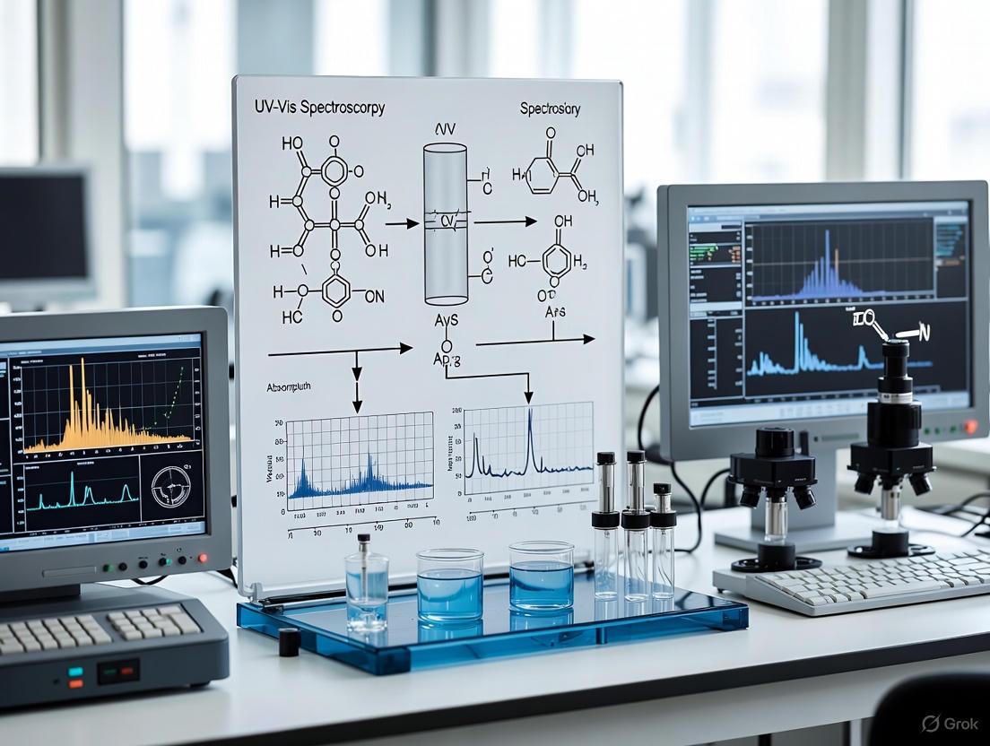
Ultraviolet-Visible (UV-Vis) spectroscopy is a fundamental analytical technique in pharmaceutical quality control (QA/QC) that relies on the interaction of light with matter. The core principle involves measuring the absorption of ultraviolet or visible light by a sample, which provides critical data for ensuring the identity, purity, potency, and stability of drug substances and products [1]. When light in the UV-Vis range (approximately 190–800 nm) interacts with a molecule, specific wavelengths are absorbed, promoting electrons to higher energy states [2] [3]. This absorption is quantitatively described by the Beer-Lambert law, forming the basis for concentration determination and other essential QA/QC analyses [4]. The technique is valued for its speed, precision, reproducibility, and non-destructive nature, making it indispensable for compliance with rigorous regulatory standards such as those outlined in ICH Q2(R1), USP Chapter <857>, and Ph. Eur. Chapter 2.2.25 [1] [5].
Core Principles
The Interaction of Light with Matter
The fundamental interaction in UV-Vis spectroscopy occurs when photons of light possess energy that matches the energy required to excite a molecule's electrons from a ground state to an excited state [3]. The energy of the photon is inversely proportional to its wavelength. The key electronic transitions involved for organic molecules are:
- π→π* and n→π* transitions: These are the most common transitions observed in UV-Vis spectroscopy, often associated with unsaturated centers in molecules, such as C=C, C=O, or aromatic rings [2].
- σ→σ* transitions: These require high energy and are typically observed in the far-UV region for saturated compounds [2].
The resulting absorption spectrum is a plot of absorbance versus wavelength, which serves as a fingerprint for qualitative analysis and provides the basis for quantitative measurement [3].
The Beer-Lambert Law
The Beer-Lambert law establishes the quantitative relationship between light absorption and the properties of the absorbing species [4]. It is mathematically expressed as:
A = εlc
Where:
- A is the Absorbance (unitless), defined as log10(I₀/I), where I₀ is the intensity of incident light and I is the intensity of transmitted light [4].
- ε is the Molar Absorptivity (L·mol⁻¹·cm⁻¹), a constant that is a characteristic of the substance at a specific wavelength [4].
- l is the Path Length (cm), the distance the light travels through the sample [4].
- c is the Concentration (mol·L⁻¹) of the absorbing species in the solution [4].
This law demonstrates that absorbance is directly proportional to concentration for a fixed path length, enabling the determination of unknown concentrations via a calibration curve [4].
Table 1: Key Parameters of the Beer-Lambert Law
| Parameter | Symbol | Units | Description |
|---|---|---|---|
| Absorbance | A | Unitless (Absorbance Units, AU) | Logarithmic measure of the amount of light absorbed by the sample. |
| Molar Absorptivity | ε | L·mol⁻¹·cm⁻¹ | A constant indicative of how strongly a chemical species absorbs light at a given wavelength. |
| Path Length | l | cm | The internal width of the cuvette or sample holder that the light beam traverses. |
| Concentration | c | mol·L⁻¹ | The concentration of the absorbing analyte in the solution. |
Applications in Pharmaceutical QA/QC
UV-Vis spectroscopy supports critical QA/QC workflows in the pharmaceutical industry, ensuring drug products meet predefined quality standards [1].
Table 2: Key Applications of UV-Vis Spectroscopy in Pharmaceutical QA/QC
| Application | Objective | Typical Protocol Summary |
|---|---|---|
| Assay and Potency Determination | Quantify the concentration of the Active Pharmaceutical Ingredient (API) in a drug product [1]. | Absorbance of the sample solution is measured at the λmax of the API and concentration is determined using a pre-established calibration curve [1]. |
| Content Uniformity | Ensure each dosage unit (e.g., tablet) contains an API amount within the specified range [1]. | Individual dosage units are dissolved in an appropriate solvent, and the absorbance of each solution is measured to calculate the API content [1]. |
| Dissolution Testing | Monitor the rate and extent of API release from a solid dosage form [1]. | Absorbance of dissolution media samples is measured at predetermined time points to calculate the cumulative percentage of API dissolved [1]. |
| Identity Testing | Confirm the identity of a raw material or finished product. | The UV absorption spectrum of the sample is compared to that of a reference standard for a match in characteristic peaks [1]. |
| Purity and Impurity Assessment | Detect and quantify potential impurities or degradation products [1]. | Absorbance is measured at a wavelength specific to an impurity, or spectral deviations from the reference standard are investigated [1]. |
An emerging application is the use of UV-Vis as a Process Analytical Technology (PAT). For instance, it can be implemented as an in-line monitoring system during continuous manufacturing processes like Hot Melt Extrusion (HME) to monitor critical quality attributes such as API concentration and solubility in a polymer matrix in real-time [6].
Experimental Protocols
Protocol 1: Standard Calibration Curve for API Quantification
This protocol details the steps for developing a calibration curve to determine the unknown concentration of an Active Pharmaceutical Ingredient (API).
1. Objective: To create a calibration curve for [API Name] in [Solvent, e.g., pH 6.8 phosphate buffer] for quantitative analysis.
2. Materials and Reagents:
- The Scientist's Toolkit for this protocol is detailed in Section 6.
3. Instrumentation:
- UV-Vis spectrophotometer (e.g., Agilent Cary Series, Shimadzu UV series) with validated performance (wavelength accuracy, photometric accuracy, stray light) [5] [7].
- Quartz cuvettes (1 cm path length).
4. Procedure:
- Step 1: Preparation of Standard Stock Solution. Accurately weigh approximately 50 mg of API reference standard. Transfer to a 50 mL volumetric flask, dissolve, and dilute to volume with solvent to obtain a stock solution of known concentration (e.g., 1 mg/mL).
- Step 2: Preparation of Working Standard Solutions. Perform serial dilutions of the stock solution to prepare a minimum of five standard solutions covering the concentration range expected for the sample (e.g., 2–20 μg/mL).
- Step 3: Spectral Scan. Scan one of the mid-concentration standards over a relevant wavelength range (e.g., 200–400 nm) to determine the wavelength of maximum absorption (λmax).
- Step 4: Measurement of Absorbance. Using the solvent as the blank, measure the absorbance of each working standard solution at the predetermined λmax.
- Step 5: Preparation of Calibration Curve. Plot the average absorbance (y-axis) against the corresponding concentration (x-axis). Perform linear regression to obtain the equation of the line (y = mx + c) and the correlation coefficient (R²).
5. Data Analysis:
- The slope of the line is related to the molar absorptivity (ε), and the concentration of an unknown sample (cunknown) is calculated as: cunknown = (Aunknown) / (εl), where Aunknown is the absorbance of the sample solution.
Protocol 2: Identity Confirmation of a Raw Material via Spectrum Comparison
1. Objective: To verify the identity of an incoming raw material (e.g., [Excipient Name]) against a qualified reference standard.
2. Procedure:
- Step 1: Sample Preparation. Prepare solutions of the test sample and the reference standard in the same solvent at equivalent concentrations.
- Step 2: Acquisition of Spectra. Scan both solutions over a specified wavelength range (e.g., 220–350 nm) using the solvent as a blank.
- Step 3: Comparison and Evaluation. Overlay the spectrum of the test sample with that of the reference standard. The identity is confirmed if the two spectra are identical in shape and show absorbance maxima and minima at the same wavelengths [1].
Method Validation
According to ICH Q2(R1) guidelines, methods used in pharmaceutical QA/QC must be validated. The following table summarizes key validation parameters for a typical UV-Vis quantitative method [8].
Table 3: Summary of Method Validation Parameters for a UV-Vis Assay
| Validation Parameter | Definition | Acceptance Criteria Example |
|---|---|---|
| Accuracy | The closeness of the measured value to the true value. | Recovery: 98.0–102.0% |
| Precision (Repeatability) | The closeness of agreement between a series of measurements under the same conditions. | %RSD ≤ 2.0% |
| Linearity | The ability of the method to obtain results directly proportional to analyte concentration. | Correlation coefficient (R²) ≥ 0.998 |
| Range | The interval between the upper and lower concentration levels over which linearity, accuracy, and precision are demonstrated. | Typically a defined range around the target concentration (e.g., 80–120%). |
| Specificity | The ability to assess the analyte unequivocally in the presence of potential interferents (e.g., impurities, excipients). | No interference observed. |
| Limit of Detection (LOD) | The lowest concentration of analyte that can be detected. | Signal-to-Noise ratio ≈ 3:1 |
| Limit of Quantitation (LOQ) | The lowest concentration of analyte that can be quantified with acceptable accuracy and precision. | Signal-to-Noise ratio ≈ 10:1 |
The Scientist's Toolkit: Essential Materials and Reagents
Table 4: Key Research Reagent Solutions and Materials
| Item | Function / Role | Critical Considerations |
|---|---|---|
| High-Purity Reference Standards | Serves as the benchmark for identity, purity, and quantity; essential for calibration [8]. | Must be of certified purity and traceable to a pharmacopoeial standard if available. |
| Spectroscopic Grade Solvents | To dissolve the analyte without introducing interference [1] [2]. | Must be transparent in the wavelength region of interest (e.g., HPLC-grade water, methanol, acetonitrile). |
| Quartz Cuvettes | Hold the sample solution for analysis. | Required for UV range analysis as quartz is transparent to UV light; standard path length is 1 cm [3]. |
| Volumetric Flasks and Pipettes | For accurate preparation and dilution of standard and sample solutions. | Class A glassware is recommended to ensure measurement accuracy and precision. |
| Filters (e.g., 0.45 μm membrane filters) | To remove particulate matter from samples, ensuring optically clear solutions and preventing light scattering [1]. | Filter material must be compatible with the solvent and must not adsorb the analyte. |
Troubleshooting and Best Practices
Successful implementation requires awareness of potential pitfalls and adherence to best practices.
- Adherence to Beer-Lambert Law: Ensure measurements are within the linear range of the instrument (typically absorbance < 1–2 AU). High absorbance can lead to deviations due to stray light or polychromatic effects [2] [3].
- Sample Preparation: Samples must be optically clear and free of particulates to prevent light scattering. Use filtration or centrifugation if necessary [1].
- Instrument Performance: Regular instrument validation is critical. Key performance checks include [7]:
- Wavelength Accuracy: Verify using holmium oxide or deuterium lamp emission lines.
- Stray Light: Check using solutions like sodium iodide.
- Photometric Accuracy: Confirm using neutral density filters or standard solutions.
Ultraviolet-Visible (UV-Vis) spectroscopy serves as a cornerstone analytical technique in pharmaceutical quality assurance and quality control (QA/QC) environments, where precision, reproducibility, and non-destructive testing are vital. This technique measures the absorption of ultraviolet or visible light by a compound as it transitions between electronic energy levels, providing a rapid, reliable means to characterize drug substances and products [1]. Within the highly regulated pharmaceutical industry, UV-Vis spectroscopy is indispensable for ensuring the identity, purity, potency, and stability of pharmaceutical compounds—critical factors in regulatory compliance, method validation, and ultimately, patient safety [1].
The fundamental principle governing quantitative UV-Vis analysis is the Beer-Lambert Law, which states that the absorbance of light by a solution is directly proportional to the concentration of the absorbing species and the path length the light takes through the solution [2]. This relationship is mathematically expressed as A = εlc, where A is the measured absorbance, ε is the molar absorptivity (a compound-specific constant), l is the path length (cm), and c is the concentration (mol/L) [2]. This law forms the basis for the quantitative determination of active pharmaceutical ingredients (APIs), impurity monitoring, and content uniformity testing that are routine in pharmaceutical control laboratories.
Core Instrumentation Components
A UV-Vis spectrophotometer is an integrated system of several key components, each playing a critical role in ensuring accurate and reproducible results. Understanding the function and specifications of each part is essential for proper instrument operation, method development, and troubleshooting in a pharmaceutical context.
The light source must provide a steady emission of light across a broad range of wavelengths. Modern instruments often use multiple lamps to cover the entire UV and visible spectrum effectively [3] [9].
| Light Source Type | Spectral Range | Key Characteristics | Pharmaceutical Application Consideration |
|---|---|---|---|
| Deuterium Lamp [3] [9] | 190 – 350 nm [9] | Intense, continuous UV spectrum; limited lifetime. | Essential for API quantification at lower UV wavelengths (e.g., nucleic acid analysis at 260 nm) [10]. |
| Tungsten-Halogen Lamp [3] [9] | 330 – 3200 nm [9] | Robust, inexpensive, stable output in visible/NIR region. | Ideal for colored compounds and dissolution testing in the visible range. |
| Xenon Lamp [3] | UV to Visible | High intensity, continuous spectrum across UV-Vis. | Higher cost and lower stability can be a concern for routine QC; often used in research instruments. |
Wavelength Selection: Monochromators
The monochromator is responsible for selecting a specific, narrow band of wavelengths from the broad output of the light source. Its core components include an entrance slit, a diffraction grating, and an exit slit [9]. The grating, which is rotated to select wavelengths, is characterized by its groove frequency (grooves per mm); a higher frequency (e.g., 1200 grooves/mm or more) provides better optical resolution [3]. The slits control the spectral bandwidth (SBW), defined as the full width at half maximum (FWHM) of the light intensity distribution exiting the monochromator [9]. A narrower SBW provides higher spectral resolution, crucial for distinguishing between closely spaced absorption peaks, while a wider SBW allows more light to pass, improving the signal-to-noise ratio for more sensitive detection [9]. The optimal SBW is typically 1/10th of the narrowest absorption peak's FWHM [9].
Sample Compartment
The sample compartment holds the analyte, typically contained within a cuvette, in the path of the light beam. The choice of cuvette material is critical and depends on the wavelength range of interest. For UV measurements below ~350 nm, quartz or fused silica cuvettes are mandatory as they are transparent to UV light [3]. For visible-only measurements, optical glass or plastic cuvettes may be sufficient and are more cost-effective [3]. Standard path lengths are 1 cm, but varying this length can help keep absorbance readings within the ideal linear range of 0.1 to 1.0 AU [1] [3]. Proper sample preparation is paramount; samples must be optically clear and free of particulate matter to prevent light scattering, which can lead to erroneously high absorbance readings [1].
Detection Systems
The detector converts the transmitted light intensity into an electrical signal, which is then processed to produce the absorbance spectrum. The selection of an appropriate detector balances sensitivity, spectral response, and cost.
| Detector Type | Operating Principle | Spectral Range | Advantages and Limitations |
|---|---|---|---|
| Photomultiplier Tube (PMT) [3] [9] | Photoelectric effect with electron multiplication via dynodes. | UV-Vis | Very high sensitivity and low noise; excellent for low-light applications. |
| Silicon Photodiode [9] | Semiconductor device generating photocurrent from incident light. | UV-Vis | Fast response, robust, and lower cost; less sensitive than PMT. |
| InGaAs Photodiode [9] | Semiconductor photodiode. | NIR | High sensitivity in NIR region; more expensive. |
Optical Configurations: Single vs. Double Beam
Spectrophotometers can have two primary optical configurations, each with distinct advantages for pharmaceutical analysis:
- Single Beam: Uses a single light path that passes through the sample. It requires measuring a blank reference sample immediately before or after the analyte measurement [9]. This design is simpler and more cost-effective but is susceptible to errors from source intensity fluctuations over time [9].
- Double Beam: Splits the monochromatic light into two beams—one passing through the sample and the other through a reference blank. The detector then measures the ratio of the two intensities simultaneously [9]. This configuration automatically compensates for drift in the light source intensity, electronic fluctuations, and stray light, providing superior stability and more reliable data, which is critical for lengthy analyses and regulated QC environments [9].
Detailed Experimental Protocol: API Potency Determination by UV-Vis Spectroscopy
This protocol details a standard procedure for quantifying the concentration of an Active Pharmaceutical Ingredient (API) in a solution using a double-beam UV-Vis spectrophotometer, a common assay in pharmaceutical potency and content uniformity testing.
Research Reagent Solutions
| Reagent/Material | Specification | Function/Purpose |
|---|---|---|
| API Reference Standard | Certified purity (e.g., USP grade) | Primary standard for calibration curve; ensures accuracy and traceability. |
| HPLC-Grade Solvent | Spectrophotometric grade, low UV absorbance | Dissolves analyte and reference; minimizes background absorbance (stray light). |
| Volumetric Flasks | Class A | Precise preparation and dilution of standard and sample solutions. |
| Quartz Cuvettes | 1 cm pathlength, matched pair | Holds sample and reference solutions; quartz is transparent in UV range. |
Step-by-Step Procedure
Instrument Preparation and Qualification:
- Power on the spectrophotometer and the computer. Allow the system to initialize and the lamp to warm up for the time recommended by the manufacturer (typically 15-30 minutes).
- Using the instrument software, set the scanning parameters: Wavelength range: 200-400 nm (or a suitable range encompassing the API's λmax); Scan speed: Medium; Spectral Bandwidth (SBW): As determined during method development (e.g., 1 nm or 2 nm).
- Ensure the sample compartment is clean and place a cuvette filled with the pure solvent (blank) in both the sample and reference holder positions.
Preparation of Standard Solutions:
- Accurately weigh approximately 50 mg of the API reference standard and transfer it quantitatively to a 100 mL volumetric flask. Dissolve and dilute to volume with the solvent to create a stock solution of known concentration (e.g., ~500 µg/mL).
- Using a serial dilution technique, prepare a minimum of five standard solutions covering the concentration range where the Beer-Lambert law is known to be linear (e.g., 5, 10, 20, 30, 40 µg/mL). Ensure all dilutions are performed with precision.
Sample Preparation:
- For a solid dosage form (tablet/capsule), weigh and finely powder not less than 10 units. Accurately weigh a portion of the powder equivalent to the weight of one unit and transfer to a suitable volumetric flask. Extract the API with the solvent, sonicate if necessary, dilute to volume, and filter.
- For a liquid formulation, dilute an accurately measured volume with solvent to obtain a concentration within the linear range of the calibration curve.
Data Acquisition:
- Perform a blank correction with the solvent in both cuvettes.
- Measure the absorbance spectrum of each standard solution, identifying the wavelength of maximum absorption (λmax) for the API.
- Measure the absorbance of the prepared sample solution at the determined λmax.
Data Analysis and Calculation:
- Construct a calibration curve by plotting the average absorbance (y-axis) of each standard against its known concentration (x-axis).
- Perform a linear regression analysis on the data. The coefficient of determination (R²) should be ≥ 0.995 for a valid calibration.
- Use the linear regression equation (y = mx + c) to calculate the concentration of the API in the sample solution based on its measured absorbance.
- Account for all dilution factors from the original sample to report the final API concentration in the drug product.
Critical Performance Parameters and Troubleshooting in Pharmaceutical Analysis
Adherence to strict performance specifications is non-negotiable in a GMP-regulated pharmaceutical laboratory.
- Stray Light: This is any light that reaches the detector without passing through the sample or is outside the selected wavelength band. It is a primary cause of deviation from the Beer-Lambert law, especially at high absorbances (>2 AU), leading to falsely low absorbance readings [2] [9]. It is controlled by using high-purity optical components and can be minimized with double-monochromator instruments [9].
- Spectral Bandwidth (SBW): As previously discussed, an inappropriate SBW can lead to poor resolution or reduced sensitivity. The selected SBW must be validated as part of the analytical method to ensure accurate quantification [9].
- Linearity and Range: The analytical method must demonstrate that the instrument's response is directly proportional to the concentration of the analyte across the specified range. This is established during method validation per ICH Q2(R1) guidelines [1].
- Wavelength Accuracy: The instrument must be regularly calibrated for wavelength accuracy (e.g., using holmium oxide filters) to ensure that the reported λmax is correct, as shifts can lead to misidentification and inaccurate quantification [1].
A thorough understanding of the key components of a UV-Vis spectrophotometer—from the stability of its light sources and the resolution of its monochromator to the sensitivity of its detector—is fundamental for scientists and researchers in drug development. This knowledge empowers them to develop robust and validated analytical methods, operate instruments effectively, troubleshoot issues, and, most importantly, generate reliable data that ensures the quality, safety, and efficacy of pharmaceutical products. As a versatile and quantitative workhorse of the QC laboratory, the proper application of UV-Vis spectroscopy remains a critical skill in the pharmaceutical scientist's toolkit.
Within pharmaceutical quality control (QC), the demand for rapid, reliable, and cost-efficient analytical techniques is paramount. Ultraviolet-Visible (UV-Vis) spectroscopy stands as a cornerstone technology in this field, offering significant advantages that align with the rigorous demands of modern drug development and manufacturing. This application note frames these advantages—speed, cost-effectiveness, and versatility—within a broader thesis on the indispensable role of UV-Vis spectroscopy in pharmaceutical QC. It provides researchers and scientists with structured quantitative data and detailed experimental protocols to facilitate implementation and underscore the technique's value in ensuring drug identity, purity, potency, and safety.
Advantages and Quantitative Data
The utility of UV-Vis spectroscopy in pharmaceutical QC is demonstrated through its performance across multiple applications. The following tables summarize key quantitative data supporting its speed, cost-effectiveness, and versatility.
Table 1: Speed and Performance Metrics of UV-Vis Spectroscopy in Pharma QC
| Application Context | Measured Parameter | Performance Outcome | Reference / Model System |
|---|---|---|---|
| In-line Content Uniformity | Measurement Frequency | ~1,000 spectra acquired in a short time [11] | Hyperspectral Imaging Prototype |
| In-line Potency Monitoring | Integration Time | Measurements in the millisecond-range [6] | Hot Melt Extrusion (HME) |
| API Quantification | Analytical Run Time | Rapid results for routine quantification [1] | General API Analysis |
| Dissolution Testing | Throughput | High-throughput capability [1] | Dissolution Profile Evaluation |
Table 2: Versatility and Analytical Scope in Pharmaceutical Applications
| Application Area | Specific Use Case | Typical Analytical Range / Performance | Reference / Model System |
|---|---|---|---|
| Identity Testing | Raw Material Identification | Spectral fingerprint comparison [1] | IR/UV-Vis for Raw Materials |
| Purity Assessment | Impurity and Degradation Detection | Detects unwanted absorption peaks [1] | General Purity Testing |
| Potency Determination | API Concentration in Tablets | R² ~0.99 for 7-13% API content [12] | In-line Theophylline Tablets |
| Process Control (PAT) | Content Uniformity Monitoring | Validated for ICH Q2(R2); Range: 7-13 wt% [12] | Continuous Manufacturing |
| Hyperspectral Imaging | API Characterization | Complete sample separation via PCA [11] | Ibuprofen, Paracetamol, Aspirin |
Experimental Protocols
Protocol: In-Line Content Uniformity Monitoring for Tablets
This protocol validates UV-Vis spectroscopy for real-time, in-line monitoring of Active Pharmaceutical Ingredient (API) content uniformity during tablet manufacturing, as per ICH Q2(R2) [12].
- 1. Objective: To implement and validate a non-destructive, rapid UV-Vis method for in-line content uniformity determination of tablets on a rotary press.
- 2. Materials:
- API: Theophylline monohydrate
- Excipients: Lactose monohydrate (filler/binder), Magnesium stearate (lubricant)
- Equipment: Rotary tablet press with integrated UV/Vis reflectance probe, data acquisition system
- Software: For data pre-treatment and calculation of relative reflectance
- 3. Methodology:
- Formulation: Prepare a powder blend with a target of 10 wt% theophylline, 0.5 wt% magnesium stearate, and the remainder as lactose monohydrate.
- Instrument Setup: Integrate a UV-Vis reflectance probe directly into the tablet press, positioning it to measure the main side of the tablets during compression.
- Data Acquisition:
- Synchronize spectrum acquisition with the tablet production cycle.
- Calculate reflectance (R) from the intensity of reflected light (I) and emitted light (I0): R = I / I₀ [12].
- Apply data pre-treatment to filter out spectra not measured from the full tablet surface.
- Calibration & Quantification:
- Use the relative reflectance at a specific, optimized wavelength (e.g., 272 nm for theophylline).
- Establish a univariate calibration model correlating the relative reflectance value to the known API concentration from off-line reference methods.
- 4. Validation:
- Specificity: Confirm the API-specific wavelength is free from excipient interference.
- Linearity: Demonstrate a linear relationship (e.g., R² ~0.99) over a defined range (e.g., 7-13 wt% API).
- Precision: Evaluate repeatability and intermediate precision under different throughputs.
- Accuracy: Compare results with a validated reference method (e.g., HPLC).
Protocol: API Solubility and Oversaturation Monitoring in Hot Melt Extrusion
This protocol uses in-line UV-Vis spectroscopy to monitor API solubility and identify oversaturation thresholds during Hot Melt Extrusion (HME) for forming amorphous solid dispersions [6].
- 1. Objective: To use UV-Vis spectroscopy as a Process Analytical Technology (PAT) tool for real-time monitoring of API-polymer solubility and detection of oversaturation during HME.
- 2. Materials:
- API: Piroxicam (PRX)
- Polymer: Kollidon VA64
- Equipment: Co-rotating twin-screw extruder with in-line UV-Vis fiber-optic probe, HPLC system for off-line validation
- 3. Methodology:
- Sample Preparation: Blend PRX and Kollidon VA64 at varying concentrations (e.g., 10-30% w/w).
- Process Setup: Install the UV-Vis probe in-line within the extruder die.
- Data Acquisition:
- Collect full-range spectra (230-700 nm) in real-time.
- Monitor the absorbance at the API-specific λ-max.
- Simultaneously calculate the lightness value (L) from the visible region of the spectrum.
- Analysis:
- Homogeneous dissolution is indicated by a stable baseline in the visible region and consistent absorbance in the UV region.
- Oversaturation is identified by a sharp increase in absorbance and a sharp decrease in L value, accompanied by baseline scattering in the visible range due to undissolved API particles.
- 4. Data Interpretation:
- Use a sequential Design of Experiments (DoE) to understand the interaction effects of CPPs (e.g., die temperature, screw speed, feed rate, API concentration) on CQAs (absorbance, L*).
- Correlate in-line UV-Vis data with off-line characterization (HPLC, DSC, XRD) to confirm the absence of degradation and the amorphous state of the dispersion.
The following workflow diagram illustrates the sequential stages of this HME monitoring process:
The Scientist's Toolkit: Essential Research Reagent Solutions
Table 3: Key Materials and Reagents for UV-Vis-Based Pharmaceutical QC
| Item | Function / Application | Critical Specifications |
|---|---|---|
| High-Purity Solvents | Dissolve samples for analysis; must not absorb in the measured wavelength range. | Spectrophotometric grade (e.g., Methanol, Acetonitrile, Water). Optically clear and free from particulates [1]. |
| Quartz Cuvettes | Hold liquid samples for transmission measurements in UV range. | High-transmission quartz; standard 1 cm path length; matched pairs for accuracy [3]. |
| Deuterated Solvents | For NMR-based confirmation of UV-Vis results in structural elucidation. | D₂O, CDCl₃, DMSO-d₆; high isotopic purity to avoid signal interference [1]. |
| Certified Reference Standards | Calibrate the spectrophotometer; validate analytical methods; quantify API. | USP/EP/JP certified; high purity and known concentration [13]. |
| Hyperspectral Imaging System | Spatially resolve API distribution in solid dosage forms (PAT). | UV wavelength range (e.g., 225-400 nm); pushbroom imager with CCD camera [11]. |
| Reflectance Probe | Enable in-line measurements on solid surfaces (e.g., powder blends, tablets). | Fiber-optic design; robust for integration into process equipment (e.g., tablet press, extruder) [12]. |
Ultraviolet-Visible (UV-Vis) spectroscopy is a fundamental analytical technique in pharmaceutical quality control research, providing a rapid, non-destructive means to verify identity, assess purity, and determine the concentration of active pharmaceutical ingredients (APIs) and finished drug products [3] [1]. The technique measures the absorption of light in the ultraviolet (typically 190-400 nm) and visible (400-700 nm) regions of the electromagnetic spectrum, which corresponds to the excitation of outer electrons in molecules to higher energy states [14] [3]. For pharmaceutical scientists, interpreting the resulting spectra—particularly the absorption maxima (λmax) and molar absorptivity (ε)—is critical for confirming molecular identity and detecting potential impurities [15] [1]. This application note details the principles and protocols for interpreting spectral data within a pharmaceutical development context.
Theoretical Principles of Electronic Transitions
The absorption of UV or visible radiation corresponds to the excitation of outer electrons from their ground state to an excited state [14]. The specific wavelengths absorbed, and the intensity of these absorptions, provide fundamental structural information about the molecule.
Table 1: Primary Electronic Transitions in UV-Vis Spectroscopy
| Transition Type | Electrons Involved | Typical λmax Range | Molar Absorptivity (ε, L·mol⁻¹·cm⁻¹) | Chromophore Example |
|---|---|---|---|---|
| π → π* | π electrons in double bonds | 200-700 nm (varies with conjugation) | 1,000 - 10,000 | Alkenes, Conjugated systems [14] [16] |
| n → π* | Non-bonding (lone pair) electrons | 250-500 nm | 10 - 100 | Carbonyl groups [14] [16] |
| n → σ* | Non-bonding electrons | 150-250 nm | ~1,000 | Alcohols, Amines [14] |
| Charge-Transfer | Electron donor to acceptor | 200-800 nm | > 10,000 | Charge-transfer complexes [14] |
The most analytically useful transitions for organic molecules in the conventional UV-Vis range (200-700 nm) involve π → π and n → π transitions [14]. The π → π transitions, typically found in molecules with conjugated double bond systems, are characterized by high molar absorptivities. In contrast, n → π transitions, occurring in molecules with heteroatoms bearing lone pairs (like carbonyl oxygen), have lower probabilities and thus weaker intensities [14] [17] [16].
Conjugation is the single most important factor affecting λmax, causing a bathochromic shift (red shift) to longer wavelengths and often a hyperchromic effect (increased intensity). This occurs because conjugation lowers the energy gap between the highest occupied molecular orbital (HOMO) and the lowest unoccupied molecular orbital (LUMO) [17] [16]. For instance, while ethene absorbs at 165 nm, the conjugated system in 1,3-butadiene absorbs at 217 nm, and beta-carotene, with its extensive 11-conjugated double-bond system, absorbs in the visible region (~450 nm), making it orange [16].
Instrumentation and Data Acquisition Workflow
A UV-Vis spectrophotometer consists of several key components: a light source, a wavelength selector, a sample holder, and a detector [3].
Diagram 1: UV-Vis Instrument Workflow
The light source typically consists of two lamps: a deuterium lamp for the UV region and a tungsten or halogen lamp for the visible region [3]. The monochromator, often containing a diffraction grating with 1200 grooves per mm or more, selects a specific wavelength of light to pass through the sample and reference [3]. For accurate measurements, the sample and a reference blank (typically the solvent used to prepare the sample) are analyzed simultaneously or sequentially. The detector then converts the transmitted light intensity into an electrical signal, and the processor calculates the absorbance at each wavelength to generate the absorption spectrum [3].
Core Quantitative Relationships
Beer-Lambert Law
The fundamental quantitative relationship in UV-Vis spectroscopy is the Beer-Lambert Law, which states that absorbance is directly proportional to the concentration of the absorbing species and the path length through the sample [14] [3].
A = ε * c * l
Where:
- A is the measured Absorbance (unitless)
- ε is the Molar Absorptivity (L·mol⁻¹·cm⁻¹)
- c is the concentration of the analyte (mol·L⁻¹)
- l is the path length of the cuvette (cm) [14] [3]
Absorbance values between 0.1 and 1.0 (or up to 2.0 for modern instruments) are generally considered within the ideal quantitative range, corresponding to 10% to 1% light transmission [3]. Values outside this range may require sample dilution or a path length adjustment.
Spectral Parameters
The key parameters extracted from a UV-Vis spectrum are:
- λmax: The wavelength at which maximum absorption occurs. This is a characteristic property of a chromophore and is highly reproducible under identical conditions [16].
- Molar Absorptivity (ε): A measure of how strongly a chromophore absorbs light at a specific wavelength. It is a intrinsic molecular property. The magnitude of ε reflects both the size of the chromophore and the probability of the electronic transition [14] [17].
Experimental Protocols for Pharmaceutical Analysis
Protocol 1: Sample Preparation and Measurement
Table 2: The Scientist's Toolkit: Essential Materials and Reagents
| Item | Function/Description | Key Considerations |
|---|---|---|
| UV-Vis Spectrophotometer | Instrument for measuring light absorption. | Must be qualified (IQ/OQ/PQ) per GMP guidelines. Wavelength accuracy and photometric accuracy should be verified [1]. |
| Quartz Cuvettes | Sample holders for liquid samples. | Quartz is essential for UV detection (<350 nm); must be matched and clean. Standard path length is 1.0 cm [3]. |
| High-Purity Solvents | To dissolve the analyte (e.g., methanol, water, buffer). | Must be optically pure, transparent in the spectral region of interest, and not react with the analyte [1]. |
| Volumetric Flasks | For precise preparation of standard and sample solutions. | Class A glassware ensures accurate volume measurements for quantitative work. |
| Analytical Balance | For accurate weighing of reference standards and samples. | Must be calibrated; critical for preparing standard solutions of known concentration. |
- Solution Preparation: Accurately weigh the API or drug product standard. Dissolve in an appropriate high-purity solvent (e.g., methanol, phosphate buffer) that is transparent in the spectral region of interest. Prepare a series of standard solutions across the expected concentration range [1].
- Blank Measurement: Fill a quartz cuvette with the pure solvent and place it in the reference compartment. This corrects for solvent absorption and reflection losses [3].
- Spectral Acquisition: Place the sample solution in a quartz cuvette and insert it into the sample holder. Scan across the desired wavelength range (e.g., 200-400 nm for UV analysis) to obtain the full absorption spectrum and identify λmax [16].
- Quantitation: Measure the absorbance of standards and samples at the predetermined λmax. Construct a calibration curve of absorbance versus concentration and use it to determine the unknown sample concentration [3] [1].
Protocol 2: Identity Confirmation via Absorbance Ratios (as per USP/EP)
This protocol is exemplified by the identification test for Ibuprofen in the USP and European Pharmacopoeia (EP) [15].
- Standard Preparation: Prepare a solution of the reference standard of Ibuprofen in the specified solvent at the concentration stated in the monograph.
- Test Sample Preparation: Prepare the test sample (ibuprofen API) identically to the standard.
- Spectrum Acquisition: Obtain the UV absorption spectrum of both the standard and test sample solutions over the specified wavelength range.
- Peak and Ratio Comparison:
- USP Test: Confirm that the test sample spectrum exhibits absorbance maxima at the same wavelengths as the standard spectrum [15].
- EP Test: Calculate the ratio of absorbances at designated wavelengths (e.g., A₁/A₂) for the test sample. Confirm that this ratio is within the acceptance range specified in the EP monograph [15].
Diagram 2: Identity Confirmation Workflow
Factors Influencing Spectral Data
Solvent Effects
The solvent can significantly influence the absorption spectrum [14]. Polar solvents can cause:
- Blue Shift (Hypsochromic Shift) for n → π* transitions: Increased solvation of lone pair electrons lowers the energy of the n orbital, increasing the transition energy [14].
- Red Shift (Bathochromic Shift) for π → π* transitions: Attractive polarization forces between the solvent and absorber lower the energy levels of both the excited and unexcited states, with a greater effect on the excited state, slightly reducing the energy difference [14].
Table 3: Common Solvents and Their UV Cut-Off Wavelengths
| Solvent | UV Cut-Off (nm) | Notes |
|---|---|---|
| Water | ~190 nm | Suitable for far-UV work. |
| Acetonitrile | ~190 nm | Excellent UV transparency. |
| n-Hexane | ~200 nm | Common for non-polar analytes. |
| Methanol | ~205 nm | Common polar solvent. |
| Chloroform | ~245 nm | Unsuitable for detection below 245 nm. |
pH and Derivatization
The pH of the solution can dramatically alter the spectrum of ionizable compounds. For example, phenols and aromatic acids may show different λmax and ε in their protonated versus deprotonated forms. This property can be exploited to enhance detection sensitivity or selectivity. Sample impurities or turbidity can cause light scattering, leading to erroneously high absorbance readings. Samples must be optically clear and free of particulates, achievable through filtration or centrifugation [1].
Advanced Applications and Computational Predictions
Beyond quantification, UV-Vis spectroscopy is used in pharmaceutical stability studies to track degradation (e.g., by observing the appearance of new absorption peaks) and in dissolution testing to monitor the release of an API from its dosage form [1].
Computational chemistry methods, particularly Time-Dependent Density Functional Theory (TD-DFT), can predict the UV-Vis spectra of molecules from first principles [18] [19]. These calculations simulate the energies and probabilities of electronic transitions, providing a theoretical λmax and oscillator strength (related to ε). While often slightly shifted compared to experimental data (by ~0.1-0.5 eV), these methods are invaluable for assigning spectral bands to specific molecular transitions and for rational molecular design in drug development [18]. Large-scale text-mining and computational efforts are underway to create vast databases of experimental and computed UV/Vis spectral attributes, facilitating data-driven materials discovery [19].
The interpretation of absorption maxima and spectral data is a cornerstone of pharmaceutical analysis. A rigorous understanding of the underlying electronic transitions, coupled with strict adherence to validated experimental protocols, allows researchers and quality control professionals to reliably identify compounds, determine their concentration, and ensure the safety and efficacy of final drug products. Mastery of this technique, supported by robust procedures and an understanding of regulatory requirements, remains essential in the modern pharmaceutical landscape.
Practical Applications in the Pharma Lab: From API Quantification to Dissolution Testing
Active Pharmaceutical Ingredient (API) Identification and Assay
Ultraviolet-Visible (UV-Vis) spectroscopy is a cornerstone analytical technique in pharmaceutical quality control and drug development, providing a reliable means for the identification and quantification of Active Pharmaceutical Ingredients (APIs). The technique operates on the principle that molecules absorb light in the ultraviolet (200-400 nm) and visible (400-780 nm) regions, resulting in electronic transitions. The fundamental relationship between absorbance and concentration is governed by the Beer-Lambert Law (A = εcl), where absorbance (A) is proportional to the concentration (c) of the analyte, its molar absorptivity (ε), and the path length (l) of the light through the sample [3]. This principle, combined with the technique's speed, sensitivity, and relative simplicity, makes it indispensable for ensuring the identity, strength, and quality of pharmaceutical products in various stages of development and manufacturing, from raw material testing to final product release [20] [21].
The application of UV-Vis spectroscopy in pharmaceuticals has evolved significantly. Traditionally used for off-line analysis of single components in solution, it is now increasingly implemented as an in-line Process Analytical Technology (PAT) tool. This shift supports the pharmaceutical industry's movement towards continuous manufacturing and the adoption of Quality by Design (QbD) and Analytical Quality by Design (AQbD) principles, which emphasize building quality into the product and process through scientific understanding and risk management [22] [12] [6]. Furthermore, the challenge of analyzing complex mixtures with overlapping spectra is being addressed through the integration of chemometrics, which uses multivariate statistical methods to extract meaningful information from spectral data [23] [24].
A comprehensive examination of UV-Vis spectrophotometric methods in pharmaceutical analysis published between 2015 and 2023 reveals clear trends in application and methodology [20]. The following tables summarize key statistical findings.
Table 1: Application Distribution of UV-Vis Spectrophotometric Methods (2015-2023)
| Sample Type | Percentage of Studies |
|---|---|
| Pharmaceutical Dosage Forms | 56% |
| Bulk Materials | 27% |
| Pure Substances | 16% |
| Biological Materials | 2% |
| Herbal Materials | 0.4% |
Table 2: Wavelength Range Utilization in UV-Vis Methods (2015-2023)
| Wavelength Range | Percentage of Studies |
|---|---|
| 200 - 240 nm | 28% |
| 240 - 300 nm | 27% |
| > 300 nm | 44% |
The data indicates a strong focus on the analysis of final dosage forms, underscoring the technique's vital role in product quality control. The significant use of wavelengths above 300 nm highlights its applicability not only to the characteristic aromatic and chromophoric group absorptions in the UV range but also to colored compounds in the visible range [20].
Experimental Protocols
This section provides detailed methodologies for two primary applications: the fundamental assay of a single API in a tablet and an advanced in-line PAT method for monitoring API content during a continuous manufacturing process.
Protocol 1: Assay of a Single API in a Tablet Formulation Using a Traditional Off-Line Approach
This protocol describes a standard method for determining the API content in a finished tablet, using theophylline as a model compound [12].
- 1. Objective: To quantify the concentration of theophylline in immediate-release tablets and validate the method according to ICH Q2(R2) guidelines.
- 2. Materials and Reagents:
- API: Theophylline monohydrate standard.
- Tablets: Immediate-release tablets containing theophylline (e.g., 10% w/w target).
- Solvent: A suitable solvent, such as 0.1 M sodium hydroxide or a buffered aqueous solution, in which the API is freely soluble.
- Volumetric Flasks: 100 mL, 250 mL.
- Ultrasonic Bath.
- Syringe Filters: 0.45 µm, nylon or PTFE.
- UV-Vis Spectrophotometer with quartz cuvettes (path length: 1 cm).
- 3. Instrumental Parameters:
- Wavelength: 272 nm (λmax for theophylline).
- Mode: Absorbance.
- Scan Range: 220 - 350 nm (for spectrum identification).
- Slit Width: 1 nm.
- Scan Speed: Medium.
- 4. Procedure:
- Standard Stock Solution: Accurately weigh approximately 25 mg of theophylline standard into a 250 mL volumetric flask. Dissolve and make up to volume with the solvent to obtain a concentration of about 100 µg/mL.
- Calibration Standards: Dilute the stock solution to prepare a series of standard solutions (e.g., 5, 10, 15, 20, 25 µg/mL).
- Sample Preparation: Weigh and finely powder not less than 20 tablets. Accurately weigh a portion of the powder equivalent to about 25 mg of theophylline into a 250 mL volumetric flask. Add approximately 150 mL of solvent, sonicate for 15-30 minutes with intermittent shaking, allow to cool, and dilute to volume. Filter a portion of the solution through a 0.45 µm syringe filter, discarding the first few mL of the filtrate.
- Blank Solution: Prepare the solvent used for extraction as the blank.
- Measurement: Measure the absorbance of the calibration standards and the sample solution against the blank at 272 nm.
- 5. Data Analysis:
- Plot a calibration curve of absorbance versus concentration of the standard solutions.
- Determine the regression equation (y = mx + c) and the coefficient of determination (R²). A value of R² > 0.995 is typically expected.
- Calculate the concentration of theophylline in the sample solution using the regression equation.
- Calculate the % label claim using the following formula:
- % Label Claim = (Calculated Concentration × Dilution Factor × Average Tablet Weight) / (Weight of Sample Taken × Label Claim) × 100%
Protocol 2: In-Line Monitoring of API Content During Hot Melt Extrusion (HME) Using AQbD Principles
This protocol outlines the development and validation of an in-line UV-Vis method for real-time monitoring of piroxicam content in a polymer matrix during HME, following AQbD principles [22] [6].
- 1. Define the Analytical Target Profile (ATP): The ATP states that the method must predict the concentration of piroxicam in Kollidon VA 64 with an accuracy (trueness and precision) within ±5% acceptance limits.
- 2. Materials and Equipment:
- API: Piroxicam (PRX).
- Polymer: Kollidon VA 64.
- Equipment: Co-rotating twin-screw hot melt extruder.
- PAT Tool: In-line UV-Vis spectrophotometer with high-temperature optical probes configured in transmission mode, installed in the extruder die.
- 3. Risk Assessment: Perform a Failure Mode and Effect Analysis (FMEA) to identify and rank risks to the analytical procedure. Critical parameters may include probe window fouling, air bubbles in the melt, and spectral interference from polymer degradation.
- 4. Method Development and Calibration:
- Setup: Install the UV-Vis probes in the extruder die. Collect a reference transmittance spectrum with an empty, clean die at the process temperature (e.g., 140°C).
- Data Collection: Collect transmittance spectra in the range of 230-780 nm with a data collection frequency of 0.5 Hz during extrusion.
- Model Development: Prepare calibration batches with known concentrations of piroxicam (e.g., 10%, 15%, 20% w/w). Use the absorbance values at the API's λmax or develop a multivariate model to correlate the spectral data with the known API concentration.
- 5. Validation Based on Accuracy Profile:
- Perform validation experiments using independent sets at various concentration levels around the target (e.g., 15% w/w).
- Calculate the β-expectation tolerance intervals for each concentration level. The method is considered valid if the 95% tolerance limits fall within the pre-defined ±5% acceptance limits.
- 6. Testing Robustness: Deliberately vary critical process parameters, such as screw speed (150-250 rpm) and feed rate (5-9 g/min), to demonstrate that the piroxicam content prediction remains accurate and precise.
Advanced Applications and Techniques
Chemometrics-Assisted UV-Vis Spectroscopy
For formulations with multiple absorbing components, such as those containing two APIs or an API with impurities, spectral overlapping poses a significant challenge. Chemometrics provides a powerful solution by applying mathematical and statistical models to extract quantitative information from complex, overlapping spectra [24].
- Common Algorithms:
- Principal Component Analysis (PCA): An unsupervised technique used for pattern recognition, classification, and outlier detection.
- Partial Least Squares (PLS) Regression: A supervised technique that builds a model between spectral data and concentration data. It is the most widely used method for quantitative analysis of complex mixtures [23].
- Application Example: Simultaneous assay of Carbetocin (API) and L-Methionine (critical excipient) in a solution for injection. A single PLS model was developed using UV-Vis spectra, allowing for the in-line monitoring of both components' concentrations during continuous liquid flow manufacturing without the need for separation [23].
UV Hyperspectral Imaging
UV hyperspectral imaging is an emerging PAT tool that combines spatial and spectroscopic information. It generates a hypercube of data, allowing for the visualization of API distribution within a solid dosage form, such as a tablet [11].
- Principle: A pushbroom imaging system scans tablets on a conveyor belt, collecting a full spectrum at every pixel of the image, typically in the 225-400 nm range.
- Data Analysis: Principal Component Analysis (PCA) is applied to the hypercube to differentiate and classify tablets based on their API content and to identify heterogeneity or potential counterfeit products.
- Advantage: This non-destructive technique provides a rapid and comprehensive analysis of the chemical composition of solid dosage forms, making it highly suitable for 100% quality control in a production setting [11].
The Scientist's Toolkit: Essential Research Reagent Solutions
Table 3: Key Materials, Reagents, and Equipment for UV-Vis API Analysis
| Item | Function / Role in Analysis | Example from Literature |
|---|---|---|
| UV-Vis Spectrophotometer | Measures the absorption of light by the sample. Can be benchtop, fiber-optic PAT, or imaging-based. | In-line system with optical probes for HME [22]; Hyperspectral imager for tablets [11]. |
| Quartz Cuvettes / High-Temp PAT Probes | Sample holders. Quartz is essential for UV range analysis. Specialized probes withstand process conditions (e.g., high temp/pressure). | Transmission probes installed in extruder die [22]; 1 mm quartz cuvette for solution analysis [11]. |
| Chemometric Software | For multivariate data analysis, model development (e.g., PLS), and data reduction (e.g., PCA). | PLS for simultaneous API/excipient assay [23]; PCA for hyperspectral image analysis [11]. |
| Chemical Standards (API, Excipients) | High-purity materials for developing and validating analytical methods, creating calibration curves, and specificity testing. | Piroxicam and Kollidon VA64 for HME [22]; Carbetocin and L-Methionine for solution assay [23]. |
| Suitable Solvents | To dissolve the sample without interfering spectrally in the region of interest (e.g., methanol, water, buffered solutions). | 0.1 M HCl:Methanol (1:1) for acetylsalicylic acid [11]; Methanol/Water for paracetamol [11]. |
Visualized Workflows
Drug Stability and Forced Degradation Studies for Shelf-Life Prediction
Within pharmaceutical quality control research, ensuring drug product stability is paramount for guaranteeing patient safety and therapeutic efficacy. A drug's shelf life, defined as the time period during which it maintains its intended identity, strength, quality, and purity, is directly determined through rigorous stability studies [25]. These studies are a regulatory requirement for all new drug applications. Forced degradation studies, also known as stress testing, are a proactive scientific approach used to intentionally degrade a drug substance or product under severe conditions. This process identifies potential degradation products, elucidates degradation pathways, and, crucially, helps develop stability-indicating analytical methods [25] [26]. UV-Vis spectroscopy serves as a cornerstone analytical technique in these endeavors due to its speed, accuracy, and economic viability for quantifying active pharmaceutical ingredients (APIs) and detecting contaminants that arise from decomposition [27]. This document details the application of UV-Vis spectroscopy within structured forced degradation protocols to accurately predict the shelf life of pharmaceutical products.
Fundamental Principles of Forced Degradation
Forced degradation is a scientific necessity that provides an insight into degradation pathways and the intrinsic stability of a molecule. It is performed under conditions more severe than those used in accelerated stability studies to generate relevant degradation products within a short timeframe [25] [26]. The primary objectives are to establish degradation pathways, elucidate the structure of degradation products, and most importantly, to develop and validate stability-indicating methods that can accurately monitor the quality of a drug product over time [25].
A key consideration in forced degradation is determining the extent of degradation. While regulatory guidelines do not specify exact limits, a degradation of the drug substance between 5% and 20% is generally considered acceptable for validating chromatographic assays, with 10% often viewed as an optimal target [25]. This ensures sufficient degradation products are generated without causing over-stressing, which may lead to secondary degradation products not representative of real-world conditions.
The following diagram illustrates the logical decision-making process and strategic workflow for designing a forced degradation study.
Experimental Protocols for Forced Degradation Studies
A minimal set of stress conditions must be investigated to comprehensively challenge the stability of a drug molecule. These typically include acid and base hydrolysis, thermal degradation, photolysis, and oxidation [25]. The following protocols provide a detailed methodology for conducting these studies, with UV-Vis spectroscopy as a primary analytical tool.
Recommended Stress Conditions
The table below summarizes the standard experimental conditions used for forced degradation studies to simulate various stress factors.
Table 1: Standard Conditions for Forced Degradation Studies [25]
| Degradation Type | Experimental Conditions | Commonly Used Storage Conditions | Sampling Time Points |
|---|---|---|---|
| Acid Hydrolysis | 0.1 M HCl | 40 °C, 60 °C | 1, 3, 5 days |
| Base Hydrolysis | 0.1 M NaOH | 40 °C, 60 °C | 1, 3, 5 days |
| Oxidation | 3% Hydrogen Peroxide (H₂O₂) | 25 °C, 60 °C | 1, 3, 5 days |
| Thermal | Solid or solution in heat chamber | 60 °C, 80 °C (with/without 75% RH) | 1, 3, 5 days |
| Photolytic | Exposure to UV/Visible light per ICH Q1B | 1x and 3x ICH energy levels | 1, 3, 5 days |
Detailed Methodological Workflow
The execution of a forced degradation study follows a systematic workflow from sample preparation to data analysis, as visualized below.
Protocol 1: Hydrolytic Degradation
- Preparation: Prepare a stock solution of the drug substance at a concentration of approximately 1 mg/mL in a suitable solvent [25].
- Stress Application: Aliquot 10 mL of the stock solution into three separate vials. Adjust one vial to 0.1 M HCl with concentrated acid, another to 0.1 M NaOH with concentrated base, and leave the third as a neutral control.
- Incubation: Store all vials at elevated temperatures (e.g., 40°C and 60°C) to accelerate the reaction [25].
- Sampling: Withdraw aliquots from each vial at predetermined time points (e.g., 24, 72, and 120 hours).
- Neutralization: For acid and base stress samples, neutralize the withdrawn aliquot immediately to stop the degradation process.
- Analysis: Dilute the samples as necessary and analyze using a UV-Vis spectrophotometer. Scan the absorption spectrum from 220 nm to 800 nm and note the absorbance at the λ_max of the API. A decrease in the API peak or the appearance of new spectral peaks indicates degradation [27].
Protocol 2: Oxidative Degradation
- Preparation: Prepare the drug solution as in Protocol 1.
- Stress Application: Add 3% (v/v) hydrogen peroxide (H₂O₂) to the drug solution. A control sample without oxidant must be prepared simultaneously.
- Incubation: Store the solutions at 25°C or 60°C [25].
- Sampling & Analysis: Withdraw aliquots at 24, 72, and 120 hours. Analyze directly via UV-Vis spectroscopy without further treatment, monitoring for changes in the spectral profile.
The Scientist's Toolkit: Key Research Reagent Solutions
The following table lists essential materials and reagents required for executing the forced degradation protocols outlined in this document.
Table 2: Essential Reagents and Materials for Forced Degradation Studies
| Item | Function / Application | Specific Examples / Notes |
|---|---|---|
| Drug Substance/Product | The analyte under investigation for stability. | High-purity active pharmaceutical ingredient (API) or prototype formulation. |
| UV-Vis Spectrophotometer | Primary analytical tool for quantifying API loss and detecting degradants via spectral changes. | Instruments with scanning capability and temperature-controlled cuvette holders are preferred [27]. |
| Hydrochloric Acid (HCl) | Used for acid hydrolysis stress testing to simulate acid-induced degradation. | Typically used at 0.1 M concentration [25]. |
| Sodium Hydroxide (NaOH) | Used for base hydrolysis stress testing to simulate base-induced degradation. | Typically used at 0.1 M concentration [25]. |
| Hydrogen Peroxide (H₂O₂) | An oxidizing agent used to simulate oxidative degradation pathways. | Commonly used at 3% concentration [25]. |
| Thermostatically-Controlled Oven | Provides elevated temperature conditions for thermal stress testing of solid and liquid samples. | Capable of maintaining temperatures at 60°C and 80°C, with humidity control for solid-state studies [25]. |
| ICH-Q1B Compliant Light Cabinet | Provides controlled UV and visible light exposure for photostability testing. | Must meet the spectral output requirements specified in ICH Q1B guideline [25]. |
Data Interpretation and Shelf-Life Prediction
UV-Vis spectroscopy provides critical quantitative data for stability assessment. The fundamental measurement is the change in absorbance at the λ_max of the API. The percentage of API remaining at each time point under stress is calculated using the formula:
% API Remaining = (At / A0) × 100
Where A0 is the initial absorbance and At is the absorbance at time t. A plot of % API remaining versus time under each stress condition provides a visual representation of the drug's susceptibility to different stressors [27].
For shelf-life prediction, the data generated from forced degradation studies at elevated temperatures are used. The degradation rate constants (k) obtained at higher temperatures (e.g., 60°C and 80°C) can be fitted to the Arrhenius equation, which describes the temperature dependence of reaction rates. This model allows for the extrapolation of the degradation rate at intended storage temperatures (e.g., 25°C or 5°C). The time taken for the API to degrade to 90% of its initial concentration (the acceptable limit for many small molecules) at the storage temperature is then estimated as the predicted shelf life [25] [28]. This model-based approach, validated by real-time stability studies, provides a scientifically sound basis for assigning expiration dates to pharmaceutical products.
Forced degradation studies are an integral part of the regulatory submission process for new drugs. Agencies like the FDA and ICH require stress testing data to understand the intrinsic stability of a molecule and to validate that the analytical methods used in stability testing are truly stability-indicating [25] [29]. A well-developed stability-indicating method, often using UV-Vis spectroscopy as a rapid screening tool, ensures that any changes in the quality of the drug product over time can be accurately detected and measured.
In conclusion, forced degradation studies are a vital predictive tool in pharmaceutical development. When combined with robust analytical techniques like UV-Vis spectroscopy, they provide invaluable insights into drug stability, guide the formulation and packaging development, and ultimately ensure that safe and effective medicines with a scientifically justified shelf life reach the patient. The protocols and methodologies detailed in this application note provide a structured framework for researchers to implement these critical studies within a pharmaceutical quality control context.
Dissolution Testing and UV Dissolution Imaging for Release Profiling
This application note provides a detailed examination of dissolution testing and UV dissolution imaging as critical tools for drug release profiling in pharmaceutical development. Intended for researchers and drug development professionals, this document outlines standardized protocols, data interpretation methods, and practical applications of these techniques within quality control frameworks. We present specific case studies and experimental data to illustrate how these methodologies enable precise characterization of drug release kinetics and performance under physiologically relevant conditions.
Dissolution testing serves as a cornerstone of pharmaceutical quality control and drug development, providing critical insights into the release characteristics of active pharmaceutical ingredients (APIs) from dosage forms. When combined with the analytical power of UV-Vis spectroscopy, this technique enables comprehensive drug release profiling essential for predicting in vivo performance, particularly for poorly soluble drugs where dissolution is the rate-limiting step for absorption [30] [31].
UV-Vis spectroscopy offers a cost-effective, simple, versatile, and non-destructive analytical technique suitable for a broad spectrum of organic compounds and some inorganic species [32]. The fundamental principle involves measuring the absorption or transmission of discrete wavelengths of UV or visible light as a function of wavelength, which provides information about sample composition and concentration [3]. When applied to dissolution testing, this enables real-time monitoring of drug release kinetics with high temporal resolution.
The emergence of UV dissolution imaging represents a significant technological advancement, generating spatially and temporally resolved absorbance maps that visualize and quantify dissolution phenomena directly [33]. This technique has expanded applications from determining intrinsic dissolution rates to monitoring drug release from complex dosage forms and profiling drug diffusion and transport processes.
Theoretical Background
Fundamentals of UV-Vis Spectroscopy in Dissolution Testing
UV-Vis spectroscopy measures the amount of discrete wavelengths of UV (100-400 nm) or visible (400-780 nm) light absorbed by or transmitted through a sample relative to a reference blank [3]. The technique operates on the principle that molecules containing π-electrons or non-bonding electrons (n-electrons) can absorb ultraviolet or visible light energy, promoting electrons to higher anti-bonding molecular orbitals [32].
The relationship between light absorption and analyte concentration is governed by the Beer-Lambert Law:
A = ε × c × L
Where:
- A = Absorbance (no units)
- ε = Molar absorptivity (L·mol⁻¹·cm⁻¹)
- c = Concentration (mol·L⁻¹)
- L = Path length (cm)
This relationship forms the quantitative foundation for determining drug concentration during dissolution testing [3]. For dissolution imaging, this principle is extended to generate spatially resolved concentration maps, providing unprecedented insight into localized dissolution phenomena at solid-liquid interfaces [33].
Critical Factors Influencing Dissolution
For weakly basic BCS Class II drugs, dissolution exhibits complex dependence on multiple physiological factors:
- pH dependence: Significantly affects solubility, especially for weakly basic drugs with pKa values in the physiological range [31]
- Buffer capacity: Influences the ability to maintain sink conditions during dissolution [31]
- Ionic strength: Impacts solubility through common ion effects and solution thermodynamics [31]
- Buffer species: May form salts with drug molecules of varying solubility, directly affecting dissolution profiles [31]
Equipment and Reagents
Research Reagent Solutions and Essential Materials
Table 1: Essential research reagents and materials for dissolution testing with UV detection
| Item | Function/Application | Specifications |
|---|---|---|
| UV-Vis Spectrophotometer | Quantification of drug concentration in dissolution media | Includes light source, wavelength selector, sample holder, detector [3] |
| Quartz Cuvettes | Sample holder for UV range measurements | Transparent to UV light; path lengths 1 mm to 10 mm [3] |
| Diffraction Grating Monochromator | Wavelength selection | 1200-2000 grooves/mm; blazed holographic for better resolution [3] |
| Phosphate Buffer Salts (NaH₂PO₄, Na₂HPO₄) | Preparation of physiologically relevant dissolution media | pH range 6.5-7.8; various molarities (6.25-100 mM) [31] |
| Simulated Gastrointestinal Fluids (FaSSGF, FeSSGF, FaSSIF, FeSSIF) | Biorelevant dissolution media | Mimics fasted/fed state conditions [31] |
| Hydrochloric Acid (HCl) | Simulated gastric fluid preparation | 0.7% HCl (pH ~1.5) to 83.7 mM (pH ~1.2) [31] |
| Acetate Buffer Components (CH₃COOH, CH₃COONa·3H₂O) | Acidic dissolution media | pH 4.5-5.0 for duodenal conditions [31] |
UV Dissolution Imaging Systems
Specialized UV imaging systems incorporate UV light sources, high-resolution CCD detectors, and flow-through cells that enable spatial resolution of dissolution phenomena. These systems provide both temporal and spatial information about drug release, allowing researchers to visualize concentration gradients and interfacial processes during dissolution [33].
Experimental Protocols
Protocol 1: pH-Dependent Solubility and Dissolution Profiling
This protocol uses carvedilol, a weakly basic BCS Class II drug (pKa 7.8), as a model compound to demonstrate the effects of physiological pH variations on dissolution behavior [31].
Materials and Preparation
- API: Carvedilol powder
- Media Preparation: Prepare simulated gastrointestinal fluids covering physiological pH range:
- Gastric fluids: 0.7% HCl (pH 1.5), SGFsp (pH 1.2), blank FaSSGF (pH 1.6), blank FeSSGF (pH 5.0)
- Intestinal fluids: Acetate buffer (pH 4.5), blank FeSSIF (pH 5.0), blank FaSSIF (pH 6.5), Phosphate buffer (pH 6.8), Phosphate buffers (pH 7.2, 7.8)
- Instrumentation: USP-compliant dissolution apparatus with UV-Vis spectrophotometer or fiber-optic detection system
Procedure
Equilibrium Solubility Determination:
- Place excess carvedilol in each dissolution medium
- Agitate at constant temperature (37±0.5°C) for 24 hours
- Filter samples through 0.45μm membrane filter
- Dilute filtrate appropriately and measure absorbance at λmax
- Calculate concentration using Beer-Lambert law with established calibration curve
Dissolution Testing:
- Use USP Apparatus I (baskets) or II (paddles) maintained at 37±0.5°C
- Set rotational speed to 50-75 rpm
- For immediate-release tablets, employ 900mL dissolution medium
- Withdraw samples automatically (via perfusion) or manually at predetermined time points
- Filter samples immediately through 0.45μm membrane filter
- Analyze drug concentration using UV-Vis spectrophotometry at λmax=285nm
- Perform measurements in triplicate for statistical significance
Data Analysis:
- Calculate mean cumulative drug release at each time point
- Plot release profiles (percentage released vs. time)
- Compute similarity factors (f2) for profile comparisons
- Determine dissolution efficiency and mean dissolution time
Expected Results
Table 2: Carvedilol solubility and dissolution across physiological pH range
| Medium | pH | Solubility (μg/mL) | % Released at 60 min | Biorelevance |
|---|---|---|---|---|
| 0.7% HCl | 1.5 | 545.1 | 95.8-98.2 | Gastric (fasted) |
| SGFsp | 1.2 | 2591.4 | 95.8-98.2 | Gastric (fasted) |
| Blank FeSSGF | 5.0 | 2591.4 | 95.8-98.2 | Gastric (fed) |
| Acetate Buffer | 4.5 | 2591.4 | 95.8-98.2 | Duodenal |
| Blank FeSSIF | 5.0 | 51.9 | 15.9-86.2 | Proximal Jejunum |
| Blank FaSSIF | 6.5 | 12.6 | 15.9-86.2 | Distal Jejunum |
| Phosphate Buffer | 6.8 | 8.7 | 15.9-86.2 | Ileum |
| Phosphate Buffer | 7.8 | 5.8 | 15.9-86.2 | Colon |
The data demonstrates the dramatic pH-dependent solubility and dissolution behavior characteristic of weakly basic drugs. Complete release occurs in gastric conditions (pH 1.2-5.0), while significantly reduced release is observed in intestinal conditions (pH 6.5-7.8) [31].
Protocol 2: IVRT Kinetic Monitoring of Semisolid Formulations Using UV-Vis Spectrophotometry
This protocol adapts Higuchi's square root approximation for rapid in vitro release testing (IVRT) of semisolid formulations using UV-Vis spectrophotometry, substantially reducing analysis time from hours to minutes [34].
Materials and Preparation
- Formulations: Hydrocortisone creams (0.5% and 1.0%)
- Receptor Solution: Methanol
- Instrumentation: UV-Vis spectrophotometer with quartz cuvette
- Membrane: Appropriate inert membrane (validated via HPLC)
Procedure
Experimental Setup:
- Place receptor solution (methanol) in spectrophotometer cuvette
- Position membrane appropriately in the diffusion cell setup
- Apply semisolid formulation uniformly to membrane
Kinetic Monitoring:
- Initiate release experiment
- Collect absorbance measurements at λ=242nm every 15 seconds for 5 minutes
- Maintain temperature control throughout experiment
- Perform six measurements per formulation over multiple days for precision assessment
Data Analysis:
- Plot absorbance at 242nm versus square root of time (√t)
- Calculate release rates from the slope of the linear region
- Determine correlation coefficient (r²) for linearity assessment
- Perform statistical comparison of release rates between different strength formulations
Method Validation
- Specificity: Demonstrate via HPLC comparison with USP traceable reference standard
- Sensitivity and Selectivity: Show statistically different release rates between 0.5% and 1.0% formulations
- Linearity: Achieve r² value ≥0.97 throughout assay duration
- Precision: Maintain %CV values ≤15% for both intra-run and inter-run experiments
Protocol 3: UV Dissolution Imaging for Intrinsic Dissolution Rate Determination
This protocol utilizes UV dissolution imaging to determine intrinsic dissolution rates (IDR) with spatial and temporal resolution [33].
Materials and Preparation
- API: Drug substance compact (prepared under controlled compression)
- Media: Physiologically relevant dissolution media
- Instrumentation: UV dissolution imaging system with appropriate flow-through cell
Procedure
Sample Preparation:
- Compress drug powder into compact using hydraulic press
- Mount compact in imaging cell ensuring flat surface exposure
Imaging Conditions:
- Set appropriate UV wavelength based on drug chromophore
- Establish flow rate to maintain sink conditions
- Calibrate system with standard solutions
Image Acquisition:
- Initiate dissolution with media flow
- Capture sequential images at predetermined intervals
- Maintain temperature at 37±0.5°C throughout experiment
Data Analysis:
- Convert absorbance maps to concentration profiles
- Determine concentration gradients at solid-liquid interface
- Calculate flux and intrinsic dissolution rates
- Model drug diffusion and transport processes
Data Analysis and Interpretation
Dissolution Profile Comparison
The similarity factor (f2) is commonly used to compare dissolution profiles:
f2 = 50 × log {[1 + (1/n) × Σ (Rt - Tt)²]⁻⁰⁵ × 100}
Where:
- n = number of time points
- Rt = reference dissolution value at time t
- Tt = test dissolution value at time t
An f2 value <50 indicates dissimilar profiles and predicts positive drug-drug interactions with acid-reducing agents, with reported prediction accuracy of 72.5% under fasted conditions [30].
Hypsochromic Shift Applications
For compounds without characteristic absorbance, the hypsochromic shift (wavelength change due to environmental variations) can monitor release kinetics. As a drug transfers from carrier phase to dissolution medium, the electron field deformation changes, altering absorption characteristics. This approach enables quantitative analysis without chemical modification of the analyte [35].
Kinetic Modeling
Drug release kinetics can be modeled using Fick's second law with appropriate initial and boundary conditions:
∂c/∂t = D × (∂²c/∂x²)
Where:
- c = concentration
- t = time
- D = diffusion coefficient
- x = spatial coordinate
Solving this equation allows determination of diffusion coefficients for predicting drug release and designing drug delivery systems [35].
Workflow Visualization
Diagram 1: Dissolution testing workflow from experiment planning to quality assessment
Diagram 2: UV-Vis spectrophotometer components and signal pathway
Applications in Pharmaceutical Development
Formulation Screening and Optimization
UV dissolution methods provide critical data for formulators to select optimal excipient combinations and manufacturing processes. The rapid feedback on release characteristics enables efficient formulation development cycles, particularly for modified-release dosage forms.
Predicting Food Effects and Drug-Drug Interactions
The pH-dependent dissolution profiling enables prediction of gastric pH-mediated drug-drug interactions, particularly with acid-reducing agents. The similarity factor (f2) comparison of dissolution profiles under different pH conditions provides valuable predictive accuracy for clinical interactions [30].
Quality Control and Regulatory Submissions
Well-validated dissolution methods form an essential component of regulatory submissions, providing evidence of product consistency and performance. The implementation of UV-based methods, including dissolution imaging, offers enhanced understanding of drug product characteristics for abbreviated new drug applications (ANDAs) [34].
Dissolution testing coupled with UV-Vis spectroscopy and UV dissolution imaging provides powerful analytical tools for comprehensive drug release profiling in pharmaceutical quality control and development. The protocols outlined in this application note demonstrate practical approaches for characterizing dissolution behavior under physiologically relevant conditions, enabling prediction of in vivo performance and ensuring product quality.
The continued advancement of UV-based dissolution methodologies, including real-time kinetic monitoring and spatially resolved imaging, offers unprecedented insights into drug release processes. These techniques support the development of robust pharmaceutical products with optimal therapeutic performance while maintaining efficiency in analytical workflows.
Impurity and Contaminant Detection and Quantification
Ultraviolet-visible (UV-Vis) spectroscopy is a cornerstone analytical technique in pharmaceutical quality assurance and quality control (QA/QC), providing a rapid, reliable, and non-destructive means to ensure the identity, purity, potency, and stability of drug substances and products [1]. This application note details the use of UV-Vis spectroscopy for the detection and quantification of impurities and contaminants, a critical requirement for patient safety and regulatory compliance. The technique measures the amount of discrete wavelengths of UV or visible light absorbed by a sample, providing information on composition and concentration [3]. Within the framework of modern pharmaceutical analysis, UV-Vis spectroscopy supports comprehensive analytical workflows from raw material testing to final product release, playing an essential role in impurity profiling, content uniformity testing, and dissolution studies [1].
Principles of UV-Vis Spectroscopy in Impurity Analysis
UV-Vis spectroscopy functions on the principle that molecules absorb specific wavelengths of light in the ultraviolet (100-400 nm) or visible (400-800 nm) regions, leading to electronic transitions from ground state to excited state [3]. The amount of light absorbed at a given wavelength follows the Beer-Lambert Law, which states that absorbance (A) is proportional to the concentration (c) of the absorbing species, the path length (L) of the sample, and its molar absorptivity (ε): A = εcL [3].
The presence of impurities or contaminants is often detected through the identification of unexpected absorption peaks or shoulders on the main analyte's spectrum, or by a deviation from the expected absorbance at the quantification wavelength for the active pharmaceutical ingredient (API) [1]. This makes UV-Vis a valuable tool for monitoring chemical degradation products, residual solvents, or other process-related contaminants that exhibit characteristic absorption profiles.
Experimental Protocols
Protocol for the Quantification of Formaldehyde and Related Carbonyl Impurities
The following is a detailed methodology adapted from a published procedure for detecting formaldehyde and its analogs, demonstrating the application of UV-Vis spectroscopy to a specific contaminant detection challenge [36].
- Principle: The method utilizes a derivatization reaction where O-(carboxymethyl) hydroxylamine (C₂H₅NO₃·0.5HCl) reacts specifically with carbonyl groups (e.g., in formaldehyde) to form an oxime bond (O-N=CH₂), with water as the only by-product. The resulting oxime product has a distinct UV absorbance that can be quantified [36].
- Reagents:
- O-(carboxymethyl) hydroxylamine hemihydrochloride (C₂H₅NO₃·0.5HCl)
- Reference standards: Formaldehyde, acetaldehyde, acetone, benzaldehyde
- Appropriate solvent (e.g., aqueous buffer or suitable organic solvent)
- Equipment:
- UV-Vis spectrophotometer with quartz cuvettes (for UV range analysis)
- Analytical balance
- Volumetric flasks and pipettes
- Procedure:
- Preparation of Detection Reagent: Prepare a fresh aqueous solution of O-(carboxymethyl) hydroxylamine at a specified concentration (e.g., 10 mM).
- Sample Derivatization: Mix a known volume of the sample solution (suspected to contain the carbonyl contaminant) with an equal volume of the detection reagent. Allow the reaction to proceed for a defined period at room temperature.
- Reference/Blank Preparation: Prepare a blank solution containing all reagents except the target analyte.
- Spectroscopic Measurement:
- Turn on the UV-Vis spectrophotometer and allow the lamp to warm up.
- Set the wavelength range, for example, from 230 nm to 300 nm.
- Place the blank solution in a quartz cuvette and run a baseline correction.
- Replace the blank with the derivatized sample and obtain the absorption spectrum.
- Calibration: Prepare a series of standard solutions of the target carbonyl compound (e.g., formaldehyde) across a known concentration range. Derivatize and measure each standard as described in steps 2-4 to construct a calibration curve of absorbance versus concentration.
- Data Analysis:
- Identify the wavelength of maximum absorbance (λmax) for the oxime derivative.
- Using the calibration curve, calculate the concentration of the carbonyl contaminant in the unknown sample based on the measured absorbance at λmax.
- The published method demonstrates a linear detection relationship with limits of detection (LOD) as low as 0.03 ppm and limits of quantitation (LOQ) of 0.03 ppm for formaldehyde, with relative standard deviations (RSDs) of 3.27-3.75%, confirming the method's feasibility, sensitivity, and precision [36].
General Protocol for Impurity Profiling in Active Pharmaceutical Ingredients (APIs)
- Objective: To assess the purity of an API by identifying and quantifying unknown impurities based on their UV absorption characteristics.
- Sample Preparation: Dissolve the API in a suitable solvent that does not absorb significantly in the spectral region of interest. For UV analysis below 300 nm, high-purity solvents and quartz cuvettes are essential. Ensure the solution is optically clear and free of particulates by filtration if necessary [1] [3].
- Procedure:
- Fill a quartz cuvette with the solvent and use it to collect a baseline spectrum.
- Replace the solvent with the prepared API solution.
- Record the UV-Vis spectrum from 200 nm to 400 nm (or a wider range if colored compounds are suspected).
- Compare the sample spectrum against a reference spectrum of a high-purity standard of the API.
- Data Analysis:
- The presence of unexpected absorption peaks or shoulders indicates potential impurities.
- For quantification, establish the molar absorptivity of the primary API at its λ_max.
- Significant deviations in absorbance at this wavelength between the test sample and the standard can indicate overall purity issues. Further investigation with hyphenated techniques like HPLC-UV may be required to separate and identify individual impurities.
Data Presentation and Performance
The following table summarizes key performance metrics for UV-Vis spectroscopy in the detection of specific contaminants, as demonstrated in the formaldehyde study and general QA/QC practice [36] [1].
Table 1: Quantitative Performance of UV-Vis Spectroscopy in Impurity Detection
| Analyte/Application | Limit of Detection (LOD) | Limit of Quantitation (LOQ) | Linear Range | Precision (RSD) | Key Wavelength(s) |
|---|---|---|---|---|---|
| Formaldehyde [36] | 0.03 ppm | 0.03 ppm | Not Specified | 3.27% | Specific to oxime derivative |
| Carbonyl Homologs (e.g., Acetaldehyde) [36] | 0.03 - 1.16 ppm | 0.03 - 5.81 ppm | Remarkably Linear | 3.27 - 3.75% | Specific to oxime derivative |
| API Potency & Content Uniformity [1] | Varies by compound | Varies by compound | Typically 0.1 - 1.0 AU* | High (Method Dependent) | λ_max of API |
*AU = Absorbance Units. Maintaining absorbance within this range ensures instrumental accuracy and adherence to the linear range of the Beer-Lambert Law [3].
Workflow and Logical Relationships
The diagram below outlines the standard workflow for impurity detection and quantification using UV-Vis spectroscopy, integrating both direct analysis and derivatization approaches.
The Scientist's Toolkit: Essential Research Reagent Solutions
Successful implementation of UV-Vis methods for impurity control relies on specific reagents and materials. The following table lists key solutions used in the featured experiments and general practice.
Table 2: Key Research Reagent Solutions for UV-Vis Impurity Analysis
| Item | Function / Application | Example / Key Characteristic |
|---|---|---|
| O-(Carboxymethyl)hydroxylamine | Derivatizing agent for specific detection of carbonyl-containing contaminants like formaldehyde [36]. | Enables formation of a quantifiable oxime derivative with a distinct UV signature. |
| Quartz Cuvettes | Sample holder for UV-Vis analysis. | Essential for measurements in the UV range (<300 nm) as quartz is transparent to UV light, unlike plastic or glass [3]. |
| High-Purity Solvents | Dissolution medium for samples and standards. | Must not absorb significantly in the spectral region of interest to avoid interference with the analyte signal [1]. |
| Certified Reference Standards | Used for calibration curve generation and method validation. | High-purity materials of the target impurity and API are critical for accurate quantification [1]. |
| Microvolume UV-Vis Systems | Spectrophotometer type for scarce samples. | Allows for measurement of small sample volumes and enables sample recovery for subsequent analyses [37]. |
Strengths and Limitations in Pharmaceutical QC
UV-Vis spectroscopy offers a suite of benefits for the pharmaceutical analyst, including speed, simplicity, cost-effectiveness, and non-destructive analysis, allowing for further testing on the same sample [1] [37]. Its suitability for quantification and routine high-throughput testing makes it indispensable for potency determination and content uniformity [1]. However, the technique also has limitations. It is generally not a stand-alone identification tool and lacks the peak resolution of chromatographic techniques, making it less effective for analyzing complex mixtures of impurities without prior separation [3]. Its effectiveness can also be limited for impurities that do not possess a strong chromophore, requiring derivatization, as demonstrated in the formaldehyde protocol [36]. Finally, results can be influenced by sample clarity, as particulates can cause light scattering, interfering with accurate absorbance measurements [1] [3].
UV-Vis spectroscopy remains a powerful, accessible, and highly relevant technique for the detection and quantification of impurities and contaminants in pharmaceutical quality control. When applied following rigorous protocols—including, where necessary, specific derivatization chemistries—it provides sensitive, precise, and reliable data. Its role in supporting broader quality frameworks, from raw material identification to stability testing, is cemented by its simplicity, non-destructive nature, and compliance with regulatory guidelines, making it an indispensable tool in the scientist's arsenal for ensuring drug safety and efficacy.
Real-Time In-Line Monitoring for Process Control (PAT)
The adoption of Process Analytical Technology (PAT) represents a paradigm shift in pharmaceutical manufacturing, moving away from traditional end-product testing toward a systematic, science-based approach for designing, analyzing, and controlling manufacturing. This framework is built on the principles of Quality by Design (QbD), which emphasizes building quality into the product through a thorough understanding of the process and its critical parameters [38]. Regulatory agencies, including the U.S. Food and Drug Administration (FDA), actively promote PAT as an innovative framework for pharmaceutical development, manufacturing, and quality assurance [39] [38]. Real-time in-line monitoring is a cornerstone of this initiative, enabling continuous oversight and control of Critical Process Parameters (CPPs) to ensure they remain within predefined limits, thereby guaranteeing the desired Critical Quality Attributes (CQAs) of the final product [38] [40]. Techniques like UV-Vis spectroscopy are particularly powerful PAT tools because they provide rapid, non-destructive, and real-time insights into the chemical and physical state of the material during processing [39].
PAT and UV-Vis Spectroscopy: Core Principles and Applications
UV-Vis spectroscopy operates on the principle that molecules absorb light in the ultraviolet and visible regions of the electromagnetic spectrum, causing electronic transitions. The resulting absorbance spectrum serves as a unique fingerprint, sensitive to changes in molecular structure, concentration, and the physical state of the material [1] [41]. When deployed as a PAT tool, a UV-Vis probe is inserted directly into the process stream (e.g., a reactor or extruder), allowing for continuous, non-invasive monitoring without the need for manual sampling [39] [42].
This capability is crucial for advanced process control, as it facilitates real-time fault detection and enables immediate corrective actions when process deviations occur [40]. The applications of in-line UV-Vis monitoring in pharmaceutical manufacturing are diverse and impactful, as shown in the following case studies.
Case Study 1: Monitoring Amorphous Solid Dispersion (ASD) Formation
The formation and stability of Amorphous Solid Dispersions (ASDs) are critical for enhancing the bioavailability of poorly soluble drugs. A 2025 study demonstrated the use of an in-line UV-Vis PAT system to monitor the hot melt extrusion of piroxicam ASDs [39].
- Critical Quality Attribute (CQA) Monitored: The study innovatively used the lightness (L*) value, derived from the UV-Vis spectrum, as a real-time indicator of product quality and uniformity [39].
- Protocol: The researchers employed a sequential Design of Experiments (DoE) approach within a QbD framework. An in-line UV-Vis probe was installed in the extruder to continuously collect spectral data during processing. Multivariate analysis, including Principal Component Analysis (PCA), was used to correlate the spectral data with the physical appearance and stability of the ASD [39].
- Outcome: The L* measurement proved highly effective, with small variations acting as early indicators of processing issues such as phase separation or bubble formation. This allowed for timely intervention and automated adjustments, ensuring consistent product quality, minimizing variability, and reducing waste [39].
Case Study 2: Controlling an API Crystallization Process
Controlling an Active Pharmaceutical Ingredient (API) crystallization step is vital to ensure precise crystal form, purity, and particle size distribution. A PAT approach using Near-Infrared (NIR) spectroscopy was developed and validated to control a new API manufacturing process on-line [43].
- Critical Quality Attribute (CQA) Monitored: The API content and residual solvent (methanol) content during crystallization [43].
- Protocol:
- An in-line NIR probe was immersed in the crystallization slurry.
- Partial Least Squares (PLS) models were developed and optimized using chemometric methods to correlate NIR spectra with reference HPLC data.
- The method was fully validated according to ICH Q2(R1) guidelines, with dosing ranges of 9.0–12.0% w/w for the API and 0.18–1.50% w/w for methanol [43].
- Outcome: The implementation of this PAT strategy enabled automatic control of the crystallization endpoint, eliminated difficult manual sampling and tedious off-line analyses, and reduced overall process time [43].
Case Study 3: Kinetic Evaluation of a Photocatalytic Reaction
Beyond traditional pharmaceutical unit operations, PAT tools are also revolutionizing reaction kinetic studies. Research published in 2020 detailed the use of in-line UV-Vis and Raman spectroscopy to monitor a photocatalytic oxidation reaction in real-time [42].
- Critical Quality Attribute (CQA) Monitored: Concentration of reactants, products, and catalyst integrity (e.g., degradation of eosin Y) [42].
- Protocol:
- A custom-designed photoreactor was equipped with both UV-Vis and Raman fiber optic probes, allowing simultaneous irradiation and monitoring.
- The reaction was monitored under various process conditions, including different stirrer speeds, oxygen pressures, and light intensities.
- Data was evaluated using an Indirect Hard Modelling (IHM) approach to resolve the contributions of individual species from the overlapping spectral data [42].
- Outcome: The methodology provided valuable kinetic insights, disclosed rate-limiting effects like oxygen mass transport, and quantified catalyst degradation, thereby supporting the development and scale-up of photocatalytic reactions [42].
Table 1: Quantitative Data from PAT Case Studies
| Application | PAT Tool | CQA Monitored | Quantitative Range / Outcome | Chemometric Method |
|---|---|---|---|---|
| ASD Extrusion [39] | In-line UV-Vis | Lightness (L*) for physical stability | L* variation as an early warning signal for defects | PCA, Sequential DoE |
| API Crystallization [43] | In-line NIR | API & solvent concentration | API: 9.0-12.0% w/w; MeOH: 0.18-1.50% w/w | PLS, ICH Q2 Validation |
| Photocatalytic Reaction [42] | In-line UV-Vis/Raman | Reaction kinetics, catalyst load | Empirical model correlating photon absorption rate with reaction rate | Indirect Hard Modelling (IHM) |
Detailed Experimental Protocol: Implementing In-Line UV-Vis for Process Monitoring
This protocol outlines the key steps for implementing an in-line UV-Vis spectroscopy system to monitor a chemical reaction or a pharmaceutical blending process.
Materials and Equipment
Table 2: The Scientist's Toolkit: Essential Research Reagent Solutions
| Item | Function / Explanation |
|---|---|
| UV-Vis Spectrophotometer | A main unit capable of supporting fiber-optic probes and continuous data acquisition. |
| In-Line Immersion Probe | A fiber-optic probe (e.g., with ATR or transmission tip) designed for direct insertion into process vessels; must be chemically compatible and withstand process conditions. |
| Calibration Standards | Certified reference materials of the target analyte for building and validating the quantitative model. |
| Data Acquisition Software | Software provided by the instrument manufacturer to control the spectrometer and collect spectral data. |
| Chemometrics Software | Software (e.g., Unscrambler, SIMCA, or custom Python/R scripts) for developing and deploying multivariate calibration models (PLS, PCA). |
Procedure
Step 1: Define Objectives and CQAs Clearly identify the critical quality attribute to be monitored (e.g., concentration of an API, endpoint of a reaction, or a physical indicator like L*). This guides all subsequent steps [38].
Step 2: Install and Interface the PAT System Safely install the UV-Vis immersion probe into the reactor or process stream. Ensure the probe is located in a region representative of the well-mixed process fluid. Connect the probe to the spectrophotometer via fiber-optic cables [42] [40].
Step 3: Develop the Calibration Model
- Collect Spectra: Perform a set of experiments that capture the expected process variation. Collect UV-Vis spectra (e.g., from 200-800 nm) in real-time from the process.
- Obtain Reference Data: Simultaneously, collect grab samples for off-line analysis using a primary reference method (e.g., HPLC).
- Model Building: Import the spectral data (X-matrix) and reference analytical data (Y-matrix) into chemometrics software. Develop a Partial Least Squares (PLS) regression model to correlate the spectral features with the CQA [43] [44].
Step 4: Validate the Model Validate the model using an independent set of data not used in calibration. Key validation parameters include Root Mean Square Error of Calibration (RMSEC) and Prediction (RMSEP), and the coefficient of determination (R²). The model must be validated according to ICH Q2(R1) guidelines for its intended use [43] [44].
Step 5: Implement Real-Time Monitoring and Control
- Deploy Model: Load the validated model into the process control system.
- Continuous Monitoring: The system continuously acquires new spectra and uses the model to predict the CQA in real-time.
- Process Control: Set up control limits for the CQA. If a prediction drifts outside these limits, the system can trigger an alarm or automatically adjust a Critical Process Parameter (CPP), such as temperature or feed rate, to bring the process back into control [38] [40].
The following workflow diagram visualizes the key steps in this PAT implementation process.
Data Analysis and Chemometrics
The raw data generated by in-line UV-Vis spectrometers is complex and multivariate. Chemometrics is the essential discipline that uses mathematical and statistical methods to extract meaningful information from this spectral data [24].
- Preprocessing: Spectral data often requires preprocessing to remove artifacts such as noise, baseline drift, or light scattering effects. Common techniques include Savitzky-Golay smoothing, derivatives (first or second), and standard normal variate (SNV) correction [44].
- Multivariate Calibration: The workhorse for quantitative analysis is Partial Least Squares (PLS) regression. PLS models the relationship between the spectral data (X) and the reference concentration values (Y), even in the presence of collinearity and noise [43] [44].
- Qualitative Analysis: Principal Component Analysis (PCA) is used for exploratory data analysis, outlier detection, and process understanding. It reduces the dimensionality of the data, helping to identify patterns and clusters in the process data [39].
The following diagram illustrates the data analysis pathway from raw spectral data to a validated process control model.
The integration of real-time in-line UV-Vis monitoring within the PAT framework represents a significant advancement in pharmaceutical manufacturing. It enables a move from static, batch-end quality control to dynamic, continuous process verification, ensuring a consistently high-quality product [38] [40]. The detailed case studies and protocols provided demonstrate the practical application and substantial benefits of this technology, including enhanced process understanding, reduced operational costs, and assured regulatory compliance. As the pharmaceutical industry continues to evolve toward continuous manufacturing and seeks greater efficiency, the role of robust, chemometrics-assisted PAT tools like in-line UV-Vis spectroscopy will undoubtedly become increasingly central to successful drug development and production.
Troubleshooting Common Issues and Optimizing Method Performance
In the pharmaceutical quality control (QC) laboratory, ultraviolet-visible (UV-Vis) spectroscopy is a cornerstone technique for ensuring the identity, purity, potency, and stability of drug substances and products [45] [1]. Its applications are widespread, ranging from drug assay and content uniformity testing to dissolution profiling and stability studies [45]. The reliability of these analyses, however, is fundamentally dependent on the integrity of the sample presented to the instrument. Challenges such as sample turbidity, inappropriate solvent selection, and incorrect cuvette choice can introduce significant errors, compromising the accuracy of absorbance measurements and leading to incorrect conclusions about drug quality [3] [46]. This application note details protocols and best practices for managing these sample-related challenges to ensure data remains reliable, reproducible, and compliant with pharmacopeial standards [47].
Understanding the Core Principles and Challenges
The fundamental principle of UV-Vis spectroscopy is the measurement of the absorption of light by a sample, which is quantitatively described by the Beer-Lambert Law (A = ε·c·l) [3] [48]. This law establishes a linear relationship between absorbance (A) and the concentration (c) of the analyte, provided the path length (l) and the molar absorptivity (ε) are constant. Sample-related imperfections directly interfere with this principle by causing light loss through mechanisms other than absorption by the target analyte, such as scattering or reflection, leading to falsely elevated absorbance readings [3] [46].
The following diagram illustrates the logical relationship between these common sample challenges, their impact on the spectroscopic measurement, and the primary strategy for mitigation.
Addressing Sample Turbidity
Turbidity, caused by suspended particulates in a solution, results in light scattering rather than true absorption. This scattering effect leads to an apparent increase in absorbance, which is particularly problematic in the ultraviolet range (240-380 nm) where it can complicate the detection of organic compounds [46]. In the visible region (380-780 nm), the scattering effect generally decreases with increasing wavelength [46].
Experimental Protocol for Turbidity Correction
Principle: This protocol uses multiplicative scatter correction (MSC) to separate the spectral effects of light scattering from those of chemical absorption, providing a reliable method to correct for turbidity interference [46].
Materials:
- UV-Vis spectrophotometer with scanning capability
- Quartz cuvettes (1 cm path length)
- Syringe filters (0.45 µm and 0.22 µm pore size, compatible with solvent)
- Centrifuge and centrifuge tubes
- Sample solutions
Procedure:
- Sample Preparation: Prepare the sample solution as per standard procedure.
- Initial Spectral Acquisition: Scan the untreated sample across the relevant UV-Vis range (e.g., 240-500 nm). Use a solvent blank for baseline correction.
- Clarification (Physical Removal of Turbidity):
- Filtration: Pass a portion of the sample through a 0.45 µm or 0.22 µm syringe filter. Discard the first few drops to avoid concentration effects from filter adsorption.
- Centrifugation: As an alternative, centrifuge a portion of the sample at a high speed (e.g., 10,000 rpm for 10 minutes) to pellet particulates.
- Spectral Acquisition of Clarified Sample: Scan the clarified (filtered or centrifuged) sample using the same parameters as in step 2.
- Data Processing with MSC:
- Export the absorbance data for the untreated and clarified samples.
- Using appropriate software, apply the MSC algorithm. The algorithm typically assumes that the average spectrum of the clarified samples represents the ideal, and it models the scattering effect for each untreated sample relative to this ideal.
- The output is a set of corrected spectra where the scattering component has been minimized, leaving the chemical absorbance features intact.
Data Analysis: The effectiveness of the correction is confirmed by the elimination of a sloping baseline and the preservation of characteristic absorption peaks for the analyte. The corrected spectrum can then be used for accurate quantitation.
Managing Solvent Effects
The solvent is not merely a diluent; it actively participates in the spectroscopic measurement. Key considerations include the solvent's UV cutoff (the wavelength below which the solvent itself absorbs strongly) and its potential chemical interactions with the analyte, which can shift the wavelength of maximum absorption (λ_max) or alter the absorbance intensity [3] [1].
Quantitative Data on Common Solvents
The table below summarizes the UV cutoffs of solvents commonly used in pharmaceutical analysis. Measurements must be performed at wavelengths sufficiently above the cutoff to ensure the solvent does not absorb the incident light.
Table 1: UV Cutoff Wavelengths of Common Solvents
| Solvent | UV Cutoff (nm) | Notes for Pharmaceutical Use |
|---|---|---|
| Water | ~190 nm [3] | Ideal for far-UV work; ensure high purity (HPLC grade). |
| Acetonitrile | ~190 nm | Common in HPLC methods; high UV transparency. |
| n-Hexane | ~200 nm | Used for resolution testing with toluene [47]. |
| Methanol | ~205 nm | Common solvent for many APIs. |
| Ethanol | ~210 nm | Commonly used in dissolution media and formulations. |
| Chloroform | ~245 nm | Use with care due to toxicity. |
| Acetone | ~330 nm | Also used as a stray light standard at 300 nm [47]. |
| Dimethyl Sulfoxide (DMSO) | ~265 nm | A common solvent for poorly soluble compounds; high cutoff can be limiting. |
Experimental Protocol for Solvent Compatibility and Blank Preparation
Principle: This protocol verifies the suitability of a solvent for a specific analytical method and establishes the correct procedure for blank measurement.
Materials:
- UV-Vis spectrophotometer
- Quartz cuvettes
- High-purity solvent
- All excipients and matrix components identical to those in the sample, but without the Active Pharmaceutical Ingredient (API)
Procedure:
- Solvent Blank Scan:
- Fill a cuvette with the pure solvent.
- Place an identical cuvette with the same solvent in the reference beam (for double-beam instruments) or use it to establish a 0 Absorbance baseline (for single-beam instruments).
- Scan the entire wavelength range of interest (e.g., 200-400 nm for UV analysis).
- The absorbance should be flat and near zero above the solvent's cutoff wavelength. Significant absorption indicates an impure or degraded solvent.
- Matrix Blank Preparation:
- Matrix Blank Scan:
- Use the matrix blank solution to zero the instrument's baseline.
- This critical step ensures that the absorbance measured for the actual sample is due only to the API, as the contributions from the formulation matrix are electronically subtracted.
- Solvent Blank Scan:
Cuvette Selection and Best Practices
The cuvette serves as the interface between the sample and the light beam. Its material and path length are critical parameters that directly influence the validity of the measurement [3].
Guidelines for Cuvette Material and Path Length Selection
Table 2: Cuvette Selection Guide for Pharmaceutical Analysis
| Cuvette Material | Transparent Wavelength Range | Key Applications | Limitations |
|---|---|---|---|
| Quartz (Fused Silica) | ~170 nm - 2.6 µm [3] | Required for UV analysis (<350 nm); high-temperature applications; far-UV studies. | Higher cost than glass or plastic. |
| Glass (Optical) | ~340 nm - 2.5 µm [3] [49] | Suitable for visible spectrum analysis only (e.g., colorimetric assays) [49]. | Absorbs UV light; not suitable for UV quantification. |
| Plastic (Polystyrene, PMMA) | ~340 nm - 800 nm | Disposable, cost-effective; suitable for visible light assays in high-throughput screens. | Absorbs UV light [3]; can be scratched by some solvents; not for organic solvents. |
- Path Length Considerations: The standard path length is 1 cm [3] [48]. To maintain absorbance within the ideal instrument linear range (0.1 - 1.0 AU) [1], adjust the path length or sample concentration:
- For high-concentration samples: Use a cuvette with a shorter path length (e.g., 1 mm) to avoid exceeding the upper limit of the Beer-Lambert linear range [3].
- For low-concentration samples: If a shorter path length is not available, concentrate the sample if possible.
Protocol for Cuvette Handling and Cleaning
Principle: Proper handling prevents scratches, contamination, and alignment errors that can affect the light path.
- Materials: Powder-free gloves, lint-free wipes, compatible cleaning solvents (e.g., water, methanol, ethanol).
- Handling Procedure:
- Always handle cuvettes by the top, non-optical surfaces to avoid fingerprints on the clear windows.
- Before use, visually inspect the cuvette for scratches, cracks, or residue.
- Ensure the cuvette is properly seated in the holder, with its clear, optical faces aligned with the light path.
- Cleaning Procedure:
- Rinse the cuvette thoroughly with a high-purity solvent that is compatible with the cuvette material and the sample (e.g., the same solvent used to prepare the sample).
- For stubborn residues, use a series of compatible solvents, ending with a volatile solvent that will evaporate quickly.
- Dry the cuvette by placing it in a clean environment or using a gentle stream of dry, inert gas. Avoid wiping the optical surfaces to prevent scratching.
The Scientist's Toolkit: Essential Research Reagents and Materials
Table 3: Key Reagents and Materials for Managing Sample Challenges
| Item | Function/Brief Explanation | Example/Reference |
|---|---|---|
| Syringe Filters | Physically remove particulates to eliminate turbidity via clarification. | 0.22 µm or 0.45 µm, nylon or PTFE membrane [46]. |
| Potassium Dichromate (K₂Cr₂O₇) | Certified reference material for verifying photometric (absorbance) accuracy of the instrument [47]. | 60 mg/L, 80 mg/L, and 140 mg/L solutions for checking accuracy up to 2 AU [47]. |
| Holmium Oxide Filter/Solution | Certified reference material for verifying wavelength accuracy across the UV-Vis spectrum [47]. | Provides sharp peaks at known wavelengths (e.g., 241, 279, 361, 536 nm) [47]. |
| Stray Light Standards | Solutions like Potassium Chloride (KCl) and Sodium Iodide (NaI) are used to quantify stray light, which is critical for accurate measurement of high-absorbance samples [47]. | KCl for testing at 198 nm, NaI for 220 nm [47]. |
| Quartz Cuvettes | Provide high transmission of UV and visible light, making them essential for any analysis involving wavelengths below ~340 nm [3] [49]. | Standard 1 cm path length is most common. |
| Matrix Blank Components | Excipients, fillers, and dissolution media without the API; used to prepare the analytical blank, which corrects for background absorbance [3] [45]. | Composition must match the final sample solution exactly, minus the analyte. |
In the pharmaceutical quality control laboratory, Ultraviolet-Visible (UV-Vis) spectroscopy serves as a cornerstone analytical technique for ensuring the identity, purity, and potency of drug substances and products. The reliability of these analyses is fundamentally dependent on the optimal configuration of key instrument parameters. This application note provides detailed protocols and data for the scientific and drug development community on optimizing wavelength selection, spectral bandwidth, and path length, framed within the rigorous requirements of pharmaceutical quality control and aligned with pharmacopeial standards [1] [47]. Proper optimization ensures method robustness, accuracy, and regulatory compliance, forming the critical link between raw spectral data and scientifically defensible results.
Core Parameter Optimization and Experimental Protocols
Wavelength Selection and Validation
Principle: Accurate wavelength selection is paramount for both qualitative identification and quantitative analysis, as it directly affects method specificity and sensitivity [50]. The absorbance maximum (λmax) of the analyte is typically chosen for quantification to maximize sensitivity and minimize the impact of minor instrumental drifts [3].
Protocol 1: Wavelength Accuracy Verification
- Objective: To verify the accuracy of the spectrophotometer's wavelength scale across the intended operational range.
- Materials:
- Method:
- Scan the certified wavelength standard (e.g., holmium oxide) according to the manufacturer's instructions.
- Record the measured peak positions of the standard.
- Compare the measured wavelengths against the certified values.
- Acceptance Criteria: The deviation between measured and certified values must not exceed the instrument specification, typically ±1 nm, to meet pharmacopeial requirements [47].
Protocol 2: Determination of Analytical Wavelength
- Objective: To identify the optimal wavelength for quantifying an active pharmaceutical ingredient (API).
- Method:
- Prepare a standard solution of the API at a known concentration using an appropriate solvent.
- Fill a quartz cuvette (for UV analysis) with the solution and place it in the spectrophotometer. Use a matched cuvette containing only solvent as the blank.
- Perform a full spectrum scan over a relevant range (e.g., 200-400 nm for many APIs).
- Identify the wavelength of maximum absorbance (λmax) from the resulting spectrum.
- Application Note: For ibuprofen identity testing per the European Pharmacopoeia, the test involves comparing the absorbance ratios of the sample at designated wavelengths against standard criteria [15].
Spectral Bandwidth Optimization
Principle: Spectral bandwidth (SBW), defined as the width of the wavelength range passed by the monochromator, impacts both resolution and the adherence to the Beer-Lambert law. A narrower SBW provides better resolution for fine spectral features but reduces light throughput, which can affect the signal-to-noise ratio [3].
Protocol: Resolution and Bandwidth Verification
- Objective: To ensure the spectrophotometer's spectral bandwidth provides sufficient resolution and does not cause deviations from the Beer-Lambert law.
- Materials: Certified 0.02% v/v toluene in hexane solution, with hexane as a blank [47].
- Method:
- Set the spectrophotometer to its narrowest available bandwidth.
- Scan the toluene in hexane solution from 265 nm to 275 nm.
- Observe the characteristic fine structure of the toluene spectrum.
- Acceptance Criteria: The spectrum should resolve the fine structure, typically showing a distinct peak or shoulder at 269 nm and a maximum at 268 nm. The ability to resolve this structure confirms the instrument's resolution is acceptable. For most pharmacopeial applications, a spectral bandwidth of ≤2 nm is required [50].
The following workflow outlines the systematic process for optimizing these core parameters:
Path Length and Absorbance Linearity
Principle: The path length (L) is the distance light travels through the sample. According to the Beer-Lambert law (A = εlc), absorbance (A) is directly proportional to path length and analyte concentration (c) [3] [51]. The standard path length for most quantitative analyses is 1 cm [3].
Protocol: Verification of Beer-Lambert Law and Path Length Selection
- Objective: To establish the linear range of the analyte and select an appropriate path length to maintain absorbance within the instrument's ideal range (0.1 - 1.0 AU) [3].
- Method:
- Prepare a series of standard solutions of the API at known, increasing concentrations.
- Using a standard 1 cm path length cuvette, measure the absorbance of each standard at the predetermined λmax.
- Plot absorbance versus concentration to generate a calibration curve.
- Analysis:
- If the absorbance of the sample exceeds 1.0 AU, the solution should be diluted or a shorter path length cuvette (e.g., 1 mm) should be used to bring the measurement back into the linear range [3].
- The correlation coefficient (R²) of the calibration curve should typically be >0.995 for quantitative work.
Table 1: Summary of Key Instrument Parameters and Pharmacopeial Standards
| Parameter | Optimal Setting / Target | Pharmacopeial Requirement / Test | Primary Impact |
|---|---|---|---|
| Wavelength Accuracy | Deviation ≤ ±1 nm | Verified using holmium oxide or mercury vapor lamp [47] | Specificity, Accuracy |
| Spectral Bandwidth | ≤ 2 nm | Resolution test with toluene in hexane [47] | Resolution, Linearity |
| Path Length | Typically 1 cm | N/A (physical attribute) | Sensitivity, Linear Range |
| Stray Light | ≤ 0.05 % | Measured with KCl (198 nm), NaI (220 nm), NaNO₂ (340 nm) [47] | Accuracy at High Absorbance |
| Photometric Accuracy | Varies by absorbance level | Tested with potassium dichromate solutions (UV) or neutral density filters (Vis) [47] | Quantification Accuracy |
The Scientist's Toolkit: Essential Research Reagent Solutions
The following reagents and materials are critical for the performance verification and daily operation of UV-Vis spectrophotometers in a regulated pharmaceutical environment.
Table 2: Key Reagents and Materials for UV-Vis Performance Verification
| Item | Function / Application | Example |
|---|---|---|
| Holmium Oxide Solution | Wavelength accuracy verification in UV-Vis range [47] | Certified holmium oxide solution for 241-641 nm [47] |
| Potassium Dichromate Solutions | Photometric (absorbance) accuracy verification in UV region [47] | Certified 60, 80, and 140 mg/L solutions [47] |
| Neutral Density Glass Filters | Photometric accuracy verification in visible region [47] | Calibrated filters at multiple wavelengths (e.g., 440, 546, 590 nm) [47] |
| Stray Light Standards | Stray light level verification at critical wavelengths [47] | KCl (for ~198 nm), NaI (for ~220 nm), NaNO₂ (for ~340 nm) [47] |
| Toluene in Hexane | Resolution and spectral bandwidth verification [47] | Certified 0.02% v/v toluene in hexane solution [47] |
| Quartz Cuvettes | Sample holder for UV range analyses [3] | Matched pair, 1 cm path length (standard) |
Advanced Application: In-line PAT for Content Uniformity
Beyond the laboratory, optimized UV-Vis systems are deployed as Process Analytical Technology (PAT) in continuous manufacturing. A validated application involves in-line monitoring of API content uniformity in tablets during compression.
Experimental Protocol Summary (as validated per ICH Q2(R2) [12]):
- Setup: A UV-Vis reflectance probe is integrated into the tablet press, measuring light reflected from the tablet surface during compression.
- Critical Parameters:
- Wavelength: Selected based on the API's absorption (e.g., 272 nm for theophylline).
- Data Treatment: Reflectance (R = I/I₀) is converted to absorbance (A = log₁₀(1/R)).
- Validation: The method was validated for specificity, linearity (7-13% API), precision, and accuracy, demonstrating that UV-Vis is a viable, simpler alternative to NIR or Raman for in-line PAT when measurement conditions are met [12].
The rigorous optimization of wavelength, spectral bandwidth, and path length is non-negotiable for generating reliable, high-quality UV-Vis data in pharmaceutical research and quality control. The protocols and parameters outlined herein, grounded in pharmacopeial requirements, provide a framework for scientists to develop robust analytical methods. By systematically controlling these factors, laboratories can ensure data integrity, comply with regulatory standards, and effectively leverage UV-Vis spectroscopy from the research bench to the manufacturing floor.
In the pharmaceutical quality control laboratory, the reliability of analytical data is paramount. UV-Vis spectroscopy is a cornerstone technique for tasks ranging from raw material identification to assay and dissolution testing of finished products. However, the integrity of this data is critically dependent on the proper performance of the spectrophotometer. Two of the most common yet frequently overlooked sources of analytical error are stray light and deviations from absorbance linearity. Stray light can lead to falsely low absorbance readings, while non-linearity compromises accurate quantification, directly impacting product quality and patient safety. This application note details the principles, monitoring protocols, and mitigation strategies for these parameters to ensure data integrity and regulatory compliance.
Understanding Stray Light: Origins and Impact on Data
What is Stray Light?
Stray light is defined as detected light of any wavelength that falls outside the bandwidth of the wavelength selected by the monochromator [52]. In a perfectly functioning instrument, only the intended wavelength reaches the detector. In practice, however, light can be scattered by optical components, reflections from dust, or imperfections in the diffraction grating, resulting in unwanted radiation at the detector. This phenomenon becomes particularly problematic when the sample has a high absorbance at the measuring wavelength, as the signal from the stray light constitutes a larger proportion of the total light detected.
Consequences of Stray Light
The presence of stray light causes a negative deviation from the Beer-Lambert law, leading to a lower than expected measured absorbance. This results in a non-linear calibration curve, especially at higher absorbances, and ultimately, an underestimation of sample concentration [53]. In pharmaceutical QC, this could lead to the incorrect acceptance of a super-potent batch or the false rejection of a conforming batch, with serious regulatory and patient safety implications.
Protocol for Stray Light Verification
Regular verification of stray light is a critical performance qualification (PQ) test. The United States (USP) and European (Ph. Eur.) Pharmacopoeias provide clear procedures for this test [47] [54].
Principle
The test involves measuring the absorbance of a solution that acts as a cut-off filter, effectively blocking all light below a specific wavelength. Any light detected at a wavelength where the solution is known to absorb all incident light must, therefore, be stray light.
Materials and Reagents
- Certified Reference Materials: The use of certified solutions is essential for audit trail and data integrity. The appropriate solution depends on the operational wavelength range [47].
- Matched Quartz Cuvettes: Ensure cuvettes are scrupulously clean and matched.
- UV-Vis Spectrophotometer: The system under test.
Table 1: Certified Reference Materials for Stray Light Testing
| Operational Wavelength Range | Recommended Solution | Certified Wavelength for Measurement |
|---|---|---|
| 190 - 210 nm | Potassium Chloride (KCl) | 198 nm |
| 210 - 270 nm | Sodium Iodide (NaI) | 220 nm |
| 250 - 330 nm | Acetone | 300 nm |
| 300 - 400 nm | Sodium Nitrite (NaNO₂) | 340 nm |
Experimental Procedure (USP <857>)
The USP describes two primary methods. The following is a detailed protocol for the most common approach [54]:
- Preparation: Fill a quartz cuvette with the appropriate certified stray light solution (e.g., 1.2% w/v KCl for testing at 198 nm). Fill a second matched cuvette with the corresponding solvent (e.g., high-purity water) to serve as the blank.
- Instrument Zero: Place the solvent blank in the sample compartment and perform a 0% T (zero transmittance) adjustment or baseline correction as per the instrument's operational standard procedure.
- Sample Measurement: Replace the blank cuvette with the cuvette containing the stray light solution.
- Absorbance Reading: Measure the absorbance of the solution at the certified wavelength (e.g., 198 nm for KCl).
- Data Recording: Record the absorbance value. The pharmacopeia requires this value to be greater than a specified minimum (e.g., A ≥ 2.0 in one method) to pass [52] [54]. A value below the threshold indicates unacceptable levels of stray light.
Troubleshooting and Mitigation Strategies
A failed stray light test indicates an instrument fault. The following flowchart outlines a logical troubleshooting workflow.
Understanding Absorbance Linearity
The Principle of Linearity
The Beer-Lambert law establishes a linear relationship between the concentration of an analyte in solution and its absorbance at a given wavelength. The linear dynamic range of an instrument is the absorbance range over which this relationship holds true. This range is limited at high absorbances by stray light and at low absorbances by instrumental noise [52]. Verifying linearity is essential to ensure that quantification is accurate across the intended operational range of the analytical method.
Consequences of Non-Linearity
Analyses performed outside the instrument's linear range will yield inaccurate concentration results. This directly compromises the validity of any quantitative measurement, including API potency, content uniformity, and dissolution testing.
Protocol for Absorbance Linearity Verification
This test verifies the photometric accuracy and linearity of the instrument's response across a range of absorbance values.
Principle
A series of standard solutions of a certified reference material at known concentrations is prepared. The absorbance of each solution is measured, and a linear regression analysis of absorbance versus concentration is performed. The correlation coefficient and the y-intercept of the resulting curve are used to evaluate linearity.
Materials and Reagents
- Certified Absorbance Standards: The choice depends on the wavelength range.
- Solvent Blank: The solvent used to prepare the standards (e.g., 0.001 M HClO₄ for potassium dichromate).
- Volumetric Flasks and Pipettes: Class A recommended for preparation of standard solutions.
- Matched Quartz Cuvettes.
Table 2: Research Reagent Solutions for Performance Verification
| Reagent/Standard | Function | Typical Use Case |
|---|---|---|
| Potassium Dichromate Solutions | Photometric accuracy and linearity in UV range | Verification of absorbance accuracy at specific wavelengths (e.g., 235, 257, 313, 350 nm) |
| Neutral Density Glass Filters | Photometric accuracy and linearity in Vis range | Verification of absorbance accuracy at visible wavelengths (e.g., 465, 546, 590 nm) |
| Holmium Oxide Filter or Solution | Wavelength accuracy verification | Checking the accuracy of the wavelength scale across the UV-Vis range |
| Toluene in Hexane Solution | Resolution power verification | Confirming the instrument's ability to resolve fine spectral features |
Experimental Procedure
- Preparation of Standards: Using certified stock solutions or preparing a dilution series from a single certified source. A minimum of three standards at different absorbance levels spanning the operational range (e.g., 0.1A, 1.0A, and 2.0A) is required, though five are recommended for a robust test [47].
- Instrument Blanking: Place the solvent blank in the cuvette holder and perform a baseline correction.
- Measurement: Measure the absorbance of each standard solution at the specified wavelengths (for potassium dichromate, typically 235, 257, 313, and 350 nm) [52] [47].
- Data Analysis: Plot the measured absorbance values against the known concentrations or established absorbance values of the standards. Perform a linear regression analysis to determine the correlation coefficient (r) and the y-intercept.
- Acceptance Criteria: The system is typically considered linear if the correlation coefficient r > 0.999 and the y-intercept is not significantly different from zero [52].
Integrated Approach for Regulatory Compliance
For laboratories working under GMP regulations, instrument qualification is mandatory. An integrated approach combining Analytical Instrument Qualification (AIQ) and Computerized System Validation (CSV) is recommended for efficiency [55]. This involves a single validation document that covers user requirements, configuration specifications, and test procedures for the instrument and its software. Furthermore, dedicated validation software is available from major instrument manufacturers to automate these performance tests, reducing operator error and ensuring consistent execution and documentation in line with pharmacopeial requirements [56].
Proactive management of stray light and absorbance linearity is not optional but a fundamental requirement for ensuring data integrity in the pharmaceutical quality control laboratory. The protocols outlined herein, grounded in current pharmacopeial standards, provide a clear roadmap for scientists to verify these critical performance parameters. By incorporating these tests into a robust instrument qualification and maintenance program, laboratories can confidently generate reliable, accurate, and defensible data, thereby safeguarding product quality and ensuring regulatory compliance.
Best Practices for Sample Preparation and Handling
Ultraviolet-Visible (UV-Vis) spectroscopy is a fundamental analytical technique in pharmaceutical quality control (QC), providing a rapid, reliable, and non-destructive means to verify the identity, purity, potency, and stability of drug substances and products [1]. Its widespread application spans from raw material identification to finished product release testing. However, the accuracy and reliability of any UV-Vis analysis are critically dependent on proper sample preparation and handling. Inadequate practices can lead to deceptive results, failed regulatory audits, and potentially compromise product safety and efficacy [57] [58]. This document outlines detailed protocols and best practices to ensure data integrity and regulatory compliance throughout the sample management lifecycle.
Core Principles of Sample Management
A robust sample management system is the foundation of reliable QC testing. It ensures sample integrity, traceability, and accountability from receipt through to final destruction [59].
- Chain of Custody: A documented record that guarantees the identity, custody, and integrity of the sample from collection through reporting and disposal. There must be no gaps in this chain [59].
- Sample Reconciliation: A process to verify that the amount of sample to be disposed of can be reconciled with the received amount and the amount used in testing. Any discrepancy must be investigated [59].
- Regulatory Foundation: Procedures must align with Good Manufacturing Practice (GMP) and Good Laboratory Practice (GLP) regulations, emphasizing strict controls, documentation, and data integrity per standards like 21 CFR Part 211 [58] [1].
The sample lifecycle in a regulated laboratory follows a strict workflow to ensure integrity and traceability.
Sample Preparation for UV-Vis Analysis
Proper preparation is critical for obtaining accurate, reproducible UV-Vis results. The two primary forms for analysis are solutions and solid thin films, each with distinct protocols [57].
Solution-State Sample Preparation
Measuring samples in solution is a straightforward method to confine a material in a controlled environment of known path length [57].
- Cuvette Selection and Handling: The sample container must be compatible with the spectral range.
- Quartz: Used for measurements in the UV and visible range (190 nm - 2500 nm) [60].
- Glass: Suitable for visible region measurements only (>320 nm) [60].
- Plastic: A disposable option for routine visible region analysis [60].
- Handling Protocol: Always handle cuvettes by the frosted or ribbed sides to prevent fingerprints on the optical surfaces. Before loading the sample, rinse the cuvette with the solvent it is dissolved in to remove residual contaminants [57] [60].
- Solvent and Concentration Considerations:
- Solvent Selection: The solvent must be transparent in the wavelength region of interest. Common choices include water, ethanol, and hexane. Solvent polarity and pH can affect the absorption spectrum and must be controlled [57] [2] [60].
- Concentration Optimization: The sample concentration must be adjusted so that absorbance readings fall within the optimal linear range of Beer's Law, typically between 0.1 and 1.0 Absorbance Units (AU) [57] [60]. If the absorbance is too high, the sample requires dilution.
- Path Length Optimization: The cuvette path length is the distance light travels through the sample. A smaller path length can be used to reduce the volume of a precious or expensive sample, or to avoid diluting a sample that is too concentrated [57].
Solid-State (Thin Film) Sample Preparation
For materials like coatings or drug delivery systems, measuring optical properties as a solid thin film is more relevant than solution measurements [57].
- Substrate Selection: Use Quartz glass substrates for measurements requiring UV light transmission. Quartz has high transmission across both UV and visible wavelengths [57].
- Film Formation and Thickness: Film thickness is a critical parameter. If the film is too thick, insufficient light is transmitted; if too thin, the sample signal is weak. Thickness can be controlled during spin-coating by varying the spin speed or precursor concentration [57].
- Film Quality: Films must be as smooth and uniform as possible. Defects, pinholes, or inconsistencies can scatter light and adversely affect spectroscopy measurements. Thoroughly clean substrates and filter solutions before deposition to minimize defects [57].
Essential Reagents and Materials
A successful UV-Vis analysis relies on high-purity reagents and calibrated equipment. The following table details key materials and their functions.
Table 1: Key Research Reagent Solutions and Materials for UV-Vis Sample Preparation
| Item | Function / Purpose | Key Considerations |
|---|---|---|
| Quartz Cuvettes | Holds liquid sample for analysis in the light path. | Path length (e.g., 1 cm); must be scrupulously clean; transparent to UV & visible light [57] [60]. |
| UV-Grade Solvents | Dissolves the analyte without interfering with measurement. | Must have low absorbance in spectral region of interest; high purity (e.g., HPLC/spectroscopic grade) [2] [60]. |
| Syringe Filters | Removes particulate matter that causes light scattering. | Pore size (0.2 µm or 0.45 µm); membrane compatibility with solvent (e.g., RC, Nylon, PTFE) [61]. |
| Ultrapure Water | Used for aqueous solution preparation, blanks, and rinsing. | Must meet Type I standards (e.g., ASTM, NCCLS, ISO, USP) for minimal impurities [61]. |
| Reference Standards | Used for instrument calibration and quantitative method validation. | Certified reference materials (CRMs) of known purity and concentration [1]. |
Quantitative Data and Instrument Optimization
Adherence to quantitative parameters is essential for method validation and regulatory compliance.
Table 2: Quantitative Parameters for UV-Vis Sample Preparation
| Parameter | Optimal Range or Value | Impact on Measurement |
|---|---|---|
| Absorbance (AU) | 0.1 - 1.0 | Ensures linearity with concentration (Beer's Law); minimizes photometric error [60]. |
| Path Length | Typically 1 cm | Standard for concentration determination; can be reduced for high-concentration samples [57]. |
| Spectral Bandwidth | 0.1 - 2 nm | Narrower bandwidth provides higher resolution but may reduce signal strength [2] [60]. |
| Stray Light | < 0.1% T | High stray light causes deviations from Beer's Law, especially at high absorbances [2]. |
Critical Instrumentation Settings
- Baseline Correction: Always measure the absorbance of the solvent alone (the "blank") in the same cuvette and subtract this spectrum from the sample spectrum to account for background absorbance [60].
- Wavelength Accuracy: Verify regularly using certified wavelength standards (e.g., Holmium Oxide filter). Inaccurate wavelength settings lead to incorrect extinction coefficients and concentration calculations [2].
- Resolution and Scanning Speed: A slower scanning speed provides a higher signal-to-noise ratio, which is critical for identifying subtle spectral features. A faster scan is sufficient for routine quantitative analysis [60].
Detailed Experimental Protocols
Protocol 1: Preparing a Drug Substance Solution for Quantification
Objective: To accurately determine the concentration of an Active Pharmaceutical Ingredient (API) using a UV-Vis spectrophotometer and a validated calibration curve.
Workflow:
Step-by-Step Procedure:
- Equipment and Reagent Preparation: Ensure all glassware and quartz cuvettes are clean. Rinse the cuvette with the intended solvent. Use high-purity solvents and filtered, ultrapure water [57] [61].
- Standard Solution Preparation:
- Accurately weigh the API reference standard using a calibrated ultra-micro balance (readability ≤ 0.1 µg) [61].
- Dissolve in the appropriate solvent to create a stock solution of known concentration.
- Serially dilute the stock solution to create at least five standard solutions covering the absorbance range of 0.1 to 1.0 AU.
- Sample Solution Preparation:
- Accurately weigh the unknown API sample.
- Dissolve and dilute to a concentration that is predicted to fall within the calibration curve range.
- Filter the solution using a 0.2 µm syringe filter (e.g., Minisart RC membrane for aqueous solutions) to remove particulates [61].
- Spectroscopic Measurement:
- Fill a clean quartz cuvette with the solvent blank, place it in the spectrometer, and record a baseline spectrum.
- Sequentially measure the absorbance of each standard and the unknown sample at the predetermined λ_max.
- Ensure all absorbance readings are within the validated linear range.
- Data Analysis:
- Plot the absorbance of the standard solutions versus their known concentrations to generate a calibration curve.
- Perform linear regression analysis. The R² value should be ≥ 0.995.
- Use the linear equation to calculate the concentration of the unknown sample based on its measured absorbance.
Protocol 2: Preparing a Thin Film Coating for Absorbance Measurement
Objective: To prepare a uniform thin film on a quartz substrate for transmission spectroscopy to simulate the material's performance in a final device application.
Step-by-Step Procedure:
- Substrate Cleaning:
- Clean quartz substrates in a sequence of detergent, deionized water, and a solvent like isopropanol.
- Use an ultrasonic bath if available. Dry the substrates with a stream of inert gas (e.g., nitrogen).
- Solution Preparation:
- Dissolve the material in a suitable solvent to form a homogeneous solution.
- Filter the solution through a 0.2 µm or 0.45 µm syringe filter to remove any aggregates or dust particles that would create film defects [57].
- Film Deposition (Spin-Coating):
- Place the cleaned substrate on the spin coater chuck.
- Dispense a consistent volume of the filtered solution onto the substrate center.
- Initiate the spin program. The final film thickness is controlled by the spin speed and solution concentration [57]. A two-stage program (low speed for spread, high speed for thin-out) is typical.
- Film Drying/Annealing:
- Depending on the material, the film may require drying on a hotplate or annealing to remove residual solvent and achieve the desired solid-state structure. This step can significantly alter the optical properties [57].
- Quality Control and Measurement:
- Visually inspect the film for uniformity, streaks, or pinholes.
- Place the substrate in the spectrophotometer's transmission holder, ensuring it is "face on" to the incoming light beam to minimize scattering effects [57].
- Measure the transmission spectrum, using a clean quartz substrate as the baseline reference.
Regulatory and Quality Considerations
In pharmaceutical QC, analytical procedures must be developed and validated according to international regulatory guidelines [1].
- Method Validation: Per ICH Q2(R1), UV-Vis methods for potency must be validated for parameters including accuracy, precision, specificity, linearity, and range [1].
- Data Integrity: All data generated must be attributable, legible, contemporaneous, original, and accurate (ALCOA+ principles). Electronic data management systems with built-in audit trails are essential for compliance with 21 CFR Part 11 [58] [61] [62].
- Equipment Qualification: UV-Vis instruments used in a GMP environment require installation (IQ), operational (OQ), and performance qualification (PQ). Regular performance verification checks for wavelength accuracy, stray light, and photometric accuracy are mandatory [1] [62].
Robust sample preparation and handling are not merely preliminary steps but are integral to the success of any UV-Vis spectroscopic method in pharmaceutical quality control. By adhering to the detailed protocols for solution and solid-film analysis, maintaining strict sample management and chain of custody, and operating within a validated and regulated quality system, scientists can ensure the generation of reliable, accurate, and defensible data. This rigorous approach is fundamental to guaranteeing the identity, strength, quality, and purity of pharmaceutical products, thereby protecting patient safety and upholding the highest standards of the industry.
Method Validation and Comparative Analysis: Ensuring Regulatory Compliance
In the highly regulated pharmaceutical industry, ensuring the accuracy and reliability of analytical methods is paramount for product quality and patient safety. Ultraviolet-Visible (UV-Vis) spectroscopy serves as a fundamental analytical technique within quality control laboratories for the identification, assay, and purity testing of drug substances and products. The validity of data generated by this technique hinges on strict adherence to international regulatory guidelines. This application note provides a detailed framework for the validation of UV-Vis spectroscopic methods and instrumentation in accordance with a triad of critical standards: the United States Pharmacopeia (USP) General Chapter <857>, the European Pharmacopoeia (EP) Chapter 2.2.25, and the International Council for Harmonisation (ICH) Q2(R1) guideline. By integrating the requirements for both instrument qualification (USP, EP) and analytical procedure validation (ICH), this document offers researchers and drug development professionals a comprehensive protocol for achieving and maintaining regulatory compliance.
The regulatory landscape for UV-Vis spectroscopy in pharmaceuticals is built upon two pillars: the qualification of the instrument itself and the validation of the analytical procedure performed using that instrument.
ICH Q2(R1): Validation of Analytical Procedures: This foundational guideline defines the validation characteristics required to demonstrate that an analytical procedure is suitable for its intended purpose [63] [64]. It applies to procedures used in the testing of chemical and biological drug substances and products, including those employing UV-Vis spectroscopy. The core validation parameters include specificity, accuracy, precision, detection limit, quantitation limit, linearity, and range [64].
USP <857> and EP 2.2.25: Spectrophotometer Qualification: These pharmacopeial chapters specify the performance verification tests required for UV-Vis instruments to ensure they generate reliable data [65] [66] [54]. Key parameters include wavelength accuracy, photometric (absorbance) accuracy, stray light, resolution, and—critically—a new requirement for photometric linearity [66] [54]. Adherence to these standards is a prerequisite before any analytical method validation per ICH can be considered valid.
The relationship between these guidelines is hierarchical: a properly qualified instrument is the foundation upon which a validated analytical procedure is built.
Instrument Qualification: Meeting USP <857> and EP 2.2.25
Regular qualification of the UV-Vis spectrophotometer is essential. The following tests must be performed at intervals defined by the user's Quality Management System, considering the instrument's use and operational environment [66].
Performance Tests and Acceptance Criteria
The tests and their acceptance criteria as mandated by USP and EP are summarized in Table 1 below. Note that both pharmacopoeias require that measurements are made at parameter values that match or 'bracket' those used in the actual analytical methods [65] [66].
Table 1: USP <857> and EP 2.2.25 Performance Tests and Acceptance Criteria
| Performance Test | Purpose | Recommended Reference Materials | USP Acceptance Criteria | EP Acceptance Criteria |
|---|---|---|---|---|
| Wavelength Accuracy | Verifies the accuracy of the wavelength scale [67]. | Holmium oxide filter or solution (Cerium for far-UV) [65] [66]. | ±1 nm (below 400 nm), ±3 nm (above 400 nm) [54]. | ±1 nm (below 400 nm), ±3 nm (above 400 nm) [66]. |
| Absorbance Accuracy | Ensures the accuracy of absorbance readings [67]. | Potassium dichromate solutions, neutral density (ND) glass filters [65] [66]. | ±0.010 A (up to 1.0 A), ±1.0% (above 1.0 A) [66] [54]. | ±0.010 A (up to 1.0 A), ±1.0% (above 1.0 A) [66]. |
| Photometric Linearity | Demonstrates the instrument's response is linear across a range of absorbance values [66]. | Potassium dichromate (multiple concentrations), ND filters [65] [66]. | Coefficient of determination (R²) ≥ 0.999 [66]. | Coefficient of determination (R²) ≥ 0.999 [66]. |
| Stray Light | Detects non-wavelength light reaching the detector, which affects absorbance accuracy [67]. | KCl (200 nm), KI (250 nm), NaI (220 nm), Acetone (300 nm), NaNO₂ (340/370 nm) [65]. | Absorbance at cutoff wavelength must meet or exceed specified value (e.g., A ≥ 2.0 for KCl at 200 nm) [65] [54]. | Absorbance at cutoff wavelength must meet or exceed specified value (e.g., A ≥ 0.7 for KCl at 200 nm) [65]. |
| Resolution | Assesses the ability to distinguish between closely spaced peaks [67]. | Toluene in hexane (0.02% v/v) [65] [54]. | Ratio of absorbance at 269 nm vs. 266 nm meets specified minimum with ≤ 2 nm bandwidth [54]. | Ratio of absorbance at 269 nm vs. 266 nm meets specified minimum [66]. |
Experimental Protocol for Instrument Qualification
Materials:
- Certified Reference Materials (CRMs) as listed in Table 1.
- Matched quartz cuvettes (e.g., 10 mm pathlength).
- Appropriate solvents (e.g., water, hexane).
Procedure:
- System Preparation: Power on the spectrophotometer and allow it to warm up for the time specified by the manufacturer. Ensure the instrument software is configured for the tests to be performed.
- Wavelength Accuracy:
- Scan the holmium oxide CRM according to the certificate's instructions, using the specified spectral bandwidth.
- Record the peak wavelengths of key absorption bands (e.g., 241.1 nm, 287.1 nm, 361.5 nm, etc.) [65].
- Calculate the difference between measured and certified values. All values must be within the acceptance criteria in Table 1.
- Absorbance Accuracy:
- Measure the absorbance of the potassium dichromate CRMs (e.g., 20, 60, and 100 mg/L) at their specified wavelengths (e.g., 235, 257, 313, 350 nm) against a solvent blank [65].
- The measured absorbance for each solution must be within ±0.010 A or ±1% of the certified value.
- Photometric Linearity:
- Using the absorbance data from the potassium dichromate solutions (or other suitable linearity set), plot the measured absorbance versus the certified absorbance (or concentration).
- Perform a linear regression analysis. The coefficient of determination (R²) must be not less than 0.999 [66].
- Stray Light:
- Fill a cuvette with the appropriate stray light solution (e.g., KCl for 200 nm). Place it in the sample compartment with a blank (water or solvent) in the reference compartment.
- Measure the absorbance at the specified wavelength (e.g., 200 nm for KCl). The measured absorbance must be greater than the minimum value stated in the pharmacopoeia (e.g., A > 2.0 for USP) [65] [54].
- Resolution:
- Scan a 0.02% v/v solution of toluene in hexane against a hexane blank.
- Identify the minimum at approximately 266 nm and the maximum at approximately 269 nm.
- Calculate the ratio of the absorbance at the maximum to the absorbance at the minimum (A₂₆₉/A₂₆₆). This ratio must meet or exceed the value required by the instrument's specified spectral bandwidth [54].
Analytical Procedure Validation: ICH Q2(R1) Requirements
Once the instrument is qualified, the analytical method itself must be validated. The following section outlines the core validation parameters defined in ICH Q2(R1) with specific considerations for UV-Vis methods.
Validation Parameters and Protocols
Table 2: ICH Q2(R1) Validation Parameters for a UV-Vis Spectrophotometric Assay
| Parameter | Definition | Protocol for UV-Vis Assay |
|---|---|---|
| Specificity | Ability to assess the analyte unequivocally in the presence of other components [64]. | Compare spectra/absorbance of the pure analyte with samples containing excipients, impurities, or placebo. There should be no interference at the analytical wavelength [68]. |
| Linearity | Ability to obtain results directly proportional to analyte concentration [64]. | Prepare and analyze a minimum of 5 concentrations of the analyte across the specified range (e.g., 50-150% of target concentration). Plot absorbance vs. concentration; calculate regression coefficient (r > 0.999 is typical) [68] [69]. |
| Range | The interval between upper and lower analyte levels with suitable precision, accuracy, and linearity [64]. | Defined from linearity studies. For assay, typically 80-120% of the test concentration [64]. |
| Accuracy | Closeness of agreement between accepted reference and found values [64]. | Perform recovery studies by spiking a placebo with known amounts of analyte at multiple levels (e.g., 80%, 100%, 120%). Calculate % recovery (should be 98-102%) [68]. |
| Precision | Repeatability (Intra-assay): Precision under the same conditions over short time [64]. | Analyze multiple preparations (n=6) of a single homogeneous sample at 100% concentration. Calculate %RSD (should be ≤ 2.0%) [68]. |
| Intermediate Precision: Within-lab variations (different days, analysts) [64]. | Repeat the assay on a different day, with a different analyst and/or instrument. Combined %RSD from both experiments should be within specified limits. | |
| LOD / LOQ | Detection Limit (LOD): Lowest detectable amount. | LOD = 3.3σ/S, LOQ = 10σ/S, where σ is the standard deviation of the blank response and S is the slope of the calibration curve [68] [64]. |
| Quantitation Limit (LOQ): Lowest quantifiable amount with precision/accuracy [64]. |
Case Study: Validation of a UV-Vis Method for Simultaneous Estimation
Research on the simultaneous estimation of Eperisone and Paracetamol provides a practical example of ICH Q2(R1) implementation [68]. The Q-Absorption Ratio method was developed and validated, demonstrating:
- Linearity: Concentrations of 5–25 μg/mL for Eperisone and 2–10 μg/mL for Paracetamol with a correlation coefficient close to 1 [68].
- Accuracy and Precision: Recovery results were close to 100% with %RSD less than 2%, meeting ICH criteria [68].
- LOD and LOQ: Values were successfully calculated using the standard deviation and slope method, confirming the method's sensitivity [68].
The Integrated Workflow: From Instrument to Validated Method
The path to regulatory compliance is a sequential process where instrument qualification is a prerequisite for successful analytical method validation. The following diagram illustrates this integrated workflow and the logical relationships between the different regulatory guidelines.
The Scientist's Toolkit: Essential Reagents and Materials
Successful implementation of these guidelines requires the use of certified materials. The following table lists key reagents and their functions.
Table 3: Essential Research Reagent Solutions for UV-Vis Compliance
| Reagent/Reference Material | Function in Qualification | Application & Notes |
|---|---|---|
| Holmium Oxide (Ho₂O₃) | Wavelength Accuracy [65] [66] | Certified reference material with multiple sharp absorption peaks across UV-Vis range (240-650 nm). |
| Potassium Dichromate (K₂Cr₂O₇) | Absorbance Accuracy & Photometric Linearity [65] [66] | Aqueous solution used at various concentrations (e.g., 20, 60, 100 mg/L) for validation in the UV region. |
| Potassium Chloride (KCl) | Stray Light Determination [65] [66] | 12 g/L solution for checking stray light at the critical far-UV wavelength of 200 nm. |
| Neutral Density (ND) Glass Filters | Absorbance Accuracy & Linearity (Visible) [65] [66] | Solid filters with certified absorbance values, ideal for validating performance in the visible range. |
| Toluene in Hexane | Resolution/Bandwidth Verification [65] [54] | 0.02% v/v solution. The ratio of its peak (269 nm) and valley (266 nm) assesses spectral resolution. |
| Nicotinic Acid Solutions | Far-UV Absorbance & Linearity [65] | Used for validating absorbance accuracy and linearity in the far-UV range (210-270 nm). |
Navigating the requirements of USP <857>, EP 2.2.25, and ICH Q2(R1) is essential for ensuring the integrity of UV-Vis spectroscopic data in pharmaceutical quality control. This application note demonstrates that compliance is a structured, two-tiered process: it begins with the rigorous qualification of the spectrophotometer using certified reference materials to verify its fundamental performance, and is followed by the thorough validation of the analytical procedure to prove its scientific soundness for a specific application. By adhering to the integrated protocols and workflows outlined herein, scientists and researchers can confidently generate reliable, high-quality data that meets global regulatory expectations, thereby upholding the highest standards of drug quality and patient safety.
In the realm of pharmaceutical quality control (QC), the reliability of analytical data is paramount for ensuring the safety, efficacy, and quality of drug products. Ultraviolet-Visible (UV-Vis) spectroscopy stands as a cornerstone technique for the quantitative analysis of active pharmaceutical ingredients (APIs) and finished dosage forms due to its simplicity, speed, and cost-effectiveness [1]. However, the generation of trustworthy data hinges on the rigorous demonstration that the analytical method is fit for its intended purpose. This process, known as method validation, provides assurance that the method consistently yields accurate and precise results. Within this framework, the core validation parameters of specificity, linearity, accuracy, and precision form the foundational pillars for establishing method suitability [70]. This application note details the experimental protocols and acceptance criteria for evaluating these critical parameters within the context of UV-Vis spectroscopy for pharmaceutical analysis, aligning with international regulatory guidelines such as the International Council for Harmonisation (ICH).
Experimental Design
Research Reagent Solutions and Materials
The following table lists essential materials and reagents required for the development and validation of a UV-Vis spectroscopic method.
Table 1: Essential Materials and Reagents for UV-Vis Method Validation
| Item | Function/Description | Key Considerations |
|---|---|---|
| UV-Vis Spectrophotometer | Instrument to measure light absorption by the sample [2]. | Requires wavelength accuracy, low stray light, and validated photometric accuracy [2]. |
| Quartz Cuvettes | Sample holder with a defined path length (typically 1 cm) [71]. | Must be matched and optically clear. Quartz is required for UV range analysis [1]. |
| Analytical Reference Standard | Highly purified analyte of known composition and purity. | Serves as the primary standard for calibration and method validation [70]. |
| High-Purity Solvents | Medium to dissolve the analyte (e.g., methanol, water, acetonitrile) [1]. | Must be transparent in the spectral region of interest and not interfere with the analyte's absorbance [17]. |
| Volumetric Glassware | For precise preparation and dilution of standard and sample solutions (e.g., volumetric flasks, pipettes) [71]. | Critical for achieving accuracy and precision in concentration-dependent measurements. |
Instrumentation and General Methodology
A double-beam UV-Vis spectrophotometer is recommended for these studies due to its enhanced stability and ability to automatically compensate for solvent and source fluctuations [72]. The fundamental steps involve:
- Preparation of Standard Solution: Accurately weigh and dissolve the API reference standard in a suitable solvent to prepare a stock solution of known concentration (e.g., 1000 µg/mL) [70].
- Determination of λmax: Dilute the stock solution appropriately and scan it over the relevant UV-Vis range (e.g., 200-400 nm) against a solvent blank. The wavelength of maximum absorption (λmax) is identified for subsequent quantitative measurements [70].
- Sample Measurement: For analysis, the absorbance of prepared standard and sample solutions is measured at the predetermined λmax.
The relationship between absorbance and concentration is governed by the Beer-Lambert Law: [A = \varepsilon l c] where (A) is the measured absorbance, (\varepsilon) is the molar absorptivity, (l) is the path length of the cuvette (cm), and (c) is the concentration of the analyte (M) [71] [73].
Protocols for Core Validation Parameters
Specificity
Objective: To demonstrate that the measured analytical response is unequivocally attributable to the target analyte and that there is no interference from other components, such as excipients, degradation products, or impurities [70].
Experimental Protocol:
- Preparation: Individually prepare solutions of the API (analyte), placebo (a mixture of all excipients without the API), and a forced degradation sample (e.g., the API subjected to acid/base hydrolysis, oxidation, heat, or light).
- Analysis: Scan the absorbance of each solution (API, placebo, and degradation sample) over a suitable wavelength range against a solvent blank.
- Evaluation: The spectrum of the API should show a clear and sharp peak at the λmax. The placebo spectrum should demonstrate no significant absorbance at this same wavelength. The forced degradation sample may show additional peaks, indicating the formation of degradation products, but the method should be able to distinguish the analyte peak from these.
The following workflow outlines the key steps in the specificity assessment:
Linearity
Objective: To verify that the analytical method produces results that are directly proportional to the concentration of the analyte within a specified range.
Experimental Protocol:
- Preparation: Prepare a series of standard solutions at a minimum of five concentration levels across the intended range (e.g., 50%, 75%, 100%, 125%, 150% of the target concentration) [71] [70].
- Measurement: Measure the absorbance of each solution at the λmax.
- Analysis: Plot the absorbance (y-axis) versus the corresponding concentration (x-axis) and perform linear regression analysis to obtain the correlation coefficient (r²), slope, and y-intercept.
Table 2: Acceptance Criteria for Linearity [70]
| Parameter | Acceptance Criterion | Description |
|---|---|---|
| Correlation Coefficient (r²) | ≥ 0.997 | Indicates the strength of the linear relationship. |
| Y-Intercept | Should be small and statistically insignificant relative to the response of the target concentration. | Assessed by evaluating the relative intercept value. |
Accuracy
Objective: To determine the closeness of agreement between the value found by the method and the value accepted as either a conventional true value or an accepted reference value [70].
Experimental Protocol (Recovery Study):
- Preparation: Prepare placebo samples and spike them with known quantities of the API at three levels covering the linear range (e.g., 80%, 100%, and 120% of the label claim). Each level should be prepared in triplicate.
- Analysis: Analyze these samples using the validated method.
- Calculation: Calculate the percentage recovery for each sample using the formula:
- % Recovery = (Measured Concentration / Spiked Concentration) × 100
Table 3: Acceptance Criteria for Accuracy (Recovery) [70]
| Analytical Level | Number of Determinations | Acceptance Criteria (% Recovery) |
|---|---|---|
| 80%, 100%, 120% of target | 3 per level (9 total) | Each value: 98.0% - 102.0% |
| Mean Recovery: 98.0% - 102.0% |
Precision
Objective: To express the degree of scatter between a series of measurements obtained from multiple sampling of the same homogeneous sample under the prescribed conditions. Precision is typically investigated at two levels: repeatability and intermediate precision.
Experimental Protocol:
- Repeatability (Intra-day Precision): Prepare and analyze six independent sample preparations from a single homogeneous batch at 100% of the test concentration by the same analyst, using the same instrument, on the same day [70].
- Intermediate Precision (Ruggedness): Demonstrate the reliability of the method under normal operational variations. This involves repeating the repeatability study on a different day, with a different analyst, and/or using a different instrument [70].
- Calculation: For both studies, calculate the % Assay for each preparation and then determine the mean, standard deviation (SD), and relative standard deviation (%RSD).
Table 4: Acceptance Criteria for Precision [70]
| Precision Level | Statistical Measure | Acceptance Criterion |
|---|---|---|
| Repeatability | %RSD | NMT (Not More Than) 1.5% for six assays |
| Intermediate Precision | %RSD | NMT 1.5% for the combined twelve assays (from both analysts/days) |
Results and Discussion
The integrated evaluation of specificity, linearity, accuracy, and precision provides a comprehensive picture of a method's suitability. A specific and linear method ensures that the signal measured is correct and predictable. Accuracy confirms the signal is true, while precision confirms it is reproducible. The data from these parameters are often interconnected; for instance, a lack of specificity can directly impact both accuracy and precision.
The following diagram illustrates the logical relationship and importance of these four core validation parameters in establishing overall method suitability:
In a practical case study, the validation of a UV-Vis method for a pyrimidine derivative (BT10M) demonstrated these principles. The method was specific at 275 nm with no placebo interference, showed excellent linearity (r² = 0.997) across 50-150 µg/mL, and achieved a mean recovery of 99.4% with an %RSD for repeatability of less than 1.5%, meeting all acceptance criteria [70]. This collective data package provides strong evidence that the method is suitable for its intended use in quality control.
Practical Applications in Pharmaceutical QC
Once validated, a UV-Vis method becomes a powerful tool for various QC applications [1]. These include:
- Assay and Potency Determination: Quantifying the API concentration in bulk drug substances and finished dosage forms to ensure they meet specification [1] [73].
- Content Uniformity Testing: Verifying that the amount of API is consistent across individual dosage units [1].
- Dissolution Testing: Monitoring the release of the API from a solid dosage form over time in a dissolution medium [1].
- Stability Testing: Tracking the chemical stability of the API and detecting the formation of degradation products over the product's shelf life [1] [74].
Adherence to these detailed protocols for establishing specificity, linearity, accuracy, and precision ensures that UV-Vis spectroscopy remains a robust, reliable, and regulatory-compliant pillar of pharmaceutical quality control, directly contributing to the delivery of safe and effective medicines to patients.
In the field of pharmaceutical quality control (QC), the Limit of Detection (LOD) and Limit of Quantification (LOQ) are two fundamental performance characteristics that define the sensitivity of an analytical procedure. The LOD represents the lowest concentration of an analyte that can be reliably detected—but not necessarily quantified—under the stated experimental conditions [75]. The LOQ is the lowest concentration that can be quantified with acceptable levels of precision and accuracy [75]. For pharmaceutical researchers and scientists, establishing these parameters is not merely a technical exercise; it is a critical component of method validation, ensuring that an analytical method can identify and measure trace impurities, degradation products, or low-dose active pharmaceutical ingredients (APIs) with confidence. The accuracy of these determinations directly impacts drug safety, efficacy, and regulatory compliance, as outlined in guidelines from regulatory bodies such as the International Council for Harmonisation (ICH) [76] [1].
Within the framework of UV-Vis spectroscopy, a technique prized for its simplicity, speed, and cost-effectiveness in pharmaceutical analysis, LOD and LOQ values are particularly crucial [51] [76]. They define the boundaries of the method's capability for applications ranging from routine API quantification to impurity monitoring in complex formulations [75] [1].
Key Concepts and Definitions
Understanding the precise meaning and distinction between LOD and LOQ is essential for proper method validation.
- Limit of Detection (LOD): The LOD is defined as the lowest concentration of an analyte that can be reliably distinguished from a blank sample or the background noise of the analytical system. At this concentration, the signal generated is statistically significant compared to the noise, confirming the analyte's presence, but it is not at a level that allows for a precise quantitative measurement [75]. It is typically calculated as a concentration that yields a signal-to-noise ratio of 3:1 [3].
- Limit of Quantification (LOQ): The LOQ is the lowest concentration of an analyte that can be quantitatively determined with acceptable precision and accuracy. Measurements at or above the LOQ provide reliable quantitative data, which is indispensable for content uniformity testing, assay determination, and low-level impurity assessment. It is typically calculated as a concentration that yields a signal-to-noise ratio of 10:1 [3].
The following table summarizes the core differences:
Table 1: Core Definitions of LOD and LOQ
| Parameter | Definition | Typical Signal-to-Noise Ratio | Primary Purpose |
|---|---|---|---|
| Limit of Detection (LOD) | The lowest concentration that can be detected, but not necessarily quantified. | 3:1 [3] | Confirms the presence of the analyte. |
| Limit of Quantification (LOQ) | The lowest concentration that can be quantified with acceptable precision and accuracy. | 10:1 [3] | Enables reliable measurement of the analyte amount. |
Calculation Methodologies
Several established methodologies exist for determining LOD and LOQ. The choice of method depends on the stage of method development, the nature of the data, and specific regulatory requirements.
Signal-to-Noise Ratio (S/N)
This approach is directly applicable to analytical techniques where a baseline signal is observable, such as in chromatographic or spectroscopic traces. It is a practical and straightforward method often used in pharmaceutical QC [3].
- Procedure: The LOD is the concentration that produces a signal-to-noise ratio (S/N) of 3:1. The LOQ is the concentration that produces a S/N of 10:1 [3]. The noise is measured from a representative section of the baseline, and the signal is measured for the analyte peak.
Standard Deviation of the Blank and Calibration Curve
This is a statistically rigorous method endorsed by ICH and other regulatory bodies. It can be executed in two ways:
- Based on Standard Deviation of the Blank: The LOD is calculated as 3 times the standard deviation (σ) of the response from multiple measurements of a blank sample. The LOQ is calculated as 10 times that same standard deviation [75].
- Based on the Calibration Curve: This method uses the standard error of the regression from a calibration curve to estimate LOD and LOQ, which is particularly useful when a blank sample does not produce a measurable signal or when the calibration curve's linearity has already been established. The formulae are:
- ( \text{LOD} = \frac{3.3 \times \sigma}{S} )
- ( \text{LOQ} = \frac{10 \times \sigma}{S} ) Where ( \sigma ) is the standard error of the regression (or the residual standard deviation of the regression line) and ( S ) is the slope of the calibration curve [76].
Table 2: Comparison of LOD and LOQ Calculation Methods
| Method | Basis of Calculation | Typical Formula | Advantages | Limitations |
|---|---|---|---|---|
| Signal-to-Noise | Measured instrument response. | LOD: S/N ≥ 3LOQ: S/N ≥ 10 [3] | Simple, intuitive, directly applicable to chromatograms/spectra. | Requires a stable, measurable baseline; can be subjective. |
| Standard Deviation of the Blank | Statistical variability of a blank sample. | LOD: Meanblank + 3σblankLOQ: Meanblank + 10σblank [75] | Statistically robust, recommended by ICH. | Requires a representative blank matrix; multiple measurements needed. |
| Calibration Curve | Statistical parameters from linear regression. | LOD: 3.3σ/SLOQ: 10σ/S [76] | Utilizes existing validation data; does not require a separate blank study. | Relies on the quality and linearity of the calibration curve. |
Experimental Protocol for LOD/LOQ Determination via Calibration Curve
This protocol outlines a detailed, step-by-step procedure for determining the LOD and LOQ of an active pharmaceutical ingredient (API) using UV-Vis spectroscopy and the calibration curve method.
Scope and Application
This procedure is applicable for the estimation of LOD and LOQ during the development and validation of UV-Vis spectroscopic methods for the quantification of APIs and related substances in pharmaceutical dosage forms.
Principle
The method is based on the linear relationship between absorbance and concentration as described by the Beer-Lambert Law (A = εlc) [51] [3]. The standard error of the regression from a linear calibration curve and its slope are used to calculate the LOD and LOQ concentrations statistically [76].
Materials and Equipment
- UV-Vis spectrophotometer (e.g., double-beam instrument) with matched quartz cuvettes (1 cm path length) [3] [1]
- Analytical balance
- Volumetric flasks (Class A)
- Micropipettes
- API reference standard of known purity
- Appropriate solvent (e.g., water, buffer, methanol) [1]
Procedure
Step 1: Preparation of Stock Standard Solution Accurately weigh approximately 10 mg of the API reference standard. Transfer it quantitatively to a 10 mL volumetric flask. Dissolve and dilute to volume with the appropriate solvent to obtain a stock solution with a concentration of about 1000 µg/mL.
Step 2: Preparation of Calibration Standards Prepare a series of at least five standard solutions covering a range of concentrations (e.g., 2, 4, 6, 8, 10 µg/mL) by performing serial dilutions of the stock solution with the solvent. Ensure all solutions are optically clear and free of particulate matter [1].
Step 3: Spectroscopic Measurement Set the spectrophotometer to the λ-max (wavelength of maximum absorption) of the API. Using the solvent as a blank, zero the instrument. Measure the absorbance of each calibration standard in triplicate [3] [1].
Step 4: Construction of Calibration Curve Calculate the mean absorbance for each concentration. Plot the mean absorbance (y-axis) against the corresponding concentration (x-axis). Perform a linear regression analysis to obtain the equation of the line (y = mx + c), the correlation coefficient (R²), the slope (S), and the standard error of the regression (σ) [76].
Step 5: Calculation of LOD and LOQ Using the parameters from the linear regression:
- ( \text{LOD} = \frac{3.3 \times \sigma}{S} )
- ( \text{LOQ} = \frac{10 \times \sigma}{S} ) Report the calculated LOD and LOQ values in concentration units (e.g., µg/mL).
Step 6: Verification (Optional but Recommended) Prepare solutions at the calculated LOD and LOQ concentrations and analyze them to verify that they meet the accepted signal-to-noise criteria of 3:1 and 10:1, respectively [3].
Diagram 1: LOD/LOQ Determination Workflow
Case Study: UV-Vis Spectrophotometric Determination of Formaldehyde
A practical example from recent literature demonstrates the application of these principles. A 2022 study developed a convenient and sensitive UV-Vis methodology for detecting formaldehyde and its analogs [36].
- Analytical Technique: Ultraviolet-Visible (UV-Vis) Spectrophotometry.
- Detection Principle: The method used O-(carboxymethyl) hydroxylamine as a derivatizing agent, which reacts specifically with formaldehyde to form an oxime bond, producing a measurable UV-Vis signal [36].
- Reported Sensitivity: The study successfully achieved very low limits of detection and quantification, as summarized below.
Table 3: Quantitative Data from Formaldehyde Detection Study [36]
| Analyte | Limit of Detection (LOD) | Limit of Quantitation (LOQ) | Remarks |
|---|---|---|---|
| Formaldehyde | 0.03 ppm | 0.03 ppm | The method exhibited remarkable linear detection relationships. |
| Various Aldehyde Homologs | 0.03 - 1.16 ppm | 0.03 - 5.81 ppm | RSDs were between 3.27-3.75%, evidencing good precision. |
This case highlights how a well-designed UV-Vis method, incorporating a specific chemical reaction, can achieve the high sensitivity required for detecting potentially hazardous substances like formaldehyde, which is highly relevant to pharmaceutical excipient and container quality control [36].
The Scientist's Toolkit: Essential Research Reagents and Materials
The following table lists key materials and reagents essential for conducting LOD/LOQ studies using UV-Vis spectroscopy in a pharmaceutical research context.
Table 4: Essential Research Reagent Solutions and Materials
| Item | Function/Application | Key Considerations |
|---|---|---|
| Certified Reference Standards [77] | To prepare calibration solutions of known purity and concentration for accurate curve construction. | Must be of high purity and traceable to a recognized standard. |
| Spectroscopic-Grade Solvents [1] | To dissolve analytes and serve as a blank; minimizes irrelevant UV absorption. | Low UV cutoff; must not absorb significantly at the analyte's λ-max. |
| Quartz Cuvettes [3] | Sample holder with a defined path length (typically 1 cm) for UV-Vis measurement. | Required for UV range as glass and plastic absorb UV light. Must be matched. |
| Buffer Salts and Reagents | To maintain constant pH, which can critical for analyte stability and absorption. | Buffer should not form complexes with the analyte or absorb at the measured wavelength. |
| Derivatization Reagents (e.g., O-(carboxymethyl) hydroxylamine) [36] | To chemically modify a non-UV-absorbing analyte into a species that absorbs strongly. | Improves selectivity and sensitivity for specific functional groups (e.g., aldehydes). |
| Filter Membranes (0.45 µm or smaller) [1] | To clarify samples by removing particulates that cause light scattering. | Essential for ensuring optically clear solutions and accurate absorbance readings. |
Advanced Considerations and Regulatory Framework
For drug development professionals, integrating LOD/LOQ determination into a broader validation and regulatory strategy is paramount.
- Method Validation: LOD and LOQ are two of several key validation parameters required by ICH Q2(R1) guidelines. Others include accuracy, precision, specificity, linearity, and robustness [76] [1]. The entire analytical method, including sample preparation, must be validated around these limits.
- Comparison with HPLC: While UV-Vis spectroscopy is fast, simple, and cost-effective for determining LOD/LOQ, it generally offers lower selectivity and sensitivity compared to High-Performance Liquid Chromatography (HPLC), especially with mass spectrometric detection [76]. HPLC is superior for analyzing complex mixtures where spectral overlaps occur, but UV-Vis remains a powerful tool for simpler assays and routine QC of APIs [76].
- Hyphenated Techniques: To enhance capability, UV-Vis detectors are often coupled with separation techniques like HPLC (HPLC-UV). This combination provides the separation power of chromatography with the quantitative detection of spectroscopy, effectively lowering the practical LOD and LOQ in complex matrices like formulated drug products [73].
Diagram 2: LOD/LOQ Roles in Pharmaceutical QC
In the rigorous field of pharmaceutical quality control, selecting the appropriate analytical technique is paramount to ensuring drug safety, efficacy, and stability. Ultraviolet-visible (UV-Vis) spectroscopy and high-performance liquid chromatography (HPLC) are two foundational pillars in the analyst's toolkit. While both techniques leverage the interaction of light with matter to quantify samples, they possess distinct operational principles, capabilities, and limitations. UV-Vis spectroscopy is a technique that measures the absorption of discrete wavelengths of UV or visible light by a sample, providing a simple and rapid means of quantification [3]. In contrast, HPLC is a separation technique where a sample mixture is passed under pressure through a column containing adsorbent material, allowing its individual components to be separated, identified, and quantified based on their different interaction rates with the column material [78].
Framed within a broader thesis on UV-Vis spectroscopy for pharmaceutical quality control research, this application note provides a structured comparative analysis. It is designed to guide researchers, scientists, and drug development professionals in making an informed choice between these two techniques by synthesizing quantitative data from direct comparison studies, detailing standardized experimental protocols, and delineating clear selection criteria based on specific analytical requirements.
Theoretical Background and Key Differences
The fundamental difference between these techniques lies in their analytical approach: UV-Vis is primarily a quantification tool for single components or simple mixtures, whereas HPLC is an integrated separation and quantification tool for complex mixtures.
UV-Vis Spectroscopy Principle: UV-Vis spectroscopy quantifies the amount of UV or visible light absorbed by a sample at specific wavelengths. This absorption is related to the electronic transitions of molecules, particularly those with chromophores. The relationship between absorbance (A), concentration (c), path length (L), and the molar absorptivity (ε) is described by the Beer-Lambert Law (A = εcL) [3]. This direct relationship allows for concentration quantification without the need for component separation, making it fast but also susceptible to interference from any other light-absorbing species in the sample.
HPLC Principle: HPLC separates the components of a mixture before quantification. The sample is carried by a mobile phase (liquid) through a stationary phase (column). Different components in the sample interact with the stationary phase to varying degrees, leading to different retention times. Once separated, components pass through a detector (often a UV-Vis detector) for identification and quantification [78]. This additional separation step is what grants HPLC its superior specificity and ability to handle complex matrices.
The following diagram illustrates the core operational workflows and logical decision points for each technique.
Quantitative Comparison of HPLC and UV-Vis Performance
Direct comparative studies highlight the performance characteristics of each technique in practical pharmaceutical scenarios. The data below summarizes key findings from two such studies investigating antibiotic and antidiabetic drugs.
Table 1: Performance Comparison of HPLC and UV-Vis from Direct Drug Analysis Studies
| Analytical Parameter | Levofloxacin in Composite Scaffolds [79] [80] | Repaglinide in Tablets [81] | ||
|---|---|---|---|---|
| Technique | HPLC | UV-Vis | HPLC | UV-Vis |
| Linearity Range | 0.05 - 300 µg/mL | 0.05 - 300 µg/mL | 5 - 50 µg/mL | 5 - 30 µg/mL |
| Regression Equation (y=mx+c) | y = 0.033x + 0.010 | y = 0.065x + 0.017 | Not Specified | Not Specified |
| Correlation Coefficient (R²) | 0.9991 | 0.9999 | > 0.999 | > 0.999 |
| Recovery (%)(Low/Medium/High Concentration) | 96.37 / 110.96 / 104.79 | 96.00 / 99.50 / 98.67 | 99.71 - 100.25 | 99.63 - 100.45 |
| Precision (% R.S.D.) | Data not fully specified | Data not fully specified | < 1.50 | < 1.50 |
| Key Conclusion | Preferred method due to accuracy in complex scaffold matrix; UV-Vis deemed inaccurate. | Both methods found reliable, simple, and fast for quality control of tablets. |
The Levofloxacin study demonstrates a critical limitation of UV-Vis. While both methods showed excellent linearity, the recovery rates for HPLC were more variable but ultimately more accurate for assessing the drug released from a complex scaffold system containing silica microspheres and nano-hydroxyapatite. The study concluded that UV-Vis was not accurate for this application because other components leaching from the biodegradable composite likely absorbed light at the same wavelength, causing interference [79] [80]. In contrast, the Repaglinide study, which analyzed the drug in a simpler tablet formulation, found both techniques to be highly accurate and precise, making UV-Vis a suitable and more economical choice for routine quality control of the active pharmaceutical ingredient (API) in an uncomplicated matrix [81].
Detailed Experimental Protocols
Protocol 1: Quantification of Levofloxacin by HPLC
This protocol is adapted from the study comparing the analysis of Levofloxacin released from mesoporous silica microspheres/nano-hydroxyapatite composite scaffolds [79] [80].
4.1.1 Research Reagent Solutions Table 2: Essential Reagents and Materials for Levofloxacin HPLC Protocol
| Item | Function / Specification |
|---|---|
| Levofloxacin Reference Standard | Primary standard for calibration curve and quantification [79]. |
| Ciprofloxacin | Internal Standard (IS) to correct for procedural losses and injection variability [79]. |
| Methanol (HPLC-grade) | Solvent for preparing standard and sample solutions, and component of mobile phase [79]. |
| Potassium Dihydrogen Phosphate (KH₂PO₄) | Buffer salt for aqueous component of mobile phase [79]. |
| Tetrabutylammonium Hydrogen Sulphate | Ion-pairing agent in mobile phase to improve chromatographic peak shape [79]. |
| Simulated Body Fluid (SBF) | Dissolution medium to mimic physiological conditions for drug release studies [79]. |
| Sepax BR-C18 Column | Stationary phase for reverse-phase chromatography (250 x 4.6 mm, 5 µm particle size) [79]. |
4.1.2 Equipment
- HPLC System (e.g., Shimadzu LC-2010AHT) with UV-Vis detector [79].
- High-speed centrifuge (e.g., Sigma D-37520) [79].
- Ultrasonic cleaner [79].
- Analytical balance [79].
4.1.3 Chromatographic Conditions
- Mobile Phase: 0.01 mol/L KH₂PO₃ : Methanol : 0.5 mol/L Tetrabutylammonium hydrogen sulphate (75:25:4, v/v/v) [79].
- Flow Rate: 1.0 mL/min [79].
- Column Temperature: 40°C [79].
- Detection Wavelength: 290 nm [79].
- Injection Volume: 10 µL [79].
4.1.4 Procedure
- Standard Solution Preparation: Precisely weigh 30.00 mg of Levofloxacin standard and dissolve in SBF in a 10 mL volumetric flask to obtain a 3 mg/mL stock solution. Perform serial dilutions with SBF to prepare standard solutions across the concentration range of 0.05 - 300 µg/mL. Prepare a 500 µg/mL Ciprofloxacin solution in methanol as the internal standard [79].
- Sample Preparation: Place the drug-loaded composite scaffold in SBF to release Levofloxacin. At predetermined time points, collect the release medium [79].
- Sample Derivatization for Analysis: To a 100 µL aliquot of standard or sample solution, add 10 µL of the Ciprofloxacin internal standard solution. Vortex-mix for 5 minutes. Add 800 µL of dichloromethane, vortex-mix for another 5 minutes, and centrifuge at 7,155 × g for 5 minutes. Transfer 750 µL of the organic supernatant, evaporate to dryness under a nitrogen stream in a 50°C water bath, and reconstitute the residue in a suitable volume of mobile phase for HPLC injection [79].
- Analysis and Quantification: Inject prepared standards and samples. Plot a calibration curve of peak area ratio (Levofloxacin/Internal Standard) against concentration. Use the regression equation to determine the concentration of Levofloxacin in unknown samples [79].
Protocol 2: Quantification of Repaglinide in Tablets by UV-Vis Spectroscopy
This protocol is adapted from the method validation study for Repaglinide tablet analysis [81].
4.2.1 Research Reagent Solutions Table 3: Essential Reagents and Materials for Repaglinide UV-Vis Protocol
| Item | Function / Specification |
|---|---|
| Repaglinide Reference Standard | Primary standard for calibration curve [81]. |
| Methanol (AR or HPLC grade) | Solvent for dissolving standards and tablet samples [81]. |
| Commercial Repaglinide Tablets | Test formulation (e.g., 2 mg label claim) [81]. |
4.2.2 Equipment
- Double-beam UV-Vis Spectrophotometer (e.g., Shimadzu 1700) with 1.0 cm quartz cells [81].
4.2.3 Procedure
- Standard Solution Preparation: Accurately weigh 10 mg of Repaglinide standard and transfer to a 10 mL volumetric flask. Dissolve and make up to volume with methanol to obtain a 1000 µg/mL primary stock solution. Dilute aliquots of this stock solution with methanol to prepare a series of standard solutions in the concentration range of 5 - 30 µg/mL [81].
- Sample Solution Preparation (Tablet Extraction): Weigh and finely powder 20 tablets. Accurately weigh a portion of the powder equivalent to 10 mg of Repaglinide and transfer to a 100 mL volumetric flask. Add about 30 mL of methanol, sonicate for 15 minutes to facilitate dissolution, cool, and dilute to volume with methanol. Filter the solution to remove insoluble excipients. Dilute an aliquot of the filtrate with methanol to obtain a final concentration within the 5 - 30 µg/mL linear range [81].
- Absorbance Measurement: Using methanol as the blank, measure the absorbance of each standard and sample solution at the λₘₐₓ of 241 nm [81].
- Analysis and Quantification: Construct a calibration curve by plotting the absorbance values of the standard solutions against their respective concentrations. The curve should demonstrate linearity (R² > 0.999). Calculate the concentration of Repaglinide in the sample solution using the regression equation from the calibration curve, and then determine the content per tablet [81].
Application Scenarios and Selection Guidelines
Choosing between HPLC and UV-Vis is not a matter of which technique is universally superior, but which is fit-for-purpose for a specific analytical challenge. The following guidelines, synthesized from the provided studies, aid in this decision.
Use UV-Vis Spectroscopy When:
- The analysis requires rapid, cost-effective quantification of a single, well-defined Active Pharmaceutical Ingredient (API) in an uncomplicated matrix, such as in raw material identification or finished tablet assay where excipients do not interfere [81] [82].
- The application involves high-throughput stability testing or dissolution profiling, where tracking changes in the concentration of a single component over time is the primary goal, and speed is critical [27].
- Resources are limited, as UV-Vis instrumentation is less expensive to acquire and maintain, requires less specialized training to operate, and has lower per-sample consumable costs compared to HPLC [83].
Use HPLC When:
- The sample is a complex mixture and you need to separate, identify, and quantify individual components, such as in impurity profiling, degradation product analysis, or herbal drug quantification where multiple flavonoids are present [79] [78] [83].
- The matrix is complex and prone to interference, as seen in the Levofloxacin-loaded scaffold study. HPLC's separation power isolates the analyte from interfering substances, providing accurate results where UV-Vis fails [79] [80].
- The analysis requires high specificity and confirmatory identity, for instance, in method validation for regulatory submission. The combination of retention time and spectral information from an HPLC detector provides a higher degree of confidence than a single absorbance value from UV-Vis [78].
Within a comprehensive pharmaceutical quality control strategy, both UV-Vis spectroscopy and HPLC hold critical but distinct roles. UV-Vis serves as an efficient and economical workhorse for routine quantitative analysis of single components in simple matrices, offering speed and operational simplicity. In contrast, HPLC stands as the powerful and indispensable tool for method development and complex analyses, delivering the separation power, specificity, and accuracy required for challenging samples like novel drug delivery systems and multi-component mixtures.
The decision to use one technique over the other must be driven by a clear understanding of the analytical objective, the complexity of the sample matrix, and the required data quality. By applying the comparative data, protocols, and guidelines outlined in this application note, researchers and drug development professionals can strategically deploy these techniques to ensure the safety, quality, and efficacy of pharmaceutical products throughout the development and manufacturing lifecycle.
Instrument Qualification and 21 CFR Part 11 Compliance for Data Security
In the highly regulated environment of pharmaceutical quality control (QC), ensuring the integrity and accuracy of analytical data is paramount. Ultraviolet-Visible (UV-Vis) spectroscopy serves as a cornerstone technique for quantitative analysis, from drug substance identification to finished product assay. The trustworthiness of this data rests on two critical pillars: rigorous instrument qualification to ensure physiological performance and strict electronic records management to guarantee data security. The U.S. Food and Drug Administration's (FDA) 21 CFR Part 11 regulation establishes the criteria under which electronic records and signatures are considered trustworthy, reliable, and equivalent to paper records [84]. This application note details the protocols for qualifying UV-Vis instrumentation and implementing the electronic controls required for full compliance within a pharmaceutical QC framework.
Analytical Instrument Qualification (AIQ) for UV-Vis Spectrophotometers
Instrument qualification is a foundational process that verifies the equipment is suitable for its intended purpose and operates within specified performance limits. The principles of Analytical Instrument Qualification (AIQ), as outlined in standards such as the United States Pharmacopeia (USP) General Chapter <1058>, provide a structured lifecycle approach [85].
Performance Verification Tests and Acceptance Criteria
Performance verification tests are defined by various pharmacopeias, including the USP, European Pharmacopoeia (EP), and Japanese Pharmacopoeia (JP) [86]. The following table summarizes the key performance parameters, their test methods, and typical acceptance criteria for a UV-Vis spectrophotometer in a regulated laboratory.
Table 1: Performance Verification Tests for UV-Vis Spectrophotometers
| Performance Parameter | Test Method & Reagents | Acceptance Criteria | Pharmacopeial Reference |
|---|---|---|---|
| Wavelength Accuracy | Holmium oxide filter or solution in 1.4M perchloric acid; scan and identify peak maxima [87]. | Deviation ≤ ±1 nm at specified emission/absorption lines (e.g., 241.15 nm, 287.15 nm) [7]. | USP, EP, JP |
| Photometric Accuracy | Potassium dichromate standard solution in 0.005M H₂SO₄; measure Abs at 235, 257, 313, 350 nm [86] [87]. | Calculated A(1%, 1 cm) values within ±1% of certified standard values. | USP, EP |
| Stray Light | Potassium chloride (1.2% w/v) in water; measure Abs at 200 nm against water blank [87]. | Absorbance > 2.0 (Transmittance < 1.0%) [7]. | USP, EP |
| Resolution | Toluene in hexane (0.02% v/v); scan from 250-300 nm; measure ratio of Abs at 269 nm (max) vs. 266 nm (min) [87]. | A269/A266 ≥ 1.5 | USP, EP |
| Photometric Noise (Stray Light) | Sodium Iodide (NaI) or Sodium Nitrate (NaNO₃) solutions for UV region stray light verification [86]. | Meets specified thresholds for low stray light. | USP, EP |
Automated instrument validation software, such as Shimadzu's UV Performance Validation Software, can significantly streamline these processes, reducing time and potential for human error [7].
The Instrument Qualification Lifecycle Workflow
The qualification of an analytical instrument follows a defined lifecycle from planning to decommissioning. The workflow below outlines the key stages and deliverables for ensuring a UV-Vis spectrophotometer remains in a validated state.
Diagram 1: Instrument qualification lifecycle showing stages from design to ongoing monitoring.
Implementing 21 CFR Part 11 Compliance for Data Security
For laboratories utilizing computerized systems, 21 CFR Part 11 compliance is mandatory. The regulation applies to electronic records that are created, modified, maintained, archived, retrieved, or transmitted under any FDA record-keeping requirement [84] [88].
Core Requirements of 21 CFR Part 11
The regulation's requirements can be distilled into several key areas for controlled systems, which are environments where system access is controlled by persons responsible for the content of the electronic records [84] [88].
Table 2: Key 21 CFR Part 11 Requirements for Controlled Systems
| Requirement Category | Regulatory Reference | Implementation Example |
|---|---|---|
| System Validation | 21 CFR 11.10(a) [84] | Documented evidence (IQ/OQ/PQ) proving the system, including software and hardware, consistently operates as intended. |
| Audit Trails | 21 CFR 11.10(e) [84] | Secure, computer-generated, time-stamped logs recording user entries and actions that create, modify, or delete electronic records. |
| User Access Controls | 21 CFR 11.10(d,g) [84] | Unique user logins with role-based permissions to ensure only authorized personnel can access, sign, or alter records. |
| Electronic Signatures | 21 CFR 11.100, 11.200 [84] [88] | Signatures must be unique to an individual, employ at least two distinct identification components (e.g., ID + password), and be permanently linked to the record. |
| Signature Manifestations | 21 CFR 11.50 [84] | The signed record must clearly display the signer's name, the date/time of signing, and the meaning of the signature (e.g., "approval"). |
| Record Protection & Retention | 21 CFR 11.10(c) [84] | Procedures to ensure electronic records are accurately and readily retrievable in a human-readable format throughout the required retention period. |
A Framework for Electronic Records Compliance
Achieving and maintaining 21 CFR Part 11 compliance requires a holistic approach that integrates technology, processes, and people. The following diagram illustrates the interconnected nature of these core components within a secure laboratory system.
Diagram 2: Core components of a 21 CFR Part 11 compliance framework, showing how technology, processes, and people support data integrity.
Many modern UV-Vis systems and software packages, such as Thermo Scientific's INSIGHT with Security software, PerkinElmer's WinLab ES, and Avantes' Panorama Pro, are designed with built-in features to facilitate this compliance [86] [89] [90]. These systems provide a secure, tamper-evident environment with comprehensive audit trails and user access management.
Integrated Protocol: Performance Verification and System Security Setup
This integrated protocol combines instrument performance checks with security configuration, providing a practical guide for QC scientists.
Experimental Protocol: Wavelength Accuracy and Stray Light Verification
Aim: To verify the wavelength accuracy and stray light performance of a UV-Vis spectrophotometer as per USP/EP guidelines. Principle: Wavelength accuracy is confirmed by measuring a reference material with known sharp spectral features. Stray light is determined by measuring a solution that absorbs all light at a target wavelength, where any transmitted signal is attributed to stray light [7] [87].
Materials & Reagents:
- Holmium oxide (Ho₂O₃) filter or solution
- Potassium chloride (KCl), analytical grade
- Volumetric flasks (100 mL, 1000 mL)
- Low-actinic glassware
Procedure: Part A: Wavelength Accuracy
- Standard Preparation: Prepare a holmium perchlorate solution by dissolving 1 g of holmium oxide in 25 ml of 1.4M perchloric acid with the aid of heating. Cool to room temperature [87].
- Instrument Setup: Allow the spectrophotometer to stabilize. Use a matched pair of quartz cuvettes.
- Measurement: Fill the sample cuvette with the holmium solution and the reference cuvette with 1.4M perchloric acid. Scan the spectrum from 200 nm to 600 nm.
- Data Analysis: Identify the wavelengths of the observed absorption maxima. Compare these values against the certified wavelengths (e.g., 241.15 nm, 287.15 nm, 361.5 nm, 536.5 nm). The deviation at each peak must be within the acceptance criteria (e.g., ±1 nm).
Part B: Stray Light
- Standard Preparation: Prepare a 1.2% w/v aqueous solution of potassium chloride (KCl) [87].
- Instrument Setup: Set the instrument to measure absorbance at a fixed wavelength of 200 nm.
- Blank Measurement: Zero the instrument using purified water as a blank.
- Sample Measurement: Replace the blank with the KCl solution and measure its absorbance at 200 nm.
- Data Analysis: The measured absorbance of the KCl solution must be greater than 2.0 Absorbance Units (AU), which corresponds to less than 1.0% transmittance.
The Scientist's Toolkit: Essential Research Reagent Solutions
Table 3: Key Reagents for UV-Vis Instrument Qualification
| Reagent Solution | Function in Qualification | Typical Application |
|---|---|---|
| Potassium Dichromate Standards | Verifies photometric accuracy and linearity across the UV and visible range [86]. | Measurement of absorbance at specific wavelengths (e.g., 235, 257, 313, 350 nm) to calculate A(1%, 1cm). |
| Holmium Oxide Solution/Filter | A primary standard for verifying wavelength accuracy due to its sharp, well-characterized absorption peaks [86] [87]. | Scanning spectrum to identify peak maxima and compare against certified values. |
| Potassium Chloride Solution | Used for stray light verification in the far-UV region, as it forms a cutoff filter that should not transmit light at 200 nm [86] [87]. | Fixed wavelength absorbance measurement at 200 nm. |
| Neutral Density Glass Filters | Used for verifying photometric accuracy and linearity in the visible wavelength range (≥400 nm) [86]. | Measurement of transmittance/absorbance at specified wavelengths. |
| Didymium Glass Filter | Serves as a wavelength standard for verification in the visible region [86]. | Scanning spectrum to identify characteristic absorption peaks. |
Robust instrument qualification and strict adherence to 21 CFR Part 11 form an inseparable foundation for data integrity in pharmaceutical quality control. By implementing the detailed performance verification protocols and electronic security controls outlined in this application note, laboratories can ensure their UV-Vis spectroscopy data is not only scientifically sound but also fully compliant with global regulatory standards. This integrated approach transforms compliance from a regulatory obligation into a key enabler for product quality and patient safety.
Conclusion
UV-Vis spectroscopy remains an indispensable, versatile, and cost-effective pillar of pharmaceutical quality control. Its foundational principles enable robust application across the drug development lifecycle, from early API quantification to final product dissolution testing. Adherence to rigorous method validation and troubleshooting protocols is paramount for ensuring data reliability and regulatory compliance. Future directions point toward deeper integration with Industry 4.0, including the use of AI and machine learning for enhanced data analysis, the expansion of real-time in-line monitoring as a Process Analytical Technology (PAT), and the development of more compact, portable systems for decentralized quality assurance. These advancements will further solidify the role of UV-Vis in developing safer, more effective medicines and streamlining pharmaceutical manufacturing processes.
References
- 1. Methods in Spectroscopic QA/ Pharma : QC - UV , IR... | Lab Manager Vis [https://www.labmanager.com/spectroscopic-methods-in-pharma-qa-qc-uv-vis-ir-and-nmr-applications-34005]
- 2. –visible Ultraviolet - Wikipedia spectroscopy [https://en.wikipedia.org/wiki/Ultraviolet%E2%80%93visible_spectroscopy]
- 3. UV-Vis Spectroscopy: Principle, Strengths and Limitations and Applications | Technology Networks [https://www.technologynetworks.com/analysis/articles/uv-vis-spectroscopy-principle-strengths-and-limitations-and-applications-349865]
- 4. Video: UV – Vis : Spectroscopy – Beer Lambert Law [https://www.jove.com/science-education/v/12290/uvvis-spectroscopy-beerlambert-law]
- 5. Application note: Pharmaceutical analysis using UV-Vis: compliance with USP Chapter and European pharmacopoeia (Ph. Eur. Chapter 2.2.25) [https://www.europeanpharmaceuticalreview.com/whitepaper/121334/application-note-pharmaceutical-analysis-using-uv-vis-compliance-with-usp-chapter-and-european-pharmacopoeia-ph-eur-chapter-2-2-25/]
- 6. In-Line UV-Vis Spectroscopy as a Fast-Working Process Analytical Technology (PAT) during Early Phase Product Development Using Hot Melt Extrusion (HME) - PMC [https://pmc.ncbi.nlm.nih.gov/articles/PMC6321000/]
- 7. Instrument Validation and Inspection Methods : Shimadzu Scientific Instruments [https://www.ssi.shimadzu.com/service-support/technical-support/analysis-basics/fundamentals-uv/validation.html]
- 8. Development and validation of UV-Visible spectrophotometric baseline manipulation methodology for simultaneous analysis of drotraverine and etoricoxib in pharmaceutical dosage forms - PMC [https://www.ncbi.nlm.nih.gov/pmc/articles/PMC3658078/]
- 9. of a Instrumentation -Visible Spectrophotometer - JASCO UV [https://jascoinc.com/learning-center/theory/spectroscopy/uv-vis-spectroscopy/instrumentation/]
- 10. UV/VIS Spectroscopy - Pacific BioLabs [https://pacificbiolabs.com/uv-vis/]
- 11. Characterization of Pharmaceutical Tablets Using UV Hyperspectral Imaging as a Rapid In-Line Analysis Tool - PMC [https://pmc.ncbi.nlm.nih.gov/articles/PMC8271527/]
- 12. UV/Vis spectroscopy as an in-line monitoring tool for tablet content uniformity - ScienceDirect [https://www.sciencedirect.com/science/article/abs/pii/S0731708523004909]
- 13. Pharmaceutical QA: USP-Compliant UV-Vis Drug Purity Testing [https://eureka.patsnap.com/article/pharmaceutical-qa-usp-compliant-uv-vis-drug-purity-testing]
- 14. - UV - Theory Vis Absorption Spectroscopy [https://teaching.shu.ac.uk/hwb/chemistry/tutorials/molspec/uvvisab1.htm]
- 15. Should UV-Vis be used to Confirm the Quality of Ibuprofen? - Behind the Bench [https://www.thermofisher.com/blog/behindthebench/should-uv-vis-be-used-to-confirm-the-quality-of-ibuprofen/]
- 16. Spectra | MCC Organic Chemistry Interpreting UV [https://courses.lumenlearning.com/suny-mcc-organicchemistry/chapter/interpreting-uv-spectra/]
- 17. -Visible UV Spectroscopy [https://www2.chemistry.msu.edu/faculty/reusch/virttxtjml/spectrpy/uv-vis/spectrum.htm]
- 18. UVVis spectroscopy - ORCA 5.0 tutorials [https://www.faccts.de/docs/orca/5.0/tutorials/spec/UVVis.html]
- 19. Comparative dataset of experimental and computational attributes of UV/vis absorption spectra | Scientific Data [https://www.nature.com/articles/s41597-019-0306-0]
- 20. A Comprehensive Examination of UV-VIS Spectrophotometric Methods in Pharmaceutical Analysis Between 2015-2023 - PubMed [https://pubmed.ncbi.nlm.nih.gov/38808707/]
- 21. FTIR and UV - Vis Spectroscopy [https://www.honeymanlaboratories.com/chemistry-testing/spectroscopy/]
- 22. Development and Validation of an in-line API Quantification Method... [https://pmc.ncbi.nlm.nih.gov/articles/PMC7076712/]
- 23. of chemometric modelling to Application - UV ... Vis spectroscopy [https://pubmed.ncbi.nlm.nih.gov/32496145/]
- 24. Chemometrics-Assisted UV - Vis Spectrophotometry for Quality Control... [https://jurnal.ugm.ac.id/ijc/article/view/74329]
- 25. Development of forced and stability indicating studies of... degradation [https://pmc.ncbi.nlm.nih.gov/articles/PMC5761119/]
- 26. and Long-Term Stability Testing... | SpringerLink Forced Degradation [https://link.springer.com/protocol/10.1007/978-1-4939-7686-7_4]
- 27. Using UV - Vis Spectrophotometry In Drug Testing... | HunterLab Stability [https://www.hunterlab.com/blog/using-uv-vis-spectrophotometry-in-drug-stability-testing-can-predict-commercial-viability/]
- 28. A dynamic shelf-life prediction method considering actual uncertainty: Application to fresh fruits in long-term cold storage - ScienceDirect [https://www.sciencedirect.com/science/article/abs/pii/S0260877423000699]
- 29. Office of Pharmaceutical Quality Research | FDA [https://www.fda.gov/about-fda/cder-offices-and-divisions/office-pharmaceutical-quality-research]
- 30. Application of Solubility and Dissolution Profile Comparison for Prediction of Gastric pH-Mediated Drug-Drug Interactions - PubMed [https://pubmed.ncbi.nlm.nih.gov/35165814/]
- 31. pH-Dependent Solubility and Dissolution Behavior of Carvedilol—Case Example of a Weakly Basic BCS Class II Drug - PMC [https://www.ncbi.nlm.nih.gov/pmc/articles/PMC4984888/]
- 32. sciencedirect.com/topics/chemistry/ uv - vis - spectroscopy [https://www.sciencedirect.com/topics/chemistry/uv-vis-spectroscopy]
- 33. Application of UV to dissolution systems imaging pharmaceutical [https://pubmed.ncbi.nlm.nih.gov/34461199/]
- 34. Kinetic Monitoring of in vitro Release Testing Using UV-Vis Spectrophotometry with Hydrocortisone Creams - PubMed [https://pubmed.ncbi.nlm.nih.gov/40602384/]
- 35. A simple spectroscopic method for the determination of the release kinetics of drugs from PHB - ScienceDirect [https://www.sciencedirect.com/science/article/abs/pii/S0142941819317039]
- 36. - UV of formaldehyde and its analogs... vis spectroscopic detection [https://pubmed.ncbi.nlm.nih.gov/35779400/]
- 37. Quantitative evaluation of true-to-life nanoplastics using UV -visible... [https://pubs.rsc.org/en/content/articlehtml/2025/en/d5en00502g]
- 38. Process Analytical Technology Tools for Monitoring Pharmaceutical Unit Operations: A Control Strategy for Continuous Process Verification - PMC [https://pmc.ncbi.nlm.nih.gov/articles/PMC8234957/]
- 39. Enhancing Process Control and Quality in Amorphous Solid... [https://pubmed.ncbi.nlm.nih.gov/40006518/]
- 40. Real-Time Monitoring: Why Have It and How to Get It [https://www.valgenesis.com/blog/real-time-process-monitoring-why-you-need-it-and-how-to-get-it]
- 41. UV-adVISor: Attention-Based Recurrent Neural Networks to Predict UV-Vis Spectra - PMC [https://pmc.ncbi.nlm.nih.gov/articles/PMC9137254/]
- 42. Process analytical technology ( PAT ) as a versatile tool for real - time ... [https://pubs.rsc.org/en/content/articlehtml/2020/re/d0re00256a]
- 43. A Process Analytical Technology (PAT) approach to control a new API manufacturing process: development, validation and implementation - PubMed [https://pubmed.ncbi.nlm.nih.gov/24468350/]
- 44. UV-Vis Spectrophotometry and UPLC–PDA Combined with Multivariate Calibration for Kappaphycus alvarezii (Doty) Doty ex Silva Standardization Based on Phenolic Compounds [https://www.mdpi.com/2218-0532/89/4/47]
- 45. Ultraviolet and Visible Spectrophotometer in Pharmaceutical Analysis » Pharmaguddu [https://pharmaguddu.com/ultraviolet-and-visible-spectrophotometer-in-pharmaceutical-analysis/]
- 46. quantitative uv-visible spectroscopy: Topics by Science.gov [https://www.science.gov/topicpages/q/quantitative+uv-visible+spectroscopy]
- 47. Pharmaceutical Standards for UV-Vis Spectrophotometers [https://www.azom.com/article.aspx?ArticleID=19393]
- 48. Monte Carlo modelling of a new sample-holder design for UV–Vis spectroscopy - ScienceDirect [https://www.sciencedirect.com/science/article/abs/pii/S0969806X25002397]
- 49. Ultraviolet-Visible Spectroscopy (UV-Vis Spectroscopy) - Laboratory Notes [https://www.laboratorynotes.com/ultraviolet-visible-spectroscopy-uv-vis-spectroscopy/]
- 50. Role of Double Beam UV Spectrophotometer in Enhancing... Vis [https://www.metashcorp.com/Role-of-UV-Vis-Spectrophotometer-in-Enhancing-Pharmaceutical-Quality.html]
- 51. The Application of Ultraviolet-Visible Spectrophotometry in Pharmaceutical Analysis [https://www.ewadirect.com/proceedings/ace/article/view/23761]
- 52. Qualification of uv -visible spectrophotometer, FTIR, dsc, HPLC | pptx [https://www.slideshare.net/slideshow/qualification-of-uvvisible-spectrophotometer-ftir-dsc-hplc/135360965]
- 53. and Performance Verification - Advances in Stray / Light ... UV VIS [https://www.chemeurope.com/en/whitepapers/126585/stray-light-and-performance-verification.html]
- 54. Validating Device Performance According to USP... - Microbioz India [https://microbiozindia.com/validating-device-performance-according-to-usp-chapter-specifications-with-specord-plus-uv-vis-spectrophotometers/]
- 55. Simple Spectrometer System, Simple Validation? [https://www.spectroscopyonline.com/view/simple-spectrometer-system-simple-validation-]
- 56. UV Validation Software - Features : SHIMADZU (Shimadzu Corporation) [https://www.shimadzu.com/an/products/uv-vis/uv-vis-nir-spectroscopy-software/uv-validation-software/features.html]
- 57. for Sample - Preparation | Ossila UV Vis Spectroscopy [https://www.ossila.com/pages/preparing-samples-uv-vis-spectroscopy]
- 58. Best Practices in the Pharmaceutical Laboratory Quality Control [https://www.linkedin.com/pulse/best-practices-pharmaceutical-quality-control-laboratory-kj8of]
- 59. Management- Quality of Control for... Sample Handling Samples [https://pharmabeginers.com/quality-control-sample-management-handling-of-samples-for-analysis/]
- 60. Instrumentation and sample | preparation ... | Fiveable Spectroscopy [https://fiveable.me/spectroscopy/unit-4/instrumentation-sample-preparation/study-guide/JcGv6j0xO0zqaTLg]
- 61. solutions designed... - Sartorius Croatia Pharmaceutical quality control [https://www.sartorius.hr/en/news/blog/pharmaceutical-quality-control-solutions-designed-in-accordance-with-gxp-standards/]
- 62. Validated UV - Vis Testing Solutions for... | Thermo Fisher Scientific - KR [https://www.thermofisher.com/kr/en/home/industrial/spectroscopy-elemental-isotope-analysis/molecular-spectroscopy/ultraviolet-visible-visible-spectrophotometry-uv-vis-vis/uv-vis-vis-instruments/uv-vis-spectroscopy-pharmaceutical-testing.html]
- 63. ( ICH 2) Q of analytical procedures - Scientific 2 R Validation guideline [https://www.ema.europa.eu/en/ich-q2r2-validation-analytical-procedures-scientific-guideline]
- 64. Q2R1.pptx [https://www.slideshare.net/slideshow/q2r1pptx/256263281]
- 65. Instrument Qualification Kits [https://www.starna.com/instrument-qualification-kits]
- 66. UV/Vis Spectrophotometry and the European Pharmacopoeia [https://www.linkedin.com/pulse/uvvis-spectrophotometry-european-pharmacopoeia-ray-wood]
- 67. Pharmaceutical QC: Validating - UV Spectrometers per Vis <857> USP [https://eureka.patsnap.com/article/pharmaceutical-qc-validating-uv-vis-spectrometers-per-usp-857]
- 68. Development and Validation of UV-Visible Spectrophotometric Method for Simultaneous Determination of Eperisone and Paracetamol in Solid Dosage Form - PMC [https://pmc.ncbi.nlm.nih.gov/articles/PMC3848213/]
- 69. (PDF) Validation of Simple Uv-Vis Spectrophotometry Method Based on Ich Q2 (R1) Guideline for the Analysis of Sodium Hypochlorite Using Rhodamine B [https://www.academia.edu/81529575/Validation_of_Simple_Uv_Vis_Spectrophotometry_Method_Based_on_Ich_Q2_R1_Guideline_for_the_Analysis_of_Sodium_Hypochlorite_Using_Rhodamine_B]
- 70. Method development and validation of potent pyrimidine derivative by... [https://orgmedchemlett.springeropen.com/articles/10.1186/s13588-014-0015-9]
- 71. 4.4: UV-Visible Spectroscopy - Chemistry LibreTexts [https://chem.libretexts.org/Bookshelves/Analytical_Chemistry/Physical_Methods_in_Chemistry_and_Nano_Science_(Barron)/04:_Chemical_Speciation/4.04:_UV-Visible_Spectroscopy]
- 72. 10.3: UV/Vis and IR Spectroscopy - Chemistry LibreTexts [https://chem.libretexts.org/Bookshelves/Analytical_Chemistry/Analytical_Chemistry_2.1_(Harvey)/10:_Spectroscopic_Methods/10.03:_UV_Vis_and_IR_Spectroscopy]
- 73. Mastering UV - Vis Techniques Spectroscopy [https://www.numberanalytics.com/blog/mastering-uv-vis-spectroscopy-techniques]
- 74. Color Analysis for Pharmaceutical Products using UV -Visible... [https://www.thermofisher.com/blog/behindthebench/color-analysis-for-pharmaceutical-products-using-uv-visible-absorption-techniques/]
- 75. Quantitative analysis and applications | Spectroscopy Class... | Fiveable [https://fiveable.me/spectroscopy/unit-4/quantitative-analysis-applications/study-guide/dKPdtSNk8G1iH0zu]
- 76. Comparative Study of UV And HPLC Methods for Estimation of Drug [https://www.ijsrtjournal.com/article/Comparative+Study+of+UV+And+HPLC+Methods+for+Estimation+of+Drug]
- 77. Spectroscopy UV-Vis Reference Standard Materials [https://www.labsertchemical.com/Spectroscopy-UV-Vis-Reference-Standard-Solution]
- 78. in HPLC and... | Lab Manager Pharmaceutical Applications [https://www.labmanager.com/hplc-in-pharmaceutical-applications-25323]
- 79. of Comparison - high and... performance liquid chromatography [https://pmc.ncbi.nlm.nih.gov/articles/PMC6425260/]
- 80. Spandidos Publications: Experimental and Therapeutic Medicine [https://www.spandidos-publications.com/10.3892/etm.2019.7238]
- 81. Comparison of UV spectrophotometry and high performance liquid chromatography methods for the determination of repaglinide in tablets - PMC [https://www.ncbi.nlm.nih.gov/pmc/articles/PMC3658086/]
- 82. Exploring the Role of UV Vis Spectroscopy in Pharmaceutical Analysis [https://microbiozindia.com/exploring-the-role-of-uv-vis-spectroscopy-in-pharmaceutical-analysis/]
- 83. (PDF) Comparative evaluation of UV / VIS and HPLC ... - Academia.edu [https://www.academia.edu/74893223/Comparative_evaluation_of_UV_VIS_and_HPLC_analytical_methodologies_applied_for_quantification_of_flavonoids_from_leaves_of_Bauhinia_forficata]
- 84. eCFR :: 21 CFR Part 11 -- Electronic Records; Electronic Signatures [https://www.ecfr.gov/current/title-21/chapter-I/subchapter-A/part-11]
- 85. Analytical Instrument - ECA Academy Qualification [https://www.gmp-compliance.org/training/gmp-course-conference/analytical-instrument-qualification]
- 86. Advanced Tools for Pharmaceuticals and High-Value QA/ QC Testing [https://www.azom.com/article.aspx?ArticleID=19392]
- 87. Calibration of uv visible spectrophotometer | PPTX [https://www.slideshare.net/slideshow/calibration-of-uv-visible-spectrophotometer/113778426]
- 88. 21 CFR Part 11: A Guide To FDA's Requirements [https://www.greenlight.guru/blog/21-cfr-part-11-guide]
- 89. CFR Part 11 compliance for UV/Vis spectroscopy | Laboratory Talk [https://laboratorytalk.com/article/168708/cfr-part-11-compliance-for-uv]
- 90. FDA Regulation 21 CFR Part 11 Compliance - Avantes [https://avantesusa.com/21-cfr-part-11-compliance-weve-got-it/]