Water Contact Angle Measurement: A Quantitative Guide for Ensuring Optical Surface Cleanliness in Biomedical Applications
This article provides a comprehensive guide for researchers and drug development professionals on using Water Contact Angle (WCA) measurement as a quantitative, non-destructive method for verifying optical surface cleanliness.
Water Contact Angle Measurement: A Quantitative Guide for Ensuring Optical Surface Cleanliness in Biomedical Applications
Abstract
This article provides a comprehensive guide for researchers and drug development professionals on using Water Contact Angle (WCA) measurement as a quantitative, non-destructive method for verifying optical surface cleanliness. It covers the foundational principles of wettability, detailing advanced methodological approaches from sessile drop to captive bubble techniques tailored for biomedical surfaces like endoscopes. The content addresses common troubleshooting challenges, including contamination sensitivity and environmental control, and offers a comparative analysis with other surface analysis techniques. By integrating validation strategies and real-world case studies, this resource empowers scientists to implement WCA as a reliable Key Performance Indicator (KPI) for process control, ultimately enhancing the reliability of critical optical components in clinical and research settings.
Water Contact Angle Fundamentals: The Science of Wettability and Surface Cleanliness
In the field of surface science, the contact angle is a fundamental quantitative measure that defines the wettability of a solid surface by a liquid. It is geometrically defined as the angle formed at the three-phase boundary where a liquid, gas, and solid intersect [1]. This measurement provides critical insights into the molecular-level interactions at a surface, making it an indispensable tool for research in optical surface cleanliness, where even microscopic contamination can disrupt performance. The value of the contact angle is a direct reflection of the balance between the surface tensions of the solid, liquid, and gas phases, as described by Young's equation [2] [1].
For researchers and drug development professionals, mastering contact angle measurement is crucial for characterizing material surfaces, validating cleaning processes, and ensuring the success of coatings and adhesives. This guide provides a comparative analysis of measurement methodologies, detailed experimental protocols, and an overview of the essential toolkit for reliable data acquisition.
Theoretical Foundation of Contact Angle
The theoretical foundation of contact angle measurement is rooted in the concept of surface free energy. A surface with high surface free energy (e.g., a clean metal) will cause a liquid droplet to spread out, resulting in a low contact angle and indicating hydrophilicity [2] [3]. Conversely, a surface with low surface free energy will cause the liquid to bead up, resulting in a high contact angle and indicating hydrophobicity [1] [4].
- Young's Model: This model describes the ideal equilibrium contact angle (θY) on a perfectly smooth, rigid, chemically homogeneous, and insoluble surface. It is defined by the balance of interfacial tensions: γSV = γSL + γLV cosθY, where γ represents the interfacial tensions between the solid (S), liquid (L), and vapor (V) phases [1] [5].
- Wenzel Model: On real, rough surfaces, the Wenzel state describes a scenario where the liquid droplet completely wets the surface texture. The model introduces a roughness factor (r) that amplifies the intrinsic wettability, making a hydrophilic surface more hydrophilic and a hydrophobic surface more hydrophobic [5].
- Cassie-Baxter Model: For surfaces where the liquid cannot penetrate the roughness, the Cassie-Baxter state occurs. The droplet rests on a composite surface of solid and trapped air, leading to exceptionally high contact angles, as seen in superhydrophobic phenomena [5].
Real-world surfaces, however, are rarely ideal. Factors like surface roughness and chemical heterogeneity mean that a droplet can reside in multiple metastable states, leading to a range of measurable contact angles rather than a single unique value [2] [1]. This reality necessitates the use of dynamic contact angle measurements to fully characterize a surface.
Figure 1: Theoretical models explaining contact angle behavior on different surface types.
Comparative Analysis of Measurement Techniques
Various methods exist for measuring contact angle, each with distinct advantages, limitations, and optimal application scenarios. The choice of technique depends on the required information, such as basic wettability screening or detailed analysis of surface heterogeneity.
Static vs. Dynamic Contact Angles
The two primary categories of measurements are static and dynamic.
- Static Contact Angle (Sessile Drop): This is the most common and simplest method. A single droplet is placed on the surface, and the contact angle is measured from an image. It is useful for initial wettability characterization and quality control [2] [1]. However, it provides limited information as the measured value can be any metastable state within the surface's energy landscape and is sensitive to evaporation and drop volume [2] [1].
- Dynamic Contact Angles: These measurements involve advancing and receding contact angles, which define the full range of possible angles on a real surface.
- Advancing Contact Angle (ACA) is the maximum stable angle measured by adding liquid to an existing droplet, causing the contact line to advance [1].
- Receding Contact Angle (RCA) is the minimum stable angle measured by removing liquid, causing the contact line to recede [1].
- Contact Angle Hysteresis is the difference between ACA and RCA (Hysteresis = ACA - RCA). A large hysteresis indicates high surface heterogeneity (roughness or chemical variation) and strong liquid adhesion to the surface [1].
Technique Comparison Table
Table 1: Comparison of primary contact angle measurement techniques.
| Technique | Measured Parameter | Key Advantages | Key Limitations | Ideal for Surface Cleanliness Research |
|---|---|---|---|---|
| Static Sessile Drop [2] [1] | Single contact angle value | Fast, simple, inexpensive, easy to automate for QC. | Only captures one metastable state; limited information on heterogeneity. | Quick verification of cleaning; hydrophilicity/phobicity classification. |
| Needle-in Dynamic Method [1] | Advancing (ACA) & Receding (RCA) Angles; Hysteresis. | Overcomes metastable states; reveals surface heterogeneity and adhesion. | More complex setup and analysis than static. | Detecting trace contamination on polymers [3]; assessing coating uniformity. |
| Wilhelmy Plate Method [1] | Advancing & Receding Angles averaged over sample perimeter. | Provides data over the entire sample immersion length; highly reproducible. | Requires uniform, flat sample with known perimeter; not for localized analysis. | Overall cleanliness assessment of well-defined substrates like glass slides or wafers. |
| Tilting Base Method [1] | Roll-off Angle and dynamic CA behavior. | Directly measures practical adhesion of droplets to a surface. | Specialized tilting stage required. | Evaluating self-cleaning surface efficacy. |
Advanced and Emerging Methodologies
Beyond traditional goniometry, advanced techniques are addressing its limitations and enhancing measurement capabilities.
- High-Resolution Optical Tensiometry: The accuracy of optical methods is highly dependent on image quality. High-resolution cameras (e.g., 5 MP) are critical as they minimize errors in baseline positioning. A one-pixel baseline shift can cause an error of up to 4° with a low-resolution camera but only 1° with a high-resolution system [6]. This is particularly important for hydrophobic surfaces where image distortion at the interface is more pronounced [6].
- Optical Coherence Tomography (OCT) with Deep Learning: OCT is an emerging, non-contact technique that acquires high-resolution, three-dimensional tomographic images of droplets from the top, overcoming the profile-view limitation of goniometry [5]. When combined with deep learning models (e.g., ConvNeXt-Tiny with Bi-LSTM), it enables highly accurate (MAE = 1.14°), automated, and material-independent contact angle prediction, even on rough or curved surfaces where traditional goniometry struggles [5].
Experimental Protocols for Surface Cleanliness
A standardized experimental protocol is essential for obtaining reliable and reproducible contact angle data in surface cleanliness research.
Workflow for Standard Contact Angle Measurement
The following workflow, based on ASTM guidelines, outlines the key steps for a static sessile drop measurement [7].
Figure 2: Standardized workflow for contact angle measurement per ASTM guidelines.
Step 1: Sample Preparation
- Cut the sample to manageable dimensions (e.g., 15mm wide strips) [7].
- Clean the surface with appropriate solvents (e.g., ethanol, isopropanol) using lint-free cloths to remove oils, fingerprints, and contaminants. Avoid touching the measurement area [7].
- For polymer films, remove static charges using an air ionizer to prevent erratic droplet behavior [7].
- Mount the sample on the goniometer stage, ensuring it is flat and level.
Step 2: Environmental Control
- Maintain the laboratory at a stable temperature of 23 ± 2°C and monitor relative humidity [7]. Fluctuations can alter liquid surface tension and affect droplet evaporation.
- Allow the sample and equipment to equilibrate to room temperature for at least 15 minutes before testing [7].
Step 3: Droplet Deposition
- Use a high-precision syringe system to dispense the probe liquid (typically high-purity deionized water). Ensure no air bubbles are present [7].
- The standard droplet volume is typically 5-8 µL, often specified as 6 µL [7].
- Position the needle 1-2 mm above the sample surface. Form a pendant droplet and gently bring the surface into contact with it before lowering the stage to deposit the droplet [7].
Step 4: Image Capture
- Capture the droplet image within 10-15 seconds of deposition to minimize evaporation effects [7].
- Ensure even, high-contrast lighting to create a sharp droplet profile with a well-defined baseline [7].
- Use a high-speed, high-resolution camera to ensure accurate edge detection [6].
Step 5: Image Analysis
- Use the instrument's software to automatically fit the droplet shape (e.g., using Young-Laplace or circle fitting algorithms) and calculate the contact angle [4].
- Manually verify that the software has correctly identified the droplet edges and the baseline (solid-liquid interface) [7].
Step 6: Data Reporting
- Perform at least 5-10 measurements at different locations on the sample surface to ensure statistical reliability [7].
- Report the individual contact angles, mean value, standard deviation, number of measurements, and all test conditions (temperature, humidity, droplet volume) as required by ASTM standards [7].
Data Interpretation for Cleanliness
- Metal Surfaces: A clean metal surface is highly energetic and hydrophilic, exhibiting a low water contact angle. Contamination (e.g., grease) drastically increases the contact angle. Effective cleaning (e.g., ultrasonic cleaning, plasma treatment) lowers the angle towards its intrinsic clean value [3].
- Polymer Surfaces: Interpretation can be less straightforward, as a clean polymer may inherently have a high contact angle. In these cases, dynamic contact angles are more informative. Contamination often significantly affects the receding contact angle, making it a more sensitive indicator of cleanliness than the static angle [3].
Table 2: Example contact angle data demonstrating the effect of cleaning on different materials.
| Material | Surface Condition | Static Water Contact Angle | Key Interpretation |
|---|---|---|---|
| Copper [3] | Contaminated (as received) | 69° | Hydrophilic, but contaminated. |
| After ultrasonic cleaning | 26° | Effective cleaning; surface is highly hydrophilic. | |
| Nickel [3] | Contaminated (as received) | 63° | Hydrophilic, but contaminated. |
| After ultrasonic cleaning | 22° | Effective cleaning; surface is highly hydrophilic. | |
| Polymer [3] | Contaminated | High RCA | Contamination pins the receding contact line. |
| After Cleaning | Low RCA | Clean surface allows the contact line to recede freely. |
The Scientist's Toolkit: Essential Research Reagents and Materials
Table 3: Key equipment and reagents for contact angle experiments.
| Item | Function/Description | Critical Specifications |
|---|---|---|
| Optical Tensiometer [8] | The core instrument for measuring contact angle via droplet image analysis. | High-resolution camera (≥5 MP) [6], automated dispensing system, software with advanced fitting algorithms. |
| High-Precision Syringe [7] | Dispenses the probe liquid with precise volume control. | Calibrated for volumes in the µL range; capable of smooth, pulsed-free liquid delivery. |
| Probe Liquid (Di-water) [7] | The liquid placed on the surface to measure wettability. | High-purity deionized water with known surface tension (e.g., 72.8 ± 0.5 mN/m at 20°C). |
| Cleaning Solvents [7] | Used to prepare and clean sample surfaces before measurement. | High-purity solvents like isopropyl alcohol, acetone; used with lint-free wipes. |
| Anti-Static Gun [7] | Neutralizes static charge on insulating samples (e.g., polymers). | Prevents distorted droplet shapes due to electrostatic forces. |
| Environmental Chamber | Encloses the measurement area to control temperature and humidity. | Maintains stable conditions (e.g., 23°C) to prevent evaporation-driven angle changes. |
Contact angle measurement remains a cornerstone technique for the quantitative assessment of surface wettability and cleanliness. While static sessile drop measurements offer a rapid and accessible method for basic characterization, dynamic advancing and receding angles provide a deeper, more reliable understanding of surface heterogeneity and contamination. The ongoing integration of advanced technologies like optical coherence tomography and deep learning is pushing the boundaries of accuracy and automation, enabling researchers to tackle more complex surface challenges. For scientists in drug development and optical surface research, a rigorous, standardized approach to contact angle measurement—selecting the appropriate technique and meticulously controlling experimental conditions—is paramount for generating meaningful data that drives innovation and ensures product quality.
Water contact angle (WCA) analysis serves as a critical, non-destructive tool for probing surface chemistry and contamination states across research and industrial applications. This technique quantifies surface wettability, where lower angles (<90°) indicate hydrophilic, high-energy surfaces typical of clean metals or contaminants with polar groups, while higher angles (>90°) signify hydrophobic, low-energy surfaces characteristic of organic contamination or low-energy materials. Despite its simplicity, WCA measurement provides exceptional sensitivity to molecular-level surface changes, enabling researchers to detect monolayer contamination, verify cleaning efficacy, and monitor surface aging. This guide examines the experimental parameters, comparative methodologies, and practical applications of WCA analysis for surface cleanliness research, providing detailed protocols and data interpretation frameworks for scientific and industrial contexts.
The Fundamental Relationship Between WCA and Surface State
Water contact angle measurement functions as a first-line diagnostic tool in surface science because it directly reflects the intermolecular forces at the solid-air interface. The underlying principle states that surfaces with high surface free energy (SFE)—typically clean metals, ceramics, and materials with polar functional groups—readily spread water droplets, resulting in low WCAs [3] [9]. Conversely, surfaces with low SFE—such as those contaminated with hydrocarbons, silicones, or comprised of non-polar polymers—resist wetting, producing high WCAs [10] [3].
The correlation between WCA and surface cleanliness is particularly pronounced on metal surfaces. Theoretically, clean metal surfaces possess extremely high surface free energy (even exceeding 1000 mN/m), but in practice, they immediately react with airborne molecules, forming a contamination layer that dramatically alters wettability [3]. Studies demonstrate that hydrocarbon adsorption can increase WCAs on flat copper samples from approximately 45° to 100° [11]. This sensitivity makes WCA an excellent indicator for verifying cleaning procedures before critical processes like coating, bonding, or painting [10] [3].
For polymer surfaces, interpretation requires greater nuance. While a contaminated surface often shows an elevated WCA, some clean polymers inherently exhibit high angles (e.g., Teflon at ~120°) [3]. In these cases, advancing and receding contact angle measurements provide more reliable cleanliness assessment, as the difference between them (contact angle hysteresis) often reveals contamination not apparent in static measurements [3].
Comparative Data: How Contamination and Materials Affect WCA
The following tables synthesize experimental WCA data from various studies, demonstrating how different contamination states and material chemistries influence measured wettability.
Table 1: WCA Changes on Metal Surfaces After Sequential Cleaning Steps [3]
| Surface Material | Initial WCA (Contaminated) | After Ultrasonic Soap Cleaning | After Additional Cleaning | Notes |
|---|---|---|---|---|
| Copper | 70° | 50° | <10° | Additional cleaning via plasma or chemical treatment |
| Nickel | 60° | 40° | <10° | Additional cleaning via plasma or chemical treatment |
Table 2: WCA Variation Due to Measurement and Environmental Factors [11]
| Factor | Condition Variation | Impact on Static WCA | Recommended Protocol |
|---|---|---|---|
| Sample Storage | Ambient air vs. controlled environment | Differences up to 60% | Store samples in inert atmosphere or clean containers |
| Droplet Evaporation | 0 vs. 10 minutes in dry air | Reduction of 30-50% | Measure within seconds of droplet placement |
| Droplet Volume | 3 µL vs. 10 µL | Significant variation | Standardize volume (typically 3-5 µL) |
| Water Grade | Different purity levels | Measurable differences | Use high-purity water (e.g., HPLC grade) |
Table 3: Classification of Surface Wettability by Water Contact Angle [9]
| WCA Range | Wettability Classification | Typical Surface Characteristics |
|---|---|---|
| ~0° - 10° | Super-hydrophilic | Immediate spreading, high-energy surfaces |
| 10° - 90° | Hydrophilic | Good wettability, clean metals, polar groups |
| 90° - 150° | Hydrophobic | Poor wettability, hydrocarbon contamination, low-energy polymers |
| >150° | Super-hydrophobic | Extreme water beading, micro-structured surfaces |
Experimental Protocols for Reliable WCA Measurements
Standard Sessile Drop Method
Principle: A water droplet is dispensed onto a surface, and the angle at the three-phase contact line (solid-liquid-vapor) is measured optically [11] [9].
Detailed Protocol:
- Sample Preparation: Clean samples in ethanol using an ultrasonic bath for 3 minutes. Dry with a stream of clean, oil-free air to prevent re-contamination [11].
- Environmental Control: Conduct measurements at stable room temperature (e.g., 22°C) and humidity. Note that low relative humidity (~20%) accelerates evaporation, significantly reducing WCA within minutes [11].
- Droplet Application: Use an automated dispensing system with a Hamilton syringe (e.g., 100 µL). Standardize droplet volume (3-5 µL) and dosing speed (e.g., 2.5 µL/s) for consistency [11].
- Image Capture & Analysis: Capture high-resolution images within 5 seconds of droplet application. Use an elliptical fit method for angles between 10° and 120°. Measure both sides of the droplet and average multiple images (e.g., 3) per droplet [11].
Captive Bubble Method for Hydrophilic Surfaces
Principle: An air bubble is injected beneath a material submerged in water, and the contact angle is measured at the bubble interface [12]. This method is particularly advantageous for hydrophilic, flexible, or fragile surfaces (e.g., free-standing graphene) where sessile drops may spread uncontrollably or damage the sample.
Detailed Protocol:
- Setup: Submerge the sample in a chamber of high-purity water. Use an inverted micro-syringe with a bent needle positioned close to the sample surface.
- Bubble Formation: Gently inject an air bubble (volume 0.2-6 µL) until it contacts the submerged surface.
- Angle Measurement: Capture images of the bubble profile. For flexible materials, account for substrate inflection by measuring angles above (θabove) and below (θbelow) the contact line. Calculate the water contact angle as: 180° − (θabove + θbelow) [12].
- Validation: This method yields equivalent results to the sessile drop technique on standard materials (e.g., 60° on HOPG) while protecting clean surfaces from airborne contamination [12].
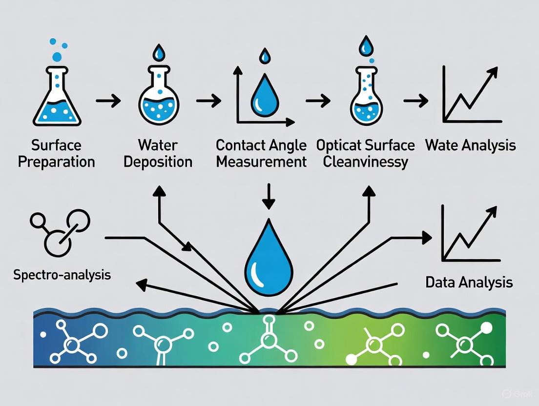
Diagram: Water Contact Angle Measurement Workflow. Two primary methods, Sessile Drop and Captive Bubble, are selected based on surface properties.
The Researcher's Toolkit: Essential Materials and Reagents
Table 4: Essential Research Reagent Solutions for WCA Measurements
| Item | Function & Application | Technical Specifications |
|---|---|---|
| High-Purity Water | Primary measurement liquid for sessile drop | HPLC grade or equivalent; low conductivity [11] |
| Ethanol (Analytical Grade) | Sample cleaning and degreasing | 99.8% purity for ultrasonic cleaning [11] |
| Hamilton Syringe | Precise droplet dispensing | 100 µL capacity; stainless steel needle [11] |
| Contact Angle Goniometer | Optical measurement system | Automated droplet dispensing & image analysis [10] [9] |
| Plasma Cleaner | Surface activation & contamination removal | Oxygen or argon plasma for ultimate cleanliness [3] |
| Environmental Chamber | Control of humidity and temperature | Prevents evaporation artifacts during measurement [11] |
Critical Limitations and Complementary Techniques
While WCA provides exceptional sensitivity to surface chemistry changes, researchers must recognize its limitations. WCA measurements cannot detect subsurface contamination that might affect bond performance, and surface roughness or curvature can introduce measurement variability if not properly accounted for [10]. Furthermore, WCA alone is not a reliable predictor of complex biological responses like cellular attachment. Studies using diverse material libraries have failed to find consistent relationships between WCA and microbial or stem cell attachment, indicating that biological interactions involve complexities beyond simple wettability [13].
For comprehensive surface characterization, WCA should be integrated with complementary analytical techniques:
- X-ray Photoelectron Spectroscopy (XPS): Provides quantitative elemental and chemical state information about the top 1-10 nm of a surface [13] [11].
- Secondary Ion Mass Spectrometry (SIMS): Offers ultra-high sensitivity for detecting organic contamination and mapping chemical distributions [13].
- Atomic Force Microscopy (AFM): Characterizes surface topography and nanoscale roughness that influences wettability [11].
Diagram: WCA Interpretation and Key Limitations. Multiple factors influence WCA, which has specific limitations for surface characterization.
Water contact angle measurement remains an indispensable technique for surface cleanliness research, providing a rapid, sensitive, and quantitative method for detecting contamination and characterizing surface chemistry. The transition from hydrophilic to hydrophobic states, as revealed by WCA, offers critical insights into the molecular-level condition of surfaces, enabling researchers to validate cleaning processes, monitor surface aging, and ensure material performance. While methodological rigor is essential for reproducible results, and complementary techniques are needed for complete surface analysis, WCA stands as a fundamental first step in the researcher's analytical toolkit for surface quality assessment.
The Critical Link Between Low Contact Angles and Optically Clean Surfaces
In the realm of high-precision optics and pharmaceutical development, surface cleanliness transcends ordinary cleanliness standards. Optical surface cleanliness refers to a state of molecular-level purity where surfaces are free from contaminants that could interfere with light transmission, reflection, or subsequent coating adhesion. This specialized form of cleanliness is critical across numerous applications, from surgical endoscope lenses to coating substrates for drug delivery systems [14]. The fundamental challenge researchers face lies in quantitatively assessing this invisible cleanliness standard—a challenge where water contact angle (WCA) measurement has emerged as an indispensable tool.
When a water droplet contacts a properly cleaned optical surface, it exhibits predictable spreading behavior characterized by a low contact angle (typically <30°). This low angle reflects the high surface energy of the clean substrate, which readily interacts with the water droplet. Conversely, contaminated surfaces, often covered with hydrophobic organic residues, exhibit high contact angles where water beads up rather than spreads [3] [10]. This quantitative relationship makes contact angle measurement a sensitive indicator of surface condition, capable of detecting monolayer contamination that would otherwise remain invisible to conventional inspection methods.
Theoretical Foundation: The Science Behind Contact Angle and Cleanliness
Wettability and Surface Energy Fundamentals
The theoretical basis for using contact angle to assess cleanliness stems from classical surface science. According to Young's equation, the contact angle (θ) formed at the solid-liquid-vapor interface is determined by the balance between three interfacial tensions: solid-vapor (γ~sv~), solid-liquid (γ~sl~), and liquid-vapor (γ~lv~) [15]:
γ~sv~ = γ~sl~ + γ~lv~cosθ
This equation establishes the direct relationship between contact angle and the surface energy of the solid (γ~sv~). For optically clean surfaces with high surface energy, the difference between γ~sv~ and γ~sl~ is large, resulting in a small θ value [15] [16]. This thermodynamic foundation validates contact angle as a quantitative measure of surface cleanliness and readiness for subsequent processing.
Surface contamination typically introduces low-energy organic layers that dramatically reduce the apparent surface energy of the substrate. Even monomolecular layers of contamination can significantly increase the water contact angle by disrupting the molecular interactions between the water droplet and the high-energy substrate [3]. This sensitivity makes contact angle measurement particularly valuable for detecting trace contamination that could compromise optical performance or coating adhesion.
Beyond Static Measurements: The Role of Hysteresis
While static contact angle provides valuable initial information, comprehensive surface characterization requires dynamic contact angle analysis, which includes advancing (θ~A~) and receding (θ~R~) angles along with their difference, known as contact angle hysteresis [17] [15]. For optically clean surfaces, both advancing and receding angles remain low, with minimal hysteresis—typically less than 10° [3].
Contact angle hysteresis arises primarily from surface chemical heterogeneity and topographical variations [17] [15]. On contaminated surfaces, the receding angle often shows greater sensitivity to hydrophobic domains, making it a particularly useful indicator for detecting patchy contamination that might not significantly affect the static contact angle [3]. This sophisticated analysis provides researchers with deeper insights into the distribution and nature of surface contaminants.
Comparative Analysis of Contact Angle Measurement Techniques
Methodological Comparison
Researchers investigating optical surface cleanliness can select from several well-established contact angle measurement techniques, each with distinct advantages and limitations for specific experimental scenarios.
Table 1: Comparison of Contact Angle Measurement Techniques
| Method | Working Principle | Optimal Use Cases | Key Advantages | Limitations |
|---|---|---|---|---|
| Optical Tensiometry (Sessile Drop) [17] [16] | Analysis of droplet profile using side-view imaging | Smooth, homogeneous surfaces; basic cleanliness verification | - Direct visualization- Simple experimental setup- Suitable for static and dynamic measurements | - Affected by surface roughness- Requires relatively flat, uniform surfaces |
| Wilhelmy Plate Method [17] | Measurement of force during immersion/emersion | Fibers, membranes, and uniform geometric samples | - Averages over entire sample- Provides inherent advancing/receding angles | - Requires specific sample geometry- Not suitable for coated-only surfaces |
| Tilting Plate Method [17] [15] | Analysis of droplet deformation on tilted surfaces | Hysteresis measurement; roll-off angle determination | - Direct hysteresis measurement- Mimics practical conditions | - More complex setup- Potential influence of inertia |
| Captive Bubble Method [17] | Analysis of air bubble under immersed surface | Highly hydrophilic surfaces; in-situ conditions | - Ideal for superhydrophilic surfaces- Mimics submerged applications | - Requires immersion cell- More complex setup |
Specialized Measurement Approaches
Beyond conventional methods, specialized techniques address unique challenges in optical surface characterization:
Roughness-Corrected Contact Angle: This approach accounts for surface topography effects using the Wenzel equation, which relates the measured contact angle to the theoretical angle through a roughness ratio (r = actual/projected surface area) [17]. This is particularly relevant for optically functional surfaces with engineered microstructures.
Picoliter Droplet Measurements: For small optical components or patterned surfaces, picoliter dispensers can produce droplets as small as 30μm in diameter, enabling measurements on confined areas as small as 0.1-0.3mm [17] [18]. This approach is invaluable for micro-optical elements and patterned substrates where traditional microliter droplets would exceed the region of interest.
Experimental Protocols for Optical Surface Cleanliness Assessment
Standardized Measurement Procedure
To ensure reproducible contact angle measurements for optical cleanliness verification, researchers should follow this standardized protocol:
Sample Preparation: Begin with representative substrate materials (typically 1cm × 1cm or larger). Handle samples with clean gloves or vacuum tweezers to prevent contamination. If comparing cleaning methods, divide samples from the same batch into experimental groups [3].
Surface Cleaning: Apply the cleaning protocol under investigation (e.g., plasma treatment, solvent washing, or ultrasonic cleaning). For metal surfaces, document cleaning sequences as even "clean" metals may instantly react with air molecules, making theoretical surface energies impossible to measure in practice [3].
Baseline Establishment: Measure reference samples with known cleanliness states to establish expected contact angle ranges. Clean glass typically exhibits angles <10°, while properly cleaned metals show angles <30° [3] [19].
Droplet Deposition: Using an automated dispensing system, place a 2-5μL deionized water droplet on the surface. Ensure consistent deposition parameters (needle size, deposition height, and velocity) to minimize measurement variability [16].
Image Acquisition: Capture the droplet image within 1-3 seconds of deposition using a high-resolution camera with appropriate backlighting. Ensure the entire droplet profile and baseline are in focus with high contrast [16].
Angle Calculation: Analyze the droplet image using appropriate fitting algorithms (Young-Laplace, circle, or polynomial fits). For asymmetric droplets, measure both left and right contact angles and report the average [17] [16].
Advanced Characterization Protocols
For comprehensive surface analysis, researchers should supplement static contact angle with these advanced protocols:
Dynamic Contact Angle Measurement:
- Using the needle method, gradually increase the droplet volume (typically 0.1-0.5μL/sec) while recording to determine the advancing angle [17].
- Subsequently decrease the volume at the same rate to measure the receding angle [17].
- Calculate hysteresis as θ~A~ - θ~R~. For optically clean surfaces, hysteresis should be minimal (<10°) [17] [15].
Surface Energy Calculation:
- Measure contact angles with at least three liquids with known polar and dispersive surface tension components (typically water, diiodomethane, and ethylene glycol) [18].
- Apply the Owens-Wendt-Rabel-Kaeble (OWRK) method to calculate the total surface energy and its polar and dispersive components [19] [18].
- Clean, high-energy surfaces typically exhibit total surface energies >40 mN/m with significant polar components [3].
Research Reagent Solutions for Contact Angle Studies
Table 2: Essential Research Materials for Contact Angle Measurements
| Category | Specific Items | Research Application | Technical Notes |
|---|---|---|---|
| Reference Liquids [18] | Deionized water, Diiodomethane, Ethylene glycol | Surface energy calculation; wettability studies | - ≥99% purity recommended- Store in sealed containers- Degas before measurements |
| Cleaning Reagents [3] [14] | Isopropyl alcohol, Acetone, Oxygen plasma, Piranha solution | Surface preparation; cleaning efficacy studies | - Sequence matters for multi-step cleaning- Plasma parameters affect results- Safety protocols critical for piranha |
| Substrate Materials [3] [19] | Fused silica, Borosilicate glass, Silicon wafers, Medical-grade metals | Method validation; substrate-specific studies | - Document surface roughness- Control storage conditions- Note substrate surface energy |
| Calibration Standards | Pre-treated reference slides, Certified surfaces | Instrument calibration; method validation | - Regular calibration schedule- Document storage conditions- Track expiration dates |
Comparative Performance Data: Contact Angle Across Surface Conditions
Empirical data demonstrates the sensitivity of contact angle measurement for detecting surface contamination and verifying cleaning effectiveness across different material classes.
Table 3: Contact Angle Variation with Surface Condition and Cleaning Methods
| Surface Material | Initial State (Contaminated) | After Basic Cleaning | After Advanced Cleaning | Cleaning Methods Employed |
|---|---|---|---|---|
| Copper [3] | 65-85° | 45-60° | 15-25° | Ultrasonic soap → Acid etching → Plasma cleaning |
| Nickel [3] | 60-80° | 40-55° | 10-20° | Ultrasonic soap → Acid etching → Plasma cleaning |
| Glass [19] | 40-60° | 20-35° | <10° | Solvent wash → Plasma treatment → UV ozone |
| PTFE [20] | 95-110° | 90-100° | 85-95° | Surfactant cleaning → Solvent rinse |
| Superhydrophobic Coating [19] | - | - | 158.2±0.7° | Fluorine-free bilayer fabrication |
The data clearly demonstrates that properly cleaned high-energy surfaces (metals and glass) consistently achieve low contact angles, while inherently low-energy materials like PTFE maintain high angles even after cleaning. Superhydrophobic coatings represent the extreme opposite of clean high-energy surfaces, with intentionally engineered high contact angles for self-cleaning applications [19].
Limitations and Complementary Analytical Techniques
While contact angle measurement provides exceptional sensitivity to surface chemical state, researchers should recognize its limitations:
- Spatial averaging provides information about the average surface condition but may miss localized contamination [10].
- Subsurface contamination that doesn't affect surface chemistry may not be detected [10].
- Surface roughness significantly influences measurements and requires specialized correction methods [17].
- Molecular specificity is limited—contact angle indicates contamination but doesn't identify specific contaminants [10].
For comprehensive surface characterization, researchers should consider integrating contact angle with complementary techniques:
- X-ray Photoelectron Spectroscopy (XPS): Provides elemental and chemical state information for contaminant identification [10].
- Atomic Force Microscopy (AFM): Maps topographical features and nanoscale heterogeneity [10].
- Optical Emission Spectroscopy: Useful for in-process monitoring of plasma cleaning treatments [21].
Water contact angle measurement stands as an indispensable tool in the researcher's arsenal for quantifying optical surface cleanliness. Its exceptional sensitivity to molecular-level contamination, rapid implementation, and quantitative output make it ideal for comparing cleaning methods, establishing cleanliness standards, and maintaining quality control across diverse optical applications. As optical technologies advance toward increasingly stringent performance requirements, the critical link between low contact angles and optically clean surfaces will continue to drive innovation in both measurement methodologies and cleaning processes.
The experimental data and methodologies presented in this guide provide researchers with a robust framework for implementing contact angle measurements in their optical cleanliness verification protocols. By selecting appropriate measurement techniques, following standardized protocols, and understanding both the capabilities and limitations of the method, scientists can leverage this powerful technique to advance optical system performance across pharmaceutical, medical, and industrial applications.
Surface cleanliness is paramount in optical research and manufacturing, where trace-level contaminants can significantly alter interfacial properties and compromise performance. This guide objectively compares the sensitivity of water contact angle measurement against other surface analysis techniques for detecting molecular contamination. We present quantitative experimental data demonstrating how sub-monolayer contaminants dramatically shift droplet behavior, supported by standardized protocols that enable researchers to correlate macroscopic wettability changes with microscopic surface condition.
In optical surface research, cleanliness is not merely about visual perfection but involves molecular-level purity that directly affects performance. Trace contaminants—including skin oils, dust particulates, and process residues—form molecular layers that fundamentally alter how light interacts with surfaces and how liquids wet them. These changes manifest quantitatively through water contact angle measurements, which provide a rapid, sensitive method for detecting surface contamination that often eludes other analytical techniques.
The significance of droplet behavior as a contamination indicator stems from direct molecular interaction at the three-phase contact line. Even fractional monolayer coverage of hydrophobic contaminants on theoretically high-energy surfaces drastically increases water contact angles, while hydrophilic contaminants produce the opposite effect. This sensitivity makes contact angle measurement an indispensable tool for quality control in optical manufacturing, pharmaceutical development, and surface science research.
Experimental Protocols for Contamination Detection
Standardized Contact Angle Measurement Procedure
Reliable contamination detection requires strict adherence to standardized measurement protocols. The following procedure, adapted from established methodologies, ensures reproducible results [22] [7]:
Sample Preparation: Cut samples to standardized dimensions (typically 15mm wide strips). Clean surfaces with appropriate solvents (isopropanol, acetone) using lint-free cloths to remove gross contamination. Handle samples with tweezers or gloves to prevent fingerprint deposition [7].
Static Contact Angle Measurement:
- Utilize an optical tensiometer/goniometer with high-speed camera system
- Dispense 5-8 μL high-purity deionized water (surface tension: 72.8±0.5 mN/m at 20°C) via precision syringe
- Position needle 1-2mm above surface, form pendant droplet, and gently contact surface
- Capture image within 10-15 seconds of deposition to minimize evaporation effects
- Analyze droplet profile using Young-Laplace fitting in specialized software [17] [7]
Dynamic Contact Angle Measurement:
Environmental Control: Maintain temperature at 23±2°C and constant humidity throughout measurements to prevent environmental artifacts [7].
Replication: Perform minimum 5-10 measurements per sample at different locations, spacing measurement points至少25mm apart [7].
Advanced Techniques for Challenging Surfaces
For rough or porous substrates that complicate traditional measurements, specialized approaches are required:
Roughness Correction: Measure surface topography simultaneously with contact angle using integrated profilometry, then apply Wenzel equation corrections to account for roughness contributions [17].
Powder Characterization: Compress powders into tablets using hydraulic presses or create thin powder layers on adhesive substrates before measurement [23].
Micro-Droplet Methods: Employ picoliter dispensers and high-magnification optics for measurements on small or patterned surfaces where standard droplets are impractical [17].
Figure 1: Experimental workflow for contamination detection via contact angle measurement. Strict protocol adherence ensures reproducible results sensitive to molecular-level surface contaminants [22] [7].
Quantitative Data: Contamination Impact on Droplet Behavior
Metallic Surface Contamination
Metal surfaces exhibit extreme sensitivity to organic contamination due to their inherently high surface energy. The table below demonstrates how successive cleaning stages remove contaminants and progressively reduce water contact angles on metal surfaces [3]:
Table 1: Water contact angle changes on metal surfaces through cleaning stages
| Surface Material | Pre-Cleaning CA (°) | After Ultrasonic Cleaning CA (°) | After Plasma Cleaning CA (°) |
|---|---|---|---|
| Copper | 74° | 16° | 8° |
| Nickel | 63° | 18° | 9° |
Polymeric Surface Contamination
Polymer surfaces present more complex wettability behavior, where receding contact angles often show greater sensitivity to contamination than static measurements:
Table 2: Advancing and receding contact angles on polymers before and after cleaning
| Polymer Type | Condition | Advancing CA (°) | Receding CA (°) | Hysteresis (°) |
|---|---|---|---|---|
| Teflon | Contaminated | 108° | 65° | 43° |
| Teflon | Cleaned | 120° | 95° | 25° |
| Polypropylene | Contaminated | 95° | 45° | 50° |
| Polypropylene | Cleaned | 102° | 85° | 17° |
The data reveals that while static angles on Teflon increased post-cleaning (consistent with contaminant removal), the receding angle provided more definitive cleanliness verification, increasing from 65° to 95° [3]. This highlights the critical importance of dynamic contact angle measurements for accurate contamination assessment.
Mineral Surface Heterogeneity Effects
Natural mineral surfaces demonstrate how localized heterogeneity affects wettability measurements. Using combined argon ion polishing and atomic force microscopy (Ar+-AFM), researchers obtained more consistent contact angle data (20.1°±2.5°) on K-feldspar homogeneous areas, versus highly variable results (10°-51°) with traditional optical methods on heterogeneous surfaces [24]. This underscores how conventional measurements can be confounded by surface heterogeneity, potentially masking contamination effects.
The Scientist's Toolkit: Essential Research Reagent Solutions
Table 3: Essential reagents and equipment for contamination detection via contact angle
| Item | Function | Specification Notes |
|---|---|---|
| Optical Tensiometer | Primary measurement instrument | High-speed camera, precision dispensing system, environmental enclosure [17] |
| Deionized Water | Primary probe liquid | High-purity (18.2 MΩ·cm), surface tension verification (72.8±0.5 mN/m at 20°C) [7] |
| Precision Syringes | Liquid dispensing | Calibrated volume, fine-gauge needles (e.g., 22-26G) for consistent droplet formation [7] |
| Solvent Cleaning Kit | Surface preparation | HPLC-grade isopropanol, acetone, lint-free wipes, gloves [7] |
| Reference Materials | Calibration | Surfaces with known contact angles (e.g., clean silicon wafer, Teflon) [22] |
| Picoliter Dispenser | Micro-scale measurements | Essential for small features, fiber analysis, patterned surfaces [17] |
| Surface Roughness Module | Topography correlation | Combined profilometry for roughness-corrected contact angles [17] |
| Anti-static Equipment | Sample handling | Ionizer guns for polymer films to prevent electrostatic artifacts [7] |
Comparative Analysis: Contact Angle vs. Alternative Techniques
While numerous surface analysis techniques exist, contact angle measurement offers unique advantages for contamination detection:
Sensitivity Comparison: Contact angle detects sub-monolayer surface contamination (0.1-1nm thickness), comparable to XPS and ToF-SIMS but with significantly lower cost and complexity [3].
Throughput Advantage: Contact angle measurements require seconds to minutes versus hours for ultra-high vacuum techniques, enabling rapid quality control decisions [4].
Limitations: Contact angle provides indirect chemical information compared to spectroscopic methods. It detects contamination presence but not specific chemical identity.
Complementary Approach: For complete surface characterization, contact angle serves as an ideal screening method followed by targeted spectroscopic analysis of suspect samples.
Figure 2: Contamination detection techniques comparison. Contact angle measurement provides sensitive, rapid detection of molecular-level surface changes caused by contaminants [24] [3] [4].
Water contact angle measurement stands as an exceptionally sensitive technique for detecting trace surface contaminants that dramatically alter droplet behavior. Through standardized protocols and proper data interpretation—particularly leveraging dynamic contact angle information—researchers can identify molecular-level contamination with precision rivaling sophisticated surface analysis instruments. The methodology provides an optimal balance of sensitivity, practicality, and quantitative output for optical surface cleanliness verification across research and industrial applications.
Within optical surface cleanliness research, the imperative to detect molecular-level contaminants without altering the test surface has driven the adoption of advanced analytical techniques. This guide objectively compares the performance of Water Contact Angle (WCA) measurement against traditional alternatives for identifying invisible residues that compromise surface quality in critical applications. While traditional methods like the water break test offer simplicity, quantitative WCA analysis provides superior sensitivity to both hydrophobic and hydrophilic contaminants, non-destructive testing capability, and data-driven traceability essential for modern quality systems. Experimental data from controlled studies demonstrate WCA's capability to detect contaminant layers as thin as 2-3 molecules, with measurement precision exceeding subjective visual methods by factors of 5-10x. The integration of WCA with machine learning platforms further enhances its predictive capability for long-term surface performance, positioning it as an indispensable tool for research and development professionals requiring non-destructive surface characterization.
Surface contamination at the molecular level represents a formidable challenge in pharmaceutical development, medical device manufacturing, and optical component production. These invisible residues—including silicone oils, surfactant layers, machining lubricants, and processing aids—often evade visual inspection yet profoundly impact adhesion, coating uniformity, and performance reliability. The limitations of traditional cleanliness assessment methods have created a technological gap in non-destructive surface characterization, particularly for detecting contaminants that do not manifest as visible films or particulates.
Water Contact Angle measurement has emerged as a powerful solution to this challenge, transitioning from a specialized research technique to a validated production tool. WCA operates on fundamental principles of surface science, measuring the angle formed at the solid-liquid-gas interface to quantify surface energy and wettability. This deceptively simple measurement provides a sensitive probe of the outermost molecular layers of a surface, detecting chemical heterogeneity that other methods miss. The technique's non-destructive nature preserves sample integrity while providing quantitative data that correlates directly with adhesion performance and coating quality.
Comparative Analysis of Surface Cleanliness Assessment Methods
Performance Comparison of Primary Assessment Techniques
Table 1: Quantitative comparison of surface cleanliness assessment methods
| Method | Detection Principle | Sensitivity | Quantitative Output | Contaminants Detected | Throughput | Capital Cost |
|---|---|---|---|---|---|---|
| Water Contact Angle | Liquid wetting behavior | 2-3 molecular layers | Numerical angle (°) | Hydrophobic & hydrophilic | Medium-high | Medium |
| Water Break Test | Visual water film continuity | Macroscopic films | Pass/fail visual assessment | Hydrophobic only | Very high | Very low |
| Dyne Pens | Surface tension inks | ~10 mN/m resolution | Semi-quantitative (mN/m) | Limited range | High | Low |
| FTIR Spectroscopy | Molecular absorption | Monolayers | Spectral peaks | Chemical identification | Low | High |
| XPS (ESCA) | Electron emission | 1-10 nm | Atomic composition | Elemental specificity | Very low | Very high |
Technical Limitations of Traditional Methods
The water break test, despite its historical prevalence and operational simplicity, suffers from critical limitations in modern precision manufacturing environments. As a purely subjective visual assessment, it depends entirely on technician interpretation, yielding results that vary significantly between operators [25]. The method fundamentally detects only hydrophobic contaminants like oils and greases, while remaining blind to hydrophilic residues such as surfactants or cleaning agent remnants that can equally compromise adhesion [25]. Perhaps most concerning is its potential to introduce contamination through non-purified test water, inadvertently depositing minerals, organics, or particulates on the validated surface [25].
Dyne pens and similar surface tension test inks provide slightly more granularity than water break tests but remain semi-quantitative at best. These methods introduce their own contaminants to the surface through the testing inks, rendering the tested area unusable for subsequent processing. Their results are influenced by application pressure and technique, introducing operator-dependent variables that compromise measurement repeatability. Like the water break test, they offer no permanent record or traceable data for process validation or quality audits [10].
Water Contact Angle Measurement: Methodologies and Experimental Protocols
Fundamental Measurement Principles
Water Contact Angle measurement quantifies the wettability of a surface by analyzing the interface where water, solid surface, and air meet. The contact angle (θ, theta) is geometrically defined as the angle formed by a liquid at the three-phase boundary where liquid, gas, and solid intersect [17]. Low contact angles (<30°) indicate high surface energy and wettability, characteristic of clean, reactive surfaces ready for bonding or coating. High contact angles (>90°) signal low surface energy surfaces, typically contaminated with hydrophobic substances or improperly prepared for adhesion processes [10].
The measurement sensitivity stems from its ability to probe the top 2-3 molecular layers of a surface, detecting chemical changes invisible to the naked eye [25]. Even trace contaminants covering less than 5% of a surface can produce measurable changes in contact angle values, providing early warning of process deviation before adhesion failures occur.
Core Measurement Methodologies
Static Sessile Drop Measurement The most common WCA approach involves depositing a precise water droplet (typically 2-10 μL) onto the surface and optically measuring the static angle using a goniometer [17]. The droplet image is analyzed with fitting algorithms (Young-Laplace, circle, or polynomial) to determine the contact angle at the three-phase boundary. This method provides a rapid, quantitative assessment of surface wettability and is suitable for relatively smooth, homogeneous surfaces [17].
Dynamic Contact Angle Analysis For more comprehensive surface characterization, dynamic measurements capture advancing (θA) and receding (θR) angles, whose difference defines contact angle hysteresis [17]. The needle method gradually increases droplet volume to measure advancing angle, then decreases volume for receding angle [17]. The tilting method places a droplet on a surface that is progressively tilted, measuring advancing angle at the droplet front and receding angle at the back just as movement begins [17]. Contact angle hysteresis and the related roll-off angle (the tilt angle at which droplets begin to move) provide critical information about surface heterogeneity and droplet adhesion [17] [26].
Table 2: Experimental protocols for WCA measurement techniques
| Method | Sample Preparation | Measurement Parameters | Data Analysis | Best For |
|---|---|---|---|---|
| Static Sessile Drop | Clean, dry, flat surface | 2-10 μL purified water, ambient conditions | Young-Laplace fitting | Quick cleanliness verification |
| Advancing/Receding (Needle) | Uniform surface chemistry | Controlled volume change 0.1-1 μL/sec | Hysteresis calculation (θA - θR) | Surface heterogeneity analysis |
| Tilting Plate | Rigid, mountable samples | Tilt rate 0.5-2°/sec, droplet volume 5-15 μL | Roll-off angle determination | Self-cleaning surfaces, drag reduction |
| Wilhelmy Plate | Uniform, rectangular samples | Known perimeter, immersion speed 0.1-5 mm/min | Force balance calculation | Fibers, porous materials, average properties |
Experimental Workflow for Contamination Detection
The following diagram illustrates the standardized experimental workflow for detecting invisible residues through WCA measurement:
Advanced Techniques for Challenging Surfaces
Roughness-Corrected Measurements Surface roughness significantly influences apparent contact angles through mechanisms described by Wenzel (complete penetration into roughness grooves) and Cassie-Baxter (composite air-solid interface) models [17]. Roughness-corrected measurements combine topographical analysis with contact angle data using the roughness ratio (actual versus projected surface area) to normalize wettability measurements [17].
Specialized Sample Protocols
- Fibers and thin materials: Employ picoliter dispensers for droplets with ~30μm diameter or meniscus measurement techniques [17]
- Powders and porous materials: Utilize Washburn method for capillary penetration analysis or compressed tablet measurements [17]
- Small or curved areas: Implement picoliter dispensing with high-magnification optics for features below standard droplet size [17]
- Extreme conditions: Employ temperature-controlled stages (-40°C to 250°C) or high-pressure chambers (up to 400 bar) for specialized applications [17]
Experimental Data: WCA Sensitivity to Specific Contaminants
Quantitative Detection Thresholds
Table 3: WCA response to common manufacturing contaminants
| Contaminant Type | Base Surface | Clean Surface WCA | Contaminated WCA | Detection Threshold | Adhesion Impact |
|---|---|---|---|---|---|
| Silicone Oil | Stainless steel | 65° ± 3° | 105° ± 8° | 0.2 mono-layer | Coating delamination |
| Fatty Acids | Glass | 42° ± 2° | 85° ± 5° | 0.5 mono-layer | 80% bond strength reduction |
| Machine Oils | Aluminum | 72° ± 4° | 112° ± 6° | 2 nm film | Paint adhesion failure |
| Surfactant Residues | Polymers | 75° ± 3° | 45° ± 7° | 0.3 mono-layer | Coating cratering |
| Oxidation Layer | Copper | 65° ± 2° | 95° ± 4° | 5 nm thickness | Solderability decrease |
Controlled contamination studies demonstrate WCA's exceptional sensitivity to surface chemistry changes. On stainless steel surfaces, silicone oil contamination as thin as 0.2 monolayers produces measurable WCA increases from 65° to over 105°, providing early detection long before adhesion failures manifest [10]. Similarly, surfactant residues that remain invisible to water break testing produce dramatic WCA decreases from 75° to 45° on polymer surfaces, signaling hydrophilic contamination that can cause coating defects [25].
Correlation with Bond Performance
The relationship between WCA measurements and adhesion performance follows a well-established non-linear correlation. Surfaces with WCAs below 40° typically exhibit excellent adhesion for most coatings and adhesives, while those exceeding 80° present increasing risk of bonding failures. The most critical process control window often falls between 50°-70°, where small WCA changes signal significant contamination events requiring intervention [10].
The Scientist's Toolkit: Essential Research Reagent Solutions
Table 4: Essential materials and reagents for WCA contamination studies
| Item | Specifications | Application | Critical Function |
|---|---|---|---|
| Ultrapure Water | Type I (18.2 MΩ·cm), TOC < 5 ppb | Reference liquid | Eliminates water-born contamination variables |
| Certified Clean Substrates | Mirror-finish stainless steel, silicon wafers | Measurement baseline | Provides validated reference surfaces |
| Contaminant Standards | Certified silicone oil, fatty acid, alkane solutions | Controlled contamination studies | Enables quantitative dose-response calibration |
| Precision Syringes | 1-10 μL, PTFE-coated tips | Droplet deposition | Ensures consistent droplet volume and placement |
| Optical Goniometer | 0.1° resolution, automated dispensing | Angle measurement | Provides quantitative, repeatable data |
| Surface Energy Kits | Multiple liquids with known surface tensions | Surface energy calculation | Enables surface energy component analysis |
Emerging Applications and Machine Learning Integration
Advanced WCA methodologies are being integrated with machine learning platforms to predict long-term surface behavior from short-term measurements. Recent studies demonstrate that ML algorithms (Random Forest, XGBoost) can accurately forecast contact angle stability under various environmental stressors by training on experimental degradation data [27] [28]. These models analyze complex, non-linear relationships between coating composition, application parameters, and WCA performance that traditional statistical methods often miss.
In co-sputtered coating development, ML models have reduced parameter optimization from months of iterative testing to days of predictive modeling. The Random Forest algorithm has demonstrated particular efficacy in predicting WCA of co-sputtered coatings, accurately forecasting superhydrophilic behavior (WCA < 5°) based on deposition parameters [27]. Similar approaches successfully predict the stability of superhydrophobic graphene-based coatings on copper substrates under mechanical stress and corrosive conditions [28].
The following diagram illustrates the integration of WCA measurement with machine learning for predictive surface analysis:
Water Contact Angle measurement represents a critical advancement in non-destructive surface characterization, providing sensitivity to invisible residues that traditional methods cannot detect. While the water break test retains limited applicability for gross contamination checks in non-critical applications, WCA offers quantitative, reproducible data essential for modern manufacturing and research environments. The technique's ability to probe the top molecular layers of a surface without alteration makes it particularly valuable for pharmaceutical development, medical device manufacturing, and optical surface research where sample preservation is paramount.
The integration of WCA with advanced analytical platforms and machine learning algorithms represents the future of predictive surface science, enabling researchers to forecast long-term performance from immediate measurements. As surface cleanliness requirements continue to tighten across industries, WCA methodology stands as an indispensable tool for the detection and quantification of invisible residues that compromise product quality and performance.
Measuring for Cleanliness: Methodologies and Direct Applications for Optical Surfaces
The contact angle is a fundamental quantitative measure used to evaluate the wettability of a solid surface by a liquid. It is defined as the angle formed between the tangent to the liquid-vapor interface and the tangent to the solid-liquid interface at the three-phase (solid-liquid-vapor) contact line [29]. The value of this angle, typically denoted as θ (theta), is a direct consequence of the balance between the cohesive forces within the liquid and the adhesive forces between the liquid and the solid [30]. In practical terms, a small contact angle (θ < 90°) signifies that the liquid spreads readily over the surface, characterizing a hydrophilic or wetting state. Conversely, a large contact angle (θ > 90°) indicates that the liquid beads up, characterizing a hydrophobic or non-wetting state [31] [29]. For instance, a contact angle of 0° denotes complete wetting, while angles exceeding 150° are associated with superhydrophobic surfaces [31].
The measurement of contact angle is not merely an academic exercise; it is a critical diagnostic tool in optical surface cleanliness research and numerous industrial and scientific fields. The wettability of a surface directly influences phenomena such as coating uniformity, adhesion, cleaning efficacy, and lubricity [31] [32]. For researchers and drug development professionals, understanding and controlling surface wettability is essential for applications ranging from the development of biocompatible implants and contact lenses to the optimization of drug delivery surfaces and diagnostic platforms [33] [34]. The three techniques detailed in this guide—Sessile Drop, Captive Bubble, and Wilhelmy Plate—represent the core methodologies for determining this crucial parameter, each with distinct principles, advantages, and limitations.
The following table provides a consolidated, data-driven comparison of the three primary contact angle measurement techniques to guide method selection.
Table 1: Comprehensive comparison of sessile drop, captive bubble, and Wilhelmy plate methods.
| Feature | Sessile Drop | Captive Bubble | Wilhelmy Plate |
|---|---|---|---|
| Basic Principle | Analyzes shape of a liquid droplet seated on a solid surface [30]. | Analyzes shape of an air bubble attached to a solid surface submerged in liquid [30] [33]. | Measures force exerted on a solid sample as it is immersed/withdrawn from liquid [30] [32]. |
| Measured Angle | Considered close to the advancing contact angle [33]. | Considered close to the receding contact angle [33]. | Directly measures both advancing (immersion) and receding (emersion) angles [34]. |
| Sample Environment | Ambient air (can lead to dehydration) [34]. | Fully immersed in liquid (preserves hydrated state) [33] [34]. | Liquid is contained in a vessel; sample is cycled in/out [30]. |
| Key Advantages | Fast, easy, versatile for sample shapes [30] [34]. | Ideal for hydrated/hydrophilic surfaces; prevents dehydration [33] [34]. | Provides highly accurate and reproducible data; measures average value and hysteresis [34] [32]. |
| Key Limitations | Susceptible to dehydration; subjective analysis for some models [34]. | Time-consuming bubble alignment; requires transparent liquid [34]. | Requires sample with uniform geometry and known perimeter; more complex setup [30] [34]. |
| Best For | Routine analysis, quality control, samples with irregular surfaces [32]. | Bio-medical materials, contact lenses, hydrogels, and any application where the surface is naturally hydrated [33] [34]. | High-accuracy research on materials with regular geometry (e.g., films, fibers); dynamic wetting studies [34] [32]. |
Detailed Methodologies and Experimental Protocols
Sessile Drop Method
The sessile drop technique is the most widely used optical method for contact angle measurement due to its straightforward setup and minimal sample preparation [30] [34].
Experimental Protocol:
- Sample Preparation: The solid sample is placed on a level stage. For consistent results, the surface may be blotted or cleaned according to protocol, though this can risk dehydration [34].
- Dispensing: A precise volume of probe liquid (e.g., water) is dispensed from a syringe equipped with a flat-tipped needle onto the sample surface, forming a pendant drop that transfers to become a sessile drop [34].
- Image Capture: A high-resolution camera, aligned orthogonally to the sample surface, captures an image of the static droplet immediately after deposition to minimize evaporation effects.
- Analysis: The contact angle is determined by software analysis of the droplet image. Common algorithms include:
- Tangent Method: Fits a tangent line to the droplet contour at the three-phase contact point. This is a local method and works even with needle-in-drop setups, but can be subjective [35].
- Circle Fitting / θ/2 Method: Assumes the droplet is a spherical cap and fits a circle to its outline. This is best for small contact angles (<20°) and small volumes where gravity is negligible [35].
- Young-Laplace Fitting: Fits the entire droplet profile to the Young-Laplace equation, which accounts for gravity-induced distortion. This is the most accurate method for larger droplets and contact angles >60°, but requires axisymmetric drops [35].
Captive Bubble Method
The captive bubble method is essentially the inverse of the sessile drop technique and is particularly valuable for studying hydrophilic and hydrated surfaces [33] [34].
Experimental Protocol:
- Sample Hydration: The solid sample is fully immersed and allowed to equilibrate in a transparent cell filled with the probe liquid (often an aqueous solution). This ensures the surface remains in a hydrated state throughout the measurement [33].
- Bubble Formation: A syringe with a J-shaped needle is positioned beneath the sample surface. A small bubble of air (or another immiscible gas/fluid) is injected from the syringe until it is "captured" on the underside of the sample surface [30] [33].
- Image Capture: A camera captures an image of the attached bubble from the side.
- Analysis: The contact angle is analyzed from the bubble image. Because the bubble is a gas-liquid interface in a liquid medium, the angle measured (θbubble) is the supplement to the conventional water-in-air angle (θwater). Therefore, θwater = 180° - θbubble [33]. The Laplace-Young equation fit is often the preferred analysis method here, especially for hydrophilic surfaces where bubble shapes are highly distorted [33] [35].
A modified captive bubble method has been developed to improve accuracy. This version places the entire setup in a pressure chamber. After the bubble is formed, the needle is retracted, and precise pressure changes in the chamber are used to control the bubble size for advancing and receding angle measurements, eliminating the interference of the needle [36].
Wilhelmy Plate Method
The Wilhelmy plate method is a force tensiometry technique that provides dynamic contact angle data [30] [32].
Experimental Protocol:
- Sample Preparation: A solid sample with a known, uniform geometry (typically a rectangular plate or a cylindrical rod) is suspended from a highly sensitive microbalance. The perimeter (P) of the sample at the immersion point must be precisely known [30] [32].
- Liquid Preparation: A vessel containing the probe liquid, with a known surface tension (γLV), is raised on a movable stage.
- Force Measurement: The liquid vessel is raised at a constant rate, immersing the sample to a set depth, and then lowered to withdraw it. The microbalance continuously records the force (F) on the sample during this cycle.
- Data Analysis: The force data is used to calculate the contact angle via the Wilhelmy equation:
- F = γLV · P · cosθ Rearranging for the contact angle: θ = cos-1[F / (γLV · P)] The force during immersion is used to calculate the advancing contact angle, while the force during withdrawal gives the receding contact angle. The difference between these two values is the contact angle hysteresis, which provides insights into surface heterogeneity and roughness [34] [32].
Decision Workflow and Application Context
The following diagram illustrates the logical decision process for selecting the most appropriate measurement technique based on sample properties and research objectives.
Essential Research Reagent Solutions
A successful contact angle measurement experiment relies on more than just the core instrument. The following table details key reagents and materials essential for preparing and conducting these analyses.
Table 2: Key research reagents and materials for contact angle measurement.
| Item | Function / Purpose | Technical Considerations |
|---|---|---|
| Probe Liquids | High-purity liquids (e.g., water, diiodomethane, ethylene glycol) used to probe surface energy and wettability. | Purity is critical to avoid contamination affecting surface tension. Multiple liquids are required for surface free energy calculations [37]. |
| Sample Preparation Kits | Tools for cleaning and pre-treating sample surfaces (e.g., plasma cleaners, UV ozone units, solvent baths). | Ensures a clean, contaminant-free surface for reliable baseline measurements. Plasma treatment can temporarily increase hydrophilicity [31]. |
| Precision Syringes & Needles | Used to generate consistent droplets (sessile drop) or bubbles (captive bubble). | Flat-tipped needles (e.g., J-shaped) are essential for proper droplet detachment and bubble capture [33] [34]. |
| Software Analysis Modules | Algorithms for image (sessile drop, captive bubble) or force (Wilhelmy plate) data processing. | Selection of correct model (e.g., Tangent, Circle, Laplace-Young) is vital for accuracy, depending on angle and droplet shape [35]. |
| Standard Reference Samples | Surfaces with known, stable contact angles (e.g., Teflon for hydrophobic, glass for hydrophilic). | Used for daily instrument calibration and validation to ensure measurement accuracy and repeatability. |
Advanced Topics in Contact Angle Measurement
Data Analysis and Theoretical Models
The accuracy of optical contact angle measurements is heavily dependent on the chosen analysis model. The Laplace-Young equation represents the most rigorous approach, as it accounts for the entire droplet shape under the influence of gravity [35] [38]. Solving this equation allows for the precise calculation of contact angle, even for large droplets where gravitational sag is significant. Simpler models, such as the Circle Fitting (or θ/2) method, assume the droplet is a spherical cap and are only accurate for small droplets (<5 µL for water) and low contact angles where gravity is negligible [35]. The Tangent method offers flexibility for non-axisymmetric drops or when a needle remains in the droplet, but it can be more subjective and less precise than whole-shape fitting methods [35].
Significance of Dynamic Angles and Hysteresis
Static contact angle measurements provide a snapshot of wettability, but real-world surfaces often exhibit contact angle hysteresis—the difference between the advancing (θA) and receding (θR) angles [34] [36]. This hysteresis is a critical parameter, as it quantifies surface heterogeneity and roughness. The Wilhelmy plate method directly measures this hysteresis during the immersion-emersion cycle [34]. In optical methods, dynamic angles are assessed by adding/removing liquid to expand/contract the droplet base, or by using the tilting plate method [30]. Understanding hysteresis is essential for applications involving moving liquid fronts, such as coating processes, lubrication, and the performance of self-cleaning surfaces.
In surface science, particularly for optical cleanliness research, the water contact angle (WCA) serves as a crucial, non-destructive indicator of surface wettability and chemical cleanliness. A contact angle is defined as the angle formed between a liquid surface and a solid surface at their point of contact, quantifying the wettability via the Young equation [16] [15]. For optical applications, even microscopic contamination or surface energy variations can significantly impact performance, making precise contact angle measurement indispensable. The fundamental challenge, however, lies in the fact that optical components are rarely ideal flat planes; they encompass a spectrum of geometries from flat lenses and prisms to curved mirrors and complex sculptured surfaces. Selecting an inappropriate measurement method for a given geometry can yield inaccurate data, leading to incorrect conclusions about surface cleanliness or coating efficacy. This guide objectively compares the performance of various contact angle measurement techniques across flat, curved, and complex optical geometries, providing researchers with the experimental protocols and data needed to make informed methodological choices.
Fundamental Principles and Measurement Methods
The core principle of contact angle measurement is based on the equilibrium of interfacial tensions at the solid-liquid-vapor boundary, described by the Young equation: γSG - γSL - γLGcosθC = 0, where γ represents the interfacial tensions between solid (S), liquid (L), and gas (G) phases, and θC is the equilibrium contact angle [15]. Real-world surfaces exhibit contact angle hysteresis, the difference between the advancing (maximal) and receding (minimal) contact angles, which provides additional information about surface heterogeneity and roughness [15].
Several established methods exist for measuring these angles, each with unique strengths and limitations, which become critically important when dealing with non-flat geometries.
- Sessile Drop: This is the most common method, where a droplet is placed on a solid sample and imaged with a high-resolution camera. The contact angle is then determined automatically by software [39]. It provides a static contact angle and is ideal for quality control and surface energy calculations.
- Needle/Tilting Methods: These dynamic methods measure advancing and receding contact angles. In the needle method, droplet volume is increased or decreased to move the contact line [39]. In the tilting method, the substrate is tilted until the drop moves, allowing measurement of both angles and the roll-off angle [39] [40]. These are crucial for studying superhydrophobic surfaces [41] [39].
- Wilhelmy Plate: This force-based method uses a sensitive balance to measure the force exerted on a sample as it is immersed in or withdrawn from a liquid. It provides an average advancing and receding contact angle for the entire immersed surface [39] [40].
- 3D Contact Angle: A newer method that reconstructs a digital spatial image of the drop from top-view reflections and laser measurements, eliminating the need for manual baseline identification [42].
- Captive Bubble: Used for solid-liquid-liquid systems or superhydrophilic surfaces, this method involves measuring the contact angle of an air bubble on a surface immersed in liquid [39].
Comparative Analysis of Methods for Different Surface Geometries
The accuracy of a contact angle measurement is highly dependent on the match between the measurement technique and the substrate geometry. The following sections and comparative table detail the performance, advantages, and limitations of each method in the context of flat, curved, and complex surfaces.
Table 1: Comprehensive Comparison of Contact Angle Methods for Different Optical Geometries
| Measurement Method | Optimal Geometry | Key Advantages | Key Limitations | Typical Accuracy/Precision |
|---|---|---|---|---|
| Sessile Drop (Standard 2D) | Flat, homogeneous surfaces [43] | Simple setup; quick; non-destructive; wide industry adoption [16] [39] | Baseline errors on curved/complex surfaces [43] [42]; assumes axisymmetric drop | Varies; highly dependent on operator and baseline definition |
| Sessile Drop (with Advanced Fitting) | Gently curved, extruded surfaces [43] | Does not require a priori information on surface tension or drop volume [43] | Complex image processing; requires multiple viewpoints for 3D surfaces | MAE of 0.8° for θ < 160° on smooth surfaces [43] |
| Tilting Stage Method | Flat, low-hysteresis surfaces [39] | Directly measures advancing/receding angles and roll-off angle [39] | Challenging for very high or low hysteresis; shear forces may influence results [15] | Provides hysteresis data critical for self-cleaning analysis [41] |
| 3D Contact Angle Method | Complex, curved, and rough surfaces [42] | No baseline finding; robust for QC; minimizes user intervention [42] | Specialized equipment required; limited to static angle measurement | High robustness; less susceptible to interference [42] |
| Wilhelmy Plate | Fibers, thin films, and uniform solids [39] [40] | Averages angle over entire sample; high sensitivity [39] [40] | Requires identical sides; destructive for some samples; not for localized analysis | Effective for fibers down to 7 µm [39] |
| Captive Bubble | Surfaces requiring immersion (e.g., contact lenses) [39] | Ideal for superhydrophilic surfaces and solid-liquid-liquid systems [39] | Requires immersion cell; not for standard hydrophobic surface analysis | Suitable for in-situ analysis of hydrated surfaces |
Methodological Protocols for Key Experiments
Protocol for Sessile Drop Measurement on Flat Surfaces
This is the foundational protocol for flat optical surfaces [16] [39].
- Sample Preparation: Clean the substrate with isopropyl alcohol and a soft, lint-free cloth to prevent scratching and remove contaminants [44].
- Droplet Deposition: Using a precision syringe pump or dosing unit, deposit a small droplet (typically 2-6 µL for precision, though larger volumes like 5 μL or 14 μL are also used) gently onto the static, level surface [44] [45].
- Image Acquisition: Illuminate the droplet from behind with a diffuse light source and capture a high-contrast, high-resolution side-view image using a CCD camera. Ensure the entire droplet is in focus with a sufficient depth of field [16] [45].
- Image Analysis:
- Define Baseline: Manually or automatically set the horizontal baseline representing the solid surface [16] [45].
- Trace Droplet Edge: Use edge-detection algorithms (e.g., binary threshold or Canny edge detection) to extract the droplet contour [16] [45].
- Calculate Contact Angle: Fit a polynomial or elliptical curve to the droplet edge and calculate the angle between the baseline and the tangent at the point of intersection [16] [45].
Protocol for Curved Surfaces Using Multi-Angle Imaging
This protocol, adapted from research on gemstones and cylinders, is suitable for spherical or cylindrical optics [43].
- Setup: Place the curved sample on a rotation stage within an anti-vibration setup.
- Multi-Angle Image Acquisition: Deposit a droplet and acquire multiple side-view images from different angles (e.g., from 0° to 360° with a 30° step), ensuring the droplet shape is not altered during rotation [43].
- 3D Shape Reconstruction: Use image processing and spline fitting to extract the drop contour in numerous cross-sections, integrating the data to reconstruct the three-dimensional air-liquid and solid-air interfaces [43].
- Contact Angle Calculation: Bypass the problematic baseline by calculating the contact angle as the angle between the tangent to the drop contour and the tangent to the base contour at their intersection points for each profile [43].
Protocol for Dynamic Contact Angle via Tilting Method
Essential for evaluating the durability and self-cleaning potential of hydrophobic optical coatings [41] [39].
- Initial Deposition: Place a droplet on the surface using a sessile drop setup integrated with a motorized tilting stage.
- Tilting: Gradually increase the tilt angle of the stage at a controlled rate.
- Image Capture and Analysis: Continuously capture images as the droplet deforms. Immediately before the droplet begins to move, the angle at the front of the drop is the advancing contact angle, and the angle at the back is the receding contact angle. The tilt angle at which motion initiates is the roll-off angle [39].
The following workflow summarizes the decision process for selecting the appropriate measurement method based on surface geometry and research objectives.
The Scientist's Toolkit: Essential Research Reagent Solutions
Successful contact angle measurement relies on precise materials and instruments. The following table details key reagents and equipment used in the featured experiments.
Table 2: Essential Materials and Reagents for Contact Angle Experiments
| Item Name | Function/Brief Explanation | Example Application in Research |
|---|---|---|
| Optical Tensiometer | Instrument comprising light source, camera, and stage for sessile drop/tilting methods [16] [40]. | Core instrument for measuring static and dynamic contact angles on flat and curved surfaces [39] [40]. |
| High-Precision Metering Pump | Generates microlitre drops of controlled volume for reproducible deposition [43] [45]. | Essential for obtaining consistent droplet size in sessile drop experiments, critical for accuracy [43]. |
| Ultrapure Water (HPLC Grade) | Standard probe liquid for wettability studies; purity eliminates surface tension variations from contaminants [45]. | Primary liquid for measuring water contact angle (WCA) to assess hydrophilicity/hydrophobicity [44] [45]. |
| Diiodomethane | Aprotic solvent with high surface tension; used with water for surface free energy calculations [45]. | Determining the polar and dispersive components of surface energy in surface free energy analysis [45]. |
| Force Tensiometer | Instrument with a sensitive balance for measuring forces via the Wilhelmy plate method [39] [40]. | Measuring average contact angles on fibers or powders, and for surface tension measurements [39] [40]. |
| Self-Assembled Monolayer (SAM) Coatings | Hydrophobic chemical coatings used to create well-defined surface chemistry on micro-structured surfaces [41]. | Fabricating superhydrophobic surfaces for durability and self-cleaning studies [41]. |
| Nanosecond Pulsed UV Laser | Used for precise surface micro-structuring and functionalization to alter wettability [45]. | Creating controlled surface topography on polymers (e.g., PE, EPDM) to study structure-wettability relationships [45]. |
The selection of a contact angle measurement method is a critical step that must be dictated by the geometry of the optical surface under investigation. For routine quality control of flat, homogeneous surfaces, the standard sessile drop method remains efficient and effective. However, for curved geometries such as lenses, advanced fitting algorithms or multi-angle imaging are necessary to avoid baseline errors. For complex, rough, or textured surfaces, and for automated quality control, the emerging 3D Contact Angle method offers significant advantages in robustness and user-independence. Furthermore, dynamic tilting or needle methods are indispensable for fully characterizing the hysteresis and self-cleaning potential of advanced hydrophobic coatings.
Future methodologies are increasingly integrating automation and machine learning. Research demonstrates the use of fully automated systems combining laser surface processing with in-situ contact angle measurement to efficiently optimize surface functionalization without prior knowledge of topography or chemistry [45]. Furthermore, supervised machine learning models, such as those using the XGBoost algorithm, are now capable of predicting water contact angles on polymers with a mean absolute error (MAE) below 5.0°, paving the way for the computational design of surfaces with tailored wettability [44]. These data-driven approaches promise to enhance the precision, speed, and predictive power of wettability analysis in optical cleanliness and beyond.
In endoscopic surgery, a clear visual field is critical for patient safety and procedural efficiency. Lens contamination from body fluids, surgical smoke, or debris disrupts surgical workflow, with surgeons spending approximately 3% of procedure time on lens cleaning interventions [14]. This article examines the validation of endoscope lens cleanliness and the performance of hydrophobic coatings within the broader research context of water contact angle measurement. We objectively compare coating performance against alternative cleaning methods and provide detailed experimental protocols for evaluating their efficacy, supporting optical surface cleanliness research for scientists and drug development professionals.
The Critical Need for Reliable Lens Cleaning in Endoscopic Surgery
Endoscope lens contamination presents significant clinical challenges. During laparoscopic procedures, surgeons experience approximately six lens cleaning events per hour, substantially disrupting surgical flow [14]. A survey of thoracoscopic surgeons found 68% considered lens soiling a troublesome issue impacting their work [14]. These contaminants include blood, ground substance, bone dust, and surgical smoke particles, with particles <500 nm causing the most significant vision deterioration [14].
Traditional cleaning methods present limitations. Manual wiping requires withdrawing the endoscope and cleaning with gauze, risking condensation formation upon reinsertion due to temperature differentials [14]. Integrated irrigation systems often clean inadequately, potentially causing fluid buildup at the surgical site and leaving bubbles or droplets on the lens [14]. These challenges underscore the need for more effective solutions, including advanced hydrophobic coatings that reduce the need for mechanical cleaning interventions.
Water Contact Angle Measurement: Fundamental Principles
Water contact angle measurement provides a quantitative, sensitive method for evaluating surface cleanliness and hydrophobicity. The contact angle represents the equilibrium formed when a liquid droplet interfaces with a solid surface, revealing critical information about surface energy and contamination levels [4].
Key Measurement Concepts
- Hydrophilic Surfaces: Characterized by contact angles <90°, indicating high surface energy and spreading behavior [39]
- Hydrophobic Surfaces: Characterized by contact angles >90°, indicating low surface energy and droplet beading [39]
- Contact Angle Hysteresis (CAH): The difference between advancing (θA) and receding (θR) contact angles, indicating surface heterogeneity [46]
- Theoretical Foundation: Young's equation describes the thermodynamic equilibrium for ideal surfaces: cosθY = (σSV - σSL)/σLV, where θY is Young's contact angle and σ represents interfacial tensions between solid (S), liquid (L), and vapor (V) phases [47]
Experimental Methodologies for Contact Angle Measurement
Core Measurement Techniques
Table 1: Standard Contact Angle Measurement Methods
| Method | Measurement Type | Key Applications | Technical Basis |
|---|---|---|---|
| Sessile Drop | Static contact angle | Surface free energy calculations, quality control [39] | High-resolution camera captures droplet image on solid sample [39] |
| Needle Method | Advancing & receding angles | Smart surfaces, surface heterogeneity [39] | Droplet size modulated via needle; angles measured at baseline movement [39] |
| Tilting Method | Advancing & receding angles, roll-off angle | Smart surfaces, self-cleaning coatings [39] | Substrate tilted until droplet movement; angles measured at front/back [39] |
| Wilhelmy Plate | Dynamic contact angle | Fibers, uniform surfaces [39] [40] | Force tensiometry measures forces during immersion/emersion [39] |
| Captive Bubble | Static or dynamic angles | Superhydrophilic surfaces, contact lenses [39] | Sample immersed in liquid; air bubble used as probe [39] |
| Washburn Method | Powder contact angle | Porous materials, powders [39] [40] | Capillary rise kinetics measured in packed powder column [39] |
Advanced Measurement Protocols
For hydrophobic coating analysis, researchers employ specialized protocols:
Dynamic Hysteresis Analysis: "The maximum value of contact angle before the drop advances is called advancing angle, and the minimum value of the contact angle before the drop retracts is called receding angle. Contact angle hysteresis (CAH) is considered to be the difference between the advancing angle and the receding angle" [46]. This hysteresis reveals critical information about surface heterogeneity and adhesion behavior.
Five-State Droplet Analysis: Research on structured hydrophobic surfaces employs a detailed protocol analyzing "the five key states of the droplet including the initial, pre-forward, forward, pre-backward and backward" to comprehensively understand wetting behavior [46].
Comparative Analysis of Endoscope Cleaning Technologies
Performance Comparison of Cleaning Methods
Table 2: Quantitative Comparison of Endoscope Lens Cleaning Technologies
| Technology | Mechanism | Contact Angle Performance | Cleaning Efficiency | Limitations |
|---|---|---|---|---|
| Manual Wiping | Physical removal via gauze/cloth | Not applicable (post-cleaning verification only) | 6 events/hour during surgery [14] | Procedure disruption, condensation risk [14] |
| Integrated Irrigation | Fluid flow across lens | Not applicable (may leave droplets) | Often unsatisfactory [14] | Fluid buildup, bubble formation [14] |
| Hydrophobic Coatings | Surface energy reduction | >90° (hydrophobic); >120° (superhydrophobic) [46] | Reduced cleaning frequency | Potential durability concerns |
| Hybrid Coating + Irrigation | Combined physical/chemical | Maintains >90° after multiple irrigations [14] | Highest efficiency [14] | System complexity |
| Micro-structured Surfaces | Topographical control | Up to 146.5° demonstrated [47] | Potential self-cleaning | Manufacturing challenges |
Surface Micro-structuring for Enhanced Hydrophobicity
Advanced manufacturing techniques enable precise surface patterning that significantly enhances hydrophobicity. Research demonstrates that "the solid fraction plays a decisive role for a surface in the transition from being hydrophilic to hydrophobic" [46]. Micro-milling technology can create rectangular microgroove structures with controlled dimensions, achieving contact angles up to 146.5° in parallel directions when convex platform width is reduced to 60μm [47].
The Gibbs free energy-based prediction model for composite states shows 98.4% accuracy in forecasting contact angles for these micro-structured surfaces [47]. This approach allows researchers to design surfaces with specific wetting properties before fabrication, optimizing parameters like groove width, depth, and spacing.
The Scientist's Toolkit: Essential Research Reagents and Materials
Table 3: Essential Materials for Contact Angle and Coating Research
| Item | Function | Application Notes |
|---|---|---|
| Optical Tensiometer | Measures static/dynamic contact angles | Automated systems (e.g., Theta Flex) enable batch processing [4] |
| Force Tensiometer | Determines contact angles via force measurement | Essential for fibers, powders via Wilhelmy method [39] [40] |
| High-Speed Camera | Captures droplet profiles and dynamics | Required for advancing/receding angle analysis [4] |
| Micro-milling Apparatus | Creates precise surface microstructures | Enables controlled topographic features [47] |
| Hydrophobic Coating Materials | Fluorinated compounds, silanes | Reduce surface energy for contamination resistance [14] |
| Reference Liquids | Ultrapure water, diiodomethane, ethylene glycol | Essential for surface free energy calculations [40] |
| Surface Characterization Tools | SEM, AFM, profilometers | Correlate topography with wetting behavior [46] |
Water contact angle measurement provides indispensable quantitative validation for endoscope lens cleanliness and coating performance. The experimental data demonstrates that hybrid approaches combining hydrophobic coatings with optimized irrigation systems offer the most promising solution for maintaining optical clarity in surgical settings [14]. Advanced micro-structured surfaces achieving contact angles >140° show particular potential for reducing lens contamination and improving surgical workflow [47]. As research progresses, standardized contact angle measurement protocols will continue to enable objective comparison of emerging technologies, driving innovations in endoscopic visualization and patient safety.
In the field of optical surface cleanliness research, the precise measurement of water contact angle serves as a fundamental diagnostic tool for quantifying surface wettability, contamination, and efficacy of cleaning protocols. The accurate dispensing of probe liquids onto surfaces constitutes a critical, yet often overlooked, foundational step in these analyses. When dealing with low surface tension liquids—a category that includes many solvents, alcohols, and surfactants used in cleaning validation—conventional dispensing tips and needles frequently fail, leading to experimental artifacts and compromised data integrity. This challenge arises because liquids with surface tension values lower than 30 mN/m readily wet most standard tip materials, such as polypropylene or stainless steel, causing the liquid to creep up the side of the tip instead of forming a discrete, pendant drop suitable for contact angle analysis or surface tension measurement [48].
Fluoropolymer tips, particularly those made from perfluoroalkoxy alkane (PFA), have emerged as a superior solution for handling these challenging liquids. Their extremely low surface free energy prevents wetting, thereby enabling the formation of well-defined drops essential for reproducible contact angle measurements [48] [49]. This guide provides a comprehensive comparison of dispensing strategies, focusing on the performance of fluoropolymer tips against other alternatives, and delivers supporting experimental data and protocols tailored for researchers in optical surface cleanliness and drug development.
Understanding Liquid Properties and Their Impact on Dispensing
The formation of a stable, symmetric drop at the end of a tip or needle is a prerequisite for reliable contact angle measurements and pendant drop surface tension analysis. The behavior of a liquid during dispensing is governed by several key properties:
- Surface Tension: This is the most significant property affecting drop formation. Low surface tension liquids (typically < 30 mN/m), such as ethanol, toluene, or surfactant solutions, exhibit strong wetting tendencies on many materials. This wetting action, driven by cohesive and adhesive force imbalances, results in the liquid spreading over and climbing up the tip surface, a phenomenon known as "creep" [48]. This prevents the formation of a clean, pendant drop.
- Density: High-density liquids like diiodomethane (density ~3.3 g/cm³), a common probe liquid for surface free energy calculations, present a different challenge. While their high surface tension supports drop formation, their density limits the maximum drop volume that can be suspended, necessitating tips with larger orifice sizes to facilitate reliable dispensing [48].
- Viscosity: High viscosity liquids experience significant resistance when flowing through small orifices. Dispensing these liquids requires tips with larger internal diameters to allow for practical flow rates without generating excessive back pressure [48].
- Chemical Compatibility: The tip material must be chemically inert to the liquid being dispensed. Harsh acids and aggressive organic solvents can degrade incompatible tip materials, leading to contamination and tip failure [48].
Table 1: Liquid Properties and Their Impact on Dispensing Strategy
| Liquid Property | Impact on Dispensing | Example Liquids | Recommended Tip Characteristic |
|---|---|---|---|
| Low Surface Tension (< 30 mN/m) | Liquid creeps up tip side; no pendant drop formed | Ethanol, Toluene, Surfactant solutions | Fluoropolymer (PFA) tip with very low surface energy [48] [49] |
| High Density (> 1 g/cm³) | Reduced maximum pendant drop volume; dispensing instability | Diiodomethane, Glycerol | Larger orifice tip (e.g., wide orifice or Gauge 14 needle) [48] [49] |
| High Viscosity (> 1500 mPa·s) | Restricted flow through tip; difficult to form and release drops | Polymer solutions, Glycerol, Silicone oils | High-viscosity tip with large orifice size [48] [49] |
| Aggressive Chemistry | Tip material degradation; liquid contamination | Harsh acids, Strong alkalis, Certain solvents | Chemically resistant material (e.g., PFA for acids/toluene) [48] |
Comparative Analysis of Dispensing Tips and Needles
Selecting the appropriate tip is a balancing act between material properties and physical design. The following section provides a performance comparison of the most common tip and needle options available for optical tensiometers.
Fluoropolymer (PFA) Tips
PFA tips are the gold standard for handling low surface tension liquids. Their primary advantage stems from their extremely low surface free energy, which is lower than the surface tension of the challenging liquids themselves. This prevents wetting, ensuring that the liquid forms a discrete droplet at the orifice. Experimental data shows that while a low surface tension liquid like ethanol (surface tension < 20 mN/m) will completely wet a standard polypropylene tip, it forms a perfect pendant drop on a PFA tip, enabling accurate surface tension and contact angle measurements [48]. PFA also offers excellent chemical resistance to acids and organic solvents, making it a versatile choice for a wide range of liquids [48] [49].
Polypropylene (PP) Tips
Standard polypropylene tips are suitable for dispensing water and other liquids with surface tensions between approximately 30 and 80 mN/m [49]. They are a cost-effective and widely available option for routine work with benign liquids. However, their moderate surface energy makes them susceptible to wetting by low surface tension liquids, rendering them ineffective for such applications. Furthermore, their chemical resistance is inferior to PFA, limiting their use with aggressive solvents [48].
Stainless Steel Needles
Stainless steel needles, often used with positive displacement dispensers, provide a durable and precise option. They are available in various gauge sizes (e.g., Gauge 22 for standard liquids, Gauge 14 for high viscosity or high-density liquids), allowing for flexibility [49]. While stainless steel has a high surface energy, it can be used with low surface tension liquids when fitted with a PTFE tubing insert or coating, which effectively creates a fluoropolymer-lined path for the liquid [49]. Needle-based systems are particularly advantageous for dispensing very small drops (from 0.1 µl) for contact angle measurements on small areas [49].
Table 2: Performance Comparison of Tip and Needle Types
| Tip/Needle Type | Optimal Surface Tension Range | Chemical Resistance | Key Advantages | Key Limitations |
|---|---|---|---|---|
| Fluoropolymer (PFA) | < 30 mN/m [48] [49] | Excellent (acids, solvents) [48] | Prevents wetting of low ST liquids; chemically inert | Higher cost than PP |
| Polypropylene (PP) | 30 - 80 mN/m [49] | Good (water, diluted acids/alkali) [49] | Low cost; suitable for water and medium ST liquids | Fails with low ST liquids; limited chemical resistance |
| Stainless Steel Needle | 30 - 80 mN/m (Gauge 22/14) [49] | Good (water, diluted acids/alkali) [49] | Durable; precise for small volumes (from 0.1 µl) [49] | Requires PTFE liner for low ST liquids [49] |
| Wide Orifice / High Viscosity Tip | N/A (Function of material) | Varies by material | Enables dispensing of viscosities up to 10,000 mPa·s [49] | Not a solution for low ST liquids on its own |
Experimental Protocols for Dispensing and Measurement
Validating the performance of a dispensing strategy requires a systematic experimental approach. The following protocols are designed to generate quantitative, comparable data on tip performance.
Protocol 1: Qualitative Wetting and Drop Formation Assay
This protocol provides a visual assessment of a tip's suitability for a given liquid.
Objective: To visually evaluate the propensity of a liquid to wet the surface of different tip materials. Materials:
- Optical tensiometer with a compatible dispensing unit (e.g., Attension dispenser) [49]
- Test liquids: Ethanol (surface tension ~22 mN/m), Diiodomethane (high density), Water (control, ~72 mN/m) [48]
- Tip varieties: PFA, standard PP, stainless steel needle
Method:
- Install the first tip type (e.g., standard PP) on the dispenser.
- Fill the reservoir with a low surface tension liquid (e.g., ethanol).
- Command the dispenser to form a pendant drop at the tip orifice.
- Record a video or series of images of the drop formation process. Note any liquid creeping up the outside of the tip.
- Repeat steps 1-4 for the same liquid using a PFA tip.
- Repeat the entire process for other liquids of interest (e.g., water, diiodomethane).
Expected Outcomes: As demonstrated in the search results, ethanol will show extensive wetting and creep on a polypropylene tip but will form a stable, well-defined pendant drop on a PFA tip [48]. This simple test is a quick and effective way to screen tip-liquid compatibility.
Protocol 2: Quantitative Pendant Drop Stability Analysis
This protocol yields numerical data on the stability and quality of the pendant drop, which is directly correlated to measurement accuracy.
Objective: To quantitatively measure the stability and symmetry of a pendant drop over time using drop shape analysis. Materials:
- Optical tensiometer with high-resolution camera and drop shape analysis software (e.g., OneAttension software) [50]
- Test liquids as in Protocol 1
- Tip varieties as in Protocol 1
Method:
- Set up the tensiometer and install the first tip type.
- Form a pendant drop of the test liquid.
- Use the software's pendant drop module to capture the drop profile and fit the Young-Laplace equation.
- Record the calculated surface tension value and the software-reported "fit error" or "deviation" for 60 seconds.
- Also, record the baseline stability (e.g., standard deviation of surface tension over time) and observe the drop's symmetry.
- Repeat for all tip and liquid combinations.
Data Analysis: Compare the average surface tension value to the known literature value. More importantly, compare the fit error and stability metrics. A tip-material combination that causes wetting will result in an asymmetric, unstable drop, leading to a higher fit error and fluctuating surface tension readings. A suitable tip like PFA for ethanol will produce a stable drop with low fit error.
Protocol 3: Advancing/Receding Contact Angle Measurement with Needle-in-Drop Method
This protocol tests the tip's performance in the dynamic contact angle measurement, which is highly sensitive to dispensing control.
Objective: To assess the precision of dynamic contact angle measurements achieved using different dispensing systems. Materials:
- Optical tensiometer with a positive displacement pipette (PDD) for superior control [49]
- A standard substrate (e.g., a polished silicon wafer)
- A low surface tension test liquid (e.g., Isopropyl Alcohol)
- PP and PFA tips compatible with the PDD
Method:
- Place a small droplet on the substrate using the standard dispensing method.
- Lower the dispensing needle into the droplet.
- Using the instrument's software, initiate an advancing contact angle measurement by slowly increasing the droplet volume (e.g., 0.1 µl/sec) while continuously recording the contact angle.
- Once the advancing volume is reached, initiate a receding contact angle measurement by slowly withdrawing liquid from the droplet.
- Repeat the measurement multiple times for each tip type.
Data Analysis: The positive displacement pipette minimizes dead volume, allowing for superior control of the dispensing rate [49]. When coupled with a PFA tip that prevents wetting, this setup should yield highly reproducible and physically accurate advancing and receding angles with low hysteresis. Using a PP tip with a low surface tension liquid could introduce artifacts due to uncontrolled wetting and liquid loss/gain at the needle, increasing hysteresis and data variability.
The Scientist's Toolkit: Essential Research Reagent Solutions
Successful experimentation in this field relies on a set of key reagents and materials. The following table details these essential components and their functions.
Table 3: Key Research Reagent Solutions for Dispensing and Contact Angle Analysis
| Item | Function / Rationale |
|---|---|
| PFA (Perfluoroalkoxy) Tips | The primary solution for dispensing low surface tension liquids; their low surface energy prevents wetting and creep [48] [49]. |
| Positive Displacement Dispenser (PDD) | A dispensing system that minimizes dead volume, enabling superior control for dynamic contact angle measurements and the dispensing of very small drops (from 0.1 µl) [49]. |
| Optical Tensiometer | The core instrument for contact angle and surface tension measurements. A high-resolution camera and advanced software (e.g., OneAttension) are critical for accuracy [17] [50]. |
| Diiodomethane | A high-density, high-surface-tension probe liquid commonly used for calculating the dispersive component of solid surface free energy [48]. Requires a wide-orifice tip. |
| Ultra-Pure Water | The standard probe liquid for wettability studies and surface free energy calculations. Its high surface tension makes it easy to dispense with standard tips [17]. |
| Compressed Duster Gas / Nitrogen | Used to gently remove dust from optical surfaces and substrates before measurement, preventing contamination that can skew contact angle results [51] [52]. |
Workflow and Decision Pathways
The following diagram summarizes the logical decision process for selecting the optimal dispensing strategy based on liquid properties and experimental goals.
Dispensing Strategy Selection Workflow
This workflow provides a systematic approach to tip selection. The primary question is whether the liquid has a low surface tension; if so, a PFA tip is immediately indicated. For other liquids, factors like density, viscosity, chemical aggressiveness, and required drop size guide the user to the most effective and reliable dispensing solution [48] [49].
The integrity of water contact angle data in optical surface cleanliness research is fundamentally dependent on the initial dispensing step. For low surface tension liquids, standard polypropylene tips and unmodified steel needles are prone to failure due to liquid creep, leading to unstable drops and inaccurate measurements. The evidence presented in this guide consistently demonstrates that fluoropolymer (PFA) tips are the most effective solution, leveraging their ultra-low surface energy to prevent wetting and enable precise, reliable drop formation. By understanding liquid properties, utilizing the provided comparison tables and experimental protocols, and following the logical selection workflow, researchers and drug development professionals can objectively select the optimal dispensing strategy, thereby ensuring the highest data quality in their surface science studies.
In surface science, the water contact angle (WCA) is a definitive metric for quantifying surface cleanliness and wettability. For researchers in optical surface cleanliness, pharmaceuticals, and material science, it provides critical insights into surface free energy and contamination levels. A low WCA indicates a hydrophilic, high-energy surface that is typically clean, whereas a high WCA suggests a hydrophobic, low-energy surface that may be contaminated with organic residues [5] [39]. Traditional WCA measurement methods, which often rely on manual goniometry, are susceptible to operator subjectivity, low throughput, and inconsistent results, making them unsuitable for rigorous quality control (QC) in research and development.
The drive towards automation is transforming this landscape. Automated, high-throughput systems are overcoming the limitations of manual methods by leveraging advanced technologies such as 3D imaging, artificial intelligence (AI), and machine learning. These operator-independent systems provide the consistency, speed, and data-driven accuracy required for modern QC protocols, enabling reliable surface characterization for applications ranging from biomedical implant design to drug formulation and coating technologies [5] [53]. This guide objectively compares the leading measurement alternatives, providing the experimental data and methodologies needed to inform their implementation.
Comparative Analysis of Measurement Techniques
The following table summarizes the core performance characteristics of modern and traditional contact angle measurement methods, highlighting their suitability for automated, high-throughput QC environments.
Table 1: Performance Comparison of Contact Angle Measurement Techniques
| Measurement Technique | Measured Parameter(s) | Throughput | Operator Independence | Key Advantages | Reported Accuracy/Performance |
|---|---|---|---|---|---|
| Deep Learning with OCT [5] | Static CA, 3D droplet profile | High | High (Fully automated) | Material-independent; Volumetric imaging; Unaffected by surface roughness | R² = 0.98; MAE = 1.14°; RMSE = 1.75° [5] |
| AI-Powered 3D AOI [54] [55] | Surface topography, defect detection | Very High | High | In-line process control; Real-time feedback; Reduces false calls | Up to 26.7% improvement in detection accuracy [54] |
| Sessile Drop [39] | Static CA | Medium | Low to Medium (Basic automation available) | Simple setup; Good for surface free energy calculations | Varies significantly with operator and sample homogeneity |
| Needle Method [39] | Advancing & Receding CA | Low | Low | Direct control of droplet volume; Measures contact angle hysteresis | Highly dependent on operator skill for needle positioning |
| Wilhelmy Plate [39] | Advancing & Receding CA (average) | Low | Medium | Provides an average CA for the entire sample; Ideal for uniform fibers | Requires identical sides on the sample; sensitive to geometry |
For true operator independence and high-throughput capability, 3D Optical Coherence Tomography (OCT) enhanced with Deep Learning and AI-powered 3D Automated Optical Inspection (AOI) represent the most advanced solutions. The deep learning model achieves metrology-grade precision with a Mean Absolute Error (MAE) of just 1.14°, a level of accuracy unattainable through manual methods [5]. Similarly, 3D AOI systems evolve inspection from a simple quality checkpoint to a proactive process control tool, integrating directly into smart factory lines to provide real-time data for optimization [55].
Detailed Experimental Protocols for Advanced Methods
Protocol: Deep Learning-Enhanced OCT for CA Measurement
This methodology, adapted from a study on automated CA measurement, uses volumetric imaging and AI to achieve high-precision, material-independent predictions [5].
Objective: To automatically and accurately predict the water contact angle on material surfaces using Spectral-Domain Optical Coherence Tomography (SD-OCT) and a ConvNeXt-Tiny deep learning model.
Materials & Reagents:
- Spectral-Domain OCT System: For high-resolution, non-contact, 3D structural imaging of droplets.
- Liquid Droplet: Typically ultrapure water, dispensed onto the sample surface.
- Sample Surfaces: Tested on High-Density Polyethylene (HDPE), Polystyrene (PS), and Polytetrafluoroethylene (PTFE).
- Computing Hardware: GPU-accelerated workstation for model training and inference.
Procedure:
- Dataset Creation: Acquire high-resolution volumetric images of liquid droplets on various material surfaces using the SD-OCT system. This creates a 3D dataset of the droplet's structure.
- Model Development: Implement a ConvNeXt-Tiny convolutional neural network architecture to extract deep features from the OCT images.
- Feature Integration: Feed the extracted features into a Bidirectional Long Short-Term Memory (Bi-LSTM) model to predict the contact angle value.
- Model Training & Validation: Train the model on the dataset and validate its performance using metrics like R² (coefficient of determination), MAE (Mean Absolute Error), and RMSE (Root Mean Square Error).
- Prediction: Deploy the trained model to automatically predict the CA from new OCT images without manual intervention.
Experimental Data: The model demonstrated superior predictive accuracy with an R² value of 0.98, an MAE of 1.14°, and an RMSE of 1.75° when using the ConvNeXt-Tiny with Bi-LSTM architecture, outperforming classical machine learning models [5].
Protocol: UV Fluorescence for Surface Cleanliness Assessment
This protocol outlines a high-throughput screening method for detecting organic contamination on metal surfaces, validated against X-ray Photoelectron Spectroscopy (XPS) [56].
Objective: To rapidly screen surface cleanliness by detecting the fluorescence of residual organic contamination, such as machining lubricants.
Materials & Reagents:
- UV-Vis Fluorescence Spectrometer: For exciting and detecting fluorescence from contaminants.
- X-ray Photoelectron Spectroscopy (XPS): Used as an absolute reference method to quantify carbonaceous film thickness and composition.
- Metallic Substrates: Stainless steel (AISI 304) and copper alloy samples.
- Contaminants: Commercial mineral oil (on SS) and tallow wax (on copper).
- Solvent: Trichloroethylene (TCE) or a modern, compliant alternative for vapour degreasing.
Procedure:
- Contamination: Apply a known contaminant (e.g., mineral oil) to the metal substrate.
- Degreasing: Clean the sample via vapour degreasing.
- UV-Vis Measurement: Immediately after degreasing, place the sample in the UV-Vis fluorescence spectrometer and measure the relative fluorescence intensity across the surface.
- XPS Validation: On the same sample, perform XPS analysis, particularly using small-area analysis mode, to measure the actual thickness and composition of the carbonaceous contamination layer. This step provides ground-truth data.
- Correlation: Correlate the fluorescence intensity from UV-Vis with the carbon film thickness measured by XPS to validate the screening results.
Experimental Data: This study found that UV fluorescence is effective for detecting certain contaminants like mineral oil but has limitations. It failed to reliably detect tallow wax, which is largely non-fluorescent due to its unconjugated, saturated chemical structure. XPS imaging revealed that vapour degreasing can leave a measurable carbon residue at the sample's edge, a feature that may be missed by UV fluorescence alone [56].
Workflow and Decision Pathways for Implementation
The following diagram illustrates the logical decision process for selecting and implementing an automated measurement system, from defining needs to final deployment.
The Scientist's Toolkit: Essential Research Reagent Solutions
Successful implementation of automated QC relies on a suite of essential materials and technologies. The table below details key solutions for researchers in this field.
Table 2: Key Research Reagent Solutions for Automated Surface Analysis
| Item | Function/Benefit | Application Notes |
|---|---|---|
| Ultrapure Water | Standard liquid for sessile drop measurements; ensures consistent surface tension and purity. | The default choice for hydrophilicity/hydrophobicity assessment and surface free energy calculations [39]. |
| Optical Coherence Tomography (OCT) | Provides high-resolution, non-contact, 3D volumetric imaging of droplets on opaque or rough surfaces. | Overcomes limitations of traditional goniometry; essential for the deep learning approach [5]. |
| Reference Materials (HDPE, PS, PTFE) | Standardized surfaces for validating and calibrating measurement systems. | Used to benchmark the performance and material-independence of automated CA systems [5]. |
| Optical Cleaning Solvents (IPA, Acetone) | High-purity solvents for maintaining pristine optical surfaces on lenses and cameras. | Critical for preventing measurement drift and artifacts; use with lint-free wipes [52]. |
| Calibration Standards (e.g., Scratch-Dig) | Quantify and qualify surface imperfections of optical components per MIL-PRF-13830B or ISO 10110. | Ensures the inspection and imaging system itself meets quality specifications [57]. |
| X-Ray Photoelectron Spectroscopy (XPS) | Gold-standard reference method for quantifying elemental surface composition and contamination thickness. | Used to validate the accuracy of high-throughput screening methods like UV fluorescence [56]. |
The transition to automated, high-throughput, and operator-independent measurement systems is no longer a luxury but a necessity for rigorous quality control in surface cleanliness research. As the data demonstrates, methods like deep learning-enhanced OCT set a new benchmark for precision with MAE values below 1.2°, while AI-powered AOI provides the seamless integration required for smart, self-optimizing research and production environments. While traditional methods like the sessile drop technique remain useful for specific applications, their susceptibility to human error limits their utility in a modern QC framework. For researchers and drug development professionals, investing in these advanced technologies is the definitive path to achieving unparalleled consistency, traceability, and reliability in surface characterization.
Achieving Reliable Data: Troubleshooting Measurement Errors and Optimizing Protocols
In optical surface cleanliness research, water contact angle (CA) measurement is a fast and sensitive technique for evaluating surface free energy and contamination. However, the accuracy of these measurements is exceptionally vulnerable to sample handling, storage, and cleaning procedures. Skewed results from improper protocols can lead to incorrect conclusions about surface cleanliness and the effectiveness of cleaning processes, ultimately compromising research integrity and product development.
The Critical Link Between Surface Cleanliness and Contact Angle
Surface cleanliness is paramount in applications like coating, painting, or printing, where poor adhesion results from contaminated substrates. Water contact angle measurement serves as a direct indicator of surface condition [3].
- High Surface Energy Surfaces (e.g., clean metals): Exhibit low water contact angles due to strong hydrophilic interactions. A theoretically clean metal surface has extremely high surface free energy, but in practice, it instantly reacts with air molecules, leading to contamination [3].
- Low Surface Energy Surfaces (e.g., polymers): Typically have high native water contact angles. Contamination can sometimes paradoxically lower the contact angle on these surfaces, complicating interpretation. For polymers, measuring advancing and receding contact angles provides a clearer picture of cleanliness, with the receding angle showing a particularly clear difference before and after cleaning [3].
Table 1: How Contact Angle Reflects Surface Cleanliness on Different Materials
| Material | Theoretical Clean State | Typical Contaminated State | Key Measurement Insight |
|---|---|---|---|
| Metals (e.g., Copper, Nickel) | Low Water Contact Angle (High Surface Energy) [3] | Higher Water Contact Angle [3] | A lower contact angle indicates more effective cleaning [3]. |
| Polymers (e.g., Teflon) | High Water Contact Angle (e.g., ~120° for Teflon) [3] | Can show uncharacteristically low contact angle [3] | Advancing and receding angles, especially the receding angle, are more sensitive cleanliness indicators [3]. |
Sample Handling: The First Link in the Chain of Errors
Improper handling is a primary source of contamination, directly altering surface chemistry and skewing CA measurements.
Common Handling Pitfalls and Protocols
- Bare Hand Contact: Skin oils permanently deposit on optical surfaces, increasing radiation absorption and creating damaging hot spots. These organic residues drastically increase the water contact angle on high-energy surfaces, falsely indicating contamination [58] [52].
- Physical Damage: Scratching delicate surfaces with improper tools changes surface topography, which can pin the contact line and lead to hysteretic CA measurements [52].
- Thermal Shock: Temperature-sensitive crystals (e.g., calcite polarizers, lithium niobate) can crack if exposed to rapid temperature changes, irreparably damaging the sample [58] [52].
Correct Handling Procedures
- Gloves and Tweezers: Always wear powder-free latex or cotton gloves. Use optical or vacuum tweezers for small components, holding them only along non-optical surfaces like ground edges [58] [52].
- Special Component Warning: The optical surfaces of holographic gratings, ruled gratings, and first-surface metallic mirrors should never be touched by any instrument, as physical contact will cause immediate damage. Cleaning is often limited to blowing with gas only [58] [52].
Sample Storage: A Persistent Source of Contamination
Improper storage introduces contaminants that confound CA measurements and shorten the lifespan of optical components.
Storage Pitfalls
- Exposure to Ambient Conditions: Dust settlement and moisture absorption (especially for hygroscopic coatings) occur when optics are stored unprotected [58] [52].
- Contact with Hard Surfaces: Placing an optic on a hard surface can grind existing contaminants into the surface, causing permanent scratches and introducing measurement errors [52].
Correct Storage Protocols
Optics should be individually wrapped in lens tissue and stored in dedicated optical storage boxes with foam or molded plastic inserts. These boxes should be kept in a temperature-controlled, low-humidity environment to prevent chemical degradation and condensation [58] [52].
Cleaning Procedures: A Double-Edged Sword
While essential, cleaning itself can skew results if performed incorrectly. The choice of method must be tailored to the sample's material and contamination type.
Cleaning Pitfalls
- Over-Cleaning: Excessive physical contact or solvent application damages surfaces. Each cleaning event carries a risk of introducing microscratches or altering surface chemistry [58].
- Incorrect Solvent Use: Pooling solvent on a surface during drying leaves streaks, and using non-optical grade solvents can deposit new residues [58] [52].
- Incorrect Wipe Technique: Dragging large abrasive particles across the surface or using dry wipes will scratch the optical surface [52].
Standardized Cleaning and Inspection Protocol
A systematic approach is required to clean effectively without causing damage. The workflow below outlines a general sequence, with specific methods detailed in the following section.
Inspection
Before and after cleaning, inspect the optic. Use a magnifier and shine a bright light at an angle to the surface. For reflective surfaces, hold the optic parallel to your line of sight to see contaminants, not reflections. For transmissive polished surfaces, hold it perpendicular to look through the optic. Use a scratch-dig paddle to categorize any surface defects against manufacturer specifications [58] [52].
Blowing Off Loose Contaminants
This non-contact method is always the first physical step and the only approved method for extremely delicate optics.
- Tool: Use a blower bulb or canister of inert dusting gas.
- Procedure: Hold the gas can upright about 6 inches (15 cm) away. Start the flow away from the optic, then wave the nozzle at a grazing angle in a figure-eight pattern. Do not use your mouth, as saliva will contaminate the surface [58] [52].
Solvent Cleaning Methods
If blowing is insufficient, use solvent methods with optical-grade solvents (e.g., acetone, methanol, isopropyl alcohol) and clean wipes (e.g., Webril Wipes, lens tissue).
Table 2: Comparison of Solvent-Based Cleaning Methods for Optics
| Method | Best For | Step-by-Step Procedure | Key Risks |
|---|---|---|---|
| Drop and Drag [58] | Flat, elevated optical surfaces. | 1. Inspect and locate contaminants.2. Hold lens tissue above the surface.3. Apply 1-2 drops of solvent to the tissue.4. Drag tissue slowly across the surface without lifting it. | Dragging sharp contaminants if not first located. |
| Lens Tissue with Forceps [58] [52] | Mounted or curved surface optics. | 1. Fold lens tissue to create a clean contact point.2. Clamp with forceps and dampen with solvent.3. Wipe optical surface in a smooth motion while slowly rotating the tissue. | Streaking if too much solvent is used or the wipe path is incorrect. |
| Webril Wipe Method [58] [52] | Most optics, especially larger surfaces. | 1. Fold a soft, pure-cotton Webril wipe to create a clean edge.2. Moisten the folded edge with solvent.3. Wipe the optic lightly and slowly across the entire surface. | Lint from unfolded edges; requires practice to avoid streaks. |
Impact on Experimental Data and Quality Control
Deviations from proper protocols directly compromise CA data, leading to false conclusions about cleaning process effectiveness and surface properties.
- Unreliable Baseline Measurements: Contamination from handling or storage prevents accurate assessment of a surface's native state. For instance, a water contact angle measurement on a metal surface that was improperly handled will not reflect its true, clean surface energy, leading to incorrect predictions about coating adhesion [3].
- Ineffective Process Optimization: If cleaning is evaluated using samples that were contaminated post-cleaning, a researcher might incorrectly conclude a cleaning process is ineffective, potentially leading to wasted resources on over-specifying a process that is already sufficient [3].
- Poor Inter-Laboratory Reproducibility: Inconsistent handling and storage practices are a significant source of variation, making it difficult to compare results across different research groups or quality control labs.
The Researcher's Toolkit for Reliable Contact Angle Measurement
Table 3: Essential Materials and Reagents for Optical Surface Handling and Cleaning
| Tool / Reagent | Function & Application |
|---|---|
| Powder-Free Gloves | Prevents contamination from skin oils during all handling steps [58] [52]. |
| Optical Tweezers | Allows for safe manipulation of small optical components without contacting the optical surface [58]. |
| Blower Bulb / Inert Gas | For non-contact removal of loose particulate matter; the sole cleaning method for delicate gratings and mirrors [58] [52]. |
| Lens Tissue | Soft tissue for wrapping optics for storage or used in solvent cleaning methods like "Drop and Drag" [58]. |
| Webril Wipes | Soft, pure-cotton wipers that hold solvent well; recommended for most solvent cleaning of optics [58] [52]. |
| Optical Grade Solvents | High-purity solvents (e.g., Acetone, Methanol, Isopropanol) for dissolving organic contaminants without leaving residues [58] [52]. |
| Scratch-Dig Paddle | A calibrated reference tool for categorizing the size of scratches and digs (defects) on an optical surface [58] [52]. |
In the precise field of optical surface research, sample storage, handling, and cleaning are not mere housekeeping tasks but integral parts of the experimental protocol. The pitfalls in these areas systematically skew water contact angle results, leading to a misunderstanding of surface cleanliness. By adopting the rigorous, standardized procedures outlined here—using the correct tools, following a disciplined workflow, and tailoring methods to the sample—researchers can ensure the integrity of their data, the reproducibility of their work, and the validity of their scientific conclusions.
In optical surface cleanliness research, the water contact angle (WCA) serves as a fundamental, quantitative measure of surface wettability and cleanliness [59] [10]. For researchers and drug development professionals, the precision of this measurement is paramount, as it predicts the success of subsequent processes such as coating, bonding, and printing [60] [61]. However, the accuracy and reproducibility of WCA data are highly susceptible to environmental conditions, particularly temperature and humidity [62]. Uncontrolled environments can lead to rapid droplet evaporation, altering the droplet volume and chemistry at the solid-liquid-vapor interface, which in turn causes significant fluctuations in the measured contact angle [62]. This introduces unwanted variability that can obscure true surface properties and compromise experimental integrity. Therefore, implementing robust strategies to mitigate evaporation and humidity effects is not merely a procedural detail but a critical prerequisite for generating reliable, reproducible data in surface science research. This guide objectively compares the performance of different environmental control methods, providing experimental data and protocols to equip scientists with the tools needed to enhance measurement validity.
The Impact of Temperature and Humidity on WCA
Environmental factors influence WCA by altering the equilibrium of interfacial tensions defined by Young's equation. Temperature and humidity have been shown to have a profound and predictable impact on the contact angle of droplets on surfaces.
A systematic study investigating the effects of external factors on the contact angle of pesticide droplets on rice leaf surfaces provides compelling quantitative data [62]. The experiments revealed a non-linear relationship between ambient temperature and contact angle; as the temperature increased from 15°C to 45°C, the contact angle first decreased, reaching a minimum value at 25°C, and then subsequently increased [62]. This suggests an optimal temperature range for wetting, beyond which evaporation or other dynamic effects may dominate.
Conversely, humidity demonstrated a more direct correlation. With a gradual increase in relative humidity from 40% to 100%, the contact angle significantly increased, reaching a maximum at 100% humidity [62]. This is likely due to the reduction in the evaporation rate of the droplet, preserving its original volume and composition. The study concluded that while pesticide formulation and concentration had a more significant effect, the influences of temperature and humidity were still substantial and must be controlled for in rigorous experimentation [62].
Table 1: Experimental Data on the Impact of Temperature and Humidity on Contact Angle
| Environmental Factor | Level/Variation | Observed Effect on Contact Angle | Experimental Context |
|---|---|---|---|
| Ambient Temperature | Increase from 15°C to 25°C | Decreased | Rice leaf surfaces, using a uniform pesticide suspension agent [62] |
| Increase from 25°C to 45°C | Increased | Rice leaf surfaces, using a uniform pesticide suspension agent [62] | |
| Relative Humidity | Increase from 40% to 100% | Significantly increased | Rice leaf surfaces, using a uniform pesticide suspension agent [62] |
| Droplet Evaporation | Volume decrease over time | Increases (for hydrophilic surfaces) / Decreases (for hydrophobic surfaces) | Alters droplet composition and interfacial tensions [17] |
Comparative Analysis of Environmental Control Methodologies
Researchers have developed several methodologies to mitigate environmental effects on WCA measurements. The performance of these alternatives varies in terms of cost, complexity, and effectiveness.
Environmental Chambers and Enclosures
The most robust solution involves performing measurements within a sealed environmental chamber. These chambers offer precise programmable control over both temperature and humidity [62]. For instance, studies utilizing a programmable constant temperature and humidity tester can maintain conditions with an accuracy of 0.1°C and 0.1% RH, effectively isolating the measurement from external fluctuations [62]. This method is ideal for fundamental research requiring highly reproducible data across different days and operators. The primary limitation is the high cost and the potential limitation in sample size that can be accommodated.
Localized Humidification and Cooling Stages
A more common and cost-effective alternative is the use of a localized enclosure or chamber that fits directly onto the sample stage of the optical tensiometer. These are often paired with passive humidification (e.g., a saturated sponge) or active, single-parameter control systems, such as a Peltier-cooled stage or a circulator [17]. These systems can control temperature over a wide range, from below zero to 250°C with specialized chambers [17]. While less comprehensive than a full environmental chamber, this setup is highly effective at reducing evaporation by maintaining a localized saturated vapor environment around the droplet, making it suitable for most quality control and applied research applications.
Advanced Droplet Placement and Measurement Techniques
Technological advancements in instrument design offer an alternative approach by minimizing the impact of evaporation. For example, ballistic deposition technology allows for consistent, repeatable droplet placement in a fraction of a second, drastically reducing the time available for evaporation to occur before the measurement is taken [10]. Furthermore, for superhydrophobic surfaces where droplet adhesion is challenging, an automated needle method is recommended [63]. In this method, the needle remains within the droplet during volume adjustment and measurement, which helps stabilize the droplet and mitigate initial evaporation effects during the critical advancing and receding angle measurement process [63] [17].
Table 2: Performance Comparison of Environmental Control Methods
| Methodology | Control Level | Relative Cost | Key Advantages | Key Limitations |
|---|---|---|---|---|
| Programmable Environmental Chamber [62] [17] | High (Active control of T & RH) | High | Provides complete isolation; essential for studying T/RH effects. | High cost; can limit sample size and accessibility. |
| Localized Stage Enclosure with Humidification/Cooling [17] | Medium (Localized control of T or RH) | Medium | Good balance of cost and effectiveness; adaptable to many instruments. | May not achieve perfect uniformity; control is less precise. |
| Advanced Droplet Placement (Ballistic Deposition) [10] | Low (Mitigates effect) | Integrated into instrument cost | Extremely fast measurement reduces evaporation time. | Does not control the environment; only minimizes exposure time. |
| Automated Needle Method [63] [17] | Low (Stabilizes droplet) | Low (as a feature) | Directly stabilizes droplet for dynamic measurements. | Specific to dynamic angle measurement; not for static alone. |
Experimental Protocols for Validating Environmental Control
Protocol: Quantifying Evaporation Rate under Different Humidities
This protocol is designed to generate data for comparing the effectiveness of different humidification methods.
- Objective: To determine the rate of droplet volume loss as a function of ambient relative humidity.
- Materials:
- Optical tensiometer with a micro-syringe dispenser.
- Environmental chamber or stage humidification enclosure.
- Inert, smooth, homogeneous test surface (e.g., silicon wafer).
- High-purity water (HPLC grade).
- Methodology:
- Place the test surface inside the environmental enclosure.
- Set the temperature to a standard 25°C and allow it to stabilize.
- Set the relative humidity to the first test condition (e.g., 30% RH).
- Dispense a 5 µL droplet of high-purity water onto the surface.
- Immediately after droplet deposition, initiate a time-lapse recording of the sessile droplet at 1 frame per second for 300 seconds.
- Using the instrument's software, analyze the droplet volume or contact angle for each frame.
- Repeat steps 3-6 for different humidity levels (e.g., 50%, 70%, 90% RH).
- Data Analysis: Plot the normalized droplet volume (V/V₀) against time for each humidity level. The slope of the linear portion of this curve represents the evaporation rate. A effective environmental control system will show a significant reduction in slope as humidity increases.
Protocol: Assessing Contact Angle Reproducibility
This protocol tests the overall robustness of a measurement setup by evaluating data variance.
- Objective: To assess the standard deviation of repeated WCA measurements under controlled versus ambient conditions.
- Materials: (Same as Protocol 4.1)
- Methodology:
- Under tightly controlled conditions (e.g., 25°C, 80% RH inside an enclosure), perform 10 successive static WCA measurements on the same spot (or identical spots) on the test surface.
- Allow sufficient time between measurements for the local environment to re-stabilize.
- Record the contact angle for each measurement.
- Repeat the same procedure under ambient laboratory conditions, recording the temperature and humidity.
- Data Analysis: Calculate the mean and standard deviation for the two data sets (controlled and ambient). A superior control method will yield a significantly lower standard deviation, demonstrating higher reproducibility. This data can be presented in a bar chart with error bars.
Visualization of Method Selection and Workflow
The following diagram illustrates the logical decision-making pathway for selecting an appropriate environmental control strategy based on research requirements and constraints.
WCA Control Method Decision Tree
The Scientist's Toolkit: Essential Reagents and Materials
The table below details key solutions and materials essential for conducting experiments on environmental effects in WCA measurement.
Table 3: Key Research Reagent Solutions for WCA Experiments
| Item Name | Function/Application | Critical Specifications |
|---|---|---|
| High-Purity Water [62] | The standard probe liquid for measuring surface cleanliness and hydrophilicity. | HPLC grade or Milli-Q purity (18.2 MΩ·cm) to avoid contamination and ensure consistent surface tension. |
| Programmable Constant Temperature & Humidity Tester [62] | To actively create and maintain a stable measurement environment for studying effects or validating control methods. | Temperature accuracy ≤ 0.1°C; Humidity accuracy ≤ 0.1% RH [62]. |
| Standardized Reference Surfaces | To provide a consistent, homogeneous surface for validating instrument performance and environmental control across experiments. | Optically smooth, chemically inert surfaces like silicon wafers or glass coverslips with a known, stable contact angle. |
| Picoliter Dispenser [17] | To enable contact angle measurements on very small areas or for creating micro-droplets where evaporation is a dominant force. | Capable of producing droplets with diameters as small as 30 μm in air. Requires high-speed camera and high-magnification optics [17]. |
| Cooling/Heating Circulator Stage [17] | To control the temperature of the sample substrate independently from the ambient air, a key factor in droplet stability. | Wide temperature range (e.g., -20°C to 150°C) with high stability. |
In the field of optical surface cleanliness research, the water contact angle (WCA) is a fundamental quantitative measure of surface wettability and cleanliness. Achieving reliable and reproducible WCA data is paramount for researchers, scientists, and drug development professionals who depend on precise surface characterization. This measurement is critically dependent on the meticulous optimization of droplet parameters—namely volume, dosing speed, and measurement timing. This guide provides a detailed, evidence-based comparison of these parameters, offering standardized experimental protocols to ensure data accuracy and robust comparison of surface properties across studies.
The Critical Role of Droplet Parameters in WCA Measurement
The contact angle is defined as the angle formed by a liquid at the three-phase boundary where the liquid, solid, and gas (typically air) intersect [17]. It is a direct indicator of the surface free energy and cleanliness; a low contact angle indicates good wetting and a hydrophilic, high-energy surface, while a high contact angle indicates poor wetting and a hydrophobic, low-energy surface [16]. For superhydrophobic surfaces, the contact angle exceeds 150° [41].
The measured value, however, is not an intrinsic property of the material alone. It is significantly influenced by the droplet's volume, the speed at which it is dispensed (dosing speed), and the time at which the measurement is taken after deposition. Variations in these parameters can lead to inconsistent results due to effects like evaporation, droplet spreading dynamics, and the distinction between static and dynamic contact angles [17]. Therefore, standardizing these variables is essential for obtaining data that is both accurate and comparable, particularly when evaluating the efficacy of surface cleaning procedures or coatings.
Comparative Analysis of Droplet Parameters
The following tables synthesize key experimental parameters and methodologies from current research and standard practices to guide your experimental design.
Table 1: Comparison of Droplet Volume and Dosing Speed in Experimental Studies
| Application Context | Typical Droplet Volume | Dosing Speed / Method | Key Findings / Rationale |
|---|---|---|---|
| General Polymer Wettability [44] | 5 µL | Static sessile drop | Provides a standard baseline for wettability screening on common polymeric materials. |
| Superhydrophobic Surface Analysis [17] | Not Specified | Needle-in method (dynamic) | Preferred for superhydrophobic surfaces; volume is increased/decreased to measure advancing/receding angles. |
| Wettability Alteration Studies [64] | N/A (Substrate immersed) | Contact angle on oil-wet substrates | Measured contact angle change (132.9° to 53.6°) to confirm wettability reversal of a surface. |
| Standard Practice (Optical Tensiometer) [17] | Variable | Tilting base method | Measures advancing/receding angles and roll-off angle simultaneously as the substrate is tilted. |
Table 2: Comparison of Measurement Timing and Experimental Protocols
| Protocol Aspect | Static Contact Angle | Dynamic Contact Angle | Specialized Measurements |
|---|---|---|---|
| Definition & Timing | Measured when the droplet is static and the three-phase boundary is not moving [17]. | Measured while the droplet's contact line is in motion [17]. | Roughness-corrected angles are measured with simultaneous topography mapping [17]. |
| Measurement Protocol | A single image is captured after droplet deposition and stabilization [17]. | Needle-in: Volume is steadily increased (advancing) or decreased (receding) while recording [17]. | Surface roughness is measured at the exact spot of contact angle analysis [17]. |
| Primary Application | Suitable for relatively smooth and homogeneous surfaces [17]. | Essential for characterizing contact angle hysteresis and surface heterogeneity [17]. | Provides the intrinsic contact angle by removing the effect of surface roughness [17]. |
| Data Output | Single value for a given location. | Yields both advancing (θA) and receding (θR) angles, and hysteresis (θA - θR). | Corrected contact angle value based on the Wenzel equation [17]. |
Experimental Protocols for Parameter Optimization
To ensure the reliability of your WCA data, follow these standardized experimental protocols.
Protocol 1: Static Contact Angle Measurement via Sessile Drop
This is the most common method for a quick assessment of surface wettability [17] [44].
- Sample Preparation: Clean the substrate thoroughly with an appropriate solvent (e.g., isopropyl alcohol) to remove any organic contaminants and ensure a reproducible initial state [44]. Handle the sample with care to prevent scratching.
- Droplet Deposition: Using a precision dispenser, place a droplet of defined volume (e.g., 5 µL) gently onto the horizontal surface. The dispensing needle should be close enough to the surface to prevent splashing but retracted after deposition to avoid influencing the droplet shape.
- Image Acquisition: Allow the droplet to stabilize for a consistent, short period (e.g., 5-10 seconds) to let vibrations dissipate. Capture a high-contrast, side-view image of the droplet using an optical tensiometer. The entire droplet profile and the solid baseline must be in sharp focus [16].
- Analysis: Use software to fit the droplet shape (e.g., using the Young-Laplace equation or a polynomial fit) and calculate the contact angle at the point of intersection with the baseline [17] [16].
Protocol 2: Dynamic Contact Angle Measurement via Needle-in Method
This protocol is crucial for understanding surface heterogeneity and hysteresis, which are critical for self-cleaning surfaces [41] [17].
- Initial Setup: Form a small droplet and bring the dispensing needle close to the sample surface, ensuring it remains within the droplet during the entire process.
- Advancing Angle Measurement: Gradually increase the droplet volume at a controlled, slow dosing speed (e.g., 0.5-1 µL/s) while continuously recording the process. The contact angle measured just as the three-phase boundary begins to advance is the advancing contact angle.
- Receding Angle Measurement: After stopping the volume increase, gradually decrease the droplet volume at the same controlled speed. The contact angle measured just as the three-phase boundary begins to recede is the receding contact angle.
- Data Processing: Report both the advancing and receding angles. The difference between them is the contact angle hysteresis, a key metric for surface slip and self-cleaning properties [17].
Visualization of Workflows and Relationships
Contact Angle Measurement Workflow
The following diagram illustrates the core decision-making process and experimental workflow for selecting the appropriate contact angle measurement method based on research objectives.
Parameter Impact on Measurement
This diagram summarizes the key cause-and-effect relationships of different droplet parameters on the final contact angle measurement result.
The Scientist's Toolkit: Essential Research Reagents and Materials
Table 3: Key Equipment and Reagents for Contact Angle Measurement
| Item | Function / Description | Application Note |
|---|---|---|
| Optical Tensiometer | Instrument for measuring static, dynamic, and roughness-corrected contact angles. Core components: camera, dispenser, sample stage, and light source [17]. | Also known as a contact angle goniometer or drop shape analyzer. Essential for all standard WCA measurements. |
| High-Precision Dispenser | A syringe-based system capable of dispensing picoliter to microliter droplets with controlled dosing speed [17]. | Critical for volume control and for performing dynamic needle-in measurements. |
| High-Speed Camera | Captures sharp images of the droplet profile for subsequent analysis [17]. | Required to "freeze" droplet motion and obtain a clear edge for fitting. |
| Picoliter Dispenser | A specialized dispenser that produces droplets with diameters as small as 30 µm [17]. | Necessary for measuring contact angles on very small areas, such as fibers or patterned surfaces. |
| Deionized Water | The standard test liquid for water contact angle measurements. | High purity is required to prevent contamination of the surface and ensure consistent surface tension. |
| Cleaning Solvents (e.g., IPA) | Used to prepare surfaces by removing organic contaminants prior to measurement [44]. | Ensures that the measured WCA reflects the intrinsic properties of the surface and not adventitious carbon. |
In research involving water contact angle measurement for optical surface cleanliness, the precision of liquid dispensing is not merely a procedural step but a foundational aspect of data integrity. Low surface tension liquids, essential in various experimental protocols, present unique challenges during deposition. Their inherent tendency to spread uncontrollably on conventional dispensing needles can compromise measurement accuracy, leading to inconsistent droplet volumes, satellite droplet formation, and ultimately, unreliable characterization of surface wettability and cleanliness [14] [65]. This guide provides an objective comparison of needle technologies, empowering researchers to select the optimal dispensing system for maintaining precision with challenging liquids.
The core issue lies in the interplay between surface tension and wettability. Liquids with low surface tension, such as alcohol mixtures or surfactant solutions, readily wet many solid surfaces, including the stainless steel (e.g., STS304) commonly used for dispensing needles [65]. This causes the liquid to spread along the needle's exterior surface before ejection, rather than forming a clean, pendant drop. This undesirable spreading directly impacts the volume and kinetic energy of the deposited droplet, which are critical parameters for reproducible contact angle analysis and the accurate assessment of optical surface cleanliness [66] [67].
Fundamental Challenges in Low Surface Tension Liquid Deposition
The Physics of Liquid-Needle Interaction
The behavior of a liquid at the needle tip is governed by the balance of cohesive forces within the liquid and adhesive forces between the liquid and the needle material. Surface tension, a manifestation of cohesive forces, acts to minimize the liquid's surface area, promoting droplet formation. Conversely, wettability, determined by adhesive forces, dictates how much the liquid spreads over the solid needle surface [67] [65].
For low surface tension liquids, cohesive forces are weak. If the needle surface is highly wettable (hydrophilic for aqueous solutions), adhesive forces dominate, causing the liquid to creep along the needle's exterior. This phenomenon is often quantified by the contact angle the liquid forms with the needle material. A low contact angle indicates strong wetting and a high tendency for spreading, which is problematic for precision dispensing [68] [65].
Consequences for Experimental Data
The practical repercussions of this spreading are significant for scientific research:
- Volume Inaccuracy: Liquid lost to spreading on the needle's exterior results in a delivered droplet volume that is less than the system's calibrated displacement volume, introducing errors in quantitative experiments [65].
- Droplet Shape Irregularity: As the liquid finally detaches, the elongated tail can break up, forming satellite droplets. These satellites may merge with the primary droplet asymmetrically or land separately, creating an irregular deposition morphology that complicates contact angle analysis [69].
- Impact Dynamics Variability: The kinetic energy and shape of a droplet at the moment of impact influence its subsequent spreading dynamics on the substrate. Inconsistent deposition due to needle wetting leads to poor reproducibility in measuring the transient contact angles that are crucial for dynamic wettability studies [70].
Needle Technology Comparison: A Performance Analysis
We objectively compare three needle approaches based on experimental data, focusing on their performance with low surface tension liquids.
Table 1: Comparative Performance of Dispensing Needle Technologies
| Needle Type | Core Technology / Principle | Advantages | Limitations | Best For |
|---|---|---|---|---|
| Conventional STS304 | Unmodified stainless steel surface. | Low cost, readily available, chemically inert. | High susceptibility to liquid spreading with low surface tension liquids. | High surface tension liquids (e.g., pure water). |
| Surface-Coated (e.g., Hydrophobic) | A thin layer of a low-surface-energy material (e.g., PTFE-like coatings) applied to the needle. | Significantly reduces liquid spreading and adhesion. | Coating can degrade over time, requiring recoating and careful maintenance; risk of contamination from coating material [65]. | Applications where periodic needle replacement/recoating is acceptable. |
| Electrochemically Etched STS304 | Micro-/nanosized porous structures created on the needle surface via controlled electrochemical etching in aqua regia [65]. | Porous structure mechanically resists liquid spread without a foreign coating; long-term durability; maintains consistent droplet volume and speed with low surface tension liquids [65]. | Fabrication requires a controlled process; potential for clogging if pores are not properly managed. | Precision dispensing in research and industries (semiconductors, pharmaceuticals) where long-term stability and contamination control are critical [65]. |
Supporting Experimental Data
A 2023 study systematically evaluated the performance of electrochemically etched needles versus conventional STS304 needles. The key quantitative findings are summarized below [65].
Table 2: Experimental Data on Etched vs. Conventional Needle Performance with Methanol Solutions
| Liquid (Methanol Concentration) | Surface Tension (mN/m) | Needle Type | Observed Spreading Behavior | Droplet Volume Consistency |
|---|---|---|---|---|
| 100% Methanol | ~22.5 [70] | Conventional | Severe spreading and crawling | Poor |
| 100% Methanol | ~22.5 [70] | Etched | Minimal spreading, clean detachment | High |
| 90% Methanol/Water | ~27 | Conventional | Significant spreading | Poor |
| 90% Methanol/Water | ~27 | Etched | Controlled flow, stable ejection | High |
| 80% Methanol/Water | ~30 | Conventional | Moderate to significant spreading | Moderate to Poor |
| 80% Methanol/Water | ~30 | Etched | Minimal spreading | High |
| 10% Methanol/Water | ~60 | Conventional | Slight spreading | Good |
| 10% Methanol/Water | ~60 | Etched | No spreading, ideal pendant drop | High |
The data demonstrates that the etched needle's performance advantage becomes increasingly critical as the liquid's surface tension decreases. The micro-/nanoporous structure effectively manages the wetting behavior, ensuring reliable dispensing even with challenging liquids like pure methanol [65].
Detailed Experimental Protocol for Needle Performance Evaluation
The following methodology, adapted from published research, allows for the systematic comparison of dispensing needles [65].
Materials and Equipment
- Dispenser System: A positive displacement dispensing system capable of precise volumetric control (e.g., pneumatic or screw-driven).
- Test Needles: Conventional STS304 needle (e.g., 21-25 gauge) and the modified needle (etched or coated) for comparison.
- Test Liquids: A series of methanol and deionized water solutions (e.g., 10%, 80%, 90%, 100% methanol by volume) to provide a range of surface tensions.
- High-Speed Camera: Capable of at least 1000 frames per second to capture the droplet ejection process.
- Image Analysis Software: For measuring droplet dimensions and volumes.
- Backlighting: A diffuse light source to ensure clear droplet imaging.
Procedure
- Needle Preparation: Clean all needles thoroughly with an appropriate solvent (e.g., isopropanol) and dry in a clean environment to remove any contaminants.
- System Setup: Mount the first test needle securely in the dispenser. Place the high-speed camera perpendicular to the needle tip, focused on the region where the droplet will form and eject. Ensure adequate backlighting for a sharp silhouette.
- Liquid Dispensing: For each test liquid:
- Fill the reservoir with the test liquid.
- Set the dispenser to a specific, low flow rate (e.g., 0.9 µL/min).
- Activate the dispenser and simultaneously trigger the high-speed camera recording.
- Record the ejection of at least 10 droplets to account for statistical variation.
- Data Collection: Repeat step 3 for each liquid in the series and for each needle type being tested.
- Image Analysis:
- Spreading Analysis: Review the footage to qualitatively assess the extent of liquid spreading on the needle's exterior surface before droplet break-off.
- Droplet Volume Calculation: For frames just before break-off, measure the diameter of the pendant drop and calculate its volume assuming an spherical cap or oblate spheroid shape. Alternatively, measure the diameter of the deposited droplet on a substrate with known wettability to back-calculate volume.
- Satellite Droplet Tracking: Note the presence and frequency of satellite droplet formation.
Data Interpretation
- Consistent droplet volumes and the absence of satellite droplets across all surface tensions indicate a high-performance needle.
- The needle that maintains performance with 80-100% methanol concentrations demonstrates superior handling of low surface tension liquids.
- Qualitative analysis of the high-speed footage will clearly show the difference in liquid crawling between a conventional and a surface-modified needle.
Connecting Needle Selection to Optical Surface Cleanliness Research
The ultimate goal in many contact angle studies is to accurately characterize the properties of a substrate, such as the cleanliness of an optical surface. Impurities or contaminants on an optical component can drastically alter its wettability and performance [14] [71]. Research into optical surface cleaning techniques, such as low-pressure plasma, relies on precise contact angle measurements to verify effectiveness [71]. An unclean or fouled surface will typically exhibit a different contact angle than a perfectly clean one.
If the deposition process itself introduces variability—through inconsistent droplet volume, kinetic energy, or the presence of satellite droplets—it becomes impossible to distinguish these artifacts from the actual surface properties being measured. A poorly performing needle adds noise to the data, potentially masking the true signal of surface cleanliness or leading to false conclusions. Therefore, selecting a needle that ensures a pristine, consistent, and reproducible droplet deposition event is a critical prerequisite for meaningful research in optical surface characterization [14] [67].
The Scientist's Toolkit: Essential Research Reagent Solutions
Table 3: Key Reagents and Materials for Deposition Studies
| Item | Function in Research |
|---|---|
| Methanol-Water Solutions | Provide a calibrated range of known surface tensions for systematic testing of needle performance and wetting behavior [65] [70]. |
| STS304 Dispensing Needles | Serve as the baseline and substrate for modification (e.g., etching); a common, inert material in dispensing systems [65]. |
| Aqua Regia (HCl/HNO₃ Mixture) | The electrolytic solution used in the electrochemical etching process to create micro-/nanoporous structures on STS304 needles [65]. |
| Optical Tensiometer / Goniometer | The primary instrument for measuring contact angles to quantify surface wettability and cleanliness of both needle and substrate surfaces [32] [67]. |
| High-Speed Camera System | Essential for visualizing and analyzing the fast dynamics of droplet formation, ejection, and impact, revealing issues like spreading and satellite formation [65] [70]. |
The selection of a dispensing needle is a critical, yet often overlooked, factor in ensuring the validity of research involving low surface tension liquids, particularly in the precise field of optical surface cleanliness. While conventional needles are sufficient for high surface tension liquids like pure water, they introduce significant error when used with alcohols, surfactant solutions, or other low tension fluids. Surface-coated needles offer an improvement but come with concerns about long-term durability and potential contamination. The development of electrochemically etched needles with integral micro-/nanostructures presents a compelling alternative, providing stable, precise performance without the need for foreign coating materials. By carefully selecting and validating their dispensing systems against the metrics outlined in this guide, researchers can eliminate a major source of experimental variability and place greater confidence in their contact angle data and subsequent conclusions about surface properties.
In the field of optical surface cleanliness research, the Water Contact Angle (WCA) serves as a fundamental, quantitative measure of surface wettability and cleanliness. Reproducible WCA measurements are not merely a procedural formality but a critical prerequisite for meaningful scientific interpretation and application. Non-reproducible data can lead to incorrect conclusions about a material’s surface properties, ultimately undermining research credibility and leading to costly errors in downstream product development, particularly in sensitive fields like drug development and medical device manufacturing [72].
The challenge of reproducibility often stems from variations in surface preparation, droplet deposition techniques, environmental conditions, and data analysis methods [72] [73]. This guide establishes a robust Standard Operating Procedure (SOP) framework to control these variables, with a specific focus on evaluating optical surface cleanliness. We will objectively compare the performance of different WCA measurement techniques, providing a structured pathway for researchers to generate reliable, comparable, and statistically sound WCA data.
A Step-by-Step SOP Framework for WCA Analysis
A comprehensive SOP for WCA analysis must encompass the entire workflow, from initial surface preparation to final data reporting. The following framework is designed to minimize variability and ensure consistency across measurements and operators.
SOP Workflow for Reproducible WCA Measurement
The following diagram outlines the core logical workflow for a robust WCA analysis SOP, integrating key decision points and control steps.
Detailed Experimental Protocols for Key Steps
1. Meticulous Solid Surface Preparation Surface preparation is arguably the most critical step for reproducible WCA, especially for optical cleanliness studies. Contamination of even a few molecular layers can drastically alter results [3].
- Cleaning: Use solvents, de-ionized water, or UV-ozone treatment to remove organic and inorganic contaminants. Ensure cleaning steps are consistent across all samples [72].
- Handling: Always handle samples with clean gloves or tweezers to prevent contamination from skin oils.
- Storage: Store prepared surfaces in clean, dust-free environments and measure immediately after preparation whenever possible [72]. For metals, note that a perfectly clean surface will react instantly with air; therefore, consistency in the time between cleaning and measurement is vital [3].
2. Ensuring Environmental Stability Environmental factors like temperature, humidity, and vibrations significantly impact measurements [72].
- Temperature & Humidity: Maintain a stable ambient temperature to avoid fluctuations in liquid properties. Keep relative humidity consistent to prevent changes in surface hydration [72].
- Vibrations: Use vibration isolation tables to minimize disruptions during droplet deposition and analysis, as vibrations can cause the contact line to move [72].
- Enclosure: Perform measurements in a closed chamber or hood to isolate the sample from air currents and contaminant exposure [72].
3. Equipment Calibration and Standardization
- Syringe and Needle: Ensure the syringe and needle are clean, as residues can alter droplet volume and shape. A hydrophobic needle tip prevents liquid from climbing up the needle surface [72].
- Camera and Optics: Regularly verify camera and optics alignment to ensure sharp, distortion-free imaging [72].
- Validation: Calibrate instruments using standard reference materials with known contact angles. Advanced setups may include self-check mechanisms [72].
4. Standardized Droplet Deposition and Measurement
- Drop Size: Use droplets with diameters >5 mm (~4–6 µL for water) to minimize edge effects and variability. Maintain consistent diameters of 5-7 mm for better reproducibility [72].
- Placement Technique: Employ automated liquid deposition if possible to minimize operator-induced variability. Depositing droplets slowly and carefully prevents distortion and oscillations [72] [73].
- Measurement Type: For cleanliness evaluation, measuring both advancing and receding contact angles is highly informative, as the receding angle is particularly sensitive to surface contamination [3]. The difference between them, known as contact angle hysteresis, provides insights into surface heterogeneity and roughness [72] [74].
5. Data Collection and Statistical Analysis
- Sampling: Perform measurements at a minimum of 3-5 locations on a substrate to account for surface variability [72].
- Statistical Rigor: Use averages and standard deviations to interpret results. Conduct Gage Repeatability and Reproducibility (GR&R) studies to evaluate the precision of both the measurement instrument and the operator [72].
- Leveraging Variability: The point-to-point variability in WCA measurements is not random; it contains important information about the surface. Track changes in the standard deviation of measurements, as this can signal process deterioration, such as worn spray nozzles in parts washers [73].
Comparative Analysis of WCA Measurement Techniques
Decision Framework for WCA Measurement Method
Selecting the appropriate measurement technique is fundamental to obtaining valid data. The diagram below aids in selecting the proper method based on sample characteristics.
Objective Comparison of Measurement Methods
Table 1: Quantitative Comparison of WCA Measurement Techniques Applied to Different Surfaces
| Measurement Method | Sample Type | Typical Contact Angle Range Obtained | Key Advantages | Key Limitations / Variability |
|---|---|---|---|---|
| Sessile Drop on Adhesive-Coated Slide [74] | Porous catalyst powders (SiO₂, Al₂O₃, TiO₂, ZrO₂) | 49° (ZrO₂) to 132° (TiO₂) with water | Provides consistent, sensible results for porous particles where other methods fail. | Sensitive to surface coverage of particles; potential liquid diffusion into pores. |
| Standard Sessile Drop [72] | Flat, non-porous solids | N/A | Simple, widely used, and well-understood. Automated image analysis reduces human error. | Prone to operator bias in manual form. Only measures two points on drop perimeter. |
| Dynamic (Advancing/Receding) [3] | Polymers & cleaned surfaces | Receding angle shows clearest difference before/after cleaning | Provides hysteresis data; receding angle is highly sensitive to surface cleanliness. | More complex procedure; requires precise liquid control. |
| Capillary Rise Method [74] | Porous powders | Highly variable, often nonsensical results | Theoretically simple setup. | Not recommended; produces highly variable and unreliable results for porous particles. |
Evaluating Surface Cleanliness: Metals vs. Polymers
The interpretation of WCA data to assess cleanliness differs significantly between high-surface-energy materials like metals and low-surface-energy materials like polymers.
Table 2: Interpreting WCA for Cleanliness on Different Material Classes
| Material Class | WCA Signature of a Clean Surface | Experimental Consideration | Supporting Data |
|---|---|---|---|
| Metals (e.g., Copper, Nickel) [3] | Low Water Contact Angle (High Surface Energy). Cleanliness is confirmed by a reduction in WCA. | Contaminated surfaces have a higher WCA. Effective cleaning (e.g., ultrasonic, plasma) drives the WCA down. | On copper, WCA can drop from ~73° (uncleaned) to ~12° after plasma cleaning [3]. |
| Polymers (e.g., Teflon) [3] | A WCA characteristic of the pure material (e.g., ~120° for Teflon). Cleanliness is confirmed by a stable, uniform WCA and a low receding angle. | A contaminated Teflon surface may show a lower-than-expected WCA. The receding angle is a more sensitive indicator of cleanliness. | A clean polymer surface will exhibit significant contact angle hysteresis, with a receding angle sensitive to residual contamination [3]. |
The Scientist's Toolkit: Essential Research Reagent Solutions
Table 3: Key Reagents and Materials for Reproducible WCA Experiments
| Item / Reagent | Function in WCA Analysis | Critical Application Note |
|---|---|---|
| Ultra-Pure Water | The primary liquid for WCA measurement; its well-characterized surface tension is the benchmark. | Verify surface tension before use. Must be free of organic contaminants that could lower surface tension [72]. |
| Reference Liquids (Diiodomethane, Glycerol) | Used in surface energy calculations alongside water via models (e.g., OWRK). | Select liquids with well-characterized properties and ensure compatibility with the substrate to avoid reactions [72]. |
| Cleaning Solvents (Isopropyl Alcohol, Acetone) | To remove organic contaminants from both the sample substrate and measurement equipment (syringe, needle). | Use high-purity grades. Ensure consistent cleaning protocols across all samples to minimize variability [72] [14]. |
| Standard Reference Materials | Calibrate the contact angle instrument to ensure measurement accuracy and reliability. | Calibration can be performed using standard reference materials with known contact angles [72]. |
| Hydrophobic Needle Tip | Prevents liquid from climbing up the needle surface during droplet deposition, ensuring consistent droplet volume and shape. | A key detail for standardizing the droplet deposition process and reducing a common source of error [72]. |
Establishing robust SOPs for Water Contact Angle analysis is a foundational element of rigorous optical surface cleanliness research. This guide has provided a comprehensive, step-by-step framework that emphasizes meticulous surface preparation, environmental control, standardized deposition techniques, and thorough statistical analysis. The comparative data clearly shows that method selection must be tailored to the sample type, with techniques like the sessile drop on an adhesive-coated slide yielding more consistent results for challenging porous materials than traditional capillary rise methods.
By adopting this structured approach and understanding the distinct WCA signatures of clean metal versus polymer surfaces, researchers and drug development professionals can transform WCA from a simple qualitative check into a powerful, quantitative, and reproducible metric for surface quality. This, in turn, enhances the reliability of research outcomes and provides a solid foundation for quality control in manufacturing processes.
Validating Performance: Comparative Analysis and Establishing Surface Quality KPIs
In the field of optical surface cleanliness research, the accurate assessment of surface wettability is a critical determinant for successful outcomes in coating, bonding, and deposition processes. For decades, manufacturers and researchers have relied on traditional methods like dyne pens and water-break tests to evaluate surface energy and cleanliness. While these methods offer simplicity and rapid implementation, they present significant limitations for scientific research and drug development applications where precision, quantification, and reproducibility are paramount. The water-break test, governed by ASTM F22 procedure, involves applying water to a surface and visually assessing its behavior—a continuous film indicates cleanliness while beading suggests contamination [25]. Similarly, dyne pens utilize felt-tipped markers containing liquids of known surface tension to provide a qualitative, threshold-based assessment of surface energy [75].
The fundamental limitation of these traditional approaches lies in their qualitative, subjective nature and inability to generate reproducible quantitative data essential for rigorous scientific research. This article presents a comprehensive comparative analysis demonstrating why water contact angle (WCA) measurement has emerged as the quantitatively superior methodology for surface characterization in research environments, particularly for optical surface cleanliness studies where molecular-level precision is required.
Fundamental Principles and Measurement Mechanisms
Water Contact Angle: A Quantitative Thermodynamic Approach
Water contact angle measurement quantifies the angle formed at the intersection of a liquid droplet, solid surface, and surrounding vapor phase. This angle, symbolically represented as θC, provides a direct thermodynamic measurement of the balance between interfacial tensions according to the Young equation: γSG - γSL - γLGcosθC = 0, where γSG, γSL, and γLG represent solid-gas, solid-liquid, and liquid-gas interfacial tensions, respectively [15]. The contact angle fundamentally reflects the relative strength of liquid, solid, and vapor molecular interactions, with lower angles indicating higher wettability and greater molecular attraction between the solid and liquid phases [15].
Modern optical tensiometers (goniometers) capture high-resolution images of precisely deposited water droplets and employ advanced algorithms to calculate contact angles with sub-degree accuracy. Beyond simple static measurements, WCA can characterize dynamic properties through advancing (θA) and receding (θR) contact angles, which represent the maximal and minimal angles a surface can exhibit, with their difference defining contact angle hysteresis—an important indicator of surface heterogeneity and molecular mobility [1].
Traditional Methods: Qualitative Approaches with Inherent Limitations
Dyne pens provide a simple, threshold-based assessment where a felt-tipped marker containing liquid of known surface tension (measured in dynes/cm) is drawn across a surface. If the liquid forms a continuous film for approximately 2 seconds, the surface energy is considered higher than the pen's value; if it beads or breaks apart, the surface energy is lower [75]. This approach yields purely qualitative, binary results (pass/fail) without quantitative gradation or sensitivity to subtle surface variations.
The water-break test involves applying a water mist or film to a surface and visually observing its behavior. A continuous, unbroken water film indicates a clean, high-energy surface, while beading or film rupture suggests hydrophobic contamination [25] [59]. Interpretation is entirely visual and subjective, with results dependent on operator judgment and environmental conditions. The test primarily detects hydrophobic contaminants but cannot identify hydrophilic residues that同样 can interfere with adhesion [25].
Comparative Analysis: Quantitative Methodological Advantages
The following comprehensive comparison delineates the fundamental advantages of WCA measurement over traditional techniques for research applications:
Table 1: Methodological Comparison of Surface Characterization Techniques
| Feature | Water Contact Angle | Dyne Pens | Water-Break Test |
|---|---|---|---|
| Data Type | Quantitative (angle, energy) | Qualitative / Threshold | Qualitative / Visual |
| Precision & Sensitivity | High (detects molecular-level changes) | Low | Low |
| Surface Suitability | Broad (metals, glass, polymers, fibers) | Best for smooth, non-porous surfaces | Best for flat, metallic surfaces |
| Operator Dependency | Low (automation possible) | High (subjective interpretation) | High (subjective interpretation) |
| Result Traceability | Full data storage and retrieval | No permanent record | No permanent record |
| Contamination Risk | Non-destructive (micro-liter volumes) | Potential ink contamination | Potential water contamination |
| Advanced Information | Surface heterogeneity, roughness, dynamic effects | No | No |
| Theoretical Foundation | Young equation, thermodynamic principles | Empirical correlation | Empirical observation |
Quantitative Data Generation and Analytical Depth
WCA provides continuous numerical data (angles from 0° to >150°) that can be statistically analyzed, trended, and correlated with surface energy through established thermodynamic models [15] [1]. This quantitative capability enables researchers to detect subtle surface modifications invisible to traditional methods, including trace contamination from solvents, silicones, or airborne hydrocarbons that can critically impact subsequent processing [10]. Furthermore, dynamic contact angle measurements (advancing/receding angles) provide insights into surface heterogeneity and molecular mobility through hysteresis analysis, offering research dimensions fundamentally inaccessible through binary pass/fail assessments [1].
Objectivity, Reproducibility, and Traceability
Unlike the subjective interpretation inherent in both dyne pen and water-break tests, automated WCA measurement eliminates operator-dependent variables, generating consistent, reproducible data across multiple operators and facilities [75] [59]. This reproducibility is particularly crucial in regulated research environments and when establishing supplier quality standards. Additionally, WCA instruments automatically store results with associated metadata, creating an auditable trail for quality control, troubleshooting, and research documentation—capabilities absent from traditional methods [10].
Versatility Across Surface Types and Conditions
WCA measurement adapts to diverse surface geometries, textures, and material compositions, including curved components, textured surfaces, and advanced composites that challenge traditional methods [75] [25]. Modern ballistic deposition technology and top-down imaging enable consistent droplet placement and accurate measurement across various geometries and surface textures, expanding applications beyond the flat, smooth surfaces required for reliable dyne pen and water-break testing [10].
Experimental Protocols and Methodologies
Water Contact Angle Measurement Protocol
Equipment and Reagents: Optical tensiometer/goniometer; high-purity water (HPLC grade or equivalent); clean, dry air or nitrogen source; sample mounting stage; precision syringe with blunt needle (typically 22-26 gauge); temperature and humidity monitoring system.
Sample Preparation: Surfaces must be free of visible particulate contamination. Handle samples with clean gloves or tweezers to prevent contamination. Condition samples at standard temperature and humidity (e.g., 23°C ± 2°C, 50% ± 5% RH) for minimum 30 minutes prior to measurement.
Measurement Procedure:
- Mount sample securely on instrument stage, ensuring horizontal leveling
- Program automated liquid dispensing system to deposit 2-10 µL droplet volume
- Position camera for optimal side-view imaging of droplet profile
- Dispense droplet onto surface using consistent velocity and height parameters
- Capture image immediately after deposition (within 1-2 seconds)
- Analyze droplet profile using Young-Laplace or circle fitting algorithm
- Repeat across minimum 5 surface locations for statistical significance
- Record all measurements with timestamp and environmental conditions
Advanced Methodologies:
- Dynamic Contact Angles: Advance/recede droplet volume by 50-100% while monitoring contact angle and baseline movement
- Surface Energy Calculation: Measure contact angles with multiple probe liquids (water, diiodomethane, ethylene glycol) and apply Owens-Wendt, van Oss, or Wu models
- Batch Processing: Program automated stage movement for high-throughput surface mapping
Traditional Methods Reference Protocols
Dyne Pen Testing:
- Select pen with appropriate surface tension value (typically 30-60 dynes/cm)
- Draw smooth, continuous line approximately 25mm long on test surface
- Observe ink behavior for 2 seconds after application
- Record whether ink remains continuous (pass) or beads/breaks (fail)
- Test different pen strengths to bracket surface energy
Water-Break Test (ASTM F22):
- Position test surface vertically or at specified angle (typically 15-30°)
- Apply fine water mist or slowly pour distilled water across surface
- Observe water film continuity for predetermined duration (typically 2-10 seconds)
- Record visual assessment: continuous film (clean), immediate breakage (contaminated), or intermediate behavior
Data Visualization and Experimental Workflows
The following diagrams illustrate the fundamental operational principles and experimental workflows for each surface characterization method, highlighting key differences in their approach and data generation capabilities.
Diagram 1: Method comparison showing quantitative WCA workflow versus qualitative traditional methods.
Diagram 2: Data output characteristics highlighting WCA's quantitative, multi-parameter capabilities versus traditional methods' qualitative limitations.
The Scientist's Toolkit: Essential Research Reagent Solutions
Table 2: Essential Research Materials and Equipment for Surface Characterization
| Item | Function/Application | Research-Grade Specifications |
|---|---|---|
| Optical Tensiometer | Primary instrument for contact angle measurement | ±0.1° accuracy; automated dispensing; environmental chamber; high-speed camera |
| High-Purity Water | Primary probe liquid for wettability assessment | HPLC grade; 18.2 MΩ·cm resistivity; TOC <5 ppb |
| Reference Surfaces | Instrument calibration and validation | Atomically smooth silicon wafers; self-assembled monolayer coatings |
| Multiple Probe Liquids | Surface energy component analysis | Diiodomethane (dispersive); ethylene glycol (polar); formamide (acid-base) |
| Precision Syringes | Consistent droplet deposition | Glass Hamilton syringes; PTFE-coated plungers; 22-26 gauge blunt needles |
| Environmental Control | Standardized measurement conditions | Temperature control ±0.5°C; humidity control ±3% RH; vibration isolation |
| Surface Energy Software | Thermodynamic modeling and calculation | Owens-Wendt; van Oss-Chaudhury-Good; Wu; Fowkes models implementation |
| Sample Preparation | Contamination-free handling | Plasma cleaner; UV-ozone reactor; solvent washing station; cleanroom gloves |
The transition from traditional qualitative methods to quantitative water contact angle measurement represents a fundamental advancement in surface science research methodology. While dyne pens and water-break tests may retain utility for basic field assessments, their inherent limitations—subjectivity, qualitative output, limited sensitivity, and inability to provide permanent, verifiable data—render them inadequate for rigorous scientific research, particularly in optical surface cleanliness applications where molecular-level characterization is essential.
Water contact angle measurement provides the quantitative, reproducible, and analytically rich data required for modern surface research, enabling precise detection of contamination, validation of surface treatments, and correlation between surface properties and subsequent performance in bonding, coating, and deposition processes. For researchers and drug development professionals requiring the highest standard of surface characterization, WCA represents not merely an incremental improvement but a fundamental transformation in capability, offering insights into surface properties that were previously inaccessible through traditional methods.
As surface science continues to advance, particularly in regulated industries and cutting-edge materials development, the adoption of quantitative WCA methodology represents an essential step toward data-driven research practices, enhanced reproducibility, and ultimately, more predictable and successful outcomes in surface-dependent processes.
In endoscopic surgery, a clear visual field is critical for patient safety and procedural efficiency. The lens of an endoscope is highly susceptible to fouling from body fluids (e.g., blood), ground substance, bone dust, and surgical smoke, leading to visual impairment [14]. Surgeons spend approximately 3% of their operative time during procedures like laparoscopic Nissen fundoplications on cleaning the endoscope lens, equating to roughly 6 cleaning events per hour [14]. A survey found that 68% of surgeons consider lens soiling troublesome during thoracoscopic procedures [14]. These disruptions not only lengthen procedure time and increase costs but may also contribute to errors in judgment and technique, potentially causing patient injury [14].
Maintaining optical surface cleanliness is therefore paramount, and the water contact angle (WCA) serves as a fundamental measurement for characterizing the wettability of endoscope lens surfaces. This metric provides valuable insight into how a surface interacts with biological fluids and contaminants. While WCA is a crucial parameter, this review explores its direct correlation with clinical outcomes through experimental data and case studies, presenting a balanced perspective on its predictive value for surgical endoscope performance.
Theoretical Foundation: Wettability and Surface Interactions
Fundamentals of Water Contact Angle Measurement
The water contact angle is a quantitative measure of a surface's wettability, describing the angle formed at the interface where a water droplet meets the solid surface.
- Hydrophilic Surfaces: Characterized by a WCA below 90°, indicating that water spreads easily across the surface [76] [77].
- Hydrophobic Surfaces: Characterized by a WCA above 90°, indicating that water beads up and is repelled from the surface [76] [77].
This simple measurement provides direct information about the surface's energy properties and its potential interactions with biological fluids. In surgical contexts, these interactions dictate how proteins, cells, and contaminants adhere to the endoscope lens during procedures [77].
The Complex Relationship Between WCA and Biological Response
While WCA measurements offer valuable surface characterization data, their relationship with biological responses is complex. Some researchers have questioned whether WCA alone is a sufficient predictor of cellular attachment to materials [13]. The initial concept that "hydrophobic surfaces attract bacteria" represents an oversimplification of carefully conducted studies on model surfaces. In reality, cellular response to a material surface depends on multiple factors beyond wettability, including surface chemistry, topography, and stiffness [13].
The confusion often arises from extrapolating results from controlled model systems to real-world material surfaces where numerous variables interact simultaneously. While protein adsorption and mammalian cell growth are directly related to surface properties, converting WCA measurements into surface energy calculations using multiple liquids of different surface tensions does not always greatly improve the predictive capacity for biological responses [13].
Diagram 1: Relationship between surface properties, WCA, and clinical outcomes. WCA influences biological responses that collectively determine endoscopic performance.
Surface Roughness and Wettability
Surface roughness significantly influences wettability measurements and their biological impact. Research has shown that surface roughness enhances the underlying wettability of a surface [76]. This means that a chemically hydrophilic surface with added roughness becomes even more hydrophilic, while a chemically hydrophobic surface becomes even more hydrophobic when rough [76]. If not properly accounted for, WCA measurements will reflect both surface chemistry and topography, making it difficult to isolate the individual effects of each parameter on clinical performance.
Experimental Approaches and Methodologies
Standardized WCA Measurement Protocols
Consistent WCA measurement is essential for correlating surface properties with clinical outcomes. The standard methodology involves:
- Instrumentation: Using an optical tensiometer equipped with a camera for drop visualization, a stage for sample placement, and a dispenser to place droplets on the sample [76].
- Measurement Conditions: Utilizing pre-set drop volumes and measurement times in software to ensure reproducibility [76].
- Surface Preparation: Ensuring consistent surface cleaning and conditioning prior to measurement to remove contaminants that could influence results.
- Environmental Control: Maintaining stable temperature and humidity conditions during measurement to prevent evaporation effects.
For more comprehensive surface characterization, researchers often calculate surface free energy from contact angle measurements with multiple liquids, which provides a property of the solid surface itself rather than being dependent on the specific liquid used [76].
Endoscope Performance Evaluation Methods
To establish meaningful correlations between WCA and clinical outcomes, researchers have developed standardized evaluation protocols:
- Cleaning Time Measurement: Quantifying the time required to restore optical clarity after intentional soiling of the endoscope lens [78].
- Contamination Frequency Assessment: Documenting the number of lens cleaning events required during specific surgical procedures [14] [78].
- Optical Clarity Metrics: Using standardized imaging systems to quantify light transmission and image distortion before and after cleaning.
- Surgeon Satisfaction Surveys: Subjective evaluation of visual clarity and workflow disruption using standardized scales.
These methodologies provide the clinical outcome data necessary for correlating with WCA measurements of endoscope lens surfaces.
Comparative Analysis of Surface Modification Techniques
Hydrophobic vs. Hydrophilic Approaches
Endoscope lens surfaces can be engineered with specific wettability properties to minimize contamination. The following table summarizes the key characteristics of these approaches:
Table 1: Comparison of hydrophobic and hydrophilic surface approaches for endoscope lenses
| Parameter | Hydrophobic Approach | Hydrophilic Approach |
|---|---|---|
| WCA Range | >90° [76] [77] | <90° [76] [77] |
| Contaminant Repellence | Effective against aqueous fluids | Effective against oily/organic residues |
| Cleaning Mechanism | Beading and rolling-off of fluids | Fluid sheeting and spreading |
| Fogging Resistance | Variable performance | Generally superior |
| Durability | May degrade with mechanical cleaning | Can withstand repeated irrigation |
| Protein Adsorption | Potentially higher for some proteins [13] | Generally reduced non-specific adsorption [77] |
Advanced Surface Modification Technologies
Beyond simple hydrophobic or hydrophilic coatings, researchers have developed more sophisticated surface modification strategies:
- Hybrid Coatings: The most promising method for achieving surface cleanliness consists of a hybrid solution combining a hydrophilic or hydrophobic coating on the endoscope lens with the use of an existing lens irrigation system [14] [79].
- PDMS Surface Modification: Polydimethylsiloxane (PDMS) surface modification techniques, including plasma treatments, ultraviolet and thermal treatments, polymer adsorption, surfactant applications, and surface grafting, can create surfaces with tailored wettability for specific clinical applications [80].
- Amphiphilic Coatings: These coatings combine both hydrophilic and hydrophobic components to create surfaces that resist multiple types of contaminants.
- Photo catalytic Coatings: Titanium dioxide (TiO₂) and other photo catalytic materials can provide self-cleaning effects under appropriate lighting conditions [14].
Case Studies in Endoscope Performance
Experimental Data on Lens Cleaning Devices
Recent research has provided quantitative data on the performance of various endoscope cleaning technologies. A 2025 study by Moriyama et al. evaluated four prototypes of washing equipment (designated A, B, C, and D) in both dry and wet laboratory settings, measuring the time required to remove blood and fat stains from endoscopes [78].
Table 2: Performance of endoscope lens cleaning prototypes in different contamination models [78]
| Prototype | Performance in Bleeding Model | Performance in Fat Deposition Model | Overall Animal Experiment Results |
|---|---|---|---|
| A | Moderate | Moderate | Moderate |
| B | Best results | Moderate | Moderate |
| C | Moderate | Best results | Most favorable outcomes |
| D | Moderate | Moderate | Moderate |
The study found that Prototype C demonstrated the most balanced and effective cleaning performance overall, highlighting that optimal cleaning solutions may vary depending on the type of contamination encountered [78].
Clinical Outcome Correlations
The correlation between lens cleaning efficiency and surgical outcomes has been quantitatively demonstrated in clinical studies:
- Procedure Time Correlation: Research found that laparoscopic assisted distal gastrectomy (LADG) required the longest cleaning time, which correlated strongly with extended operative time (r = 0.89, p < 0.05) [78].
- Cleaning Time Reduction: Implementation of specialized cleaning devices significantly reduced the duration of endoscopic surgeries, consequently alleviating surgeon stress, optimizing medical resources, and enhancing surgical safety [78].
- Workflow Impact: Traditional cleaning methods that require withdrawal of the endoscope for manual wiping disrupt surgical workflow, whereas modern cleaning systems that operate in situ minimize these disruptions [14] [78].
The Researcher's Toolkit: Essential Materials and Methods
Table 3: Essential research reagents and equipment for WCA and endoscope performance studies
| Item | Function | Application Context |
|---|---|---|
| Optical Tensiometer | Measures static and dynamic contact angles | Quantifying surface wettability |
| Surface Free Energy Calculation Software | Determines polar and dispersive surface energy components | Advanced surface characterization |
| PDMS Coating Solutions | Creates reproducible surfaces with modifiable wettability | Surface modification studies [80] |
| Plasma Treatment Equipment | Modifies surface chemistry and wettability | Surface activation and cleaning |
| Standardized Contamination Solutions | Creates consistent soiling for cleaning tests | Performance comparison studies |
| Optical Clarity Measurement Systems | Quantifies light transmission and image quality | Objective performance assessment |
| Endoscopic Irrigation Systems | Delicates controlled fluid flow for cleaning | Irrigation-based cleaning studies [14] |
| Borescope Inspection Systems | Visualizes internal instrument surfaces | Validation of cleaning efficacy [81] [82] |
The correlation between water contact angle measurements and clinical outcomes in surgical endoscope performance represents a complex but valuable relationship. While WCA alone provides an incomplete picture of how a surface will perform in actual surgical environments, it serves as a crucial starting point for surface characterization and design.
The most promising approaches combine surface engineering with mechanical cleaning systems. As research by Kreeft et al. concluded, "the most promising method for achieving surface cleanliness consists of a hybrid solution, namely, that of a hydrophilic or hydrophobic coating on the endoscope lens and the use of the existing lens irrigation system" [14] [79]. This integrated approach addresses the multifactorial nature of lens contamination while leveraging the predictive value of WCA measurements for specific contamination types.
Future research directions should focus on developing more sophisticated surface modifications that respond dynamically to different contaminants, creating "smart" surfaces that optimize their properties in real-time during surgical procedures. Additionally, standardized testing protocols that correlate WCA with specific clinical outcome measures will enhance the translational value of basic surface science research to clinical application.
Diagram 2: Integrated approach to endoscope lens cleaning combining surface modification guided by WCA measurements with mechanical cleaning systems.
In the realm of optical surface cleanliness research, establishing precise pass/fail criteria is not merely a quality control formality—it is a fundamental requirement for ensuring system performance and reliability. Optical components, from laser systems to imaging devices, are exceptionally vulnerable to performance degradation from both particulate contamination and organic thin films [83] [84]. These contaminants cause beam obscuration, scatter losses, and can significantly alter the surface properties that water contact angle measurements precisely quantify [83]. This guide provides a comprehensive framework for defining cleanliness specifications based on water contact angle analysis, comparing methodological approaches, and implementing scientifically-grounded pass/fail criteria tailored to your application's requirements.
Understanding Contact Angle Fundamentals
The contact angle formed between a water droplet and a solid surface serves as a sensitive indicator of surface energy and cleanliness. This measurement provides critical insights into wettability and contamination levels that directly impact optical performance [85] [86].
Low Contact Angle (<90°): Indicates hydrophilic surfaces with high surface energy, characteristic of properly cleaned optical components where water spreads readily across the surface.
High Contact Angle (>90°): Reveals hydrophobic surfaces with low surface energy, typically resulting from organic contamination or surface treatments that cause water to bead up [87].
The relationship between contact angle and surface cleanliness stems from the balance of interfacial tensions described by Young's equation, where the measured angle reflects the chemical composition and topography of the outermost surface layer [85].
Methodological Comparison: Static vs. Dynamic Contact Angle Measurements
Selecting the appropriate measurement methodology is crucial for establishing meaningful pass/fail criteria. The table below summarizes the key characteristics of static and dynamic approaches:
Table 1: Comparison of Static and Dynamic Contact Angle Measurement Methods
| Parameter | Static Contact Angle | Dynamic Contact Angle |
|---|---|---|
| Measurement Focus | Single equilibrium measurement | Advancing (θA) and receding (θR) angles during liquid motion |
| Data Output | Single value representing basic wettability | Two values plus hysteresis (θA - θR) |
| Measurement Speed | Fast (approximately 30 seconds) | Moderate (2-5 minutes) |
| Information Depth | Basic surface wettability | Comprehensive surface behavior including heterogeneity |
| Contamination Sensitivity | Moderate | High - detects subtle contamination through hysteresis |
| Primary Applications | Quality control, rapid screening | R&D, contamination analysis, treatment optimization |
| Cost & Complexity | Lower | Higher |
Static Contact Angle Methodology
The sessile drop method represents the standard static approach, where a single water droplet is placed on the surface and allowed to reach equilibrium before measurement [85] [87]. Modern instruments utilize high-resolution cameras and automated analysis software to capture precise measurements within seconds [85].
Experimental Protocol for Static Measurements:
- Surface preparation and cleaning according to standardized protocols
- Placement of 2-10µL deionized water droplet using precision syringe
- Image capture within 1-3 seconds of droplet deposition
- Automated angle calculation using Young-Laplace curve fitting or tangent methods
- Multiple measurements across different surface regions for statistical significance
Dynamic Contact Angle Methodology
Dynamic measurements capture the complexity of real-world surface interactions by measuring both advancing and receding angles [85]. This approach typically employs the "needle-in" method, where liquid is systematically added to and withdrawn from a sessile droplet while capturing angle data throughout the process.
Experimental Protocol for Dynamic Measurements:
- Initial droplet deposition (typically 2-5µL)
- Gradual addition of liquid (0.2-1µL increments) until contact line advances
- Measurement of advancing angle at maximum volume before advancement
- Controlled withdrawal of liquid until contact line recedes
- Measurement of receding angle at minimum volume before retreat
- Calculation of hysteresis (θA - θR) as indicator of surface heterogeneity
Establishing Pass/Fail Criteria for Optical Cleanliness
Defining Specification Limits Based on Application Requirements
Pass/fail criteria must align with the performance requirements of your specific application. The following table illustrates typical contact angle specifications for different optical applications:
Table 2: Application-Specific Contact Angle Pass/Fail Criteria
| Application Context | Target Contact Angle Range | Maximum Allowable Hysteresis | Critical Considerations |
|---|---|---|---|
| High-Power Laser Optics (e.g., NIF) | 5-15° (per surface specification) | <10° | Scatter loss <2.5×10⁻⁵ per surface; minimal organic films [83] [84] |
| Medical Device Optics | 15-45° (depending on coating) | <15° | Biocompatibility; consistent adhesion properties |
| Consumer Electronics Coatings | 90-120° (oleophobic/hydrophobic) | <25° | Uniform appearance; fingerprint resistance |
| Research-Grade Optics | 0-10° (super-hydrophilic) | <5° | Maximum light transmission; minimal scatter |
Statistical Basis for Pass/Fail Decisions
Establishing robust pass/fail criteria requires understanding the statistical risks associated with quality decisions:
Type I Error (False Rejection): Incorrectly rejecting a properly cleaned component, incurring unnecessary rework costs [86]
Type II Error (False Acceptance): Failing to identify a contaminated component, potentially resulting in system failure or performance degradation [86]
The acceptable risk levels for these errors should reflect the criticality of the optical component. For high-value applications, implementing multiple measurements per part statistically minimizes both error types without excessive inspection costs [86].
Implementation Framework for Cleanliness Verification
Define Baseline Performance: Characterize contact angles for known-clean surfaces specific to your material and coating type
Establish Control Limits: Determine mean and standard deviation of acceptable parts, setting pass/fail thresholds based on ±σ variations from the mean [86]
Validate Measurement Protocol: Ensure consistent methodology across operators and time through standardized protocols
Document Decision Criteria: Explicitly document the contact angle values and hysteresis ranges that constitute pass/fail outcomes [88]
Implement Continuous Monitoring: Regularly review criteria effectiveness and adjust based on performance data
Experimental Data and Case Studies
Contamination Detection Sensitivity
Studies demonstrate that dynamic contact angle measurements, particularly through hysteresis analysis, detect surface contamination at levels where static measurements show minimal change [85]. In one documented case, hydrocarbon contamination increasing from 0.1% to 1.0% monolayer coverage caused only a 5° change in static contact angle but increased hysteresis from 12° to 42°—clearly signaling contamination presence [85].
Cleanroom Correlation Research
Research at Lawrence Livermore National Laboratory established direct correlations between cleanroom classifications, particulate settling rates, and contact angle measurements on optical surfaces [83] [84]. Their findings enabled predicting optical performance degradation based on contact angle shifts resulting from cleanroom exposure duration.
Decision Framework for Method Selection
The following workflow diagram illustrates the systematic process for selecting the appropriate contact angle measurement method based on application requirements:
The Scientist's Toolkit: Essential Research Reagent Solutions
Table 3: Key Reagents and Materials for Contact Angle Research
| Item | Function | Application Notes |
|---|---|---|
| High-Purity Water (HPLC Grade) | Primary test liquid for measurements | Low conductivity ensures accurate angle measurement without ionic effects |
| Reference Standards (Teflon, Silicon Wafer) | Instrument calibration and validation | Provides known angle values for measurement verification |
| Optical Cleaning Solvents (ACS Grade) | Surface preparation and contamination study | Isopropanol, acetone for controlled contamination studies |
| Surface Energy liquids (Diodomethane, Ethylene Glycol) | Surface energy component analysis | Enables calculation of dispersive and polar surface energy components |
| Plasma Treatment System | Surface activation and controlled modification | Creates defined surface energy states for method validation [85] |
| Vibration Isolation Table | Measurement stability | Essential for precise dynamic angle measurements |
Establishing scientifically-grounded pass/fail criteria for optical surface cleanliness requires careful consideration of measurement methodology, application requirements, and statistical decision-making. Static contact angle measurements provide efficient quality control for routine monitoring, while dynamic methods deliver comprehensive surface characterization for contamination analysis and research applications. By implementing the frameworks and protocols outlined in this guide, researchers and development professionals can define cleanliness specifications that reliably ensure optical performance while minimizing both false rejection and false acceptance risks. The resulting specifications bridge laboratory measurements with real-world performance, creating a robust foundation for optical system reliability.
Integrating WCA with Complementary Techniques (FTIR, XPS) for Root-Cause Analysis
In optical surface cleanliness research, achieving and maintaining a pristine surface is paramount. While water contact angle (WCA) analysis is a powerful, rapid technique for assessing surface wettability and cleanliness, it has a fundamental limitation: it reveals that a surface has changed, but not why. For true root-cause analysis, WCA must be integrated with techniques that provide chemical identification, such as X-ray Photoelectron Spectroscopy (XPS) and Fourier-Transform Infrared Spectroscopy (FTIR). This integrated approach transforms surface quality control from a reactive to a proactive discipline, enabling researchers to not only detect contamination but also identify its molecular origin and formulate a targeted corrective action.
This guide objectively compares the performance of WCA, XPS, and FTIR, detailing how their synergy provides a complete picture of surface state—from macroscopic wettability to nanoscale chemical composition.
Fundamental Principles and Individual Strengths and Limitations
What Each Technique Measures
Water Contact Angle (WCA): WCA measures the angle a water droplet forms at the point of contact with a solid surface. This angle is a direct indicator of the surface's wettability and effective surface energy. A low contact angle indicates high wettability (hydrophilicity), typically associated with a clean, high-energy surface, while a high contact angle indicates low wettability (hydrophobicity), often a sign of organic contamination or poor surface preparation [10] [75]. It is a fast, non-destructive, and quantitative method sensitive to subtle, invisible surface chemistry changes [10].
X-ray Photoelectron Spectroscopy (XPS): XPS is a surface-sensitive quantitative spectroscopic technique that measures the elemental composition, empirical formula, and chemical state of elements within the top 5-10 nm of a material. It works by irradiating a sample with X-rays and measuring the kinetic energy of ejected electrons [89].
Fourier-Transform Infrared Spectroscopy (FTIR): FTIR spectroscopy identifies molecular functional groups and specific chemical bonds present on a surface or in a material. It does this by absorbing infrared radiation at specific frequencies that are characteristic of molecular vibrations [89]. It can be configured in attenuated total reflectance (ATR) mode for surface-specific analysis.
Comparative Performance of WCA, XPS, and FTIR
The table below provides a direct, objective comparison of the core characteristics of these three techniques.
Table 1: Core Characteristics of WCA, XPS, and FTIR
| Feature | Water Contact Angle (WCA) | X-ray Photoelectron Spectroscopy (XPS) | Fourier-Transform Infrared Spectroscopy (FTIR) |
|---|---|---|---|
| Primary Information | Wettability, surface free energy | Elemental composition, chemical state | Molecular functional groups, chemical bonds |
| Detection Sensitivity | Monolayer sensitivity for wettability changes | ~0.1 - 1 at% (high for elements) | Can be sensitive to sub-monolayer coverage |
| Analysis Depth | Top molecular layer (indirect) | 5 - 10 nm | ~0.5 - 5 µm (deeper than XPS) |
| Quantitative Nature | Quantitative for angle/energy | Highly quantitative | Semi-quantitative |
| Sample Throughput | High (seconds per measurement) | Low (minutes to hours per sample) | Medium (minutes per sample) |
| Key Limitation | Does not identify chemical species | Requires high vacuum, complex data analysis | Less sensitive to non-IR-active groups; can miss trace elements |
Experimental Protocols for Integrated Analysis
Workflow for Combined Technique Root-Cause Analysis
A robust root-cause analysis follows a logical sequence, starting with a broad screening and progressively focusing on specific chemical identification. The following diagram illustrates this integrated workflow.
Detailed Methodologies for Key Experiments
Protocol for WCA Measurement and Surface Screening
- Objective: To rapidly quantify surface wettability variations across multiple samples or locations to identify potential contamination or inconsistent surface treatment [10].
- Materials: Optical tensiometer (goniometer) or portable surface analyzer [90], high-purity deionized water, syringe with precision needle (if manual), solid samples.
- Step-by-Step Procedure:
- Sample Preparation: Clean samples with solvent to remove gross contamination. Ensure samples are dry before measurement.
- Droplet Deposition: Using a syringe or automated dispenser, place a small, consistent water droplet (typically 2-10 µL) onto the sample surface. The patented Ballistic Deposition technology can be used for consistent droplet placement on complex geometries [10] [90].
- Image Capture: Immediately after droplet deposition, capture a high-contrast image of the static droplet profile using a digital camera connected to the instrument.
- Angle Calculation: Use instrument software to automatically calculate the contact angle by fitting the droplet shape (Young-Laplace or circle fitting method). For surfaces with roughness, the Ballistic Deposition method can provide more consistent results by overcoming the interference of surface texture [90].
- Data Collection: Perform measurements in at least triplicate at different locations on the sample to ensure reproducibility and account for surface heterogeneity.
Protocol for XPS Analysis of Surface Composition
- Objective: To determine the elemental composition and chemical bonding states of the top ~10 nm of a surface to identify inorganic contaminants or chemical modifications [89] [91].
- Materials: XPS instrument, sample holder, conductive tape if sample is insulating.
- Step-by-Step Procedure:
- Sample Loading: Mount the sample securely on the XPS sample holder. Use conductive tape to minimize charging for non-conductive samples.
- Vacuum Establishment: Introduce the sample into the ultra-high vacuum (UHV) chamber of the XPS system (typically better than 10⁻⁸ mbar) to minimize surface contamination and allow electron detection without scattering.
- Survey Spectrum Acquisition: Expose the sample to a monochromatic X-ray beam (e.g., Al Kα) and collect a wide energy range "survey" spectrum (e.g., 0-1100 eV binding energy) to identify all elements present (except H and He).
- High-Resolution Scans: For elements of interest identified in the survey scan, collect high-resolution spectra over a narrow energy range. This allows for accurate determination of the chemical state (e.g., C-C/C-H vs C-O vs O-C=O for carbon).
- Data Analysis: Calculate atomic percentages from peak areas. Deconvolute high-resolution spectra using fitting software to quantify different chemical states.
Protocol for FTIR Analysis of Molecular Functional Groups
- Objective: To identify organic functional groups and specific molecular contaminants on a surface [89] [91].
- Materials: FTIR spectrometer with ATR accessory, pressure clamp.
- Step-by-Step Procedure (ATR-FTIR):
- Background Collection: Place the ATR crystal (e.g., diamond) in the beam path and collect a background spectrum with a clean crystal.
- Sample Placement: Place the sample in firm, uniform contact with the ATR crystal using the pressure clamp. Good contact is critical for obtaining a strong signal.
- Sample Spectrum Acquisition: Collect the infrared spectrum of the sample over a standard range (e.g., 4000-600 cm⁻¹). The infrared beam penetrates a few micrometers into the sample in contact with the crystal.
- Data Processing: Subtract the background spectrum from the sample spectrum to isolate the sample's absorption signature. The result is a spectrum showing absorption peaks corresponding to specific molecular vibrations (e.g., C=O stretch ~1700 cm⁻¹, O-H stretch ~3300 cm⁻¹).
- Peak Assignment: Identify the chemical bonds and functional groups present by comparing the observed absorption peaks to reference databases of known compounds.
Case Studies in Integrated Root-Cause Analysis
Case Study 1: Identifying Organic Contamination on a Medical Implant Surface
- Problem: A DLC-coated medical implant showed inconsistent WCA readings, with some batches exhibiting unexpectedly high angles, suggesting hydrophobic contamination that could impact biocompatibility [89].
- Integrated Analysis Workflow:
- WCA Screening: Measured WCA on good and problematic batches. Confirmed that problematic batches had WCAs >90°, while good batches had WCAs <70° [10] [89].
- FTIR Analysis: ATR-FTIR was performed on both samples. The spectrum from the problematic batch showed new, distinct absorption peaks that were assigned to C=O and C–O stretches, suggesting the presence of an organic contaminant with ester or carboxylic acid groups [89].
- XPS Analysis: XPS survey scans showed a significant increase in oxygen content on the problematic surface. High-resolution C 1s spectra were deconvoluted, revealing a substantial increase in the component associated with O-C=O bonds, confirming the FTIR findings and quantitatively showing a 35% increase in this functional group compared to the control [89].
- Root Cause Identified: The contamination was traced to a specific cleaning solvent that was leaving a residual organic layer on the implant surface. The combined data confirmed its chemical nature.
- Corrective Action: The cleaning procedure was modified, and WCA was implemented as a rapid, 100% batch screening method to prevent recurrence.
Case Study 2: Verifying Surface Modification of Wheat Seeds
- Problem: To understand the mechanism by which cold plasma treatment improves the wettability and germination of wheat seeds [91].
- Integrated Analysis Workflow:
- WCA Measurement: Treated seeds showed a significant decrease in WCA from ~120° to ~40°, confirming enhanced hydrophilicity [91].
- XPS Analysis: XPS revealed a chemical change: the O/C atomic ratio on the seed surface increased from 0.2 to 0.4 after plasma treatment. It also detected new elements (K, Ca, Si) not present on the untreated seed surface, suggesting the plasma etched away the outer hydrophobic lipid layer, exposing the more hydrophilic inner layers [91].
- FTIR Analysis: FTIR spectra showed a decrease in the intensity of C-H stretches (associated with long-chain lipid hydrocarbons) after plasma treatment, directly confirming the removal of the hydrophobic layer inferred from XPS [91].
- AFM/SEM: Microscopy confirmed a change in surface morphology and increased roughness, which contributed to the wettability change [91].
- Root Cause Identified: Plasma treatment functionalizes the seed surface with oxygen-containing groups and removes/etches the natural hydrophobic lipid layer, leading to dramatically improved wettability.
- Outcome: The research provided a scientific basis for optimizing plasma treatment parameters for agricultural applications.
Table 2: Summary of Quantitative Data from Case Studies
| Case Study | Technique | Key Quantitative Finding on Problematic Surface | Interpretation |
|---|---|---|---|
| Medical Implant Contamination | WCA | Increase from <70° to >90° | Presence of hydrophobic contaminant |
| XPS | 35% increase in O-C=O component in C 1s spectrum | Confirmation of organic ester/acid contaminant | |
| FTIR | Appearance of C=O stretch peak at ~1730 cm⁻¹ | Detection of carbonyl functional groups | |
| Plasma-Treated Seeds | WCA | Decrease from ~120° to ~40° | Successful hydrophilization |
| XPS | O/C ratio increased from 0.2 to 0.4 | Surface oxidation and functionalization | |
| FTIR | Decrease in C-H stretch peak intensity (~2900 cm⁻¹) | Removal of surface lipid layer |
The Scientist's Toolkit: Essential Research Reagent Solutions
Table 3: Essential Materials and Reagents for Surface Analysis
| Item | Function/Brief Explanation |
|---|---|
| Optical Tensiometer / Goniometer | Instrument for performing baseline WCA measurements via the sessile drop method [90]. |
| Portable Surface Analyzer | Enables WCA measurement on production floors or complex part geometries using ballistic deposition [10] [90]. |
| XPS Spectrometer | Instrument for performing quantitative elemental and chemical state analysis of the top ~10 nm of a surface [89]. |
| FTIR Spectrometer with ATR | Instrument for identifying molecular functional groups on surfaces without complex sample preparation [89]. |
| High-Purity Deionized Water | Standard liquid for WCA measurements; purity is critical for reproducible results [75]. |
| Reference Materials (e.g., Silicon Wafer, Teflon) | Surfaces with known, consistent wettability for instrument calibration and validation. |
| UHV-Compatible Sample Holders & Tape | For securely mounting samples within the XPS vacuum chamber without introducing contamination [89]. |
The integration of WCA, XPS, and FTIR creates a powerful diagnostic toolkit for root-cause analysis in surface science. WCA serves as the essential, high-throughput canary in the coal mine, signaling a change in surface state. XPS and FTIR then act as the forensic team, providing deep chemical intelligence to identify the exact nature of the change. By following the structured workflows and protocols outlined in this guide, researchers and drug development professionals can move beyond simply observing problems to efficiently diagnosing and eliminating their underlying causes, thereby ensuring the highest standards of surface cleanliness and performance.
Within optical surface cleanliness research, the integrity of a surface is paramount. This guide objectively evaluates Water Contact Angle (WCA) analysis as a key performance indicator (KPI) against other surface characterization techniques. While WCA provides a rapid, quantitative measure of surface wettability and cleanliness, its predictive power is context-dependent. We present comparative experimental data and detailed methodologies to help researchers in scientific and drug development fields identify the appropriate role for WCA within a broader surface quality strategy, particularly for validating supplier-provided optical components and controlling in-house cleaning processes.
In research and drug development, the performance of optical components—from microscope lenses to microplate readers and specialized sensors—is fundamentally governed by their surface cleanliness. Contamination, whether from airborne hydrocarbons, processing residues, or handling, can alter the surface energy state, leading to suboptimal performance, flawed data, or device failure.
For decades, verifying surface cleanliness relied on subjective methods like visual inspection or water break tests, which offer limited, non-quantitative feedback. The transition to quantitative, data-driven KPIs has become essential for rigorous process control and supplier validation [10]. Among these KPIs, Water Contact Angle (WCA) has emerged as a deceptively simple yet powerful tool. It quantifies the wettability of a surface by measuring the angle formed at the solid-liquid-vapor interface, which is directly influenced by the outermost molecular layers of a material [15]. A low WCA indicates a hydrophilic, high-energy surface that is typically clean and ready for bonding or coating, whereas a high WCA suggests a hydrophobic, low-energy surface, often indicative of organic contamination or the presence of a hydrophobic coating [10].
This article provides a comparative analysis of WCA, examining its strengths and limitations alongside alternative techniques. By framing this within optical surface cleanliness research, we aim to equip scientists with the data and protocols necessary to leverage WCA effectively.
Theoretical Foundations of Water Contact Angle
The theoretical basis for WCA is rooted in surface thermodynamics. The equilibrium contact angle (θC) for an ideal, smooth, and chemically homogeneous surface is described by the Young equation, which balances the interfacial tensions at the three-phase contact line [15]:
γ_SG - γ_SL - γ_LG * cosθC = 0
where γ_SG, γ_SL, and γ_LG represent the solid-gas, solid-liquid, and liquid-gas interfacial tensions, respectively.
However, real-world surfaces used in research and industry deviate from this ideal. This leads to the phenomenon of contact angle hysteresis, where the measured angle depends on whether the triple line is advancing or receding. The advancing contact angle (θA) is typically larger and is a measure of liquid-solid cohesion, while the receding contact angle (θR) is smaller and relates to liquid-solid adhesion [15]. The most stable, experimentally accessible contact angle lies between these two values and can be calculated from them [92]. This hysteresis is critical for applications involving moving contact lines, such as the clearing of fluids from an optical surface.
Modern interpretations also consider molecular-level interactions. A recent molecular energetic perspective explains the classical 90° hydrophobicity threshold by quantifying a system's ability to compensate for hydrogen-bond defects it induces in its hydration layer. When compensation is less favorable than the defect cost in bulk water, the system exhibits hydrophobic behavior (WCA > 90°), providing a non-arbitrary molecular basis for the macroscopic observation [15].
Comparative Analysis of Surface Characterization Techniques
No single technique provides a complete picture of surface cleanliness and properties. The following table compares WCA to other common analytical methods, highlighting their respective capabilities.
Table 1: Comparison of Surface Characterization Techniques for Cleanliness Research
| Technique | Measured Parameters | Sensitivity Depth | Key Advantages | Key Limitations |
|---|---|---|---|---|
| Water Contact Angle (WCA) | Surface wettability, apparent surface energy [15] | 1-2 nm (molecular top layer) [13] | Fast, non-destructive, quantitative, inexpensive, portable systems available [10] | Does not identify chemical species; influenced by roughness/heterogeneity [10] [13] |
| X-ray Photoelectron Spectroscopy (XPS) | Elemental composition, chemical state, empirical formula [13] | 1-10 nm | Provides specific chemical identification; quantitative | Expensive; requires high vacuum; complex data interpretation |
| Time-of-Flight Secondary Ion Mass Spectrometry (ToF-SIMS) | Elemental and molecular composition of the outermost surface | 1-2 nm | Extremely high surface sensitivity; detailed molecular information | Semi-quantitative; complex spectra; requires high vacuum |
| Atomic Force Microscopy (AFM) | Topography, roughness, nanomechanical properties | Surface topology | High-resolution 3D mapping; can measure in liquid | Slow imaging; small scan area; does not provide chemistry |
| Attenuated Total Reflectance Fourier-Transform Infrared Spectroscopy (ATR-FTIR) | Chemical functional groups, molecular identity | 0.5-5 µm | Can identify organic contaminants and coatings | Lower surface sensitivity than XPS/SIMS |
WCA's primary strength as a KPI lies in its speed and sensitivity to changes in the outermost surface chemistry, making it an excellent frontline tool for process control. For instance, a consistently low WCA reading on a cleaned glass lens confirms the effectiveness of the cleaning protocol. A sudden increase in WCA can signal process deviation or contamination before it impacts downstream applications [10].
However, a significant limitation noted in biomaterials research is that WCA alone is not a reliable predictor of complex biological responses, such as protein adsorption or cell attachment, across diverse material libraries [13]. While trends may exist within closely related material families, extrapolating these to all materials is an oversimplification. Therefore, for applications in drug development involving bio-interfaces, WCA should be supplemented with biologically relevant assays.
Experimental Protocols for WCA Analysis
Standard Sessile Drop Method
This is the most common protocol for measuring static WCA.
Materials & Reagents:
- Research-Grade Water: Type I deionized water (18.2 MΩ·cm) is essential to avoid contaminants that alter surface tension.
- Solid Substrate: The optical component or material under test (e.g., glass, silicon, polymer lens).
- Contact Angle Goniometer: A system comprising a light source, sample stage, precision syringe, and high-resolution camera.
- Software: Image analysis software for fitting the droplet profile and calculating the angle.
Procedure:
- Sample Preparation: Handle samples with clean gloves or tweezers to prevent contamination. For supplier validation, test multiple parts from a single batch.
- Droplet Deposition: Using the automated syringe, dispense a ultra-pure water droplet (typical volume 2-5 µL) onto the substrate surface. The deposition should be controlled to avoid impacting the surface, which can cause splashing and pinning.
- Image Acquisition: Immediately after the droplet settles, capture a high-contrast side-view image.
- Angle Calculation: Use the instrument's software to fit the droplet profile (typically via tangent, circle, or Young-Laplace fitting methods) and determine the contact angle. Report the average and standard deviation of at least 5 measurements at different locations on the sample.
Dynamic Contact Angle Hysteresis Measurement
This protocol provides advancing (θA) and receding (θR) angles, offering more information about surface heterogeneity.
Procedure:
- Initial Droplet Formation: Place a small droplet (e.g., 2 µL) on the surface using the syringe.
- Advancing Angle: Slowly add volume (e.g., 1-2 µL) to the droplet. The contact line will expand. The angle measured just before this expansion occurs is the advancing angle (θA).
- Receding Angle: Slowly withdraw liquid from the droplet. The angle measured just before the contact line retracts is the receding angle (θR).
- The difference (θA - θR) is the contact angle hysteresis, a key indicator of surface homogeneity and a critical parameter for applications like lens cleaning, where fluid movement is involved [92] [15].
Diagram 1: Experimental workflow for static and dynamic WCA measurement.
WCA as a KPI for Process Control and Supplier Validation
Establishing a WCA-based Process Control System
In optical surface cleanliness research, WCA can be integrated into a statistical process control (SPC) framework to monitor and stabilize cleaning and coating processes.
Application Example: Validating a Lens Cleaning Process
- Define Specification: Establish a target WCA range for a "clean" lens (e.g., < 10° for a pristine hydrophilic glass surface).
- Measure Baseline: Measure WCA on newly received lenses from a supplier to establish a baseline.
- Monitor Critical Control Points: Measure WCA after each cleaning step (e.g., solvent wash, plasma treatment) to identify which step introduces variation or contamination.
- Track Over Time: Use control charts to plot WCA data over time. Any data point outside the control limits signals a process deviation that requires investigation [10].
Table 2: WCA Response to Different Surface Conditions and Treatments
| Surface Condition / Treatment | Expected WCA Range | Interpretation in Optical Context |
|---|---|---|
| Pristine, Cleaned Glass | < 10° - 30° | Hydrophilic; clean surface, high energy, ready for bonding or coating. |
| Airborne Hydrocarbon Contamination | 60° - 90° | Indicates adsorption of organic contaminants, a common failure mode. |
| Silicone Contamination | > 100° | Presence of low-surface-energy silicones, which can severely impact optical coatings. |
| Plasma Treatment (O2) | < 10° | Effective cleaning and activation, creating a hydrophilic surface. |
| Hydrophobic Coating (e.g., FDTS) | > 100° | Successful application of an anti-stiction or anti-fog coating. |
Using WCA for Supplier Validation and Incoming Inspection
WCA provides an objective, quantitative metric for qualifying new suppliers and conducting incoming inspections of optical components. A supplier's ability to consistently deliver parts with a WCA within a specified range is a powerful indicator of their process control and quality.
Protocol for Supplier Validation:
- Define Acceptance Criteria: Contractually define the acceptable WCA range for supplied components.
- Statistical Sampling: Perform WCA measurements on a statistically significant sample size from each batch received.
- Data-Driven Decisions: Accept or reject the batch based on the data. Consistently out-of-spec batches can trigger corrective action requests or supplier audits.
This data-driven approach moves beyond subjective "certificate of conformance" reviews to active validation, reducing the risk of introducing contaminated or improperly processed components into sensitive research or production workflows [10].
The Scientist's Toolkit: Essential Research Reagent Solutions
The following table details key materials and reagents essential for conducting rigorous WCA experiments in optical surface research.
Table 3: Essential Research Reagents and Materials for WCA Analysis
| Item | Function / Rationale | Critical Specifications |
|---|---|---|
| Type I Ultrapure Water | The standard liquid for WCA measurement. | Resistivity of 18.2 MΩ·cm at 25°C; filtered to 0.22 µm to remove particulates. |
| Optical Grade Solvents | For sample cleaning and preparation prior to WCA measurement. | HPLC or Optima grade Acetone, Isopropanol, Ethanol to ensure no residue. |
| Reference Calibration Slides | To verify the accuracy and calibration of the goniometer. | Slides with known, stable contact angles (e.g., low angle for hydrophilic, high for hydrophobic). |
| High-Precision Syringe & Needle | For consistent, reproducible droplet deposition. | Flat-tipped needle (e.g., 22-26 gauge) to ensure symmetric droplet detachment. |
| Plasma Cleaner (O2 or Air) | To generate a pristine, hydrophilic surface for baseline measurements and instrument calibration. | Low-pressure RF or atmospheric plasma systems. |
Water Contact Angle analysis stands as a robust, accessible, and highly sensitive KPI for monitoring surface cleanliness in optical research and development. Its primary power lies in its ability to provide rapid, quantitative feedback for process control and objective data for supplier validation. However, it is not a panacea. Researchers must be aware of its limitations, particularly its inability to identify specific chemical contaminants and its poor performance as a sole predictor of complex biological interactions. The most effective surface quality strategy employs WCA as a frontline, go/no-go gauge, complemented by other analytical techniques like XPS and AFM for root-cause analysis when WCA values deviate from specifications. By integrating WCA into a holistic data-driven framework, scientists and drug developers can achieve unprecedented control over the critical interface where light meets matter.
Conclusion
Water Contact Angle measurement has evolved from a research tool into an indispensable, quantitative method for ensuring optical surface cleanliness in biomedical research and drug development. By understanding its foundational principles, applying the correct methodologies, and implementing rigorous troubleshooting protocols, researchers can transform surface quality from a subjective assumption into a verifiable, data-driven KPI. The future of this technique lies in its deeper integration into automated production and quality control systems, particularly for validating the cleanliness of critical components like surgical endoscopes and medical device optics. Adopting WCA as a standard practice will significantly enhance product reliability, reduce adhesion-related failures, and ultimately contribute to higher standards of patient safety and clinical efficacy. Future directions should focus on developing standardized protocols specific to biomedical applications and exploring the correlation between WCA values and long-term biofouling resistance.
References
- 1. Contact Angle Measurements and Wettability [https://www.nanoscience.com/techniques/tensiometry/contact-angle-measurements-and-wettability/]
- 2. How to utilize contact angles in surface characterization [https://www.biolinscientific.com/blog/how-to-utilize-contact-angles-in-surface-characterization-static-contact-angles?update_2025=1]
- 3. How to evaluate surface cleanliness through contact angle ... [https://www.biolinscientific.com/blog/how-to-evaluate-surface-cleanliness-through-contact-angle-measurements]
- 4. Cleanliness and quality control testing by contact angle [https://www.nanoscience.com/applications/cleanliness-and-quality-control-testing-by-contact-angle-how-clean-is-my-surface/]
- 5. Automated and highly precise surface wetting contact ... [https://www.sciencedirect.com/science/article/abs/pii/S0263224125011479]
- 6. Why is Camera Resolution important in Optical Tensiometry? [https://www.nanoscience.com/blogs/why-is-camera-resolution-important-in-optical-tensiometry/]
- 7. ASTM Contact Angle Test: Complete Step-by-Step Guide [https://www.keylinktech.com/blog/contact-angle/guide-to-astm-contact-angle-test/]
- 8. OCA Optical contact angle goniometers and drop shape ... [https://www.dataphysics-instruments.com/us/products/oca/]
- 9. The Science of Hydrophobicity - IGL Coatings Blog [https://blog.iglcoatings.com/the-science-of-hydrophobicity/]
- 10. Why Water Contact Angle Became the Benchmark for ... [https://www.brighton-science.com/blog/why-water-contact-angle-became-the-benchmark-for-surface-quality]
- 11. An in-depth evaluation of sample and measurement ... [https://www.nature.com/articles/s41598-022-23341-3]
- 12. Contact angle measurement of free-standing square ... [https://pmc.ncbi.nlm.nih.gov/articles/PMC6180012/]
- 13. is not a good predictor of biological responses to... Water contact angle [https://pmc.ncbi.nlm.nih.gov/articles/PMC7613460/]
- 14. Review of Techniques to Achieve Optical Surface ... [https://pmc.ncbi.nlm.nih.gov/articles/PMC5603965/]
- 15. Contact angle [https://en.wikipedia.org/wiki/Contact_angle]
- 16. Contact Angle: A Guide to Theory and Measurement [https://www.ossila.com/pages/contact-angle-theory-measurement]
- 17. Contact Angle | Measurements [https://www.biolinscientific.com/measurements/contact-angle]
- 18. Contact Angle [https://www.eag.com/techniques/contact-angle/]
- 19. Non-fluorinated, anti-reflective, self-cleaning and durable ... [https://www.sciencedirect.com/science/article/abs/pii/S0254058425005267]
- 20. Experimental and theoretical investigation of contact-angle ... [https://www.sciencedirect.com/science/article/abs/pii/S0017931016000181]
- 21. Advances in finishing of optical complex substrates [https://www.sciencedirect.com/science/article/abs/pii/S0030399224003967]
- 22. Surface-wetting characterization using contact-angle ... [https://pubmed.ncbi.nlm.nih.gov/29988109/]
- 23. Optical Tensiometry Solutions for Contact Angle ... [https://www.nanoscience.com/blogs/measuring-contact-angle-on-challenging-substrates-porous-and-powder-materials/]
- 24. Study of the contact angle of water droplet on the surface ... [https://www.sciencedirect.com/science/article/abs/pii/S0009250921002700]
- 25. The Water Break Test as a Surface Measurement Gauge [https://www.brighton-science.com/blog/the-water-break-test-as-a-surface-measurement-gauge]
- 26. The relative importance of contact-angle hysteresis and ... [https://pubs.rsc.org/en/content/articlelanding/2025/cc/d5cc03046c]
- 27. Flat superhydrophilic coating and machine learning of co- ... [https://www.sciencedirect.com/science/article/pii/S3050585225000199]
- 28. Machine learning-driven stability analysis of eco-friendly ... [https://www.nature.com/articles/s41598-025-18155-y]
- 29. 接觸角原理 [https://www.amtech.com.tw/yellowpage/custom_61349.html]
- 30. 接触角的测试方法(一) [https://www.shiyanjia.com/knowledge-articleinfo-4825.html]
- 31. 解析接触角测量仪在光学膜亲疏水性能分析中的应用-昆山北斗精密 ... [http://www.ksbdyq.com/news_x/1066.html]
- 32. Optical vs. Force Tensiometry [https://www.nanoscience.com/applications/optical-vs-force-tensiometry/]
- 33. 俘泡法(Captive Bubble method)测量接触角的特点和应用 [http://www.nb-si.com/knowledge-base/captive-bubble-method/]
- 34. A review of contact angle techniques [https://contactlensupdate.com/2017/01/31/a-review-of-contact-angle-techniques/]
- 35. 接觸角測量方法 [https://www.sindatek.com/Method.htm]
- 36. A modified captive bubble method for determining ... [https://www.sciencedirect.com/science/article/abs/pii/S0169433214000889]
- 37. 光学法接触角测量方法-材料测试 [https://shiyanjia.com/knowledge/articleinfo-462.html]
- 38. 光学俯视法接触角测量以及原理 - NBSI [http://www.nb-si.com/knowledge-base/skx-method/]
- 39. 7 ways to measure contact angle [https://www.biolinscientific.com/blog/7-ways-to-measure-contact-angle]
- 40. Contact Angle Goniometry & Surface Tension Measurements [https://www.nanoscience-analytical.com/capabilities/contact-angle-measurements/]
- 41. Evaluation and prediction of superhydrophobic surface ... [https://www.sciencedirect.com/science/article/pii/S0264127525004009]
- 42. 3D Contact Angle method [https://www.kruss-scientific.com/en-US/know-how/glossary/3d-contact-angle-method]
- 43. Drop shape visualization and contact angle measurement ... [https://www.sciencedirect.com/science/article/abs/pii/S0021979711010022]
- 44. A Machine Learning Model for the Prediction of Water ... [https://pmc.ncbi.nlm.nih.gov/articles/PMC11912489/]
- 45. Surface Wettability Control Without Knowledge of ... [https://www.mdpi.com/2304-6732/12/11/1055]
- 46. Contact angle hysteresis analysis on superhydrophobic ... [https://www.sciencedirect.com/science/article/abs/pii/S0264127517305919]
- 47. Prediction of Contact Angle for Oriented Hydrophobic ... [https://www.mdpi.com/2079-6412/13/8/1305]
- 48. Which tip or needle is most suitable for your liquid? [https://www.biolinscientific.com/blog/which-tip-or-needle-is-most-suitable-for-your-liquid?update_2025=1]
- 49. New Attension Dispenser [https://www.biolinscientific.com/attension-1]
- 50. Attension® Optical Tensiometers [https://www.nanoscience.com/products/attension-tensiometers/optical-tensiometers/]
- 51. Technical Note: How to Clean Optics [https://www.newport.com/n/how-to-clean-optics]
- 52. Optics Handling and Care Tutorial [https://www.thorlabs.com/newgrouppage9.cfm?objectgroup_id=9025]
- 53. The Future of Commercial Cleaning: AI and Automation in ... [https://www.vanguardozarks.com/2025/03/the-future-of-commercial-cleaning-ai-and-automation-in-2025/]
- 54. Latest Developments in Automated Optical Inspection ... [https://smtmachineline.com/latest-trends-automated-optical-inspection-market-2025/]
- 55. AI-Powered AOI (Automated Optical Inspection) [https://kohyoungamerica.com/ai-powered-aoi-automated-optical-inspection-the-key-to-higher-yields-and-smarter-factories/]
- 56. UV fluorescence as a method of high throughput surface ... [https://www.sciencedirect.com/science/article/abs/pii/S0143749620302025]
- 57. Understanding Surface Quality Specifications [https://www.edmundoptics.com/knowledge-center/application-notes/lasers/understanding-surface-quality-specifications/?srsltid=AfmBOoofjI1b6QfUggG6RFNahAmcn8UbWluwZaTdCOMELqYOKK7TB4Mt]
- 58. Application Note: Handling & Cleaning Optical Components [https://wavelength-oe.com/handling-and-cleaning-procedures-for-optical-components/]
- 59. Why should you use the water contact angle measurement ... [https://www.biolinscientific.com/blog/why-should-you-use-the-water-contact-angle-measurement-instead-of-the-water-break-test]
- 60. How Contact Angle Affects Adhesion and Coating [https://www.keylinktech.com/blog/contact-angle/how-contact-angle-affects-adhesion-and-coating/]
- 61. Understanding Contact Angle and Its Applications in ... [https://universallab.org/blog/understanding_contact_angle_and_Its_applications_in_material_science]
- 62. Effects of temperature and humidity on the contact angle ... [https://pmc.ncbi.nlm.nih.gov/articles/PMC9184250/]
- 63. Comparison of contact angle measurements on ... [https://www.biolinscientific.com/blog/comparison-of-contact-angle-measurements-on-superhydrophobic-surfaces?update_2025=1]
- 64. Optimization of flooding parameters and enhanced-efficiency ... [https://journals.plos.org/plosone/article?id=10.1371/journal.pone.0326805]
- 65. Performance Improvement of an STS304-Based ... [https://www.mdpi.com/2072-666X/14/12/2183]
- 66. Effect of gravity on the spreading of a droplet deposited ... [https://pmc.ncbi.nlm.nih.gov/articles/PMC10284902/]
- 67. Contact Angle Measurement – Definitive Guide [https://dropletlab.com/surface-science-hub/contact-angle-measurement/]
- 68. How to utilize contact angles in surface characterization [https://www.biolinscientific.com/blog/how-to-utilize-contact-angles-in-surface-characterization-receding-contact-angle?update_2025=1]
- 69. Numerical simulations of droplet forming, breaking and ... [https://www.sciencedirect.com/science/article/abs/pii/S1526612522002134]
- 70. Experimental study of splashing patterns and the ... [https://www.sciencedirect.com/science/article/abs/pii/S089417771200235X]
- 71. Research on low-pressure plasma cleaning of typical ... [https://www.spiedigitallibrary.org/conference-proceedings-of-spie/13511/135112U/Research-on-low-pressure-plasma-cleaning-of-typical-optical-components/10.1117/12.3056828.full]
- 72. 10 Essential Steps for Achieving Reproducible Contact Angle [https://dropletlab.com/blog/10-steps-reproducible-contact-angle/]
- 73. Leveraging the Variability in Contact Angle Measurements ... [https://www.brighton-science.com/blog/leveraging-the-variability-in-contact-angle-measurements-to-improve-surface-quality-and-process-control]
- 74. A comparison of contact angle measurement techniques ... [https://www.sciencedirect.com/science/article/abs/pii/S0032591012005979]
- 75. Why should you replace dyne pens with the contact angle ... [https://www.biolinscientific.com/blog/why-should-you-replace-dyne-pens-with-the-contact-angle-measurement]
- 76. Wettability measurements to evaluate biocompatibility [https://www.biolinscientific.com/blog/wettability-measurements-to-evaluate-biocompatibility?update_2025=1]
- 77. Why measure contact angles on biomedical device? [https://www.biolinscientific.com/blog/why-measure-contact-angles-on-biomedical-device]
- 78. Endoscopic Lens Cleaning Device Without In-And-Out ... [https://pubmed.ncbi.nlm.nih.gov/40533931/]
- 79. Delft University of Technology Review of techniques to ... [https://www.semanticscholar.org/paper/9b4a8b37e338344e5fc93370fdacc6bd324beb58]
- 80. PDMS surface wettability modification and its applications [https://www.sciencedirect.com/science/article/pii/S0167732225011559]
- 81. Unseen threats: Lumens 2.0 study reveals the hidden ... [https://www.sciencedirect.com/science/article/pii/S0196655325000574]
- 82. Lumens 2.0 Study Results: Borescopes Reveal Hidden ... [https://www.infectioncontroltoday.com/view/lumens-2-0-study-results-borescopes-reveal-hidden-challenges-of-cleaning-lumened-surgical-instruments]
- 83. Optical cleanliness specifications and cleanliness verification [https://digital.library.unt.edu/ark:/67531/metadc621180/]
- 84. Optical cleanliness specifications and cleanliness verification [https://www.osti.gov/biblio/14657]
- 85. Dynamic vs. Static Contact Angle Test: Which to Choose [https://www.keylinktech.com/blog/contact-angle/dynamic-vs-static-contact-angle-test/]
- 86. Contact Angle Measurements Guide Better, Data-Driven ... [https://www.brighton-science.com/blog/data-informed-use-of-contact-angle-measurements-minimize-the-risk-of-making-the-wrong-decision-about-product-quality]
- 87. Experimental basis for determining contact angle in the ... [https://www.sciencedirect.com/science/article/abs/pii/S0167198722002884]
- 88. The Comedy of Pass/Fail Criteria in Software Development [https://startup-house.com/glossary/passfail-criteria]
- 89. Comparison between FTIR and XPS characterization of ... [https://www.sciencedirect.com/science/article/abs/pii/S0169433213003887]
- 90. Contact Angle Goniometer 101: A Comprehensive Guide to ... [https://www.brighton-science.com/blog/materials-science/surface-analyst-vs-goniometer-measurements]
- 91. The Influence of Glow and Afterglow Cold Plasma ... [https://pmc.ncbi.nlm.nih.gov/articles/PMC9266853/]
- 92. contact angle wca: Topics by Science.gov [https://www.science.gov/topicpages/c/contact+angle+wca.html]